Embed Size (px)
Citation preview

RESEARCH ARTICLE Open Access
RACK1, a clue to the diagnosis of cutaneousmelanomas in horsesCécile Campagne1,2, Sophia Julé1,2,6, Florence Bernex1,2,3, Mercedes Estrada4, Geneviève Aubin-Houzelstein1,2,5,Jean-Jacques Panthier1,2,5 and Giorgia Egidy1,2,5*
Abstract
Background: Melanocytic proliferations are common in horses but the diagnosis of malignancy is not alwaysstraightforward. To improve diagnosis and prognosis, markers of malignancy are needed. Receptor for activated Ckinase 1 (RACK1) protein may be such a marker. RACK1 was originally found to characterize malignant melanocyticlesions in the Melanoblastoma-bearing Libechov minipig (MeLiM) and, later, in human patients. Our purpose was toinvestigate the value of RACK1 in the classification of cutaneous melanocytic proliferations in horses.
Results: Using immunofluorescence, we report here that both MITF (Microphthalmia-associated transcription factor)and PAX3 (Paired box 3) allow the identification of melanocytic cells in horse skin samples. Importantly, RACK1 wasdetected in melanocytic lesions but not in healthy skin melanocytes. Finally, we found that RACK1 labeling can beused in horses to distinguish benign melanocytic tumors from melanomas. Indeed, RACK1 labeling appeared moreinformative to assess malignancy than individual histomorphological features.
Conclusions: This study confirms that horses provide an interesting model for melanoma genesis studies. Itestablishes MITF and PAX3 as markers of horse melanocytic cells. RACK1 emerges as an important marker ofmalignancy which may contribute to progress in the diagnosis of melanomas in both human and veterinarymedicine.
Keywords: Melanoma, Diagnosis, RACK1, MITF, PAX3
BackgroundMelanocytic tumors are common in horses. Up to 18%of all skin tumors are melanocytic [1]. The true inci-dence may be even higher since a number of epidemio-logical studies do not include a histological report.Equine melanocytic tumors may occur in any body arearegardless of age, sex or coat color. Most of thesetumors are clinically benign at initial presentation, buttwo-thirds are thought to progress to malignancy andmetastasize [2]. Accordingly, setting a diagnosis onhistopathological analysis alone can be challenging [2].The term “melanoma” is used for malignant melanocytictumors, whereas “melanocytoma” refers to the benign
forms, with the corresponding restrictions. Indeed mor-phological criteria are not always predictive of clinicalfeatures [1]. In human melanoma, morphology-basedmelanoma classification has presented limited clinicalrelevance. Nevertheless, Bastian and colleagues have re-cently shown that clinical and morphologic featuresassociated with known mutations could be used to iden-tify biologically related disease groups [3,4]. Coat colorgenetic studies in horses identified genes responsible forassociated pathologies [5,6], like melanoma in grayhorses [7]. More studies are needed to determine themutation status of horse melanocytic proliferations.At present, a molecular marker of malignancy would
be of great interest to distinguish benign from malignantmelanocytic tumors. We found that immunodetection ofRACK1 (Receptor for activated C kinase 1) proteindeserves consideration as such a marker. Indeed RACK1is strongly detected in melanoma cells of primarytumors and metastases developed in MeLiM minipigs aswell as in human patients. In contrast, RACK1 is not
* Correspondence: [email protected], UMR955 de Génétique fonctionnelle et médicale, Ecole NationaleVétérinaire d’Alfort, 7 avenue du Général de Gaulle, Maisons-Alfort F-94704,France2Université Paris Est, Ecole Nationale Vétérinaire d’Alfort, 7 avenue du Généralde Gaulle, Maisons-Alfort F-94704, FranceFull list of author information is available at the end of the article
© 2012 Campagne et al.; licensee BioMed Central Ltd. This is an Open Access article distributed under the terms of theCreative Commons Attribution License (http://creativecommons.org/licenses/by/2.0), which permits unrestricted use,distribution, and reproduction in any medium, provided the original work is properly cited.
Campagne et al. BMC Veterinary Research 2012, 8:95http://www.biomedcentral.com/1746-6148/8/95
detected in normal skin melanocytes or in benigntumoral proliferations [8].RACK1 is a 36 kDa scaffold protein containing seven
internal WD40 repeats, originally identified as ananchoring protein for protein kinase C (PKC) [9]. It isnow well established that RACK1 is ubiquitous, with atightly regulated expression, and that it interacts with alarge number of proteins. Through its ability to coordin-ate the interaction of key signaling molecules, RACK1 iswidely perceived as playing a central role in critical bio-logical responses both in normal cell physiology and intumorigenesis [10]. Several in vitro studies have shownthat RACK1 could be implicated in cancer hallmarks[10-14]. Particularly, there is evidence for a role ofRACK1 in the pathogenesis of melanoma. In the MeWohuman melanoma cell line, RACK1 serves as an adaptorprotein for PKC-mediated JNK (c-Jun NH2-terminalkinase) activation and increases the survival to UVinduced-apoptosis [15]. RACK1 may allow cross-talksbetween several pathways involved in melanoma devel-opment through the orchestration of protein-proteininteractions.In this study, we tested the value of RACK1 detection
in the diagnosis of horse melanoma.
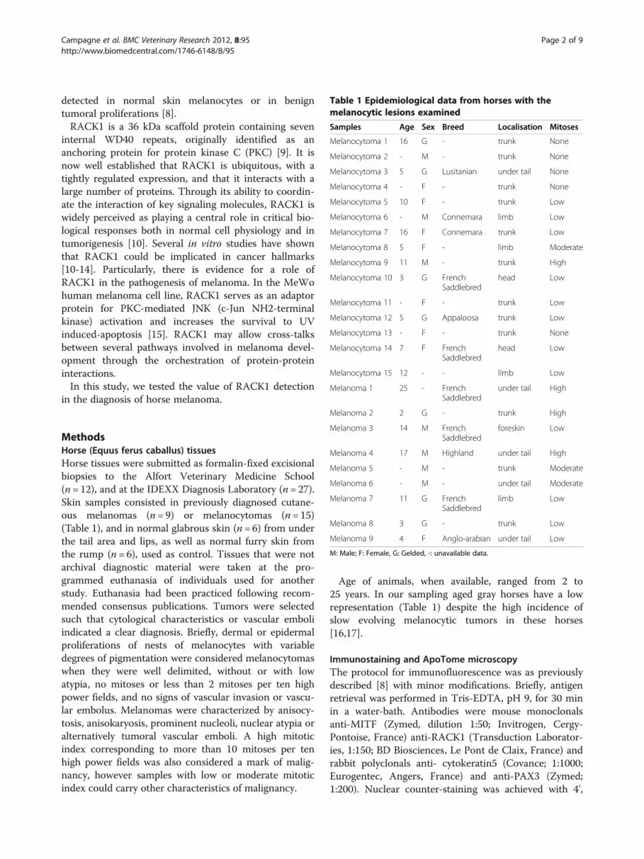
MethodsHorse (Equus ferus caballus) tissuesHorse tissues were submitted as formalin-fixed excisionalbiopsies to the Alfort Veterinary Medicine School(n = 12), and at the IDEXX Diagnosis Laboratory (n= 27).Skin samples consisted in previously diagnosed cutane-ous melanomas (n = 9) or melanocytomas (n= 15)(Table 1), and in normal glabrous skin (n = 6) from underthe tail area and lips, as well as normal furry skin fromthe rump (n = 6), used as control. Tissues that were notarchival diagnostic material were taken at the pro-grammed euthanasia of individuals used for anotherstudy. Euthanasia had been practiced following recom-mended consensus publications. Tumors were selectedsuch that cytological characteristics or vascular emboliindicated a clear diagnosis. Briefly, dermal or epidermalproliferations of nests of melanocytes with variabledegrees of pigmentation were considered melanocytomaswhen they were well delimited, without or with lowatypia, no mitoses or less than 2 mitoses per ten highpower fields, and no signs of vascular invasion or vascu-lar embolus. Melanomas were characterized by anisocy-tosis, anisokaryosis, prominent nucleoli, nuclear atypia oralternatively tumoral vascular emboli. A high mitoticindex corresponding to more than 10 mitoses per tenhigh power fields was also considered a mark of malig-nancy, however samples with low or moderate mitoticindex could carry other characteristics of malignancy.
Age of animals, when available, ranged from 2 to25 years. In our sampling aged gray horses have a lowrepresentation (Table 1) despite the high incidence ofslow evolving melanocytic tumors in these horses[16,17].
Immunostaining and ApoTome microscopyThe protocol for immunofluorescence was as previouslydescribed [8] with minor modifications. Briefly, antigenretrieval was performed in Tris-EDTA, pH 9, for 30 minin a water-bath. Antibodies were mouse monoclonalsanti-MITF (Zymed, dilution 1:50; Invitrogen, Cergy-Pontoise, France) anti-RACK1 (Transduction Laborator-ies, 1:150; BD Biosciences, Le Pont de Claix, France) andrabbit polyclonals anti- cytokeratin5 (Covance; 1:1000;Eurogentec, Angers, France) and anti-PAX3 (Zymed;1:200). Nuclear counter-staining was achieved with 4',
Table 1 Epidemiological data from horses with themelanocytic lesions examined
Samples Age Sex Breed Localisation Mitoses
Melanocytoma 1 16 G - trunk None
Melanocytoma 2 - M - trunk None
Melanocytoma 3 5 G Lusitanian under tail None
Melanocytoma 4 - F - trunk None
Melanocytoma 5 10 F - trunk Low
Melanocytoma 6 - M Connemara limb Low
Melanocytoma 7 16 F Connemara trunk Low
Melanocytoma 8 5 F - limb Moderate
Melanocytoma 9 11 M - trunk High
Melanocytoma 10 3 G FrenchSaddlebred
head Low
Melanocytoma 11 - F - trunk Low
Melanocytoma 12 5 G Appaloosa trunk Low
Melanocytoma 13 - F - trunk None
Melanocytoma 14 7 F FrenchSaddlebred
head Low
Melanocytoma 15 12 - - limb Low
Melanoma 1 25 - FrenchSaddlebred
under tail High
Melanoma 2 2 G - trunk High
Melanoma 3 14 M FrenchSaddlebred
foreskin Low
Melanoma 4 17 M Highland under tail High
Melanoma 5 - M - trunk Moderate
Melanoma 6 - M - under tail Moderate
Melanoma 7 11 G FrenchSaddlebred
limb Low
Melanoma 8 3 G - trunk Low
Melanoma 9 4 F Anglo-arabian under tail Low
M: Male; F: Female, G: Gelded, -: unavailable data.
Campagne et al. BMC Veterinary Research 2012, 8:95 Page 2 of 9http://www.biomedcentral.com/1746-6148/8/95

6'-diamidino-2-phénylindole (DAPI) (Invitrogen, 1:1000).Sections were examined with a Zeiss Axio ObserverZ1M ApoTome microscope (Carl Zeiss S.A.S. ; Le Pecq,France). Controls without the first antibodies showed nounspecific labeling. Images were processed with theAxioVision computer program version 4.6 (Carl Zeiss).Figures are representative of the skin samples evaluated.All images shown are individual sections of z seriesstack. Final figures were assembled with Adobe Photo-shop CS3 (Adobe Systems; USA).
Analysis of RACK1 staining distributionRACK1 staining distribution was analyzed at the tissularand cellular levels. Distribution within the cytoplasmwas graded 0 when homogeneous and 1 when heteroge-neous. Samples were graded blindly without reference topathology reports.
Analysis of elementary histological featuresAll histopathological evaluations were carried out onroutinely stained hematoxylin-eosin-safran sections. Thesize of the tumors was missing in some clinical files; thedimensions of the lesions on histological sections rangedfrom 4 to 80 mm. To highlight the histological specificityof horse melanomas and melanocytomas, all tissue sec-tions were examined at 3 different magnifications (10, 20,40 high power field) and classified according to the elevenhisto-morphometric criteria previously defined by Viroset al. [4]: scatter of intraepidermal melanocytes, nest for-mation of intraepidermal melanocytes, cytoplasmic pig-mentation of neoplastic melanocytes, size and shape ofcells, nuclei and nucleoli, epidermal contour, lateral cir-cumscription, thickness of normal epidermis and presenceof ulceration. Grading was carried out like in Viros et al.[4] except for ulceration which was graded 0 for absenceand 1 for presence. The samples were graded blindly
Figure 1 PAX3 and MITF immunolabeling in horse skin and cutaneous melanocytic proliferations. (1) control horse skin, (2) cutaneousmelanocytoma, (3) cutaneous melanoma (A): PAX3 protein immunolabeling (green) with corresponding bright-field photographs (B). A specificnuclear PAX3 labeling is identified in melanocytes (arrows) and melanocytic cells (A1-A3) with low background signal. (C): MITF proteinimmunolabeling (red) with corresponding bright-field photographs (D). A specific nuclear MITF labeling is observed in melanocytic cells incontrol skin and lesions (C1-C3) with very low background. Nuclear counterstaining is shown in blue. Dotted lines (A1 and C1) indicate epidermis-dermis boundary. e, epidermis; d, dermis. Magnification is the same in all images, bar: 10 μm.
Campagne et al. BMC Veterinary Research 2012, 8:95 Page 3 of 9http://www.biomedcentral.com/1746-6148/8/95

independently by two of us without reference to pathologyreports, Two groups –melanomas and melanocytomas–were subsequently made based on these reports. Sectionswere observed with a Leica DMLB microscope (LeicaMicrosystems S.A.S., Nanterre, France). Images were pro-cessed with the MetaVue Imaging System (MolecularDevices; St Grégoire, France) computer program. Histo-logical pictures were taken with AxioImager.ZI through aAxioCam HRc camera and processed with the AxioVision4.6.3 SPI software (Carl Zeiss).
Statistical analysisStatistical differences between means taken in pairs wereevaluated by Student’s t test. The test was adapted for anumber of samples below 30 [18]. A P-value <0.05 wasconsidered as statistically significant.
ResultsMITF is a sensitive marker to identify melanocytic cells inhorsesIn order to analyze melanocytic proliferations, we firstneeded a marker to identify melanocytic cells within tis-sue sections. Both MITF and PAX3 transcription factorsare expressed by melanocytes and their precursors[19,20]. Comparison of horse and human proteinsequences for MITF and PAX3 resulted in more than90% identity. On tissue sections, melanocytes at thebasal layer of healthy skin were labeled by a specific nu-clear signal using a rabbit PAX3 antibody (Figure 1 A1)or a mouse MITF antibody (Figure 1 C1). Unspecific la-beling was not detected (not shown). Moreover, MITFand PAX3 are expressed by melanocytic cells withintumoral proliferations [8,21]. PAX3 and MITF-positivecells were identified both in melanocytomas and in mel-anomas regardless of the pigmentation of the lesion(Figure 1 A2, A3, C2, C3). Both MITF and PAX3 anti-bodies proved to be helpful in identifying the melanocyticlineage in horse tissues. Nevertheless, MITF antibodydetected the nucleus of melanocytic cells with more sen-sitivity than did the PAX3 antibody. MITF antibody wasthus used for further analyses.
RACK1 protein distinguishes melanoma frommelanocytoma, but also from normal melanocytes inhorsesWe analyzed the tissue and cellular distribution ofRACK1 protein, in healthy skin and melanocytic lesions,after double immunostaining for RACK1 and MITF. Inhealthy control skins (n = 12), RACK1 protein was highlyexpressed in the cytoplasm of keratinocytes whichwere used as positive controls (Figure 2A). By contrast,MITF-positive melanocytes were negative for RACK1(Figure 2A). Triple immunostaining against RACK1,MITF and cytokeratin 5 (CK5), a marker of basal
Figure 2 RACK1, CK5 and MITF immunolabelings in horse skin.RACK1 protein labeling (green), MITF (red) and CK5 (magenta) incontrol horse skin. Cytoplasm of basal keratinocytes is positive forCK5 signal and RACK1 (B). Melanocytes are positive for MITF, butnegative for CK5 and RACK1 (A, B). Dotted line indicates epidermis-dermis boundary. e, epidermis; d, dermis. Bar: 10 μm.
Campagne et al. BMC Veterinary Research 2012, 8:95 Page 4 of 9http://www.biomedcentral.com/1746-6148/8/95

keratinocytes, was performed in order to better identifythe cell type containing RACK1 in the epidermis. Everysignal for RACK1 in the vicinity of melanocytes coloca-lized with CK5 (Figure 2B). Thus, in these labeling condi-tions, RACK1 was not detected in horse normalmelanocytes.In all melanocytic lesions examined (n = 24), RACK1
was extensively detected in MITF-positive cells, with twodistinct distribution patterns. RACK1 was distributed ei-ther heterogeneously (Figure 3A) or homogeneously overthe lesion, whether tumors were pigmented or not(Figure 3B, 3C). In lesions with heterogeneous distribu-tion, RACK1 was detected as a granular cytoplasmicstaining in melanocytic cells. In sharp contrast, in lesionswith homogeneous distribution, RACK1 staining was dif-fuse, perinuclear and cytoplasmic (compare Figure 3Awith Figure 3B and 3C). We tested whether the
cytoplasmic distribution of RACK1 staining could be ofhelp in classifying melanoma lesions. For this purposethe distribution of RACK1 was graded 0 whenhomogenous, and 1 when heterogeneous. Samples weregraded independently by two of us, blindly without refer-ence to pathology reports. Subsequently, melanocytomaand melanoma samples were grouped based on path-ology reports. Comparison of RACK1 grading betweenthe two groups resulted in a statistical difference (1 ± 0vs. 0.2 ± 0.36 respectively; P< 0.001). All samples histolo-gically classified as melanocytomas (n = 15) stained het-erogeneously for RACK1. Among melanomas (n = 9),two samples also stained heterogeneously for RACK1(data not shown). Noteworthy, these two melanomas hadseveral characteristics of low histopathological aggres-siveness, which was low mitotic rate and no vascular em-bolisation but high anisokaryosis. All other melanoma
Figure 3 RACK1 and MITF immunolabelings in cutaneous melanocytic proliferations from horses. MITF labeling is shown in red andRACK1 in green. (A) melanocytoma, (B) melanoma, (C) achromic melanoma. In melanocytoma, RACK1 cytoplasmic expression is heterogeneous(A1 and A2). Arrowheads point to melanocytic cells that express variable amounts of RACK1. By contrast, in pigmented or achromic melanoma(B1 to C2), all MITF-positive cells display a strong and homogeneous cytoplasmic RACK1 signal (B2, C2). Nuclear counterstaining is shown in blue.Corresponding bright-field photographs are presented (A3 to C3). Magnification is the same in all images, scale bar represents 10 μm.
Campagne et al. BMC Veterinary Research 2012, 8:95 Page 5 of 9http://www.biomedcentral.com/1746-6148/8/95

samples stained homogeneously for RACK1. Thus, cyto-plasmic RACK1 labeling may be helpful in distinguishingmelanoma from benign melanocytic skin tumors.
RACK1 detection is more informative than individualhistomorphological featuresMelanocytic lesions in our samples displayed a largehistological variety. Figures 4A and 4B show various pig-mentation patterns and cell shapes in two representativemelanomas. Interestingly enough, RACK1 distributionpattern was constant throughout the whole of thelesions, as illustrated in Figure 4C. To better characterizethe lesions, we performed a detailed analysis based onthe morphological criteria Bastian’s group used onhuman samples [4], summarized in Table 2. We foundthat equine melanomas were characterized by: high pig-mentation (Table 2, 2nd column), abrupt lateral circum-scription (Table 2, 4th column) at the transition frominvolved area to adjacent normal tissue (Figure 4A) and athicker epidermal contour (Table 2, 5th column), indica-tive of epidermal hyperplasia. Moreover, we oftenobserved an absence of junctional component, as previ-ously described [17]. However none of these characteris-tics reached statistical significance when compared tothose of melanocytomas. Finally, we checked every mor-phological criterion and the RACK1 distribution patternagainst the malignancy status. RACK1 signal pattern ofdistribution was more frequently indicative of malig-nancy than any single morphological criterion, pointingout its usefulness as a diagnostic marker.
DiscussionEarly identification of malignant melanocytic lesions iscrucial for patient survival in human and veterinarymedicine. RACK1 is a scaffold protein found to integratevarious metabolic pathways involved in tumorigenesis[22,23]. It was proposed as a marker of malignancy inpig and human melanomas [8]. We extend these obser-vations to melanomas in horses which displayed an over-expression of RACK1 when compared to normalcutaneous melanocytes.In an attempt to find a more refined morphological clas-
sification to distinguish benign from malignant lesions, weused the criteria defined for human melanoma in equinemelanocytomas and melanomas. Although none of themorphological criteria taken separately were powerfulenough to distinguish between benign and malignant tis-sue, we highlight specific histological features of equinemelanoma that recapitulate those seen in atypical rareforms of human malignant nævi and melanomas such aspigment synthesizing melanoma, desmoplastic melan-oma, primary dermal melanoma, and malignant bluenævus [24-27]. This makes the study of equine melan-oma a source of information to understand developmentof atypical melanoma in mammals.We show that PAX3 and MITF immunolabeling can
be used to identify melanocytes as well as melanocytictransformed cells within a tumor bulk. MITF is knownto mark melanocytic proliferations in humans and pigs[8]. More specifically, we show that PAX3 is expressedby mature melanocytes in horses, as in humans [21]. Inevery sample from healthy and tumoral tissues, the
Figure 4 Variability of histological features of horse melanomas and uniformity of RACK1/MITF labeling. (A-B): Sections of two differentmelanomas with hematoxylin-eosin-safran staining, with their respective low magnification in the inserts. Note the variability in pigmentationbetween tumors (A, B) and in different areas of the same tumor (A1, A2). (C): Histological staining of a pigmented area from A with ovoid andspindled cells (C1), and low power capture of RACK1 (green) - MITF (red) labeling in the adjacent section (C2), (C3) bright-field corresponding toC2. Note RACK1 signal uniformity in the different areas. Bar: 50 μm.
Campagne et al. BMC Veterinary Research 2012, 8:95 Page 6 of 9http://www.biomedcentral.com/1746-6148/8/95

melanocytic cells displayed a specific nuclear MITF la-beling. Achromic tumoral samples were also positivelystained for MITF. We therefore propose MITF immuno-labeling as a diagnosis marker for melanocytic tumors inhorses.All the samples identified as melanocytomas by histo-
pathologic analysis stained heterogeneously for RACK1.On the other hand, a homogeneous staining for RACK1only appeared on melanomas. Two samples identified as
melanomas also stained heterogeneously for RACK1.Both had morphological characteristics of low aggres-siveness as a low mitotic index. This is a common pitfallin histological analysis of equine melanoma, and reportson antigens related to the cell cycle gave controversialresults [17,28]. However, no classification based ondetailed staging [29] is available for equine melanomas.We extended previous observations on RACK1 expres-sion in melanoma from humans and pigs to melanocytic
Table 2 Histomorphological features and RACK1 pattern in horse melanocytic lesions
Scattera Pigmentb Nestingc Circumd Epid.Contoure
Cellsizef
Cellshapeg
Nuclearsizeh
Nuclearshapei
Nucleolarsizej
Ulcerk RACK1patternl
Melanocytomas
1 3 4 3 - - 3 0.5 2 - - 0 1
2 - 4 0 - - 2 1 1 - 1 - 1
3 0 1 2 2 2 2 2 2 1 1 0 1
4 1 3 3 1 4 1.5 3 3 1.5 1 1 1
5 1 3.5 0 2 2 2 1 1 0 1 0 1
6 3 4 1 1 2 3 0.5 2 0 1 1 1
7 1 2 2 0 2 3 0 3 0 1 0 1
8 0 1 1 2 2 3 1.5 2.5 1.5 3 0 1
9 0 3 1 0 2 3 1 2 0 1 0 1
10 2 3 1 1 2 2 1.5 3 0 1 0 1
11 1 4 0 1 3 2.5 2.5 1 1 1 0 1
12 0 3.5 0 0 2 3 1.5 2 0 1 1 1
13 2 4 0 2 3 1 2 3 0 1 0 1
14 1 4 2.5 0 2 2.5 1.5 2 0 1.5 0 1
15 - 4 0 1 0 2 1.5 2 0 1 1 1
Melanomas
1 3 2 3 1 - 3 1 3 - 3 1 0
2 3 4 2 2 3 3 2.5 3 0 1 0 0
3 2 1 1 2 4 2 2.5 3 2 1 1 0
4 - 1 0 1 - 3 1 3 0 3 0 0
5 3 4 1 1 1 3 1 3 0 2.5 1 0
6 1 4 1 2 3 2 2 3 1.5 2 0 0
7 1 4 3 2 2 3 1 2.5 - 2 0 1
8 0 3 0 2 2 3 1 3 0 2.5 0 1
9 2 3 2 1 3 3 3 3 1 3 0 0aScatter of intraepidermal melanocytes: 0, absent; 1, slight; 2, medium; 3, prominent.bCytoplasmic pigmentation of neoplastic melanocytes: 0,absent; 1, slight; 2, medium; 3, high; 4, very high.cNesting of intraepidermal melanocytes: 0, absent; 1, slight; 2, medium; 3, prominent.dLateral circumscription: 0, discontinuous; 1, continuous; 2, abrupt.eEpidermal contour: 0, atrophy; 1, effacement; 2, normal; 3, thickening; 4, hyperplasia.fCell size: 1, small; 2, medium; 3, large.gCell shape: 0, round; 1, ovoid; 2, elongated; 3, spindled.hNuclear size: 1, small; 2, medium; 3, large.iNuclear shape: 0, round; 1, ovoid; 2, elongated; 3, spindled.jNucleolar size: 1, small; 2, medium; 3, large.kUlceration: 0, absent; 1, present.lRACK1 distribution pattern: 0, homogenous; 1, heterogenous.-: Impossible evaluation.x.5: means of two different authors observations.
Campagne et al. BMC Veterinary Research 2012, 8:95 Page 7 of 9http://www.biomedcentral.com/1746-6148/8/95

lesions in horses. We show here that the cellular distri-bution of RACK1 reflects melanoma progression andaggressiveness: the more homogenous and diffuse thecytoplasmic RACK1 labeling is, the more aggressive themelanoma. Thus, we propose RACK1 as a marker ofmalignancy in equine melanocytic proliferations.Furthermore, our data show that the cellular distribution
pattern of RACK1 on melanocytes in tumors, i.e.homogenous if malignant and heterogeneous if benign, isconstant throughout the whole lesion. RACK1 distributionpattern analyzed in a punch biopsy appears as informativefor diagnosis and less invasive than a complete biopsy. Thisis a particularly interesting element since skin biopsies inhorses are not that easy to carry out. A quick grading couldinfluence on the decision of treating melanomas more ur-gently, even if they are less suited for surgical therapies.In domestic animals, veterinary pathologists use the
term melanocytomas to describe benign melanocytictumors [30]. In humans, such benign melanocytic tumorsare designated as nævi and the term melanocytoma is sel-dom used. It defines melanocytic tumors with uncertainmalignancy status [31,32]. Our data show that the distri-bution of RACK1 labeling is heterogeneous in horse mela-nocytomas while it is absent or faint in human nævi [8].This confirms the difference between melanocytomas andnævi at the molecular level. In horses, the second mostcommon clinical presentation of melanoma is the malig-nant transformation of a melanocytoma [33]. Based onRACK1 distribution, we hypothesize that horse melanocy-tomas may be premalignant entities ready to switch tomalignancy. RACK1 detection would reveal this switch tomalignancy, if any.
ConclusionsRACK1 protein was detected with intense, diffuse andhomogenous staining in MITF-positive cells of equinemelanomas. This observation is consistent with stainingsin human melanomas and MeLiM minipig melanocytictumors. We now confirm the usefulness of RACK1 label-ing as a diagnosis marker for melanoma. The homogenousdistribution pattern of RACK1 signal shared by human,pig, and horse melanomas strongly suggests a function forRACK1 in melanoma progression in mammalian skin.
AbbreviationsCK5: Cytokeratin 5; IGF1R: Insulin-like growth factor-1 receptor; JNK: c-JunNH2-terminal kinase; MITF: Microphtalmia-associated transcription factor;Pax3: Paired box 3 gene; RACK1: Receptor for activated C-kinase 1.
Competing interestsThe authors declare that they have no competing interests.
Authors’ contributionsCC carried out immunolabelling experiments and analysis, histo-morphological analysis, statistical analysis and drafted the manuscript. SJcarried out microtome sections of samples, HES staining andimmunolabelling experiments. FB participated to histo-morphological
analysis. ME provided and selected samples. FB, GAH and JJP providedintellectual input and revised the manuscript. GE designed and supervisedthe overall study, and drafted the manuscript. All authors read and approvedthe final manuscript.
AcknowledgementsWe would like to thank Céline Robert and Narcisse Towanou for providingcontrol horses and Kevin Cheesman for its contribution to the initial steps ofthe project. We are grateful to Agnès Champeix and Patricia Wattier forcontrol sample preparation, to Sophie Château-Joubert and Jacky Ezagal fortechnical assistance, to Edouard Reyes-Gomez for pictures with AxioImager.ZIand to Marc Chodkiewicz for careful reviewing of the manuscript. This workwas supported by grants from Institut National de la RechercheAgronomique, Agence Nationale de la Recherche Emergence Bio andAssociation pour la Recherche contre le Cancer. CC received a Mitjavillescholarship from the Académie Nationale de Médecine (2008–2009) and agrant (Allocation de Recherche MENRT) from the French Ministry of Research(2009–2012).
Author details1INRA, UMR955 de Génétique fonctionnelle et médicale, Ecole NationaleVétérinaire d’Alfort, 7 avenue du Général de Gaulle, Maisons-Alfort F-94704,France. 2Université Paris Est, Ecole Nationale Vétérinaire d’Alfort, 7 avenue duGénéral de Gaulle, Maisons-Alfort F-94704, France. 3Ecole NationaleVétérinaire d’Alfort, Service d’Anatomie pathologique, 7 avenue Général deGaulle, Maisons-Alfort F-94704, France. 4Laboratoire IDEXX Alfort, 17 alléeJean-Baptiste Preux, Alfortville F-94140, France. 5Département de Biologie duDéveloppement, Institut Pasteur, Unité de Génétique Fonctionnelle de laSouris; CNRS URA 2578, USC INRA, 25 rue du Dr. Roux, Paris F-75724, France.6Present address: CRC – UMRS 872, 15 rue de l’Ecole de Médecine, ParisF-75006, France.
Received: 31 January 2012 Accepted: 15 June 2012Published: 29 June 2012
References1. MacGillivray KC, Sweeney RW, Del Piero F: Metastatic melanoma in horses.
J Vet Intern Med 2002, 16:452–456.2. Smith SH, Goldschmidt MH, McManus PM: A comparative review of
melanocytic neoplasms. Vet Pathol 2002, 39:651–678.3. Broekaert SM, Roy R, Okamoto I, van den Oord J, Bauer J, Garbe C, Barnhill
RL, Busam KJ, Cochran AJ, Cook MG, et al: Genetic and morphologicfeatures for melanoma classification. Pigment Cell Melanoma Res 2010,23:763–770.
4. Viros A, Fridlyand J, Bauer J, Lasithiotakis K, Garbe C, Pinkel D, Bastian BC:Improving melanoma classification by integrating genetic andmorphologic features. PLoS Med 2008, 5:e120.
5. Andersson LS, Axelsson J, Dubielzig RR, Lindgren G, Ekesten B: Multiplecongenital ocular anomalies in Icelandic horses. BMC Vet Res 2011, 7:21.
6. Bellone RR: Pleiotropic effects of pigmentation genes in horses. AnimGenet 2010, 41(Suppl 2):100–110.
7. Rosengren Pielberg G, Golovko A, Sundstrom E, Curik I, Lennartsson J,Seltenhammer MH, Druml T, Binns M, Fitzsimmons C, Lindgren G, et al: Acis-acting regulatory mutation causes premature hair graying andsusceptibility to melanoma in the horse. Nat Genet 2008, 40:1004–1009.
8. Egidy G, Jule S, Bosse P, Bernex F, Geffrotin C, Vincent-Naulleau S, Horak V,Sastre-Garau X, Panthier JJ: Transcription analysis in the MeLiM swinemodel identifies RACK1 as a potential marker of malignancy for humanmelanocytic proliferation. Mol Cancer 2008, 7:34.
9. Ron D, Chen CH, Caldwell J, Jamieson L, Orr E, Mochly-Rosen D: Cloning ofan intracellular receptor for protein kinase C: a homolog of the betasubunit of G proteins. Proc Natl Acad Sci U S A 1994, 91:839–843.
10. Adams DR, Ron D, Kiely PA: RACK1, A multifaceted scaffolding protein:Structure and function. Cell communication and signaling: CCS 2011, 9:22.
11. Berns H, Humar R, Hengerer B, Kiefer FN, Battegay EJ: RACK1 is up-regulated in angiogenesis and human carcinomas. FASEB J 2000,14:2549–2558.
12. Hanahan D, Weinberg RA: Hallmarks of cancer: the next generation.Cell 2011, 144:646–674.
Campagne et al. BMC Veterinary Research 2012, 8:95 Page 8 of 9http://www.biomedcentral.com/1746-6148/8/95
13. Zhang J, Zhu F, Li X, Dong Z, Xu Y, Peng C, Li S, Cho YY, Yao K, Zykova TA,Bode AM: Rack1 protects N-terminal phosphorylated c-Jun fromFbw7-mediated degradation. Oncogene 2012, 31:1835-44.
14. Zhang W, Cheng GZ, Gong J, Hermanto U, Zong CS, Chan J, Cheng JQ,Wang LH: RACK1 and CIS mediate the degradation of BimEL in cancercells. J Biol Chem 2008, 283:16416–16426.
15. Lopez-Bergami P, Habelhah H, Bhoumik A, Zhang W, Wang LH, Ronai Z:RACK1 mediates activation of JNK by protein kinase C [corrected]. MolCell 2005, 19:309–320.
16. Fleury C, Berard F, Leblond A, Faure C, Ganem N, Thomas L: The study ofcutaneous melanomas in Camargue-type gray-skinned horses (2):epidemiological survey. Pigment Cell Res 2000, 13:47–51.
17. Seltenhammer MH, Simhofer H, Scherzer S, Zechner R, Curik I, Solkner J,Brandt SM, Jansen B, Pehamberger H, Eisenmenger E: Equine melanoma ina population of 296 grey Lipizzaner horses. Equine Vet J 2003, 35:153–157.
18. Schwartz D: Les petits échantillons. In Méthodes statistiques à l'usage desmédecins et des biologistes. 4th edition. Edited by Flammarion M-S. Paris;1996:151–162.
19. He S, Yoon HS, Suh BJ, Eccles MR: PAX3 Is extensively expressed in benignand malignant tissues of the melanocytic lineage in humans. J InvestDermatol 2010, 130:1465–1468.
20. Hou L, Panthier JJ, Arnheiter H: Signaling and transcriptional regulation inthe neural crest-derived melanocyte lineage: interactions between KITand MITF. Development 2000, 127:5379–5389.
21. Medic S, Rizos H, Ziman M: Differential PAX3 functions in normal skinmelanocytes and melanoma cells. Biochem Biophys Res Commun 2011,411:832–837.
22. He X, Wang J, Messing EM, Wu G: Regulation of receptor for activated Ckinase 1 protein by the von Hippel-Lindau tumor suppressor in IGF-I-induced renal carcinoma cell invasiveness. Oncogene 2011, 30:535–547.
23. Serrels B, Sandilands E, Serrels A, Baillie G, Houslay MD, Brunton VG, CanelM, Machesky LM, Anderson KI, Frame MC: A complex between FAK,RACK1, and PDE4D5 controls spreading initiation and cancer cellpolarity. Curr Biol 2010, 20:1086–1092.
24. Antony FC, Sanclemente G, Shaikh H, Trelles AS, Calonje E: Pigmentsynthesizing melanoma (so-called animal type melanoma): aclinicopathological study of 14 cases of a poorly known distinctivevariant of melanoma. Histopathology 2006, 48:754–762.
25. Barnhill RL, Gupta K: Unusual variants of malignant melanoma. ClinDermatol 2009, 27:564–587.
26. Ludgate MW, Fullen DR, Lee J, Rees R, Sabel MS, Wong SL, Johnson TM:Animal-type melanoma: a clinical and histopathological study of 22cases from a single institution. Br J Dermatol 2010, 162:129–136.
27. Swetter SM, Ecker PM, Johnson DL, Harvell JD: Primary dermal melanoma:a distinct subtype of melanoma. Arch Dermatol 2004, 140:99–103.
28. Roels S, Tilmant K, Van Daele A, Van Marck E, Ducatelle R: Proliferation,DNA ploidy, p53 overexpression and nuclear DNA fragmentation in sixequine melanocytic tumours. J Vet Med A Physiol Pathol Clin Med 2000,47:439–448.
29. Balch CM, Gershenwald JE, Soong SJ, Thompson JF, Atkins MB, Byrd DR,Buzaid AC, Cochran AJ, Coit DG, Ding S, et al: Final version of 2009 AJCCmelanoma staging and classification. J Clin Oncol 2009, 27:6199–6206.
30. Goldschmidt MH, Hendrick MJ: Tumors of the skin and soft tissues. InTumors in Domestic Animals. 4th edition. Edited by Meuden DJ. Iowa: IowaState Press; 2002:45–118.
31. Quatresooz P, Pierard-Franchimont C, Pierard GE: Molecular histology onthe diagnostic cutting edge between malignant melanomas andcutaneous melanocytomas (Review). Oncol Rep 2009, 22:1263–1267.
32. Zembowicz A, Scolyer RA: Nevus/Melanocytoma/Melanoma: an emergingparadigm for classification of melanocytic neoplasms? Arch Pathol LabMed 2011, 135:300–306.
33. Jeglum KA: Melanomas. In Current therapy in equine medecine. 3rd edition.Edited by Robinson NE. Philadelphia: Saunders W; 1997:399–400.
doi:10.1186/1746-6148-8-95Cite this article as: Campagne et al.: RACK1, a clue to the diagnosis ofcutaneous melanomas in horses. BMC Veterinary Research 2012 8:95.
Submit your next manuscript to BioMed Centraland take full advantage of:
• Convenient online submission
• Thorough peer review
• No space constraints or color figure charges
• Immediate publication on acceptance
• Inclusion in PubMed, CAS, Scopus and Google Scholar
• Research which is freely available for redistribution
Submit your manuscript at www.biomedcentral.com/submit
Campagne et al. BMC Veterinary Research 2012, 8:95 Page 9 of 9http://www.biomedcentral.com/1746-6148/8/95


