Embed Size (px)
Citation preview

JOURNAL OF VIROLOGY, Oct. 2003, p. 10270–10279 Vol. 77, No. 190022-538X/03/$08.00�0 DOI: 10.1128/JVI.77.19.10270–10279.2003Copyright © 2003, American Society for Microbiology. All Rights Reserved.
Exploitation of Microtubule Cytoskeleton and Dynein duringParvoviral Traffic toward the Nucleus
Sanna Suikkanen, Tuula Aaltonen, Marjukka Nevalainen, Outi Valilehto,Laura Lindholm, Matti Vuento, and Maija Vihinen-Ranta*
Department of Biological and Environmental Science, University of Jyvaskyla, FIN-40500 Jyvaskyla, Finland
Received 21 April 2003/Accepted 30 June 2003
Canine parvovirus (CPV), a model virus for the study of parvoviral entry, enters host cells by receptor-mediated endocytosis, escapes from endosomal vesicles to the cytosol, and then replicates in the nucleus. Weexamined the role of the microtubule (MT)-mediated cytoplasmic trafficking of viral particles toward thenucleus. Immunofluorescence and immunoelectron microscopy showed that capsids were transported throughthe cytoplasm into the nucleus after cytoplasmic microinjection but that in the presence of MT-depolymerizingagents, viral capsids were unable to reach the nucleus. The nuclear accumulation of capsids was also reducedby microinjection of an anti-dynein antibody. Moreover, electron microscopy and light microscopy experimentsdemonstrated that viral capsids associate with tubulin and dynein in vitro. Coprecipitation studies indicatedthat viral capsids interact with dynein. When the cytoplasmic transport process was studied in living cells bymicroinjecting fluorescently labeled capsids into the cytoplasm of cells containing fluorescent tubulin, capsidswere found in close contact with MTs. These results suggest that intact MTs and the motor protein dynein arerequired for the cytoplasmic transport of CPV capsids and contribute to the accumulation of the capsid in thenucleus.
To begin a successful infection, viruses have developed astrategy that involves adsorption to cell surface receptors, pen-etration into the cytosol, uncoating of the viral genome, andtargeting of the genome and accessory proteins to the correctcell area for nucleic acid replication. Most DNA viruses rep-licate in the nucleus, which provides the cellular factors re-quired for the amplification and transcription of the viral ge-nomes and for posttranscriptional processing of the viralmRNA. This suggests that after crossing the plasma membraneor endocytic membrane, released viruses or their componentsmust also traverse the cytoplasm to enter the nucleus.
The cytoplasm imposes a diffusion barrier caused by highviscosity and steric obstacles. Cytoplasmic solutes and macro-molecules, along with the lattice-like mesh of microtubules(MTs), actin, and intermediate filament networks, restrict thefree diffusion of macromolecular complexes larger than 500kDa (25, 44), indicating that virus-sized particles are unlikelyto move efficiently through the cytosol by diffusion alone. It islikely that viruses would need to be actively transported duringtheir cytoplasmic trafficking. MTs are polarized structures witha fast-growing plus end extending toward the cell peripheryand a slow-growing minus end located at the centrosome orMT organizing center (MTOC), which is typically found in aperinuclear position (27). Directed transport of cellular com-ponents is linked to large complexes that form molecular mo-tors. Cytoplasmic dynein and kinesin are known to mediateorganelle movement in opposite directions along MTs. Cyto-plasmic dynein, a minus-end-directed, MT-based motor, is amultisubunit protein complex of 1,270 kDa consisting of two
heavy chains (530 kDa), two or three intermediate chains (74kDa), and a variable number of small subunits (19, 20). TheATPase and MT motor domains are located within the dyneinheavy chains, whereas the specific cargo-binding activity in-volves the intermediate chains and several classes of lightchains (7, 51). In many cases the MT-dependent transport ofmaterial is facilitated by the dynein activator protein dynactin,which mediates dynein binding to MTs (2, 18). Dynein, inconjunction with dynactin, facilitates membrane transportfrom the early endosomes to late endosomes and lysosomes (4,17, 33, 50) and from the endoplasmic reticulum to the Golgiapparatus (40).
Ubiquitous as it is, the detailed process by which virusestransport their genome and associated proteins through thecytoplasm is still relatively poorly characterized. The involve-ment of MTs in cytoplasmic traffic has been reported for anumber of viruses, and dynein-mediated transport has beendescribed for adenovirus (22, 47, 48), human foamy virus (42),herpes simplex virus type 1 (HSV-1) (14, 45, 59), and Africanswine fever virus (ASFV) (3). In the case of HSV-1, the viralnucleocapsid protein (UL34) interacts with a cytoplasmic dy-nein intermediate chain (59), while for ASFV, the viral proteinp54 interacts with a cytoplasmic dynein light chain (3). Inaddition, vaccinia virus exploits MTs to enhance its exit frominfected cells. Vaccinia virus particles, using MT plus-end-directed kinesin as a motor, are transported along MTs fromthe perinuclear site of assembly to the site of exit at the plasmamembrane (38, 41).
The icosahedral, nonenveloped parvoviruses are among thesmallest of the animal DNA viruses. The atomic structure ofthe canine parvovirus (CPV) capsid shows that the matureparticle has a diameter of about 26 nm. The virion containsthree capsid proteins (VP1, VP2, and VP3) with molecularsizes of 83, 67, and 65 kDa, respectively (1, 49, 58).
* Corresponding author. Mailing address: Department of Biologicaland Environmental Science, P.O. Box 35, University of Jyvaskyla,Survontie 9, FIN-40500 Jyvaskyla, Finland. Phone: (358) 14 2604209.Fax: (358) 14 2602271. E-mail: [email protected].
10270

CPV uses the transferrin receptor as its cell surface receptor(34), and the infectious pathway involves clathrin-mediated,MT-dependent endocytosis followed by accumulation of viralcapsids within perinuclear vesicles (35, 46, 52). After cell entry,capsids remain within endocytic compartments for severalhours (35, 46). Although the mechanism of capsid escape fromendosomal vesicles is still unknown, once in the cytosol, capsidsmove toward the nuclear membrane and enter the nucleus in aprocess which can be blocked by intracytoplasmic injection ofantiviral antibodies up to several hours after virus inoculation(54, 55). MTs are required for nuclear localization of viralcapsids, as evidenced by the fact that this process could beblocked by depolymerization of MTs in the presence of no-codazole. However, removal of nocodazole led to repolymer-ization of MTs and to nuclear transport of capsids (55).
The nuclear import of capsids appears to require modifica-tions of viral capsids exposing potential nuclear localizationsequences (NLS) within the VP1 N-terminal unique region(54). The N-terminal sequence (PAKRARRGYK) betweenresidues 4 and 13 functions in nuclear transport when conju-gated to bovine serum albumin (BSA) (53), and some specificchanges in that VP1 N-terminal basic sequence reduced therelative infectivity of the capsids (54). Although the VP1 Nterminus is inaccessible in capsids of CPV and minute virus ofmice (MVM), that sequence is exposed in vitro without capsiddisassembly (12, 54, 61). Both VP1 and VP2 are transportedinto the nucleus, and in MVM there are two NLS mapped nearthe VP1-specific N terminus, while transport of VP2 appears toinvolve an internal basic sequence (KGKLTMRAKLR) (23, 24).
Here we examine further the translocation process of CPVcapsids toward the nucleus. To test whether CPV capsids usean MT-associated mechanism for their transport, we first in-jected antibodies against dynein into the cell cytoplasm andsubsequently injected cells with CPV; then we monitored cap-sid transport and viral infection. The anti-dynein antibody re-duced nuclear transport relative to that in untreated cells.Furthermore, in vitro reconstitution and immunoprecipitationexperiments suggested interactions between viral capsids anddynein. Immuno-electron microscopic (immuno-EM) studiesshowed that viral capsids entered the nuclei of cells after mi-croinjection of capsids. These studies suggest that the cytoplas-mic trafficking of the CPV capsids toward the nucleus is anMT-dependent process using cellular dynein and that viralcapsids can enter the nucleus without extensive capsid dissas-sembly.
MATERIALS AND METHODS
Cells and viruses. Norden Laboratory feline kidney (NLFK) cells were grownin Dulbecco�s modified Eagle medium supplemented with 10% fetal calf serum(Gibco, Paisley, United Kingdom). CPV-d isolates were derived from an infec-tious plasmid clone by transfection of NLFK cells (34, 37). To prepare fullcapsids, viruses were propagated in NLFK cells; the capsids were purified byisopycnic centrifugation in a 45% CsCl gradient as described previously (46). Fullcapsids were used for preparation of Oregon green (OG)-labeled capsids ac-cording to the instructions for amine-reactive probes (Molecular Probes, Eu-gene, Oreg.). Immunoprecipitation experiments were carried out with virusesisolated from cell cultures infected for 5 to 7 days. Infected cells were frozen,thawed, and detached with a rubber policeman, and cell debris was removed byspinning at 2,000 rpm for 20 min. Viruses from the supernatant were concen-trated by ultrafiltration (with a 500-kDa filter; Millipore Corp., Bedford, Mass.),and full CPV capsids were pelleted by ultracentrifugation at 173,000 � g for 2 hand resuspended in 1 ml of phosphate-buffered saline, pH 7.4 (PBS). The
suspension was sonicated at low power on ice and extracted with chloroform, andthe aqueous phase was collected.
Antibodies and chemicals. A rabbit antibody to CPV capsid and a mousemonoclonal antibody (hybridoma culture medium; MAb 8) (36, 57) to CPVcapsid were gifts from Colin Parrish (Cornell University, Ithaca, N.Y.). A mouseMAb to nonstructural protein 1 (NS1) was obtained from Caroline Astell (60)and conjugated to Texas red (TxR) (Molecular Probes). MTs were visualized byusing a mouse MAb (Amersham, Little Chalfont, Buckinghamshire, UnitedKingdom) to tubulin. Anti-dynein antibodies (against the 74-kDa intermediatechain of cytoplasmic dynein) were obtained from Sigma (St. Louis, Mo.) orChemicon (Temecula, Calif.). An anti-kinesin MAb was obtained from Chemi-con. In the double-labeling studies, Alexa-546- or Alexa-488-conjugated anti-mouse antibodies and Alexa-488- or Alexa-546-conjugated anti-rabbit antibodiesfrom Molecular Probes were used. Nanogold-conjugated polyclonal rabbit anti-mouse immunoglobulin G (IgG) was purchased from Nanoprobes (Yaphank,N.Y.). Epon LX-112 was obtained from Ladd Research industries (Williston,Vt.), and alkaline phosphatase-conjugated swine anti-rabbit immunoglobulinsand aminoethylcarbazole substrate were from Dako (Glostrup, Denmark).
Nocodazole (Sigma) and vinblastine (Lilly France S.A., Fegersheim, France)were used for the depolymerization of MTs. Cycloheximide was obtained fromSigma, and OG-labeled bovine tubulin and taxol (paclitaxel) were obtained fromMolecular Probes. Nanogold and HQ-silver enhancement reagents were ob-tained from Nanoprobes.
Microinjection antibody and drug treatments. Microinjection into NLFK cellswas carried out using a semiautomatic system comprising Transjector 5246 andMicromanipulator 5171 (Eppendorf, Hamburg, Germany) on an inverted micro-scope. Cells were grown to 80% confluency on microgrid coverslips (grid size,175 nm; Eppendorf) and then injected into cytoplasm with 0.1 to 0.5 pl ofantibody or viral capsids at 2.5 to 5 mg/ml. Anti-dynein and anti-kinesin anti-bodies, control mouse IgG, and viral capsids were concentrated and dialyzedagainst microinjection buffer (10 mM Tris-HCl–120 mM KCl [pH 7.4]).
Full capsids were injected together with the anti-dynein or anti-kinesin anti-bodies or with control mouse IgG. After 6 h of incubation, cells were fixed with4% paraformaldehyde (PFA) (for 20 min at room temperature [RT]) and thenincubated with PBS containing 0.1% Triton X-100, 1% BSA, and 0.01% sodiumazide for 20 min at RT prior to immunolabeling of capsids and injected antibody.
To test for changes in the organization of the MT network for cytoplasmictrafficking of capsids, cells were incubated in a medium containing 60 �Mnocodazole, 20 �M vinblastine, or 2 �M taxol for 30 min prior to capsid injec-tion. The drug was then maintained until fixation in methanol. Cells were stainedwith an anti-tubulin antibody to confirm the effect of the drug on the MTstructure. Laser scanning microscopy (LSM) was conducted on a Zeiss LSM 510inverted microscope. Cycloheximide (0.2 mM) was added to the medium toprevent synthesis of new viral proteins and progeny virus during incubations ofmore than 6 h involving analysis of the localization of incoming virus particles ininoculated cells or in microinjected cells.
CPV infection was detected by staining for the viral NS1 protein with aTxR-conjugated anti-NS1 MAb after methanol fixation (for 6 min at �20°C).The proportion of NS1-expressing cells in normally inoculated cells or in cellsinjected with capsids was determined at different time points. In addition, theeffects of MT-depolymerizing drugs on the number of NS1-expressing cells wereexamined in normally inoculated or microinjected cells.
Preembedding labeling for electron microscopy. NLFK cells on 35-mm-diam-eter plastic culture dishes were grown to �80% confluency. In injection exper-iments, cells were incubated for 2 or 6 h at 37°C after injection of full capsids (2.5mg/ml). For infection assays, cells were incubated with 0.05 �g of capsids/ml for10 or 12 h prior to fixation. Cells were washed twice with PBS and then fixed for2 h at RT in periodate-lysine-PFA fixative (9). Fixed cells were prepared forpreembedding EM as described previously (39, 43). Cells were treated with0.01% saponin and 0.1% BSA in 0.1 M phosphate buffer, pH 7.4 (buffer A),before addition of the anti-capsid MAb diluted in buffer A. Half of the sampleswere treated with 0.05% Triton X-100 in buffer A to ensure that the nuclearmembrane was permeabilized. After 1 h of incubation at RT and washes withbuffer A, nanogold-conjugated polyclonal rabbit anti-mouse IgG was applied for1 h, followed by washes with buffer A and 0.1 M phosphate buffer (pH 7.4). Cellswere postfixed with 1% glutaraldehyde in phosphate buffer for 10 min at RT,quenched with 50 mM NH4Cl in phosphate buffer, and then washed with phos-phate buffer and water. Cells were treated in the dark with HQ-silver for 2 min,followed by washes with water and gold toning (exposure to 2% sodium acetate[three times, for 5 min each time], 0.05% gold chloride [for 10 min on ice], and0.3% sodium thiosulfate [twice, for 10 min each time, on ice]). After washes withwater, the cell cultures were reduced in 1% osmium tetroxide in 0.1 M phosphatebuffer for 1 h at 4°C, dehydrated with a graded series of ethanol, and then stained
VOL. 77, 2003 CYTOPLASMIC TRAFFICKING OF CANINE PARVOVIRUS CAPSID 10271

with 2% uranyl acetate. Plastic capsules filled with Epon LX-112 were placedupside down on top of the cells. After polymerization, the capsules were warmedup to 100°C and removed carefully, and sections parallel to the bottom were cutwith an ultramicrotome (Ultracut 8008; Reichert-Jung) set to 50 nm, picked upon a copper grid, stained with 2% uranyl acetate and lead citrate, and viewedwith an EM.
MT binding assays. In vitro microscopic assays were set up to observe theinteraction between the CPV, MT, and dynein. TxR-labeled �-tubulin was po-lymerized and stabilized with taxol as recommended by the manufacturer. Forthe LSM microscopy, OG-labeled capsids were mixed with polymerized MTs andthe postnuclear supernatant (PNS) in a 1:1:1 ratio (giving a final concentrationof 1.5 �g/ml each) in FDB buffer [35 mM piperazine-N,N-9-bis(2-ethanesulfonicacid) (PIPES), 1 mM EGTA, 0.5 mM EDTA, 1 to 4 mM GTP] containing 10 �Mtaxol (20) and incubated for 30 min at room temperature. The mixture wastransferred to a glass chamber coated with DEAE-dextran (2 mg/ml) (31) andviewed without fixation.
For EM, concentrated capsids (containing dynein according to immuno-EMand immunoprecipitation studies) were mixed with MTs in a 1:1 ratio. Themixture was incubated on the grids for 1 h and stained with uranyl oxalate,followed by a methyl cellulose-uranyl acetate embedding step (16). In parallel,samples were fixed with 4% PFA and immunolabeled. CPV was labeled with ananti-capsid antibody followed by gold-conjugated protein A (5-nm-diameterbeads), after which free antibodies were blocked with protein A. Dynein waslabeled with an anti-dynein MAb, followed by gold-conjugated protein A (10-nm-diameter beads). Then samples were negatively stained and embedded withmethyl cellulose-uranyl acetate.
Finally, the distribution of capsids and their relationship to the MT networkwere studied in the cytoplasm of living cells. Cells were first injected withrhodamine-labeled tubulin diluted in PBS (2.5 mg/ml). After incubation at 37°Cfor 3 to 4 h, the same cells were then reinjected with OG-labeled viral capsids(2.5 mg/ml) and incubated for 1 to 2 h at 37°C. The cells were then monitored byLSM without fixation.
Coimmunoprecipitation and Western blotting. For coprecipitation experi-ments, concentrated viral capsids were immunoprecipitated with anti-dyneinantibodies. Lysates prepared from noninfected cells were used as a control.Before precipitation, viruses and cell lysates were mixed with protein A beads(Prosep-A High Capacity; Bioprocessing Ltd., Durham, England) for 1 h at 4°C,followed by centrifugation at 12,000 � g for 20 s. Supernatants were used forimmunoprecipitation. Precleared viruses were immunoprecipitated with a mix-ture of two anti-dynein MAbs or with a control MAb against trout Ig (2 to 5 �gof antibody/1 mg of protein). The mixture was incubated overnight at 4°C; thenimmune complexes were collected by absorption onto protein A beads (for 1.5 hat 4°C). The precipitates were washed (four times) with PBS containing 0.1%Tween 20 (PBS-Tween). For Western blotting, precipitated proteins were sep-arated on a sodium dodecyl sulfate-polyacrylamide gel electrophoresis runninggel (7.5% acrylamide) (21). The proteins were transferred to nitrocellulosemembranes and incubated overnight with 10 mM Tris-buffered saline, pH 7.4(TBS), containing 3% BSA; then capsids were detected with a rabbit antibodyagainst denatured CPV capsids. After a wash with TBS and 0.2% Tween, themembranes were incubated first with horseradish peroxidase-conjugated goatanti-rabbit immunoglobulins and then with an aminoethylcarbazole substrate(Dako).
RESULTS
It has been shown previously that capsids injected into thecytoplasm of cells were transported through the cytoplasmtoward and into the nucleus in a process that could be blockedby nocodazole treatment (55). Here we further examined therole of the cytoskeleton in cytoplasmic trafficking of viral cap-sids and the connection between capsids, dynein, and MTs andthe nuclear import of the capsids.
Cytoskeleton-dependent, nucleus-directed transport of cyto-solic virus. To determine the efficiency of the nuclear transportof cytoplasmic capsids, viral capsids were injected into cells inthe presence or absence of drugs affecting MTs and in thepresence of 0.2 mM cycloheximide. In nontreated cells after10 h of incubation, approximately 42% of injected cells showedstrong nuclear labeling, suggesting that many capsids had ac-
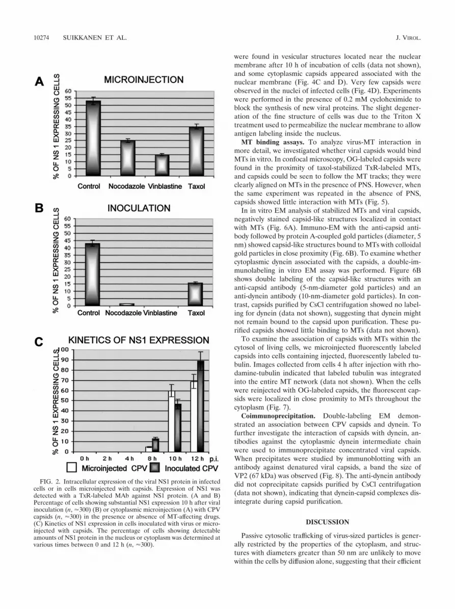
cumulated in the nucleus (Fig. 1). In the remaining injectedcells, numerous small fluorescent spots that represented eitherindividual capsids or capsid clusters were distributed through-out the cell. Disruption of the MT network by nocodazole orvinblastine treatments reduced the amount of nuclear importof viruses, so that only 8 or 1% of cells showed nuclear accu-mulation of viral capsids. Cells treated with taxol, an MT sta-bilizer, also had reduced nuclear import levels, showing 22%fluorescent nuclei (Fig. 1).
To demonstrate whether drug-treated or nontreated cells(without cycloheximide) injected with capsids became infected,cells were also immunolabeled for newly produced viral NS1protein. Interestingly, 25% of cells treated with nocodazoleand 15% of cells treated with vinblastine showed detectableNS1 protein at 12 h postinjection (Fig. 2A), indicating that thetreatments did not inhibit the nuclear import of capsidsenough to prevent infection. Furthermore, 35% of injectedcells in the presence of taxol and 53% of injected cells in theabsence of drugs showed detectable levels of NS1 expression(Fig. 2A).
In addition, to confirm whether the above results with mi-croinjected capsids were consistent with productive infection,we analyzed the NS1 production of CPV-inoculated cells at10 h after normal viral infection. Only 1.5% of nocodazole-treated cells and 0% of vinblastine-treated cells expressed NS1,whereas 43% of untreated and 16% of taxol-treated cellsshowed the presence of NS1 (Fig. 2B). The lower expression ofNS1 in inoculated, drug-treated cells may be due to the com-bination of blockage of the endocytic pathway of the virus andeffects on cytoplasmic transport.
The kinetics of NS1 expression was examined by injectingcapsids into the cytoplasm or by inoculating the cells with thevirus, and then determining the proportions of NS1-expressingcells at various time points. An increase in the proportion ofNS1-expressing cells was observed after 8 h in both injectedcells and inoculated cells, and the maximum percentage ofinfected cells was reached after 12 h (Fig. 2C).
Transport of capsids to the nucleus in the presence of an-tibodies against MT motor proteins. We tested whether anti-bodies against motor proteins would affect the nuclear trans-port of capsids. We coinjected target cells with capsids and aMAb to the 74-kDa intermediate chain of the dynein motorcomplex. The presence of the anti-dynein antibody causes thedisruption of the minus end-directed movement of dynein, ashas been observed for adenovirus capsids (22). The cytoplas-mic anti-dynein MAb decreased the amount of nuclear capsids,whereas in parallel sets of cells injected with capsids and eitheran anti-kinesin MAb or control mouse IgG, no effect on nu-clear transport was seen (Fig. 3). Seven to 13% of cells coin-jected with an anti-dynein MAb and capsids showed traceamounts of capsids accumulating in the nucleus, suggestingthat nuclear transport was mostly blocked by the antibody.However, some trafficking might still have occurred, or thatcapsids reached the nucleus by diffusion or as a result of themicroinjection process. When the capsids were coinjected withan anti-kinesin MAb or control mouse IgG, clear nuclear lo-calization of capsids was detected in 40 to 50% of the injectedcells.
EM analysis of nuclear transport of capsids. To monitorintracellular transport, capsids were labeled with a MAb
10272 SUIKKANEN ET AL. J. VIROL.

against intact viral capsids, and nanogold-immunolabeling EMwith a silver enhancement technique was used. By use of thismethod, intracellular capsids were visualized as small, in-tensely labeled, grainy spots (Fig. 4). Purified full CPV capsidswere injected into the cytosol of cells, and capsid antigen was
detected inside the nucleus at 2 h after injection (Fig. 4A).Capsids recognized by MAb 8, which binds intact capsids (56),were also seen inside the cytoplasm. In some of the images,capsids seemed to be adjacent to the nuclear membrane (Fig.4B). In cells inoculated with capsids, the incoming CPV capsids
FIG. 1. Intracellular localization of CPV capsids 10 h after injection into cytoplasm of cells in the presence or absence of drugs affecting theMT cytoskeleton. (A) Localization of capsids after cytoplasmic injection. Bars, 10 �m. (B) Percentages of cells injected with capsids showingsubstantial nuclear localization (n, �300). All experiments were performed in the presence of 0.2 mM cycloheximide, and 2.5 mg of virus/ml wasused for injections. The injected capsids were detected with a specific rabbit anti-capsid IgG followed by Alexa-488-labeled anti-rabbit IgG (green),and MTs were detected with an antibody against tubulin followed by an Alexa-546-conjugated anti-mouse antibody (red).
VOL. 77, 2003 CYTOPLASMIC TRAFFICKING OF CANINE PARVOVIRUS CAPSID 10273
were found in vesicular structures located near the nuclearmembrane after 10 h of incubation of cells (data not shown),and some cytoplasmic capsids appeared associated with thenuclear membrane (Fig. 4C and D). Very few capsids wereobserved in the nuclei of infected cells (Fig. 4D). Experimentswere performed in the presence of 0.2 mM cycloheximide toblock the synthesis of new viral proteins. The slight degener-ation of the fine structure of cells was due to the Triton Xtreatment used to permeabilize the nuclear membrane to allowantigen labeling inside the nucleus.
MT binding assays. To analyze virus-MT interaction inmore detail, we investigated whether viral capsids would bindMTs in vitro. In confocal microscopy, OG-labeled capsids werefound in the proximity of taxol-stabilized TxR-labeled MTs,and capsids could be seen to follow the MT tracks; they wereclearly aligned on MTs in the presence of PNS. However, whenthe same experiment was repeated in the absence of PNS,capsids showed little interaction with MTs (Fig. 5).
In in vitro EM analysis of stabilized MTs and viral capsids,negatively stained capsid-like structures localized in contactwith MTs (Fig. 6A). Immuno-EM with the anti-capsid anti-body followed by protein A-coupled gold particles (diameter, 5nm) showed capsid-like structures bound to MTs with colloidalgold particles in close proximity (Fig. 6B). To examine whethercytoplasmic dynein associated with the capsids, a double-im-munolabeling in vitro EM assay was performed. Figure 6Bshows double labeling of the capsid-like structures with ananti-capsid antibody (5-nm-diameter gold particles) and ananti-dynein antibody (10-nm-diameter gold particles). In con-trast, capsids purified by CsCl centrifugation showed no label-ing for dynein (data not shown), suggesting that dynein mightnot remain bound to the capsid upon purification. These pu-rified capsids showed little binding to MTs (data not shown).
To examine the association of capsids with MTs within thecytosol of living cells, we microinjected fluorescently labeledcapsids into cells containing injected, fluorescently labeled tu-bulin. Images collected from cells 4 h after injection with rho-damine-tubulin indicated that labeled tubulin was integratedinto the entire MT network (data not shown). When the cellswere reinjected with OG-labeled capsids, the fluorescent cap-sids were localized in close proximity to MTs throughout thecytoplasm (Fig. 7).
Coimmunoprecipitation. Double-labeling EM demon-strated an association between CPV capsids and dynein. Tofurther investigate the interaction of capsids with dynein, an-tibodies against the cytoplasmic dynein intermediate chainwere used to immunoprecipitate concentrated viral capsids.When precipitates were studied by immunoblotting with anantibody against denatured viral capsids, a band the size ofVP2 (67 kDa) was observed (Fig. 8). The anti-dynein antibodydid not coprecipitate capsids purified by CsCl centrifugation(data not shown), indicating that dynein-capsid complexes dis-integrate during capsid purification.
DISCUSSION
Passive cytosolic trafficking of virus-sized particles is gener-ally restricted by the properties of the cytoplasm, and struc-tures with diameters greater than 50 nm are unlikely to movewithin the cells by diffusion alone, suggesting that their efficient
FIG. 2. Intracellular expression of the viral NS1 protein in infectedcells or in cells microinjected with capsids. Expression of NS1 wasdetected with a TxR-labeled MAb against NS1 protein. (A and B)Percentage of cells showing substantial NS1 expression 10 h after viralinoculation (n, �300) (B) or cytoplasmic microinjection (A) with CPVcapsids (n, �300) in the presence or absence of MT-affecting drugs.(C) Kinetics of NS1 expression in cells inoculated with virus or micro-injected with capsids. The percentage of cells showing detectableamounts of NS1 protein in the nucleus or cytoplasm was determined atvarious times between 0 and 12 h (n, �300).
10274 SUIKKANEN ET AL. J. VIROL.

intracellular transport requires interactions with the host trans-port system (25, 26, 44). Earlier results showed that the 26-nm-diameter CPV particles require intact MTs during the in-fection. Although nocodazole blocked the viral infection (46,52), it was quite difficult to determine the steps in viral entryaffected and whether the block was due to effects on endocy-tosis or to direct interaction between viral components and theMTs. Direct microinjection of capsids resulted in a relativelyslow but efficient nuclear uptake of capsids, which was blockedby nocodazole treatment, suggesting that MTs controlled thetransport of capsids across the cytoplasm to the nucleus (55).These experiments did not define the mechanism responsiblefor the cytoplasmic transport of capsids.
To define the role of MTs in nuclear localization, we tested
three drugs that affect the MT cytoskeleton for their effects onthe nuclear transport of cytoplasmically injected capsids. In thepresence of nocodazole, which depolymerizes MTs, and vin-blastine, which causes MT paracrystal formation, injected viralcapsids were found dispersed in the cytoplasm. They did notreach the perinuclear region in significant amounts, suggestingthat an essential MT-dependent step preceded the nuclearimport of capsids (Fig. 1A). There was also a slightly decreasedlevel of nuclear import of capsids in the presence of taxol,which stabilizes MTs (Fig. 1B). These data imply that MTs canserve as tracks to mediate the transport of virus toward thenucleus and that the polymerization and depolymerizationevents at MT ends might also affect nucleus-oriented viralmovements.
FIG. 3. Intracellular localization of CPV capsids microinjected into cytoplasm along with antibodies against the motor protein dynein orkinesin. The cells were incubated for 6 h after injection, after which they were fixed and stained for capsids with rabbit polyclonal anti-capsid IgGfollowed by Alexa-546-conjugated goat anti-rabbit IgG (red). The presence of the injected anti-dynein MAb, the anti-kinesin MAb, and controlmouse IgG was detected using Alexa-488-conjugated goat anti-mouse IgG (green).
VOL. 77, 2003 CYTOPLASMIC TRAFFICKING OF CANINE PARVOVIRUS CAPSID 10275

Successful infection of cells was monitored by immunostain-ing for the viral NS1 protein. NS1 protein serves both as aninitiator protein for viral DNA replication and as a transcrip-tional activator of the viral promoters (11, 13, 32). Thus, NS1is found in infected cells before the synthesis of structuralproteins (10, 30). NS1 protein is an efficient marker for theimport of capsids to the nucleus, leading to viral gene expres-sion and productive infection.
For cells injected with capsids, expression of NS1 proteinwas significantly decreased, although not completely absent, inthe presence of MT-destroying drugs (Fig. 2A). The NS1 ex-pression may be due to some of the injected capsids reachingthe nucleus in an MT-independent fashion, perhaps as a directresult of the microinjection, or due to the large numbers ofcapsids injected. In cells inoculated with virus, NS1 expressionwas inhibited almost completely by nocodazole or vinblastinetreatment (Fig. 2B), most likely due to the multilevel inhibitionof transport caused by the MT disruption. Since intact MTsand functional motor proteins are essential for endosomalvesicular trafficking, endosomal transport and sorting, andmaintenance of the location of late endosomes and recyclingendosomes (5, 8, 17, 28, 29, 33), the disintegration of MTsmight cause a disturbance in the endocytic entry of the virus
capsids. Our studies suggest that CPV escapes into the cytosolduring the late steps of the endocytic pathway (Suikkanen etal., unpublished data). In addition, it has been shown thatinoculation of cells with depleted MT-dependent transportcauses entering capsids to be retained in vesicles dispersed tothe peripheries of the cells (46). Our present results seem tosuggest that capsids entering MT-deficient cells were not ableto traffic through the endosomal pathway to the site of releasebut instead remained inside endosomal structures without be-ing able to cause a productive infection. Taken together, theseresults suggest that in the absence of MT, fewer viral genomeswere transported to the cell nucleus and the level of viral NS1protein synthesis was significantly reduced.
The production of NS1 started 8 h after either injection orinfection of cells (Fig. 2C). Even though cytoplasmically mi-croinjected viruses were detected within the nucleus 2 h post-microinjection (Fig. 4A and B), they appear to require severalhours to reach levels of NS1 expression detectable with afluorescent microscope.
To accomplish the movement toward the nucleus, viral cap-sids are likely to benefit from the inherent polarity of MTs byusing MT-associated motors. Since minus ends of MTs areanchored at the perinuclear MTOC, it seems likely that dynein
FIG. 4. Intracellular distribution of CPV capsids in cytoplasmically microinjected or in normally infected cells. Capsids were detected by useof a preembedding immunolabeling technique where labeling with MAb 8, an antibody generated against intact capsids, was followed bysilver-enhanced nanogold (1.4-nm-diameter particles) and gold toning treatments. (A) Cells injected with capsids (2.5 mg/ml) and then incubatedfor 2 h. (B) Close-up shows intranuclear capsids (arrowheads) and a nuclear membrane with a viral capsid attached to it (arrow). (C) In infectedcells fixed at 10 h postinfection, viral capsids were detected on the nuclear membrane (arrowheads). (D) Infected cells fixed at 12 h postinfectionshowed cytoplasmic localization of capsids in addition to trace amount of nuclear capsids (arrowheads). All experiments were performed in thepresence of cycloheximide (0.2 mM). Bars, 200 nm.
10276 SUIKKANEN ET AL. J. VIROL.

is used to transport incoming viral capsids toward the nucleus.Coinjection of antibodies against dynein along with capsidsshowed that the transport of capsids into the nucleus wasmarkedly decreased in the presence of the antibody, suggestingthat the intermediate chain of dynein is involved in capsidtransport toward the nucleus. Antibodies against the plus-end-directed motor kinesin did not affect CPV trafficking to the
FIG. 5. In vitro binding of CPV capsids to MTs. Shown is the interaction of OG-labeled purified viral capsids with TxR-labeled MTs stabilizedwith taxol in the presence or in the absence of PNS.
FIG. 6. Interaction between the CPV viral capsids, MTs, and dy-nein in vitro. (A) EM image of negatively stained preparation contain-ing taxol-stabilized MTs (arrowhead) and purified capsids (arrows).(B) Immunogold-labeled viral capsids (5-nm-diameter gold particles)containing the intermediate chain of dynein (10-nm-diameter goldparticles) (arrowheads) bound to MT. Bars, 50 nm.
FIG. 7. Localization of CPV capsids in living cells. Cells were mi-croinjected with rhodamine-labeled tubulin and incubated for 4 h at37°C to label MTs; then they were injected with OG-labeled capsidsand incubated for 2 h at 37°C. Cells were visualized with LSM withoutfixation.
VOL. 77, 2003 CYTOPLASMIC TRAFFICKING OF CANINE PARVOVIRUS CAPSID 10277

nucleus (Fig. 3). Whether the binding of dynein to CPV cap-sids involved other cellular components remains unclear.
NLS-mediated translocation through the nuclear pore com-plex (NPC) of the nuclear envelope occurs by sequential stepsthat involve a series of interactions between cargo, solubletransport factors, and nuclear pore proteins of the centraltransport channel (6). The mechanism of nuclear import re-stricts the passive diffusion of molecules of approximately 25 A(2.5 nm) but allows NLS-containing substrates as large as 300A or more to rapidly enter the nucleus (15). Parvoviruses arepotentially small enough to pass through the nuclear porewithout capsid disassembly or deformation. It has not yet beenestablished whether parvoviruses are able to pass through theNPC intact, without capsid dissassembly, during the early stepsof infection. The VP1 N-terminal end, containing several po-tential NLS, appears to play a role in the nuclear importprocess. After injection into the cytoplasm, the VP1 N-termi-nal sequence of the capsid was exposed in a manner which wascorrelated with the nuclear transport of the capsids (54). TheEM studies presented here demonstrate further that viral cap-sids injected into cytoplasm and entering the nucleus are rec-ognized by a MAb recognizing only intact capsids (56), indi-cating that nuclear entry occurs without extensive uncoating(Fig. 4A and B). During normal infection, some cells showedtrace amounts of intranuclear labeling, suggesting that only afew virions are imported to the nucleus, whereas most capsidsremain in the endosomal vesicles or in the cytoplasm (Fig. 4Cand D).
Both in vitro and in vivo assays were used to analyze thebinding of capsids to MTs. Capsids actively bound to MTs invitro became aligned along MT tracks (Fig. 5). In vitro bindingassays using EM confirmed the MT-binding activity of capsidsand showed that capsids could be simultaneously bound toboth dynein and MTs (Fig. 6A and B). The association ofcapsids with dynein was further verified by coprecipitation ofcapsids isolated from infected cells by using antibodies to dy-nein (Fig. 8). The interaction of capsids with MTs in vitro is
consistent with the in vivo observation that cytoplasmic viruseswere associated MTs in living cells (Fig. 7). Taken together,these data imply that capsids were bound to MTs both in vitroand in vivo, in addition to the involvement of dynein in thisinteraction.
Taken together, the data described here confirm that cyto-plasmic trafficking of capsids requires intact MTs and demon-strate the ability of dynein MT motor proteins to bind capsids,suggesting that dynein may play an important role during thebinding and transporting of capsids along MTs to the site oftheir nuclear import. Our results suggest that parvoviral cap-sids released to the cytoplasm are likely to traffic along MTstoward the nucleus during productive infection. It will now beimportant to determine how the interactions among capsids,dynein, and MTs are regulated in order to explain the molec-ular mechanism leading to infection.
ACKNOWLEDGMENTS
We are especially grateful to Colin Parrish for fruitful discussions,helpful comments on the manuscript, and antibodies. We thankGareth Griffiths for help with EM and Pirjo Kauppinen, Jonna Hirsi-maki, Einari Niskanen, Paavo Niutanen, and Raimo Pesonen for ex-cellent technical assistance.
This work was supported by grants from the Academy of Finland(contract 10210), the National Technology Agency (TEKES; contract70026/00), and the Finnish Cultural Foundation.
REFERENCES
1. Agbandje, M., R. McKenna, M. G. Rossmann, M. L. Strassheim, and C. R.Parrish. 1993. Structure determination of feline panleukopenia virus emptyparticles. Proteins 16:155–171.
2. Allan, V. 1996. Motor proteins: a dynamic duo. Curr. Biol. 6:630–633.3. Alonso, C., J. Miskin, B. Hernaez, P. Fernandez-Zapatero, L. Soto, C. Canto,
I. Rodriguez-Crespo, L. Dixon, and J. M. Escribano. 2001. African swinefever virus protein p54 interacts with the microtubular motor complexthrough direct binding to light-chain dynein. J. Virol. 75:9819–9827.
4. Aniento, F., N. Emans, G. Griffiths, and J. Gruenberg. 1993. Cytoplasmicdynein-dependent vesicular transport from early to late endosomes. J. CellBiol. 123:1373–1387.
5. Bananis, E., J. W. Murray, R. J. Stockert, P. Satir, and A. W. Wolkoff. 2000.Microtubule and motor-dependent endocytic vesicle sorting in vitro. J. CellBiol. 151:179–186.
6. Bayliss, R., A. H. Corbett, and M. Stewart. 2000. The molecular mechanismof transport of macromolecules through nuclear pore complexes. Traffic1:448–456.
7. Boylan, K., M. Serr, and T. Hays. 2000. A molecular genetic analysis of theinteraction between the cytoplasmic dynein intermediate chain and the glued(dynactin) complex. Mol. Biol. Cell 11:3791–3803.
8. Brown, K. D., M. Friedkin, and E. Rozengurt. 1980. Colchicine inhibitsepidermal growth factor degradation in 3T3 cells. Proc. Natl. Acad. Sci. USA77:480–484.
9. Brown, W. J., and M. G. Farquhar. 1989. Immunoperoxidase methods forthe localization of antigens in cultured cells and tissue sections by electronmicroscopy. Methods Cell Biol. 31:553–569.
10. Cotmore, S. F., and P. Tattersall. 1987. The autonomously replicating par-voviruses of vertebrates. Adv. Virus Res. 33:91–169.
11. Cotmore, S. F., J. Christensen, J. P. F. Nuesch, and P. Tattersall. 1995. TheNS1 polypeptide of the murine parvovirus minute virus of mice binds toDNA sequences containing the motif [ACCA]2–3. J. Virol. 69:1652–1660.
12. Cotmore, S. F., A. M. D’Abramo, Jr., C. M. Ticknor, and P. Tattersall. 1999.Controlled conformational transitions in the MVM virion expose the VP1N-terminus and viral genome without particle disassembly. Virology 254:169–181.
13. Cotmore, S. F., and P. Tattersall. 1994. An asymmetric nucleotide in theparvoviral 3� hairpin directs segregation of a single active origin of DNAreplication. EMBO J. 13:4145–4152.
14. Dohner, K., A. Wolfstein, U. Prank, C. Echeverri, D. Dujardin, R. Vallee,and B. Sodeik. 2002. Function of dynein and dynactin in herpes simplex viruscapsid transport. Mol. Biol. Cell 13:2795–2809.
15. Feldherr, C. M., D. Akin, and R. J. Cohen. 2001. Regulation of functionalnuclear pore size in fibroblasts. J. Cell Sci. 114:4621–4627.
16. Griffiths, G., A. McDowall, R. Back, and J. Dubochet. 1984. On the prepa-ration of cryosections for immunocytochemistry. J Ultrastruct. Res. 89:65–78.
FIG. 8. Coimmunoprecipitation of CPV capsids with an anti-dy-nein antibody. Viruses concentrated from cells infected with CPV(lane 2) and a cell extract from noninfected cells (lane 3) were immu-noprecipitated with MAbs against dynein. A control precipitation(lane 1) was performed with a MAb against trout Ig, and all sampleswere immunoblotted with an antibody against viral capsid protein. Themigration positions of CPV capsid proteins VP1 (Mr, 83,000) and VP2(Mr, 67,000) are shown in lane 4.
10278 SUIKKANEN ET AL. J. VIROL.

17. Gruenberg, J., G. Griffiths, and K. E. Howell. 1989. Characterization of theearly endosome and putative endocytic carrier vesicles in vivo and with assayof vesicle fusion in vitro. J. Cell Biol. 108:1301–1316.
18. Holleran, E. A., S. Karki, and E. L. Holzbaur. 1998. The role of the dynactincomplex in intracellular motility. Int. Rev. Cytol. 182:69–109.
19. Holzbaur, E. L., and R. B. Vallee. 1994. Dyneins: molecular structure andcellular function. Annu. Rev. Cell Biol. 10:339–372.
20. King, S. J., and T. A. Schroer. 2000. Dynactin increases the processivity ofthe cytoplasmic dynein motor. Nat. Cell Biol. 2:20–24.
21. Laemmli, U. K. 1970. Cleavage of structural proteins during the assembly ofthe head of bacteriophage T4. Nature 227:680–685.
22. Leopold, P. L., G. Kreitzer, N. Miyazawa, S. Rempel, K. K. Pfister, B. E.Rodriguez, and R. G. Crystal. 2000. Dynein- and microtubule-mediatedtranslocation of adenovirus serotype 5 occurs after endosomal lysis. Hum.Gene Ther. 11:151–165.
23. Lombardo, E., J. C. Ramirez, M. Agbandje-McKenna, and J. M. Almendral.2000. A beta-stranded motif drives capsid protein oligomers of the parvovi-rus minute virus of mice into the nucleus for viral assembly. J. Virol. 74:3804–3814.
24. Lombardo, E., J. C. Ramirez, J. Garcia, and J. M. Almendral. 2002. Com-plementary roles of multiple nuclear targeting signals in the capsid proteinsof the parvovirus minute virus of mice during assembly and onset of infec-tion. J. Virol. 76:7049–7059.
25. Luby-Phelps, K. 2000. Cytoarchitecture and physical properties of cyto-plasm: volume, viscosity, diffusion, intracellular surface area. Int. Rev. Cytol.192:189–221.
26. Luby-Phelps, K. 1994. Physical properties of cytoplasm. Curr. Opin. CellBiol. 6:3–9.
27. Mandelkow, E., and E. M. Mandelkow. 1995. Microtubules and microtubule-associated proteins. Curr. Opin. Cell Biol. 7:72–81.
28. Matteoni, R., and T. E. Kreis. 1987. Translocation and clustering of endo-somes and lysosomes depends on microtubules. Eur. J. Cell Biol. 105:1253–1265.
29. McGraw, T. E., K. W. Dunn, and F. R. Maxfield. 1993. Isolation of atemperature-sensitive variant Chinese hamster ovary cell line with a mor-phologically altered endocytic recycling compartment. J. Cell. Physiol. 155:579–594.
30. Molitor, T. W., H. S. Joo, and M. S. Collett. 1985. Identification and char-acterization of a porcine parvovirus nonstructural polypeptide. J. Virol.55:554–559.
31. Murray, J. W., E. Bananis, and A. W. Wolkoff. 2000. Reconstitution ofATP-dependent movement of endocytic vesicles along microtubules in vitro:an oscillatory bidirectional process. Mol. Biol. Cell 11:419–433.
32. Nuesch, J. P., S. F. Cotmore, and P. Tattersall. 1995. Sequence motifs in thereplicator protein of parvovirus MVM essential for nicking and covalentattachment to the viral origin: identification of the linking tyrosine. Virology209:122–135.
33. Oda, H., R. J. Stockert, C. Collins, H. Wang, P. M. Novikoff, P. Satir, andA. W. Wolkoff. 1995. Interaction of the microtubule cytoskeleton with endo-cytic vesicles and cytoplasmic dynein in cultured rat hepatocytes. J. Biol.Chem. 270:15242–15249.
34. Parker, J. S., W. J. Murphy, D. Wang, S. J. O’Brien, and C. R. Parrish. 2001.Canine and feline parvoviruses can use human or feline transferrin receptorsto bind, enter, and infect cells. J. Virol. 75:3896–3902.
35. Parker, J. S., and C. R. Parrish. 2000. Cellular uptake and infection bycanine parvovirus involves rapid dynamin-regulated clathrin-mediated endo-cytosis, followed by slower intracellular trafficking. J. Virol. 74:1919–1930.
36. Parker, J. S. L., and C. R. Parrish. 1997. Canine parvovirus host range isdetermined by the specific conformation of an additional region of thecapsid. J. Virol. 71:9214–9222.
37. Parrish, C. R. 1991. Mapping specific functions in the capsid structure ofcanine parvovirus and feline panleukopenia virus using infectious plasmidclones. Virology 183:195–205.
38. Ploubidou, A., V. Moreau, K. Ashman, I. Reckmann, C. Gonzalez, and M.Way. 2000. Vaccinia virus infection disrupts microtubule organization andcentrosome function. EMBO J. 19:3932–3944.
39. Pohl, K., and Y. D. Stierhof. 1998. Action of gold chloride (“gold toning”) onsilver-enhanced 1 nm gold markers. Microsc. Res. Tech. 42:59–65.
40. Presley, J. F., N. B. Cole, T. A. Schroer, K. Hirschberg, K. J. Zaal, and S. J.
Lippincott. 1997. ER-to-Golgi transport visualized in living cells. Nature389:81–85.
41. Rietdorf, J., A. Ploubidou, I. Reckmann, A. Holmstrom, F. Frischknecht, M.Zettl, T. Zimmermann, and M. Way. 2001. Kinesin-dependent movement onmicrotubules precedes actin-based motility of vaccinia virus. Nat. Cell Biol.3:992–1000.
42. Saib, A., M. Neves, M. L. Giron, M. C. Guillemin, J. Valla, J. Peries, and M.Canivet. 1997. Long-term persistent infection of domestic rabbits by thehuman foamy virus. Virology 228:263–268.
43. Sawada, H., and M. Esaki. 2000. A practical technique to postfix nanogold-immunolabeled specimens with osmium and to embed them in Epon forelectron microscopy. J. Histochem. Cytochem. 48:493–498.
44. Seksek, O., J. Biwersi, and A. S. Verkman. 1997. Translational diffusion ofmacromolecule-sized solutes in cytoplasm and nucleus. J. Cell Biol. 138:131–142.
45. Sodeik, B., M. W. Ebersold, and A. Helenius. 1997. Microtubule-mediatedtransport of incoming herpes simplex virus 1 capsids to the nucleus. J. CellBiol. 136:1007–1021.
46. Suikkanen, S., K. Saajarvi, J. Hirsimaki, O. Valilehto, H. Reunanen, M.Vihinen-Ranta, and M. Vuento. 2002. Role of recycling endosomes andlysosomes in dynein-dependent entry of canine parvovirus. J. Virol. 76:4401–4411.
47. Suomalainen, M., M. Y. Nakano, K. Boucke, S. Keller, and U. F. Greber.2001. Adenovirus-activated PKA and p38/MAPK pathways boost microtu-bule-mediated nuclear targeting of virus. EMBO J. 20:1310–1319.
48. Suomalainen, M., M. Y. Nakano, S. Keller, K. Boucke, R. P. Stidwill, andU. F. Greber. 1999. Microtubule-dependent plus- and minus end-directedmotilities are competing processes for nuclear targeting of adenovirus.J. Cell Biol. 144:657–672.
49. Tsao, J., S. Chapman, M. Agbandje, W. Keller, K. Smith, H. Wu, M. Luo,T. J. Smith, M. G. Rossman, R. W. Compans, and C. R. Parrish. 1991. Thethree-dimensional structure of canine parvovirus and its functional implica-tions. Science 251:1456–1464.
50. Valetti, C., D. M. Wetzel, M. Schrader, M. J. Hasbani, S. R. Gill, T. E. Kreis,and T. A. Schroer. 1999. Role of dynactin in endocytic traffic: effects ofdynamitin overexpression and colocalization with CLIP-170. Mol. Biol. Cell10:4107–4120.
51. Vaughan, P. S., J. D. Leszyk, and K. T. Vaughan. 2001. Cytoplasmic dyneinintermediate chain phosphorylation regulates binding to dynactin. J. Biol.Chem. 276:26171–26179.
52. Vihinen-Ranta, M., A. Kalela, P. Makinen, L. Kakkola, V. Marjomaki, andM. Vuento. 1998. Intracellular route of canine parvovirus entry. J. Virol.72:802–806.
53. Vihinen-Ranta, M., L. Kakkola, A. Kalela, P. Vilja, and M. Vuento. 1997.Characterization of a nuclear localization signal of canine parvovirus capsidproteins. Eur. J. Biochem. 1:389–394.
54. Vihinen-Ranta, M., D. Wang, W. S. Weichert, and C. R. Parrish. 2002. TheVP1 N-terminal sequence of canine parvovirus affects nuclear transport ofcapsids and efficient cell infection. J. Virol. 76:1884–1891.
55. Vihinen-Ranta, M., W. Yuan, and C. R. Parrish. 2000. Cytoplasmic traffick-ing of the canine parvovirus capsid and its role in infection and nucleartransport. J. Virol. 74:4853–4859.
56. Weichert, W. S., J. S. L. Parker, A. T. M. Wahid, S.-F. Chang, E. Meier, andC. R. Parrish. 1998. Assaying for structural variation in the parvovirus capsidand its role in infection. Virology 250:106–117.
57. Wikoff, W. R., G. Wang, C. R. Parrish, R. H. Cheng, M. L. Strassheim, T. S.Baker, and M. G. Rossmann. 1994. The structure of a neutralized virus:canine parvovirus complexed with neutralizing antibody fragment. Structure2:595–607.
58. Xie, Q., and M. S. Chapman. 1996. Canine parvovirus capsid structure,analyzed at 2.9 A resolution. J. Mol. Biol. 264:497–520.
59. Ye, G. J., K. T. Vaughan, R. B. Vallee, and B. Roizman. 2000. The herpessimplex virus 1 UL34 protein interacts with a cytoplasmic dynein intermedi-ate chain and targets nuclear membrane. J. Virol. 74:1355–1363.
60. Yeung, D. E., G. W. Brown, P. Tam, R. H. Russnak, G. Wilson, I. Clark-Lewis, and C. R. Astell. 1991. Monoclonal antibodies to the major nonstruc-tural nuclear protein of minute virus of mice. Virology 181:35–45.
61. Yuan, W., and C. R. Parrish. 2000. Comparison of two single-chain antibod-ies that neutralize canine parvovirus: analysis of an antibody-combining siteand mechanisms of neutralization. Virology 269:471–480.
VOL. 77, 2003 CYTOPLASMIC TRAFFICKING OF CANINE PARVOVIRUS CAPSID 10279


