Embed Size (px)
Citation preview

2409Development 124, 2409-2419 (1997)Printed in Great Britain © The Company of Biologists Limited 1997DEV8417
The microtubule motor cytoplasmic dynein is required for spindle orientation
during germline cell divisions and oocyte differentiation in Drosophila
Maura McGrail† and Thomas S. Hays*
Department of Genetics and Cell Biology, University of Minnesota, 1445 Gortner Avenue, St. Paul, MN 55108, USA†Present address: Howard Hughes Medical Institute, Division of Cellular and Molecular Medicine, UCSD School of Medicine, 9500 Gilman Drive, La Jolla, CA 92093,USA*Author for correspondence (e-mail: [email protected])
During animal development cellular differentiation is oftenpreceded by an asymmetric cell division whose polarity isdetermined by the orientation of the mitotic spindle. In thefruit fly, Drosophila melanogaster, the oocyte differentiatesin a 16-cell syncytium that arises from a cystoblast whichundergoes 4 synchronous divisions with incomplete cytoki-nesis. During these divisions, spindle orientation is highlyordered and is thought to impart a polarity to the cyst thatis necessary for the subsequent differentiation of the oocyte.Using mutations in the Drosophila cytoplasmic dyneinheavy chain gene, Dhc64C, we show that cytoplasmicdynein is required at two stages of oogenesis. Early inoogenesis, dynein mutations disrupt spindle orientation in
dividing cysts and block oocyte determination. The local-ization of dynein in mitotic cysts suggests spindle orienta-tion is mediated by the microtubule motor cytoplasmicdynein. Later in oogenesis, dynein function is necessary forproper differentiation, but does not appear to participatein morphogen localization within the oocyte. These resultsprovide evidence for a novel developmental role for thecytoplasmic dynein motor in cellular determination anddifferentiation.
Key words: dynein, cell fate, asymmetric division, Drosophila,spindle orientation, oogenesis
SUMMARY
INTRODUCTION
A central question in the fields of cell biology and develop-mental genetics concerns how a cell chooses to adopt a partic-ular cell fate and differentiate into a specific cell type. Whilethe mechanisms that contribute to the decision process are stillemerging, it is known that one pathway to cellular differen-tiation involves asymmetric cell division in which cell fatedeterminants are partitioned unequally between daughter cells.The orientation of the mitotic spindle has long been known tobe important in directing asymmetric cell divisions duringembryogenesis (reviewed by Strome, 1993). More recently,molecular genetic analyses in C. elegans (Kemphues et al.,1988; Cheng et al, 1995; Guo and Kemphues, 1995; Etemad-Moghadam et al., 1995) and Drosophila (Kraut et al., 1996)have identified molecules that couple spindle orientation to theasymmetric localization of cell fate determinants (reviewed byRhyu and Knoblich, 1995; White and Strome, 1996; Doe,1996). Members of the myosin family of actin-based molecularmotors have been implicated in the asymmetric segregation offactors necessary for cell fate determination in yeast (Jansen etal., 1996; Bobola et al., 1996) and C. elegans (Guo andKemphues, 1996). Less is known about the pathways whichestablish the initial cellular asymmetry that directs spindle ori-entation and polarized transport. Given the importance of themicrotubule cytoskeleton in cytoplasmic organization andintracellular transport in polarized cell types, microtubules and
their associated motor molecules may be part of the primarymechanism that establishes this cytoplasmic asymmetry.
Oogenesis in Drosophila melanogaster is an attractivesystem for studying the role of microtubule motors in cellulardifferentiation (for a comprehensive review of oogenesis, seeSpradling, 1993). Cytological and pharmacological studieshave suggested that the microtubule cytoskeleton is essentialfor oocyte differentiation through its roles in intercellulartransport and in the asymmetric localization of determinantswithin the developing oocyte that influence axis specification(reviewed by Theurkauf, 1994; St Johnston, 1995). Oocytedevelopment occurs in a cyst of 16 interconnected cells thatarises from a single cystoblast which undergoes 4 synchronousrounds of division with incomplete cytokinesis. One cell dif-ferentiates as the oocyte, while the remaining 15 becomehighly polyploid nurse cells. The nurse cells synthesizematerials which are transferred through the cytoplasmic con-nections (ring canals) into the differentiating oocyte (reviewedby Mahajan-Miklos and Cooley, 1994). The asymmetrictransport of materials is thought to occur along a polarizedmicrotubule array established shortly after the 16-cell cyst isformed (Mahowald and Strassheim, 1970; Theurkauf et al.,1993). Microtubules originate in the pro-oocyte and extendtheir plus ends through the ring canals into the nurse cells,creating a polarized transport system on which a minus-enddirected microtubule motor could function.
Within the germline cyst a polarity must be established in

2410 M. McGrail and T. S. Hays
order to define the pro-oocyte as the site of microtubule nucle-ation in the assembly of the polarized microtubule array. Thispolarity may be reflected in the asymmetric segregation of thespectrin-rich, multivesicular fusome during the four mitoticdivisions (Lin and Spradling, 1995) and in the resulting fixedpattern of cytoplasmic connections among the 16 cystocytes(King et al., 1982). The asymmetric cystocyte divisions resultfrom the behavior of the fusome and its interaction with spindlepoles during mitotic division. How anchorage of the mitoticspindles to the fusome ensures the stereotypical pattern of cystcell divisions, which creates a polarized cyst leading to oocytedetermination, reorganization of the microtubule cytoskeleton,and oocyte differentiation, is still unclear.
The dynein ATPases constitute a family of minus-enddirected microtubule-based motors. The best characterizedmembers of the dynein family are the axonemal dyneins whichpower ciliary and flagellar motility (reviewed by Porter, 1996).Since the identification of a cytoplasmic form of dynein in non-ciliated cells (Paschal and Vallee, 1987), the function, regula-tion, and subcellular targeting of the cytoplasmic dynein motorhave been the subject of intense study (reviewed by Schroer,1994; Holzbaur and Vallee, 1994; Vallee and Sheetz, 1996).Our previous characterization of the asymmetric localizationof cytoplasmic dynein to and within the developing oocytesuggested that cytoplasmic dynein might function in multiplemicrotubule-dependent processes in Drosophila oogenesis (Liet al., 1994). The identification of mutations in the Drosophilacytoplasmic dynein heavy chain gene Dhc64C (Gepner et al.,1996) has allowed us to investigate the role of cytoplasmicdynein in oocyte determination and differentiation. Our resultsprovide evidence for a novel role of the cytoplasmic dynein inasymmetric cell division and cellular differentiation.
MATERIALS AND METHODS
Fly stocks and genetic analysesThe mutations in the Drosophila cytoplasmic dynein heavy chainDhc64C used in this study are described by Gepner et al. (1996).Adult flies, heteroallelic for Dhc64C mutations, were generated at theexpected frequency relative to sibling classes. The female-sterilephenotype of the dynein mutants was shown to be rescued by the wild-type Dhc64C transgene by introducing P[Dhc+]X (Gepner et al., 1996)into the mutant background. Examination of the localization of theDrosophila kinesin heavy chain khc motor domain-β-galactosidasefusion protein was performed by introducing the second chromosomekinesin-β-gal insert KZ32 (Clark et al., 1994), provided by I. Clark(Princeton University), into a Dhc64C mutant background using theappropriate crosses. Flies heterozygous for KZ32 and heteroallelic forDhc64C, ie., KZ32/+; Dhc64C6-6/Dhc64C6-12, were selected for ovarydissection and analysis.
Germline clonal analysis was performed using the FRT/FLP recom-binase method (Golic and Lindquist, 1989; Golic, 1991) together withthe ovoD1-Dominant Female Sterile technique (Chou et al., 1993). Thethird chromosome P[ovoD1]2X48 P[w+FRT]2A (Chou et al., 1993) wasprovided by N. Perrimon (Harvard University). The X chromosomeFLP recombinase stock P[ry+, hsFLP] (Golic and Lindquist, 1989),and the third chromosome FRT stock P[w+ FRT]2A (Chou et al., 1993),were obtained from the Bloomington Stock Center (Bloomington,IN). A Dhc64C4-19 P[w+ FRT]2A recombinant chromosome was con-structed using standard recombination techniques. The recombinantchromosome was shown to be free of secondary lethal mutations byrescue of the Dhc64C4-19 recessive lethal phenotype by the wild-type
P[Dhc+]X transgene. For clonal analysis, 0-24 hour eggs werecollected from the appropriate crosses, aged for 4 days to the thirdinstar larval stage, and the larvae heat shocked in a 37°C water bathfor 2 hours to induce expression of the FLP recombinase. Under theseconditions the frequency of control females that contained clones inat least one ovary was between 70-90%. Ovaries were examined underthe dissecting microscope and clones were identified by the presenceof mature eggs and vitellogenic egg chambers. To determine thenumber of germline cells/mosaic egg chamber, the clonal analysiswith the Dhc64C4-19 was repeated in the absence of ovoD1, and ovarieswere then prepared for immunofluorescence and probed with thedynein heavy chain antibody P1H4 and the nuclear stain OliGreen(Molecular Probes, Inc., Eugene, OR).
Antibodies and immunofluorescence localizationA purified, bacterially expressed fusion protein from Dhc64C cDNAclone pTR13 (Li et al., 1994), encoding amino acid residues 128-422of the cytoplasmic dynein heavy chain, was used as antigen in thepreparation of hybridoma cell lines and the P1H4 ascites (Immuno-logical Resource Center, U of IL Urbana/Champaign). Microtubule-associated proteins were prepared from wild-type OregonR ovaries,and SDS-PAGE and blotting were carried out essentially as describedby McGrail et al. (1995) except that 15 µg total protein, and approx.2 µg ATP-MAPS, was loaded per lane. For blotting the P1H4 mono-clonal was used at a dilution of 1:10,000.
Ovaries were dissected, fixed and processed for immunocytochem-istry as described by McGrail et al. (1995). For immunofluoresence,the P1H4 anti-dynein heavy chain mouse monoclonal was diluted1:500, the anti-Bicaudal-D mouse monoclonal (Suter and Steward,1991) was diluted 1:3, the rabbit polyclonal anti-β-galactosidaseantibody (Cappel/Organon Teknika Corp., Westchester, PA) wasdiluted 1:200, and ring canals were detected with the anti-phospho-tyrosine monoclonal antibody (ICN Biomedical, Inc., Aurora, OH)diluted 1:50. Dynein and staufen localization were examined inovaries double-labelled with the anti-dynein heavy chain rabbit poly-clonal antibody PEP1 (Li et al., 1994) diluted 1:50, the rat polyclonalanti-staufen antiserum diluted 1:100, and FITC-conjugated goat anti-rabbit and Texas Red-conjugated goat anti-rat secondary antibodies.Mitotic spindles and fusomes were detected in ovaries double labelledwith a rat monoclonal anti-α-tubulin antibody (Accurate ChemicalCo., Westbury, NY) diluted 1:10, the rabbit polyclonal anti-α-spectrinantibody (Dubreuil et al., 1987; Byers et al., 1987) diluted 1:200, andTexas Red-conjugated goat anti-rat and FITC-conjugated goat anti-rabbit secondary antibodies. For triple labelling ovaries wereincubated with a rabbit polyclonal anti-dynein heavy chain antibody(Hays et al., 1994) diluted 1:3, the rat monoclonal anti-α-tubulinantibody diluted 1:10 (Accurate Chemical Co., Westbury, NY), thechromatin marker OliGreen diluted 1:200 (Molecular Probes, Inc.,Eugene, OR), and Cy5-conjugated goat-anti-rabbit and Texas Red-conjugated goat-anti-rat secondary antibodies. A similar pattern ofdynein localization in mitotic cysts was observed with the P1H4 mon-oclonal and colocalized with α-spectrin on the fusome. The secondaryantibodies Texas Red-conjugated goat anti-rat and Texas Red-conju-gated goat anti-mouse (Jackson ImmunoResearch Labs, West Grove,PA), FITC-conjugated goat-anti-rabbit (Boehringer Mannheim, Indi-anapolis, IN) and Cy5-conjugated goat anti-rabbit (Amersham,Arlington Heights, IL) were diluted 1:100. Confocal z series imagefiles of labelled tissues were acquired with a BioRad 1000 laserscanning confocal microscope, and processed using NIH Image 1.59and Adobe Photoshop 3.0 (Adobe Systems, Inc., Mountain View, CA)software.
In situ hybridizationWhole-mount in situ hybridization of digoxigenin-labelled antisenseRNA probes to egg chambers was performed according to the methodof Tautz and Pfeifle (1989) with modifications. The oskar cDNA(Ephrussi et al., 1991) was provided by R. Lehmann (Skirball

2411Dynein function in oocyte differentiation
Table 1. Cytoplasmic dynein function is required in thegermline for oocyte development
No. of ovaries with eggs*No. of Total no. of ovaries scored
Genotype females analyzed (% of ovaries with eggs)
Dhc64C4-19/P[ovoD1] 295 12/590 (2)P[Dhc+]; Dhc64C4-19/ 139 267/278 (96)
P[ovoD1]
*Mosaic egg chambers which lack dynein function in the germline fail todevelop an oocyte or mature egg. Egg chambers with germline cellshomozygous mutant for the dynein allele Dhc64C4-19 (Gepner et al., 1996)were produced with the FRT/FLP recombinase system (Golic, 1991; Golicand Lindquist, 1989). On the homologous third chromosome was thetransgene P[ovoD1]2X48 (Chou et al., 1993). ovoD1 is a dominant female-sterile allele of the ovo locus which prevents the germline fromdifferentiating, therefore eggs are not normally produced in the ovaries offemales carrying the ovoD1 transgene (Chou et al., 1993). Consequently, anyeggs present must have arisen from a mitotic recombination event whichproduced a stem cell that has lost the chromosome bearing the P[ovoD1]transgene and is now homozygous mutant for the Dhc64C4-19 allele. In testfemales of the genotype Dhc64C4-19/P[ovoD1], a total of 12 eggs were foundin 590 ovaries scored. None of the ovaries contained egg chambers withdeveloping oocytes. In contrast, in control females of the genotype P[Dhc+]X;Dhc64C4-19/P[ovoD1], which carried a copy of the wildtype Dhc64Ctransgene P[Dhc+] on the X chromosome, 96% of the ovaries had at least oneovariole with developing oocytes and many mature eggs. The almostcomplete absence of oocytes and eggs in the test ovaries demonstrates thatcytoplasmic dynein function is necessary in the germline for oocytedevelopment.
Fig. 1. Specificity of the mouse monoclonal anti-dynein heavy chainantibody P1H4. A Coomassie blue-stained gel (lanes 1-4), andduplicate gel blotted and probed with the P1H4 monoclonal (lanes 5-8), of fractions from a microtubule-associated proteins (MAPS)preparation from ovary extracts. Lanes 1 and 5, homogenate; lanes 2and 6, 125,000 g supernatant; lanes 3 and 7, taxol-stabilizedmicrotubule pellet; lanes 4 and 8, cytoplasmic dynein enriched ATP-eluted MAPS. The P1H4 monoclonal antibody specifically detectsthe dynein heavy chain (arrow, DHC) in the homogenate and highspeed supernatant, and reveals the enrichment of cytoplasmic dyneinin the extraction of ATP-dependent MAPS from the microtubulepellet. Molecular mass standards are indicated to the left of lane 1.
Institute, New York University Medical School), the bicoid cDNA(Berleth et al., 1988) by E. Stephenson (University of Alabama), andthe gurken cDNA (Neuman-Silberberg and Schüpbach, 1993) by T.Schüpbach (Princeton University). Digoxigenin-labelled antisenseRNA probes were synthesized from cDNAs using the RNA Tran-scription Kit (Stratagene, La Jolla, CA) and digoxigenin-11-UTP(Boehringer Mannheim, Indianapolis, IN). Probes were hydrolyzedto approximately 200 nucleotides in length by incubation in 60 mMNa2CO3/40 mM NaHCO3 pH 10.2 (Cox et al., 1984). Ovaries weredissected in EBR buffer (130 mM NaCl, 5 mM KCl, 2 mM CaCl2,10 mM Hepes pH 6.9) and fixed for 20 minutes in 200 µl 4%paraformaldehyde in PBS containing 10% DMSO plus 600 µlheptane. The fixed ovaries were rinsed 3 times in PBS/0.1% Tween-20 (PBTw), washed in PBTw for 2 hours at room temperature, anddissected into individual ovarioles. Ovarioles were treated with 10-50 µg/ml Proteinase K at room temperature for 30 minutes up to 2hours, depending on the probe. Prehybridization, hybridization andwashes were carried out at 55°C. Hybridized tissues were incubatedwith an anti-digoxigenin alkaline phosphatase-conjugated antibody(Boehringer Mannheim Co., Indianapolis, IN) and the color reactionperformed with BCIP and NBT (Sigma Chemical Co., St. Louis,MO) in alkaline phosphatase buffer (100 mM NaCl, 5 mM MgCl2,100 mM Tris pH 9.5). After staining, tissues were washed in PBTw,mounted on slides in PBTw, and examined on a Zeiss Axioskop 10microscope using 20×/0.5 and 40×/0.75 plan-neofluar lenses and dif-ferential interference contrast optics. Images were recorded on KodakEktachrome 160T color slide film. Composites were made from slideimages scanned into a Macintosh Power PC 8100 (Apple ComputerCo., Cupertino, CA) using a Polaroid Sprintscan 35 film scanner(Polaroid Co., Cambridge, MA) and Adobe Photoshop 3.0 (AdobeSystem, Inc., Mountain View, CA) software.
RESULTS
Cytoplasmic dynein is required in the germline foroocyte differentiationTo investigate whether dynein function was specificallyrequired in the germline for oocyte development, we mademosaic egg chambers whose germline cyst lacked wild-typedynein. We used the FRT/FLP recombinase method (Golic andLindquist, 1989; Golic, 1991) together with the ovoD1
Dominant-Female Sterile technique (Chou et al., 1993) togenerate dynein mutant germline clones. We previously deter-mined that the recessive lethal dynein heavy chain alleleDhc64C4-19 is a strong loss-of-function mutation (Gepner et al.,1996). Mosaic egg chambers whose germline was homozygousfor the Dhc64C4-19 allele failed to produce mature eggs. Outof 590 ovaries examined from females that could potentiallycontain clonally derived eggs, only 12 mature eggs were foundand none of the ovaries contained egg chambers with devel-oping oocytes (Table 1). The clonal Dhc64C4-19 egg chamberscontained 16 differentiated nurse cells (see below) and weredistinguished from the undifferentiated cells contained withinthe ovoD1 egg chambers. In contrast, 96% of the 267 ovariesexamined from control females, which carried one copy of thewild-type Dhc64C transgene P[Dhc+]X (Gepner et al., 1996),contained many mature eggs and egg chambers with develop-ing oocytes (Table 1) and produced viable adult progeny. Theability of the wild-type Dhc64C transgene to rescue the pro-duction of normal, fertilized eggs demonstrates that cytoplas-mic dynein is required in the germline for oocyte development.
The mosaic analysis was repeated in the absence of ovoD1
to further characterize the germline requirement for dynein
function in oocyte determination. To identify the mosaic eggchambers, we used an anti-dynein heavy chain monoclonalantibody (P1H4) that specifically detects the cytoplasmicdynein heavy chain in extracts and microtubule-associatedproteins isolated from ovaries (Fig. 1). The egg chambers weredouble-labelled with the chromatin marker OliGreen todetermine the number and size of nuclei. Immunofluorescence

2412 M. McGrail and T. S. Hays
Fig. 2. Cytoplasmic dynein is required in the germline for oocytedifferentiation. (A,B) The same egg chambers probed with the anti-dynein heavy chain monoclonal antibody P1H4 (A) and stained withthe chromatin marker OliGreen (B). (C,D) Identical egg chambersprobed with a monoclonal antibody specific for phosphotyrosineepitopes to label ring canals (C) and stained with the chromatinmarker OliGreen (D). (A-D) In mosaic egg (top right in each panel)chambers in which the germline is homozygous mutant for thedynein allele Dhc64C4-19 (arrows, A-D), the oocyte fails todifferentiate and each of the cells adopt the nurse cell fate. In wild-type egg chambers (bottom left in each panel) the oocyte is identifiedby the reduced amount of chromatin in the nucleus (arrowheads,B,D), the accumulation of dynein within the oocyte (arrowhead, A),and the presence of 4 ring canals at the anterior surface (arrowheads,C). In contrast, in the mosaic chambers dynein (as well as, Bic-D;not shown) failed to accumulate (arrow, A) and contained a polyploidnucleus in each germline cell (arrows, B,D), indicating the failure ofthe oocyte to differentiate. The disorganized pattern of ring canals inthe mosaic egg chambers (arrow, C) indicates that the initial polarityof the mosaic germline cyst is disrupted. Magnification is identical inall panels. Scale bars in B and D, 50 µm.
Table 2. Female-sterile phenotypes of Dhc64C mutantsanalyzed in this study
Genotype Phenotype
Dhc64C3-2/Dhc64C6-12 The ovaries do not produce mature eggs; the egg chambers do not contain a developing oocyte and each cell develops with a polyploid nucleus like a nurse cell
Dhc64C6-6/Dhc64C6-12 The ovaries produce mature eggs, however, the eggs are fragile and show variable defects in size, shape, and number and orientation of the chorionic appendages
Genotype refers to the genotype of the adult female doubly heterozygousfor the two Dhc64C mutations. The Dhc64C alleles Dhc64C3-2, Dhc64C6-6
and Dhc64C6-12 are described by Gepner et al. (1996).
localization with the P1H4 monoclonal antibody revealed thatthe mosaic egg chambers lacked dynein signal in the germline(arrow Fig. 2A). 96% of the 85 mosaic egg chambers examinedcontained the normal number of 16 cells, however, each of thecells appeared to develop as nurse cells, as indicated by theirpolyploid nuclei (arrows, Fig. 2B,D). In addition, the Bicaudal-D protein (Bic-D; Suter and Steward, 1991) did not accumu-late in a single cell in the mosaic chambers.
The polarity of the germline cyst is disrupted incytoplasmic dynein mutantsTo understand how oocyte determination is blocked in dyneinmutant egg chambers, we first examined the polarity of the eggchambers in germline clones. An antibody against a phospho-tyrosine epitope present in ring canals was used to reveal thesize and position of the intercellular bridges in wild-type andmutant egg chambers. In the mosaic egg chambers the pattern(e.g., size and location) of the ring canal connections appearedabnormal. The most posterior cell in the egg chamber did nothave the typical cluster of the 4 largest ring canals (Fig. 2C,Darrow). One explanation of this phenotype is based on the dis-ruption of spindle orientation and division pattern (see below),but we do not exclude the possibility that a cell containing fourring canals is mislocalized within the 16-cell cyst. Nonethe-
less, the mosaic egg chambers always lacked an oocytenucleus, and instead contained 16 polyploid nurse cell nuclei.By contrast, in wild-type egg chambers an oocyte invariablywas positioned at the posterior of the egg chamber, and waseasily distinguished by the four largest ring canals which resideat the anterior margin of the oocyte (arrowheads, Fig. 2C,D).These results suggest that dynein function is required early ingermline cyst formation to establish cyst polarity. The disrup-tion of cyst polarity can also explain the failure of Bic-D toaccumulate in a single cell in the germline clones. The absenceof polarity in the cyst correlates with a failure in oocyte fatedetermination and the differentiation of 16 polyploid nursecells.
The polarity of the germline cyst is also aberrant in ovariesfrom the female-sterile combination of Dhc64C alleles,Dhc64C3-2/Dhc64C6-12. Females doubly heterozygous for therecessive lethal alleles Dhc64C3-2 and Dhc64C6-12 are viable butsterile (Gepner et al., 1996; Table 2). In contrast to the mosaicegg chambers, only 15% of 700 Dhc64C3-2/Dhc64C6-12
chambers scored contained 16 cells. The remaining eggchambers contained predominantly 8 (approx. 45%), 4(approx.32%), or 2 (approx.5%) cells, while a small percentage(>5%) of single cell chambers were also noted. However, regard-less of cell number an oocyte was never observed in the mosaicor transheterozygous mutant egg chambers and each cellappeared to develop along the nurse cell pathway. The reducedand even number of cells in the transheterozygous egg chamberssuggests an additional functional contribution of dynein to stemcell division. The predominant 16-cell cysts in clonal eggchambers may indicate a perdurance of wild type Dhc64Cproduct that is sufficient for these mitotic divisions.
The monoclonal antibody P1H4 revealed a low level of cyto-plasmic dynein evenly distributed throughout the germlinecompartment of the mutant egg chambers (Fig. 3A). Similar tothe mosaic egg chambers, Bicaudal-D also failed to accumu-late in a single cell within these mutant egg chambers (Fig. 3C).Moreover, Bicaudal-D accumulation was never observed to beenriched in a single cell within mutant germarial cysts. Recentmolecular studies have identified the membrane skeletalproteins α- and β-spectrin and the Drosophila adducin-like hu-li tai shao gene product (Yue and Spradling, 1992; Lin et al.,1994b; de Cuevas et al., 1996) as components of the fusome.We examined fusomes in the Dhc64C3-2/Dhc64C6-12 germariaby immunofluorescence microscopy using an anti-α-spectrinantibody (Dubreuil et la., 1987; Byers et al., 1987). We foundthat fusomes were present in the mutant germline cysts (Fig.

2413Dynein function in oocyte differentiation
Fig. 3. The heteroallelic female-sterile combination of Dhc64Calleles Dhc64C3-2/Dhc64C6-12, blocks oocyte differentiation. InDhc64C3-2/Dhc64C6-12 egg chambers the oocyte fails to differentiateas in Dhc64C4-19 mosaic egg chambers. (A,B) Immunofluorescencelocalization with the anti-dynein heavy chain monoclonal antibodyP1H4. (A) In Dhc64C3-2/Dhc64C6-12 chambers dynein signal wasuniformly distributed throughout all cells in the germline cysts of thegermarium and in the developing egg chambers.(C,D) Immunofluorescence localization of Bicaudal-D. (C) Bicaudal-D did not accumulate in a single cell in the Dhc64C3-2/Dhc64C6-12
germline cysts or egg chambers, indicating the oocyte fails todifferentiate in the dynein mutant cysts. (E,F) Fusome labelling withan anti-α-spectrin antibody (Dubreuil et al., 1987; Byers et al.,1987). (E) Fusomes are present in the germline cysts in the Dhc64C3-2/Dhc64C6-12 germaria, however, the fusomes appear lessbranched in comparison to control, wild-type fusomes (F). (B,D,F) Control ovaries in which the localization of dynein (B) andBicaudal-D (D) to the oocyte, and normal fusome morphology (F)were rescued by introducing the wild-type Dhc64C transgeneP[Dhc+]X into Dhc64C3-2/Dhc64C6-12 females. Magnification isidentical in A and B; C and D; and E and F. Scale bars (A and B), (C and D) (E and F), 50 µm.
3E), although the branching of the fusomes was reducedcompared to the control (Fig. 3F). The wild-type Dhc64Ctransgene was able to rescue each aspect of the mutantphenotype, including cyst cell number, dynein (Fig. 3B) andBicaudal-D (Fig. 3D) localization to the pro-oocyte and oocyte,fusome morphology (Fig. 3F) and fertility (data not shown).
Spindle orientation during asymmetric cell divisionrequires cytoplasmic dyneinThe orientation and attachment of mitotic spindles to thefusome during cystocyte divisions is thought to ensure the fixedpattern of cell divisions necessary to create the polarized cyst(Telfer, 1975; King et al., 1982; Storto and King, 1989; Linand Spradling, 1995). We found a striking defect in spindle ori-entation in the dynein mutant cysts. Ovaries from Dhc64C3-2/Dhc64C6-12 females, and control P[Dhc+]X ; Dhc64C3-2/
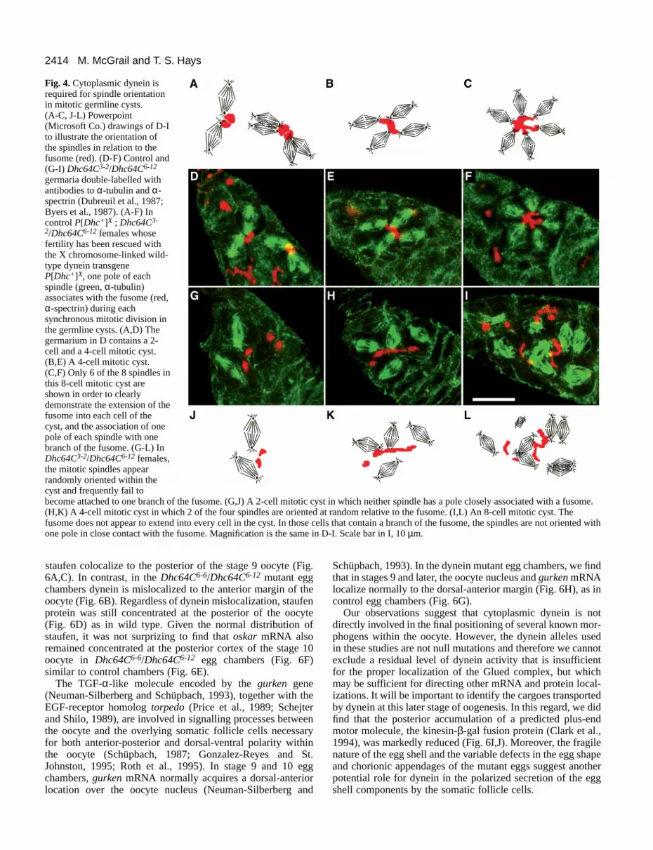
Dhc64C6-12 females carrying one copy of the wild-typeDhc64C transgene, were double-labelled with antibodies to α-tubulin and α-spectrin. As has been reported previously forwild-type cysts (Storto and King, 1989; Lin and Spradling,1995), in control 2-cell (Fig. 4A,D), 4-cell (Fig. 4B,E) and 8-cell (Fig. 4C,F) mitotic cysts the spindles were arranged incompact clusters with one spindle pole in close contact with asingle lobe or branch of the fusome. In contrast, in the mutantcysts the spindles frequently did not appear to contact thefusome, but instead were randomly oriented in the cyst (Fig.4G-L). In the 2-cell mutant cyst shown in Fig. 4G, neitherspindle has a pole in close contact with fusome. α-spectrinlocalization revealed that occasionally the fusome was lessbranched and failed to extend into every cell of the cyst (Fig.4H,I). However, in those cells that contained a branch of thefusome, the spindles were not oriented with one pole close tothe fusome (Fig. 4 arrows in H,L).
The defect in spindle orientation suggested that dyneinfunction might be directly involved in the mechanism ofspindle orientation. We examined dynein localization in wild-type mitotic cysts and detected an enriched, punctate patternof dynein centrally located in a region of the cyst that wouldcorrespond to the position of the fusome (Fig. 5A-H). We didnot detect an enrichment of dynein on a fusome-like structurein interphase cysts (data not shown). In the 2-cell anaphase cystand the 4-cell metaphase cyst, each spindle (green, Fig. 5A,E)was oriented with one pole in close association to the area ofdynein staining (red, Fig. 5A,E). The individual dynein (Fig.5B,F), tubulin (Fig. 5C,G), and chromatin (Fig. 5D,H) imagesare shown. These data suggest that spindle attachment to thefusome is mediated by cytoplasmic dynein. This mechanismwould ensure the fixed pattern of cystocyte divisions that leadto a polarized germline cyst.
Cytoplasmic dynein is required in late stage eggchambers for oocyte differentiation In addition to its role in the early events of oocyte determina-tion and differentiation, the microtubule cytoskeleton has beenimplicated in the establishment of axial polarity and the local-ization of morphogens within the developing oocyte(Pokrywka and Stephenson, 1991; Theurkauf et al., 1992; Laneand Kalderon, 1994). Female adults doubly heterozygous forthe dynein alleles Dhc64C6-6 and Dhc64C6-12 produce latestage egg chambers but are female sterile (Table 2). We showedpreviously that the localization of dynein to the posterior of thestage 9 oocyte is disrupted in egg chambers derived fromDhc64C6-6/Dhc64C6-12 females. Instead dynein accumulates ina punctate pattern at the anterior margin of the oocyte (McGrailet al., 1995). We further demonstrated that the posterior accu-mulation of a dynein-activating complex, the Glued complex,is dependent on cytoplasmic dynein. The wild-type Gluedcomplex is mislocalized along with the dysfunctional dyneinto the oocyte anterior (McGrail et al., 1995). Using this mutantbackground, we have investigated whether the cytoplasmicdynein motor is required in later stages of oocyte differen-tiation for the positioning of known morphogens within theoocyte.
The staufen gene encodes a protein with a double-strandedRNA-binding motif which is required for localization of oskarmRNA to the posterior of the oocyte (St. Johnston et al., 1991).In egg chambers derived from control females, dynein and
2414 M. McGrail and T. S. Hays
Fig. 4. Cytoplasmic dynein isrequired for spindle orientationin mitotic germline cysts. (A-C, J-L) Powerpoint(Microsoft Co.) drawings of D-Ito illustrate the orientation ofthe spindles in relation to thefusome (red). (D-F) Control and(G-I) Dhc64C3-2/Dhc64C6-12
germaria double-labelled withantibodies to α-tubulin and α-spectrin (Dubreuil et al., 1987;Byers et al., 1987). (A-F) Incontrol P[Dhc+]X ; Dhc64C3-
2/Dhc64C6-12 females whosefertility has been rescued withthe X chromosome-linked wild-type dynein transgeneP[Dhc+]X, one pole of eachspindle (green, α-tubulin)associates with the fusome (red,α-spectrin) during eachsynchronous mitotic division inthe germline cysts. (A,D) Thegermarium in D contains a 2-cell and a 4-cell mitotic cyst.(B,E) A 4-cell mitotic cyst.(C,F) Only 6 of the 8 spindles inthis 8-cell mitotic cyst areshown in order to clearlydemonstrate the extension of thefusome into each cell of thecyst, and the association of onepole of each spindle with onebranch of the fusome. (G-L) InDhc64C3-2/Dhc64C6-12 females,the mitotic spindles appearrandomly oriented within thecyst and frequently fail tobecome attached to one branch of the fusome. (G,J) A 2-cell mitotic cyst in which neither spindle has a pole closely associated with a fusome.(H,K) A 4-cell mitotic cyst in which 2 of the four spindles are oriented at random relative to the fusome. (I,L) An 8-cell mitotic cyst. Thefusome does not appear to extend into every cell in the cyst. In those cells that contain a branch of the fusome, the spindles are not oriented withone pole in close contact with the fusome. Magnification is the same in D-I. Scale bar in I, 10 µm.
staufen colocalize to the posterior of the stage 9 oocyte (Fig.6A,C). In contrast, in the Dhc64C6-6/Dhc64C6-12 mutant eggchambers dynein is mislocalized to the anterior margin of theoocyte (Fig. 6B). Regardless of dynein mislocalization, staufenprotein was still concentrated at the posterior of the oocyte(Fig. 6D) as in wild type. Given the normal distribution ofstaufen, it was not surprizing to find that oskar mRNA alsoremained concentrated at the posterior cortex of the stage 10oocyte in Dhc64C6-6/Dhc64C6-12 egg chambers (Fig. 6F)similar to control chambers (Fig. 6E).
The TGF-α-like molecule encoded by the gurken gene(Neuman-Silberberg and Schüpbach, 1993), together with theEGF-receptor homolog torpedo (Price et al., 1989; Schejterand Shilo, 1989), are involved in signalling processes betweenthe oocyte and the overlying somatic follicle cells necessaryfor both anterior-posterior and dorsal-ventral polarity withinthe oocyte (Schüpbach, 1987; Gonzalez-Reyes and St.Johnston, 1995; Roth et al., 1995). In stage 9 and 10 eggchambers, gurken mRNA normally acquires a dorsal-anteriorlocation over the oocyte nucleus (Neuman-Silberberg and
Schüpbach, 1993). In the dynein mutant egg chambers, we findthat in stages 9 and later, the oocyte nucleus and gurken mRNAlocalize normally to the dorsal-anterior margin (Fig. 6H), as incontrol egg chambers (Fig. 6G).
Our observations suggest that cytoplasmic dynein is notdirectly involved in the final positioning of several known mor-phogens within the oocyte. However, the dynein alleles usedin these studies are not null mutations and therefore we cannotexclude a residual level of dynein activity that is insufficientfor the proper localization of the Glued complex, but whichmay be sufficient for directing other mRNA and protein local-izations. It will be important to identify the cargoes transportedby dynein at this later stage of oogenesis. In this regard, we didfind that the posterior accumulation of a predicted plus-endmotor molecule, the kinesin-β-gal fusion protein (Clark et al.,1994), was markedly reduced (Fig. 6I,J). Moreover, the fragilenature of the egg shell and the variable defects in the egg shapeand chorionic appendages of the mutant eggs suggest anotherpotential role for dynein in the polarized secretion of the eggshell components by the somatic follicle cells.

2415Dynein function in oocyte differentiation
Fig. 5. Cytoplasmic dynein is enriched on a structure resembling the fusome in mitotic cysts. (A,E) Wild-type Oregon R germaria triplelabelled with antibodies to the cytoplasmic dynein heavy chain (Hays et al., 1994) and α-tubulin, and the chromatin stain OliGreen. In wild-type mitotic cysts cytoplasmic dynein (red) accumulates in an area of the cyst where the fusome normally resides. One pole of each spindle(green) in the 2-cell anaphase cyst (A) and the 4-cell metaphase cyst (E) appears close to the area of enriched cytoplasmic dynein staining.Chromatin is shown in blue. Below panels A and E the individual dynein heavy chain (B,F), α-tubulin (C,G), and chromatin (D,H) images areshown. Magnification is the same in A and E. Scale bar in E, 10 µm.
DISCUSSION
In this report we have presented evidence that the microtubulemotor cytoplasmic dynein is required at two stages ofDrosophila oogenesis. Early in oogenesis dynein functionappears to mediate spindle orientation during the asymmetriccell divisions that give rise to the germline cyst. The disrup-tion of spindle orientation in dynein mutant backgrounds cor-relates with a defect in cyst polarity and a failure to differen-tiate an oocyte in the dynein mutant cyst. Other dynein allelesdo not block these early stages of oocyte determination, but
Fig. 6. Cytoplasmic dynein is not directly involved in morphogenlocalization. In heteroallelic dynein mutant Dhc64C6-6/Dhc64C6-12
females the localization patterns of mRNAs and proteins required foraxis specification are not disrupted. (A,C,E,G,I) Control and(B,D,F,H,J) mutant Dhc64C6-6/Dhc64C6-12 egg chambers labelledwith probes to dynein (A,B) and staufen (C,D), oskar mRNA (E,F),gurken mRNA (G,H), and β-galactosidase (I,J). (A-D) Stage 9 eggchambers double-labelled with the dynein antibody PEP1 (A,B; Li etal., 1994) and an antibody to staufen (C,D). In the mutant eggchambers dynein is mislocalized to the anterior margin (B) whilestaufen is enriched at the posterior pole of the oocyte (D). In controlegg chambers, dynein (A) and staufen (C) co-localize at the oocyteposterior. (E, F) oskar mRNA is enriched at the oocyte posterior inDhc64C6-6/Dhc64C6-12 stage 10 egg chambers (F) as in control eggchambers (E). G, H gurken mRNA is localized in a dorsal anteriorposition over the oocyte nucleus in the mutant egg chambers (H) asin control chambers (G). I, J In the Dhc64C6-6/Dhc64C6-12 eggchambers the kinesin-β-gal fusion protein is localized to the oocyteposterior (J), however the amount of staining is reduced compared tocontrol chambers (I). Magnification is the same in all panels. Scalebar in H, 50 µm.
still result in female sterility and demonstrate a second dyneinfunction in the subsequent differentiation of the oocyte. Ourresults provide a direct demonstration that the microtubule

2416 M. McGrail and T. S. Hays
motor cytoplasmic dynein is required for oocyte differen-tiation. These studies significantly extend the evidence thatmicrotubule-based transport is central to the process of oocytedifferentiation.
Cytoplasmic dynein is required for oocytedifferentiationMicrotubule-based transport has been implicated in the deter-mination of oocyte fate and the subsequent specification ofaxial polarity within the oocyte (reviewed by St Johnston,1995; Lehmann, 1995; Theurkauf, 1994). The establishment ofoocyte fate and axial polarity can both be characterized asmultistep processes. In the case of oocyte fate determination,the initial step may be the asymmetric segregation of thefusome precursor, or spectrosome, during the first division ofthe cystoblast (Lin and Spradling, 1995). In a second step,asymmetric cell division generates a polarized 16 cell germlinecyst. The spectrin-rich fusome organelle serves to anchor asingle mitotic spindle pole at each of four divisions and ensuresa fixed pattern of interconnections between the 16 cells in acyst (Telfer, 1975; Storto and King, 1989). The pattern of inter-connections within the cyst is reflected in the branching of thefusome material that extends into and connects each cell of thecyst. As a final step, a polarized array of microtubules isnucleated from a single microtubule organizing center(MTOC) within the pro-oocyte and extends into the nurse cellcomplex (Mahowald and Strassheim, 1970; Theurkauf et al.,1993). One hypothesis is that the assembly of this polarizedmicrotubule array may depend on the fusome (Lin andSpradling, 1995). Directed transport of specific mRNAs andcytoplasmic constituents along these microtubules and into thepro-oocyte is required for the continued differentiation of theoocyte (reviewed by Spradling, 1993).
Our previous studies on cytoplasmic dynein led us topropose that the minus-end motor might participate in thetransport of determinants along the polarized microtubule arrayfrom the nurse cell cytoplasm to the pro-oocyte (Li et al.,1994). This was based on the observation that the dynein heavychain accumulated in the pro-oocyte, in contrast to the pre-dominant expression of the Dhc64C dynein heavy chain tran-script in the nurse cell complex (Li et al., 1994). In addition,the temporal and spatial pattern of dynein accumulation to thepro-oocyte was similar to Bicaudal-D, and was dependent onthe products of both the Bicaudal-D and egalitarian genes.Given that mutations in Bicaudal-D and egalitarian appear todisrupt the establishment and maintenance of the polarizedmicrotubule array in the 16-cell cyst (Theurkauf et al., 1993),the failure to accumulate dynein in a single cell might reflectthe absence of a microtubule network along which the dyneinmotor would translocate. In this case, the block in oocyte deter-mination by mutations in Dhc64C, egl, or Bic-D would reflecta disruption of the polarized transport system and the failureto accumulate some necessary determinant in the pro-oocyte.
The analyses reported here indicate that dynein function isrequired within the germline of the 16-cell cyst, but at an earlierstep in oocyte determination. Our observation that Bicaudal-Ddoes not accumulate in a single cell in dynein mutant cysts,together with the absence of a clearly defined pro-oocyte pos-itioned at the posterior of the mutant cysts, suggests that cyto-plasmic dynein is required to generate a polarized germlinecyst. The aberrant pattern of ring canals that we observed in
mosaic egg chambers can be explained by disruption of thefixed pattern of cystocyte divisions. In support of this expla-nation, we observed disrupted spindle orientations in germarialmitotic cysts from dynein mutant ovaries and suggest that onefunction of dynein during cyst formation is to mediate properspindle orientation. Spindle orientation is important fordirecting the fixed pattern of cell divison and the asymmetricsegregation of existing ring canals as well as fusome materialduring the formation of the polarized germline cyst. In theabsence of the proper polarity within the cyst, the transportsystem may fail to assemble or assemble incorrectly and oocytedetermination could be blocked. Our studies have not elimi-nated the possibility that dynein also functions shortly aftercyst formation as part of a transport system that delivers factorsinto the pro-oocyte which are necessary for maintaining thepathway toward oocyte differentiation. However, if Bic-D isrequired to establish the polarized microtubule substrate alongwhich dynein is proposed to translocate, then it is not clear whyBic-D accumulation is blocked in dynein mutant eggchambers. In the absence of motor function, why should micro-tubule assembly be disrupted? Perhaps dynein is requiredtogether with Bic-D and egl to organize the polarized micro-tubule array? Further work is necessary to address this questiondirectly and to identify potential determinants as cargoes of thedynein motor.
Once oocyte fate is established, polarized transport alongmicrotubules could direct the positioning of molecules withinthe oocyte in later stages of differentiation. For example,microtubules have been implicated in the positioning of theoocyte nucleus to the dorsal-anterior corner of the oocyte, thelocalization of oskar mRNA and staufen protein to theposterior pole plasm, and the enrichment of bicoid mRNA atthe anterior margin of the oocyte (reviewed by Theurkauf,1994; St. Johnston, 1995). We previously showed that dyneinaccumulates at the posterior end of the growing oocyte at atime when other morphogenetic proteins and mRNAs alsobecome enriched in the posterior pole plasm (Li et al., 1994).Moreover, dynein accumulation is disrupted by microtubuleinhibitors (J. T. Robinson and T. S. Hays, unpublished obser-vations) and exhibits gene product requirements (e.g., capu andspire) similar to those observed for the posterior localizationof known determinants (Li et al., 1994).
The functional significance of the posterior accumulation ofthe dynein motor is suggested by a female-sterile heteroalleliccombination of Dhc64C alleles, Dhc64C6-6/Dhc64C6-12, inwhich dynein is enriched at the anterior, rather than posterior,pole of the oocyte. This mislocalization of the mutant dyneinmotor disrupts the posterior positioning of the associatedcomplex, Glued (McGrail et al., 1995). Despite these obser-vations, in the present study we found no evidence for mislo-calization of staufen protein, oskar mRNA, or gurken mRNAin the Dhc64C6-6/Dhc64C6-12 background. One interpretationof these results is that dynein motor function may not becritical to the positioning of morphogens during oogenesis.Alternatively, given that the Dhc64C alleles Dhc64C6-6 andDhc64C6-12 are not null mutations, our results cannot excludethe possibility that a residual level of dynein function is suf-ficient for oskar mRNA, gurken mRNA, and staufen proteinlocalization, but insufficient for Glued localization. Interest-ingly, we did observe an apparent reduction in the accumula-tion of the kinesin-β-gal fusion protein at the posterior pole of

2417Dynein function in oocyte differentiation
the oocyte in the dynein mutant egg chambers. This observa-tion raises the possibility that dynein may be required for someother aspect of cytoplasmic organization (e.g., microtubulearchitecture) within the oocyte. While the underlying basis ofthe sterile phenotype remains to be elucidated, sterility is notapparently related to a disruption of the proper accumulationof oskar mRNA, gurken mRNA, or staufen protein in thedeveloping oocyte. Further studies will need to addresswhether a somatic function for dynein in follicle cell secretionmay contribute to the late defect in oogenesis. The presentwork provides evidence that cytoplasmic dynein may servemultiple functions during distinct stages of oocyte differen-tiation. Future studies of cytoplasmic dynein function inDrosophila oogenesis may help to reveal how a single motoraccomplishes multiple tasks.
A novel role for cytoplasmic dynein in asymmetriccell division Storto and King (1989) proposed that spindle positioningensures the unequal inheritance of the ring canals and associ-ated fusome material at each round of mitosis. This mechanismis believed to account for the production of a branched chainof interconnected cystocytes, in which the two cells with fourring canals and the largest amount of fusome material becomepro-oocytes. How the selection is made for one of the two cellsto continue to differentiate as the oocyte, while the other takesup a nurse cell fate is not clear. However, a recent model hasaddressed this issue and proposed that the future single pro-oocyte is initially determined by the asymmetric segregation ofthe spectrosome at the first cystoblast division. The identity ofthe pro-oocyte is maintained at each division by the asymmet-ric inheritance of the fusome material, which leads to apolarized fusome (Lin and Spradling, 1995). The polarizedfusome may provide the information necessary to subsequentlyassemble a polarized microtubule array and establish directedtransport to the pro-oocyte. Mutations that disrupt the assemblyof the fusome and oocyte differentiation, including mutationsin the fusome components α-spectrin and the adducin-like hu-li tai shao gene product, foster the view that fusome functionis critical in oocyte differentiation (Yue and Spradling, 1992;Lin et al., 1994b; de Cuevas et al., 1996). Our analysis ofmitotic germline cysts in the dynein mutant germaria revealedthat mitotic spindles fail to acquire the proper orientation withrespect to the fusome, and the normal pattern of ring canal con-nections and cyst polarity is disrupted. These results supportthe hypothesis that spindle orientation and asymmetric celldivision are critical for oocyte determination.
Localization of cytoplasmic dynein to the fusomeand the mechanism of spindle orientationThe localization of cytoplasmic dynein to the fusome suggestsa simple model for the mechanism of asymmetric cell divisionin Drosophila germline mitotic cysts. Anchored to the fusomethrough its interaction with spectrin and adducin, the dyneinmotor acts upon astral microtubules to rotate the mitoticspindles into alignment. An analogous role has been proposedfor cytoplasmic dynein in the positioning of the nucleus andspindle into the bud neck during mitosis in S. cerevisae (Li etal., 1993; Eschel et al., 1993; Yeh et al., 1995). Moreover, recentstudies have suggested that dynein attachment to membranevesicles in mammalian cells is mediated through the binding of
dynactin to a spectrin and adducin-containing membraneskeleton (Holleran et al., 1996). The observation that cytoplas-mic dynein appeared enriched on the fusome only in mitoticcysts suggests a cell cycle-dependent regulation of dynein asso-ciation with the fusome. Previous investigators have reportedthat levels of phosphorylation correlate with the cell cycle-dependent association of dynein with membrane vesicles(Niclas et al., 1996), as well as the redistribution of dynein froma vesicular to a diffuse cytoplasmic pool (Lin et al., 1994a).
An alternative interpretation of dynein enrichment on thefusome is that dynein participates in the assembly and functionof the fusome during cell division and cyst formation. Defectsin the assembly or integrity of the fusome could also result inaberrant spindle orientation and an altered pattern of celldivison. Dynein association with the fusome could reflectdynein attachment to microtubules within the fusome andminus-end motor activity could act to organize the polarizedmicrotubule array during cyst formation. Mutations in thefusome components α-spectrin (de Cuevas et al., 1996) and theDrosophila adducin homolog hu-li tai shao (Yue and Spradling,1992; Lin et al., 1994b) disrupt cyst formation and oocytedifferentiation. However, in these mutant cysts the fusomeappears absent, or nearly absent. In dynein mutant cysts themorphology of the fusome was frequently abnormal, howeverspindle poles present in proximity to branched arms of thefusome still often failed to attach. This result suggests thatwhile dynein association with the fusome may serve otherfunctions, it appears to be required for attachment of themitotic spindle pole to the fusome.
Our previous analyses of cytoplasmic dynein in Drosophilademonstrated the requirement for dynein function in cellviability during development (Gepner et al., 1996). The resultspresented here demonstrate a specific developmental role forcytoplasmic dynein in cellular differentiation, and provideevidence for a novel function for cytoplasmic dynein in asym-metric cell division that is critical to the differentiation of theDrosophila oocyte. Together with recent studies on the role ofmyosins in asymmetric cell division in yeast and C. elegans(Jansen et al., 1996; Bobolo et al., 1996; Guo and Kemphues,1996) our observations on dynein function in Drosophilaoogenesis underscore the importance of cytoskeletal motors incellular differentiation during development.
We thank S. Miklasz of the Immunological Resource Center (Uni-versity of IL Urbana/Champaign), S. O’Rourke and M. Serr for prepa-ration and characterization of the P1H4 ascites. We also thank K.Boylan for the affinity purification of the dynein heavy chain antiserum,M. Sanders, J. Sedgewick and J. Oja for advice and assistance with NIHImage and Photoshop, and P. Lefebvre, J. Yost and J. Essner forcomments on the manuscript. We are grateful to T. Schüpbach, R.Lehmann and E. Stephenson for cDNAs, I. Clark and N. Perrimon forfly stocks, and D. Kiehart, P. Macdonald and R. Steward for antibodies.This work was supported by the NIH and the March of Dimes (T. S.H.) and the NSF Training Grant in Cytoskel-etal Biology (NSF/DBI-9602237) at the University of Minnesota (M. M.).
REFERENCES
Berleth, T., Burri, M., Thoma, G., Bopp, D., Richstein, S., Frigerio, G.,Noll, M. and Nüsslein-Volhard, C. (1988). The role of localization of bicoidRNA in organizing the anterior pattern of the Drosophila embryo. EMBO J.7, 1749-1756.

2418 M. McGrail and T. S. Hays
Bobola, N., Jansen, R.-P., Shin, T. H. and Nasmyth, K. (1996). Asymmetricaccumulation of Ash1p in post anaphase nuclei depends on a myosin andrestricts yeast mating-type switching to mother cells. Cell 84, 699-709.
Byers, T. J., Dubreuil, R. R., Branton, D., Keihart, D. P. and Goldstein, L. S.B. (1987). Drosophila spectrin. II. Conserved features of the alpha-subunitare revealed by analysis of cDNA clones and fusion proteins. J. Cell Biol.105, 2103-2110.
Cheng, N. N., Kirby, C. M. and Kemphues, K. J. (1995). Control of cleavagespindle orientation in C. elegans: the role of the genes par-2 and par-3.Genetics 139, 549-559.
Chou, T.-B., Noll, E. and Perrimon, N. (1993). Autosomal P[ovoD1] dominantfemale-sterile insertions in Drosophila and their use in generating germ-linechimeras. Development 119, 1359-1369.
Clark, I., Giniger, E., Ruohola-Baker, H., Jan, L. Y. and Jan, Y. N. (1994).Transient posterior localization of a kinesin fusion protein reflectsanteroposterior polarity of the Drosophila oocyte. Curr. Biol. 4, 289-300.
Cox, K., DeLeon, D. V., Angerer, L. M. and Angerer, R. C. (1984). Detectionof mRNAs in sea urchin embryos by in situ hybridization using asymmetricRNA probes. Dev. Biology 101, 485-502.
de Cuevas, M., Lee, J. K. and Spradling, A. C. (1996). alpha-spectrin isrequired for germline cell division and differentiation in the Drosophilaovary. Development 122, 3959-3968.
Doe, C. Q. (1996). Spindle orientation and asymmetric localization inDrosophila: both inscuteable? Cell 86, 695-697.
Dubreuil, R. R., Byers, T. J., Branton, D., Goldstein, L. S. B. and Kiehart,D. P. (1987). Drosophila spectrin. I. Characterization of the purified protein.J. Cell Biol. 105, 2095-2102.
Ephrussi, A., Dickinson, L. and Lehmann, R. (1991). oskar organizes thegerm plasm and directs localization of the posterior determinant nanos. Cell66, 37-50.
Eschel, D., Urrestarazu, L. A., Vissers, S., Jauniaux, J.-C., van Vliet-Reedijk, J. C., Planta, R. J. and Gibbons, I. R. (1993). Cytoplasmic dyneinis required for normal nuclear segregation in yeast. Proc. Natl. Acad. Sci.USA 90, 11172-11176.
Etemad-Moghadam, B., Guo, S. and Kemphues, K. J. (1995).Asymmetrically distributed PAR-3 protein contributes to cell polarity andspindle alignment in early C. elegans embryos. Cell 83, 743-752.
Gepner, J., Li, M.-g., Ludmann, S., Kortas, C., Boylan, B., Iyadurai, S. J. P.,McGrail, M. and Hays, T. S. (1996). Cytoplasmic dynein function isessential in Drosophila. Genetics 142, 865-878.
Golic, K. G. and Lindquist, S. (1989). The FLP recombinase of yeastcatalyzes site-specific recombination in the Drosophila genome. Cell 59,499-509.
Golic, K. G. (1991). Site-specific recombination between homologouschromosomes in Drosophila. Science 252, 958-961.
Gonzalez-Reyes, A., Elliott, H. and St. Johnston, D. (1995). Polarization ofboth major body axes in Drosophila by gurken-torpedo signalling. Nature375, 654-658.
Guo, S. and Kemphues, K. J. (1995). par-1, a gene required for establishingpolarity in C. elegans embryos encodes a putative ser/thr kinase that isasymmetrically distributed. Cell 81, 611-620.
Guo, S. and Kemphues, K. J. (1996). A non-muscle myosin required forembryonic polarity in Caenorhabditis elegans. Nature 382, 455-458.
Hays, T. S., Porter, M. E., McGrail, M., Grissom, P., Gosch, P., Fuller, M. T.and McIntosh, J. R. (1994). A cytoplasmic dynein motor in Drosophila:identification and localization during embryogenesis. J. Cell Sci. 107, 1557-1569.
Holleran, E., Tokito, M. K., Karki, S. and Holzbaur, E. L. F. (1996).Centractin (ARP1) associates with spectrin revealing a potentialmechanism to link dynactin to intracellular organelles. J. Cell Biol.135, 1815-1829.
Holzbaur, E. L. F. and Vallee, R. B. (1994). Dyneins: molecular structure andcellular function. Annu. Rev. Cell Biol. 10, 339-372.
Jansen, R.-P., Dowzer, C., Michaelis, C., Galova, M. and Nasmyth, K.(1996). Mother cell-specific HO expression in budding yeast depends on theunconventional myosin Myo4p and other cytoplasmic proteins. Cell 84, 687-697.
Kemphues, K. J., Priess, J. R., Morton, D. G. and Cheng, N. (1988).Identification of genes required for cytoplasmic localization in early C.elegans embryos. Cell 52, 311-320.
King, R. C., Cassidy, J. D. and Rousset, A. (1982). The formation of clones ofinterconnected cells during oogenesis in insects. In Insect Ultrastructure (ed.R. C. King and H. Akai), pp. 3-31. Plenum Press, New York.
Kraut, R., Chia, W., Jan, L. Y., Jan, Y. N. and Knoblich, J. A. (1996). Role of
inscuteable in orienting asymmetric cell divisions in Drosophila. Nature 383,50-55.
Lane, M. E. and Kalderon, D. (1994). RNA localization along theanteroposterior axis of the Drosophila oocyte requires PKA-mediatedtransduction to direct normal microtubule organization. Genes Dev. 8, 2986-2995.
Lehmann, R. (1995). Cell-cell signaling, microtubules and the loss ofsymmetry in the Drosophila oocyte. Cell 83, 353-356.
Li, Y. Y., Yeh, E., Hays, T. S. and Bloom, K. (1993). Disruption of mitoticspindle orientation in a yeast dynein mutant. Proc. Natl. Acad. Sci. USA 90,10096-10100.
Li, M.-g., McGrail, M., Serr, M. and Hays, T. S. (1994). Drosophilacytoplasmic dynein, a microtubule motor that is asymmetrically localized inthe oocyte. J. Cell Biol. 126, 1475-1494.
Lin, S. X., Ferro, K. L. and Collins, C. A. (1994a). Cytoplasmic dyneinundergoes intracellular redistribution concommitant with phosphorylation ofthe heavy chain in response to serum starvation and okadaic acid. J. Cell Biol.127, 1009-1019.
Lin, H., Yue, L. and Spradling, A. C. (1994b). The Drosophila fusome, agermline-specific organelle, contains membrane skeletal proteins andfunctions in cyst formation. Development 120, 947-956.
Lin, H. and Spradling, A. C. (1995). Fusome asymmetry and oocytedetermination in Drosophila. Dev. Genetics 16, 6-12.
Mahajan-Miklos, S and Cooley, L. (1994). Intercellular cytoplasm transportduring Drosophila oogenesis. Dev. Biol. 165, 336-351.
Mahowald, A.P. and Strassheim, J. M. (1970). Intercellular migration ofcentrioles in the germarium of Drosophila melanogaster. An electronmicroscopic study. J. Cell Biol. 45, 306-320.
McGrail, M., Gepner, J., Silvanovich, A., Ludmann, S., Serr, M. and Hays,T. S. (1995). Regulation of cytoplasmic dynein function in vivo by theDrosophila Glued complex. J. Cell Biol. 131, 411-425.
Neuman-Silberberg, F. S. and Schüpbach, T. (1993). The Drosophiladorsoventral patterning gene gurken produces a dorsally localized RNA andencodes a TGFα-like protein. Cell 75, 165-174.
Niclas, J., Allan, V. J. and Vale, R. D. (1996). Cell cycle regulation of dyneinassociation with membranes modulates microtubule-based organelletransport. J. Cell Biol. 133, 585-593.
Paschal, B. M. and Vallee, R. B. (1987). Retrograde transport by themicrotubule-associated protein MAP1C. Nature 330, 181-183.
Price, J. V., Clifford, R. J. and Schupbach, T. (1989). The maternalventralizing locus torpedo is allelic to faint little ball, an embryonic lethaland encodes the Drosophila EGF receptor homolog. Cell 56, 1085-1092.
Pokrywka, N. J. and Stephenson, E. C. (1991). Microtubules mediate thelocalization of bicoid RNA during Drosophila oogenesis. Development 113,55-66.
Porter, M. E. (1996). Axonemal dyneins: assembly, organization andregulation. Curr. Opin. Cell Biol. 8, 10-17.
Roth, S., Neuman-Silberberg, F. S., Barcelo, G. and Schüpbach, T. (1995).cornichon and the EGF receptor signaling process are necessary for bothanterior-posterior and dorsal-ventral pattern formation in Drosophila. Cell81, 967-978.
Rhyu, M. S. and Knoblich, J. A. (1995). Spindle orientation and asymmetriccell fate. Cell 82, 523-526.
St. Johnston, D. (1995). The intracellular localization of messenger RNAs.Cell 81, 161-170.
St. Johnston, D., Beuchle, D. and Nüsslein-Volhard, C. (1991). staufen, agene required to localize maternal RNAs in the Drosophila egg. Cell 66, 51-63.
Schejter, E. D. and Shilo, B.-Z. (1989). The Drosophila EGF receptorhomologue (DER) is allelic to faint little ball, a locus essential for embryonicdevelopment. Cell 56, 1091-1101.
Schüpbach, T. (1987). Germ line and soma cooperate during oogenesis toestablish the dorsoventral pattern of egg shell and embryo in Drosophilamelanogaster. Cell 49, 699-707.
Schroer, T. A. (1994). Structure, function and regulation of cytoplasmicdynein. Curr. Opin. Cell Biol. 6, 69-73.
Spradling, A. C. (1993). Developmental genetics of oogenesis. In TheDevelopment of Drosophila melanogaster (ed. M. Bate and A. Martinez-Arias), pp. 1-70. Cold Spring Harbor Laboratory Press, Cold Spring Harbor,New York.
Storto, P. D. and King, R. C. (1989). The role of polyfusomes in generatingbranched chains of cystocytes during Drosophila oogenesis. Dev. Genet. 10,70-86.
Strome, S. (1993). Determination of cleavage planes. Cell 72, 3-6.

2419Dynein function in oocyte differentiation
Suter, B. and Steward, R. (1991). Requirement for phosphorylation andlocalization of the Bicaudal-D protein in Drosophila oocyte differentiation.Cell 67, 917-926.
Tautz, D. and Pfeifle, C. (1989). A non-radioactive in situ hybridizationmethod for the localization of specific RNAs in Drosophila embryos revealstranslational control of the segmentation gene hunchback. Chromosoma 98,81-85.
Telfer, W. H. (1975). Development and physiology of the oocyte-nurse cellsyncytium. Adv. Insect Physiol. 11, 223-319.
Theurkauf, W. E., Smiley, S., Wong, M. L. and Alberts, B. M. (1992).Reorganization of the cytoskeleton during Drosophila oogenesis:implications for axis specification and intercellular transport. Development115, 923-936.
Theurkauf, W. E., Alberts, B. M., Jan, Y. N. and Jongens, T. A. (1993). Acentral role for microtubules in the differentiation of Drosophila oocytes.Development 118, 1169-1180.
Theurkauf, W. E. (1994). Microtubules and cytoplasm organization duringDrosophila oogenesis. Dev. Biol. 165, 352-360.
Vallee R. B. and Sheetz, M. P. (1996). Targeting of motor proteins. Science271, 1539-1544.
White, J and Strome, S. (1996). Cleavage plane specification in C. elegans:how to divide the spoils. Cell 84, 195-198.
Yeh, E., Skibbens, R. V., Cheng, J. W., Salmon, E. D. and Bloom K. (1995).Spindle dynamics and cell cycle regulation of dynein in the budding yeast,Saccharomyces cerevisiae. J. Cell Biol. 130, 687-700.
Yue, L. and Spradling, A. (1992). hu-li tai shao, a gene required for ring canalformation during Drosophila oogenesis, encodes a homolog of adducin.Genes Dev. 6, 2443-2454.
(Accepted 9 April 1997)


