Embed Size (px)
Citation preview

Citation: Schweitzer-Stenner, R.
Exploring Nearest Neighbor
Interactions and Their Influence on
the Gibbs Energy Landscape of
Unfolded Proteins and Peptides. Int.
J. Mol. Sci. 2022, 23, 5643. https://
doi.org/10.3390/ijms23105643
Received: 22 April 2022
Accepted: 13 May 2022
Published: 18 May 2022
Publisher’s Note: MDPI stays neutral
with regard to jurisdictional claims in
published maps and institutional affil-
iations.
Copyright: © 2022 by the author.
Licensee MDPI, Basel, Switzerland.
This article is an open access article
distributed under the terms and
conditions of the Creative Commons
Attribution (CC BY) license (https://
creativecommons.org/licenses/by/
4.0/).
International Journal of
Molecular Sciences
Review
Exploring Nearest Neighbor Interactions and Their Influence onthe Gibbs Energy Landscape of Unfolded Proteins and PeptidesReinhard Schweitzer-Stenner
Department of Chemistry, Drexel University, Philadelphia, PA 19104, USA; [email protected];Tel.: +1-215-895-2268
Abstract: The Flory isolated pair hypothesis (IPH) is one of the corner stones of the random coilmodel, which is generally invoked to describe the conformational dynamics of unfolded and intrinsi-cally disordered proteins (IDPs). It stipulates, that individual residues sample the entire stericallyallowed space of the Ramachandran plot without exhibiting any correlations with the conformationaldynamics of its neighbors. However, multiple lines of computational, bioinformatic and experimentalevidence suggest that nearest neighbors have a significant influence on the conformational samplingof amino acid residues. This implies that the conformational entropy of unfolded polypeptides andproteins is much less than one would expect based on the Ramachandran plots of individual residues.A further implication is that the Gibbs energies of residues in unfolded proteins or polypeptides arenot additive. This review provides an overview of what is currently known and what has yet to beexplored regarding nearest neighbor interactions in unfolded proteins.
Keywords: unfolded proteins; isolated pair hypothesis; nearest neighbor interactions; chemical shiftsand J-coupling; conformational entropy
1. Introduction
For a long period of time the unfolded states of proteins have not attracted considerableinterest in the protein community because their basic properties seemed to be obvious andgeneric. Following early models of Flory, it was assumed that unfolded peptide chains aredescribable by the random coil model irrespective of its amino acid residues sequence [1–3].Locally, it was assumed that the latter sample the entire sterically allowed region of theRamachandran plot, which is rather similar for different amino acid residues, glycine andproline being the exceptions (cf. Figure 1). Hence, the unfolded state has been thought tobe just a reservoir of conformational entropy that has to be overcome for a protein to fold.
This view has changed over the last 25 years for a variety of reasons. First, thediscovery of intrinsically disordered proteins (IDPs) that perform biological functionsrevealed that the nature of their amino acid residues must play a role, since the results ofbioinformatic analyses suggested that polar and charged amino acid residues occur morefrequently in IDPs than they do in folded proteins [4–7]. IDPs are of particular relevancefor cell signaling processes that frequently involve a transition between disordered andfolded states. Intrinsically disordered segments of otherwise folded proteins are involvedin pivotal protein–DNA and ligand–receptor interactions [5,8]. Some IDPs are of greatbiomedical relevance in that they are prone to self-assembly into amyloid fibrils [9–11].Canonical examples are the amyloid β-peptides Aβ1-40 and Aβ1-42, α-synuclein and thetau protein [12–14].
The second reason for the growing interest in unfolded proteins and peptides isbased on the discovery that, contrary to Flory’s assumption and a belief cultivated overdecades, individual amino acid residues do sample a space less than the one dictated bysteric constraints. Moreover, experimental data and coil library analyses revealed thatthe Ramachandran distribution depends very much on the nature of the amino acid side
Int. J. Mol. Sci. 2022, 23, 5643. https://doi.org/10.3390/ijms23105643 https://www.mdpi.com/journal/ijms

Int. J. Mol. Sci. 2022, 23, 5643 2 of 33
chains [15–23]. Multiple lines of evidence lead to the conclusion that the amino acid residuespecificity of conformational distributions is due to specific backbone/side chain–waterinteractions [24–28]. All these results suggest the thermodynamics of unfolded proteinsand IDPs are more complicated than originally anticipated.
Int. J. Mol. Sci. 2022, 23, x FOR PEER REVIEW 2 of 35
The second reason for the growing interest in unfolded proteins and peptides is
based on the discovery that, contrary to Flory’s assumption and a belief cultivated over
decades, individual amino acid residues do sample a space less than the one dictated by
steric constraints. Moreover, experimental data and coil library analyses revealed that the
Ramachandran distribution depends very much on the nature of the amino acid side
chains [15–23]. Multiple lines of evidence lead to the conclusion that the amino acid resi-
due specificity of conformational distributions is due to specific backbone/side chain–wa-
ter interactions [24–28]. All these results suggest the thermodynamics of unfolded pro-
teins and IDPs are more complicated than originally anticipated.
Figure 1. Sterically allowed φ, ψ space proposed by Ramachandran. Solid lines enclose the region
allowed by hard-sphere bumps at standard radii; dashed lines show the region allowed with re-
duced radii; dotted lines add regions allowed if τ (N-Cα-C′) is relaxed slightly. Ψ and φ values run
from −180° to 180°. Taken from [29].
The third reason for the increasing attractiveness of unfolded proteins is that they do
not always behave like an ideal random coil. While the scaling laws obtained for the radii
of gyration and end-to-end distances of a large number of foldable proteins subjected to
denaturing conditions (i.e., high urea concentration) indicate a nearly perfect self-avoid-
ing random walk (i.e., an exponents close to ν = 0.59) [30], IDPs or disordered protein
segments can exhibit scaling laws with exponents spreading over a large range, depend-
ing on the apparent net charge of the investigated sequence [31–35]. Exponents below 0.5
indicate a compact structure, while values above 0.6 reflect extended coils preferably in-
teracting with water [34]. It should be noted in this context that it is known for quite some
time that proteins such as cytochrome c prefer some type of compact molten globule state
under certain denaturing conditions (low or high pH, high temperature, on membrane
surfaces, even in the presence of urea and guanidine hydrochloride at neutral pH) [36,37].
Interestingly, even scaling coefficients between 0.53 and 0.6, indicating ensembles some-
where between an ideal and self-avoiding random coil, do not exclude the possibility that
Figure 1. Sterically allowed ϕ, ψ space proposed by Ramachandran. Solid lines enclose the regionallowed by hard-sphere bumps at standard radii; dashed lines show the region allowed with reducedradii; dotted lines add regions allowed if τ (N-Cα-C′) is relaxed slightly. Ψ and ϕ values run from−180◦ to 180◦. Taken from [29].
The third reason for the increasing attractiveness of unfolded proteins is that theydo not always behave like an ideal random coil. While the scaling laws obtained forthe radii of gyration and end-to-end distances of a large number of foldable proteinssubjected to denaturing conditions (i.e., high urea concentration) indicate a nearly perfectself-avoiding random walk (i.e., an exponents close to ν = 0.59) [30], IDPs or disorderedprotein segments can exhibit scaling laws with exponents spreading over a large range,depending on the apparent net charge of the investigated sequence [31–35]. Exponentsbelow 0.5 indicate a compact structure, while values above 0.6 reflect extended coilspreferably interacting with water [34]. It should be noted in this context that it is known forquite some time that proteins such as cytochrome c prefer some type of compact moltenglobule state under certain denaturing conditions (low or high pH, high temperature,on membrane surfaces, even in the presence of urea and guanidine hydrochloride atneutral pH) [36,37]. Interestingly, even scaling coefficients between 0.53 and 0.6, indicatingensembles somewhere between an ideal and self-avoiding random coil, do not exclude thepossibility that the unfolded proteins exhibit local transient structures [38]. Calculationsreported by Fitzkee and Rose revealed that an unfolded protein can behave like a self-avoiding random coil while still showing considerable local order [39].
Irrespective of the complexities indicated above one might arrive at the conclusionthat if the intrinsic structural propensities of the amino acid residues are known, then theconformational sampling of an unfolded protein could be predicted, and its energeticsand entropic content assessed. Different propensity scales are available in the literature,which could be used to this end [23,40–45]. However, such a modeling of unfolded statesrequires the absence of non-local contacts between the amino acid residues and the validityof Flory’s isolated pair hypothesis (IPH), which states that the conformational dynamics of

Int. J. Mol. Sci. 2022, 23, 5643 3 of 33
residues do not correlate. If both requirements were met residue energetics and entropieswould be additive. Unfortunately, none of the above conditions are generally met. Whilenon-local contacts might be negligible in extended IDPs (solvation energy dominates overintrapeptide interactions), several lines of evidence suggest that the isolated pair hypothesisis not valid irrespective of the specific coil state of the protein.
While the influence of non-local contacts on the compactness and local order ofIDPs and unfolded proteins has been explored in some detail [14,46–49], work on nearestneighbor interactions (NNIs) appears somewhat fragmentized. It is the goal of this reviewto provide an overview over the rather diverse work performed over the last 25 yearsthat was aimed at exploring how NNIs affect conformational propensities of amino acidresidues. We will compare the results of these efforts and shed some light on their relevancefor a thorough understanding of unfolded and disordered proteins.
Even though the term random coil is clearly defined in the polymer literature (as nicelydelineated in [50]), the term is often used as a label for unfolded and disordered proteinsin an indiscriminate way. Strictly speaking, the term can only be used for sufficientlylong polymers comprised of rigid monomers (peptide groups) and freely rotatable linkers,the length dependence of which can be described by a power law for its mean radiusof hydration; i.e., 〈Rh〉 ∼ Nν, where N is the number of peptide units. As indicatedabove, the exponent is 0.59 in a good solvent. Locally, the random coil model is basedon the assumption that amino acid residue conformations are restricted solely by sterichindrances between the side chains as well as between side chains and backbone groupsand by electrostatic interactions [51]. However, as indicated above, peptide/protein-solventinteractions reduce the available conformational space further and increase the anisotropy of theresidue orientations [24,25,27,28,52–55]. Moreover, unfolded and disordered proteins frequentlycontain segments that can transiently adopt regular secondary structures [14,38,49,56]. Hence,real unfolded polypeptides and proteins do not meet the requirements for a random coilon a local level while they still might do so on a more global level [30,39,57]. Therefore,we follow the late Harold Scheraga in that we use the term statistical coil instead, whichcorrectly reflects the fact that different chain conformations differ in terms of their Gibbsfree energies [56,57].
This review is organized as follows. In the first step some basic thermodynamicaspects of NNIs are delineated. The main part of the review is divided into three sections.Part 1 provides some general description of the statistical thermodynamics of NNIs. Thisis followed by Part 2, in which we review the coil library analyses that extracted theconformational propensities and NNI effects from the structure of the denatured proteins.In Part 3, we provide an overview of the computational studies that explored the underlyingphysical mechanism of NNIs. Part 4 describes the experimental studies, mostly on shortpeptides, that were specifically aimed at quantifying the NNIs between different types ofresidues. A summary and outlook finish the review.
2. Thermodynamic Aspects of Nearest Neighbor Interactions
It is now well established that the Ramachandran plot of amino acid residues in un-folded peptides and proteins contains several basins associated with different secondarystructures. The most prominent ones are shown in Figure 1. Generally, based on ex-perimental studies with blocked dipeptides and unblocked GxG guest–host tripeptides(x: host amino acid residue), all residues predominantly populate two basins in the up-per left part of the Ramachandran plot, which are assignable to polyproline II (pPII) andβ-strand (β) (between 70 and 100%) [23,40,42,43]. The basin of the former can be foundin a region between ϕ-values of −90◦ and −60◦ while the ψ-values might vary between130◦ and 180◦. The center of β-strand basins can vary over a range between ϕ-values of−100◦ and −140◦. Corresponding ψ-values vary again between 130◦ and 180◦. This areaencompasses backbone structures associated with different types of β-sheet structures. Thesampling of other basins by residues in the above peptides is somewhat more limited. Themost prominent one is assignable to right-handed helical structures (−70◦ < ϕ < −30◦,
Int. J. Mol. Sci. 2022, 23, 5643 4 of 33
−60◦ < ψ < −20◦), which encompasses canonical α-helical and 310-like conformations.Quite a few residues sample the region of γ (30◦ < ϕ < 70◦, −80◦ < ψ < −50◦) and inverseγ-turns (ϕ, ψ with opposite sign). In specific cases residues were found to sample left-handed helical structures (30◦ < ϕ < 70◦, 20◦ < ψ < 60◦), and conformations in the bridgeregion around ϕ = −60◦ and ψ = 0, which are generally found at the i + 2 position of type Iand II’ β-turns [43,45,58,59]. In addition, aspartic acid and asparagine residues can formasx-turns, supporting the conformations (50◦ < ϕ < 80◦, 120◦ < ψ < 180◦). Asx-turns consistof a sequence of three residues, the first of which is either aspartic acid or asparagine [60].
The population χi,k of these conformations can be calculated as
χi,k =eGi,k/RT
∑k eGi,k/RT (1)
where Gi,k is the Gibbs free energy if the k-th conformation of the i-th residue. R is gasconstant and T the temperature in Kelvin. It can be decomposed as follows:
Gi,k = G0i,k + δG0
ij,k + δGij,kl (2)
where G0i,k is the Gibbs energy of the kth conformation of the ith residue in the absence of
nearest neighbor interactions, δG0ij,k is the Gibbs energy contribution by the jth neighbor
that reflect the steric and physico-chemical properties of the neighbor irrespective of itsadopted conformation and δGij,kl is the conformation-dependent contribution of the jthneighbor in its lth conformation.
Distinguishing between these two contributions to the NNI–Gibbs energy is tech-nically difficult (vide infra) but nevertheless of conceptual significance. If the NNI wascomprised only of δG0
ij,kl contributions, there would be no cooperativity between the in-teracting residues. In such a case, which is comparable to the first-order contribution tothe eigenenergies of quantum mechanical systems, the internal energies, entropies andthus also the Gibbs energies of residues in an unfolded polypeptide would still be additiveand thus the isolated pair hypothesis would still be valid. Therefore, only conformationdependent NNIs are a real game changer in that the population of a certain conformationof a considered residue becomes dependent on the conformation adopted by its neigh-bors. To illuminate the significance of conformation dependent NNIs, let us consider anoligopeptide with only two interacting residues. In the absence of any cooperativity, themole fraction of a conformation pair k, l is calculated as
χkl =e(G
′1,k+G′2,l)/RT
∑k eG′1,k/RT ∑l eG′2,l /RT (3)
where the Gibbs energy terms G′1,k and G′2,l contain the first two terms of Equation (2). Inthe presence of cooperative NNIs, the corresponding equation for χkl looks rather different:
χkl =e(G
′1,k+G′2,l+δG12,kl+δG21,kl)/RT
∑k ∑l e(G′1,k+G′2,l+δG12,kl+δG21,kl)/RT(4)
where we consider the mutual influence of residue 2 on 1 and residue 1 on 2 in thenumerator. The partition sum in the denominator runs over all peptide conformations,which is different from the product of partition sums of individual residues in Equation (3).Obviously, the corresponding conformational Gibbs entropies of the two systems are alsodifferent, namely,
S = −R
∑k(χ1,klnχ1,k) +∑
l(χ1,l lnχ1,l)
(5)

Int. J. Mol. Sci. 2022, 23, 5643 5 of 33
in the absence andS = −R∑
k∑
l(χkl lnχkl) (6)
in the presence of cooperativity.The theory introduced thus far solely considers the possibility that NNIs affect the
Gibbs energies of residue conformations. However, it is equally likely that NNIs changethe equilibrium position of a basin. This can be seen from a simple example. Assume thatthe potential function associated with a basin can be approximated by a harmonic potentialof the type V(q) = 1/2·k(q− q0)
2 where q represents one of the two dihedral coordinates.If the corresponding residue is subjected to a perturbing potential V(q) = aq + b, theequilibrium position shifts from q = q0 to q′0 = a/k + q0. If the q-dependence of theperturbing potential is more complicated, that expression for q0
′ becomes more complicatedas well.
In what follows, we will have to keep the theoretical thoughts of this section in mindwhen we go over the NNI literature.
3. Coil Library Studies
In 1995, two important articles reported what the authors called conformationalpropensities of amino acid residues in coil regions of proteins. Swindells et al. determinedthe Ramachandran distributions of amino acid residues in coil regions of 85 structuresobtained from the protein data bank [15]. They excluded any residues incorporated inright-handed helices and β-sheets to eliminate local and non-local interactions associatedwith the stabilization of these secondary structures. Moreover, they assumed that the thusobtained Ramachandran distributions could reflect intrinsic structural propensities and thusbe representative of the respective conformational distributions in unfolded/denaturedproteins. This conceptual model is based on the assumption that non-local interactionsin these coils are random in nature and averaged out by considering a sufficiently largedata set. Contrary to one of the basic assumptions of Flory’s random coil model, theyfound significant differences between the population distributions of different amino acidresidues. They reported integrated propensities for four regions of the Ramachandranplot shown in Figure 2. If one converts their propensity values into fractions by dividingthe reported propensity values by their sum, one obtains 0.35, 0.21 and 0.33 for the pPII,β-strand and right-handed helical region, respectively, of alanine. For valine, however, thecorresponding fractions are 0.24, 0.45 and 0.22. Swindells et al. found that the obtainedpropensity values correlated with secondary structure propensities for the β-strand whilethe correlation with right-handed helical propensities was found to be weak [15].
Int. J. Mol. Sci. 2022, 23, x FOR PEER REVIEW 6 of 35
Figure 2. Ramachandran distribution of the sterically allowed region of amino acid residues in coil
libraries. The two regions in the upper left and lower left quadrant are associated with β-strand and
polyproline II (p), respectively. The region crossing the ψ = 0 line in the left part of the plot contains
right-handed helical and turn-supporting conformations. Taken from [15] and modified.
In earlier days, Ramachandran plots were indiscriminately derived from protein data
sets because of the belief that all non-intrinsic influences could be averaged out by a suf-
ficiently large data set, irrespective of its content of regular secondary structures. That this
is not the case was demonstrated by Serrano [16]. Figure 3 shows the distribution of ala-
nine obtained from an unrestricted data set (Figure 3A) and from a restricted coil set (hel-
ices and where sheets were omitted (Figure 3B). The difference is striking. While the for-
mer is dominated by an intense peak in the right-handed helical region the latter becomes
maximal in the pPII region. Hence, Serrano’s data clearly reveal the propensity of alanine
for polyproline II six years before experimental results obtained with an hepta-alanine
peptide suggested the same and triggered a highly controversial debate. Details of this
debate are summarized in an earlier review of Toal and Schweitzer-Stenner [22]. Serrano’s
analysis, which was augmented by a comparison of the chemical shifts and 3J(HNHCα),
observed for short model peptides and calculated for coil library distributions, clearly cor-
roborated the notion that different amino acid residues have different intrinsic propensi-
ties for specific conformations.
Nearest neighbor effects were first considered in a third paper by Penkett et al. that
appeared in 1997 [61]. Keeping in mind the well-stablished fact that the helical and β-sheet
propensities of amino acid residues are context dependent they were wondering whether
such context dependencies would also be observable in the unfolded proteins for which
they assumed minimal non-local interactions. To this end, they used 1H NMR to deter-
mine the 3J(HNHCα) coupling constants for a 130-residue fragment of the fribronectin-bind-
ing proteins from Staphyloccus aureus. They observed that 9 of 16 glutamic acid residues
preceded by residues with either branched or aromatic side chains exhibited coupling
constants between 6.2 and 7.0 Hz, while the other 7 with asparagine or glutamate as up-
stream neighbors lie between 5.7 and 6.3 Hz.
Further insight into the underlying NNIs came from the average 3J(HNHCα) values
calculated for the coil library distributions of Swindells et al. [15]. These calculations were
performed with a Karplus equation that relates the coupling constant to the dihedral back-
bone angle φ as follows [58]:
𝐽(𝑥, 𝑦, η) = 𝐴𝑐𝑜𝑠2(η + θ1) + 𝐵𝑐𝑜𝑠(η + θ2) + 𝐶 (7)
where x and y denote the interacting nuclei x and y, η = φ, ψ, and θi (I = 1,2) are phase
angles. A, B and C are empirical Karplus parameters obtained by fitting Equation (9) to
the J-coupling data sets obtained for proteins with a known crystal structure [59,62–65].
Alternatively, these parameters could be obtained with density functional theory calcula-
tions but this has been accomplished thus far only for alanine [66]. It is likely that the exact
Figure 2. Ramachandran distribution of the sterically allowed region of amino acid residues in coillibraries. The two regions in the upper left and lower left quadrant are associated with β-strand andpolyproline II (p), respectively. The region crossing the ψ = 0 line in the left part of the plot containsright-handed helical and turn-supporting conformations. Taken from [15] and modified.

Int. J. Mol. Sci. 2022, 23, 5643 6 of 33
In earlier days, Ramachandran plots were indiscriminately derived from protein datasets because of the belief that all non-intrinsic influences could be averaged out by asufficiently large data set, irrespective of its content of regular secondary structures. Thatthis is not the case was demonstrated by Serrano [16]. Figure 3 shows the distributionof alanine obtained from an unrestricted data set (Figure 3A) and from a restricted coilset (helices and where sheets were omitted (Figure 3B). The difference is striking. Whilethe former is dominated by an intense peak in the right-handed helical region the latterbecomes maximal in the pPII region. Hence, Serrano’s data clearly reveal the propensityof alanine for polyproline II six years before experimental results obtained with an hepta-alanine peptide suggested the same and triggered a highly controversial debate. Detailsof this debate are summarized in an earlier review of Toal and Schweitzer-Stenner [22].Serrano’s analysis, which was augmented by a comparison of the chemical shifts and3J(HNHCα), observed for short model peptides and calculated for coil library distributions,clearly corroborated the notion that different amino acid residues have different intrinsicpropensities for specific conformations.
Int. J. Mol. Sci. 2022, 23, x FOR PEER REVIEW 7 of 35
parameters are side-chain dependent. Empirical values should therefore be considered as
an average. In the case of unfolded and disordered proteins and peptides, the measured
Karplus parameters represent a conformational average:
⟨𝐽(𝑥, 𝑦)⟩ =∑ 𝐽(𝑥, 𝑦, η𝑖)𝑃(η𝑖)𝑁𝑖=1
∑ 𝑃(η𝑖)𝑁𝑖+1
(8)
where P(ηi) is the probability for the residue to adopt a dihedral angle φi or ψi.
Figure 3. Conformational distribution of alanine residues inferred from the protein data sets. (A)
The complete data set including residues in well-defined secondary structures. (B) Only alanine
residues in coils outside of right-handed helical and β-sheet structures were considered. Each num-
ber on the abscissae represents an 18° interval starting at −180°. Taken from [16] with permission,
1995, Academic Press.
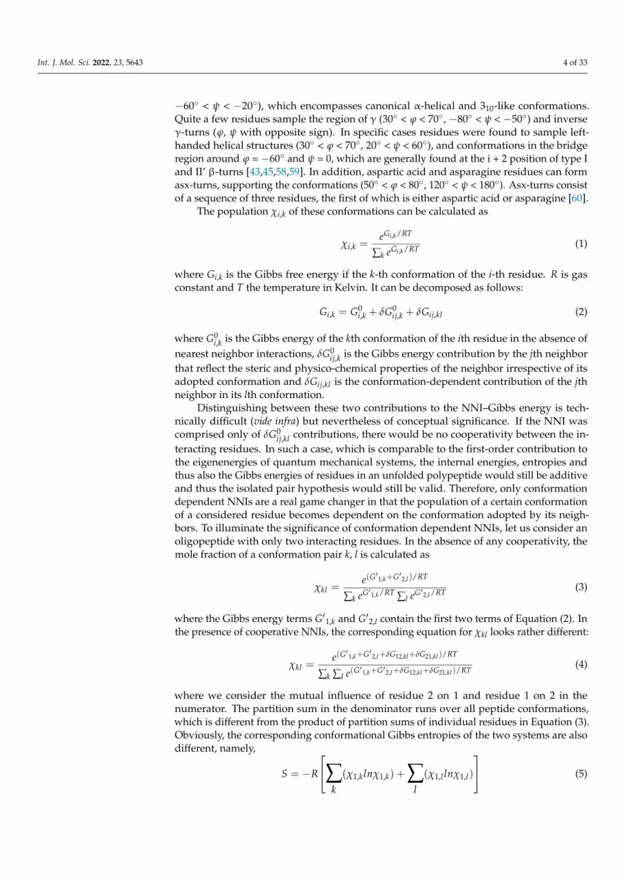
The results of the J-coupling analysis of Penkett et al. are shown in Figure 4 [61]. The
average 3J(HNHCα) values of 18 amino acid residues can apparently be subdivided into two
groups. Residues preceded by F, H, I, T, V, W, and Y (L-group) exhibit systematically
higher 3J(HNHCα) values than corresponding residues preceded by representatives of the
complementary group (G excluded). These results seem to indicate that sterically de-
manding aliphatic and aromatic residues move the overall distribution to lower (more
negative) φ values. While important as a first data-based insight regarding the occurrence
of NNIs, the results can be interpreted either as reflecting shifts of basins associated with
different secondary structures and as redistribution between different basins. Experi-
mental results to be discussed below reveal that NNIs can indeed cause both.
Figure 3. Conformational distribution of alanine residues inferred from the protein data sets. (A) Thecomplete data set including residues in well-defined secondary structures. (B) Only alanine residuesin coils outside of right-handed helical and β-sheet structures were considered. Each number onthe abscissae represents an 18◦ interval starting at −180◦. Taken from [16] with permission, 1995,Academic Press.

Int. J. Mol. Sci. 2022, 23, 5643 7 of 33
Nearest neighbor effects were first considered in a third paper by Penkett et al. thatappeared in 1997 [61]. Keeping in mind the well-stablished fact that the helical and β-sheetpropensities of amino acid residues are context dependent they were wondering whethersuch context dependencies would also be observable in the unfolded proteins for whichthey assumed minimal non-local interactions. To this end, they used 1H NMR to determinethe 3J(HNHCα) coupling constants for a 130-residue fragment of the fribronectin-bindingproteins from Staphyloccus aureus. They observed that 9 of 16 glutamic acid residuespreceded by residues with either branched or aromatic side chains exhibited couplingconstants between 6.2 and 7.0 Hz, while the other 7 with asparagine or glutamate asupstream neighbors lie between 5.7 and 6.3 Hz.
Further insight into the underlying NNIs came from the average 3J(HNHCα) valuescalculated for the coil library distributions of Swindells et al. [15]. These calculationswere performed with a Karplus equation that relates the coupling constant to the dihedralbackbone angle ϕ as follows [58]:
J(x, y,η) = Acos2(η+ θ1) + Bcos(η+ θ2) + C (7)
where x and y denote the interacting nuclei x and y, η = ϕ, ψ, and θi (I = 1, 2) are phase angles.A, B and C are empirical Karplus parameters obtained by fitting Equation (9) to the J-couplingdata sets obtained for proteins with a known crystal structure [59,62–65]. Alternatively, theseparameters could be obtained with density functional theory calculations but this has beenaccomplished thus far only for alanine [66]. It is likely that the exact parameters are side-chain dependent. Empirical values should therefore be considered as an average. In thecase of unfolded and disordered proteins and peptides, the measured Karplus parametersrepresent a conformational average:
〈J(x, y)〉 = ∑Ni=1 J(x, y,ηi)P(ηi)
∑Ni+1 P(ηi)
(8)
where P(ηi) is the probability for the residue to adopt a dihedral angle ϕi or ψi.The results of the J-coupling analysis of Penkett et al. are shown in Figure 4 [61]. The
average 3J(HNHCα) values of 18 amino acid residues can apparently be subdivided intotwo groups. Residues preceded by F, H, I, T, V, W, and Y (L-group) exhibit systematicallyhigher 3J(HNHCα) values than corresponding residues preceded by representatives ofthe complementary group (G excluded). These results seem to indicate that stericallydemanding aliphatic and aromatic residues move the overall distribution to lower (morenegative) ϕ values. While important as a first data-based insight regarding the occurrenceof NNIs, the results can be interpreted either as reflecting shifts of basins associated withdifferent secondary structures and as redistribution between different basins. Experimentalresults to be discussed below reveal that NNIs can indeed cause both.
In what follows, we will focus on two different coil library analyses reported by theSosnick and Dunbrack group, which both deal with NNIs in explicit terms [19,67]. Other re-ported libraries put a focus on individual propensities and conformational sampling, whichis of lesser interest in this context [15,16,68,69]. Jha et al. conducted a very comprehensivecoil library analysis of 2020 chains with more than twenty residues [18,67]. The authorsproduce Ramachandran plots for four different data sets: one set with no restrictions (allsecondary structure sequences included), a second one from which helices and sheets weretaken out, a third one for which turns were taken out as well and a fourth one from whichflanking residues were also eliminated. The authors’ analysis clearly showed that takinghelices and sheets out of the data set produce rather different Ramachandran distributions.Moreover, their analysis yielded rather different Ramachandran distributions for individualresidues and revealed significant nearest neighbor influences.
Int. J. Mol. Sci. 2022, 23, 5643 8 of 33Int. J. Mol. Sci. 2022, 23, x FOR PEER REVIEW 8 of 35
Figure 4. (a) Population of the extended β-region (b-region in Figure 2) by the indicated amino acid
residue in the presence of an L-type (F,H,I,TV,W,Y) or S-type (remaining amino acid set, G ex-
cluded) upstream neighbor. (b) Corresponding average 3J(HNHCα) values (Equations (6) and (7)).
Taken from [61] with permission, 1997, Academic Press.
In what follows, we will focus on two different coil library analyses reported by the
Sosnick and Dunbrack group, which both deal with NNIs in explicit terms [19,67]. Other
reported libraries put a focus on individual propensities and conformational sampling,
which is of lesser interest in this context [15,16,68,69]. Jha et al. conducted a very compre-
hensive coil library analysis of 2020 chains with more than twenty residues [18,67]. The
authors produce Ramachandran plots for four different data sets: one set with no re-
strictions (all secondary structure sequences included), a second one from which helices
and sheets were taken out, a third one for which turns were taken out as well and a fourth
one from which flanking residues were also eliminated. The authors’ analysis clearly
showed that taking helices and sheets out of the data set produce rather different Rama-
chandran distributions. Moreover, their analysis yielded rather different Ramachandran
distributions for individual residues and revealed significant nearest neighbor influences.
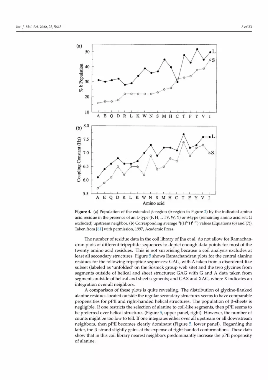
The number of residue data in the coil library of Jha et al. do not allow for Ramachan-
dran plots of different tripeptide sequences to depict enough data points for most of the
twenty amino acid residues. This is not surprising because a coil analysis excludes at least
all secondary structures. Figure 5 shows Ramachandran plots for the central alanine resi-
dues for the following tripeptide sequences: GAG, with A taken from a disordered-like
subset (labeled as ‘unfolded’ on the Sosnick group web site) and the two glycines from
Figure 4. (a) Population of the extended β-region (b-region in Figure 2) by the indicated aminoacid residue in the presence of an L-type (F, H, I, TV, W, Y) or S-type (remaining amino acid set, Gexcluded) upstream neighbor. (b) Corresponding average 3J(HNHCα) values (Equations (6) and (7)).Taken from [61] with permission, 1997, Academic Press.
The number of residue data in the coil library of Jha et al. do not allow for Ramachan-dran plots of different tripeptide sequences to depict enough data points for most of thetwenty amino acid residues. This is not surprising because a coil analysis excludes atleast all secondary structures. Figure 5 shows Ramachandran plots for the central alanineresidues for the following tripeptide sequences: GAG, with A taken from a disordered-likesubset (labeled as ‘unfolded’ on the Sosnick group web site) and the two glycines fromsegments outside of helical and sheet structures; GAG with G and A data taken fromsegments outside of helical and sheet segments; and GAX and XAG, where X indicates anintegration over all neighbors.
A comparison of these plots is quite revealing. The distribution of glycine-flankedalanine residues located outside the regular secondary structures seems to have comparablepropensities for pPII and right-handed helical structures. The population of β-sheets isnegligible. If one restricts the selection of alanine to coil-like segments, then pPII seems tobe preferred over helical structures (Figure 5, upper panel, right). However, the number ofcounts might be too low to tell. If one integrates either over all upstream or all downstreamneighbors, then pPII becomes clearly dominant (Figure 5, lower panel). Regarding thelatter, the β-strand slightly gains at the expense of right-handed conformations. These datashow that in this coil library nearest neighbors predominantly increase the pPII propensityof alanine.
Int. J. Mol. Sci. 2022, 23, 5643 9 of 33
Int. J. Mol. Sci. 2022, 23, x FOR PEER REVIEW 9 of 35
segments outside of helical and sheet structures; GAG with G and A data taken from seg-
ments outside of helical and sheet segments; and GAX and XAG, where X indicates an
integration over all neighbors.
Figure 5. Ramachandran plots of the central residue in the indicated tripeptide sequences. Upper
panel: (Left) only alanine residues in coils were considered; (Right) only alanine residues outside of
helices and β-sheets were considered. Lower panel: X indicates the summation over all nearest
neighbors outside of helices and β-sheets. The plots were directly taken from the website of the
Sosnick group [70].
A comparison of these plots is quite revealing. The distribution of glycine-flanked
alanine residues located outside the regular secondary structures seems to have compa-
rable propensities for pPII and right-handed helical structures. The population of β-sheets
is negligible. If one restricts the selection of alanine to coil-like segments, then pPII seems
to be preferred over helical structures (Figure 5, upper panel, right). However, the number
of counts might be too low to tell. If one integrates either over all upstream or all down-
stream neighbors, then pPII becomes clearly dominant (Figure 5, lower panel). Regarding
the latter, the β-strand slightly gains at the expense of right-handed conformations. These
data show that in this coil library nearest neighbors predominantly increase the pPII pro-
pensity of alanine.
The Sosnick group utilized their coil library distributions to explore the chemically
denatured state of apomyoglobin, ubiquitin, the SNase fragment Δ131Δ and eglin C, for
which they tried to reproduce experimentally determined NMR-based residual dipolar
constants [18]. Jha et al. did so with coil library-based Ramachandran distributions with
Figure 5. Ramachandran plots of the central residue in the indicated tripeptide sequences. Upperpanel: (Left) only alanine residues in coils were considered; (Right) only alanine residues outsideof helices and β-sheets were considered. Lower panel: X indicates the summation over all nearestneighbors outside of helices and β-sheets. The plots were directly taken from the website of theSosnick group [70].
The Sosnick group utilized their coil library distributions to explore the chemicallydenatured state of apomyoglobin, ubiquitin, the SNase fragment ∆131∆ and eglin C, forwhich they tried to reproduce experimentally determined NMR-based residual dipolarconstants [18]. Jha et al. did so with coil library-based Ramachandran distributions withand without specific nearest neighbor interactions. Residues were taken from regions thatcontain neither helices nor sheets nor turn conformations. NNIs were considered solely forresidue dimers for which the total internal energy was written as
U(ai, bi, ai+1, bi+1) = U(ai, bi) + U(ai+1, bi+1) + δU(ai, bi, ai+1, bi+1) (9)
where ai labels the identity of the ith-residue that samples the basin bi. The interactionenergy δU accounts for the cooperativity or anti-cooperativity between conformations biand bi+1 of the two residues. It is related to the conditional probability P(ai,bi,ai+1,bi+1) by
δU(aj, bj, aj+1, bj+1
)= −RT
[P(aj, bj, aj+1, bj+1
)P(aj, bj
)P(aj+1, bj+1
)] (10)
The coil data basis of the authors was not large enough to allow for an empiricaldetermination of NNIs. To gain information about the latter they performed Monte Carlo

Int. J. Mol. Sci. 2022, 23, 5643 10 of 33
simulations with an energy functional for each basin with and without NNIs. Energyminimization was constrained by the utilized coil library distributions for individualresidues. The energy functional did not contain any protein–solvent interaction; basically,the authors employed excluded volume effects. This procedure was carried out withand without nearest neighbor interactions. As one can infer from the results obtainedfor apomyoglobin, shown in Figure 6, calculations with NNIs achieved a much betterreproduction of the experimental residue coupling constants. Equally interesting is thefact that the experimental values do not at all follow predictions based on an idealizedrandom coil model (Figure 6B). The latter used three major isoenergetic basins for eachresidue of an A50 polypeptide (pPII, β-strand, right-handed helical) with a populationof 0.33. The obtained V-shape reflect the decreased correlation with the molecular axisfor residues closer to the termini. An analysis of the conformational ensemble of theinvestigated unfolded/denatured proteins reveals a dominance of what Jha et al. calledstretched conformations in which individual residues sample predominantly pPII andβ-basins. For the set of coil library residues used for the residual dipolar coupling analysisthey obtained a mole fraction ratio of <pPII>:<β>:<right-handed helical> = 0.33:0.36:0.27.While the numbers seem to be reminiscent of a random coil supporting distribution (i.e.,sampling of all sterically allowed regions), the very existence of distinguishable pPII and β-basins is not. Despite the deviation from an ideal random coil behavior on a local level, theradii of gyration calculated for the above and six additional unfolded/denatured proteinsindicate self-avoiding random coils. This result is in line with computational results ofFitzkee et al., who showed that an ensemble of rods connected by flexible linkers can stillreproduce the scaling law for self-avoiding random coils [39]. All these results show that itis necessary to distinguish between local and global aspects of random coils, as very earlyon emphasized by de Gennes [71] for polymers and by Toal and Schweitzer-Stenner forpeptides and proteins [22].
Int. J. Mol. Sci. 2022, 23, x FOR PEER REVIEW 11 of 35
Figure 6. (A) Experimental (black) and calculated (grey) residual dipole coupling values for apo-
myoglobin in 10% acrylamide. The employed model combined coil library data with MD simula-
tions. Left: Calculations performed without considering NNIs. Right: Calculations performed with
NNIs obtained from a comparison of Ramachandran plots. (B) Residual dipole coupling values of
an idealized random coil ensemble of an A50 polypeptide generated without nearest neighbor cou-
pling. Equal population (1/3) was assumed for pPII, β-strand and right-handed helical basins. Taken
from [18] (free-access article).
An even more thorough and residue-specific analysis of coil libraries have been un-
dertaken by Ting et al. [19]. Their data set contained 3038 proteins from the Uppsala Elec-
tron Density server. In line with the protocol of the Sosnick group they obtained different
coil libraries by employing different restrictions regarding the selection of residues. The
largest set contained loop residues (no regular secondary structure elements) for which
all backbone atoms appear in the data set. Each residue is at least three residues away
from the regular structures. This set was termed TCBIG, which contain the single letter
designation of turn, coil, β-bridge, π-helix and 310-helix. The second, reduced data set did
not contain π- and 310 helices (TCB). The authors classified their Ramachandran distribu-
tions in terms of the following five conformations (Table 1): α-helical, β-strand, polypro-
line II, left-handed helical and extended. Since considering all combinations of a given
residue with its neighbors is an impossible task (203 combinations) the authors confined
themselves to selected ‘dimers’ where they changed the neighbor either upstream or
downstream from the considered residue and averaged over all neighbors for the other
side. If the influence of the two neighbors is not additive—a notion for which experimental
evidence exist in the literature (vide infra)—the information obtained from Ramachandran
plots of different pairs might not necessarily represent the NNIs between the pairs. In
order to permit a thorough mathematical analysis of the obtained Ramachandran plots,
the authors represented the latter by a continuous functional that can be ascribed to a
combination of a two-dimensional Gaussian distribution located in close proximity to ba-
sins associated with secondary structures. While this modeling bears some similarity with
the Gaussian model of Schweitzer-Stenner [72], differences should be emphasized. While
the latter works with 1:1 assignments of Gaussians to the assumed basins of the Rama-
chandran plot (which would be five for the above set of conformations assumed by Ting
et al.), the former functional is entirely based on the distributions of data points inferred
from the coil library sets. In both cases, the functionals facilitate the mathematic analyses
of distributions.
A closer look at the residue dimer distributions of Ting et al. reveal that some neigh-
bors have a significant influence on specific residues. Let us again focus on alanine. Com-
pared with X-AG, F, V and Q as downstream neighbors substantially increase the right-
handed helical population of alanine at the expense of pPII (Figure 7). On the contrary,
proline as a downstream neighbor stabilizes pPII. The nearest downstream neighbors of
valine (including valine itself) increase the right-handed helical populations as well. While
Figure 6. (A) Experimental (black) and calculated (grey) residual dipole coupling values for apo-myoglobin in 10% acrylamide. The employed model combined coil library data with MD simulations.Left: Calculations performed without considering NNIs. Right: Calculations performed with NNIsobtained from a comparison of Ramachandran plots. (B) Residual dipole coupling values of anidealized random coil ensemble of an A50 polypeptide generated without nearest neighbor coupling.Equal population (1/3) was assumed for pPII, β-strand and right-handed helical basins. Takenfrom [18] (free-access article).
While of great insight for an understanding of the relevance of NNIs, the above studiesdo not provide much specific information about how NNIs depend on the physico-chemicalproperties of the involved residues. The coil library analysis of Jha et al. suggests a stronganti-cooperativity between pPII and right-handed helical conformations of alanine andalanine-like as well as β-branched upstream neighbors, respectively. Aromatic residuespositioned downstream seem to cause anti-cooperative interactions between the pPII statesof the interacting residues [67].

Int. J. Mol. Sci. 2022, 23, 5643 11 of 33
An even more thorough and residue-specific analysis of coil libraries have beenundertaken by Ting et al. [19]. Their data set contained 3038 proteins from the UppsalaElectron Density server. In line with the protocol of the Sosnick group they obtaineddifferent coil libraries by employing different restrictions regarding the selection of residues.The largest set contained loop residues (no regular secondary structure elements) forwhich all backbone atoms appear in the data set. Each residue is at least three residuesaway from the regular structures. This set was termed TCBIG, which contain the singleletter designation of turn, coil, β-bridge, π-helix and 310-helix. The second, reduced dataset did not contain π- and 310 helices (TCB). The authors classified their Ramachandrandistributions in terms of the following five conformations (Table 1): α-helical, β-strand,polyproline II, left-handed helical and extended. Since considering all combinations ofa given residue with its neighbors is an impossible task (203 combinations) the authorsconfined themselves to selected ‘dimers’ where they changed the neighbor either upstreamor downstream from the considered residue and averaged over all neighbors for the otherside. If the influence of the two neighbors is not additive—a notion for which experimentalevidence exist in the literature (vide infra)—the information obtained from Ramachandranplots of different pairs might not necessarily represent the NNIs between the pairs. Inorder to permit a thorough mathematical analysis of the obtained Ramachandran plots,the authors represented the latter by a continuous functional that can be ascribed to acombination of a two-dimensional Gaussian distribution located in close proximity tobasins associated with secondary structures. While this modeling bears some similaritywith the Gaussian model of Schweitzer-Stenner [72], differences should be emphasized.While the latter works with 1:1 assignments of Gaussians to the assumed basins of theRamachandran plot (which would be five for the above set of conformations assumedby Ting et al.), the former functional is entirely based on the distributions of data pointsinferred from the coil library sets. In both cases, the functionals facilitate the mathematicanalyses of distributions.
Table 1. List of Hellinger distances between the indicated amino acid residues independent ofneighbors (left value) and in the presence of a glutamine residue at the upstream position (rightvalue). Hellinger distance values indicating at least moderately different distributions are typed inbold. All values were taken from Ting et al. [19] and divided by 100.
F Y Q K V I A N
F 1 0.08 0.1/0.15 0.12/0.16 0.21/0.14 0.22/0.17 0.19/0.15 0.19/0.12
Y 0.08 1 0.11/0.13 0.12/0.14 0.21/0.15 0.21/0.18 0.19/0.12 0.19/0.08
Q 0.10/0.15 0.11/0.13 1 0.09 0.21/0.09 0.22/0.11 0.16/0.07 0.18/0.09
K 0.12/0.16 0.12/0.14 0.09 1 0.20/0.09 0.20/0.12 0.16/0.08 0.22/0.09
V 0.21/0.14 0.21/0.15 0.21/0.09 0.20/0.09 1 0.09/0.10 0.28/0.12 0.32/0.11
I 0.22/0.17 0.21/0.18 0.22/0.11 0.20/0.12 0.09/0.10 1 0.16/0.13 0.32/0.13
A 0.19/0.15 0.19/0.12 0.16/0.07 0.16/0.08 0.28/0.12 0.16/0.13 1 0.25/0.09
N 0.19/0.12 0.190.08 0.18/0.09 0.22/0.09 0.32/0.11 0.32/0.13 0.25/0.09 1
A closer look at the residue dimer distributions of Ting et al. reveal that some neighborshave a significant influence on specific residues. Let us again focus on alanine. Comparedwith X-AG, F, V and Q as downstream neighbors substantially increase the right-handedhelical population of alanine at the expense of pPII (Figure 7). On the contrary, prolineas a downstream neighbor stabilizes pPII. The nearest downstream neighbors of valine(including valine itself) increase the right-handed helical populations as well. While thelatter is also significant in the coil library of the Sosnick group, the neighbor-induced helicalpopulation seems to be more pronounced in the Ting et al. library. This is a very surprising
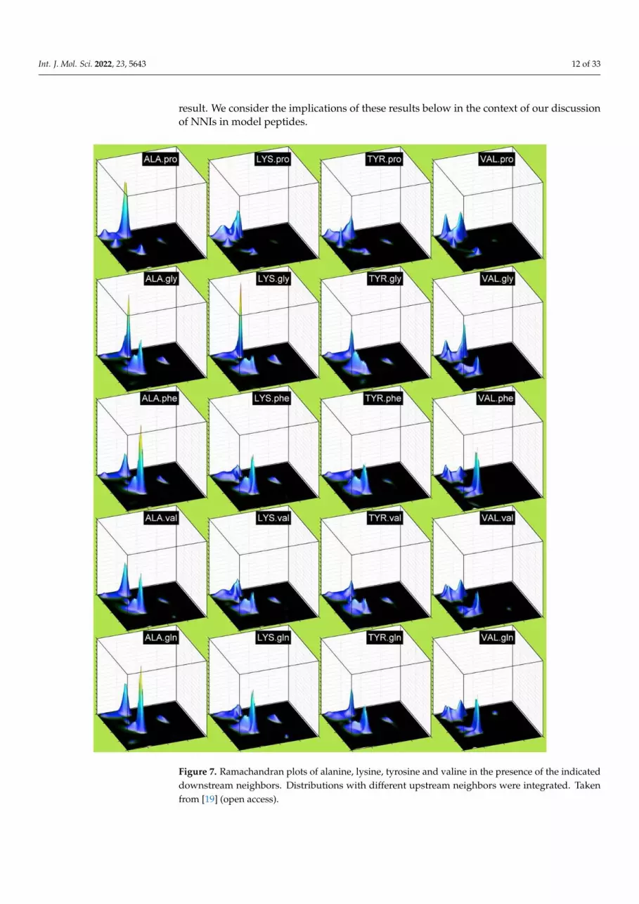
Int. J. Mol. Sci. 2022, 23, 5643 12 of 33
result. We consider the implications of these results below in the context of our discussionof NNIs in model peptides.
Int. J. Mol. Sci. 2022, 23, x FOR PEER REVIEW 13 of 35
Figure 7. Ramachandran plots of alanine, lysine, tyrosine and valine in the presence of the indicated
downstream neighbors. Distributions with different upstream neighbors were integrated. Taken
from [19] (open access).
4. Simulations
The first thorough computational investigation of NNIs was carried out by Pappu et
al. [68]. The authors confined themselves on exploring the interactions between alanine
residues in a blocked oligo-alanine peptide. The authors sub-divided the Ramachandran
plot into 6 × 6 equally sized mesostates (60° × 60°). Their Monte-Carlo calculations were
Figure 7. Ramachandran plots of alanine, lysine, tyrosine and valine in the presence of the indicateddownstream neighbors. Distributions with different upstream neighbors were integrated. Takenfrom [19] (open access).

Int. J. Mol. Sci. 2022, 23, 5643 13 of 33
Ting et al. used Hellinger distances as a measure of dissimilarity between the Ra-machandran plots. The latter can be calculated as
H(
PR(φi, ψi), P′R(φi, ψi))=
∣∣∣∣∣∣∣∣∣∣∣∣12
π∫−π
π∫−π
(√PR(φi, ψi)−
√PR′(φi, ψi)
)dφdψ
∣∣∣∣∣∣∣∣∣∣∣∣ (11)
where PR and P′R are the two Ramachandran distributions to be compared with each other.An H value of zero means that the two distributions are identical, whereas a value of1 indicate they are orthogonal. However, even very dissimilar Ramachandran distributionswill not be able to produce values close to 1, because they cover only a limited fraction ofthe Ramachandran space. Schweitzer-Stenner and Toal employed the following criteria:H values between 0 and 0.1 indicate that two distributions are similar [73]. Values between0.1 and 0.25 as well as between 0.25 and 0.4 indicate that they are modestly similar anddissimilar, respectively. Values above 0.4 reflect very dissimilar distributions (note, thatTing et al. multiplied their H-values with 100). Since the Hellinger distance is practically ameasure of orthogonality, it is more sensitive to changes of basin position than it is to theredistribution of populations [73].
Table 1 lists the Hellinger distances for pairs of eight amino acid residues. The leftvalues represent the H-distances for pairs irrespective of their neighbors (Ramachandranplots for different neighbors were added up) whereas the right values represent H-distancesif Q is present as an upstream neighbor. Only a few residue pairs fall in the category‘modestly dissimilar’. They mostly contain valine and asparagine. If only Q is consideredas upstream neighbors, then all H-values are in the similar or modestly similar range. Thesignificance of these values is not entirely clear. The integration over all neighbors mighthide a strong influence of a few residue types. Valine and asparagine seem to be goodcandidates, as is, most likely, proline (values for P are reported by Ting et al.). We willreturn to the use of Hellinger distances below when we discuss investigations of shortpeptides in water.
4. Simulations
The first thorough computational investigation of NNIs was carried out byPappu et al. [68]. The authors confined themselves on exploring the interactions be-tween alanine residues in a blocked oligo-alanine peptide. The authors sub-divided theRamachandran plot into 6 × 6 equally sized mesostates (60◦ × 60◦). Their Monte-Carlocalculations were performed with a rather simple hard sphere model by means of whichthey just explored the sterically available space, very much in line with the classical Ra-machandran approach [69]. They identified clashes between nearest neighbors samplingmesostates in the right helical region, while nearest neighbors sampling mesostates in thepPII and β-strand regions do not interfere with each other. Their results led the authors toconclude that an increasing chain length (of their oligo-alanine peptide) leads to a reductionin conformational space in the unfolded state, which reduces the conformational entropyand thus facilitates the folding into an overall right-handed α-helical conformation.
In a subsequent paper, Tran et al. investigated how steric-based NNIs depend ondifferent type of neighbors [74]. They explored the conformational propensities of 22 aminoacid residues (norvaline and norleucine in addition to the natural ones) in N-acetyl-(host)L-x-(host)L-N-methylacetamide (L: number of host residues) host–guest blocked tetrapeptides.Glycine, alanine, valine, phenylalanine and proline were selected as hosts. The result oftheir analysis is shown in Figure 8. While the influence of glycine on the guest residue isnegligible (as one would expect), all other hosts (including proline) shift conformationalsampling from the lower (all types of right-handed helical conformations) to the upperleft quadrant (pPII and all types of β-strand). Interestingly, the underlying NNIs seemto be more pronounced for L = 2 (influence of nearest and second nearest neighbor) forA, F and, in part, V hosts. Altogether, the NNIs identified by Tran et al. produce morestretched peptide and protein conformations in the unfolded state than expected based
Int. J. Mol. Sci. 2022, 23, 5643 14 of 33
on Ramachandran-type distributions. In that regard, their results are at variance with thenearest neighbor effects inferred from the coil library of Ting et al. [19]. However, for anincreasing chain length, conformational entropy causes the chain to depart from a rod-likestructure. Consequently, longer polypeptides and denatured proteins still obey the scalinglaw for a self-avoiding random coil.
Int. J. Mol. Sci. 2022, 23, x FOR PEER REVIEW 14 of 35
performed with a rather simple hard sphere model by means of which they just explored
the sterically available space, very much in line with the classical Ramachandran approach
[69]. They identified clashes between nearest neighbors sampling mesostates in the right
helical region, while nearest neighbors sampling mesostates in the pPII and β-strand re-
gions do not interfere with each other. Their results led the authors to conclude that an
increasing chain length (of their oligo-alanine peptide) leads to a reduction in conforma-
tional space in the unfolded state, which reduces the conformational entropy and thus
facilitates the folding into an overall right-handed α-helical conformation.
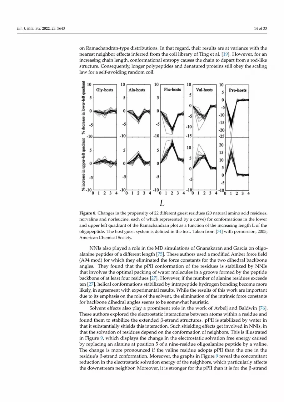
In a subsequent paper, Tran et al. investigated how steric-based NNIs depend on
different type of neighbors [74]. They explored the conformational propensities of 22
amino acid residues (norvaline and norleucine in addition to the natural ones) in N-acetyl-
(host)L-x-(host)L-N-methylacetamide (L: number of host residues) host–guest blocked
tetrapeptides. Glycine, alanine, valine, phenylalanine and proline were selected as hosts.
The result of their analysis is shown in Figure 8. While the influence of glycine on the
guest residue is negligible (as one would expect), all other hosts (including proline) shift
conformational sampling from the lower (all types of right-handed helical conformations)
to the upper left quadrant (pPII and all types of β-strand). Interestingly, the underlying
NNIs seem to be more pronounced for L = 2 (influence of nearest and second nearest
neighbor) for A, F and, in part, V hosts. Altogether, the NNIs identified by Tran et al.
produce more stretched peptide and protein conformations in the unfolded state than ex-
pected based on Ramachandran-type distributions. In that regard, their results are at var-
iance with the nearest neighbor effects inferred from the coil library of Ting et al. [19].
However, for an increasing chain length, conformational entropy causes the chain to de-
part from a rod-like structure. Consequently, longer polypeptides and denatured proteins
still obey the scaling law for a self-avoiding random coil.
Figure 8. Changes in the propensity of 22 different guest residues (20 natural amino acid residues,
norvaline and norleucine, each of which represented by a curve) for conformations in the lower and
upper left quadrant of the Ramachandran plot as a function of the increasing length L of the oligo-
peptide. The host guest system is defined in the text. Taken from [74] with permission, 2005, Amer-
ican Chemical Society
Figure 8. Changes in the propensity of 22 different guest residues (20 natural amino acid residues,norvaline and norleucine, each of which represented by a curve) for conformations in the lowerand upper left quadrant of the Ramachandran plot as a function of the increasing length L of theoligopeptide. The host guest system is defined in the text. Taken from [74] with permission, 2005,American Chemical Society.
NNIs also played a role in the MD simulations of Gnanakaran and Garcia on oligo-alanine peptides of a different length [75]. These authors used a modified Amber force field(A94 mod) for which they eliminated the force constants for the two dihedral backboneangles. They found that the pPII conformation of the residues is stabilized by NNIsthat involves the optimal packing of water molecules in a groove formed by the peptidebackbone of at least four residues [27]. However, if the number of alanine residues exceedsten [27], helical conformations stabilized by intrapeptide hydrogen bonding become morelikely, in agreement with experimental results. While the results of this work are importantdue to its emphasis on the role of the solvent, the elimination of the intrinsic force constantsfor backbone dihedral angles seems to be somewhat heuristic.
Solvent effects also play a prominent role in the work of Avbelj and Baldwin [76].These authors explored the electrostatic interactions between atoms within a residue andfound them to stabilize the extended β-strand structures. pPII is stabilized by water inthat it substantially shields this interaction. Such shielding effects get involved in NNIs, inthat the solvation of residues depend on the conformation of neighbors. This is illustratedin Figure 9, which displays the change in the electrostatic solvation free energy causedby replacing an alanine at position 5 of a nine-residue oligoalanine peptide by a valine.The change is more pronounced if the valine residue adopts pPII than the one in theresidue’s β-strand conformation. Moreover, the graphs in Figure 9 reveal the concomitantreduction in the electrostatic solvation energy of the neighbors, which particularly affectsthe downstream neighbor. Moreover, it is stronger for the pPII than it is for the β-strand

Int. J. Mol. Sci. 2022, 23, 5643 15 of 33
conformation of valine. This important result suggests a cooperative interaction betweenthe pPII state of valine and the β-strand conformation of the neighbor. Besides valine,Avbelj and Baldwin investigated the influence of the remaining 18 amino acid residues.They found the decrease in the electrostatic solvation free energy of the guest residue(compared with alanine) is particularly pronounced (>6.2 kJ/mol for pPII) for aromatic andaliphatic/β-branched residues (V, I, W, Y, F, H and T) and always larger if the guest residueadopts pPII. This work reveals that NNIs between residues adopting conformations in theupper left quadrant of the Ramachandran space are mostly solvent mediated.
Int. J. Mol. Sci. 2022, 23, x FOR PEER REVIEW 15 of 35
NNIs also played a role in the MD simulations of Gnanakaran and Garcia on oligo-
alanine peptides of a different length [75]. These authors used a modified Amber force
field (A94 mod) for which they eliminated the force constants for the two dihedral back-
bone angles. They found that the pPII conformation of the residues is stabilized by NNIs
that involves the optimal packing of water molecules in a groove formed by the peptide
backbone of at least four residues [27]. However, if the number of alanine residues exceeds
ten [27], helical conformations stabilized by intrapeptide hydrogen bonding become more
likely, in agreement with experimental results. While the results of this work are im-
portant due to its emphasis on the role of the solvent, the elimination of the intrinsic force
constants for backbone dihedral angles seems to be somewhat heuristic.
Solvent effects also play a prominent role in the work of Avbelj and Baldwin [76].
These authors explored the electrostatic interactions between atoms within a residue and
found them to stabilize the extended β-strand structures. pPII is stabilized by water in that
it substantially shields this interaction. Such shielding effects get involved in NNIs, in that
the solvation of residues depend on the conformation of neighbors. This is illustrated in
Figure 9, which displays the change in the electrostatic solvation free energy caused by
replacing an alanine at position 5 of a nine-residue oligoalanine peptide by a valine. The
change is more pronounced if the valine residue adopts pPII than the one in the residue’s
β-strand conformation. Moreover, the graphs in Figure 9 reveal the concomitant reduction
in the electrostatic solvation energy of the neighbors, which particularly affects the down-
stream neighbor. Moreover, it is stronger for the pPII than it is for the β-strand confor-
mation of valine. This important result suggests a cooperative interaction between the
pPII state of valine and the β-strand conformation of the neighbor. Besides valine, Avbelj
and Baldwin investigated the influence of the remaining 18 amino acid residues. They
found the decrease in the electrostatic solvation free energy of the guest residue (com-
pared with alanine) is particularly pronounced (>6.2 kJ/mol for pPII) for aromatic and al-
iphatic/β-branched residues (V, I, W, Y, F, H and T) and always larger if the guest residue
adopts pPII. This work reveals that NNIs between residues adopting conformations in the
upper left quadrant of the Ramachandran space are mostly solvent mediated.
Figure 9. Changes in the electrostatic solvation free energy as a result of substituting alanine at po-
sition 5 of an A9 peptide by valine. The free energy is expressed in units of kcal/mol. Taken from
[76] (open access).
Figure 9. Changes in the electrostatic solvation free energy as a result of substituting alanine atposition 5 of an A9 peptide by valine. The free energy is expressed in units of kcal/mol. Takenfrom [76] (open access).
The group of Sosnick has substantially contributed to our current understanding ofNNIs. Besides their work on coil libraries [18,77], they conducted a thorough MD studyon xAA and AAx tripeptides in water where x denotes the guest residue. To this end theyemployed three force fields in implicit water: Amber 94, the modified Amber force fieldof Garcia (G-S-94), and OPLS-AA-2001 [78]. Simulation with these three force fields yieldrather different propensities for the central alanine of AAA. Amber 94 produces the well-known preference for right-handed helical structures while the other force field yield a morebalanced distribution. The authors could not reproduce the high pPII propensity for alaninewith the G-S-94 force field, which can certainly be attributed to their use of an implicitsolvent model. They also observed substantial differences between the Ramachandrandistributions in AAA and in the alanine dipeptide in that the residue of the latter spendsmore time in pPII and β. These results are at variance with experimental results that show ahigher pPII preference for A in AAA than in the alanine dipeptide, in qualitative agreementwith Garcia’s work [79]. Again, this discrepancy points to different solvent models used byGarcia and Zaman et al.
Figure 10 displays the propensities for four different representative neighbors obtainedwith the G-S-94 and OPLS-AA-2001 force field. The predicted changes are considerable butvery much force-field dependent. For GAA, which one could use as a reference system, theG-S-94 force field produces a Ramachandran plot for the central alanine that is dominatedby right-handed helical conformations. On the contrary, the OPLS force field produces a

Int. J. Mol. Sci. 2022, 23, 5643 16 of 33
dominance of pPII and β-strand. With G-S-94, replacing G by L, N or D keeps the highhelical propensity while causing some redistribution between pPII and β-strand. The OPLSforce field yields an increased sampling of the right-handed helical and bridge region forG→L and an overall increase in the helical population for G→D. The distributions obtainedwith Amber 94 are not indicative of massive NNI influence, as for all the guest residuesthe α-helix population is dominant. There is no doubt that the results of these calculationsare important in that they suggest that NNIs can produce substantial changes in theGibbs energy landscape of residues and their conformational entropy. However, withoutexperimental validation, it is problematic to employ the obtained population changes for aquantitative assessment of the influence of NNIs on the energetics of unfolded polypeptidesand proteins.
Int. J. Mol. Sci. 2022, 23, x FOR PEER REVIEW 17 of 35
Figure 10. Ramachandran plots of the central alanine residues in the xAA peptides for the indicated
guest residues as obtained from MD simulations with G-S-94 (left) and OPLS-AA-01 (right) force
fields. Details are described in the text. Taken from [78] and modified.
5. Experimental Results
5.1. NMR on Denatured Proteins
As indicated above, the first experimental results indicating that NNIs affect the
structural distributions of denatured proteins came from NMR studies. They rely to a sig-
nificant extent on the use of a J-coupling constant, which reflect the degree of through-
bond interaction between two nuclear spins, which are generally one, two or three bonds
apart. Their general dependence on dihedral backbone angles is described by Equation (6)
(vide supra).
Penkett et al. used 3J(HNHCα) coupling constants of a denatured fibronectin binding
protein to conclude that β-branched and aromatic neighbors shift these values up [61].
The authors interpreted this observation as indicating a shift to more negative average φ-
values of the respective conformational ensemble. An even more thorough study was con-
ducted by Peti et al., who analyzed 3J(HNHCα) coupling and chemical shifts of three dena-
tured proteins, namely, ubiquitin, disulfide reduced, carboxymethylated lysozyme and a
so-called all-A-α-lactalbumin (all-A means that all cysteines were replaced by alanines)
[80]. 1H,15N-HSQC spectra were interpreted as indicative of a random coil conformation
in which right-handed helical and pPII/β-regions are populated. This notion was further
Figure 10. Ramachandran plots of the central alanine residues in the xAA peptides for the indicatedguest residues as obtained from MD simulations with G-S-94 (left) and OPLS-AA-01 (right) forcefields. Details are described in the text. Taken from [78] and modified.
5. Experimental Results5.1. NMR on Denatured Proteins
As indicated above, the first experimental results indicating that NNIs affect thestructural distributions of denatured proteins came from NMR studies. They rely to asignificant extent on the use of a J-coupling constant, which reflect the degree of through-bond interaction between two nuclear spins, which are generally one, two or three bonds

Int. J. Mol. Sci. 2022, 23, 5643 17 of 33
apart. Their general dependence on dihedral backbone angles is described by Equation (6)(vide supra).
Penkett et al. used 3J(HNHCα) coupling constants of a denatured fibronectin bindingprotein to conclude that β-branched and aromatic neighbors shift these values up [61].The authors interpreted this observation as indicating a shift to more negative averageϕ-values of the respective conformational ensemble. An even more thorough study wasconducted by Peti et al., who analyzed 3J(HNHCα) coupling and chemical shifts of threedenatured proteins, namely, ubiquitin, disulfide reduced, carboxymethylated lysozymeand a so-called all-A-α-lactalbumin (all-A means that all cysteines were replaced byalanines) [80]. 1H,15N-HSQC spectra were interpreted as indicative of a random coilconformation in which right-handed helical and pPII/β-regions are populated. This notionwas further supported by their observation that the average 15N chemical shifts of theamino acid residues (taken over all residue of a given type in the investigated proteins)correlate with the corresponding chemical shifts derived from the (restricted) coil library ofSmith et al. [81]. However, if these proteins were really sampling a random coil typeensemble there should be no NNIs of significance. This, however, does not seem to bethe case. Peti et al. showed that the nearest neighbor-induced chemical shift changesreported by Braun et al. [82], based on 15N measurements of unblocked GGxA peptides(x represents all natural amino acid residues), correlate with the nearest neighbor effectson leucine residues in the set of unfolded proteins [83]. They attributed these changes toconformational changes. Correlations between 15N chemical shift and 3J(HNHCα) couplingconstant changes support this notion. These results seem to confirm the observation ofPenkett et al., that branched and aromatic neighbors produce more negative ϕ-values [61].If Peti et al. interpreted the neighbor dependence of the 15N chemical shifts correctly,their results invalidate the isolated pair hypothesis, which implies that the conformationalensemble of the investigated proteins cannot be an ideal random coil.
5.2. Structural Analysis of Homopeptides
The above NMR based analyses rely very much on averages over many amino acidresidues in the considered denatured proteins and in the utilized coil libraries. We wonderwhether such a procedure could obfuscate information about the conformational propen-sities of residues and their dependence on nearest neighbors. Moreover, averaging overdifferent ensembles might lead to very similar coupling constants, so that changes in thelatter are difficult to interpret, particularly if one relies only on a single type of couplingparameter. An alternative approach in this regard utilizes short peptides, which, owingto their limited length, cannot adopt any regular secondary structure. For a long periodof time, blocked dipeptides were considered suitable model systems to explore intrinsicconformational propensities of amino acid residues. Ramachandran and Flory used them toexplore the sterically allowed region of the Ramachandran plot [3,69]. The alanine dipeptidehas been the system of choice for multiple MD simulations [84–89]. More recently, they havebeen used to experimentally determine conformational preferences in water and relatedblocked tripeptides even for the investigation of nearest neighbor interactions [17,23,90–94].The preference for blocked dipeptides over, e.g., unblocked tripeptides, was generallybased on the assumption that the charged terminal groups of, e.g., tripeptides, could af-fect conformational propensities [95,96]. However, experimental evidence reported byToal et al. revealed that this is not the case for trialanine (A3) and trivaline (V3) [79]. Kallen-bach and coworkers chose AcG2xG2NH2 host–guest peptides [97,98]. Our research grouphas embarked on a thorough investigation of unblocked tri-, tetra-, and pentapeptidesto determine the intrinsic conformational propensities and NNI effects [24,42–45,99–101].Contrary to blocked dipeptides, this choice provides some more natural context to theinvestigated amino acid residue. In what follows, we will review these works with anemphasis on NNIs.
We start this discussion with a focus on alanine. The respective amino acid residuehas long served as model system for the exploration of the Ramachandran space. The

Int. J. Mol. Sci. 2022, 23, 5643 18 of 33
Ramachandran plot for the alanine dipeptide that solely reflects steric exclusion andelectrostatic interactions looks very much like Figure 1. Thus, it fully represents the localaspect of an ideal random coil behavior. Very similar plots were obtained with moresophisticated molecular dynamics simulations in explicit water [91,92,102,103]. Hence, itcame as a surprise when Shi et al., based on 1H NMR and UVCD data for a hepta-alaninepeptide (XAO-peptide, Ac-X2A7O2-NH2, X represents aminobutyric acid) in water, arrivedat the conclusion that the peptide predominantly samples a basin assignable to the pPIIconformation [104]. Up to this point this conformation had been associated with the transconformation of proline in oligo- and poly-proline peptides, even though some early UVCDstudies of Tiffany and Krimm had indicated that poly-L-lysine and poly-L-glutamic acidcould adopt this conformation [105]. Their results were later corroborated by vibrationalcircular dichroism studies [106].
Since there was no obvious reason for alanine to prefer pPII, the results of Shi et al.became highly controversial in the field. Scheraga, Liwo and coworkers re-analyzed thedata of Shi et al. based on the results of MD simulations and arrived at the conclusionthat there is no specific preference of alanine for pPII [102,103,107]. Small-angle X-rayscattering data were found to be inconsistent with a conformational ensemble dominatedby pPII [103]. This discussion overlooked the fact that early coil library studies had alreadyindicated the very high pPII propensity of alanine (vide supra), thus lending credibility tothe results of Shi et al. [40].
After the paper of Shi et al. was published, their results were sometimes interpretedas indicating that the alanine sequences could adopt a stable pPII-helix [108–110]. Somewording chosen by the authors certainly facilitated this reading, but in a follow-up paperthey actually found no evidence for any cooperative nearest neighbor interactions betweenalanines in GGAAGG and GGAAAGG peptides [98], which would be needed for theformation of a stable pPII helix. Nevertheless, their work triggered a discussion of the so-called reconciliation problem, namely, the apparent contradiction between the occurrenceof pPII helices in denatured proteins and their well-established behavior as a self-avoidingrandom coil [30,74].
Spectroscopic studies on alanine-based oligopeptides suggest that some cooperativitybetween the pPII states of alanine residues actually exists, in line with the MD results ofGarcia (vide supra). At an early stage of the debate about the alleged pPII propensity ofalanine, Schweitzer-Stenner et al. combined IR, polarized Raman and vibrational circulardichroism (VCD) data to show that the unblocked tetra-alanine A4 has a higher pPIIpropensity than tri-alanine (A3) [111]. This result was corroborated by a Raman opticalactivity study of McColl et al. [112]. However, both studies were qualitative in nature inthat they did not report any numbers reflecting conformational propensities. This gap wasfilled later by more quantitative studies that utilized NMR J-coupling constants in additionto the amide I’ band profiles in IR, Raman and VCD spectra. The obtained results suggestthat the central alanine residue in A3 has a slightly higher pPII propensity than the one inGAG, namely, 0.84–0.9 for the former and 0.72–0.8 for the latter [76,84,113]. This differencelooks small, but it is indicative of a Gibbs free energy change of ca. 3 kJ/mol in favor ofpPII (for A3).
Graf et al. measured the 3J(HNHCα) and 2J(NCα) coupling constants for a series of Anpeptides (n = 3–7) [114]. While the former reflects average ϕ-values, the latter depends onthe ψ-value of the residue that precedes the utilized amide nitrogen. The authors used themeasured coupling constants in constrained MD simulations from which they obtained aslight stabilization of β-strand over pPII with increasing residue number. However, theobtained changes might be within the error limits of their analysis. Two other spectroscopicstudies suggest that an increasing length of alanine sequences stabilizes non-extendedstructures over pPII, in line with predictions [75]. Verbaro et al. combined vibrationalspectroscopy and fluorescence energy transfer experiments with the coupling constantsof Graf et al., to show that the individual alanine residues of the unblocked A5W peptideexhibit pPII fractions between 0.65 and 0.75 [115]. These values are lower than the those

Int. J. Mol. Sci. 2022, 23, 5643 19 of 33
observed for trialanine, but they are still way higher than any predictions obtained withsteric exclusion and MD calculations. The slight destabilization of pPII benefits right-handed helical conformations for residues 2–4, in line with predictions of Gnanakaran andGarcia. The fifth alanine residue behaves differently in that it exhibits a more pronouncedβ-strand population. The latter could reflect interactions with the aromatic W-residue atthe C-terminal, which exhibits a mixture of pPII and β-strand.
Shi et al. had initiated the debate about the pPII propensity of alanine with a spectro-scopic analysis of the XAO peptide (vide supra). They deduced a rather high propensity(mole fraction) value of ~0.9 from their data. While later studies on shorter peptides yieldedsimilar values, it is higher than the values Verbaro et al. reported for the alanine residues inA5W. Moreover, a conformational ensemble of XAO totally dominated by pPII samplingwould be inconsistent with the radius of gyration obtained from the SAXS experiments [103].A more realistic picture emerged from the study of Schweitzer-Stenner and Measey [116],who combined IR, Raman and VCD band profiles with the 3J(HNHCα) values of Shi et al.with the results of MD simulations of Scheraga, Liwo and collaborators [116]. The authorsobtained pPII propensities in the 0.55–0.7 range for the three central alanine residues of thepeptide, while the values are much lower for the residue in proximity of X and O. Theywere found to sample an exceptional large number of different turn-supporting structuresand to exhibit larger β-strand propensities than alanine in short peptides. The results ofthis study are in line with those of Verbaro et al. [115], but they additionally suggest thatalanine can be heavily influenced by the conformational distribution of neighbors. Thiswas later confirmed by Toal et al. [99] (vide infra). Regarding the debate about the pPIIpropensity of alanine, the results of Schweitzer-Stenner and Measey hit a middle groundbetween two extreme views, namely, that of a nearly all pPII XAO peptide [104] and theresults of MD simulations and short-angle X-ray studies that suggested that there is noexceptional pPII propensity of alanine at all [103,107].
We now move to a discussion of other homopeptide sequences. Early work of Eker et al.suggested that the central valine in unblocked V3 has a very high β-propensity [117,118]. Thelatter was later confirmed by Graf et al. who reported a value of 0.52 for β, 0.19 for pPIIand 0.29 for right-handed helical conformations [114]. Schweitzer-Stenner, who combinedthe J-coupling values of Graf et al. with amide I profiles, reported an even higher β-strand value (0.68), which comes at the expense of pPII and right-handed helical sampling(0.16 for both) [72]. These values seem to be more in line with the absence of a significantsignal in the UVCD spectrum of V3 [118]. They should be compared with the confor-mational distributions of valine in GVG, for which Hagarman et al. reported a β-strandpropensity of just 0.38 [42]. The remaining population of V is distributed over pPII andseveral turn-forming conformations, including γ-turn. The Ramanchandran plots of GVGand VVV are shown in Figure 11. They illustrate the large influence that the two terminalvaline residues have on the central valine residue, which involves changes in the populationand basin position. Taken together, these results are indicative of strong NNIs that causea predominance of β-strands, which, with respect to pPII, is stabilized by ca. 3.7 kJ/molin VVV.
Results obtained for other homo-tripeptides and related GxG peptides are notewor-thy. For GKG, Hagarman et al. reported a rather balanced distribution in the upper leftpanel of the Ramachandran plot (mole fractions of 0.5 and 0.41 for pPII and β-strand,respectively) [42]. The distribution of the central residue of KKK reported by Verbaro et al.is significantly different [113]. The usual two basin distribution comprising pPII and βis merged into one broad basin centered at ϕ,ψ = −95◦, 170◦. The authors termed thisa distorted pPII conformation. This notion seems to be justified by a UVCD spectrumthat is still very much pPII like, though with a more symmetric couplet. The result is atleast qualitatively consistent with earlier findings for poly-L-lysine and an hepta-lysinepeptide [105,119]. Apparently, the changes caused by NNIs in this peptide are quantitativeand qualitative in that they can change the populations as well as the positions of the basins.

Int. J. Mol. Sci. 2022, 23, 5643 20 of 33
Int. J. Mol. Sci. 2022, 23, x FOR PEER REVIEW 20 of 35
residues in A5W. Moreover, a conformational ensemble of XAO totally dominated by pPII
sampling would be inconsistent with the radius of gyration obtained from the SAXS ex-
periments [103]. A more realistic picture emerged from the study of Schweitzer-Stenner
and Measey [116], who combined IR, Raman and VCD band profiles with the 3J(HNHCα)
values of Shi et al. with the results of MD simulations of Scheraga, Liwo and collaborators
[116]. The authors obtained pPII propensities in the 0.55–0.7 range for the three central
alanine residues of the peptide, while the values are much lower for the residue in prox-
imity of X and O. They were found to sample an exceptional large number of different
turn-supporting structures and to exhibit larger β-strand propensities than alanine in
short peptides. The results of this study are in line with those of Verbaro et al. [115], but
they additionally suggest that alanine can be heavily influenced by the conformational
distribution of neighbors. This was later confirmed by Toal et al. [99] (vide infra). Regard-
ing the debate about the pPII propensity of alanine, the results of Schweitzer-Stenner and
Measey hit a middle ground between two extreme views, namely, that of a nearly all pPII
XAO peptide [104] and the results of MD simulations and short-angle X-ray studies that
suggested that there is no exceptional pPII propensity of alanine at all [107,103].
We now move to a discussion of other homopeptide sequences. Early work of Eker
et al. suggested that the central valine in unblocked V3 has a very high β-propensity
[117,118]. The latter was later confirmed by Graf et al. who reported a value of 0.52 for β,
0.19 for pPII and 0.29 for right-handed helical conformations [114]. Schweitzer-Stenner,
who combined the J-coupling values of Graf et al. with amide I profiles, reported an even
higher β-strand value (0.68), which comes at the expense of pPII and right-handed helical
sampling (0.16 for both) [72]. These values seem to be more in line with the absence of a
significant signal in the UVCD spectrum of V3 [118]. They should be compared with the
conformational distributions of valine in GVG, for which Hagarman et al. reported a β-
strand propensity of just 0.38 [42]. The remaining population of V is distributed over pPII
and several turn-forming conformations, including γ-turn. The Ramanchandran plots of
GVG and VVV are shown in Figure 11. They illustrate the large influence that the two
terminal valine residues have on the central valine residue, which involves changes in the
population and basin position. Taken together, these results are indicative of strong NNIs
that cause a predominance of β-strands, which, with respect to pPII, is stabilized by ca.
3.7 kJ/mol in VVV.
Figure 11. Ramachandran plots of the central valine residue of GVG (left) and VVV (right). The
plots were produced using the Gaussian model of Schweitzer-Stenner et al. with the parameters
reported for these peptides [42,72].
Results obtained for other homo-tripeptides and related GxG peptides are notewor-
thy. For GKG, Hagarman et al. reported a rather balanced distribution in the upper left
Figure 11. Ramachandran plots of the central valine residue of GVG (left) and VVV (right). The plotswere produced using the Gaussian model of Schweitzer-Stenner et al. with the parameters reportedfor these peptides [42,72].
Another GxG-x3 comparison has been carried out for aspartic acid. The Ramachandranof GDG is rather peculiar. The pPII population is low (0.2), whereas the β-strand iscomparatively highly populated (0.48). In addition, aspartic acid was found to sample typeII’ β-turn-supporting conformations and to a significant degree asx-turn conformations,which lie in the upper right quadrant of the Ramachandran plot (Figure 11) [43,45,59]. Thelatter do not appear in coil library Ramachandran plots for these aspartic acid residues, butthey occur frequently in proteins [60]. In fully protonated DDD, the β-strand population ofthe central residue is nearly identical with the one of GDG, but the shape of the distributionwas found to be different [120]. However, NNIs populate right-handed helical (310)-supporting conformations at the expense of pPII and asx. The former lies slightly belowthe type II′ β-turn-supporting conformation populated in GDG [45]. Interestingly, upondeprotonation of the D-residues, the distribution looks very much like the one observed forKKK, but with a less negative (more pPII-like) ϕ-angle. A similar result was obtained forionized GDG by Rybka et al., though with more separated pPII and β-basins [45].
Recently, our research group has explored the conformational landscape of unblockedGxxG and GxxxG peptides. Contrary to the above tripeptides, the presence of terminalglycine residues allowed us to determine the Ramachandran plots of all x-residues. Here,we start with x = D (protonated). The Ramachandran plots are shown in Figure 12. There arenumerous noteworthy observations. First, the corresponding peptide Ramachandran plotsare rather different, which already indicates that the NNIs are operative. For all D-residues,we obtained a comparatively high population of type I/II′ (i + 2)-turn-forming structures.The asx-turn basin is still populated for residue D1 of GDDG and for D1 and D3 of GDDDG.pPII and β-strand populations are comparable, with the exception of the second D residueof GDDG, where β-strand is even more populated than it is in GDG. An investigationinto the aspartic acid dipeptide at acidic and neutral pH led to the conclusion that theabove differences between ionized and protonated DDD reflect interactions between theterminal carboxylate group and the aspartate side chains, which are naturally absent indenatured proteins. Hence, it is not surprising that, e.g., the coil library distribution ofaspartic acid in DDD segments resembles more the one observed for protonated DDD [120].The results obtained with fully protonated D-containing peptides should be consideredas representing the properties of D-containing homo-segments in numerous disorderedsegments or proteins. A detailed discussion can be found in Milorey et al. [101].

Int. J. Mol. Sci. 2022, 23, 5643 21 of 33
Int. J. Mol. Sci. 2022, 23, x FOR PEER REVIEW 22 of 35
Hellinger distance analysis of Milorey et al. suggests that the distributions of the tetra-
and pentapeptides are distinct from that of the respective GxG, while they show some
similarity with respect to each other [100,101]. This analysis reveals that the basin posi-
tions in the Ramachandran plots of the investigated GxxG and GxxxG peptides are not
very different. For trivaline and oligo-alanines, the NNIs seem to stabilize the already
dominant conformer, but the length-dependent propensity of alanine for right-handed
helices leads to stabilization of the helical conformations [27,75].
Figure 12. Ramachandran plots of non-terminal (D) residues of the indicated fully protonated oligo-
L-aspartic acid peptides. Figure reproduced from [101] with permission, 2021, American Chemical
Society.
Recently, Schweitzer-Stenner et al. used NMR spectroscopy to investigate the cati-
onic state of GKKG. They found that the downstream lysine residue is more effected by
NNIs than the upstream one. The Ramachandran for the former looks very much like the
one obtained for KKK, but with pPII and β-strand bassines clearly separated [121].
5.3. Structural Analysis of Heteropeptides
In this paragraph we discuss two experimental approaches aimed at exploring NNIs
in short peptides. We start with the work of Cho and colleagues. Oh et al. used UV CD
and 3J(HNHCα)-values of blocked tripeptides to assess the nearest neighbor interactions
between all 20 natural amino acid residues (the authors used the term dipeptides, but that
is not in line with literature terminology for blocked peptides) [93,122]. Since they did not
assign individual amide proton signals, they used an average of the two 3J(HNHCα) cou-
pling values. They compared the average pPII population of the corresponding xy and yx
pairs of the residues, which they deduced from the average 3J(HNHCα) coupling by apply-
ing a simple two-state (pPII-β strand) model. The large aspect ratio of the standard devi-
ation along the diagonal (0.169) and the antidiagonal (0.03) in Figure 13 was interpreted
as indicating that the effective propensities of the corresponding xy and yx pairs are very
similar. In other words, the amino acid residue type rather than the sequence matters.
Figure 12. Ramachandran plots of non-terminal (D) residues of the indicated fully protonatedoligo-L-aspartic acid peptides. Figure reproduced from [101] with permission, 2021, AmericanChemical Society.
What do the results of the studies on the above homopeptide sequences have incommon? For the D- and R-sequences, NNIs seem to produce a distribution with anearly equipartition between pPII and β. The respective mole fractions of, e.g., GRRGand GRRRG, suggest that while the N-terminal R resembles to some extent the centralresidue of GRG (i.e., pPII dominance over β), the distributions of the other R-residues aremuch more balanced [100]. A similar equipartition effect seems to be operative in trilysine,where it is accompanied by a merger of pPII and β-basins. For D, this conformationalbalancing appears only in GDDDG. NNIs cause a reorganization of the population ofturn-forming peptides for both R and D. The central R residue of GRRRG features somemeasurable populations of right-handed helical conformations, while the central residueof GDDDG exhibits an increased population of type II’/I β-turn-forming structures. AHellinger distance analysis of Milorey et al. suggests that the distributions of the tetra-and pentapeptides are distinct from that of the respective GxG, while they show somesimilarity with respect to each other [100,101]. This analysis reveals that the basin positionsin the Ramachandran plots of the investigated GxxG and GxxxG peptides are not verydifferent. For trivaline and oligo-alanines, the NNIs seem to stabilize the already dominantconformer, but the length-dependent propensity of alanine for right-handed helices leadsto stabilization of the helical conformations [27,75].
Recently, Schweitzer-Stenner et al. used NMR spectroscopy to investigate the cationicstate of GKKG. They found that the downstream lysine residue is more effected by NNIsthan the upstream one. The Ramachandran for the former looks very much like the oneobtained for KKK, but with pPII and β-strand bassines clearly separated [121].
5.3. Structural Analysis of Heteropeptides
In this paragraph we discuss two experimental approaches aimed at exploring NNIsin short peptides. We start with the work of Cho and colleagues. Oh et al. used UV CDand 3J(HNHCα)-values of blocked tripeptides to assess the nearest neighbor interactionsbetween all 20 natural amino acid residues (the authors used the term dipeptides, but thatis not in line with literature terminology for blocked peptides) [93,122]. Since they didnot assign individual amide proton signals, they used an average of the two 3J(HNHCα)

Int. J. Mol. Sci. 2022, 23, 5643 22 of 33
coupling values. They compared the average pPII population of the corresponding xy andyx pairs of the residues, which they deduced from the average 3J(HNHCα) coupling byapplying a simple two-state (pPII-β strand) model. The large aspect ratio of the standarddeviation along the diagonal (0.169) and the antidiagonal (0.03) in Figure 13 was interpretedas indicating that the effective propensities of the corresponding xy and yx pairs are verysimilar. In other words, the amino acid residue type rather than the sequence matters.
Int. J. Mol. Sci. 2022, 23, x FOR PEER REVIEW 23 of 35
Figure 13. Correlation plot of the pPII propensities of amino acid residues in blocked tripeptides
with the residue sequence xy versus the respective pPII propensities of the corresponding yx pep-
tides. Proline residues were excluded. The figure was taken from [122] and modified.
In a follow-up paper, Jung et al. investigated the same large set of peptides, but this
time they assigned chemical shifts and thus J-coupling constants to the N- and C-terminal
residues [94]. Here we focus on their 3J(HNHCα) data because this parameter has a clearly
established structure dependence. Figure 14 (upper panel) shows the average 3J(HNHCα)
of their x and y residues for the 19 investigated amino acid residues (proline omitted). The
averaging was done over all neighbors. Several aspects of the plots are noteworthy. Just
based on their 3J(HNHCα) values there seem to be three classes of residues. The first one
contains residues with values significantly below the respective average (6.7 Hz for x and
7.4 Hz for y). It solely contains alanine and glycine. The second one contains values sig-
nificantly above the averages. Its members are N, I, V, H and T for x and N, Y,F, I, V, H
and T for y. If changes of 3J(HNHCα) would solely reflect pPII/β-ratios, class 1 residues
would have a high pPII propensity, while class 2 members would prefer β-strand. The
remaining residues (class 3) fluctuate around the respective average values. The second
observation is that the standard deviations of the average values are small. This seems to
suggest that the nearest neighbor dependence of 3J(HNHCα) is weak. Third, the averages
over all J-coupling constants are different (larger for y than for x). This seems to reflect
some end effects, which is astonishing because one of the constantly stated arguments for
the use of blocked peptides is the proposed absence of end effects.
The plots in the lower panel of Figure 14 convey a slightly different message. It de-
picts the average change of 3J(HNHCα) caused by the respective residues that constitute the
abscissa. The data suggest that if positioned at x most of the residues exert nearly the same
rather moderate influence, with the aromatic residues Y, F and W as exceptions. The latter
all increase the 3J(HNHCα) values of their neighbors. More variations were observed for the
y position, where K, R, Y, H and N cause a reduction in their neighbors 3J(HNHCα), while
Y, F and W at position x again cause a substantial increase in this coupling constant.
Figure 13. Correlation plot of the pPII propensities of amino acid residues in blocked tripeptides withthe residue sequence xy versus the respective pPII propensities of the corresponding yx peptides.Proline residues were excluded. The figure was taken from [122] and modified.
In a follow-up paper, Jung et al. investigated the same large set of peptides, but thistime they assigned chemical shifts and thus J-coupling constants to the N- and C-terminalresidues [94]. Here we focus on their 3J(HNHCα) data because this parameter has a clearlyestablished structure dependence. Figure 14 (upper panel) shows the average 3J(HNHCα)of their x and y residues for the 19 investigated amino acid residues (proline omitted).The averaging was done over all neighbors. Several aspects of the plots are noteworthy.Just based on their 3J(HNHCα) values there seem to be three classes of residues. The firstone contains residues with values significantly below the respective average (6.7 Hz for xand 7.4 Hz for y). It solely contains alanine and glycine. The second one contains valuessignificantly above the averages. Its members are N, I, V, H and T for x and N, Y, F, I, V, Hand T for y. If changes of 3J(HNHCα) would solely reflect pPII/β-ratios, class 1 residueswould have a high pPII propensity, while class 2 members would prefer β-strand. Theremaining residues (class 3) fluctuate around the respective average values. The secondobservation is that the standard deviations of the average values are small. This seems tosuggest that the nearest neighbor dependence of 3J(HNHCα) is weak. Third, the averagesover all J-coupling constants are different (larger for y than for x). This seems to reflectsome end effects, which is astonishing because one of the constantly stated arguments forthe use of blocked peptides is the proposed absence of end effects.

Int. J. Mol. Sci. 2022, 23, 5643 23 of 33Int. J. Mol. Sci. 2022, 23, x FOR PEER REVIEW 24 of 35
Figure 14. Upper panel: Average J-coupling constants of the indicated amino acid residues in
blocked xy-tripeptides. Lower panel: Average effect of the upstream and downstream neighbors on
the 3J(HHHCα) constant of the indicated residues of blocked xy-tripepetides. Note that the authors’
x1 and x2 correspond to x and y of the notation used in this article. The character C in the upper left
corner indicates that these figures are part of a figure set in [94], from where they were taken and
modified.
Now, we turn to the work on unblocked peptides carried out in our research group
and in the Schwalbe laboratory in Frankfurt. Toal et al. conducted a large number of in-
vestigations into GxyG-type tetrapeptides by using vibrational and NMR spectroscopy.
Here, we focus on the most important aspects of their results. Figure 15 shows the mole
fraction of a series of GxAG, GAyG, GxDG and GDyG peptides. The Ramachandran plots
for the alanine-containing series are depicted in Figure 16. In all these cases, the influence
of the x- and y-neighbors is obvious. For alanine they reduce the pPII fraction quite sub-
stantially. The effect is most pronounced for valine. In the case of aspartic acid, the nearest
neighbors increase the pPII propensity of D at the expense to turn conformations. On both
cases, NNIs move the system towards equipartition, though to a different degree.
Toal et al. found that propensity-related NNI effects are much less pronounced for
pairs of lysine, leucine and valine [99]. Interestingly, however, NNIs were found to cause,
in part, substantial changes in the basin coordinates. These results thus underscore the
notion that changes in individual coupling constants can be very misleading in that they
could reflect changes in populations and basin coordinates. Only the use of multiple cou-
pling constants and vibrational spectroscopy data lead to a meaningful result.
In addition to exploring the conformational distributions of GxyG peptides at room
temperature, Toal et al. measured the temperature dependence of the 3J(HNHCα) constants.
Earlier 1H NMR experiments on GxG peptides had shown that these data sets can be an-
alyzed rather accurately in terms of a two-state model that describes the equilibrium be-
tween pPII and β-strand [45]. This yields very informative values for ΔH and TΔS. Figure
17 compares the thermodynamic parameters of L and V residues in the presence of differ-
ent neighbors. For L the ΔG-values of pPII/β-strand equilibria at room temperature vary
Figure 14. Upper panel: Average J-coupling constants of the indicated amino acid residues in blockedxy-tripeptides. Lower panel: Average effect of the upstream and downstream neighbors on the3J(HHHCα) constant of the indicated residues of blocked xy-tripepetides. Note that the authors’x1 and x2 correspond to x and y of the notation used in this article. The character C in the upperleft corner indicates that these figures are part of a figure set in [94], from where they were takenand modified.
The plots in the lower panel of Figure 14 convey a slightly different message. It depictsthe average change of 3J(HNHCα) caused by the respective residues that constitute theabscissa. The data suggest that if positioned at x most of the residues exert nearly the samerather moderate influence, with the aromatic residues Y, F and W as exceptions. The latterall increase the 3J(HNHCα) values of their neighbors. More variations were observed forthe y position, where K, R, Y, H and N cause a reduction in their neighbors 3J(HNHCα),while Y, F and W at position x again cause a substantial increase in this coupling constant.
Now, we turn to the work on unblocked peptides carried out in our research groupand in the Schwalbe laboratory in Frankfurt. Toal et al. conducted a large number ofinvestigations into GxyG-type tetrapeptides by using vibrational and NMR spectroscopy.Here, we focus on the most important aspects of their results. Figure 15 shows the molefraction of a series of GxAG, GAyG, GxDG and GDyG peptides. The Ramachandranplots for the alanine-containing series are depicted in Figure 16. In all these cases, theinfluence of the x- and y-neighbors is obvious. For alanine they reduce the pPII fractionquite substantially. The effect is most pronounced for valine. In the case of aspartic acid, thenearest neighbors increase the pPII propensity of D at the expense to turn conformations.On both cases, NNIs move the system towards equipartition, though to a different degree.
Toal et al. found that propensity-related NNI effects are much less pronounced forpairs of lysine, leucine and valine [99]. Interestingly, however, NNIs were found to cause, inpart, substantial changes in the basin coordinates. These results thus underscore the notionthat changes in individual coupling constants can be very misleading in that they couldreflect changes in populations and basin coordinates. Only the use of multiple couplingconstants and vibrational spectroscopy data lead to a meaningful result.
In addition to exploring the conformational distributions of GxyG peptides at roomtemperature, Toal et al. measured the temperature dependence of the 3J(HNHCα) constants.Earlier 1H NMR experiments on GxG peptides had shown that these data sets can be

Int. J. Mol. Sci. 2022, 23, 5643 24 of 33
analyzed rather accurately in terms of a two-state model that describes the equilibriumbetween pPII and β-strand [45]. This yields very informative values for ∆H and T∆S.Figure 17 compares the thermodynamic parameters of L and V residues in the presence ofdifferent neighbors. For L the ∆G-values of pPII/β-strand equilibria at room temperaturevary between 0.5 and−0.5 kJ/mol (~0.2*RT), in line with the reported very limited influenceof NNIs on L at room temperature. However, except for GDLG, the corresponding enthalpicand entropic differences of the investigated GxLG and GLyG peptides are considerable.This notion particularly applies to GSLG and GVLG. For V-containing tetrapeptides, thethermodynamic parameters convey a different message. While ∆H and T∆S values arelarge for GVG, corresponding values of V-containing tetrapeptides are small, with thenotable exception of GVLG. If both enthalpic and entropic values are high, then the entropywill win at temperatures at which proteins thermally unfold. This leads to a stabilizationof β-strand conformations. The rather small Gibbs energy differences between pPII andβ-strand at room temperature obtained for all residue pairs, depicted in Figure 17, are dueto enthalpy–entropy compensation and the close proximity of the isoequilibrium point toroom temperature. Toal et al. showed that the Gibbs energy difference of a rather largenumber of GxG peptides (x = L, V, I, S, K, Y, W and F) become practically identical ata temperature of 302 K. The isoequilibrium of another group (x = E, R, M and N) wasobserved at 312 K [24]. The occurrence of isoequilibria points can be related to an enthalpy–entropy compensation and a common origin of enthalpic and entropic differences, namely,solute–solvent interactions [123–125], which in the case of GxyG peptides can obscure NNIeffects. What this implies for our understanding of thermal unfolding of proteins has stillto be understood.
Int. J. Mol. Sci. 2022, 23, x FOR PEER REVIEW 25 of 35
between 0.5 and −0.5 kJ/mol (~0.2*RT), in line with the reported very limited influence of
NNIs on L at room temperature. However, except for GDLG, the corresponding enthalpic
and entropic differences of the investigated GxLG and GLyG peptides are considerable.
This notion particularly applies to GSLG and GVLG. For V-containing tetrapeptides, the
thermodynamic parameters convey a different message. While ΔH and TΔS values are
large for GVG, corresponding values of V-containing tetrapeptides are small, with the
notable exception of GVLG. If both enthalpic and entropic values are high, then the en-
tropy will win at temperatures at which proteins thermally unfold. This leads to a stabili-
zation of β-strand conformations. The rather small Gibbs energy differences between pPII
and β-strand at room temperature obtained for all residue pairs, depicted in Figure 17, are
due to enthalpy–entropy compensation and the close proximity of the isoequilibrium
point to room temperature. Toal et al. showed that the Gibbs energy difference of a rather
large number of GxG peptides (x = L, V, I, S, K, Y, W and F) become practically identical
at a temperature of 302 K. The isoequilibrium of another group (x = E, R, M and N) was
observed at 312 K [24]. The occurrence of isoequilibria points can be related to an en-
thalpy–entropy compensation and a common origin of enthalpic and entropic differences,
namely, solute–solvent interactions [123–125], which in the case of GxyG peptides can ob-
scure NNI effects. What this implies for our understanding of thermal unfolding of pro-
teins has still to be understood.
Figure 15. Mole fractions of pPII, β-strand and turn-forming conformation of alanine (upper panel)
and aspartic acid (lower panel) in the indicated tri- and tetrapeptides. The turn fraction was calcu-
lated as the sum over the occupation of all non-pPII/β-strand conformations. Note, that the color
codes for the two panels are different. Taken from Toal [126].
Figure 15. Mole fractions of pPII, β-strand and turn-forming conformation of alanine (upper panel)and aspartic acid (lower panel) in the indicated tri- and tetrapeptides. The turn fraction was calcu-lated as the sum over the occupation of all non-pPII/β-strand conformations. Note, that the colorcodes for the two panels are different. Taken from Toal [126].

Int. J. Mol. Sci. 2022, 23, 5643 25 of 33Int. J. Mol. Sci. 2022, 23, x FOR PEER REVIEW 26 of 35
Figure 16. Ramachandran plots of alanine residues in the indicates peptides. Produced with data
reported in [99].
Figure 17. Plots of the Gibbs free energy, enthalpy and entropic contribution to the pPII-β strand
equilibrium of aspartic acid, serine (upper panel), leucine and valine (lower panel) in the presence
of indicated neighbors in GxyG tetrapeptides. In the lower panel the character ξ represents ΔH
(black) and TΔS (grey). These figure merges two different figures from [99].
Figure 16. Ramachandran plots of alanine residues in the indicates peptides. Produced with datareported in [99].
Int. J. Mol. Sci. 2022, 23, x FOR PEER REVIEW 26 of 35
Figure 16. Ramachandran plots of alanine residues in the indicates peptides. Produced with data
reported in [99].
Figure 17. Plots of the Gibbs free energy, enthalpy and entropic contribution to the pPII-β strand
equilibrium of aspartic acid, serine (upper panel), leucine and valine (lower panel) in the presence
of indicated neighbors in GxyG tetrapeptides. In the lower panel the character ξ represents ΔH
(black) and TΔS (grey). These figure merges two different figures from [99].
Figure 17. Plots of the Gibbs free energy, enthalpy and entropic contribution to the pPII-β strandequilibrium of aspartic acid, serine (upper panel), leucine and valine (lower panel) in the presence ofindicated neighbors in GxyG tetrapeptides. In the lower panel the character ξ represents ∆H (black)and T∆S (grey). These figure merges two different figures from [99].

Int. J. Mol. Sci. 2022, 23, 5643 26 of 33
In a follow-up study, Schweitzer-Stenner and Toal compared Ramachandran plots ofGxG and GxyG by means of Hellinger distance calculations [73]. We reiterate that Hellingerdistances are more sensitive to changes in basin coordinates than to redistributions ofpopulations. Their values suggest that, e.g., distributions of GAG are only modestly dis-similar from those of the alanine residues in the above tetrapeptides. Compared with GDG,however, K, L and V as neighbors produce very dissimilar distributions of D. Interestingly,a significant dissimilarity was also obtained for valine if flanked by D, S and L, whichpredominantly reflects basin rather than population changes. Moreover, the authors calcu-lated the Hellinger distances for corresponding pairs in the coil library of Ting et al. Theyobtained much lower values, which suggests that for the investigated residue pairs theaveraging over either the upstream or downstream residues obfuscates to some extent theNNIs between residues.
While the experimental data presented in this and preceding paragraphs reveal sub-stantial NNI effects between amino acid residues in short peptides and in coil libraries theydo not per se imply a violation of the isolated pair hypothesis. In principle, it is thinkablethat just the different nature of neighbors causes the observed changes in basin populationand coordinates. A breakdown of the isolate pair hypothesis requires that the populationof a conformation of a residue depends on the conformation adopted by its neighbors.Recently, Schweitzer-Stenner and Toal showed that the available GxyG data set is diagnosticof an anticooperative pPII-β-strand interaction where the pPII of one residue stabilizes theβ-strand of neighbors, thus destabilizing the respective pPII conformation [127]. For someresidue pairs, this interaction becomes significant only at high protein-melting tempera-tures, because of the large enthalpic and entropic differences between pPII and β-strand(vide infra). The authors utilized the derived temperature dependence of the NNIs betweenK and V in GKVG to predict the temperature dependence of the UVCD spectrum of theunfolded Max3 peptide (VK)4VDPPTKKV(KV)2.
The work of Toal et al. as well as the homopeptide studies discussed in the precedingparagraph revealed the necessity to use a set of J-coupling constants in conjunction withvibrational spectroscopy data to arrive at a quantitative assessment of NNI. Just probing3J(HNHCα) and its changes in the presence of different neighbors does not allow for a validstructural analysis. In order to substantiate this notion, a closer look at J-coupling constantsis helpful. Let us start with alanine. If one averages over all alanine neighbors the obtainedvalue for 3J(HNHCα) is 6.36 Hz and carries a standard deviation of 0.4 Hz, which mightpoint to moderate NNIs (data were taken from Toal et al. [99]). For 3J(HNC′) the averagevalue is 1.23 Hz with a standard deviation of 0.08 Hz. These values seem to suggest weakNNIs. However, a closer look informs that these averages have very limited meaning.Valine as a neighbor increases 3J(HNHCα) from 6.1 to 6.6 Hz, which is a significant increase.Concomitantly, 3J(HNC′) increases from 1.18 to 1.27 Hz. These correlated changes combinedwith an even more significant increase in 3J(HCαC′) from 1.02 to 2.56 Hz reflect a drasticdecrease in the pPII propensity from 0.8 to 0.38. Again, if one would be only concernedabout the average 3J(HCαC′) value (2.15± 0.32 Hz), one would not expect such a significantchange in the propensities by any of the investigated neighbors. The J-coupling plots forD-containing peptides underscore this conclusion. If one looks solely at the 3J(HNHCα)values, a standard deviation of 0.47 Hz (relative value = 0.06) for an average of 7.42 Hzsuggest that the nearest neighbor-induced changes are significantly smaller than, e.g.,the differences between the average values obtained for A and D. However, respective3J(HNC′) and 3JHNCαC′) variations are way more pronounced, thus indicating significantNNI-induced changes, in line with the analysis of Toal et al. [99]. For the series of argininehomopeptides investigated by Milorey et al., the standard deviation for 3J(HNHCα) is verysmall (the relative value is just 0.04) [100], which would lead to the conclusion that themutual influence of arginines on each other is weak. However, a look at the large standarddeviation of the average 3J(HNC′) reveals that such a conclusion would be incorrect. Itshould be noted in this context that the changes in the VCD strength of the excitonically

Int. J. Mol. Sci. 2022, 23, 5643 27 of 33
coupled amide I′ modes of GxyG peptides, as reported by Toal et al., are further indicatorsof substantial NNI-induced structural changes [99].
Taken together, the studies described in this paragraph corroborate the occurrence ofresidue specific NNIs. The observed correlation between the pPII and β-strand propen-sities of the neighbors indicate that NNIs are conformation dependent, which implies abreakdown of the isolated pair hypothesis.
6. Summary and Outlook
Nearest neighbor interactions between amino acid residues in unfolded and denaturedproteins started to attract attention nearly 25 years ago. Their relevance stems from the factthat their occurrence could indicate a violation of the isolated pair hypothesis of Flory, onwhich his random coil model for unfolded polypeptides and proteins was built. At thebeginning information about nearest neighbor interactions came from coil library analyses,MD simulations and, to a limited extent, from NMR data on denatured proteins. The resultsof coil library analyses provided the clearest evidence for a substantial influence of NNIs onconformational propensities of amino acid residues in coil-like structures. Only recently hasexperiments on model peptides provided a more quantitative assessment of NNI in termsof propensity changes and thermodynamic interaction parameters. Unfortunately, a unify-ing picture did not yet emerge from the available data. Early coil library and NMR dataindicate that the upstream presence of aliphatic and aromatic residues might cause a confor-mational redistribution to more extended structures. The extensive set of coil library-basedRamachandran plots of Ting et al. suggest that NNI mostly affect the population of thebasin associated with right-handed helical conformations. The few MD-simulation studiesaimed at exploring NNI effects reveal that the emerging results depend on the choice of theforce fields. Experimental studies on tetra- and a few pentapeptides reveal a complicatedpicture in that they show that NNI effects are highly residue and sequence dependent.While they mostly cause redistributions between pPII and β-strand conformations, modestpopulations of turn-forming conformations occur in homopeptide sequences.
In view of the complexity indicated above one might wonder whether studying NNIsis worth the effort. While there has been considerable initial interest in the subject over aperiod of ca. 15 years after the emergence of first evidence for detectable NNIs in denaturedproteins, it seems that it has been put on the backburner, particularly by the computationalcommunity. Recent force-field developments were guided by conformational analyses onblock dipeptides, which thus explicitly excludes NNIs [128]. Conformational studies onpoly-peptide sequences with different net charges were predominantly aimed at exploringthe classical polymer physics parameters, such as the radius of gyration, average inter-residue distance and scaling factor, rather than on conformational propensities and nearestneighbor interactions. We think that an explicit consideration of NNIs is necessary fora variety of reasons. First, even though our experimental studies suggest that NNIs fre-quently lead to a randomization of individual Ramachandran distributions and correlationeffects between pPII and β-strand conformations, they cause a reduction in conformationalentropies. This notion is in line with computational studies on unfolded proteins [18,77]. Inview of the relevance of the conformational entropy for protein–protein and protein–DNAinteractions associated with disorder->order or order->disorder transitions, a correct assess-ment of the conformational entropies of the involved IDPs or disordered segments seemsto be pivotal. Second, since solvation plays a major role in determining conformationalpropensities and NNIs, the notion that the solvation Gibbs energy of an unfolded proteinis just the sum of the residue solvation energies can no longer be maintained [129]. Third,given the relevance of MD simulations for the study of IDPs and their biological functions,the utilized force field should be able to account for the influence of NNIs. In view of thefact that most of the current force fields cannot even properly reproduce experimental dataof model peptides [25,26,130], the field is not even close to achieving this goal. Fourth, it isvery likely that residual structures of protein segments are relevant for the initial phase ofprotein folding and peptide/protein self-assembly [131–133].

Int. J. Mol. Sci. 2022, 23, 5643 28 of 33
When it comes to the study of NNIs one might wonder whether a further explorationof coil libraries or studies of short peptides can fill the void. The advantage of the formeris the large data sets; its disadvantage is the fact that coil distributions might still not berepresentative of residues in unfolded proteins, because the protein context and differentdegrees of solvent accessibility cannot be ignored [45]. The latter issues are addressed withstudies on short model peptides, but performing an extensive analysis, such as the one byToal et al., for all combinations of amino acid residues is out of question. Moreover, onehas to take into account that studies on short peptides may not provide the full picture,even for longer polypeptides that are incapable of folding. The reason for this deficiency isthat cooperative effects that support the population of right-handed helical structures aredifficult to obtain from an analysis of tetra- and pentapeptides. According to the Zimm–Bragg or Lifson–Roig model, cooperative NNIs between residues in helical conformationscan become more relevant with increasing length of the oligo/poly-peptide [134,135]. Thiscould actually explain the observation that coil library distributions indicate more right-handed helical content than the Ramachandrans of short peptides [44]. To maneuver in thisrather complex landscape, a reduction strategy is called for that would identify the NNIsbetween a limited number of residues, representing groups with aromatic, polar, chargedand aliphatic side chains. Possible candidates are F, S, R and V. One might add L to thealiphatic group since β- and γ-branched aliphatic residues behave differently [99].
Available data sets for A, L and V are already considerable but might need to becomplemented by studies on pentapeptides, since the influences of up- and downstreamneighbors are apparently not additive. D plays a special role because its side chain caninteract with the backbone. Once the NNIs for a complete set of sequences with theseresidues are determined, one could study longer peptides composed of residues for whichhigh (Zimm–Bragg) s-parameters have been reported to elucidate the interplay between thehelix and extended structures supporting NNIs. Such a data set could provide a sufficientbasis for the development of a suitable MD force field and water models and substantiallyincrease our understanding of unfolded and intrinsically disordered proteins.
Funding: Part of the research described in this article was funded by the National Science Foundation(CHE 0804492 and MCB-1817650).
Acknowledgments: I would like to acknowledge the invaluable contributions of members of theBiospectroscopy Research Group at Drexel University to the research reviewed in this article. Workon GxG tripeptide was conducted by Andrew Hagarman, Thomas Measey, and Daniel Verbaro.Siobhan Toal and Bridget Milorey performed the work on tetra- and pentapeptides. I acknowledgelongstanding collaborations with Harald Schwalbe (Johann Wolfgang Goethe Universität, Frankfurt)and Brigita Urbanc (Department of Physics, Drexel University).
Conflicts of Interest: The author declares no conflict of interest.
References1. Brant, D.A.; Flory, P.J. The Configuration of Random Polypeptide Chains. I. Experimental Results. J. Am. Chem. Soc. 1965, 87,
2788–2791. [CrossRef]2. Flory, P.J. Statistical Mechanics of Chain Molecules; Cornell University Press: Ithaca, NY, USA, 1953.3. Brant, D.A.; Flory, P.J. The Configuration of Random Polypeptide Chains. II. Theory. J. Am. Chem. Soc. 1965, 87, 2791–2800.
[CrossRef]4. Uversky, V.N. What Does It Mean to Be Natively Unfolded? Eur. J. Biochem. 2002, 269, 2–12. [CrossRef] [PubMed]5. Uversky, V.N.; Dunker, A.K. Why Are We Interested in the Unfolded Peptides and Proteins? In Protein Pept. Folding, Misfolding,
Non-Folding; Schweitzer-Stenner, R., Ed.; Wiley & Sons, Inc.: Hoboken, NJ, USA, 2012; pp. 1–54, ISBN 978-0-470-59169-7.6. Romero, P.; Obradovic, Z.; Li, X.; Garner, E.C.; Brown, C.J.; Dunker, A.K. Sequence Complexity of Disordered Protein. Proteins
Struct. Funct. Genet. 2001, 42, 38–48. [CrossRef]7. Dunker, A.K.; Obradovic, Z. The Protein Trinity-Linking Function and Disorder. Nat. Biotechnol. 2001, 19, 805–806. [CrossRef]
[PubMed]8. Oldfield, C.J.; Cue, B.; Dunker, A.K.; Uversky, V.N. Binding Promiscurity of Unfolded Peptides. In Protein and Peptide Fold-
ing, Misfolding and Non-Folding; Schweitzer-Stenner, R., Ed.; Wiley & Sons, Inc.: Hoboken, NJ, USA, 2012; pp. 241–278,ISBN 978-0-470-59169-7.

Int. J. Mol. Sci. 2022, 23, 5643 29 of 33
9. Crick, S.L.; Pappu, R.V. Thermodynamic and Kinetic Models for Aggregation of Intrinsically Disordered Proteins. In Protein andPeptide Folding, Misfolding, and Non-Folding; Schweitzer-Stenner, R., Ed.; Wiley & Sons, Inc.: Hoboken, NJ, USA, 2012; pp. 413–440,ISBN 9780470591697.
10. Vitale, R.M.; Andreotti, G.; Amodeo, P.; Motta, A. Structural Elements Regulating Interactions in the Early Stages of Fibrillogenesis:A Human Calcitonin Model System. In Protein and Peptide Folding, Misfolding, and Non-Folding; Schweitzer-Stenner, R., Ed.;Wiley & Sons, Inc.: Hoboken, NJ, USA, 2012; pp. 351–388, ISBN 978-0-470-59169-7.
11. Dobson, C.M. Protein Misfolding, Evolution and Disease. Trends Biochem. Sci. 1999, 24, 329–332. [CrossRef]12. Rochet, J.C.; Lansbury, P.T. Amyloid Fibrillogenesis: Themes and Variations. Curr. Opin. Struct. Biol. 2000, 10, 60–68. [CrossRef]13. Bernadó, P.; Bertoncini, C.W.; Griesinger, C.; Zweckstetter, M.; Blackledge, M. Defining Long-Range Order and Local Disorder in
Native α-Synuclein Using Residual Dipolar Couplings. J. Am. Chem. Soc. 2005, 127, 17968–17969. [CrossRef]14. Schwalbe, M.; Ozenne, V.; Bibow, S.; Jaremko, M.; Jaremko, L.; Gajda, M.; Jensen, M.R.; Biernat, J.; Becker, S.; Mandelkow, E.; et al.
Predictive Atomic Resolution Descriptions of Intrinsically Disordered HTau40 and α-Synuclein in Solution from NMR and SmallAngle Scattering. Structure 2014, 22, 238–249. [CrossRef]
15. Swindells, M.B.; Macarthur, M.W.; Thornton, J.M. Intrinsic ϕ, ψ Propensities of Amino Acids, Derived from the Coil Regions ofKnown Structures. Nat. Struct. Biol. 1995, 2, 569–603. [CrossRef]
16. Serrano, L. Comparison between the ϕ Distribution of the Amino Acids in the Protein Database and NMR Data Indicates ThatAmino Acids Have Various ϕ Propensities in the Random Coil Conformation. J. Mol. Biol. 1995, 254, 322–333. [CrossRef][PubMed]
17. Avbelj, F.; Grdadolnik, S.G.; Grdadolnik, J.; Baldwin, R.L. Intrinsic Backbone Preferences Are Fully Present in Blocked AminoAcids. Proc. Natl. Acad. Sci. USA 2006, 103, 1272–1277. [CrossRef] [PubMed]
18. Jha, A.K.; Colubri, A.; Freed, K.F.; Sosnick, T.R. Statistical Coil Model of the Unfolded State: Resolving the Reconciliation Problem.Proc. Natl. Acad. Sci. USA 2005, 102, 13099–13104. [CrossRef] [PubMed]
19. Ting, D.; Wang, G.; Shapovalov, M.; Mitra, R.; Jordan, M.I.; Dunbrack, R.L. Neighbor-Dependent Ramachandran ProbabilityDistributions of Amino Acids Developed from a Hierarchical Dirichlet Process Model. PLoS Comput. Biol. 2010, 6, e1000763.[CrossRef] [PubMed]
20. Shi, Z.; Chen, K.; Liu, Z.; Kallenbach, N.R. Conformation of the Backbone in Unfolded Proteins. Chem. Rev. 2006, 106, 1877–1897.[CrossRef] [PubMed]
21. Schweitzer-Stenner, R. Conformational Propensities and Residual Structures in Unfolded Peptides and Proteins. Mol. Biosyst.2012, 8, 122–133. [CrossRef]
22. Toal, S.; Schweitzer-Stenner, R. Local Order in the Unfolded State: Conformational Biases and Nearest Neighbor Interactions.Biomolecules 2014, 4, 725–773. [CrossRef]
23. Grdadolnik, J.; Mohacek-Grosev, V.; Baldwin, R.L.; Avbelj, F. Populations of the Three Major Backbone Conformations in 19Amino Acid Dipeptides. Proc. Natl. Acad. Sci. USA 2011, 108, 1794–1798. [CrossRef]
24. Toal, S.E.; Verbaro, D.J.; Schweitzer-Stenner, R. Role of Enthalpy-Entropy Compensation Interactions in Determining theConformational Propensities of Amino Acid Residues in Unfolded Peptides. J. Phys. Chem. B 2014, 118, 1309–1318. [CrossRef]
25. Meral, D.; Toal, S.; Schweitzer-Stenner, R.; Urbanc, B. Water-Centered Interpretation of Intrinsic PPII Propensities of Amino AcidResidues: In Vitro-Driven Molecular Dynamics Study. J. Phys. Chem. B 2015, 119, 13237–13251. [CrossRef]
26. Zhang, S.; Schweitzer-Stenner, R.; Urbanc, B. Do Molecular Dynamics Force Fields Capture Conformational Dynamics of Alaninein Water? J. Chem. Theory Comput. 2020, 16, 510–527. [CrossRef] [PubMed]
27. Garcia, A.E. Characterization of Non-Alpha Helical Conformations in Ala Peptides. Polymer 2004, 45, 669–676. [CrossRef]28. Kentsis, A.; Mezei, M.; Gindin, T.; Osman, R. Unfolded State of Polyalanine Is a Segmented Polyproline II Helix. Proteins Struct.
Funct. Genet. 2004, 55, 493–501. [CrossRef] [PubMed]29. Richardson, J.S. Ramachandran Plot. Available online: https://commons.wikimedia.org/wiki/File:Ramachandran_plot_
original_outlines.jpg (accessed on 15 April 2022).30. Plaxco, K.W.; Morton, C.J.; Grimshaw, S.B.; Jones, J.A.; Pitkeathly, M.; Campbell, L.D.; Dobson, C.M. The Effects of Guanidine
Hydrochloride on the “random Coil” Conformations and NMR Chemical Shifts of the Peptide Series GGXGG. J. Biomol. NMR1997, 10, 221–230. [CrossRef] [PubMed]
31. Uversky, V.N. Natively Unfolded Proteins: A Point Where Biology Waits for Physics. Protein Sci. 2002, 11, 739–756. [CrossRef]32. Hofmann, H.; Soranno, A.; Borgia, A.; Gast, K.; Nettels, D.; Schuler, B. Polymer Scaling Laws of Unfolded and Intrinsically
Disordered Proteins Quantified with Single-Molecule Spectroscopy. Proc. Natl. Acad. Sci. USA 2012, 109, 16155–16160. [CrossRef]33. Müller-Späth, S.; Soranno, A.; Hirschfeld, V.; Hofmann, H.; Rüegger, S.; Reymond, L.; Nettels, D.; Schuler, B. Charge Interactions
Can Dominate the Dimensions of Intrinsically Disordered Proteins. Proc. Natl. Acad. Sci. USA 2010, 107, 14609–14614. [CrossRef]34. Mao, A.H.; Crick, S.L.; Vitalis, A.; Chicoine, C.L.; Pappu, R.V. Net Charge per Residue Modulates Conformational Ensembles of
Intrinsically Disordered Proteins. Proc. Natl. Acad. Sci. USA 2010, 107, 8183–8188. [CrossRef]35. Holehouse, A.S.; Garai, K.; Lyle, N.; Vitalis, A.; Pappu, R.V. Quantitative Assessments of the Distinct Contributions of Polypeptide
Backbone Amides versus Side Chain Groups to Chain Expansion via Chemical Denaturation. J. Am. Chem. Soc. 2015, 137,2984–2995. [CrossRef]
36. Alvarez-Paggi, D.; Hannibal, L.; Castro, M.A.; Oviedo-Rouco, S.; Demicheli, V.; Tórtora, V.; Tomasina, F.; Radi, R.; Murgida, D.H.Multifunctional Cytochrome c: Learning New Tricks from an Old Dog. Chem. Rev. 2017, 117, 13382–13460. [CrossRef]

Int. J. Mol. Sci. 2022, 23, 5643 30 of 33
37. Schweitzer-Stenner, R.; Hagarman, A.; Verbaro, D.; Soffer, J.B. Conformational Stability of Cytochrome C Probed by OpticalSpectroscopy. Methods Enzymol. 2009, 466, 109–153. [CrossRef] [PubMed]
38. Schwalbe, H.; Fiebig, K.M.; Buck, M.; Jones, J.A.; Grimshaw, S.B.; Spencer, A.; Glaser, S.J.; Smith, L.J.; Dobson, C.M. Structural andDynamical Properties of a Denatured Protein. Heteronuclear 3D NMR Experiments and Theoretical Simulations of Lysozyme in8 M Urea. Biochemistry 1997, 36, 8977–8991. [CrossRef] [PubMed]
39. Fitzkee, N.C.; Rose, G.D. Reassessing Random-Coil Statistics in Unfolded Proteins. Proc. Natl. Acad. Sci. USA 2004, 101,12497–12502. [CrossRef] [PubMed]
40. Shi, Z.; Chen, K.; Liu, Z.; Ng, A.; Bracken, W.C.; Kallenbach, N.R. Polyproline II Propensities from GGXGG Peptides Reveal anAnticorrelation with β-Sheet Scales. Proc. Natl. Acad. Sci. USA 2005, 102, 17964–17968. [CrossRef] [PubMed]
41. Elam, W.A.; Schrank, T.P.; Hilser, V.J. Experimental and Computational Studies of Polyproline II Propensity. In Protein andPeptide Folding, Misfolding, and Non-Folding; Schweitzer-Stenner, R., Ed.; Wiley & Sons: Chichester, UK, 2012; pp. 159–185,ISBN 9780470591697.
42. Hagarman, A.; Measey, T.J.; Mathieu, D.; Schwalbe, H.; Schweitzer-Stenner, R. Intrinsic Propensities of Amino Acid Residues inGxG Peptides Inferred from Amide I’ Band Profiles and NMR Scalar Coupling Constants. J. Am. Chem. Soc. 2010, 132, 540–551.[CrossRef]
43. Hagarman, A.; Mathieu, D.; Toal, S.; Measey, T.J.; Schwalbe, H.; Schweitzer-Stenner, R. Amino Acids with Hydrogen-BondingSide Chains Have an Intrinsic Tendency to Sample Various Turn Conformations in Aqueous Solution. Chem.-A Eur. J. 2011, 17,6789–6797. [CrossRef]
44. Schweitzer-Stenner, R.; Hagarman, A.; Toal, S.; Mathieu, D.; Schwalbe, H. Disorder and Order in Unfolded and DisorderedPeptides and Proteins: A View Derived from Tripeptide Conformational Analysis. I. Tripeptides with Long and PredominantlyHydrophobic Side Chains. Proteins Struct. Funct. Bioinform. 2013, 81, 955–967. [CrossRef]
45. Rybka, K.; Toal, S.E.; Verbaro, D.J.; Mathieu, D.; Schwalbe, H.; Schweitzer-Stenner, R. Disorder and Order in Unfolded andDisordered Peptides and Proteins: A View Derived from Tripeptide Conformational Analysis. II. Tripeptides with Short SideChains Populating Asx and β-Type like Turn Conformations. Proteins Struct. Funct. Bioinform. 2013, 81, 968–983. [CrossRef]
46. Meng, W.; Lyle, N.; Luan, B.; Raleigh, D.P.; Pappu, R.V. Experiments and Simulations Show How Long-Range Contacts Can Formin Expanded Unfolded Proteins with Negligible Secondary Structure. Proc. Natl. Acad. Sci. USA 2013, 110, 2123–2128. [CrossRef]
47. Lyle, N.; Das, R.K.; Pappu, R.V. A Quantitative Measure for Protein Conformational Heterogneity. J. Chem. Phys. 2013, 139, 121907.[CrossRef]
48. Das, R.K.; Pappu, R.V. Conformations of Intrinsically Disordered Proteins Are Influenced by Linear Sequence Distributions ofAppositely Charged Residues. Proc. Natl. Acad. Sci. USA 2013, 110, 13392–13397. [CrossRef] [PubMed]
49. Salmon, L.; Nodet, G.; Ozenne, V.; Yin, G.; Jensen, M.R.; Zweckstetter, M.; Blackledge, M. NMR Characterization of Long-RangeOrder in Intrinsically Disordered Proteins. J. Am. Chem. Soc. 2010, 132, 8407–8418. [CrossRef] [PubMed]
50. Harmon, T.S.; Holehouse, A.S.; Rosen, M.K.; Pappu, R.V. Intrinsically Disordered Linkers Determine the Interplay between PhaseSeparation and Gelation in Multivalent Proteins. eLife 2017, 6, e30294. [CrossRef] [PubMed]
51. Ho, B.K.; Thomas, A.; Brasseur, R. Revisiting the Ramachandran Plot: Hard-Sphere Repulsion, Electrostatics, and H-Bonding inthe Alpha-Helix. Protein Sci. 2003, 12, 2508–2522. [CrossRef] [PubMed]
52. Ilawe, N.V.; Raeber, A.E.; Schweitzer-Stenner, R.; Toal, S.E.; Wong, B.M. Assessing Backbone Solvation Effects in the Con-formational Propensities of Amino Acid Residues in Unfolded Peptides. Phys. Chem. Chem. Phys. 2015, 17, 24917–24924.[CrossRef]
53. Fleming, P.J.; Fitzkee, N.C.; Mezei, M.; Srinivasan, R.; Rose, G.D. A Novel Method Reveals That Solvent Water Favors PolyprolineII over β-Strand Conformation in Peptides and Unfolded Proteins: Conditional Hydrophobic Accessible Surface Area (CHASA).Protein Sci. 2009, 14, 111–118. [CrossRef]
54. Lanza, G.; Chiacchio, M.A. Interfacial Water at the Trialanine Hydrophilic Surface: A DFT Electronic Structure and Bottom-upInvestigation. Phys. Chem. Chem. Phys. 2015, 17, 17101–17111. [CrossRef]
55. Diguiseppi, D.; Milorey, B.; Lewis, G.; Kubatova, N.; Farrell, S.; Schwalbe, H.; Schweitzer-Stenner, R. Probing the Conformation-Dependent Preferential Binding of Ethanol to Cationic Glycylalanylglycine in Water/Ethanol by Vibrational and NMR Spec-troscopy. J. Phys. Chem. B 2017, 121, 5744–5758. [CrossRef]
56. Tanaka, S.; Scheraga, H.A. Statistical Mechanical Treatment of Protein Conformation. 4. A Four-State Model for Specific-SequenceCopolymers of Amino Acids. Macromolecules 1976, 9, 812–833. [CrossRef]
57. Tanaka, S.; Scheraga, H.A. Statistical Mechanical Treatment of Protein Conformation. 5. A Multistate Model for Specific SequenceCopolymers of Amino Acids. Macromolecules 1977, 10, 9–20. [CrossRef]
58. Karplus, M. Theoretical Calculation Links NMR Coupling Constant to Molecular Geometry. J. Chem. Phys. 1959, 30, 11. [CrossRef]59. Wang, A.C.; Bax, A. Determination of the Backbone Dihedral Angles Φ in Human Ubiquitin from Reparametrized Empirical
Karplus Equations. J. Am. Chem. Soc. 1996, 118, 2483–2494. [CrossRef]60. Duddy, W.J.; Nissink, J.W.M.; Allen, F.H.; Milner-White, E.J. Mimicry by Asx- and ST-Turns of the Four Main Types of β-Turn in
Proteins. Protein Sci. 2008, 13, 3051–3055. [CrossRef] [PubMed]61. Penkett, C.J.; Redfield, C.; Dodd, I.; Hubbard, J.; McBay, D.L.; Mossakowska, D.E.; Smith, R.A.G.; Dobson, C.M.; Smith, L.J. NMR
Analysis of Main-Chain Conformational Preferences in an Unfolded Fibronectin-Binding Protein. J. Mol. Biol. 1997, 274, 152–159.[CrossRef] [PubMed]

Int. J. Mol. Sci. 2022, 23, 5643 31 of 33
62. Hu, J.S.; Bax, A. Determination of ϕ and χ1 Angles in Proteins from 13C-13C Three-Bond J Couplings Measured by Three-Dimensional Heteronuclear NMR. How Planar Is the Peptide Bond? J. Am. Chem. Soc. 1997, 119, 6360–6368. [CrossRef]
63. Vuister, G.W.; Bax, A. Quantitative J Correlation: A New Approach for Measuring Homonuclear Three-Bond J(HNHα) CouplingConstants in 15N-Enriched Proteins. J. Am. Chem. Soc. 1993, 115, 7772–7777. [CrossRef]
64. Wirmer, J.; Schwalbe, H. Angular Dependence of 1J(Ni,Cαi) and 2J(Ni,Cα(i-1)} Couplings Constants Measured in J-ModulatedHSQCs. J. Biomol. NMR 2002, 23, 47–55. [CrossRef]
65. Hähnke, M.J.; Richter, C.; Heinicke, F.; Schwalbe, H. The HN(COCA)HAHB NMR Experiment for the Stereospecific Assignmentof H β-Protons in Non-Native States of Proteins. J. Am. Chem. Soc. 2010, 132, 918–919. [CrossRef]
66. Case, D.A.; Scheurer, C.; Bruschweiler, R. Static and Dynamic Effects on Vicinal Scalar J Couplings in Proteins and Peptides: AMD/DFT Analysis. J. Am. Chem. Soc. 2000, 122, 10390–10397. [CrossRef]
67. Jha, A.K.; Colubri, A.; Zaman, M.H.; Koide, S.; Sosnick, T.R.; Freed, K.F. Helix, Sheet, and Polyproline II Frequencies and StrongNearest Neighbor Effects in a Restricted Coil Library. Biochemistry 2005, 44, 9691–9702. [CrossRef]
68. Pappu, R.V.; Srinivasan, R.; Rose, G.D. The Flory Isolated-Pair Hypothesis Is Not Valid for Polypeptide Chains: Implications forProtein Folding. Proc. Natl. Acad. Sci. USA 2000, 97, 12565–12570. [CrossRef] [PubMed]
69. Ramachandran, G.N.; Ramakrishnan, C.; Sasisekharan, V. Stereochemistry of Polypeptide Chain Configurations. J. Mol. Biol.1963, 7, 95–99. [CrossRef]
70. Sosnick, T.R. Sampling Library. Available online: http://godzilla.uchicago.edu/cgi-bin/rama.cgi (accessed on 10 April 2022).71. de Gennes, P.-G. Scaling Concepts in Polymer Physics; Cornell University Press: Ithaca, NY, USA, 1979.72. Schweitzer-Stenner, R. Distribution of Conformations Sampled by the Central Amino Acid Residue in Tripeptides Inferred from
Amide i Band Profiles and NMR Scalar Coupling Constants. J. Phys. Chem. B 2009, 113, 2922–2932. [CrossRef] [PubMed]73. Schweitzer-Stenner, R.; Toal, S.E. Construction and Comparison of the Statistical Coil States of Unfolded and Intrinsically
Disordered Proteins from Nearest-Neighbor Corrected Conformational Propensities of Short Peptides. Mol. Biosyst. 2016, 12,3294–3306. [CrossRef] [PubMed]
74. Tran, H.T.; Wang, X.; Pappu, R.V. Reconciling Observations of Sequence-Specific Conformational Propensities with the GenericPolymeric Behavior of Denatured Proteins. Biochemistry 2005, 44, 11369–11380. [CrossRef]
75. Gnanakaran, S.; Garcia, A.E. Validation of an All-Atom Protein Force Field: From Dipeptides to Larger Peptides. J. Phys. Chem. B2003, 107, 12555–12557. [CrossRef]
76. Avbelj, F.; Baldwin, R.L. Origin of the Neighboring Residue Effect on Peptide Backbone Conformation. Proc. Natl. Acad. Sci. USA2004, 101, 10967–10972. [CrossRef]
77. Baxa, M.C.; Haddadian, E.J.; Jha, A.K.; Freed, K.F.; Sosnick, T.R. Context and Force Field Dependence of the Loss of ProteinBackbone Entropy upon Folding Using Realistic Denatured and Native State Ensembles. J. Am. Chem. Soc. 2012, 134, 15929–15936.[CrossRef]
78. Zaman, M.H.; Shen, M.Y.; Berry, R.S.; Freed, K.F.; Sosnick, T.R. Investigations into Sequence and Conformational Dependence ofBackbone Entropy, Inter-Basin Dynamics and the Flory Isolated-Pair Hypothesis for Peptides. J. Mol. Biol. 2003, 331, 693–711.[CrossRef]
79. Toal, S.; Meral, D.; Verbaro, D.; Urbanc, B.; Schweitzer-Stenner, R. PH-Independence of Trialanine and the Effects of TerminiBlocking in Short Peptides: A Combined Vibrational, NMR, UVCD, and Molecular Dynamics Study. J. Phys. Chem. B 2013, 117,3689–3706. [CrossRef]
80. Peti, W.; Smith, L.J.; Redfield, C.; Schwalbe, H. Chemical Shifts in Denatured Proteins: Resonance Assignments for DenaturedUbiquitin and Comparisons with Other Denatured Proteins. J. Biomol. NMR 2001, 19, 153–165. [CrossRef] [PubMed]
81. Smith, L.J.; Bolin, K.A.; Schwalbe, H.; MacArthur, M.W.; Thornton, J.M.; Dobson, C.M. Analysis of Main Chain Torsion Angles inProteins: Prediction of NMR Coupling Constants for Native and Random Coil Conformations. J. Mol. Biol. 1996, 255, 494–506.[CrossRef] [PubMed]
82. Braun, D.; Wider, G.; Wüthrich, K. Sequence-Corrected $ˆ15$N “Random Coil” Chemical Shifts. J. Am. Chem. Soc. 1994, 116,8466–8469. [CrossRef]
83. Peti, W.; Hennig, M.; Smith, L.J.; Schwalbe, H. NMR Spectroscopic Investigation of ψ Torsion Angle Distribution in UnfoldedUbiquitin from Analysis of 3J(Cα,Cα) Coupling Constants and Cross-Correlated Γ(HNN1CαHα)/(C) Relaxation Rates. J. Am.Chem. Soc. 2000, 122, 12017–12018. [CrossRef]
84. MacKerell, A.D.; Bashford, D.; Bellott, M.; Dunbrack, R.L.; Evanseck, J.D.; Field, M.J.; Fischer, S.; Gao, J.; Guo, H.; Ha, S.; et al.All-Atom Empirical Potential for Molecular Modeling and Dynamics Studies of Proteins. J. Phys. Chem. B 1998, 102, 3586–3616.[CrossRef] [PubMed]
85. Ren, P.; Ponder, J.W. Consistent Treatment of Inter- and Intramolecular Polarization in Molecular Mechanics Calculations. J.Comput. Chem. 2002, 23, 1497–1506. [CrossRef] [PubMed]
86. Mackerell, A.D.; Feig, M.; Brooks, C.L. Extending the Treatment of Backbone Energetics in Protein Force Fields: Limitations ofGas-Phase Quantum Mechanics in Reproducing Protein Conformational Distributions in Molecular Dynamics Simulation. J.Comput. Chem. 2004, 25, 1400–1415. [CrossRef]
87. Kaminski, G.A.; Friesner, R.A.; Tirado-Rives, J.; Jorgensen, W.L. Evaluation and Reparametrization of the OPLS-AA Force Fieldfor Proteins via Comparison with Accurate Quantum Chemical Calculations on Peptides. J. Phys. Chem. B 2001, 105, 6474–6487.[CrossRef]

Int. J. Mol. Sci. 2022, 23, 5643 32 of 33
88. Cruz, V.; Ramos, J.; Martínez-Salazar, J. Water-Mediated Conformations of the Alanine Dipeptide as Revealed by DistributedUmbrella Sampling Simulations, Quantum Mechanics Based Calculations, and Experimental Data. J. Phys. Chem. B 2011, 115,4880–4886. [CrossRef]
89. Kwac, K.; Lee, K.K.; Han, J.B.; Oh, K.I.; Cho, M. Classical and Quantum Mechanical/Molecular Mechanical Molecular DynamicsSimulations of Alanine Dipeptide in Water: Comparisons with IR and Vibrational Circular Dichroism Spectra. J. Chem. Phys.2008, 128, 105106. [CrossRef]
90. Kim, Y.S.; Wang, J.; Hochstrasser, R.M. Two-Dimensional Infrared Spectroscopy of the Alanine Dipeptide in Aqueous Solution. J.Phys. Chem. B 2005, 109, 7511–7521. [CrossRef] [PubMed]
91. Poon, C.D.; Samulski, E.T.; Weise, C.F.; Weisshaar, J.C. Do Bridging Water Molecules Dictate the Structure of a Model Dipeptide inAqueous Solution? J. Am. Chem. Soc. 2000, 122, 5642–5643. [CrossRef]
92. Grdadolnik, J.; Grdadolnik, S.G.; Avbelj, F. Determination of Conformational Preferences of Dipeptides Using VibrationalSpectroscopy. J. Phys. Chem. B 2008, 112, 2712–2718. [CrossRef]
93. Oh, K.-I.; Jung, Y.-S.; Hwang, G.-S.; Cho, M. Conformational Distributions of Denaturated and Unstructured Proteins Are Similarto Those of 20 × 20 Blocked Dipeptides. J. Biomol. NMR 2012, 53, 25–41. [CrossRef]
94. Jung, Y.S.; Oh, K.I.; Hwang, G.S.; Cho, M. Neighboring Residue Effects in Terminally Blocked Dipeptides: Implications forResidual Secondary Structures in Intrinsically Unfolded/Disordered Proteins. Chirality 2014, 26, 443–452. [CrossRef] [PubMed]
95. He, L.; Navarro, A.E.; Shi, Z.; Kallenbach, N.R. End Effects Influence Short Model Peptide Conformation. J. Am. Chem. Soc. 2012,134, 1571–1576. [CrossRef] [PubMed]
96. Lee, O.; Roberts, G.M.; Diem, M. IR Vibrational CD in Alanyl Tripeptide: Indication of a Stable Solution Conformer. Biopolymers1989, 28, 1759–1770. [CrossRef] [PubMed]
97. Ding, L.; Chen, K.; Santini, P.A.; Shi, Z.; Kallenbach, N.R. The Pentapeptide GGAGG Has PII Conformation. J. Am. Chem. Soc.2003, 125, 8092–8093. [CrossRef]
98. Chen, K.; Liu, Z.; Kallenbach, N.R. The Polyproline II Conformation in Short Alanine Peptides Is Noncooperative. Proc. Natl.Acad. Sci. USA 2004, 101, 15352–15357. [CrossRef]
99. Toal, S.E.; Kubatova, N.; Richter, C.; Linhard, V.; Schwalbe, H.; Schweitzer-Stenner, R. Randomizing the Unfolded State ofPeptides (and Proteins) by Nearest Neighbor Interactions between Unlike Residues. Chem. Eur. J. 2015, 21, 5173–5192, Erratum inChemistry 2017, 23, 18084–18087. [CrossRef]
100. Milorey, B.; Schweitzer-Stenner, R.; Andrews, B.; Schwalbe, H.; Urbanc, B. Short Peptides as Predictors for the Structure ofPolyarginine Sequences in Disordered Proteins. Biophys. J. 2021, 120, 662–676. [CrossRef] [PubMed]
101. Milorey, B.; Schwalbe, H.; O’Neill, N.; Schweitzer-Stenner, R. Repeating Aspartic Acid Residues Prefer Turn-like Conformationsin the Unfolded State: Implications for Early Protein Folding. J. Phys. Chem. B 2021, 125, 11392–11407. [CrossRef] [PubMed]
102. Makowska, J.; Rodziewicz-Motowidło, S.; Baginska, K.; Vila, J.A.; Liwo, A.; Chmurzynski, L.; Scheraga, H.A. Polyproline IIConformation Is One of Many Local Conformational States and Is Not an Overall Conformation of Unfolded Peptides andProteins. Proc. Natl. Acad. Sci. USA 2006, 103, 1744–1749. [CrossRef]
103. Zagrovic, B.; Lipfert, J.; Sorin, E.J.; Millett, I.S.; Van Gunsteren, W.F.; Doniach, S.; Pande, V.S. Unusual Compactness of aPolyproline Type II Structure. Proc. Natl. Acad. Sci. USA 2005, 102, 11698–11703. [CrossRef] [PubMed]
104. Shi, Z.; Anders Olson, C.; Rose, G.D.; Baldwin, R.L.; Kallenbach, N.R. Polyproline II Structure in a Sequence of Seven AlanineResidues. Proc. Natl. Acad. Sci. USA 2002, 99, 9190–9195. [CrossRef]
105. Tiffany, M.L.; Krimm, S. New Chain Conformations of Poly(Glutamic Acid) and Polylysine. Biopolymers 1968, 6, 1379–1382.[CrossRef]
106. Dukor, R.K.; Keiderling, T.A. Reassessment of the Random Coil Conformation: Vibrational CD Study of Proline Oligopeptidesand Related Polypeptides. Biopolymers 1991, 31, 1747–1761. [CrossRef]
107. Makowska, J.; Rodziewicz-Motowidło, S.; Baginska, K.; Makowski, M.; Vila, J.A.; Liwo, A.; Chmurzynski, L.; Scheraga, H.A.Further Evidence for the Absence of Polyproline II Stretch in the XAO Peptide. Biophys. J. 2007, 92, 2904–2917. [CrossRef]
108. Sahoo, H.; Roccatano, D.; Hennig, A.; Nau, W.M. A 10-Å Spectroscopic Ruler Applied to Short Polyprolines. J. Am. Chem. Soc.2007, 129, 9762–9772. [CrossRef]
109. Rucker, A.L.; Pager, C.T.; Campbell, M.N.; Qualls, J.E.; Creamer, T.P. Host-Guest Scale of Left-Handed Polyproline II HelixFormation. Proteins Struct. Funct. Genet. 2003, 53, 68–75. [CrossRef]
110. Mikhonin, A.V.; Myshakina, N.S.; Bykov, S.V.; Asher, S.A. UV Resonance Raman Determination of Polyproline II, Extended2.5 1-Helix, and β-Sheet ψ Angle Energy Landscape in Poly-L-Lysine and Poly-L-Glutamic Acid. J. Am. Chem. Soc. 2005, 127,7712–7720. [CrossRef] [PubMed]
111. Schweitzer-Stenner, R.; Eker, F.; Griebenow, K.; Cao, X.; Nafie, L.A. The Conformation of Tetraalanine in Water Determined byPolarized Raman, FT-IR, and VCD Spectroscopy. J. Am. Chem. Soc. 2004, 126, 2768–2776. [CrossRef] [PubMed]
112. McColl, I.H.; Blanch, E.W.; Hecht, L.; Kallenbach, N.R.; Barron, L.D. Vibrational Raman Optical Activity Characterization ofPoly(l-Proline) II Helix in Alanine Oligopeptides. J. Am. Chem. Soc. 2004, 126, 5076–5077. [CrossRef] [PubMed]
113. Verbaro, D.J.; Mathieu, D.; Toal, S.E.; Schwalbe, H.; Schweitzer-Stenner, R. Ionized Trilysine: A Model System for Understandingthe Nonrandom Structure of Poly- l -Lysine and Lysine-Containing Motifs in Proteins. J. Phys. Chem. B 2012, 116, 8084–8094.[CrossRef]

Int. J. Mol. Sci. 2022, 23, 5643 33 of 33
114. Graf, J.; Nguyen, P.H.; Stock, G.; Schwalbe, H. Structure and Dynamics of the Homologous Series of Alanine Peptides: A JointMolecular Dynamics/NMR Study. J. Am. Chem. Soc. 2007, 129, 1179–1189. [CrossRef]
115. Verbaro, D.; Ghosh, I.; Nau, W.M.; Schweitzer-Stenner, R. Discrepancies between Conformational Distributions of a PolyalaninePeptide in Solution Obtained from Molecular Dynamics Force Fields and Amide I′ Band Profiles. J. Phys. Chem. B 2010, 114,17201–17208. [CrossRef]
116. Schweitzer-Stenner, R.; Measey, T.J. The Alanine-Rich XAO Peptide Adopts a Heterogeneous Population, Including Turn-like andPolyproline II Conformations. Proc. Natl. Acad. Sci. USA 2007, 104, 6649–6654. [CrossRef]
117. Eker, F.; Cao, X.; Nafie, L.; Schweitzer-Stenner, R. Tripeptides Adopt Stable Structures in Water. A Combined Polarized VisibleRaman, FTIR, and VCD Spectroscopy Study. J. Am. Chem. Soc. 2002, 124, 14330–14341. [CrossRef]
118. Eker, F.; Griebenow, K.; Schweitzer-Stenner, R. Stable Conformations of Tripeptides in Aqueous Solution Studied by UV CircularDichroism Spectroscopy. J. Am. Chem. Soc. 2003, 125. [CrossRef]
119. Rucker, A.L.; Creamer, T.P. Polyproline II Helical Structure in Protein Unfolded States: Lysine Peptides Revisited. Protein Sci.2002, 11, 980–985. [CrossRef]
120. Duitch, L.; Toal, S.; Measey, T.J.; Schweitzer-Stenner, R. Triaspartate: A Model System for Conformationally Flexible DDD Motifsin Proteins. J. Phys. Chem. B 2012, 116, 5160–5171. [CrossRef] [PubMed]
121. Schweitzer-Stenner, R.; Milorey, B.; Schwalbe, H. Randomizing of Oligopeptide Conformations by Nearest Neighbor Interactionsbetween Amino Acid Residues. Biomolecules 2022, 12, 684. [CrossRef]
122. Oh, K.-I.; Lee, K.-K.; Park, E.-K.; Jung, Y.-S.; Hwang, G.-S.; Cho, M. A Comprehensive Library of Blocked Dipeptides RevealsIntrinsic Backbone Conformational Propensities of Unfolded Proteins. Proteins Struct. Funct. Genet. 2012, 80, 977–990. [CrossRef][PubMed]
123. Lumry, R.; Shyamala, R. Enthalpy-Entropy Compensation Phenomena in Water Solutions of Proteins and Small M Olecules: AUbiquitous Property of Water. Biopolymers 1970, 9, 1125–1227. [CrossRef]
124. Dunitz, J.D. Win Some, Lose Some: Enthalpy-Entropy Compensation in Weak Intermolecular Interactions. Chem. Biol. 1995, 2,709–712. [CrossRef]
125. Liu, L.; Guo, Q.X. Isokinetic Relationship, Isoequilibrium Relationship, and Enthalpy-Entropy Compensation. Chem. Rev. 2001,101, 673–695. [CrossRef]
126. Toal, S.E. Defining Local Order in the Unfolded State Using Short Peptide Model Systems and Spectroscopic Methods: Conforma-tional Biases, Meditation by Solvation and Nearest Neighbor Effects. Ph.D. Thesis, Drexel University, Philadelphia, PA, USA,2014.
127. Schweitzer-Stenner, R.; Toal, S.E. Anticooperative Nearest-Neighbor Interactions between Residues in Unfolded Peptides andProteins. Biophys. J. 2018, 114, 1046–1057. [CrossRef]
128. Vitalis, A.; Pappu, R.V. ABSINTH: A New Continuum Solvation Model for Simulations of Polypeptides in Aqueous Solutions. J.Comput. Chem. 2009, 30, 673–699. [CrossRef]
129. Dill, K.A. Additivity Principles in Biochemistry. J. Biol. Chem. 1997, 272, 701–704. [CrossRef]130. Andrews, B.; Guerra, J.; Schweitzer-Stenner, R.; Urbanc, B. Do Molecular Dynamics Force Fields Accurately Model Ramachandran
Distributions of Amino Acid Residues in Water? Phys. Chem. Chem. Phys. 2022, 24, 3259–3279. [CrossRef]131. Wright, P.E.; Dyson, H.J. Intrinsically Unstructured Proteins: Re-Assessing the Protein Structure-Function Paradigm. J. Mol. Biol.
1999, 293, 321–331. [CrossRef] [PubMed]132. Dyson, H.J.; Wright, P.E. Peptide Conformation and Protein Folding. Curr. Opin. Struct. Biol. 1993, 3, 60–65. [CrossRef]133. Dyson, H.J.; Wright, P.E. Defining Solution Conformations of Small Linear Peptides. Annu. Rev. Biophys. Biophys. Chem. 1991, 20,
519–538. [CrossRef] [PubMed]134. Zimm, B.H.; Bragg, J.K. Theory of the Phase Transition between Helix and Random Coil in Polypeptide Chains. J. Chem. Phys.
1959, 31, 526–535. [CrossRef]135. Lifson, S.; Roig, A. On the Theory of Helix-Coil Transitions in Polypeptides. J. Chem. Phys. 1961, 34, 1963–1974. [CrossRef]


