Embed Size (px)
Citation preview
10.1128/JVI.74.24.11557-11565.2000.
2000, 74(24):11557. DOI:J. Virol. LandolfoAngeretti, Marisa Gariglio, Lars Thelander and SantoRiera, Maura Cornaglia, Alessandra Mondo, Alessandra David Lembo, Giorgio Gribaudo, Anders Hofer, Ludovica Quiescent FibroblastsReplication of Murine Cytomegalovirus inReductase Activity Associated with the Expression of an Altered Ribonucleotide
http://jvi.asm.org/content/74/24/11557Updated information and services can be found at:
These include:
REFERENCEShttp://jvi.asm.org/content/74/24/11557#ref-list-1at:
This article cites 65 articles, 39 of which can be accessed free
CONTENT ALERTS more»articles cite this article),
Receive: RSS Feeds, eTOCs, free email alerts (when new
http://journals.asm.org/site/misc/reprints.xhtmlInformation about commercial reprint orders: http://journals.asm.org/site/subscriptions/To subscribe to to another ASM Journal go to:
on Septem
ber 28, 2014 by guesthttp://jvi.asm
.org/D
ownloaded from
on S
eptember 28, 2014 by guest
http://jvi.asm.org/
Dow
nloaded from
JOURNAL OF VIROLOGY,0022-538X/00/$04.0010
Dec. 2000, p. 11557–11565 Vol. 74, No. 24
Copyright © 2000, American Society for Microbiology. All Rights Reserved.
Expression of an Altered Ribonucleotide Reductase ActivityAssociated with the Replication of Murine Cytomegalovirus
in Quiescent FibroblastsDAVID LEMBO,1 GIORGIO GRIBAUDO,1 ANDERS HOFER,2 LUDOVICA RIERA,1
MAURA CORNAGLIA,3 ALESSANDRA MONDO,1 ALESSANDRA ANGERETTI,1
MARISA GARIGLIO,4 LARS THELANDER,2 AND SANTO LANDOLFO3*
Department of Public Health and Microbiology, University of Torino,1 Immunogenetics and Experimental OncologyCenter, C.N.R.,3 Turin, and Department of Medical Sciences, University of Novara, Novara,4 Italy, and
Department of Medical Biosciences, Medical Biochemistry, Umea University, Umea, Sweden2
Received 16 June 2000/Accepted 14 September 2000
Ribonucleotide reductase (RNR) is an essential enzyme for the de novo synthesis of both cellular and viralDNA and catalyzes the conversion of ribonucleoside diphosphates into the corresponding deoxyribonucleosidediphosphates. The enzyme consists of two nonidentical subunits, termed R1 and R2, whose expression is verylow in resting cells and maximal in S-phase cells. Here we show that murine cytomegalovirus (MCMV)replication depends on ribonucleotide reduction since it is prevented by the RNR inhibitor hydroxyurea.MCMV infection of quiescent fibroblasts markedly induces both mRNA and protein corresponding to thecellular R2 subunit, whereas expression of the cellular R1 subunit does not appear to be up-regulated. Theincrease in R2 gene expression is due to an increase in gene transcription, since the activity of a reporter genedriven by the mouse R2 promoter is induced following virus infection. Cotransfection experiments revealedthat expression of the viral immediate-early 1 protein was sufficient to mediate the increase in R2 promoteractivity. It was found that the viral gene M45, encoding a putative homologue of the R1 subunit, is expressed24 and 48 h after infection. Meanwhile, we observed an expansion of the deoxyribonucleoside triphosphate poolbetween 24 and 48 h after infection; however, neither CDP reduction nor viral replication was inhibited bytreatment with 10 mM thymidine. These findings indicate the induction of an RNR activity with an alteredallosteric regulation compared to the mouse RNR following MCMV infection and suggest that the virus R1homologue may complex with the induced cellular R2 protein to reconstitute a new RNR activity.
The replication of both cellular and DNA virus genomesrequires a balanced supply of deoxyribonucleoside triphos-phates (dNTPs). In eukaryotic cells, conversion of ribonucle-oside diphosphates to the corresponding deoxyribonucleosidediphosphates is catalyzed by ribonucleotide reductase (RNR),the rate-limiting enzyme in DNA precursor biosynthesis (56,60, 61). Ribonucleotide reduction is the first of a series ofmetabolic reactions leading to DNA synthesis and as such iscontrolled at several levels. The same enzyme reduces all fourribonucleotides, and both substrate specificity and overall ac-tivity are tightly controlled by binding of NTP allosteric effec-tors. Substrate specificity is controlled by binding of ATP ordATP (CDP/UDP reduction), dTTP (GDP reduction), ordGTP (ADP reduction) to a specificity site in the R1 protein,while overall activity is controlled by binding ATP (activation)or dATP (inactivation) to an activity site (39). The activity ofRNR is cell cycle regulated and is very low or not detectable inresting cells and maximal in S-phase cells (56, 61). This iscontrolled both at the level of transcription and by regulationof protein stability (6, 13, 22, 24).
Three RNR classes have been characterized based on themechanism for generation of the protein radical, metal cofac-tor requirement, and subunit composition (39). Human cells,like most eukaryotic cells, contain a class Ia RNR. This formalso exists in some prokaryotes, e.g., the well-studied nrdA/
nrdB encoded enzyme of Escherichia coli. Class Ia has an a2b2form of RNR consisting of two homodimeric subunits, proteinsR1 (a2) and R2 (b2). The R1 protein is the business end of theenzyme and contains the active site and the binding sites forallosteric effectors. The R2 protein is a radical storage devicecontaining an iron center-generated tyrosyl free radical.
Among the Herpesviridae family, several alpha- and gamma-herpesviruses, including herpes simplex virus type 1 (HSV-1),HSV-2, varicella-zoster virus, Epstein-Barr virus, pseudorabiesvirus, and equine herpesviruses 1, 3, and 4, induce a novel,distinct RNR activity (4, 17, 19, 35, 43). The viral enzyme maybe required for virus growth in nondividing cells and for viralpathogenesis and reactivation from latency in infected hosts(12, 20, 28, 29, 34, 37). The HSV-1 RNR enzyme is the mostextensively characterized and, like the mammalian and E. colienzymes, belongs to class Ia. However, it differs from thecellular enzyme in that it completely lacks allosteric regulationas well as most of the residues involved in effector binding inthe E. coli and mammalian enzymes at both the activity andspecificity sites (16, 42). Therefore, CDP reduction by the HSVRNR is not inhibited by dTTP or dATP, as it is for the mam-malian RNR. Furthermore, the N-terminal end of the HSV R1protein contains a transmembrane helical segment followed bya Ser/Thr protein kinase (18).
Analysis of the protein-coding content of the human andmurine cytomegalovirus (HCMV and MCMV) genomes re-veals the presence of an open reading frame (ORF), termedUL45 and M45, respectively (14, 55), which shows homology tothe R1 subunit of other herpesviruses. For instance, sequencealignment of UL45 or M45 to that of HSV-1 R1, chosen as a
* Corresponding author. Mailing address: Department of PublicHealth and Microbiology, University of Torino, Via Santena 9, 10126Turin, Italy. Phone: 39.011.6706604. Fax: 39.011.6636436. E-mail:[email protected].
11557
on Septem
ber 28, 2014 by guesthttp://jvi.asm
.org/D
ownloaded from
representative of herpesvirus R1 proteins, reveals a 25 and a22% amino acid identity, respectively. However, since the pu-tative HCMV and MCMV R1 subunit lacks certain amino acidresidues that are believed to be critical for enzymatic functionand are highly conserved among the R1 proteins of other classIa RNRs, it is not clear whether it acts as an enzyme subunit.One such structural element is the redox-active dithiol on theflexible C-terminal tail of other class Ia R1 proteins, where theCMV R1 has only one cysteine residue.
Like other betaherpesviruses, such as human herpesvirus 6(HHV-6) and HHV-7 CMV genomes do not carry an ORF forthe R2 subunit. It follows that these viruses do not express afunctional RNR enzyme.
HCMV and MCMV efficiently replicate in vitro in growth-arrested fibroblasts (21, 44). Since the dNTP concentrationsare very low in nondividing cells and limit viral replication, it isstill unknown how HCMV and MCMV ensure a sufficientsupply of dNTPs to their polymerase in the absence of a func-tional RNR enzyme. To solve this paradox, one may hypoth-esize that during their evolution CMV have acquired the abil-ity to force a quiescent cell to express the R1 and R2 subunitsof the cellular RNR. Alternatively, the virally encoded R1subunit may complex with the virus-induced cellular R2 sub-unit to reconstitute a functional enzyme. A third possible ex-planation would be salvage of the neccessary deoxynucleosides.
This paper addresses these questions by evaluating the ex-pression and activity of the cellular RNR in quiescent cellsduring MCMV infection.
MATERIALS AND METHODS
Cells and culture conditions. NIH 3T3 murine fibroblasts were grown asmonolayers in Dulbecco’s modified Eagle’s medium (DMEM) (Gibco-BRL)supplemented with 10% calf serum (Gibco-BRL). Quiescent NIH 3T3 cells(arrested in the G0/G1 phase) were obtained by culturing the subconfluentcultures for 48 h in DMEM medium plus 0.5% calf serum (low-serum medium).Flow cytometry at this time demonstrated that more than 90% of the cellsarrested in G0/G1.
Virus preparation and infections. MCMV (mouse salivary gland virus, strainSmith; ATCC VR.194) was purchased from the American Type Culture Collec-tion (Rockville, Md.). Virus stocks were first produced in salivary glands ofBALB/c mice and then propagated in vitro by infecting C57BL/6 mouse embryofibroblasts (C57BL/6-MEF) at a virus-to-cell ratio of 0.01. Cells were incubatedin DMEM supplemented with 2% heat-inactivated calf serum, and virus washarvested by sonication, depending on the cytopathology, at about 1 week postin-fection and clarified by centrifugation. Mock-infected fluid was prepared fromC57BL/6-MEF by the procedure used to prepare MCMV. A virus stock solutioncontaining approximately 107 PFU/ml (as determined by plaque assay on theB6MEF cell line, an embryonic fibroblast cell line derived from C57BL/6 miceand immortalized through several culture passages) was used in all experiments.
For RNA and protein level determinations, transfection assays, and enzymeassays, quiescent NIH 3T3 cells were infected with MCMV at a multiplicity ofinfection (MOI) of 5 PFU/cell unless otherwise stated. Mock-infected controlcultures were exposed to an equal volume of mock-infecting fluid. Virus adsorp-tions were carried out for 1 h at 37°C, and 0 h postinfection (p.i.) was defined asthe time immediately following this period. At the end of the adsorption, thelow-serum medium removed from the cells before infection was returned to theplates to avoid any cellular stimulation that could have resulted from the additionof fresh serum growth factors.
Inactivation of virus by UV light. MCMV stock or mock-infecting fluid in anuncovered 60-mm-diameter dish was placed in a UV linker (Pbi International)and irradiated with one pulse of UV light at 0.6 J/cm2. Preliminary experimentsdemonstrated that under these conditions no MCMV gene expression could bedemonstrated in UV-irradiated MCMV-infected NIH 3T3 cells (44). The virusstock or the mock-infecting fluid was irradiated just prior to use and then placedon ice. To minimize light exposure and prevent light-induced repair mechanisms,irradiated stocks were kept covered with aluminum foil and infections wereperformed in the absence of fluorescent lights.
Plasmids. pET28a(1)R2(1) contains the human R2 cDNA cloned into the E.coli expression vector pET28a(1) (Novagen). p3I contains a fragment (nucleo-tides 113 to 2825) of the human R1 cDNA. pGL3R2 1.5 contains a 1,517-bpPvuII-to-PvuII fragment of the mouse R2 promoter (nucleotides-1500 to 117relative to the major transcription start) linked to the luciferase coding region ofpGL3 (Promega) (13). pGL3R1 5.7 contains the mouse R1 promoter linked tothe luciferase coding region of pGL3 (38). pCMVCAT contains a 1.2kb PstI-
NdeI segment from HindIII fragment L of MCMV DNA, positioned upstreamfrom the bacterial chloramphenicol acetyltransferase (CAT) reporter gene ofpSVOCAT. The viral genomic segment contains the immediate-early (IE) en-hancer and the IE1/3 promoter of MCMV (32).
pIE100/1 and pIE3 contain MCMV genome fragments which encode the pIE1and pIE3 proteins, respectively. Their expression is driven by the MCMV IEenhancer and the IE1/3 promoter (50).
Transient-transfection and reporter gene assays. All plasmids were purified bycesium chloride centrifugation. For transient gene expression assay, the daybefore transfection cells were plated in growth medium at a density of 2 3 105
cells/60-mm-diameter dish. The medium was changed 4 h before transfection.The cells were transfected by the calcium phosphate procedure, and the amountof DNA of each transfection was standardized to 12 mg with carrier DNA (theinert pBluescript SK plasmid) (Stratagene). The DNA-calcium precipitates wereadded to the culture medium, and the cells were incubated for 18 h. Thereafter,the transfectants were washed twice with medium and incubated for 48 h inDMEM supplemented with 0.5% calf serum. To measure the luciferase activity,the cells were washed twice with phosphate-buffered saline (PBS), scraped fromthe plates in PBS containing 1 mM EDTA, and collected by centrifugation. Thepellets were resuspended in 100 ml of reporter lysis buffer (Promega), and solubleproteins were recovered after centrifugation. Supernatants were quantified forprotein concentration, and aliquots were assayed with 100 ml of luciferine sub-strate (Promega) in a 1600CA Tri-Carb liquid scintillation analyzer (Packard).Reporter gene activity was normalized to the amount of plasmid DNA intro-duced into recipient cells by DNA dot blot analysis as described by Abken andReifenrath (1).
Preparation of RNA and Northern analysis. At the indicated times, cells wererinsed twice with ice-cold PBS and total cellular RNA was isolated by homoge-nization in 4 M guanidium isothiocyanate and centrifugation through a 5.7 Mcesium chloride cushion, as described by Chirgwin et al. (15).
Total RNA (30 mg) was fractionated on a 1% agarose–2.2 M formaldehyde geland then blotted onto nitrocellulose membrane (Hybond C-Super; Amersham).The filters were baked for 2 h at 80°C and prehybridized for 4 h at 42°C in 50%formamide–750 mM NaCl–48.5 mM Na2HPO4–5 mM EDTA (pH 7.4)–23 Den-hardt’s solution–0.1% sodium dodecyl sulfate (SDS)–200 mg of denaturedsalmon sperm DNA per ml. The hybridizations were carried out at 42°C over-night with denatured probes at 106 cpm/ml. The filters were then washed twicefor 30 min at room temperature with 23 SSC (13 SSC is 0.15 M NaCl plus 0.015M sodium citrate) –0.1% SDS and twice for 30 min at 42°C with 0.53 SSC–0.1%SDS. After autoradiography, the hybridization signals were quantitated by den-sitometric scanning.
Northern blot analysis was performed with random-primed radiolabeledprobes corresponding to (i) a 1.8-kb BamHI segment of human R1 cDNA, (ii) a703-kb EcoRI-EcoRV fragment of human R2 cDNA, and (iii) the mouse glyc-eraldehyde-3-phosphate dehydrogenase (G3PDH) full-length cDNA. The full-length M45 gene was obtained by PCR amplification of MCMV DNA andcompletely sequenced.
RT-PCR analysis of MCMV M45 transcription. Reverse transcriptase PCR(RT-PCR) was employed to analyze the transcription of MCMV M45 followingMCMV. Total cellular RNA isolated and purified as described above was treatedwith RNase-free DNase, repurified, and quantitated spectrophotometrically. A2-mg quantity of RNA was retrotranscribed at 42°C for 60 min in PCR buffer(1.5 mM MgCl2) containing 5 mM random primers, 0.5 mM each dNTP, and100 U of Moloney murine leukemia virus reverse transcriptase (Ambion) in afinal volume of 20 ml. The resulting cDNAs were amplified with the followingprimers for MCMV M45: upstream primer, 59 ATG GCT CGC ATC CGC CGCTAC-39; downstream primer, 59 GGC CGA GTA GAA CTG AGC GCG-39. Thefollowing primers were used for b-actin: upstream primer, 59 TGG AAT CCTGTG GCA TCC ATG AAA-39; downstream primer, 59 TAA AAC GCA GCTCAG TAA CAG TCC-39. Amplification was performed at 94°C for 1 min, 55°Cfor 1 min, and 72°C for 1 min for a total of 30 cycles, and the products were an-alyzed by agarose gel electrophoresis (2% agarose).
Expression of recombinant R2 in E. coli and generation of a rabbit antiserum.The human R2 subunit was expressed in E. coli BL21(DE3) transformed withpET28a(1)R2(1) as a fusion protein tagged with six residues of histidine and an11-amino-acid sequence from the T7 capsid protein. Purification of the recom-binant R2 and rabbit immunization were performed as described elsewhere (41).The sera were obtained after bleeding at 1 week after the fourth immunizationand precipitated with ammonium sulfate at 45% saturation. The precipitateswere then resuspended in PBS and further purified on a protein A affinitycolumn (Pharmacia) as specified by the manufacturer.
Preparation of protein extracts and immunoblotting. Whole-cell extracts wereprepared by resuspending pelletted cells in lysis buffer containing 125 mMTris-HCl (pH 6.8), 3% SDS, 20 mM dithiothreitol, 1 mM phenylmethylsulfonylfluoride, 4 mg of leupeptin per ml, 4 mg of aprotinin per ml, and 1 mg of pepstatinper ml. After a brief sonication, soluble proteins were collected by centrifugationat 15,000 3 g. Supernatants were quantified for protein concentration with a Dcprotein assay kit (Bio-Rad Laboratories) and stored at 270°C in 10% glycerol.For immunoblotting, after SDS-polyacrylamide gel electrophoresis (PAGE) theproteins were transferred to Immobilon-P membranes (Millipore). The filterswere then blocked in 5% nonfat dry milk in 10 mM Tris-HCl (pH 7.5)–100 mMNaCl–0.1% Tween 20 and immunostained with the anti-R1 monoclonal antibody
11558 LEMBO ET AL. J. VIROL.
on Septem
ber 28, 2014 by guesthttp://jvi.asm
.org/D
ownloaded from
AD203 (49), the anti-R2 polyclonal antibodies, the anti-MCMV IE1 polyclonalantibodies (27), or the anti-actin mouse monoclonal antibody (Boehringer).Immune complexes were then detected by means of sheep anti-mouse immuno-globlin or goat anti-rabbit immunoglobulin antibodies, both conjugated to horse-radish peroxidase (Amersham), and visualized by using enhanced chemiolumi-nescence (Super Signal; Pierce) as specified by the manufacturer.
Cytotoxicity assay. Cells were grown to subconfluence in 24-well plates andthen incubated in low-serum medium for 48 h. Thereafter the medium wasreplaced by low-serum medium containing increasing concentrations of hy-droxyurea (HU) (Sigma). After 48 h, cell viability was detemined by the 3-(4,5-dimethylthiazol-2-yl)-2,5-diphenyltetrazolium bromide (MTT) method, as previ-ously described (53).
Inhibition of viral replication and DNA synthesis. Inhibition of viral replica-tion was determined in cells grown to subconfluence in 24-well plates and thenincubated in low-serum medium for 48 h. Thereafter they were infected withMCMV at a MOI of 1 PFU/cell. One column per plate was mock infected andserved as a cell control. The infected cultures were treated in low-serum mediumwith increasing concentrations of HU or thymidine (TdR) (Sigma) in duplicatewells. One column per plate was left untreated and served as a virus control.Cultures were incubated until the control cultures displayed an evident cytopa-thology. Thereafter, the cells and the supernatants from the anti-CMV assaywere harvested and disrupted by sonication. The disrupted cells were centrifugedat 500 3 g for 10 min, and the supernatant was assayed for infectivity by astandard plaque assay for MCMV on the B6MEF cell line. The number ofplaques was plotted as a function of drug concentration, and the concentrationproducing 50% reduction in plaque formation, i.e., the 50% effective concentra-tion (EC50) was determined.
To evaluate the inhibition of MCMV DNA synthesis, cells were grown tosubconfluence in six-well plates and then incubated in low-serum medium for48 h. Thereafter the cells were infected with MCMV at a MOI of 1 PFU/cell.One well per plate was mock infected and served as a cell control. The infectedcultures were treated in low-serum medium with different concentrations of HUor TdR. One well per plate was not treated and served as a virus control. At 48 hp.i., the cells were harvested and total DNA was isolated by resuspending cellpellets in lysis buffer (10 mM Tris-HCl [pH 8.0], 25mM EDTA, 100 mM NaCl,0.5% SDS, 100 mg of proteinase K per ml) and incubating the mixtures at 50°Cfor 18 h. The digestion was then followed by phenol-chloroform extraction,ethanol precipitation, and RNase treatment (1 mg of RNase A per ml for 1 h at37°C). Two-fold dilutions of the DNA samples were then immobilized on aZeta-Probe hybridization membrane (Bio-Rad). DNA samples were sequentiallyhybridized with a 32P-labeled 1,104-bp XbaI-AvaI DNA fragment which containsa portion of the fourth exon of the MCMV IE1 gene and with a 32P-labeledmouse G3PDH full-length cDNA. The membranes were autoradiographed, andhybridization signals were quantitated with the Bio-Rad molecular imaging anal-ysis system.
Determination of nucleotide pools in MCMV and mock-infected quiescentNIH 3T3 cells. Cell cultures with or without 10 mM thymidine were extractedwith ice-cold trichloroacetic acid. The extracted nucleotides were separateddirectly by high-pressure liquid chromatography (NTPs) or first run through aborate affinity column (dNTPs) as described by Hofer et al. (36). The nucleotidepools are given as percentages of the total NTP pool (CTP 1 UTP 1 ATP 1GTP 1 dCTP 1 dTTP 1 dATP 1 dGTP) to minimize variations due to smalldifferences in cell numbers in the samples.
RNR assay. MCMV- or mock-infected quiescent NIH 3T3 cells were extractedas described previously (2). The crude extracts (600 mg of protein) were assayedfor reduction of [3H]CDP to [3H]dCDP at 37°C as described previously (23) afterthe addition of an excess of pure recombinant mouse R2 protein (47) (5 mg) toeach assay tube.
RESULTS
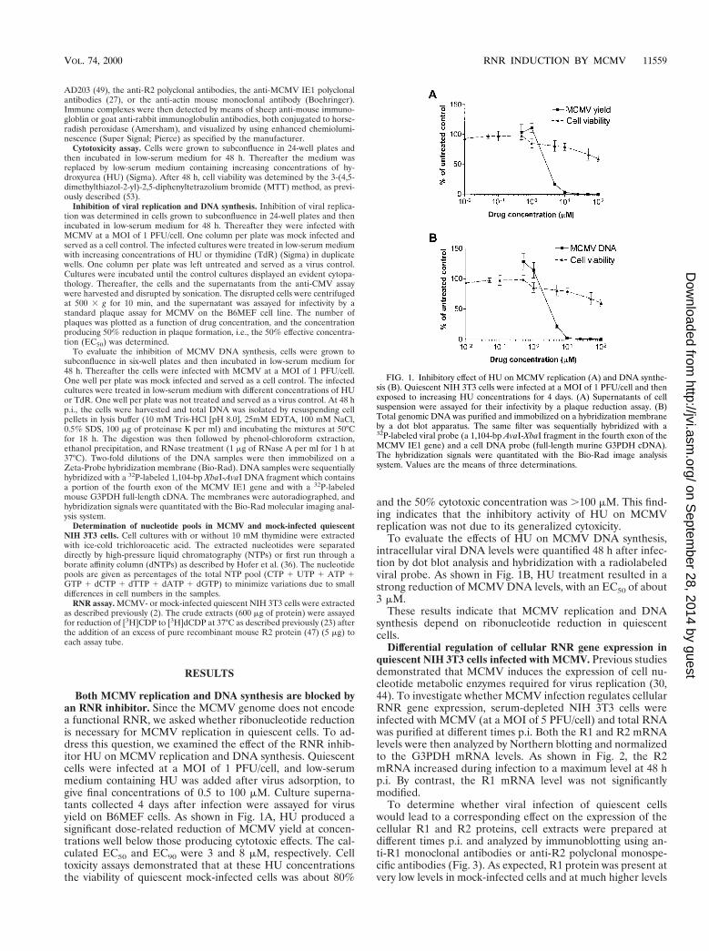
Both MCMV replication and DNA synthesis are blocked byan RNR inhibitor. Since the MCMV genome does not encodea functional RNR, we asked whether ribonucleotide reductionis necessary for MCMV replication in quiescent cells. To ad-dress this question, we examined the effect of the RNR inhib-itor HU on MCMV replication and DNA synthesis. Quiescentcells were infected at a MOI of 1 PFU/cell, and low-serummedium containing HU was added after virus adsorption, togive final concentrations of 0.5 to 100 mM. Culture superna-tants collected 4 days after infection were assayed for virusyield on B6MEF cells. As shown in Fig. 1A, HU produced asignificant dose-related reduction of MCMV yield at concen-trations well below those producing cytotoxic effects. The cal-culated EC50 and EC90 were 3 and 8 mM, respectively. Celltoxicity assays demonstrated that at these HU concentrationsthe viability of quiescent mock-infected cells was about 80%
and the 50% cytotoxic concentration was .100 mM. This find-ing indicates that the inhibitory activity of HU on MCMVreplication was not due to its generalized cytoxicity.
To evaluate the effects of HU on MCMV DNA synthesis,intracellular viral DNA levels were quantified 48 h after infec-tion by dot blot analysis and hybridization with a radiolabeledviral probe. As shown in Fig. 1B, HU treatment resulted in astrong reduction of MCMV DNA levels, with an EC50 of about3 mM.
These results indicate that MCMV replication and DNAsynthesis depend on ribonucleotide reduction in quiescentcells.
Differential regulation of cellular RNR gene expression inquiescent NIH 3T3 cells infected with MCMV. Previous studiesdemonstrated that MCMV induces the expression of cell nu-cleotide metabolic enzymes required for virus replication (30,44). To investigate whether MCMV infection regulates cellularRNR gene expression, serum-depleted NIH 3T3 cells wereinfected with MCMV (at a MOI of 5 PFU/cell) and total RNAwas purified at different times p.i. Both the R1 and R2 mRNAlevels were then analyzed by Northern blotting and normalizedto the G3PDH mRNA levels. As shown in Fig. 2, the R2mRNA increased during infection to a maximum level at 48 hp.i. By contrast, the R1 mRNA level was not significantlymodified.
To determine whether viral infection of quiescent cellswould lead to a corresponding effect on the expression of thecellular R1 and R2 proteins, cell extracts were prepared atdifferent times p.i. and analyzed by immunoblotting using an-ti-R1 monoclonal antibodies or anti-R2 polyclonal monospe-cific antibodies (Fig. 3). As expected, R1 protein was present atvery low levels in mock-infected cells and at much higher levels
FIG. 1. Inhibitory effect of HU on MCMV replication (A) and DNA synthe-sis (B). Quiescent NIH 3T3 cells were infected at a MOI of 1 PFU/cell and thenexposed to increasing HU concentrations for 4 days. (A) Supernatants of cellsuspension were assayed for their infectivity by a plaque reduction assay. (B)Total genomic DNA was purified and immobilized on a hybridization membraneby a dot blot apparatus. The same filter was sequentially hybridized with a32P-labeled viral probe (a 1,104-bp AvaI-XbaI fragment in the fourth exon of theMCMV IE1 gene) and a cell DNA probe (full-length murine G3PDH cDNA).The hybridization signals were quantitated with the Bio-Rad image analysissystem. Values are the means of three determinations.
VOL. 74, 2000 RNR INDUCTION BY MCMV 11559
on Septem
ber 28, 2014 by guesthttp://jvi.asm
.org/D
ownloaded from
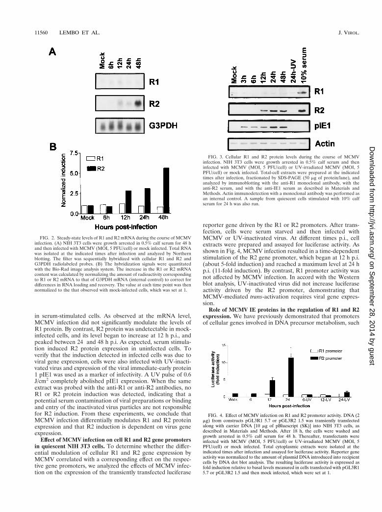
in serum-stimulated cells. As observed at the mRNA level,MCMV infection did not significantly modulate the levels ofR1 protein. By contrast, R2 protein was undetectable in mock-infected cells, and its level began to increase at 12 h p.i., andpeaked between 24 and 48 h p.i. As expected, serum stimula-tion induced R2 protein expression in uninfected cells. Toverify that the induction detected in infected cells was due toviral gene expression, cells were also infected with UV-inacti-vated virus and expression of the viral immediate-early protein1 pIE1 was used as a marker of infectivity. A UV pulse of 0.6J/cm2 completely abolished pIE1 expression. When the sameextract was probed with the anti-R1 or anti-R2 antibodies, noR1 or R2 protein induction was detected, indicating that apotential serum contamination of viral preparations or bindingand entry of the inactivated virus particles are not responsiblefor R2 induction. From these experiments, we conclude thatMCMV infection differentially modulates R1 and R2 proteinexpression and that R2 induction is dependent on virus geneexpression.
Effect of MCMV infection on cell R1 and R2 gene promotersin quiescent NIH 3T3 cells. To determine whether the differ-ential modulation of cellular R1 and R2 gene expression byMCMV correlated with a corresponding effect on the respec-tive gene promoters, we analyzed the effects of MCMV infec-tion on the expression of the transiently transfected luciferase
reporter gene driven by the R1 or R2 promoters. After trans-fection, cells were serum starved and then infected withMCMV or UV-inactivated virus. At different times p.i., cellextracts were prepared and assayed for luciferase activity. Asshown in Fig. 4, MCMV infection resulted in a time-dependentstimulation of the R2 gene promoter, which began at 12 h p.i.(about 5-fold induction) and reached a maximum level at 24 hp.i. (11-fold induction). By contrast, R1 promoter activity wasnot affected by MCMV infection. In accord with the Westernblot analysis, UV-inactivated virus did not increase luciferaseactivity driven by the R2 promoter, demonstrating thatMCMV-mediated trans-activation requires viral gene expres-sion.
Role of MCMV IE proteins in the regulation of R1 and R2expression. We have previously demonstrated that promotersof cellular genes involved in DNA precursor metabolism, such
FIG. 2. Steady-state levels of R1 and R2 mRNA during the course of MCMVinfection. (A) NIH 3T3 cells were growth arrested in 0.5% calf serum for 48 hand then infected with MCMV (MOI, 5 PFU/cell) or mock infected. Total RNAwas isolated at the indicated times after infection and analyzed by Northernblotting. The filter was sequentially hybridized with cellular R1 and R2 andG3PDH radiolabeled probes. (B) The hybridization signals were quantitatedwith the Bio-Rad image analysis system. The increase in the R1 or R2 mRNAcontent was calculated by normalizing the amount of radioactivity correspondingto R1 or R2 mRNA to that of G3PDH mRNA (internal control) to correct fordifferences in RNA loading and recovery. The value at each time point was thennormalized to the that observed with mock-infected cells, which was set at 1.
FIG. 3. Cellular R1 and R2 protein levels during the course of MCMVinfection. NIH 3T3 cells were growth arrested in 0.5% calf serum and theninfected with MCMV (MOI, 5 PFU/cell) or UV-irradiated MCMV (MOI, 5PFU/cell) or mock infected. Total-cell extracts were prepared at the indicatedtimes after infection, fractionated by SDS-PAGE (50 mg of protein/lane), andanalyzed by immunoblotting with the anti-R1 monoclonal antibody, with theanti-R2 serum, and with the anti-IE1 serum as described in Materials andMethods. Actin immunodetection with a monoclonal antibody was performed asan internal control. A sample from quiescent cells stimulated with 10% calfserum for 24 h was also run.
FIG. 4. Effect of MCMV infection on R1 and R2 promoter activity. DNA (2mg) from constructs pGL3R1 5.7 or pGL3R2 1.5 was transiently transfectedalong with carrier DNA [10 mg of pBluescript (SK)] into NIH 3T3 cells, asdescribed in Materials and Methods. After 18 h, the cells were washed andgrowth arrested in 0.5% calf serum for 48 h. Thereafter, transfectants wereinfected with MCMV (MOI, 5 PFU/cell) or UV-irradiated MCMV (MOI, 5PFU/cell) or mock infected. Total cytoplasmic extracts were isolated at theindicated times after infection and assayed for luciferase activity. Reporter geneactivity was normalized to the amount of plasmid DNA introduced into recipientcells by DNA dot blot analysis. The resulting luciferase activity is expressed asfold induction relative to basal levels measured in cells transfected with pGL3R15.7 or pGL3R2 1.5 and then mock infected, which were set at 1.
11560 LEMBO ET AL. J. VIROL.
on Septem
ber 28, 2014 by guesthttp://jvi.asm
.org/D
ownloaded from
as the dihydrofolate reductase (DHFR) and thymidylate syn-thase (TS) promoters, are trans-activated by MCMV pIE1 butnot by pIE3 (30, 44).
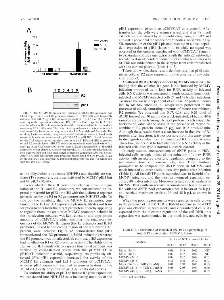
To see whether these IE gene products play a role in regu-lation of the R1 and R2 promoters, we cotransfected an ex-pression plasmid for pIE1 or pIE3 with the luciferase reportergenes driven by the R1 or R2 promoters into NIH 3T3 cells. Torule out the possibility that the MCMV IE promoter, con-tained in the IE1 or IE3 expression plasmids, titrates out tran-scription factors from the target promoters, thereby appearingto regulate them, the amount of MCMV promoter included inthe transfection mixtures was kept constant and appropriateamounts of pCMVCAT, which contains the regulatory se-quences of the MCMV IE region (the IE enhancer and IE1-3promoter) linked to the coding region of the irrelevant CATprotein, were included. Figure 5A demonstrates that pIE1transactivated the R2 promoter (6.5-fold induction) whereasthe R1 promoter was not affected. By contrast, pIE3 expressionhad no effect on R1 or R2 promoter activity. The ability of theIE1 or the IE3 constructs to express functional proteins wasverified by cotransfection assays with the pCMVCAT orpE1CAT indicator plasmid, respectively. As previously ob-served (50), pIE1 expression increased the activity of theMCMV IE enhancer and IE1-3 promoter of pCMVCATwhereas pIE3 expression resulted in trans-activation of theMCMV E1 early promoter of pE1CAT (data not shown).
To confirm the ability of pIE1 to induce R2 gene expression,we transfected into NIH 3T3 cells increasing amounts of the
pIE1 expression plasmid or pCMVCAT as a control. Aftertransfection the cells were serum starved, and after 48 h cellextracts were analyzed by immunoblotting using anti-R2 andanti-pIE1 polyclonal monospecific antibodies. As shown in Fig.5B, transfection of the pIE1 plasmid resulted in a dose-depen-dent expression of pIE1 (lanes 4 to 6) while no signal wasobserved in the samples transfected with pCMVCAT (lanes 1to 3). Analysis of the same extracts with the anti-R2 antibodiesrevealed a dose-dependent induction of cellular R2 (lanes 4 to6). This was undetectable in the samples from cells transfectedwith the control plasmid (lanes 1 to 3).
Taken as a whole, these results demonstrate that pIE1 stim-ulates cellular R2 gene expression in the absence of any otherviral product.
An altered RNR activity is induced by MCMV infection. Thefinding that the cellular R1 gene is not induced by MCMVinfection prompted us to look for RNR activity in infectedcells. RNR activity was measured in crude extracts from mock-infected and MCMV-infected cells 24 and 48 h after infection.To make the assay independent of cellular R2 protein, induc-ible by MCMV infection, all assays were performed in thepresence of added, saturating amounts of mouse recombinantR2 protein. We observed that 0.07, 0.18, and 0.20 nmol ofdCDP formed per 30 min in the mock-infected, 24-h, and 48-hsamples, respectively, using 0.6 mg of protein in each assay. Thebackground value of the assay in the presence of mouse re-combinant R2 protein alone (5 mg) was 0.02 nmol/30 min.Although these results show a clear increase in the level of R1protein after infection, it is not possible from this assay aloneto distinguish cellular from virus-induced R1 protein activity.Therefore, we decided to find whether the RNR activity in theinfected cells displayed a normal allosteric control.
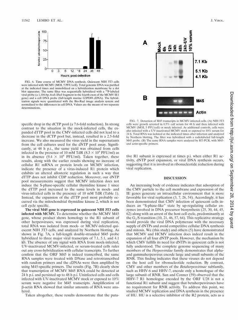
In early studies, measurements of dNTP pools in HSV-infected cells strongly indicated the induction of a new RNRactivity with an altered allosteric regulation compared to themammalian host cell enzyme (16, 42). These findingsprompted us to compare the dNTP pools in MCMV- andmock-infected quiescent cells at two time points after infection(Table 1). All four dNTP pools expanded two- to fivefold afterMCMV infection, and the most pronounced expansion oc-curred 48 h after infection. Moreover, a time course analysis ofMCMV DNA synthesis revealed a considerable temporal over-lap with the dNTP pool expansion since it began at 24 h p.i.and reached maximum levels at 36 and 48 h p.i. as shown inFig. 6.
When the pool measurements were repeated in cells grownin the presence of 10 mM TdR, a 10-fold increase in the dTTPpool was observed in both mock- and virus-infected cells. Asexpected from the allosteric regulation of the cell RNR, thisexpansion was accompanied in the mock-infected cells by a
FIG. 5. The MCMV IE protein pIE1 stimulates cellular R2 expression. (A)Effect of pIE1 on R1 and R2 promoter activity. NIH 3T3 cells were transientlycotransfected with 2 mg of the indicator plasmids pGL3R1 5.7 or pGL3R2 1.5and 1 mg of the expression vectors for pIE1, pIE3, or CAT respectively. At 18 hafter transfection, the cells were washed and then maintained for 48 h in mediumcontaining 0.5% calf serum. Thereafter, total cytoplasmic extracts were isolatedand assayed for luciferase activity, as described in Materials and Methods. Theresulting luciferase activity is expressed as fold induction relative to basal levelsmeasured in cells cotransfected with pGL3R1 5.7 or pGL3R2 1.5 and the valuefor the CAT-expressing vector, which was set at 1. (B) Effect of pIE1 expressionon cell R2 protein levels. NIH 3T3 cells were transiently transfected with 0.5, 1,and 4 mg of the CAT expression vector (lanes 1, 2, and 3 respectively) or the pIE1expression vector (lanes 4, 5, and 6 respectively). At 18 h after transfection, thecells were washed and then maintained in medium containing 0.5% calf serum.After 48 h, total-cell extracts were prepared, fractionated by SDS-PAGE (50 mgof protein/lane), and analyzed by immunoblotting with the anti-R2 serum andwith the anti-pIE1 serum.
TABLE 1. Distribution of individual dNTPs as a percentage oftotal NTP content after MCMV infection
Infection% of total NTP accounted for by:
dCTP dTTP dATP dGTP
Mock (24 h) 0.08 0.18 0.02 NDa
Mock (48 h) 0.07 0.15 0.02 NDMCMV (24 h) 0.08 0.16 0.04 0.01MCMV (48 h) 0.31 0.41 0.12 0.03Mock (24 h) 1 TdR (10 mM) 0.01 1.91 0.03 0.03MCMV (24 h) 1 TdR (10 mM) 0.05 2.08 0.04 0.06MCMV (48 h) 1 TdR (10 mM) 0.18 1.58 0.06 0.05
a ND, not detectable.
VOL. 74, 2000 RNR INDUCTION BY MCMV 11561
on Septem
ber 28, 2014 by guesthttp://jvi.asm
.org/D
ownloaded from
specific drop in the dCTP pool (a 7.6-fold reduction). In strongcontrast to the situation in the mock-infected cells, the ex-panded dTTP pool in the CMV-infected cells did not lead to adecrease in the dCTP pool but, instead, resulted in a 2.5-foldincrease. We also measured the virus yield in the supernatantsfrom the cell cultures used for the dNTP pool assay. Signifi-cantly, at 48 h p.i., the same yield was obtained from cellsinfected in the presence of 10 mM TdR (8.3 3 104 PFU/ml) asin its absence (9.4 3 104 PFU/ml). Taken together, theseresults, along with the earlier results showing no increase ofcellular R1 mRNA or protein levels on MCMV infection,indicate the presence of a virus-induced R1 protein whichexhibits an altered allosteric regulation in such a way thatdTTP does not inhibit CDP reduction. Moreover, our dNTPpool measurements suggest that MCMV infection does notinduce the S-phase-specific cellular thymidine kinase 1 sincethe dTTP pool increased to the same levels in mock- andvirus-infected cells in the presence of 10 mM TdR (Table 1).Instead, the expansion of the dTTP pool most probably oc-curred via the mitochondrial thymidine kinase 2, which is notcell cycle specific.
The viral M45 gene is expressed in quiescent NIH 3T3 cellsinfected with MCMV. To determine whether the MCMV M45gene, whose product shows homology to the R1 subunit ofother herpesviruses, was expressed during viral replication,total RNA was isolated from mock- or MCMV-infected qui-escent NIH 3T3 cells, and analyzed by Northern blotting. Asshown in Fig. 7A, a full-length double-stranded M45 probehybridized to three major viral transcripts of 7.1, 5.1, and 4.1kb. The absence of any signal with RNA from mock-infected,UV-inactivated MCMV-infected, or serum-treated cells rulesout any cross-hybridization with cellular transcripts. To furtherconfirm that the ORF M45 is indeed transcribed, the sameRNA samples were treated with DNase and retrotranscribedwith random primers and the cDNAs were then amplified byusing M45-specific primers. The results (Fig. 7B) clearly showthat transcription of MCMV M45 RNA could be detected at24 h p.i. and persisted up to 48 h p.i. Uninfected cells and cellsinfected with UV-inactivated MCMV stock or exposed to 10%serum were negative for M45 transcripts. Amplification ofb-actin RNA showed that similar amounts of RNA were ana-lyzed.
Taken altogether, these results demonstrate that the puta-
tive R1 subunit is expressed at times p.i. when either R1 ac-tivity, dNTP pool expansion, or viral DNA synthesis occurs,suggesting that it is involved in ribonucleotide reduction duringviral replication.
DISCUSSION
An increasing body of evidence indicates that adsorption ofthe CMV particle to the cell membrane and expression of theIE genes generate an intracellular environment that is morefavorable for viral replication (3, 7, 26, 54). Accordingly, it hasbeen demonstrated that CMV infection of quiescent cells in-duces an “S-phase-like” state by up-regulating cellular en-zymes involved in DNA precursor biosynthesis (25, 30, 44, 45,62) along with an arrest of the host cell cycle, predominantly atthe G1/S transition (10, 21, 46, 57, 64). This replicative strategywould provide the viral DNA polymerase with the necessarysupply of dNTPs and avoid competitive cellular DNA synthesisand mitosis. We (this study) and others (5) have demonstratedthat MCMV and HCMV infection does indeed result in theexpansion of all four dNTP pools. However, the mechanism bywhich CMV fulfills its need for dNTPs in quiescent cells is notfully understood. The complete genome sequencing of manymembers of the Herpesviridae family demonstrates that alpha-and gammaherpesvirus encode large and small subunits of theRNR. This finding indicates that these viruses do not dependon the host cell for ribonucleotide reduction. By contrast,CMV and other members of the Betaherpesvirinae subfamily,such as HHV-6 and HHV-7, encode only a homologue of thelarge subunit of RNR. Sun and Conner (59) observed that theHHV-7 R1 homologue encoded by the ORF U28 is not afunctional R1 subunit and suggest that betaherpesviruses haveno requirement for RNR activity. To address this point, westudied MCMV replication and DNA synthesis in the presenceof HU. HU is a selective inhibitor of the R2 protein, acts as a
FIG. 6. Time course of MCMV DNA synthesis. Quiescent NIH 3T3 cellswere infected with MCMV (MOI, 5 PFU/cell). Total genomic DNA was purifiedat the indicated times and immobilized on a hybridization membrane by a dotblot apparatus. The same filter was sequentially hybridized with a 32P-labeledviral probe (a 1,104-bp AvaI-XbaI fragment in the fourth exon of the MCMV IE1gene) and a cell DNA probe (full-length murine G3PDH cDNA). The hybrid-ization signals were quantitated with the Bio-Rad image analysis system andnormalized to the differences in cell DNA. Values are the means of two separatedeterminations.
FIG. 7. Detection of M45 transcripts in MCMV-infected cells. (A) NIH 3T3cells were growth arrested in 0.5% calf serum for 48 h and then infected withMCMV (MOI, 5 PFU/cell) or mock infected. As additional controls, cells werealso infected with a UV-inactivated MCMV stock or exposed to 10% serum for24 h. Total RNA was isolated at the indicated times after infection and analyzedby Northern blotting. The filter was hybridized with a radiolabeled full-lengthM45 probe. (B) The same RNA samples were analyzed by RT-PCR, with M45-and actin-specific primers.
11562 LEMBO ET AL. J. VIROL.
on Septem
ber 28, 2014 by guesthttp://jvi.asm
.org/D
ownloaded from
radical scavenger, and increases the rate of iron loss frommammalian R2 proteins. Inhibition of MCMV replication andDNA synthesis by HU in quiescent cells clearly shows that denovo synthesis of dNTPs by an iron-radical RNR is needed andrules out the possibility that the DNA precursors are obtainedthrough the induction of the deoxyribonucleoside salvage path-way. Since CMV does not encode a functional RNR, one wayof inducing ribonucleotide reduction in a quiescent cell is tostimulate an unscheduled expression of its R1 and R2 proteins.Here we demonstrated that while MCMV infection does notaffect R1 expression, it strongly induces R2 promoter activityand mRNA and protein levels. We reason that R2 stimulationis caused by the infection on the basis of the following piecesof evidence: (i) dependence of the effect on MOI (data notshown); (ii) dependence of the effect on viral infectivity, sinceUV-inactivated virus cannot trigger any stimulation of the R2promoter and protein expression; and (iii) ability of a specificMCMV genome fragment encoding the pIE1 to trans-activatethe R2 promoter and induce R2 protein expression.
Several reports have shown that CMV infection stimulatesthe expression of a number of cellular genes important for cellcycle regulation and DNA synthesis. This regulation has beenreported to depend on either viral binding to the cell surface(8, 9, 65) or viral IE protein expression (11, 31, 33, 40, 48, 54,58, 62, 66). The observation that inactivation of MCMV by UVexposure abolished the induction of R2 protein as well astrans-activation of the R2 promoter suggests that virus geneexpression, rather than interaction of viral particles with thecell surface, is required to stimulate R2 gene expression. As wehave previously observed for the DHFR (44), TS (30) andfolylpolyglutamate synthetase (FPGS) (unpublished data) pro-moters, both MCMV infection and pIE1 transactivated the R2promoter, and virus-dependent transactivation was observedduring the time frame when pIE1 protein was expressed (Fig.3). Furthermore, transient transfection of a pIE1 expressionvector induced R2 protein expression. Taken together, theseresults indicate that R2 induction by MCMV occurs at least inpart via pIE1 expression.
We next asked which R1 protein (cellular or viral) is respon-sible for ribonucleotide reduction in MCMV-infected cells.Although the cellular R1 protein is barely detectable in bothuninfected and infected quiescent cells, we cannot exclude thepossibility that these low levels could be sufficient to supportMCMV replication. In keeping with the low level of cellularR1 protein in mock-infected cells, its activity, measured in thepresence of saturating amounts of recombinant mouse R2 pro-tein, is slightly above the background value (0.07 and 0.02 nmolof dCDP/30 min, respectively). On the other hand, the RNRassay clearly shows an increase of R1 protein activity inMCMV-infected cells (0.18 and 0.2 nmol of dCDP/30 min at 24and 48 h p.i., respectively), which is in contrast to the unin-duced levels of cell R1 mRNA and protein. However, since allthe activity values were low, it is not possible from this assay todefinitely distinguish a cellular R1 protein from a virus-in-duced R1 protein.
If we assume that the RNR activity in MCMV-infected cellsis the result of cellular R1 and R2 association, the low enzymeactivity might reflect the low level of R1 protein in quiescentcells. Alternatively, if the viral R1 associates with the cellularR2, this hybrid RNR might have a different allosteric controland would require different assay conditions. Early studiesdemonstrated that HSV-1 induced an altered RNR in extractsof infected cells, since pyrimidine nucleotide reduction by the“new” reductase activity was highly resistant to dTTP inhibi-tion (16, 42). Moreover, HSV-1 replication was not affected incells in which cellular DNA synthesis was inhibited by TdR
treatment. In the mammalian cell, TdR is converted to dTTP,which acts as an allosteric inhibitor of the cellular RNR andsuppresses CDP reduction (51, 52, 63). dCTP depletion resultsin blocked DNA synthesis and cell proliferation. As observedwith HSV-1, our dNTP pool data support the existence of analtered RNR in the MCMV-infected cells. As expected, mea-surement of dNTP pools demonstrated that mock-infectedcells contain an RNR that is sensitive to dTTP inhibition, sincewe observed a specific drop in the dCTP pool following theaddition of 10 mM TdR whereas in TdR-treated MCMV-infected cells the level of dTTP remained high; also, the levelof dCTP increased and MCMV replication was not inhibited.Thus, the MCMV-induced reductase activity allows the virus tooverride the dTTP-inhibited normal cellular RNR and makedCTP. Taken together, our data demonstrate that MCMVinfection stimulates expression of the cellular R2 protein andinduces an RNR activity with an altered allosteric regulationcompared to the mouse RNR. Whether this latter effect is theconsequence of the association of the R2 protein with theproduct of the M45 gene of MCMV remains to be demon-strated. However, several observations suggest involvement ofthe M45 gene product in ribonucleotide reduction in the in-fected cells. First, we have demonstrated that the M45 gene isindeed expressed in MCMV-infected cells. Temporal studieshave shown that a significant accumulation of the M45 tran-scripts can be detected at 24 and 48 h p.i. Moreover, there is aconsiderable temporal overlap between the increase in M45mRNA levels, R2 expression, increased R1 activity, expansionof the dNTP pool, and synthesis of the viral DNA. Whenconsidered together with the data demonstrating an alteredallosteric regulation of RNR following MCMV infection, thesefindings support the hypothesis that the product of M45 maycomplex with R2 to form a functional version of the enzyme.Studies are under way to verify this hypothesis.
CMV can replicate in quiescent cells that have shut downtheir machinery for synthesizing DNA. The reactions catalyzedby RNR and by the enzymes involved in the biosynthesis ofthymidylate (dTMP) are highly repressed in cells that are notundergoing DNA synthesis. We have previously demonstratedthat MCMV infection of quiescent cells leads to the coordi-nated stimulation of the cell enzymes FPGS, DHFR, and TS,involved in dTMP synthesis. Here we present evidence that anRNR activity with altered allosteric control is induced in qui-escent cells by MCMV infection. The induction of this set ofenzymes releases the virus from normal cell control and allowsdNTP biosynthesis and viral replication to take place duringperiods of the cell cycle other than the S phase.
ACKNOWLEDGMENTS
We thank Timothy J. Kinsella for providing plasmid pET28a(1)R2(1)and Nigel Parker for providing plasmid p3I.
This work was supported by grants from the Italian AIDS ResearchProject (grant 50B.25), from MURST-CNR Biotechnology Program L.95/95, from A.I.R.C., from the Italian Ministry of Public Health, andfrom the Swedish Natural Science Research Council.
REFERENCES
1. Abken, H., and B. Reifenrath. 1992. A procedure to standardize CAT re-porter gene assay. Nucleic Acids Res. 20:3527.
2. Akerblom, L., A. Ehrenberg, A. Graslund, H. Lankinen, P. Reichard, and L.Thelander. 1981. Overproduction of the free radical of ribonucleotide re-ductase in hydroxyurea-resistant mouse fibroblast 3T6 cells. Proc. Natl.Acad. Sci. USA 78:2159–2163.
3. Albrecht, T., I. Boldogh, M. Fons, S. AbuBakar, and C. Z. Deng. 1990. Cellactivation signals and the pathogenesis of human cytomegalovirus. Intervi-rology 31:68–75.
4. Averett, D. R., C. Lubbers, G. B. Elion, and T. Spector. 1983. Ribonucleotidereductase induced by herpes simplex type 1 virus. Characterization of a
VOL. 74, 2000 RNR INDUCTION BY MCMV 11563
on Septem
ber 28, 2014 by guesthttp://jvi.asm
.org/D
ownloaded from
distinct enzyme. J. Biol. Chem. 258:9831–9838.5. Biron, K. K., J. A. Fyfe, S. C. Stanat, L. K. Leslie, J. A. Sorrell, C. U. Lambe,
and D. M. Coen. 1986. A human cytomegalovirus mutant resistant to thenucleoside analog 9-[2-hydroxy-1-(hydroxymethyl)ethoxy]methylguanine(BW B759U) induces reduced levels of BW B759U triphosphate. Proc. Natl.Acad. Sci. USA 83:8769–8773.
6. Bjorklund, S., S. Skog, B. Tribukait, and L. Thelander. 1990. S-phase-specific expression of mammalian ribonucleotide reductase R1 and R2 sub-unit mRNAs. Biochemistry 29:5452–5458.
7. Boldogh, I., S. AbuBakar, and T. Albrecht. 1990. Activation of protoonco-genes: an immediate early event in human cytomegalovirus infection. Sci-ence 247:961–964.
8. Boldogh, I., S. AbuBakar, D. Millinoff, C. Z. Deng, and T. Albrecht. 1991.Cellular oncogenes activation by human cytomegalovirus. Lack of correla-tion with virus infectivity and immediate early gene expression. Arch. Virol.118:163–177.
9. Boyle, K. A., R. L. Pietropaolo, and T. Compton. 1999. Engagement of thecellular receptor for glycoprotein B of human cytomegalovirus activates theinterferon-responsive pathway. Mol. Cell. Biol. 19:3607–3613.
10. Bresnahan, W. A., I. Boldogh, E. A. Thompson, and T. Albrecht. 1996.Human cytomegalovirus inhibits cellular DNA synthesis and arrests produc-tively infected cells in late G1. Virology 224:150–160.
11. Bresnahan, W. A., T. Albrecht, and E. A. Thompson. 1998. The cyclin Epromoter is activated by human cytomegalovirus 86-kDa immediate earlyprotein. J. Biol. Chem. 273:22075–22082.
12. Cameron, J. M., I. McDougall, H. S. Marsden, V. G. Preston, D. M. Ryan,and J. H. Subak-Sharpe. 1988. Ribonucleotide reductase encoded by herpessimplex virus is a determinant of the pathogenicity of the virus in mice anda valid antiviral target. J. Gen. Virol. 69:2607–2612.
13. Chabes, A., and Thelander, L. 2000. Controlled protein degradation regu-lates ribonucleotide reductase activity in proliferating mammalian cells dur-ing the normal cell cycle and in response to DNA damage and replicationblocks. J. Biol. Chem. 275:17747–17753.
14. Chee, M. S., A. T. Bankier, S. Beck, R. Bohni, C. M. Brown, R. Cerny, T.Horsnell, C. A. Hutchison, T. Kouzarides, J. A. Martignetti, et al. 1990.Analysis of the protein-coding content of the sequence of human cytomeg-alovirus strain AD169. Curr. Top. Microbiol. Immunol. 154:125–169.
15. Chirgwin, J. M., A. E. Przybyla, R. J. MacDonald, and W. J. Rutter. 1979.Isolation of biologically active ribonucleic acid from sources enriched inribonucleases. Biochemistry 18:5294–5299.
16. Cohen, G. H. 1972. Ribonucleotide reductase activity of synchronized KBcells infected with herpes simplex virus. J. Virol. 9:408–418.
17. Conner, J., H. Marsden, and J. B. Clements. 1994. Ribonucleotide reductaseof herpes viruses. Rev. Med. Virol. 4:25–34.
18. Cooper, J., J. Conner, and J. B. Clements. 1995. Characterization of thenovel protein kinase activity present in the R1 subunit of herpes simplexvirus ribonucleotide reductase. J. Virol. 69:4979–4985.
19. Davison, A. J., and J. E. Scott. 1986. The complete DNA sequence ofvaricella-zoster virus. J. Gen. Virol. 67:1759–1816.
20. de Wind, N., A. Berns, A. Gielkens, and T. Kimman. 1993. Ribonucleotidereductase-deficient mutants of pseudorabies virus are avirulent for pigs andinduce partial protective immunity. J. Gen. Virol. 74:351–359.
21. Dittmer, D., and E. S. Mocarski. 1997. Human cytomegalovirus infectioninhibits G1/S transition. J. Virol. 71:1629–1634.
22. Engstrom, Y., S. Eriksson, I. Jildevik, S. Skog, L. Thelander, and B. Tribu-kait. 1985. Cell cycle-dependent expression of mammalian ribonucleotidereductase. Differential regulation of the two subunits. J. Biol. Chem. 260:9114–9116.
23. Engstrom, Y., S. Eriksson, L. Thelander, and M. Akerman. 1979. Ribonu-cleotide reductase from calf thymus. Purification and properties. Biochem-istry 18:2941–2948.
24. Eriksson, S., A. Graslund, S. Skog, L. Thelander, and B. Tribukait. 1984.Cell cycle-dependent regulation of mammalian ribonucleotide reductase.The S phase-correlated increase in subunit M2 is regulated by de novoprotein synthesis. J. Biol. Chem. 259:11695–11700.
25. Estes, J. E., and E.-S. Huang. 1977. Stimulation of cellular thymidine kinaseby human cytomegalovirus. J. Virol. 24:13–21.
26. Fortunato, E. A., A. K. McElroy, I. Sanchez, and D. H. Spector. 2000.Exploitation of cellular signaling and regulatory pathways by human cyto-megalovirus. Trends Microbiol. 8:111–119.
27. Gariglio, M., P. Foresta, C. Sacchi, M. Lembo, L. Hertel, and S. Landolfo.1997. Suppression of high mobility group protein T160 expression impairsmouse cytomegalovirus replication. J. Gen. Virol. 78:665–670.
28. Goldstein, D. J., and S. K. Weller. 1988. Factor(s) present in herpes simplexvirus type 1-infected cells can compensate for the loss of the large subunit ofthe viral ribonucleotide reductase: characterization of an ICP6 deletionmutant. Virology 166:41–51.
29. Goldstein, D. J., and S. K. Weller. 1988. Herpes simplex virus type 1-inducedribonucleotide reductase activity is dispensable for virus growth and DNAsynthesis: isolation and characterization of an ICP6 lacZ insertion mutant.J. Virol. 62:196–205.
30. Gribaudo, G., L. Riera, D. Lembo, M. De Andrea, M. Gariglio, T. L. Rudge,
L. F. Johnson, and S. Landolfo. 2000. Murine cytomegalovirus stimulatescellular thymidylate synthase gene expression in quiescent cells and requiresthe enzyme for replication. J. Virol. 74:4979–4987.
31. Gribaudo, G., S. Ravaglia, L. Guandalini, R. Cavallo, M. Gariglio, and S.Landolfo. 1996. The murine cytomegalovirus immediate early 1 proteinstimulates NF-kB activity by transactivating the NF-kB p105/p50 promoter.Virus Res. 45:15–27.
32. Gribaudo, G., S. Ravaglia, M. Gaboli, M. Gariglio, R. Cavallo, and S.Landolfo. 1995. Interferon-a inhibits the murine cytomegalovirus immedi-ate-early gene expression by down-regulating NF-kB activity. Virology 211:251–260.
33. Hayhurst, G. P., L. A. Bryant, R. C. Caswell, S. M. Walker, and J. H.Sinclair. 1995. CCAAT box-dependent activation of the TATA-less humanDNA polymerase a promoter by the human cytomegalovirus 72-kilodaltonmajor immediate-early protein. J. Virol. 69:182–188.
34. Heineman, T. C., and J. I. Cohen. 1994. Deletion of the varicella-zoster viruslarge subunit of ribonucleotide reductase impairs growth of virus in vitro.J. Virol. 68:3317–3323.
35. Henry, B. E., R. Glaser, J. Hewetson, and D. J. O’Callaghan. 1978. Expres-sion of altered ribonucleotide reductase activity associated with the replica-tion of the Epstein-Barr virus. Virology 89:262–271.
36. Hofer, A., J. T. Ekanem, and L. Thelander. 1998. Allosteric regulation ofTrypanosoma brucei ribonucleotide reductase studied in vitro and in vivo.J. Biol. Chem. 273:34098–34104.
37. Jacobson, J. G., D. A. Leib, D. J. Goldstein, C. L. Bogard, P. A. Schaffer,S. K. Weller, and D. M. Coen. 1989. A herpes simplex virus ribonucleotidereductase deletion mutant is defective for productive acute and reactivatablelatent infections of mice and for replication in mouse cells. Virology 173:276–283.
38. Johansson, E., K. Hjortsberg, and L. Thelander. 1998. Two YY-1-bindingproximal elements regulate the promoter strength of the TATA-less mouseribonucleotide reductase R1 gene. J. Biol. Chem. 273:29816–29821.
39. Jordan, A., and P. Reichard. 1998. Ribonucleotide reductases. Annu. Rev.Biochem. 67:71–98.
40. Koszinowski, U. H., G. M. Keil, H. Volkmer, M. R. Fibi, A. Ebeling-Keil, andK. Munch. 1986. The 89,000-Mr murine cytomegalovirus immediate-earlyprotein activates gene transcription. J. Virol. 58:59–66.
41. Kuo, M.-L., and T. J. Kinsella. 1997. Overexpression of a hexa-histidine andT7 peptide tagged human ribonucleotide reductase small subunit, R2 inEscherichia coli and the generation of human R2 antibodies. Int. J. Oncol.10:515–520.
42. Langelier, Y., and G. Buttin. 1981. Characterization of ribonucleotide re-ductase induction in BHK-21/C13 Syrian hamster cell line upon infection byherpes simplex virus (HSV). J. Gen. Virol. 57:21–31.
43. Lankinen, H., A. Graslund, and L. Thelander. 1982. Induction of a newribonucleotide reductase after infection of mouse L cells with pseudorabiesvirus. J. Virol. 41:893–900.
44. Lembo, D., A. Angeretti, M. Gariglio, and S. Landolfo. 1998. Murine cyto-megalovirus induces expression and enzyme activity of dihydrofolate reduc-tase in quiescent cells. J. Gen. Virol. 78:2803–2808.
45. Lembo, D., G. Gribaudo, R. Cavallo, L. Riera, A. Angeretti, L. Hertel, and S.Landolfo. 1999 Human cytomegalovirus stimulates cellular dihydrofolatereductase activity in quiescent cells. Intervirology 42:30–36.
46. Lu, M., and T. Shenk. 1996. Human cytomegalovirus infection inhibits cellcycle progression at multiple points, including the transition from G1 toS. J. Virol. 70:8850–8857.
47. Mann, G. J., A. Graslund, E. Ochiai, R. Ingemarson, and L. Thelander. 1991.Purification and characterization of recombinant mouse and herpes simplexvirus ribonucleotide reductase R2 subunit. Biochemistry 30:1939–1947.
48. Margolis M. J., S. Pajovic, E. L. Wong, M. Wade, R. Jupp, J. A. Nelson, andJ. Clifford Azizkhan. 1995. Interaction of the 72-kilodalton human cytomeg-alovirus IE1 gene product with E2F1 coincides with E2F-dependent activa-tion of dihydrofolate reductase transcription. J. Virol. 69:7759–7767.
49. McClarty, G. A., A. K. Chan, Y. Engstrom, J. A. Wright, and L. Thelander.1987. Elevated expression of M1 and M2 components and drug-inducedposttranscriptional modulation of ribonucleotide reductase in a hydroxy-urea-resistant mouse cell line. Biochemistry 26:8004–8011.
50. Messerle, M., B. Buhler, G. M. Keil, and U. H. Koszinowski. 1992. Structuralorganization, expression, and functional characterization of the murine cy-tomegalovirus immediate-early gene 3. J. Virol. 66:27–36.
51. Morris, N. R., and G. A. Fisher. 1963. Studies concerning the inhibition ofcellular reproduction by deoxyribonucleosides. I. Inhibition of the synthesisof deoxycytidine by a phosphorylated derivative of thymidine. Biochim. Bio-phys. Acta 68:84–92.
52. Morris, N. R., P. Reichard, and G. A. Fisher. 1963. Studies concerning theinhibition of cellular reproduction by deoxyribonucleosides. II. Inhibition ofthe synthesis of deoxycytidine by thymidine, deoxyadenosine and deox-yguanosine. Biochim. Biophys. Acta 68:93–99.
53. Pauwels, R., J. Balzarini, M. Baba, R. Snoeck, D. Schols, P. Hederwijin, J.Desmyter, and E. De Clerq. 1988. Rapid and automated tetrazolium-basedcolorimetric assay for the detection of anti HIV compounds. J. Virol. Meth-ods 20:309–321.
11564 LEMBO ET AL. J. VIROL.
on Septem
ber 28, 2014 by guesthttp://jvi.asm
.org/D
ownloaded from
54. Poma, E. E., T. F. Kowalik, L. Zhu, J. H. Sinclair, and E.-S. Huang. 1996.The human cytomegalovirus IE1–72 protein interacts with the cellular p107protein and relieves p107-mediated transcriptional repression of an E2F-responsive promoter. J. Virol. 70:7867–7877.
55. Rawlinson, W. D., H. E. Farrell, and B. G. Barrell. 1996. Analysis of thecomplete DNA sequence of murine cytomegalovirus. J. Virol. 70:8833–8849.
56. Reichard, P. 1988. Interactions between deoxyribonucleotide and DNA syn-thesis. Annu. Rev. Biochem. 57:349–374
57. Salvant, B. S., E. A. Fortunato, and D. H. Spector. 1998. Cell cycle dysregu-lation by human cytomegalovirus: influence of the cell cycle phase at the timeof infection and effects on cyclin transcription. J. Virol. 72:3729–3741.
58. Schickedanz, J., L. Philipson, W. Ansorge, R. Pepperkork, R. Klein, andU. H. Koszinowski. 1988. The 89,000-Mr murine cytomegalovirus immediate-early protein stimulates c-fos expression and cellular DNA synthesis. J. Virol.62:3341–3347.
59. Sun, Y., and J. Conner. 1999. The U28 ORF of human herpesvirus-7 doesnot encode a functional ribonucleotide reductase R1 subunit. J. Gen. Virol.80:2713–2718.
60. Thelander, L., and A. Gräslund. 1994. Ribonucleotide reductase in mam-
malian cells. Metal Ions Biol. Syst. 30:109.61. Thelander, L., and P. Reichard. 1979. Reduction of ribonucleotides. Annu.
Rev. Biochem. 48:133–158.62. Wade, M., T. F. Kowalik, M. Mudryj, E.-S. Huang, and J. Clifford Azizkhan.
1992. E2F mediates dihydrofolate reductase promoter activation and multi-protein complex formation in human cytomegalovirus infection. Mol. Cell.Biol. 12:4364–4374.
63. Whittle, E. D. 1966. Effect of thymidine on deoxyribonucleic acid synthesisand cytidine metabolism in rat thymus cells. Biochim. Biophys. Acta 114:44–60.
64. Wiebusch, L, and C. Hagemeier. 1999. Human cytomegalovirus 86-kilodal-ton IE2 protein blocks cell cycle progression in G1. J. Virol. 73:9274–9283.
65. Yurochko, A. D., E.-S. Hwang, L. Rasmussen, S. Keay, L. Pereira and E.-S.Huang. 1997. The human cytomegalovirus UL55 (gB) and UL 75 (gH)glycoprotein ligands initiate the rapid activation of Sp1 and NF-kB duringinfection. J. Virol. 71:5051–5059.
66. Yurochko, A. D., T. F. Kowalik, S. M. Huong, and E.-S. Huang. 1995. Humancytomegalovirus upregulates NF-kB activity by transactivating the NF-kBp105/p50 and p65 promoter. J. Virol. 69:5391–5400.
VOL. 74, 2000 RNR INDUCTION BY MCMV 11565
on Septem
ber 28, 2014 by guesthttp://jvi.asm
.org/D
ownloaded from


