Embed Size (px)
Citation preview

J. Membrane Biol. 143, 219-226 (1995) The Journal of
Membrane Biology �9 Springer-Verlag New York Inc. 1995
Expression of Potassium Channels in Epithelial Cells Depends on Calcium-activated Cell-Cell Contacts
D. Talavera, A. Ponce, R. Fiorentino, L. Gonzfilez-Mariscal, R.G. Contreras, S.H. S~inchez, M.R. Garcia-Villegas, J. Vald6s, M. Cereijido Center for Research and Advanced Studies. Apartado Postal 14-740, 07000 M~xico, D.F. M6xico
Received: 7 June 1994
Abstract. Harvesting MDCK cells with trypsin-EDTA reduces potassium currents (IK) to a mere 10%, presum- ably by hydrolysis of K + channels, but replating at con- fluence restores them in 12-18 hr, through a process that requires transcription, translation and exocytic fusion o f intracellular membrane vesicles to the plasma membrane (Ponce & Cereijido, 1991; Ponce et al., 1991a). In the present work we find that this restoration of I K also re- quires cell-cell contacts and the presence of 1.8 rnM Ca 2§ The role of extracellular Ca 2§ may be substituted by 2.0 gM TRH, 10 nM PMA or 200 gg/ml DiC8, drugs that stimulate the system of phospholipase C (PLC) and pro- tein kinase C (PKC). Conversely, the recovery of IK triggered by Ca-dependent contacts can be blocked by 110 gM neomycin, 2.0 gM H7, and 250 nM staurosporine, inhibitors of PLC and PKC. These results suggest that the expression of new K + channels depends on Ca 2§ activated contacts with neighboring cells and that the information is conveyed through PLC and PKC, a pro- cess in keeping with changes in its enzymatic activity and cellular distribution of PKC. Plasma membrane is also reduced and restored upon harvesting and replating, and depends on Ca2+-activated contracts. However, the effects of the chemicals tested on I K differ from the ones they elicit on the recovery of plasma membrane, suggest- ing that cells can independently regulate their population of K + channels and the surface of their membrane.
Key words: Epithelia - - K + channels - - Expression of ion channels - - Ca z+ - - Membrane area - - Cell-cell contacts
Correspondence to: M. Cereijido
Introduction
Cells of transporting epithelia establish tight junctions (TJs) and translocate substances vectorially from one compartment to another, thanks to a polarized distribu- tion of transporting mechanisms in their apical and ba- solateral domains (Cereijido et al., 1980). The under- standing of the mechanisms that form TJs and polarize the cells has been appreciably advanced by the use of model systems constituted by epithelial cell lines cul- tured as monolayers (Cereijido et al., 1978, 1980). In a previous work we found that MDCK cells (epithelioid of renal origin) express five types of K + channels and one of CI-, which are distributed in a strictly polarized manner: four types of K + channels are apical, a fifth is basolateral, and the only type of CI- channel found is exclusively located on the basolateral side (Bolivar & Cereijido, 1987; Ponce & Cereijido, 1991; Ponce et al., 1991a; Ponce, Contreras & Cereijido, 1991b).
Harvesting with trypsin-EDTA reduces the overall potassium current (Ii0 to a mere 10% of pretrypsination levels, presumably by hydrolysis of the extracellular do- mains of the peptides constituting the channels. Resto- ration of I K is achieved in 12-20 hr upon replating the cells at confluence, by a process that is blocked by in- hibitors of transcription (actimomycin D), translation (cycloheximide), exocytic fusion (chloroquine), micro- filaments (cytochalasin B) and microtubule assembly (colchicine), suggesting that it requires the synthesis of new K + channels (Ponce et al., 1991a). This indicates that the expression of new K § channels follows the same time course and depends basically on the same cellular mechanisms responsible for TJ formation (Contreras et al., 1992a). Junction formation also requires calcium- dependent contacts between neighboring cells (Gonzfi- lez-Mariscal, Chfivez de Ramirez & Cereijido, 1985;

220 D. Talavera et al.: K + Channel Expression in Epithelial Cells
Gon2alez-Mariscal et al., 1990). In the case of junction formation, Ca 2+ is needed on an extracellular site (Con- treras et al., 1992a, b) probably constituted by a uvo- morulin molecule (Gumbiner & Simons, 1987), and the information that cell-cell contact has been established is transduced to the intracellular mechanisms that assemble and seal the TJ through a cascade of reactions involving phospholipase C (PLC) and protein kinase C (PKC) (AC) (Balda et al., 1991, 1993). Therefore, in the present work we investigate whether the expression of K § channels depends likewise on cell-cell contacts and the presence of Ca 2+, and involves PLC, and PKC.
Materials and Methods
CELL CULTURE
Starter MDCK cultures were obtained from the American Type Culture Collection (MDCK, CCL-34). In most experiments, cells were be- tween 60-90th passage. They were grown at 36.5~ in disposable plastic bottles (Costar 3150, Cambridge, MA) with an air-5% CO 2 atmosphere (VIP CO 2 incubator 417, Lab. Line Instruments, New Brunswick, NY) and 20 ml of Dulbecco's modified Eagle's basal me- dium (DMEM, GIBCO, 430-1600, Grand Island, NY) with 100 U/ml of penicillin, 100 gg/ml of streptomycin (In Vitro, MExico, D.F.) and 10% fetal calf serum (Boehringer Mannheim, Germany).
MEMBRANE CURRENTS AND CAPACITANCE
Monolayers of cells cultured on glass coverslips were deposited on a flat chamber fixed to the stage of an inverted Diavert microscope (Leitz, Wetzlar) equipped with Hoffman optics. Membrane currents were studied in the whole-cell clamp configuration as previously de- scribed (Bolivar & Cereijido, 1987). Recordings were made through a Dagan 8900 amplifier (Minneapolis, MN) using micropipettes pulled from borosilicate glass (Coming, I.D. 1.1-1.2 ram) prepared in a Brown-Flamming type micropipette puller (Sutter Instruments, Novato, CA). Micropipettes were fire-polished and had a resistance of 2-3 Mr2. They were mounted in a pipette holder attached to a hydraulic micromanipulator (Narishige MF83, Tokyo, Japan). All recordings re- ported were obtained after seals of at least 5 GfL Sealing and record- ings were monitored with a Tektronix 2212 oscilloscope (Beaverton, OR). Whole-cell currents were recorded through Ag-AgC1 electrodes and filtered at 1 kHz. Signals were digitalized through an analogue/ digital converter (Labmaster TM-125, DMA, Scientific Solutions) and analyzed with a 80486-33 MHz based PC type computer. Stimulation was made with the Clampex program and analysis was performed with the Clampan program (pCLAMP version 5.5, Axon Instruments, Foster City, CA). Capacitive currents used to estimate the area of plasma membrane were filtered at 10 kHz and recorded at 100 kHz.
PROTEIN KINASE C (PKC) ASSAY
PKC specific activity of cells extracts was determined with the Protein Kinase C Assay System (GIBCO BRL, 3161SA), which measures the transference of 7 phosphate from [7-32p]ATP (neg-002A, DuPont) to a synthetic peptide containing the PKC substrate consensus sequence, as described by Yasuda et al. (1990).
FLUORESCENCE CYTOLOGICAL DETECTION OF PKC
We used a rhodamine-conjugated bisindolylmaleimide PKC inhibitor (rim-l) (Teflabs, Austin, TX) that targets the catalytic site of this en- zyme. Glass coverslips containing MDCK monolayers, cultured under several of the conditions described below, were rinsed twice with PBS, fixed and permeabilized with -20~ methanol for 45 sec, washed with PBS, incubated with 3% fetal bovine serum in PBS for 30 min, and treated with rim-1 for 1.0 hr. They were then washed twice with PBS and mounted in p-phenytdiamine-glycerol (1:9) and examined with a confocal microscope (MRC-600, Bio-Rad).
SOLUTIONS, ABBREVIATIONS AND SOURCE OF CHEMICALS
Intracellular solutions contain (mM): K + 141, Na + 5.0, Ca 2+ 1.0, EGTA 2.3 (Free Ca 2+ 3 x 10⁻⁷), Mg 2+ 1.0, glucose 14, HEPES 10, methane- sulfonic acid 153, pH 7.4. The bathing medium contains (raN): K § 5.0, Na § 141, Ca 2+ 5.0, Mg a+ 1.0, glucose 16, HEPES 10, methanesulfonic acid 158, pH 7.4.
DiC8 (1,2-dioctanoylglycerol) and EGD (ethylene glycol di- octanoate) were from Molecular Probes (Eugene, OR). dB-cAMP (N6,-O2"-dibutyryladenosine 3'-5'-cyclic monophosphate) and stanro- sporine were from Sigma Chemical (St. Louis, MO). NC and LC refer to media with low (1-5 gM) and normal Ca 2+ (1.8 mM); respectively.
Results are expressed as mean _+ SE (number of observations).
Results
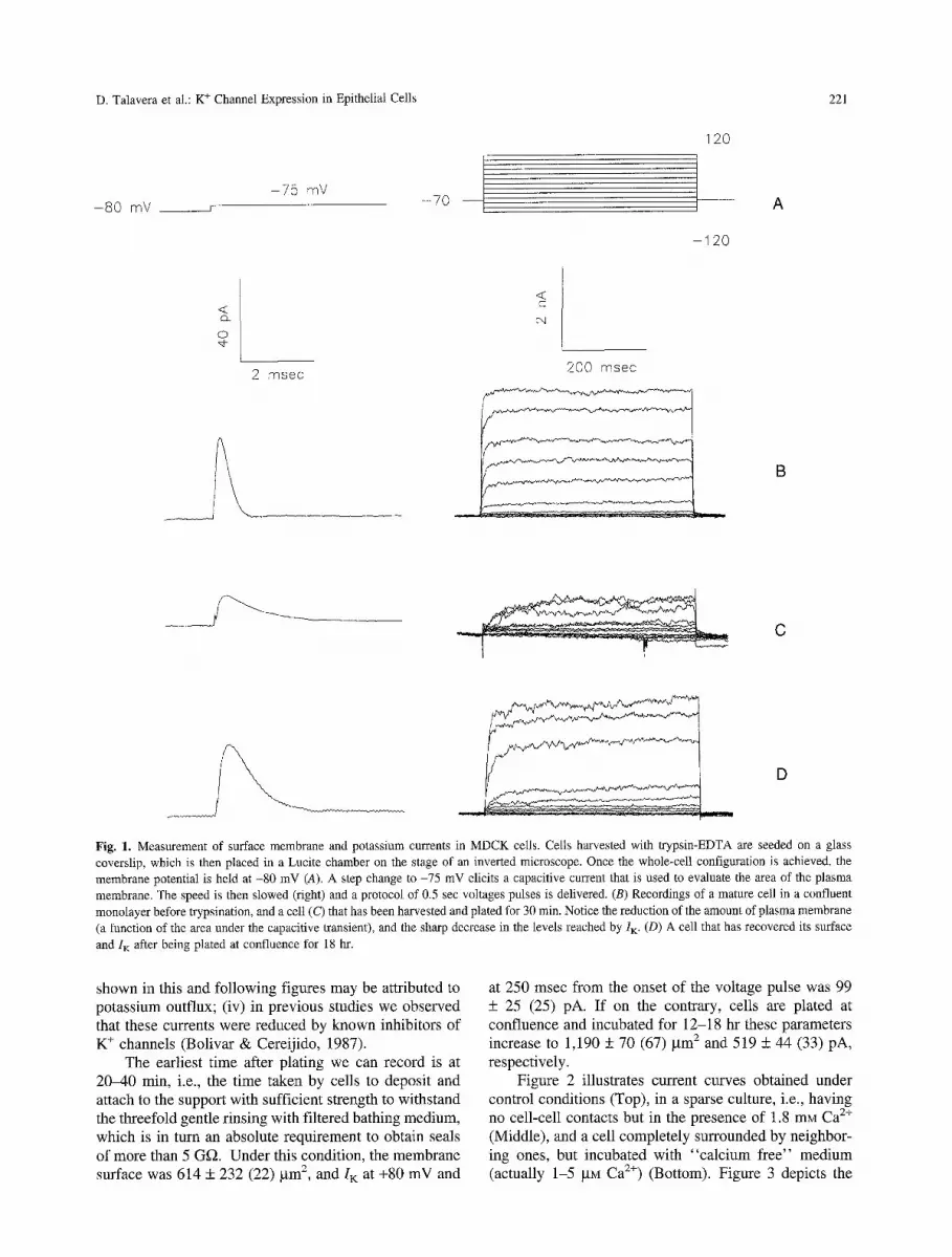
As mentioned above, MDCK cells harvested from con- fluent monolayers with trypsin-EDTA lose half of their surface membrane and 90% of their I K, but recover them in 12-20 hr after replating at confluence. Accordingly, to investigate the effect of cell-cell contacts and Ca 2§ on this recovery, we delivered the protocol of pulses shown in Fig. 1A to cells that had been just plated, and to cells that had been allowed to recover for a given length of time. Protocols consisted of two parts. In the first one, the holding potential was suddenly switched from -80 to -75 mV to elicit a transient of capacitive current, and sampling was made at 100 khz (Fig. 1B, left). This ma- neuver was repeated 10 times and averaged. By dividing the value of the area under the transient by the voltage step, we calculated membrane capacitance. Using the equivalence of 1 gF/cm 2 of membrane we then trans- formed capacitance into membrane area. The second protocol consisted of a series of voltage steps, each last- ing 500 msec, delivered to record the current response. A P/4 procedure was used to eliminate linear compo- nents (Fig. 1A, right). Figure 1 shows capacitive tran- sients (left) and ion currents recorded from a cell in a mature monolayer (B), a cell recently plated (C), and a cell plated and allowed to recover for 18 hr (D). Con- sidering that (i) records exhibited a series of outward currents; (ii) experiments were performed in the absence of permeable anions; (iii) the concentrations of K + in the cell and the bathing solution were 141 and 5 raM, respec- tively, and those of Na § were 5 and 141 raM, currents
D. Talavera et al.: K + Channel Expression in Epithelial Cells 221
- 8 0 mV _ _ J - 7 5 mV
- 7 0
120
- 1 2 0
A
< d)_
o
2 I~l $8C
<
cq
200 msec
B
C
% D
Fig. 1. Measurement of surface membrane and potassium currents in MDCK ceils. Cells harvested with trypsin-EDTA are seeded on a glass coverslip, which is then placed in a Lucite chamber on the stage of an inverted microscope. Once the whole-cell configuration is achieved, the membrane potential is held at -80 mV (A). A step change to -75 mV elicits a capacitive current that is used to evaluate the area of the plasma membrane. The speed is then slowed (right) and a protocol of 0.5 sec voltages pulses is delivered. (B) Recordings of a mature cell in a confluent monolayer before trypsination, and a celt (C) that has been harvested and plated for 30 min. Notice the reduction of the amount of plasma membrane (a function of the area under the capacitive transient), and the sharp decrease in the levels reached by I K. (D) A cell that has recovered its surface
and I K after being plated at confluence for 18 hr.
shown in this and following figures may be attributed to potassium outflux; (iv) in previous studies we observed that these currents were reduced by known inhibitors of K + channels (Bolivar & Cereijido, 1987).
The earliest time after plating we can record is at 20-40 min, i.e., the time taken by cells to deposit and attach to the support with sufficient strength to withstand the threefold gentle rinsing with filtered bathing medium, which is in turn an absolute requirement to obtain seals of more than 5 Gf~. Under this condition, the membrane surface was 614 + 232 (22) gm 2, and I K at +80 mV and
at 250 msec from the onset of the voltage pulse was 99 + 25 (25) pA. If on the contrary, cells are plated at confluence and incubated for 12-18 hr these parameters increase to 1,190 + 70 (67) gm 2 and 519 + 44 (33) pA, respectively.
Figure 2 illustrates current curves obtained under control conditions (Top), in a sparse culture, i.e., having no cell-cell contacts but in the presence of 1.8 mM Ca 2§ (Middle), and a cell completely surrounded by neighbor- ing ones, but incubated with "calcium free" medium (actually 1-5 gM Ca 2§ (Bottom). Figure 3 depicts the

222 D. Talavera et at.: K + Channel Expression in Epithelial Cells
<~ d~ o 0
200 msec
I
2 + 1 . 5 �9 c o n f l u e n t w i t h Ca
2 + [] subeonfluent, with Ca
�9 confluent, without Ca2+ T
/ / r-
Z w 1.0 n l cd
c )
DA
/ .<
m
0.5 i,i
- 1 0 0 - 5 0 0 50 100
MEMBRANE POTENTIAL (mY)
Fig. 3. Current/voltage relationship in cells treated as in Fig. 2.
Fig. 2. (Top) Potassium currents recorded in cells that have been plated for 18-20 hr as confluent monolayers, and incubated in a me- dium with 1.8 mM Ca 2+ (NC). (Middle) Potassium currents recorded in a cell in a sparse culture, i.e., without neighbors, but in NC. (Bottom) I K recorded in a cell plated at confluence, but incubated thereafter in the absence of Ca 2+ (actually 1-5 btM).
current/voltage relationship under these three experimen- tal conditions. It may be noticed that their left branch is essentially identical, indicating that harvesting does not increase the leakage, and differences are almost exclu- sively observed in the voltage-stimulated part. The size of the currents in the last two experimental conditions is much smaller. Even the shape of the current curves var- ies (Fig. 2), as in the condition of confluency without Ca 2+ (Fig. 2, Bottom) the kinetic of activation appears faster and I K shows some tendency to inactivate.
Figure 4 summarizes the measurements of mem- brane surface (Top) and potassium currents (Bottom). It is clear that their expression depends on the simulta- neous presence of cell-cell contacts and Ca 2+. The re- quirement of Ca-dependent cell-cell contacts was also observed in the de novo assembly and sealing of TJs (Gonz~lez-Mariscal et al., 1990; Contreras et al., 1992b). The effect of these contacts on the expression of TJs is mediated by PLC, PKC via G proteins (Balda et al., 1991). To explore whether PLC and PKC are also in- volved in the expression of K + channels, we used inhib- itors and stimulators of these enzymes. To facilitate comparisons, we express the values obtained as percent- age of control, a procedure that requires two specifica- tions: (1) Control values are different for inhibitors than for stimulators, because inhibitors were tested in conflu-
ent monolayers incubated with 1.8 inM Ca 2+, but stimu- lators were added instead to isolated cells with 1.8 mM Ca 2+ or to confluent cells in the absence of this ion, to bypass the step in which Ca-activated cell-cell contacts should have acted. Therefore, 100% in the first condi- tion corresponds to a higher value, Controls are shown in Fig. 4. (2) As stated in the Introduction, full recovery of K + channels takes 12-18 hr; therefore, the longer the interval between recordings in recently plated cells and those in cells allowed to recover, the more significant the difference between values obtained. However, prelimi- nary experiments indicated that cells could not withstand exposure to some of the chemicals used (e.g., staurospo- rine) for more than some 6 hr (results not shown). Ac- cordingly, the length of time allowed for recovery was decided as a compromise between the "longest period of recovery" and the "shortest exposure to a toxic." Since every experiment included control recordings (i.e., with- out inhibitors or stimulators), data were normalized as follows:
v = V m ( C . d c )
where v is the normalized value, %, is the value actually measured, c is the average of control experiments run in parallel, and C a l I is the mean value of all controls of the same conditions (i.e., one of the three shown in Fig. 4).
PLC transforms phosphatidyl 4,5-diphosphoinositol into inositol 1,4,5-triphosphate (IP 3) and diacylglycerol (DAG), and this substance stimulates PKC. To explore whether the effect of cell-cell contacts and Ca 2+ on the recovery of surface membrane and IK is mediated by

D. Talavera et al.: K + Channel Expression in Epithelial Cells 223
E
LLI rO 4 : LI_ rY ::D 03
n = 54 15 67
T
1000
500
0
n =
-~ T
34 15 33
6OO
o_
400
Z Ld r~ rY
200
o
CONFLUENCY
CALCIUM
- +
+
+
+
Fig. 4. Membrane surface (Top) and potassium currents (Bottom) of cells that have been plated at subconfluence in 1.8 m g Ca 2+ (left), at confluence without Ca > (center), and cells at confluence with NC (right). The surface of the plasma membrane was obtained by dividing the area under the capacitive curve by the size of the voltage step (5 mV), and using the equivalence l gF = 1 cm 2. I K is the value taken at +80 mV and at 250 msec of starting the voltage pulse.
PLC, we assayed the effect of 2.0 gM TRH, a stimulator of this enzyme (Martin et al., 1986), and observed a significant increase in the expression of K channels (Fig. 5, hatched bar) without affecting membrane area (open bar). Correspondingly, 110 gM neomycin, a substance that binds to PIP 2 and prevents its convertion to IP 3 plus DAG by PLC, produces a drastic inhibition of channel expression. However, this drug does block restoration of membrane area. This lack of effect of the stimulator may not be attributed to insufficient concentration, as the use of 4.0 ~tM TRH, i.e., doubling the dose, fails to increase the amount of plasma membrane (control: 648 + 83 [10] vs . TRH: 456 + 48 [8] gm2).
Next, we explored the participation of PKC by as- saying the effect of 200 gg/ml DiC8 (a permeable ana- logue of DAG) in cells in confluent monolayers cultured without Ca 2+. Since this substance is rapidly metabo- lized to phosphatidic acid (May, Lapetina & Cuatrecasas,
n = 4 12
_J O
I-- Z 0 (D
LI_ 0
I"- Z Ld (.3 Cd I..d 0....
700
600
500
400
300
200
100
0
TRH
I -r--] v//~///A
NEOMYC
Fig. 5. Effect of stimulation and inhibition of PLC in the recovery of plasma membrane (open bars) and I K (shaded bars). In this and fol- lowing figures the horizontal dashed line represents the control value obtained for each condition (Fig. 4). The first two bars correspond to cells incubated at confluence without Ca >, treated with 2.0 ~tM TRH. The last two bars correspond to cells incubated at confluency with Ca 2+.
1986) we protected its decay with 200 gM EGD (Balda et al., 1991). Figure 6 shows that 200 gg/ml of DiC8 per- mits a recovery of the amount of plasma membrane, and stimulates the restitution of ion channels. PMA (10 riM) another inhibitor of PKC, produces essentially the same effect, albeit at a lower degree. In another series of stud- ies (Fig. 6) we tested the effect of 250 nM staurosporine and 2.0 gM H7, inhibitors of PKC (Tamaoki et al., 1986; Knight, Sugden & Baker, 1988). In this case cells were plated at confluence and in the presence of 1.8 rnM Ca 2§ i.e., a condition in which cells recover their plasma mem- brane and Iir It may be noticed that the blockade of PKC suppresses the expression of both parameters.
The Table shows the content of PKC under the three experimental conditions. The absence of Ca 2§ increases the content of PKC as if the cell were posed to phos- phorylate a crucial molecular component upon arrival of this ion, so that in confluent cells with Ca 2§ having performed its task, the amount of PKC was reduced. Furthermore, Ca 2+ and cell-cell contacts modify the dis- tribution of this enzyme (Fig. 7). Thus, in subconfluent cells incubated in the presence of Ca 2+ or at confluence but in the absence of this ion, the enzyme remains in the perinuclear area. On the contrary, in confluent cells with Ca a§ PKC seems to be contained in vesicles spread over the cytoplasm and presents a distinct accumulation in the membrane region, a position that in some cells was shown to be required for the participation of this enzyme in membrane events.
The amount of plasma membrane, measured in the

224 D. Talavera et al.: K + Channel Expression in Epithelial Cells
J
0 rY" I ,- Z 0 (.3
LI_ 0
I-- Z bJ 0 rY i , i EL
n =
7 0 0 L
6 0 0 -
5 0 0 -
4 0 0 -
3 0 0 -
2 0 0 -
1 0 0 -
O -
2 4 17 9 8 9 5 9 8
I I SURFACE
K+-CURRENT
DiG8 PMA H7 STAUR
Fig. 6. Membrane surface and potassium currents of confluent cells incubated without Ca 2§ in the presence of PKC stimulators (200 gg/ml DiC8 plus 200 gM EGD, and 100 nM PMA) and with Ca 2+ in the presence of PKC inhibitors (2.0 gM H7 and 250 ng staurosporine).
Table. Effect of Ca 2+ and cell-cell contacts on PCK activity
Ca z+ Confluency n Activi ty
+ + 12 1
- + 10 1.99 +_ 0.28 + - 4 0.77 _+ 0.21
MDCK cells were harvested, plated in medium with 1.8 mM Ca 2+ for 1.0 hr and subsequently transferred to the same Ca-containing medium or to a "Ca-free" one (actually 1-5 p,M). After 18 hr, monolayers were scraped and PKC activity was measured with the GIBCO BRL kit as described in Materials and Methods.
same cells, is also dependent on Ca-act iva ted cel l -cel l
contacts, and is sensi t ive to the chemica ls used to modi fy
the effect of PLC and PKC (open bars in Figs. 4 -6) , a p h e n o m e n o n expec ted f rom the fact that, as demon-
strated in previous work (Ponce et al., 1991a), channels
are inserted in the p lasma membrane through a process
o f exocyt ic fusion. However , exocyt ic fus ion is a com- mon mechan i sm for the insert ion o f all other transporting
mechanisms, and is not expected to vary to the same
extent as I K. Furthermore, the amount o f p lasma m e m - brane in a g iven m o m e n t is the balance o f exocyt ic fu-
sion and retrieval, which are processes that may have different sensit ivi ty to the several exper imenta l condi- t ions tested in the present work. This is ref lected in Fig.
8, where Iir and membrane area measured in all series o f exper iments reported, show little correlat ion.
Discussion
Cation currents in the strain o f M D C K cells that we use are a lmost exc lus ive ly accounted for by K + channels, as
Fig. 7. Fluorescence cytological detection of PKC. Cells stained with rhodamine conjugated rim-1 were observed with a confocal fluores- cence microscope. (A) In confluent cells incubated without Ca 2+, PKC remains accumulated in the perinuclear area. (B) In sparse cells incu- bated in a Ca-containing medium PKC appears accumulated around the perinuclear area. (C) In cells incubated at confluency in the presence of Ca 2+ PKC appears to be contained in vesicles spread over the cyto- plasm, and along the cell borders (arrows). Intensity was adjusted to show details and is not proportional to the amount of enzyme the cells showed. B is a composite of different fields, grouped to show six sparse cells at the same magnification as in the other two pictures.
Na + and Ca 2+ currents and the corresponding channels for these ions were never detected (Stefani & Cerei j ido,
1983; Ponce & Cerei j ido, 1991). Likewise , studies f rom other laboratories have exc lus ive ly found K + currents and K + channe l s (Kolb , P a u l m i c h l & Lang , 1987; Fr iedr ich et al., 1988). M D C K cells also have C1- cur- rents (Lang, Def regger & Paulmichl , 1986; Ponce et al.,

D. Talavera et al.: K + Channel Expression in Epithelial Cells 225
<~ ~D_
~-. Z LLI r'Y
r,D
O9 09 <:: k-- 0 0_
800
500
400
200
O O
o o o
0
0 oo
0 I I I I I I
0 500 1000 1500 2000 2500
MEMBRANE AREA (/zm 2)
Fig. 8. Relationship between I K and the amount of plasma membrane. Each circle represents the value of both parameters measured in the same group of cells under the same experimental conditions described in the present work. Accordingly, the graph includes cells with high and low Ca 2+, confluent and sparse, and with and without drugs, to show that the different experimental situations may affect I K and sur- face independently.
1991b) but these are absent in the experimental condi- tions used in the present work, because CI- has been replaced by nonpermeable methanesulfonic acid.
Harvesting with trypsin-EDTA abolishes most of the IK. It is not clear whether the small amount of IK mea- sured as soon as one can start making recordings (20-40 min) is due to old channels that have escaped clipping by the enzyme, or whether it is due to channels reinstalled during the 20-40 min period in which, for technical rea- sons (waiting for cell attachment to the substrate, gentle washing to permit gigasealing), we cannot make record- ings. Anyhow, the extent of recovery during the 6-18 hr period chosen for the present studies is sufficiently large to study the factors involved.
Judging on the basis of conductance, voltage depen- dence and other kinetic parameters, we have previously found five different types of K + channels in the mature stage (1-3 days after plating at confluence) in the clone of MDCK cells used in the present work (Ponce & Cereijido, 1991). Since the physiological role of each type of channel in these cells is still unknown, it is not certain that each type would be needed in the same pro- portion along the whole-cell cycle. While the fact that trypsinization reduces I K by 90% suggests that all of them are almost totally eliminated by harvesting, the ki- netics of re-expression of the different types might not be necessarily the same for each channel type.
In previous work we found that restoration of K + channels requires synthesis of mRNA and proteins, the participation of microtubules and microfilaments, and depends also on exocytic fusion of membrane vesicles to the plasma membrane, presumably to incorporate
newly synthesized channels (Ponce et al., 1991a). In the present work we observe that restoration of K § channels requires also Ca2§ cell-cell contact and a chain of reactions in which PLC and PKC are involved.
Therefore, the mechanism for the expression of K § channels seems to be part of a general mechanism that results in the making of a cell from transporting epithelia. Thus, Vega-Salas, Salas and Rodrfguez-Boulan (1987) have shown that MDCK cells plated as monolayers, but incubated without Ca 2§ accumulate apical markers in a cytoplasmic compartment (VAC = vesicular apical com- partment). Upon transferring these cells to Ca 2+- containing media, the VAC is fused to the plasma mem- brane and becomes their apical domain. Likewise, in a previous work, we have demonstrated that MDCK cells incubated without Ca 2§ have their Na-K-ATPases dis- tributed at random in the plasma membrane, and a large fraction of this enzyme is in a cytoplasmic compartment; yet when 1.8 rnM Ca 2§ is added, Na-K-ATPase molecules are directed to and fused with the plasma membrane (Contreras et al., 1989). Finally, monolayers kept at con- fluence in the absence of Ca 2+ do not assemble and seal TJs, but these start to form as soon as the monolayers are transferred to media with 1.8 mM Ca 2§ (Gonzfilez- Mariscal et al., 1985).
In the case of the expression of TJs, the requirement of Ca 2§ is very specific, as it cannot be replaced by any of the other multivalent cations tested: Mn 2+, Mg 2+, Sr 2+, Cd 2+ and La 3+ (Contreras et al., 1992b). Ca 2§ elicits this effect by acting on the outside of the cell membrane (Gonzglez-Mariscal et al., 1990; Contreras et al., 1992a), probably on an uvomorulin molecule (Gumbiner & Si- mons, 1987), and the information that Ca 2+ has activated this cell-attaching-molecule (CAM) is conveyed to the cytoplasmal side through a cascade involving at least two G proteins (one excitatory and one inhibitory) acting on the PLC-PKC system (Balda et al., 1991). In the present work we observe that recovery of membrane area and ion channels depends also on cell-cell contacts and Ca 2§ and involves PLC and PKC, as the chemicals affecting these molecules have a clear effect on the expression of I K.
Figure 7 and the Table show that the distribution and activity of PKC require the presence of cell-cell contacts and Ca 2+. Interestingly, Kraft and Anderson (1983) and Myers, Lazo and Pitt (1989) have shown that in order to phosphorylate membrane components in EL4 mouse thy- moma cells and cells from the pulmonary artery of calves, part of the PKC must be transferred from the cytoplasm to the plasma membrane.
Taken together, the present results plus those we have obtained in earlier studies (Ponce & Cereijido, 1991; Ponce et al., 1991 a,b), suggest that to express most of its K + channels, a newly plated cell requires transcrip- tion, translation, exocytic and a fusion of membrane ves- icles guided by the cytoskeleton, and that PLC, PKC and AC participate in these processes.

226 D. Talavera et al.: K + Channel Expression in Epithelial Cells
Our resul ts also show that cel ls are able to con t ro l
i n d e p e n d e n t l y the a m o u n t of surface m e m b r a n e and the i r
popu la t i on of channe l s . Th i s is no t surpr i s ing because
ep i the l ia l cel ls and rena l cells, in par t icular , h a v e to adap t
the expres s ion of t r ans loca t ing m e c h a n i s m s to acidif ica-
t ion, dehydra t ion , K over load , and o ther phys io log ica l
r e q u i r e m e n t s tha t pose en t i re ly d i f fe ren t p r o b l e m s and
requi re d i f fe ren t ce l lu lar responses .
We wish to express our gratitude to Amparo Lazaro, Catalina Flores and Elizabeth del Oso for their efficient help, and to the National Research Council of MExico (CONACYT) and the Volkswagen Stiff- tung of Germany for their economic support.
References
Balda, M.S., Gonz~lez-Mariscal, L., Contreras, R.G., Cereijido, M. 1991. Assembly and sealing of tight junctions: Possible participa- tion of G-proteins, phospholipase C, protein kinase C and calmod- ulin. J. Membrane Biol. 122:193-202
Balda, M.S., Gonzfilez-Mariscal, L., Matter, K., Cereijido, M., Ander- son, J.M. 1993. Assembly of the tight junction: the role of diacyl- glycerol. J. Cell Biol. 123:293-302
Bolfvar, J.J., Cereijido, M. 1987. Voltage and Ca2+-activated K + channel in cultured epithelial cells (MDCK). J. Membrane Biol. 97:43-51
Cereijido, M., Ehrenfeld, J., Meza, I., Mart/nez-Palomo, A. 1980. Structural and functional membrane polarity in cultured monolay- ers of epithelioid MDCK cells. J. Membrane Biol. 52:147-159
Cereijido, M., Robbins, E.S., Dolan, W.J., Rotunno, C.A., Sabatini, D.D. 1978. Polarized monolayers formed by epithelial cells on a permeable and translucent support. J. Cell Biol. 77:853-880
Contreras, R.G., Avila, G., Guti6rrez, C., Bolfvar, J.J., Gon~aIez- Mariscal, L., Darzon, A., Beaty, G., Rodrfguez-Boulan, E., Cereijido, M. 1989. Repolarization of Na+-K + pumps during estab- lishment of epithelial monolayers. Am. J. Physiol. 257:C896-C905
Contreras, R.G., Gonz~lez-Mariscal, L., Balda, M.S., Garc/a-Villegas, M.R., Cereijido, M. 1992a. The role of calcium in the making of a transporting epithelium. NIPS 7:105-108
Contreras, R.G., Miller, JM., Zamora, M., Gonzfilez-Mariscal, L., Cereijido, M. 1992b. Interaction of calcium with plasma membrane of epithelial (MDCK) cells during junction formation. Am. J. Phys- iol. 263:C313-C318
Friedrich, F., Panlmichl, M., Kolb, H.A., Lang, F. 1988. Inward recti- fier K + channels in renal epithelioid cells (MDCK) activated by serotonin. J. Membrane Biol. 106:149-155
Gonzfilez-Mariscal, L., Chfivez de Ramlrez, B., Cereijido, M. 1985. Tight junction formation in cultured epithelial cells (MDCK). J. Membrane Biol. 86:113-125
Gonzfilez-Mariscal, L., Contreras, R.G., BoIfvar, J.J., Ponce, A., Ch~ivez de Ram/rez, B., Cereijido, M. 1990. Role of calcium in
tight junction formation between epithelial ceils. Am. J. Physiol. 259:C978-C986
Gumbiner, B., Simons, K. 1987. The role of uvomornlin in the forma- tion of epithelial occluding junctions. In: Junctional Complexes of Epithelial Cells. pp. 169-186. Wiley, Chichester, UK
Knight, D.E., Sugden, D., Baker, P.F. 1988. Evidence implicating pro- tein kinase C in exocytosis from electropermeabilized bovine chro- maffin cells. J. Membrane Biol. 104:21-34
Kolb, H.A., Paulmicbl, M., Lang, F. 1987. Epinephrine activates out- ward rectifying K + channels in Madin-Darby canine kidney cells. Pfluegers Arch. 408:548-591
Kraft, A.S., Anderson, W.B. 1983. Phorbol esters increase the amount of Ca a+, phospholipid-dependent protein kinase associated with plasma membrane. Nature 301:621-623
Lang, F., Defregger, M., Paulmichl, M. 1986. Apparent chloride con- ductance of subconfluent Madin Darby canine kidney cells. Pfluegers Arch. 407"i58-162
Martin, T.F.J., Lucas, D.O., Bajjaleih, S.M., Kowalchyk, J.A. 1986. Thyrotropin-releasing hormone activates a Ca++-dependent poly- phosphoinositide phosphodiesterase in permeable GH3 cells. GTP gamma S potentiation by cholera and pertussis toxin insensitive mechanism. J. Biol. Chem. 261:2918-2927
May, W.S., Lapetina, E.G., Cuatrecasas, P. 1986. Intracellular activa- tion of protein kinase C and regulation of surface transferrin recep- tor by diacylglycerol is a spontaneous reversible process that is associated with rapid formation of phosphatidic acid. Proc. Natl. Acad. Sci. USA 83"1281-1284
Myers, C.L., Lazo, J.S., Pitt, B.R. 1989. Translocation of protein kinase C is associated with inhibition of 5-HT uptake by cultured endo- thelial cells. Am. J. Physiol. 257:L253-L258
Ponce, A., Bolivar, J.J., Vega, J., Cereijido, M. 1991a. Synthesis of plasma membrane and potassium channels in epithelial (MDCK) cells. Cell Physiol. Biochem. 1:195-204
Ponce, A., and Cereijido, M. 1991. Polarized distribution of cation channels in epithelial cells. Cell Physiol. Biochem. 1:13-23
Ponce, A., Contreras, R.G., Cereijido, M. 1991b. Polarized distribution of chloride channels in epithelia1 cells (MDCK). Cell Physiol. Bio-
chem. 1:160-169 Stefani, E., Cereijido, M. 1983. Electrical properties of cultured epi-
thelioid cells (MDCK). J. Membrane Biol. 73:177-184 Tamaoki, T., Nomoto, H., Takahashi, I., Kato, Y., Morimoto, M., To-
mita, F. 1986. Staurosporine, a potent inhibitor of phospholipid/ Ca ++ dependent protein kinase. Biochem. Biophys. Res. Commun. 135:397-402
Vega-Salas, D.E., Salas, P.J., Rodrfguez-Boulan, E. 1987. Modulation of the expression of an apical plasma membrane protein of Madin- Darby Canine Kidney epithelial cells: cell-cell interactions control the appearance of a novel intracellular storage compartment. J. Cell. Biol. 104:1249-1259
Yasuda, I., Kishimoto, A., Tanaka, S., Tominaga, M., Sakurai, A., Nishiznka, Y. 1990. A synthetic peptide substrate for selective assay of protein kinase C. Biochem. Biophys. Res. Commun. 166"t220-i227


