Embed Size (px)
Citation preview

Epithelial and Mesenchymal Cell Biology
Integrity of Cell-Cell Contacts Is a Critical Regulatorof TGF-�1-Induced Epithelial-to-MyofibroblastTransition
Role for �-Catenin
Andras Masszi,*† Lingzhi Fan,* Laszlo Rosivall,†‡
Christopher A. McCulloch,§ Ori D. Rotstein,*Istvan Mucsi,‡¶ and Andras Kapus*From the Department of Surgery,* University Health Network and
University of Toronto, Ontario, Canada; the Institute of
Pathophysiology,† Semmelweis University, Budapest, Hungary; the
Hungarian Academy of Sciences and Semmelweis University
Nephrology Research Group,‡ Budapest, Hungary; the Canadian
Institutes of Health Research Group (CIHR) in Matrix Dynamics,§
University of Toronto, Toronto, Ontario, Canada; and the First
Department of Internal Medicine,¶ Semmelweis University,
Budapest, Hungary
Injury of the tubular epithelium and TGF-�1-inducedconversion of epithelial cells to �-smooth muscle ac-tin (SMA)-expressing myofibroblasts are key featuresof kidney fibrosis. Since injury damages intercellularjunctions and promotes fibrosis, we hypothesizedthat cell contacts are critical regulators of TGF-�1-triggered epithelial-to-mesenchymal transition(EMT). Here we show that TGF-�1 was unable to in-duce EMT in intact confluent monolayers, but threedifferent models of injury-induced loss of epithelialintegrity (subconfluence, wounding, and contact dis-assembly by Ca2�-removal) restored its EMT-inducingeffect. This manifested in loss of E-cadherin, in-creased fibronectin production and SMA expression.TGF-�1 or contact disassembly alone only modestlystimulated the SMA promoter in confluent layers, buttogether exhibited strong synergy. Since �-catenin isa component of intact adherens junctions, but whenliberated from destabilized contacts may act as a tran-scriptional co-activator, we investigated its role inTGF-�1-provoked EMT. Contact disassembly alone in-duced degradation of E-cadherin and �-catenin, butTGF-�1 selectively rescued �-catenin and stimulatedthe �-catenin-driven reporter TopFLASH. Moreover,chelation of free �-catenin with the N-cadherin cyto-
plasmic tail suppressed the TGF-�1 plus contact dis-assembly-induced SMA promoter activation and pro-tein expression. These results suggest a �-catenin-dependent two-hit mechanism in which both aninitial epithelial injury and TGF-�1 are required forEMT. (Am J Pathol 2004, 165:1955–1967)
Epithelial-to-mesenchymal transition (EMT) plays a cen-tral role in development and carcinogenesis.1 Recently,the revolutionary concept has emerged that EMT is one ofthe key mechanisms underlying organ fibrosis includingkidney.2–4 Indeed, tubulointerstitial fibrosis (TIF) is thecommon pathological pathway through which chronickidney diseases progress to end-stage renal failure. TIFis characterized by the gradual loss of the tubular epi-thelium, concomitant with the progressive accumulationof fibroblasts and �-smooth muscle actin (SMA)-positivemyofibroblasts, leading to an excessive production anddeposition of extracellular matrix components.5 The keyimportance of myofibroblasts in TIF is further supportedby the strong positive correlation between SMA expres-sion and the loss of kidney functions.6
Both clinical studies and animal models of TIF indicatethat fibroblasts and myofibroblasts can be derived fromthe tubular epithelium undergoing EMT7–9 in response toinflammatory cytokines, predominantly transforminggrowth factor-�1 (TGF-�1).10,11 The most compelling ev-idence for EMT in fibrosis has been provided by Iwano et
Supported by grants from the Canadian Institutes of Health Research(CIHR) and Kidney Foundation of Canada (to A.K.), and the HungarianMinistry of Education OTKA/T042651 (to I.M.) and OTKA/34409, ETT/564(to R.L.). I.M. is a Bekesy scholar and a recipient of NATO ScientificFellowship 2006/NATO/02. A.K. is a CIHR scholar.
Accepted for publication August 18, 2004.
Address reprint requests to Dr. Andras Kapus, Toronto General Hos-pital, NUG-001, 200 Elizabeth Street, Toronto, Ontario, Canada, M5G2C4. E-mail: [email protected].
American Journal of Pathology, Vol. 165, No. 6, December 2004
Copyright © American Society for Investigative Pathology
1955

al,12 who found that in mice with genetically tagged prox-imal tubular cells, 36% of renal fibroblasts originated fromthe epithelium in a TIF model.
To explore the underlying mechanisms, several groupsincluding our own have developed cellular models ofTGF-�1-induced epithelial-fibroblast/myofibroblast tran-sition.13–15 These studies showed that the key features ofEMT include the early loss of cell-cell contacts due to thedown-regulation of ZO-1 and E-cadherin, reorganizationof the cytoskeleton, acquisition of spindle-like morphol-ogy, and finally the expression of SMA, the hallmark of themyofibroblast phenotype. This process takes severaldays and requires the interplay of a multitude of TGF-�1-induced pathways, including SMAD proteins,16 integrin-linked kinase,17 and Rho-family GTPases.15,18
Extensive research in tumor and developmental biol-ogy has solidified the concept that intracellular contactsare not only passive targets, but also are active regula-tors of EMT. Accordingly, the loss of E-cadherin promotesEMT, while forced E-cadherin expression can restore theepithelial phenotype in transformed tumor cells.19–22 Fur-thermore, recent studies by Zeisberg and Kalluri23 pro-vide strong support for the importance of the loss ofE-cadherin in EMT in tubular cells. These authors showedthat inhibition of the TGF-�1-induced down-regulation ofE-cadherin reverses EMT. Accordingly, epithelial injury andsubsequent repair, which involve damage or loss of inter-cellular contacts, are known to facilitate tissue fibrosis.24
Regarding the underlying mechanism, �-catenin, the intra-cellular binding partner of E-cadherin appears to be a goodcandidate to participate in contact-dependent regulation ofEMT, because of its dual function. Namely, in cells withintact intercellular contacts, �-catenin is an integral compo-nent of the adherens junctions, however when freed fromthe contacts, it can act as a transcriptional co-activator bybinding to members of the T cell factor/lymphoid enhancerfactor (TCF/LEF) family of transcription factors.25 Indeed,�-catenin signaling has been implicated in EMT progres-sion in tumor cells,26–28 although its role in promoting organfibrosis remains to be defined. Consistent with its potentialinvolvement, we and others have shown that TGF-�1 in-duces increased nuclear accumulation of �-catenin in tu-bular cells,15,29 and �-catenin has been reported to interactwith SMAD proteins.30,31 Further, epithelial cells from pa-tients with pulmonary fibrosis showed strong nuclear stain-ing for �-catenin.32 Finally �-catenin targets certain genes,which have been implicated in EMT.33–35 Nonetheless, itremains controversial whether TGF-�1 induces �-catenin-dependent transcription in nonmalignant epithelialcells,29,30,36 and if so, whether such a process can impacton epithelial-myofibroblast transition. Accordingly, we hy-pothesized that the integrity of cell-cell contacts in general,and �-catenin signaling in particular might be importantregulators of TGF-�1-induced myofibroblast generation.
Our present studies provide evidence that cell con-tacts are critical determinants of EMT susceptibility toTGF-�1. We show that a partial absence or disassemblyof cell-cell junctions is a prerequisite for the TGF-�1-provoked EMT. Investigating the underlying mechanism,we found �-catenin plays an important role in the TGF-�1and cell contact-dependent, synergistic regulation of the
SMA promoter and protein expression. These findingssuggest a two-hit model in which both an initial tissueinjury and TGF-�1 are required for EMT.
Materials and Methods
Materials and Antibodies
The following primary antibodies were used: anti-�-catenin,anti-Rho (Santa Cruz Biotechnology, Santa Cruz, CA), anti-E-cadherin (Transduction Laboratories, Mississauga, ON),anti-SMA (1A4), anti-fibronectin, anti-�-actin, and anti-tubu-lin (Sigma, St. Louis, MO). Rhodamine-phalloidin was fromCytoskeleton (Denver, CO). Horseradish peroxidase-conju-gated anti-mouse and anti-rabbit IgG antibodies were pur-chased from Amersham Biosciences (Baic d’Urfé, QC), andanti-goat antibody from Santa Cruz. Fluorescein isothiocya-nate and Cy3-labeled anti-goat, anti-rabbit, and anti-mousesecondary antibodies were obtained from Jackson Immu-noresearch Laboratories (West Grove, PA).
Cells
For most of our studies we used LLC-PK1 CL4 cellsstably expressing the AT1 receptor, as we characterizedEMT in detail in this proximal tubular cell line in ourprevious studies.15 However, the process of EMT wasqualitatively and quantitatively similar in wild-type LLC-PK1 cells as well. Cells were cultured in Dulbecco’smodified Eagle’s medium (DMEM), containing 10% fetalbovine serum (FBS), 1% penicillin/streptomycin (Gibco,Burlington, ON) at 37°C under 5% CO2 in a humidifiedincubator. Cells were grown to 30% or 100% confluence,and then treated with vehicle only (4 mmol/L HCl and0.1% albumin) or 4 ng/ml human recombinant TGF-�1(Sigma) for the indicated times. In certain experiments,the medium of confluent layers grown on plastic or per-meable support (Corning, Acton, MA) was replaced withnominally Ca2�-free DMEM (Gibco) without FBS.
Western Blotting
After treatments, cells were scraped into Triton lysis buffer(30 mmol/L N-2-hydroxyethylpiperazine-N-2-ethanesulfonicacid (HEPES) (pH 7.4), 100 mmol/L NaCl, 1 mmol/L ethyl-ene glycol-bis (2-aminoethylether)-N, N N� N�-tetraaceticacid (EGTA), 20 mmol/L NaF, 1% Triton X-100, 1 mmol/Lphenylmethylsulfonyl fluoride (PMSF), 20 �l/ml Protease In-hibitory Cocktail (Pharmingen BD Biosciences, San Diego,CA), and 1 mmol/L Na3VO4). A portion of the samples wassaved as total lysates, then the cytosolic (Triton soluble) andcytoskeletal (Triton insoluble) fractions were separated bycentrifugation. Samples were dissolved in Laemmli bufferand boiled for 5 minutes. Equal amount of protein wereseparated on SDS-polyacrylamide gels and transferred tonitrocellulose membranes (Bio-Rad, Hercules, CA). Immu-noblotting was performed as described previously.15 Immu-noreactive bands were visualized by enhanced chemilumi-nescence reaction kit (Amersham).
1956 Masszi et alAJP December 2004, Vol. 165, No. 6

Preparation of GST-RBD Beads andMeasurement of Rho Activity
Rho activity was determined as described previously15,37
using a pull-down affinity assay based on the ability of theRho-binding domain (RBD) of Rhotekin to capture theGTP-loaded form of Rho. Recombinant glutathione-S-transferase (GST)-RBD fusion protein was prepared fromE. coli, and adsorbed onto glutathione-covered beads.Confluent monolayers on 10-cm Petri dishes were serum-starved for 3 hours in normal or calcium-free DMEM, andthen exposed to 10 ng/ml TGF-�1 for 10 minutes. Sub-sequently, cells were washed with ice-cold phosphate-buffered saline (PBS) and scraped into 800 �l lysis buffersupplemented with 0.1% SDS, 0.5% Na-Deoxycholate.The supernatants were incubated with GST-RBD beadsfor 45 minutes at 4°C. After washing, the beads wereboiled in Laemmli buffer. Bead-associated Rho proteinwas detected by Western blotting.
Wound Assay
Confluent monolayers grown on 25-mm glass coverslipswere wounded with a rubber policeman generating an�3-mm gap. After extensive washing, the cells wereplaced into DMEM, and treated with 4 ng/ml TGF-�1 orvehicle for 3 days.
Immunofluorescence Microscopy
Cells grown on coverslips were fixed in 4% paraformal-dehyde, incubated with PBS containing 100 mmol/L gly-cine, and washed with PBS. Cells were permeabilized inPBS containing 0.1% Triton X-100, blocked with 5% al-bumin for 1 hour, and incubated with the primary anti-bodies for another hour. After extensive washing, fluores-cently labeled secondary antibodies were added for 1hour, the coverslips were washed again and mounted onslides using Fluorescence Mounting Medium (DAKO Di-agnostics, Mississauga, ON). Samples were analyzedusing a Nikon Eclipse TE200 microscope (�100 objec-tive) (Nikon, Mississauga, ON) and a Hamamatsu cooledCCD camera (C4742–95) (Hamamatsu, Bridgewater, NJ)controlled by the Simple PCI software.
Plasmids
A 765-bp piece of the rat SMA promoter containing sev-eral cis-elements including the serum response elementbinding motifs (CArG A and CArG B boxes), a TGF-�1control element (TCE), a TATA box, and two E-boxes wasligated into PA3-Luc luciferase vector (pSMA-Luc). Theconstruct was a kind gift from Dr. R. A. Nemenoff (De-partment of Medicine, Renal Division, University of Colo-rado Health Sciences Center, Denver, CO).38 The LEF/TCF reporter plasmid, TopFLASH and its mutant control,FopFLASH were purchased from Upstate Biotechnology(Charlottesville, VA). The thymidine kinase-driven Renillaluciferase vector (pRL-TK, Promega, Madison, WI) wasused as an internal control. The construct coding the
cytoplasmic tail of N-cadherin ligated to GFP (N-cad-GFP) was a generous gift of Dr. B. Geiger (Department ofChemical Immunology, The Weizmann Institute of Sci-ence, Rehovot, Israel).39 Dominant-negative TCF4 con-struct (dN-TCF4) cloned into pcDNA3 vector was fromDrs. O. Tetsu and F. McCormick (Cancer Reserch Insti-tute, University of California, School of Medicine, SanFrancisco, CA).40 This construct encodes for a Myc-epitope-tagged truncation mutant of TCF-4, in which theregion between amino acids 2–53 has been deleted.pEGFP vector was from Clontech Laboratories (Palo Alto,CA) and pcDNA3 was from Invitrogen (Burlington, ON).
Transient Transfection and Luciferase Assay
Cells plated onto 6-well plates were transfected at 100%or 30% of confluence using FuGENE6 (Roche MolecularBiochemicals, Indianapolis, IN; reagent:DNA ratio 2.5 �l/�g). For promoter activity measurements, cells were co-transfected with 0.5 �g promoter plasmid, 0.05 �gpRL-TK along with 2 �g of the specific inhibitory con-struct or pcDNA3 per well. After 16 hours, the cells werewashed with Hank’s balanced salt solution (HBSS) andincubated in serum-free normal or low calcium DMEM for3 hours, followed by treatment with vehicle or 10 ng/mlTGF-�1 for 24 hours. Subsequently, the cells were lysedin 250 �l Passive Lysate Buffer (Promega), exposed tofreezing/thawing, and the samples were clarified by cen-trifugation. Firefly and Renilla luciferase activities weredetermined using the Dual-Luciferase Reporter Assay Kit(Promega) and a Berthold Lumat LB 9507 luminometeraccording to the manufacturers’ instructions. Resultswere normalized by dividing the Firefly luciferase activitywith the Renilla luciferase activity of the same sample.Each transfection was done in duplicate, and determina-tions for each group were repeated at least three times.
Retroviral Infection
For generating the pFB-N-cad and pFB-GFP vectors, thecoding region of N-cad-GFP and pEGFP plasmids wereinserted into the pFB-Neo plasmid (Stratagene, La Jolla,CA) using EcoRI and Xho enzymes. To produce retrovi-ruses, 293T packaging cells were transfected with pFB-N-cad, pFB-GFP, or pFB-Neo vectors, respectively,along with pVpack-GP and pVpack-VSVG. After 24 to 36hours, supernatants were collected and filtered through a0.45-�m filter. LLC-PK1 cells plated on 6-well plates weretransduced with the virus at 20% confluence. After a16-hour transduction, cells were further incubated infresh medium for 36 hours and treated with vehicle or 4ng/ml TGF-�1. Three days later, cell lysates were pre-pared and samples were analyzed by Western blot.
Statistical Analysis
Data are presented as representative blots from threesimilar experiments or as the means � SEM for the num-ber of experiments (n) indicated. Statistical significancewas determined by Student’s t-test or analysis of variance
�-Catenin Impacts on Myofibroblast Transition 1957AJP December 2004, Vol. 165, No. 6
(one-way analysis of variance, SPSS Inc., Chicago, IL),using Bonferroni and Tukey post-hoc tests. P � 0.05value is accepted to be significant.
Results
Cell Confluence Is a Critical Determinant ofTGF-�1-Induced EMT in LLC-PK1 Tubular Cells
To assess the role of cell contacts in TGF-�1-inducedEMT, we first compared the effect of the cytokine inconfluent and sparse cultures, ie, under conditions wherethe cells have mature intercellular junctions or less well-
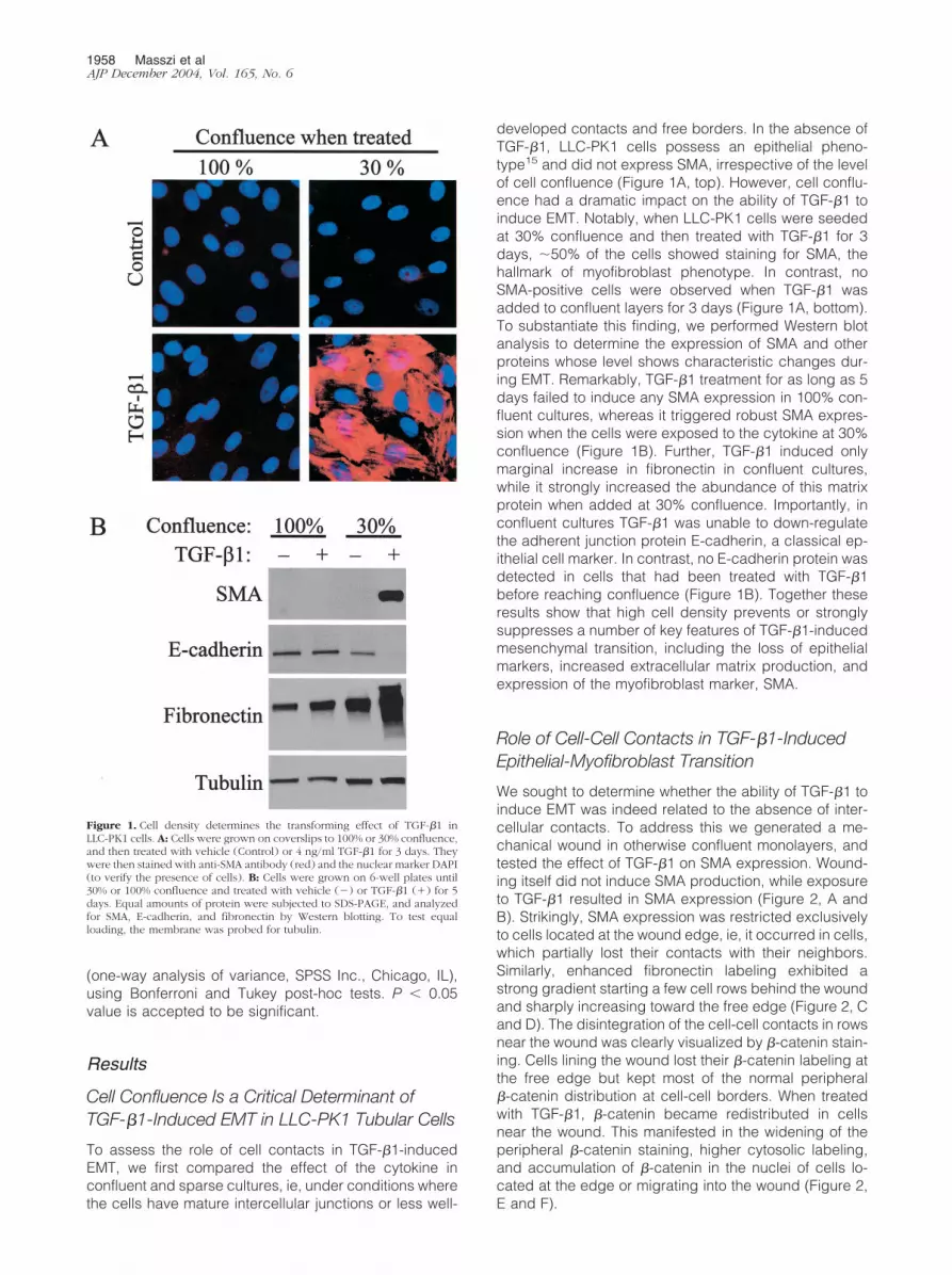
developed contacts and free borders. In the absence ofTGF-�1, LLC-PK1 cells possess an epithelial pheno-type15 and did not express SMA, irrespective of the levelof cell confluence (Figure 1A, top). However, cell conflu-ence had a dramatic impact on the ability of TGF-�1 toinduce EMT. Notably, when LLC-PK1 cells were seededat 30% confluence and then treated with TGF-�1 for 3days, �50% of the cells showed staining for SMA, thehallmark of myofibroblast phenotype. In contrast, noSMA-positive cells were observed when TGF-�1 wasadded to confluent layers for 3 days (Figure 1A, bottom).To substantiate this finding, we performed Western blotanalysis to determine the expression of SMA and otherproteins whose level shows characteristic changes dur-ing EMT. Remarkably, TGF-�1 treatment for as long as 5days failed to induce any SMA expression in 100% con-fluent cultures, whereas it triggered robust SMA expres-sion when the cells were exposed to the cytokine at 30%confluence (Figure 1B). Further, TGF-�1 induced onlymarginal increase in fibronectin in confluent cultures,while it strongly increased the abundance of this matrixprotein when added at 30% confluence. Importantly, inconfluent cultures TGF-�1 was unable to down-regulatethe adherent junction protein E-cadherin, a classical ep-ithelial cell marker. In contrast, no E-cadherin protein wasdetected in cells that had been treated with TGF-�1before reaching confluence (Figure 1B). Together theseresults show that high cell density prevents or stronglysuppresses a number of key features of TGF-�1-inducedmesenchymal transition, including the loss of epithelialmarkers, increased extracellular matrix production, andexpression of the myofibroblast marker, SMA.
Role of Cell-Cell Contacts in TGF-�1-InducedEpithelial-Myofibroblast Transition
We sought to determine whether the ability of TGF-�1 toinduce EMT was indeed related to the absence of inter-cellular contacts. To address this we generated a me-chanical wound in otherwise confluent monolayers, andtested the effect of TGF-�1 on SMA expression. Wound-ing itself did not induce SMA production, while exposureto TGF-�1 resulted in SMA expression (Figure 2, A andB). Strikingly, SMA expression was restricted exclusivelyto cells located at the wound edge, ie, it occurred in cells,which partially lost their contacts with their neighbors.Similarly, enhanced fibronectin labeling exhibited astrong gradient starting a few cell rows behind the woundand sharply increasing toward the free edge (Figure 2, Cand D). The disintegration of the cell-cell contacts in rowsnear the wound was clearly visualized by �-catenin stain-ing. Cells lining the wound lost their �-catenin labeling atthe free edge but kept most of the normal peripheral�-catenin distribution at cell-cell borders. When treatedwith TGF-�1, �-catenin became redistributed in cellsnear the wound. This manifested in the widening of theperipheral �-catenin staining, higher cytosolic labeling,and accumulation of �-catenin in the nuclei of cells lo-cated at the edge or migrating into the wound (Figure 2,E and F).
Figure 1. Cell density determines the transforming effect of TGF-�1 inLLC-PK1 cells. A: Cells were grown on coverslips to 100% or 30% confluence,and then treated with vehicle (Control) or 4 ng/ml TGF-�1 for 3 days. Theywere then stained with anti-SMA antibody (red) and the nuclear marker DAPI(to verify the presence of cells). B: Cells were grown on 6-well plates until30% or 100% confluence and treated with vehicle (�) or TGF-�1 (�) for 5days. Equal amounts of protein were subjected to SDS-PAGE, and analyzedfor SMA, E-cadherin, and fibronectin by Western blotting. To test equalloading, the membrane was probed for tubulin.
1958 Masszi et alAJP December 2004, Vol. 165, No. 6

As a further method to test the role of cell-cell contactsin EMT, we used an approach that did not involve me-chanical injury. We induced disassembly of mature cell-cell contacts in confluent cultures by Ca2�-removal, amaneuver known to disrupt the homotypic adhesion be-tween cadherins.41 Remarkably, Ca2�-removal com-pletely restored the ability of TGF-�1 to provoke EMT.During the course of a 3-day experiment in the presenceof Ca2�, TGF-�1 neither induced SMA expression, norcaused a significant reduction in E-cadherin, as reportedabove (Figure 3A). Removal of Ca2� without TGF-�1addition did not cause SMA expression and only margin-ally affected fibronectin expression, but it induced a sub-stantial reduction in E-cadherin. Importantly, TGF-�1added in the absence of Ca2� triggered a graduallyincreasing SMA expression, led to complete eliminationof E-cadherin, and strongly stimulated fibronectin pro-duction (Figure 3A). In accordance with these findings, ina Ca2�-free medium epithelial cells still preserved theirpolygonal shape. However, exposure of these cells toTGF-� resulted in mesenchymal morphology (Figure 3B).
Thus, the absence of stable intercellular junctions, irre-spective of whether it is brought about by non-confluenceor mechanical or chemical disruption of the cell contactsis a prerequisite for the EMT-inducing effect of TGF-�1.
We considered the possibility that, in confluent layers,TGF-�1 cannot promote EMT when added from the api-cal side, because it cannot access the basolateral mem-brane, where TGF-�1 receptors might reside.29 To testthis, we performed control experiments using LLC-PK1cells grown on permeable support to provide free accessto both membrane domains. As shown on Figure 4A, inconfluent monolayers neither apically nor basolaterallyadded TGF-�1 induced SMA expression. However, whencell contacts were disassembled by Ca2�-removal, api-cal or basolateral administration of TGF-�1 provoked arobust and similar SMA response. These data precludedthat resistance to TGF-�1 was due to differential acces-sibility. Next, we asked whether confluent cultures exhib-ited general unresponsiveness to TGF-�1, or only thetransforming effect was suppressed. To address this, wedetermined if TGF-�1 remained able to stimulate RhoGTPase, an important signal for cytoskeletal remodeling.In agreement with our previous findings,15 TGF-�1 in-duced a sizable Rho activation in confluent cultures (Fig-ure 4B), which was similar in the presence or absence ofCa2�. This suggests that contact disassembly was not
Figure 2. Loss of cell-cell contacts by wounding restores the ability ofTGF-�1 to induce EMT. Cells were allowed to grow until 100% confluenceand then a wound was generated with a rubber policeman in the monolayer.Subsequently cells were treated with vehicle (Control; A, C, and E) or 4ng/ml TGF-�1 (B, D, and F) for 3 days, then fixed, permeabilized, andstained for SMA (A and B), fibronectin (C and D), or �-catenin (E and F). Themagnification was �40 for A to D, and �100 for E and F.
Figure 3. Disassembly of intercellular contacts by low calcium restores thetransforming effect of TGF-�1 in confluent monolayers. A: Confluent cellswere washed and incubated in serum-free normal or Ca2�-free DMEM, and3 hours later treated with vehicle (Control) or 4 ng/ml TGF-�1 for theindicated times. Expression of SMA, E-cadherin, and fibronectin were deter-mined by Western blotting. B: TGF-�1 treatment (24 hours) induced mor-phological changes in low calcium DMEM (phase-contrast microscopy, mag-nification, �40).
�-Catenin Impacts on Myofibroblast Transition 1959AJP December 2004, Vol. 165, No. 6

required for this response. Consistent with these results,exposure of confluent layers to TGF-�1 stimulated stressfiber formation (Figure 4C). Together these data showthat TGF-�1 signaling pathways are functional in conflu-ent LLC-PK1 cells, suggesting that failed transformation isnot due to general unresponsiveness, but rather due to theabsence of cell contact-regulated permissive factor(s).
Cell-Cell Contacts Regulate the SMA Promoter
To assess whether cell contacts might impact the regu-lation of the SMA promoter, we investigated the effect ofTGF-�1 on a SMA promoter luciferase construct underconditions where cell junctions were manipulated by theabove-described means (Figure 5). In confluent cultureswith stable cell contacts (100%, normal Ca2� medium),TGF-�1 induced a modest �3-fold increase in the activityof the SMA promoter, confirming the basal responsive-ness to TGF-�1 in confluent layers. Reduction of Ca2�
without TGF-�1 addition resulted in �4-fold rise in SMApromoter activity. Importantly, TGF-�1 added to confluentlayers in which the cell junctions had been disassembledby Ca2�-removal elicited a 14-fold increase in SMA pro-moter activity. Thus, contact disassembly and TGF-�1acted in a strongly synergistic manner as evidenced bythe multiplicative effect. We next tested the impact of
subconfluence in the presence of normal Ca2�. Thebasal activity of the SMA promoter was fivefold higher in30% than in 100% confluent cultures. TGF-�1 addition to30% confluent cells resulted in a 20-fold increase in SMApromoter activity, compared to confluent, unstimulatedcells. These data show that the overall stimulation of theSMA promoter by TGF-�1 strongly depends on the stateof cell-cell contacts in both models, irrespective ofwhether the contacts have been disassembled in alreadyconfluent layers or have not been fully formed yet ingrowing subconfluent cultures.
TGF-�1 Stimulates the Transcriptional Activity of�-Catenin in LLC-PK1 Tubular Cells
Since �-catenin has a dual function as an adherent junc-tion component and a transcriptional co-activator, and itredistributes during EMT in LLC-PK1 cells (15 and Figure2), it might act as a mediator of contact-dependent tran-scriptional responses. To address this possibility, we firstdetermined whether TGF-�1 could stimulate �-catenin-dependent transcription. Cells at 30% confluence weretransfected with the �-catenin-dependent reporter Top-FLASH or its inactive mutant FopFLASH, and then ex-posed to vehicle or TGF-�1. The basal TopFLASH activitywas 10-fold higher than FopFLASH, indicating that Top-FLASH expression reflects �-catenin-specific transcrip-tion, and that there is readily detectable, �-catenin-dependent transcription even in non-stimulated,subconfluent cells (Figure 6A). Importantly, TGF-�1 in-duced a twofold increase in TopFLASH activity, while ithad no significant effect on the marginal FopFLASH ac-tivity. Moreover, LiCl, an inhibitor of �-catenin degrada-tion also stimulated TopFLASH activity. Western blotanalysis verified that LiCl indeed increased the steadystate level of �-catenin in LLC-PK1 cells (Figure 6A).Together these findings suggest that the continuous deg-radation of �-catenin limits �-catenin signaling in non-
Figure 4. Confluent cells remain responsive to TGF-�1. A: Cells were cul-tured until confluence on porous tissue culture inserts (pore size, 3 �m) thenserum-deprived in normal (�) or low calcium medium (�). After 3 hours,cells were treated with vehicle (�) or 4 ng/ml TGF-�1 from the upper (U) orthe lower side (L) for 3 days. Total cell lysates were prepared and SMAexpression was examined by Western blots. B: To determine Rho activitywere serum-starved for 3 hours in normal (�) or low Ca2� (�) DMEM, andthen treated with vehicle (�) or 10 ng/ml TGF-�1 (�) for 10 minutes. Afterlysis, active Rho was captured with GST-Rhotekin beads and detected byWestern blotting. Aliquots of the same lysates were probed for total Rho. C:Confluent cultures were treated with vehicle (Control) or 4 ng/ml TGF-�1.F-actin was visualized by rhodamine phalloidin.
Figure 5. Subconfluence or disassembly of cell-cell contacts potentiates theeffect of TGF-�1 on SMA promoter activity. LLC-PK1 cells grown to 100% or30% confluence were co-transfected with pSMA-Luc (0.5 �g/well), and theRenilla luciferase construct (pRL-TK, 0.05 �g/well). After 16 hours, cells werewashed and incubated with serum-free normal (�) or low Ca2� (�) DMEMfor 4 hours, and then treated with vehicle (�) or 10 ng/ml TGF-�1 (�) for24 hours. After lysis, Firefly and Renilla luciferase activities were determinedby luminometry. Values were normalized to the basal activity measured inthe 100% confluent vehicle-treated group in the presence of normal calcium(n � 6).
1960 Masszi et alAJP December 2004, Vol. 165, No. 6

stimulated cells. We then examined the kinetics of theTGF-�1-induced TopFLASH response. Figure 6B showsthat the TopFLASH activity gradually increased over thefirst 24 hours following TGF-�1 treatment, and thenreached a plateau.
In subsequent experiments, we tested whether thestate of cell-cell contacts affected �-catenin-dependenttranscription. After reaching confluence, the basal Top-FLASH activity was �60% of the level observed in sub-
confluent cultures (Figure 6C). Addition of TGF-�1 toconfluent layers induced only a slight (non-significant)increase in TopFLASH from this reduced baseline. Dis-assembly of junctions by removal of Ca2� did not exert asignificant effect itself, but it significantly potentiated theTGF-�1-induced increase in TopFLASH activity. Togetherthese results show that TGF-�1 induced �-catenin-de-pendent transcription, and the amplitude of this effectwas dependent on the state of intercellular contacts.
TGF-�1 Rescues �-Catenin But Not E-Cadherinafter Destabilization of Cell Contacts
To explore the mechanism whereby TGF-�1 stimulates�-catenin-dependent transcription, we examined the fateof junctional proteins after disassembly of cell contacts.Ca2�-removal resulted in a dramatic reduction of E-cad-herin and a substantial decrease in �-catenin protein,indicating that dissociation of the contacts promoted thedegradation of their components (Figure 7A). In intactmonolayers, TGF-�1 did not affect the level of theseproteins. However, when TGF-�1 was added to monolay-ers kept in Ca2�-free medium, it exerted grossly differenteffects on the two junctional proteins: while it did notinfluence the degradation of E-cadherin, it largely pre-vented the loss of �-catenin. The massive down-regula-tion of E-cadherin, the transmembrane binding partner of�-catenin, together with the selective rescue of �-cateninshould increase the level of free �-catenin. In agreementwith this assumption, in intact cells �-catenin was presentboth in the cytosolic and the cytoskeletal (Triton-insolu-ble) fraction, while after low Ca2� plus TGF-�1 treatmentthe rescued �-catenin was not bound to the cytoskeleton(Figure 7A, right).
To gain insight into the mechanism responsible for thedecrease in �-catenin protein on contact disassembly,we tested if degradation by the proteasomal pathwaycontributes to this process (Figure 7B). MG132, a potentproteasome inhibitor strongly reduced the Ca2�-removal-induced degradation of �-catenin. Furthermore, LiCl, aninhibitor of glycogen synthase kinase-3�, the enzymewhich phosphorylates and thereby facilitates proteaso-mal processing of �-catenin, also strongly reduced theCa2� removal-induced drop in �-catenin protein (Figure7C). Together these findings suggest that proteasomaldegradation is involved in �-catenin elimination, andTGF-�1 inhibits this process. Interestingly, MG132 didnot inhibit the contact disassembly-induced loss of E-cadherin, suggesting that E-cadherin down-regulationmay be due to alternative degradation pathways and/orthe inhibition of its synthesis. Pharmacological inhibitionof �-catenin degradation is expected to reduce the lossof �-catenin even if contact disassembly does not accel-erate �-catenin degradation per se, but inhibits �-cateninsynthesis. To address whether contact disassembly in-deed impacts on �-catenin degradation, we inhibitedprotein synthesis by cycloheximide, and tested the effectof Ca2� removal under these conditions, when anychange should be due to a difference in degradation.Cycloheximide added alone modestly reduced the level
Figure 6. TGF-�1 activates �-catenin-dependent TCF/LEF reporter constructTopFLASH. A: Cells grown to 30% confluence on 6-well plates were trans-fected either with TopFLASH (0.5 �g/well) or its inactive mutant, FopFLASH(0.5 �g/well), along with pRL-TK (0.05 �g/well). Sixteen hours after trans-fection, the medium was replaced with serum-free DMEM for 4 hours,followed by exposure to vehicle (Control), 10 ng/ml TGF-�1 or 30 mmol/LLiCl. Cells were lysed after 24 hours, and luciferase activities determined.Values were normalized to the relative luciferase activity measured in theTopFLASH-transfected control group, and expressed as means � SEM (n �8). The blot shows the effect of LiCl treatment on the steady state level of�-catenin. Equal loading was detected by probing the blot for tubulin. B:Thirty percent confluent cultures transfected with TopFLASH, as in A, weretreated with vehicle or 10 ng/ml TGF-�1 and lysed at the indicated timepoints. C: The effect of subconfluence or the disassembly of cell-cell contactson TopFLASH activity was examined by transfecting cells at the indicatedconfluence with DNA mixtures as above. After 16 hours, cells were washedand further incubated in serum-free normal (�) or low calcium medium (�)for 4 hours before treatment with vehicle or 10 ng/ml TGF-�1. Twenty-fourhours later, cells were lysed, and luciferase activities were measured. Resultsare expressed as means � SEM, normalized to the activity measured in the30% confluent vehicle-treated group in normal calcium medium (n � 6).
�-Catenin Impacts on Myofibroblast Transition 1961AJP December 2004, Vol. 165, No. 6

of �-catenin after 24 hours. Importantly, Ca2� removalstrongly facilitated the loss of �-catenin even in the pres-ence of cycloheximide, indicating that the disruption ofthe contacts indeed promotes degradation of �-catenin.These results do not exclude the possibility that alteredsynthesis also contributes to the reduction in �-catenin.
�-Catenin Is Involved in the TGF-�1-InducedSMA Promoter Activation and ProteinExpression
While these observations are consistent with a potentialrole for �-catenin in epithelial-myofibroblast transition,
they do not provide evidence for the involvement of�-catenin in the process. Therefore, we intended to inter-fere with �-catenin, and test the effect of this manipulationon SMA promoter activity and protein expression. Weused a construct that encodes a chimera of green fluo-rescence protein (GFP) and the cytosolic tail of N-cad-herin. This construct offers a uniquely selective way toinhibit �-catenin-dependent signaling: it binds to free�-catenin but does not disturb the function of �-catenin inthe cell junctions.39 Confluent cells were co-transfectedwith control or N-cad plasmid plus the SMA-luciferasereporter system, and the promoter activity was testedunder conditions where contact integrity was manipu-lated with Ca2� in the absence or presence of TGF-�1(Figure 8). In the control group, the promoter exhibitedthe same highly synergistic behavior as shown in Figure5. In N-cad-transfected cells, TGF-�1 alone caused weakactivation similar as in empty plasmid-transfected con-trols, while the Ca2�-removal-induced activation wassomewhat reduced. Importantly, N-cad suppressed therobust activation on Ca2�-removal plus TGF-�1 by 65%.To substantiate that interference with �-catenin signalinginhibits SMA promoter activation, we used another con-struct, dominant-negative dN-TCF4. This construct inter-feres with �-catenin-dependent transcription by compet-ing with the endogenous TCF transcription factors.40 dN-TCF4 slightly reduced the SMA promoter activationinduced by TGF-�1 or Ca2�-removal, and strongly(�50%) suppressed promoter activation elicited by thecombination of the two stimuli. Together these data sug-gest that �-catenin is involved in SMA promoter regula-
Figure 7. TGF-�1 rescues �-catenin but not E-cadherin from contact disas-sembly-induced degradation. A: Confluent LLC-PK1 cells were incubated inserum-free normal (�) or low calcium (�) DMEM for 16 hours, and thentreated with vehicle (�) or 4 ng/ml TGF-�1 (�) for 24 hours. Subsequentlycells were washed and lysed in Triton lysis buffer. Aliquots from total celllysates and Triton-insoluble extracts were subjected to SDS-PAGE and thecomponents of adherent junction were detected by Western blotting. Equalloading was verified by reprobing the membranes for �-actin. B: Confluentcells were treated with vehicle dimethylsulfoxide (DMSO) or proteasomeinhibitor, MG132 (1 �mol/L) followed by incubation in normal (�) or lowcalcium (�) medium in the presence of DMSO or MG132 for 24 hours.�-catenin and E-cadherin were measured by Western blotting in total celllysates. The membrane was reprobed for tubulin. C: Confluent cells wereincubated in normal or low calcium (�) medium, in the presence or absenceof 30 mmol/L LiCl for 30 minutes or 24 hours, as indicated. Subsequently thecells were lysed and �-catenin and tubulin (as loading control) were detectedby Western blotting. D: To assess the role of contact disassembly in �-catenindegradation, protein synthesis was blocked in confluent monolayers by 100�g/ml cycloheximide, and the cells were incubated for 30 minutes or 24hours in the presence or absence of Ca2�. Note that Ca2�-removal stronglystimulated the loss of �-catenin also in the presence of cycloheximide,indicating that it accelerated the degradation of �-catenin.
Figure 8. Interference with �-catenin signaling inhibits TGF-�1 plus lowcalcium-induced activation of the SMA promoter. Cells grown until conflu-ence were transfected with pSMA-Luc (0.5 �g/well) along with pRL-TK (0.05�g/well) and either empty vector (pcDNA3, 2 �g/well) or N-cad-GFP ordominant-negative TCF4 (dN-TCF4). A day after transfection, cells werewashed and incubated in serum-free normal (�) or low calcium (�) DMEMfor 4 hours and then exposed to vehicle (�) or 10 ng/ml TGF-�1 (�) for 24hours. Subsequently, luciferase activities were determined and normalized tothe activities measured in vehicle-treated cells in normal calcium medium ineach group, transfected with either pcDNA3 or the different test plasmids(n � 6). Cell lysates were prepared from each transfected group and probedwith the corresponding antibodies ie, anti-GFP for N-cad-GFP and anti-mycfor the myc-tagged dN-TCF4. The treatments (TGF-�1, Ca2�-removal), whichwere initiated after 24 hours of transfection, did not affect the expression ofthe transfected constructs.
1962 Masszi et alAJP December 2004, Vol. 165, No. 6

tion during EMT, and show that inhibition of �-cateninsignaling disrupts the synergism between the TGF-�1and cell contact disassembly-induced SMA promoter ac-tivation.
Next, we examined whether �-catenin signaling im-pacts on SMA protein as well. This is an important ques-tion, since robust (14- to 20-fold) but not modest (�5-fold) activation of the promoter corresponded to SMAprotein expression, suggesting the existence of a thresh-old. To follow SMA expression, 30% confluent cultureswere transfected with GFP or N-cad-GFP, exposed toTGF-�1 and stained for SMA (Figure 9A). After a 3-daytreatment with TGF-�1, �45% of the GFP-transfectedcells were SMA-positive. In contrast, only 21% of N-cad-GFP-expressing cells stained for SMA, indicating that
N-cad-GFP exerted a strong ( 50%) inhibition of SMAexpression compared to GFP (Figure 9B).
We detected SMA expression biochemically as well.This approach is more accurate than counting trans-fected cells, because in the latter case the evaluation isbased on an all-or-none response (SMA� versus SMA�),and partially suppressed expression cannot be exactlyassessed. To achieve this, we generated a retrovirusexpressing N-cad-GFP. Viral transduction resulted in 80to 90% efficiency of gene delivery as judged by GFPpositivity (not shown). We used both model systems (sub-confluence and Ca2�-removal) to test the effect of N-cador GFP on SMA protein. Western blots shown in Figure 9,C and D demonstrate that infection with N-cad virusefficiently suppressed TGF-�1-induced SMA expressionin both EMT models.
Finally, we considered whether insufficient �-cateninsignaling in confluent cultures is an important factor forthe reduced potency of TGF-�1 to stimulate the SMApromoter. If so, increasing free �-catenin by inhibition ofits degradation should increase TGF-�1-induced SMApromoter activity. To test this idea, we used LiCl, as itstimulated TopFLASH activity even stronger than TGF-�1(Figure 5). Confluent cells in normal Ca2� medium wereexposed to 30 mmol/L NaCl (as control) or LiCl in theabsence or presence TGF-�1, and SMA promoter activity
Figure 10. The effect of LiCl on TGF-�1-induced SMA promoter activationand protein expression in confluent cultures. A: Confluent cells were trans-fected with pSMA-Luc and pRL-TK, and then exposed to 30 mmol/L NaCl (�,control) or LiCl (�) for 1 hour followed by a 24-hour treatment with vehicleor TGF-�1. SMA promoter activity was normalized to the NaCl and vehicle-treated group (n � 8). B: Confluent cells were treated as above except SMAprotein was determined by Western blotting after a 3-day exposure to vehicleor TGF-�1.
Figure 9. Chelation of �-catenin inhibits TGF-�1-induced SMA protein ex-pression. A: Cells grown on coverslips were transfected with GFP alone orN-cad-GFP (2 �g/well) at 30% confluence, 16 hours before treatment with 4ng/ml TGF-�1. After 3 days, cells were fixed, permeabilized, and stained forSMA. Successfully transfected cells were identified by GFP-positivity (GFP,bottom). Arrows indicate identical cells. No SMA expression was detectedin cells transfected with either GFP or N-cad-GFP without the addition ofTGF-�1 (not shown). B: For quantification, a hundred GFP-positive cellswere counted in each of three independent experiments. Values are means �SD, P � 0.02. C: N-cad-GFP was cloned into the pFB-neo retrovirus (pFB-N-cad-GFP), which provided at least 80% infection rate. Cells at 30% conflu-ence were transduced with the pFB-Neo (Control) or pFB-GFP or pFB-N-cad-GFP. After 16 hours cells were treated with vehicle (�) or 4 ng/mlTGF-�1 (�) for 3 days, then lysed and equal amounts of total cell lysateswere probed for SMA. The membrane was reprobed for tubulin to test equalloading. SMA protein was quantified by densitometry and normalized to thelevel of tubulin. D: Cells grown until 100% confluence were transduced withpFB-GFP or pFB-N-cad-GFP virus. After 16 hours cells were washed andincubated in normal (�) or low calcium (�) DMEM. After a 3-day treatmentwith vehicle (�) or TGF-�1 cells were lysed and SMA expression wasmeasured.
�-Catenin Impacts on Myofibroblast Transition 1963AJP December 2004, Vol. 165, No. 6

was determined (Figure 10A). While LiCl had only a mod-est effect in the absence of TGF-�1, it significantly po-tentiated the TGF-�1-induced activation of the SMA pro-moter. LiCl caused a 2- to 3-fold increase in the TGF-�1effect, raising the overall activation of the promoter to 10-to 15-fold. Since this activation approached the levelwhere SMA expression occurred after Ca2�-removal, wechecked for SMA protein by Western blots. Remarkably,LiCl restored the ability of TGF-�1 to induce detectableSMA production in confluent layers, although the effectwas substantially weaker than after complete disruptionof the cell junctions (Figure 10B). Together these datashow that specific inhibition of �-catenin function sub-stantially reduces the TGF-�1-induced SMA promoter ac-tivity and protein expression, while enhancing �-cateninsignaling restores promoter activity.
Discussion
Our studies reveal novel insights into the pathophysiol-ogy of epithelial-myofibroblast transition, a process cen-tral to tubulointerstitial fibrosis. We provide evidence thatan initial absence or loss of cell-cell adhesions is a pre-requisite for TGF-�1 to induce complete myofibroblasttransition, and that �-catenin is a key component of thecontact-dependent regulation of EMT. Specifically,�-catenin signaling plays a strong potentiating and syn-ergistic role in the TGF-�1-induced activation of the SMApromoter and protein expression. TGF-�1-has beenshown to down-regulate key cell contact proteins13–15,42
and the TGF-�1-triggered loss of cell contacts has beenidentified as an early step in EMT.14 Moreover, our datasuggest the existence of reciprocal regulatory relation-ship as well. Specifically, topical injury of cell junctionsmay be necessary for the initial transforming effect ofTGF-�1. This notion is supported by our findings thatwhile intact monolayers are resistant to full transformationby TGF-�1, three different methods that interfere with cellcontacts (subconfluence, wounding, and Ca2�-removal)restore the ability of TGF-�1 to induce three major fea-tures of myofibroblast transition (E-cadherin down-regu-lation, enhanced fibronectin production, and SMA ex-pression). Based on these observations, we propose atwo-hit model for EMT. An initial loss of epithelial integrityoccurs, which, in vivo, may be due to various insultsknown to trigger fibrosis, including immuncomplex dep-osition,8 hypoxic damage,43 ureteral obstruction or phys-ical injury.44 When these injured sites are exposed toTGF-�1 (second hit), they serve as initial foci for EMTwherefrom it spreads in a self-augmenting manner.These local groups of cells undergo transition, leading toenhanced TGF-�1 production and extracellular matrix(ECM) deposition, which in turn disrupts neighboring ar-eas. It is noteworthy that in other tubular cell systemsTGF-�1 caused EMT in apparently confluent layers(eg, 10,13). However, in most of these studies TGF-�1 wasadded before the monolayer reached confluence, and itonly became confluent by the end of the few-day expo-sure to TGF-�1. Interestingly Strutz et al45 remarked thatthe EMT in mouse proximal epithelial tubule (MCT) cells
was more pronounced when TGF-�1 treatment started atlower density. Further, tissue fibroblasts have beenshown to spontaneously differentiate into myofibroblastswith much higher frequency, when plated at low densi-ty.46 Our data together with these observations suggestthat the contact-dependent sensitivity to TGF-�1 is ageneral phenomenon. However, it is conceivable thatvarious tubular cells or segments are differentially sensi-tive to the inhibitory effect of contact integrity on theinduction of EMT by TGF-�1 or other stimuli.47
We found that the inability of TGF-�1 to induce fulltransition in confluent cultures is not due to general un-responsiveness. These findings are in agreement withprevious observations showing that certain features ofEMT are present in apparently confluent cells.29 How-ever, in our model, specific components required for thefull effect are missing, and we have identified one ofthese as free �-catenin.
There is a substantial body of literature suggesting that�-catenin signaling plays important roles in EMT duringtumorigenesis and metastasis (eg,48–50). Consistent withthis notion, the loss of E-cadherin, which likely increasesthe level of free �-catenin, facilitates EMT, whereas theexpression of E-cadherin can reverse the transformedphenotype.19–22 As a more direct indication for the role of�-catenin, Eger et al26 have shown that in mammaryepithelial cells that express the Fos-estradiol receptorfusion protein, estradiol caused enhanced �-catenin-de-pendent transcription concomitant with EMT. A few daysbefore the submission of the present study, the samegroup reported that �-catenin and TGF-�1 signaling co-operated to maintain the mesenchymal phenotype in themammary tumor cells.28 Furthermore, overexpression ofLEF-1 was found to provoke EMT in epithelial tumorcells.51 Recent studies have indicated that TGF-�1 inhib-its E-cadherin expression at the transcriptional level alsoin kidney tubular cells.42 However, it remained question-able, and in fact controversial,29,30,36 whether in normalepithelial cells, without overexpression of signaling pro-teins, �-catenin contributes to TGF-�1-induced EMT, andespecially to myofibroblast differentiation. Subconfluentcultures may contain less E-cadherin than mature fullyconfluent cultures, which itself may facilitate �-catenin-dependent signaling. However, to prove the involvementof �-catenin in TGF-�1-induced epithelial-myofibroblasttransition one should demonstrate that the cytokine in-duces enhanced �-catenin signaling and that interfer-ence with �-catenin results in suppression of TGF-�1-induced transition. Regarding the first criterion, ourprevious15 and current results as well as Tian et al29 haveshown that in normal kidney epithelial cells TGF-�1 in-duces �-catenin dissociation from contacts and translo-cation to the nucleus. Contact disassembly by Ca2�-removal also results in the release of �-catenin from thejunctions, however this in itself does not lead to EMT. Thisfact is perfectly consistent with our finding that, in theabsence of TGF-�1, both E-cadherin and �-catenin arerapidly degraded following contact disassembly. How-ever, we have shown that TGF-�1 selectively rescues�-catenin, thereby allowing it to exert downstream ef-fects. Recent studies offer plausible mechanisms
1964 Masszi et alAJP December 2004, Vol. 165, No. 6

whereby TGF-�1 may stabilize �-catenin; TGF-�1 hasbeen shown to stimulate Akt kinase52 and integrin-linkedkinase (ILK),17 both of which are involved in EMT. Re-cently TGF-�1-induced ILK activation has been found tobe necessary and possibly sufficient for EMT in kidneytubular cells.17 Importantly, both kinases can activate�-catenin/LEF-dependent transcription, by inhibiting gly-cogen synthase kinase-3�, the enzyme that phosphory-lates �-catenin, targeting it for degradation.53
We show that the TGF-�1-induced rise in free �-cate-nin is sufficient to exert TCF/LEF-dependent transcription(TopFLASH). This finding differs from those obtained inhepatoma30 and HK2 tubular cells,29 where TGF-�1 didnot stimulate TopFLASH. There are, however, importantdifferences between the experimental systems. Apartfrom the fact that hepatoma cells have abnormal �-cate-nin signaling, a notable point is that without transfectionof LEF-1, TopFLASH activity was marginal in HK2 cells.Thus, in these cells, LEF-1 and not �-catenin is the limit-ing factor. This is obviously not the case in our model,where both TGF-�1 and LiCl activate TopFLASH, indicat-ing that there is sufficient TCF/LEF present. In this re-spect, various tubular cells or tubule segments may showdifferences in their susceptibility to myofibroblast transi-tion. A second important point is that the HK2 cells wereconfluent when challenged with TGF-�1, and, as we haveshown, under these conditions the activation of �-cate-nin-dependent transcription is very weak. Accordingly,basal TopFLASH activity was inversely related to cellconfluence, a finding consistent with recent studiesshowing that cell density regulates the cellular localiza-tion and transcriptional activity of �-catenin.54,55 Thus,the state of cell contacts determines the overall magni-tude of TCF/LEF-dependent transcription induced byTGF-�1. To address whether �-catenin-dependent sig-naling impacts myofibroblast transition, we used two con-structs, which interfere with �-catenin signaling: the N-cadherin cytosolic tail (N-cad) and dN-TCF4. Both ofthese strongly suppressed the synergistic effect exertedby cell contact disassembly plus TGF-�1 on the SMApromoter. These data, together with the finding that LiClpartially restores the effect of TGF-�1 in confluent cellsprovide evidence for the involvement of the �-catenin-TCF/LEF pathway in the regulation of the SMA promoter.However, it is worth noting that TGF-�1 may activateTCF/LEF signaling not only by enhancing the binding of�-catenin to TCF/LEF. In addition, TGF-�1 and �3 havebeen shown to stimulate the association of LEF-1 withSMAD proteins, and this complex may induce transcrip-tion through SMAD-binding elements.30,56 Moreover,TGF-�1 promotes the association of SMAD4 and 3 with�-catenin,31 which may also recruit TCF/LEF, initiating acooperative regulation that involves both SMAD and TCF/LEF-dependent motifs. These interactions clearly point toa collaborative, synergistic relationship between theSMAD and �-catenin pathways. In accordance with this,while inhibition of �-catenin strongly reduced the effect ofTGF-�1 on SMA promoter and protein expression, theseactions were not complete. Interestingly, intact contactshad a stronger inhibitory effect than elimination of �-cate-nin, suggesting that other components of the cell junc-
tions also participate in this regulation. In this regard,expression of truncation mutants of the tight junctionprotein ZO-1 has been reported to cause EMT48 andinduce SMA expression.57 However, truncated ZO-1 mayalso act through �-catenin, as its transforming effect canbe prevented by �-catenin down-regulation.48 Futurestudies should address which other junctional proteinscontribute to EMT.
Finally, the central questions remains: how does�-catenin regulate SMA transcription? �-catenin mayform a complex with SMAD proteins and act throughSMAD-dependent motifs, and/or its effect is indirect. Inagreement with the latter mechanism, the SMA promoteritself does not contain TCF/LEF binding motifs, and nei-ther LiCl nor overexpression of �-catenin is sufficient toinduce SMA expression in the absence of TGF-�1. How-ever, there are a number of known �-catenin-dependenttarget genes whose products may be critically importantfor SMA expression. Of these, potential candidates aregenes encoding fibronectin and metalloproteinases,which are regulated by �-catenin34,58 and have beensuggested to play important roles in the regulation ofSMA expression.59,60 Future studies are warranted toaddress these possibilities.
Once upregulated, �-catenin may modify the expres-sion of a whole array of key proteins during EMT: forexample, it can accelerate the down-regulation of E-cadherin,33 the up-regulation of vimentin35 and fibronec-tin.34 Indeed, Wnt proteins, the extracellular activators ofthe canonical �-catenin signaling have been implicatedas mediators of renal fibrosis.44
In summary, we provide evidence that cell contactintegrity and �-catenin signaling regulate SMA expres-sion during TGF-�1-induced EMT. The �-catenin path-way may offer new therapeutic targets to lessen progres-sive organ fibrosis.
Acknowledgments
We thank Drs. K. Szaszi and B. Alman and Ms. C. DiCiano-Oliveira for valuable discussions.
References
1. Thiery JP: Epithelial-mesenchymal transitions in tumour progression.Nat Rev Cancer 2002, 2:442–454
2. Strutz F, Okada H, Lo CW, Danoff T, Carone RL, Tomaszewski JE,Neilson EG: Identification and characterization of a fibroblast marker:FSP1. J Cell Biol 1995, 130:393–405
3. Kalluri R, Neilson EG: Epithelial-mesenchymal transition and its impli-cations for fibrosis. J Clin Invest 2003, 112:1776–1784
4. Liu Y: Epithelial to mesenchymal transition in renal fibrogenesis:pathologic significance, molecular mechanism, and therapeutic inter-vention. J Am Soc Nephrol 2004, 15:1–12
5. Eddy AA: Molecular basis of renal fibrosis. Pediatr Nephrol 2000,15:290–301
6. Boukhalfa G, Desmouliere A, Rondeau E, Gabbiani G, Sraer JD:Relationship between alpha-smooth muscle actin expression andfibrotic changes in human kidney. Exp Nephrol 1996, 4:241–247
7. Ng YY, Huang TP, Yang WC, Chen ZP, Yang AH, Mu W, Nikolic-Paterson DJ, Atkins RC, Lan HY: Tubular epithelial-myofibroblast
�-Catenin Impacts on Myofibroblast Transition 1965AJP December 2004, Vol. 165, No. 6

transdifferentiation in progressive tubulointerstitial fibrosis in 5/6 ne-phrectomized rats. Kidney Int 1998, 54:864–876
8. Jinde K, Nikolic-Paterson DJ, Huang XR, Sakai H, Kurokawa K, AtkinsRC, Lan HY: Tubular phenotypic change in progressive tubulointer-stitial fibrosis in human glomerulonephritis. Am J Kidney Dis 2001,38:761–769
9. Rastaldi MP, Ferrario F, Giardino L, Dell’Antonio G, Grillo C, Grillo P,Strutz F, Muller GA, Colasanti G, D’Amico G: Epithelial-mesenchymaltransition of tubular epithelial cells in human renal biopsies. Kidney Int2002, 62:137–146
10. Okada H, Danoff TM, Kalluri R, Neilson EG: Early role of Fsp1 inepithelial-mesenchymal transformation. Am J Physiol 1997, 273:F563–F574
11. Bottinger EP, Bitzer M: TGF-beta signaling in renal disease. J Am SocNephrol 2002, 13:2600–2610
12. Iwano M, Plieth D, Danoff TM, Xue C, Okada H, Neilson EG: Evidencethat fibroblasts derive from epithelium during tissue fibrosis. J ClinInvest 2002, 110:341–350
13. Fan JM, Ng YY, Hill PA, Nikolic-Paterson DJ, Mu W, Atkins RC, Lan HY:Transforming growth factor-beta regulates tubular epithelial- myofibro-blast transdifferentiation in vitro. Kidney Int 1999, 56:1455–1467
14. Yang J, Liu Y: Dissection of key events in tubular epithelial to myofi-broblast transition and its implications in renal interstitial fibrosis.Am J Pathol 2001, 159:1465–1475
15. Masszi A, Di Ciano C, Sirokmany G, Arthur WT, Rotstein OD, Wang J,McCulloch CA, Rosivall L, Mucsi I, Kapus A: Central role for Rho inTGF-beta1-induced alpha-smooth muscle actin expression during epi-thelial-mesenchymal transition. Am J Physiol 2003, 284:F911–F924
16. Li JH, Zhu HJ, Huang XR, Lai KN, Johnson RJ, Lan HY: Smad7inhibits fibrotic effect of TGF-beta on renal tubular epithelial cells byblocking smad2 activation. J Am Soc Nephrol 2002, 13:1464–1472
17. Li Y, Yang J, Dai C, Wu C, Liu Y: Role for integrin-linked kinase inmediating tubular epithelial to mesenchymal transition and renal in-terstitial fibrogenesis. J Clin Invest 2003, 112:503–516
18. Bhowmick NA, Ghiassi M, Bakin A, Aakre M, Lundquist CA, EngelME, Arteaga CL, Moses HL: Transforming growth factor-beta1 medi-ates epithelial to mesenchymal transdifferentiation through a RhoA-dependent mechanism. Mol Biol Cell 2001, 12:27–36
19. Vleminckx K, Vakaet Jr L, Mareel M, Fiers W, van Roy F: Geneticmanipulation of E-cadherin expression by epithelial tumor cells re-veals an invasion suppressor role. Cell 1991, 66:107–119
20. Hay ED, Zuk A: Transformations between epithelium andmesenchyme: normal, pathological, and experimentally induced.Am J Kidney Dis 1995, 26:678–690
21. Cano A, Perez-Moreno MA, Rodrigo I, Locascio A, Blanco MJ, delBarrio MG, Portillo F, Nieto MA: The transcription factor snail controlsepithelial-mesenchymal transitions by repressing E-cadherin expres-sion. Nat Cell Biol 2000, 2:76–83
22. Auersperg N, Pan J, Grove BD, Peterson T, Fisher J, Maines-Ban-diera S, Somasiri A, Roskelley CD: E-cadherin induces mesenchy-mal-to-epithelial transition in human ovarian surface epithelium. ProcNatl Acad Sci USA 1999, 96:6249–6254
23. Zeisberg M, Kalluri R: The role of epithelial-to-mesenchymal transitionin renal fibrosis. J Mol Med 2004, 82:175–181
24. Saika S, Kono-Saika S, Ohnishi Y, Sato M, Muragaki Y, Ooshima A,Flanders KC, Yoo J, Anzano M, Liu CY, Kao WW, Roberts AB: Smad3signaling is required for epithelial-mesenchymal transition of lensepithelium after injury. Am J Pathol 2004, 164:651–663
25. Behrens J, von Kries JP, Kuhl M, Bruhn L, Wedlich D, Grosschedl R,Birchmeier W: Functional interaction of beta-catenin with the tran-scription factor LEF-1. Nature 1996, 382:638–642
26. Eger A, Stockinger A, Schaffhauser B, Beug H, Foisner R: Epithelialmesenchymal transition by c-Fos estrogen receptor activation in-volves nuclear translocation of beta-catenin and upregulation of beta-catenin/lymphoid enhancer binding factor-1 transcriptional activity.J Cell Biol 2000, 148:173–188
27. Stockinger A, Eger A, Wolf J, Beug H, Foisner R: E-cadherin regulatescell growth by modulating proliferation-dependent beta-catenin tran-scriptional activity. J Cell Biol 2001, 154:1185–1196
28. Eger A, Stockinger A, Park J, Langkopf E, Mikula M, Gotzmann J,Mikulits W, Beug H, Foisner R: beta-Catenin and TGFbeta signallingcooperate to maintain a mesenchymal phenotype after FosER-in-duced epithelial to mesenchymal transition. Oncogene 2004, 23:2672–2680
29. Tian YC, Fraser D, Attisano L, Phillips AO: TGF-beta1-mediated al-terations of renal proximal tubular epithelial cell phenotype. Am JPhysiol 2003, 285:F130–F142
30. Labbe E, Letamendia A, Attisano L: Association of Smads with lym-phoid enhancer binding factor 1/T cell-specific factor mediates co-operative signaling by the transforming growth factor-beta and wntpathways. Proc Natl Acad Sci USA 2000, 97:8358–8363
31. Tian YC, Phillips AO: Interaction between the transforming growthfactor-beta type II receptor/Smad pathway and beta-catenin duringtransforming growth factor-beta1-mediated adherens junction disas-sembly. Am J Pathol 2002, 160:1619–1628
32. Chilosi M, Poletti V, Zamo A, Lestani M, Montagna L, Piccoli P, PedronS, Bertaso M, Scarpa A, Murer B, Cancellieri A, Maestro R, Semen-zato G, Doglioni C: Aberrant Wnt/beta-catenin pathway activation inidiopathic pulmonary fibrosis. Am J Pathol 2003, 162:1495–1502
33. Mann B, Gelos M, Siedow A, Hanski ML, Gratchev A, Ilyas M, BodmerWF, Moyer MP, Riecken EO, Buhr HJ, Hanski C: Target genes of beta-catenin-T cell-factor/lymphoid-enhancer-factor signaling in human colo-rectal carcinomas. Proc Natl Acad Sci USA 1999, 96:1603–1608
34. Gradl D, Kuhl M, Wedlich D: The Wnt/Wg signal transducer beta-catenin controls fibronectin expression. Mol Cell Biol 1999, 19:5576–5587
35. Gilles C, Polette M, Mestdagt M, Nawrocki-Raby B, Ruggeri P, Birem-baut P, Foidart JM: Transactivation of vimentin by beta-catenin inhuman breast cancer cells. Cancer Res 2003, 63:2658–2664
36. Letamendia A, Labbe E, Attisano L: Transcriptional regulation bySmads: crosstalk between the TGF-beta and Wnt pathways. J BoneJoint Surg Am 2001, 83:S31–S39
37. Di Ciano-Oliveira C, Sirokmany G, Szaszi K, Arthur WT, Masszi A, Peter-son M, Rotstein OD, Kapus A: Hyperosmotic stress activates Rho: dif-ferential involvement in Rho kinase-dependent MLC phosphorylationand NKCC activation. Am J Physiol 2003, 285:C555–C566
38. Garat C, Van Putten V, Refaat ZA, Dessev C, Han SY, Nemenoff RA:Induction of smooth muscle alpha-actin in vascular smooth musclecells by arginine vasopressin is mediated by c-Jun amino-terminalkinases and p38 mitogen-activated protein kinase. J Biol Chem 2000,275:22537–22543
39. Sadot E, Simcha I, Shtutman M, Ben-Ze’ev A, Geiger B: Inhibition ofbeta-catenin-mediated transactivation by cadherin derivatives. ProcNatl Acad Sci USA 1998, 95:15339–15344
40. Tetsu O, McCormick F: Beta-catenin regulates expression of cyclinD1 in colon carcinoma cells. Nature 1999, 398:422–426
41. Gumbiner BM: Regulation of cadherin adhesive activity. J Cell Biol2000, 148:399–404
42. Zeisberg M, Hanai J, Sugimoto H, Mammoto T, Charytan D, Strutz F,Kalluri R: BMP-7 counteracts TGF-beta1-induced epithelial-to-mes-enchymal transition and reverses chronic renal injury. Nat Med 2003,9:964–968
43. Manotham K, Tanaka T, Matsumoto M, Ohse T, Inagi R, Miyata T,Kurokawa K, Fujita T, Ingelfinger JR, Nangaku M: Transdifferentiationof cultured tubular cells induced by hypoxia. Kidney Int 2004, 65:871–880
44. Surendran K, McCaul SP, Simon TC: A role for Wnt-4 in renal fibrosis.Am J Physiol 2002, 282:F431–F441
45. Strutz F, Zeisberg M, Ziyadeh FN, Yang CQ, Kalluri R, Muller GA,Neilson EG: Role of basic fibroblast growth factor-2 in epithelial-mesenchymal transformation. Kidney Int 2002, 61:1714–1728
46. Masur SK, Dewal HS, Dinh TT, Erenburg I, Petridou S: Myofibroblastsdifferentiate from fibroblasts when plated at low density. Proc NatlAcad Sci USA 1996, 93:4219–4223
47. Zuk A, Matlin KS, Hay ED: Type I collagen gel induces Madin-Darbycanine kidney cells to become fusiform in shape and lose apical-basal polarity. J Cell Biol 1989, 108:903–919
48. Reichert M, Muller T, Hunziker W: The PDZ domains of zonula occlu-dens-1 induce an epithelial to mesenchymal transition of Madin-Darby canine kidney I cells: evidence for a role of beta-catenin/Tcf/Lef signaling. J Biol Chem 2000, 275:9492–9500
49. Lu Z, Ghosh S, Wang Z, Hunter T: Downregulation of caveolin-1function by EGF leads to the loss of E-cadherin, increased transcrip-tional activity of beta-catenin, and enhanced tumor cell invasion.Cancer Cell 2003, 4:499–515
50. Oloumi A, McPhee T, Dedhar S: Regulation of E-cadherin expressionand beta-catenin/Tcf transcriptional activity by the integrin-linked ki-nase. Biochim Biophys Acta 2004, 1691:1–15
1966 Masszi et alAJP December 2004, Vol. 165, No. 6

51. Kim K, Lu Z, Hay ED: Direct evidence for a role of beta-catenin/LEF-1signaling pathway in induction of EMT. Cell Biol Int 2002, 26:463–476
52. Bakin AV, Tomlinson AK, Bhowmick NA, Moses HL, Arteaga CL:Phosphatidylinositol 3-kinase function is required for transforminggrowth factor beta-mediated epithelial to mesenchymal transition andcell migration. J Biol Chem 2000, 275:36803–36810
53. Delcommenne M, Tan C, Gray V, Rue L, Woodgett J, Dedhar S:Phosphoinositide-3-OH kinase-dependent regulation of glycogensynthase kinase 3 and protein kinase B/AKT by the integrin-linkedkinase. Proc Natl Acad Sci USA 1998, 95:11211–11216
54. Dietrich C, Scherwat J, Faust D, Oesch F: Subcellular localization ofbeta-catenin is regulated by cell density. Biochem Biophys Res Com-mun 2002, 292:195–199
55. Conacci-Sorrell M, Simcha I, Ben-Yedidia T, Blechman J, Savagner P,Ben-Ze’ev A: Autoregulation of E-cadherin expression by cadherin-cadherin interactions: the roles of beta-catenin signaling, Slug, andMAPK. J Cell Biol 2003, 163:847–857
56. Nawshad A, Hay ED: TGFbeta3 signaling activates transcription ofthe LEF1 gene to induce epithelial mesenchymal transformation dur-ing mouse palate development. J Cell Biol 2003, 163:1291–1301
57. Ryeom SW, Paul D, Goodenough DA: Truncation mutants of the tightjunction protein ZO-1 disrupt corneal epithelial cell morphology. MolBiol Cell 2000, 11:1687–1696
58. Takahashi M, Tsunoda T, Seiki M, Nakamura Y, Furukawa Y: Identi-fication of membrane-type matrix metalloproteinase-1 as a target ofthe beta-catenin/Tcf4 complex in human colorectal cancers. Onco-gene 2002, 21:5861–5867
59. Serini G, Bochaton-Piallat ML, Ropraz P, Geinoz A, Borsi L, Zardi L,Gabbiani G: The fibronectin domain ED-A is crucial for myofibroblas-tic phenotype induction by transforming growth factor-beta1. J CellBiol 1998, 142:873–881
60. Cheng S, Lovett DH: Gelatinase A (MMP-2) is necessary and suffi-cient for renal tubular cell epithelial-mesenchymal transformation.Am J Pathol 2003, 162:1937–1949
�-Catenin Impacts on Myofibroblast Transition 1967AJP December 2004, Vol. 165, No. 6


