Embed Size (px)
Citation preview
processes
Article
Extraction of Type I Collagen from Tilapia Scales Using AceticAcid and Ultrafine Bubbles
Junko Kuwahara
�����������������
Citation: Kuwahara, J. Extraction of
Type I Collagen from Tilapia Scales
Using Acetic Acid and Ultrafine
Bubbles. Processes 2021, 9, 288.
https://doi.org/10.3390/pr9020288
Academic Editors: Kenji Usui and
Kin-ya Tomizaki
Received: 30 December 2020
Accepted: 28 January 2021
Published: 2 February 2021
Publisher’s Note: MDPI stays neutral
with regard to jurisdictional claims in
published maps and institutional affil-
iations.
Copyright: © 2021 by the author.
Licensee MDPI, Basel, Switzerland.
This article is an open access article
distributed under the terms and
conditions of the Creative Commons
Attribution (CC BY) license (https://
creativecommons.org/licenses/by/
4.0/).
Department of Life, Environment and Applied Chemistry, Fukuoka Institute of Technology, Higashi-ku,Fukuoka 811-0295, Japan; [email protected]; Tel.: +81-92-606-4038
Abstract: Type I collagen is commonly used in medical materials and cosmetics. While it can beextracted from the skin and bones of mammals, marine collagen has attracted attention recently,since the use of mammalian collagen could result in zoonosis, and products containing mammaliancollagen are avoided due to some religious beliefs. Chemical extractions using strong acids and alkalis,thermal extractions, and other nonconventional methods have been used for collagen extraction.However, there are few reports on environmentally friendly methods. Although heat extractionsprovide higher yields of collagen, they often cause collagen denaturation. Therefore, dilute acetic acidand ultrafine bubbles of oxygen, carbon dioxide, and ozone were used to extract type I collagen fromtilapia scales. The extraction performance of the different conditions employed was qualitativelyanalyzed by SDS-PAGE electrophoresis, and the collagen concentration was quantified using circulardichroism spectroscopy by monitoring the peak intensity at 221 nm, which is specific to the triplehelix of type I collagen. Collagen was extracted from tilapia scales with a yield of 1.58% by theaeration of ultrafine bubbles of carbon dioxide gas in a 0.1 M acetic acid solution for 5 h.
Keywords: collagen; circular dichroism; tilapia scales; ultrafine bubbles; Sirius Red
1. Introduction
The extracellular matrix is commonly defined as noncellular component tissue thatprovides both biochemical and essential structural support to its cellular constituents [1].Collagen is an extracellular protein that accounts for 25–30% of the total protein contentwithin the human body [2]. In fact, most invertebrates [3] and vertebrates [4] are richin collagen. Collagen is actively involved in cell differentiation and proliferation, and issynthesized in the human body by fibroblasts and osteoblasts [5,6]. Human skin is primarilycomposed of type I collagen (about 75–80%), though it also contains a small amount oftype III collagen [7]. Type I collagen is also present in tendons and ligaments [8,9], whilethe majority type found in human cartilage is type II collagen [10]. Furthermore, collagenis present in other mammals, such as those belonging to bovine and porcine species [4],and in fish. Based on the organism of origin, collagen is classified as either mammalian ormarine; however, the triple-helix structure consisting of three polypeptide chains containingrepeating units of three amino acids—(Gly-Pro-Xaa)n—is common to both mammalianand marine collagen [11]. The levels of proline and hydroxyproline in the collagen ofdifferent fish species vary significantly depending on the environmental temperature inwhich the fish lives, as the amounts of these amino acids affect the thermal stability of thecollagen [12,13]. Collagen also has a slightly different amino-acid composition dependingon its site of origin. As a result, 28 different types of collagen have been identified thus far,the most common types being types I–V, and can be categorized by their macromolecularstructures [14–16]. For example, the type I collagen molecule has a triple-helix structurecomposed of two α1 chains and one α2 chain. A cross-link between two α chains iscalled a β chain, which is a dimer of chains, while a cross-link between three chains iscalled a γ chain (trimer) [17]. From the perspective of materials science, collagen is oneof the most common polymeric biomaterials and is used as such in a wide variety of
Processes 2021, 9, 288. https://doi.org/10.3390/pr9020288 https://www.mdpi.com/journal/processes
Processes 2021, 9, 288 2 of 11
fields ranging from foods to cosmetics to drugs [18,19]. Compared to synthetic polymers,collagen has a high biological affinity, prompting its recent use in the field of regenerativemedicine as a scaffolding material for cell culturing [20,21]. Type I collagen is the mostabundant collagen type and is suitable as a raw material for cosmetics and biomaterials.The main source of type I collagen is bovine or porcine dermis [4], although, type I collagenof fish origin, commonly extracted from Atlantic cod [22], salmon [23], sea bream [24],and tilapia [25,26], has recently attracted attention. Marine collagen is inexhaustibleand is safer than mammalian collagen because it is not exposed to zoonotic diseases.Furthermore, employing discarded fish scales from food processing for the extraction oftype I collagen is a particularly attractive means to effectively recycle organic resources andlimit waste. However, most of the methods for extracting type I collagen from fish scales areenzymatic [26,27], require heat [28], or use chemical reagents [29], resulting in a mixture ofgelatin (denatured collagen) and collagen. Although the enzymatic method is convenient,it is not suitable for processing large amounts of raw materials due to the cost of enzymes.While chemical reagents are cheaper, some substances can be harmful or toxic. Recently, acollagen extraction method using pressurized CO2 gas has been reported [30], and moreenvironmentally friendly extraction methods are also gaining increasing attention.
Therefore, to develop an environmentally friendly method for extracting collagen fromfish scales, the present work employed a combination of dilute acetic acid (0.1 M and 0.5 M)with ultrafine bubbles in place of conventional, toxic substances like hydrochloric acidand sodium hydroxide. Gas-based extraction may be useful in chemical-effluent treatmentprocesses, as it does not require the removal of chemicals and enzymes. Furthermore,oxygen and ozone gas are strong oxidizers, with the former being easily soluble in water,and carbon dioxide gas can acidify aqueous solutions. Therefore, the effectiveness of eachof these three gases for collagen extraction were determined and compared. Ultrafinebubbles, or nanobubbles, are defined as gas-filled spherical bubbles with diameters of lessthan 1000 nm. The International Standards Organization is currently evaluating standardsfor ultrafine bubbles (ISO/TC 281) [31], and many details related to the mechanism ofultrafine bubbles remain unresolved. However, various application technologies, suchas sterilization [32] and material decomposition [33], have been developed by applyingcavitation generation. Studies on the extraction of collagen with dilute acetic acid solutionshave been previously reported [5,34–37], but an extraction method using a combinationof dilute acetic acid and ultrafine bubbles has not been reported thus far. In this study,type I collagen was extracted from tilapia scales using dilute acetic acid in combinationwith ultrafine bubbles formed using different gases. An improved collagen quantificationmethod involving circular dichroism (CD) spectroscopy was also developed, which wasused to assess the extracted type I collagen.
2. Materials and Methods2.1. Materials
Acid-demineralized tilapia (Oreochromis niloticus) scales were provided by the ShinryoCorporation (Fukuoka, Japan) for use in this study. General reagents were purchased fromFUJIFILM Wako Pure Chemical Industries, Ltd. (Osaka, Japan). WIDE-VIEW™ PrestainedProtein Size Marker III from FUJIFILM Wako Pure Chemicals (Osaka, Japan) was used as amolecular-weight marker for electrophoresis. The β-mercaptoethanol sample treatment forTris-SDS, the running buffer for SDS-Tris-glycine (10X), and Page Blue 83 Stain Reagent(CBB-R250) were purchased from Cosmo Bio Co. The running buffer for SDS-Tris-glycinewas diluted from 10X to 1X before use. The final concentration of the running buffer (1X)was 25 mM Tris, 0.192 M Glycine, 0.1% SDS, pH 8.4. Ready-to-use gel MULTIGEL® II mini7.5, 13 W (7.5% gel) from Cosmo Bio Co., Ltd. was used for SDS-PAGE. The quantitativeanalysis of collagen was conducted using the Sirius Red Total Collagen Detection Kit fromChondrex, Inc., (Woodinville, WA, USA). Nitrogen, carbon dioxide, and oxygen gases wereobtained from Fukuoka Oxygen Corporation (Fukuoka, Japan). Cellcampus AQ-03A (from
Processes 2021, 9, 288 3 of 11
tilapia scales, 0.3% type I collagen solution, Taki Chemical Co., Ltd.) was used to preparethe calibration curve for the CD analysis.
2.2. Extraction of Collagen from Fish Scales
The acetic acid solution (5 L, 0.1 M or 0.5 M) was added to 125 g of tilapia scales, andthe immersed scales were then stored at 25 ◦C for 12 h, during which neither mechanicalstirring nor gas-aeration treatment were performed. The scales were then supplied withO2, CO2, or O3 in the form of ultrafine bubbles at a gas flow rate of 3 L/min for 6 h. Eachsample was monitored every hour for up to 6 h at 25 ◦C. An ozone cube, SK-W005L,manufactured by Shoken Corporation (Hiroshima, Japan) was used to generate ozone.In addition, ultrafine bubbles were generated using a small, desktop-model ultrafinebubbler (0.1 kW) manufactured by ZEC FIELD.INC (Fukuoka, Japan). To separate the scalesfrom the solution after the reaction, natural filtration was performed with a nonwovencloth commonly used for food preparation. The filtrate samples were immediately storedat −20 ◦C and then analyzed after thawing.
2.3. SDS-PAGE of Extracted Collagen Samples
SDS-PAGE analysis employing the Laemmli method [38] was used to estimate themolecular weight of the proteins present in the marine collagen isolated from the tilapiascales. The pH values of the samples were adjusted to neutral pH by adding the appropriateamount of 1.0 M NaOH. The sample solution was mixed with β-mercaptoethanol solutionof the reducing reagent in a ratio of 1:1 (v/v) and heated at 95 ◦C for 10 min to completelydenature the sample. The Cassette Electrophoresis Unit Model DPE-1020 from Cosmo BioCo., Ltd. was used. The sample solutions (10 µL) and 10 µL of protein marker were loadedonto the gel, and electrophoresis was performed at 30 mA for 1 h. The gels were stainedwith a Page Blue 83 Stain Reagent (CBB-R250) solution for 1 h and then de-stained viashaking in a solution containing water, methanol, and acetic acid (875:50:75, v/v/v).
2.4. Quantitative Analysis of Soluble Collagen Using the Sirius Red Total Collagen Detection Kit
The quantitative analysis of soluble collagen was performed according to the manualof detection kit. In a microtube, 100 µL of the extraction sample described in Section 2.2and 900 µL of ultrapure water were mixed. Next, 500 µL of Sirius Red solution includedin the kit was added to the mixed solution, and the tubes were stirred in a vortex mixerand then stored at room temperature for 20 min. After centrifugation (10,000 rpm, 3 min,4 ◦C), the supernatant was removed, and the red pellet at the bottom of the tube was stored.A washing solution (500 µL) was added to the pellet, which was then stirred in a vortexmixer before being re-suspended in the washing solution. After centrifugation (10,000 rpm,3 min, 4 ◦C), the supernatant was removed. The extraction buffer (250 µL) was added tothe pellet, which was completely dissolved using a vortex mixer. The absorbance of thesolution was measured at 540 nm using a UV-vis spectrophotometer. The standard of thedetection kit, Bovine type I collagen (0.5 mg/mL), was used to construct a calibration curvefor calculating the sample concentrations.
The yield of type I collagen by the Sirius Red method was calculated according tothe protocol supplied with the collagen detection kit; likewise, a calibration curve wasprepared. Using the calibration curve, the weight concentration (g/mL) of type I collagenper 1 mL of the acetic acid-extracted sample solution was calculated. In addition, the yield(%) based on the weight of the raw material (dried tilapia scales) was calculated using thefollowing formula:
Yield (%) = [concentration of collagen (g/mL) × total volume of 0.1 M acetic acid (mL)/total weight ofimmersed fish scales (g)]× 100
(1)
Here, acetic acid refers to the aqueous acetic acid solution used in the preparation ofthe immersion. The 0.1 M solution is listed here, but the calculation when using 0.5 Macetic acid is the same. The denominator of this equation is the total weight of the scales
Processes 2021, 9, 288 4 of 11
immersed in acetic acid, and the numerator is the product of the sample concentration andthe total volume of the acetic acid immersion solution.
2.5. Circular Dichroism Measurements
To analyze the secondary structure of collagen solubilized in acetic acid, CD measure-ments were performed using a J-805 CD spectrophotometer (JASCO Corp., Tokyo, Japan) at25 ◦C. Measurements were obtained at wavelengths of 200–250 nm, and a cylindrical quartzcell with an optical path length of 1 mm was used. Due to the large baseline associated withthe 0.5 M acetic acid solution, only the 0.1 M acetic acid solution samples were measured.
3. Results and Discussion3.1. Analysis of Collagen by SDS-PAGE Profiles
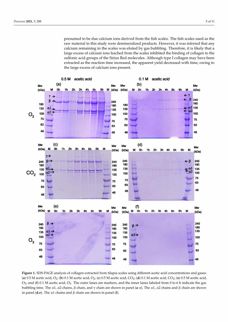
The electrophoretic pattern of collagen exhibited two thick bands and two thin bands.There are several trimers of α chains in tilapia skin that are (α1)2α2 type [39]. The α1 andα2 bands are 136 and 127 kDa, respectively [40], and the band intensity of α1 is twice thatof α2, indicative of type I collagen. One of the three chains that comprise the collagentriple helix is called the α chain and has a molecular weight of approximately 100–130 kDa.Other bands representing γ chains (300 kDa) and β chains (200 kDa) were also identified,further supporting the presence of type I collagen. A γ chain is a heterotrimer formed fromtwo α1 chains and one α2 chain, and a β chain is a dimer of α chains. These data are inagreement with the results obtained from tilapia, carp [41], zebrafish [42], and other fishspecies [24].
When comparing the gas types, thick bands were observed for oxygen and carbondioxide, with both gases efficiently extracting collagen after 1 h of aeration, as shown inFigure 1a,b. However, no thick bands were observed with ozone, suggesting that collagenwas not extracted by ozone gas, as indicated in Figure 1e,f. The thin band observed at 0 hmay have been decomposed by the strong oxidizing power of ozone. When evaluatingthe effect of acetic acid concentration on the effectiveness of extraction, it is clear that morecollagen was extracted in a shorter amount of time by 0.5 M acetic acid than by 0.1 Macetic acid. The bands became thicker as the gas aeration time increased, indicating that alarger amount of collagen was being extracted. However, it is difficult to determine fromthe SDS-PAGE profile, alone, whether it is gelatin or collagen. [43] In general, SDS-PAGEband images provide only a qualitative evaluation; therefore, other methods are needed toquantitatively analyze collagen extraction.
3.2. Evaluation of Collagen Extraction Rate Using the Sirius Red Total Collagen Detection Kit
The Sirius Red method was used to determine the content of the extracted col-lagen. This method for the quantitative estimation of collagen was first reported byMarotta et al. [44]. Sirius Red is also frequently used to distinguish between type I andtype III collagen in tissue sections. According to previous reports, type I to type III collagenin fish, amphibian, reptile, bird, and mammalian organs can be stained by Sirius Red andidentified using polarized light microscopy experiments [45]. Picrosirius Red, a mixtureof picric acid and Sirius Red, also stains collagen fibrils in cellular tissue [46]. Specifically,the sulfonate groups of the Sirius Red molecules bind to the side chains of the basic aminoacids of the collagen [47].
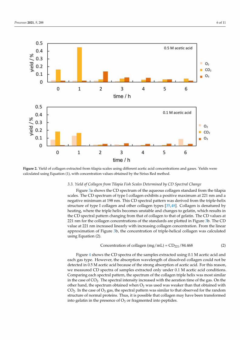
Figure 2 summarizes the yield of collagen extracted with respect to the time each gaswas supplied as ultrafine bubbles. When 0.5 M acetic acid and CO2 were used, the yieldincreased to approximately 0.2% after 0 h and 0.4% after 1 h, but the yield rapidly decreasedafter bubbling for longer periods of time. When 0.1 M acetic acid was used, the yields ofcollagen extracted in the presence of CO2 and O2 gases after 0 h and 1 h were approximately0.1%, exhibiting lower yields than those obtained with 0.5 M acetic acid. In addition, theyield of type I collagen decreased further with prolonged bubbling. The results of the SiriusRed method did not agree with the results of the SDS-PAGE profile, where the bands hadbecome thicker with increasing bubbling time. The reason for the decrease in yield was
Processes 2021, 9, 288 5 of 11
presumed to be due calcium ions derived from the fish scales. The fish scales used as theraw material in this study were demineralized products. However, it was inferred that anycalcium remaining in the scales was eluted by gas bubbling. Therefore, it is likely that alarge excess of calcium ions leached from the scales inhibited the binding of collagen to thesulfonic acid groups of the Sirius Red molecules. Although type I collagen may have beenextracted as the reaction time increased, the apparent yield decreased with time, owing tothe large excess of calcium ions present.
Figure 1. SDS-PAGE analysis of collagen extracted from tilapia scales using different acetic acid concentrations and gases:(a) 0.5 M acetic acid, O2; (b) 0.1 M acetic acid, O2; (c) 0.5 M acetic acid, CO2; (d) 0.1 M acetic acid, CO2; (e) 0.5 M acetic acid,O3; and (f) 0.1 M acetic acid, O3. The outer lanes are markers, and the inner lanes labeled from 0 to 6 h indicate the gasbubbling time. The α1, α2 chains, β chain, and γ chain are shown in panel (a–c). The α1, α2 chains and β chain are shownin panel (d,e). The α1 chains and β chain are shown in panel (f).
Processes 2021, 9, 288 6 of 11
Figure 2. Yield of collagen extracted from tilapia scales using different acetic acid concentrations and gases. Yields werecalculated using Equation (1), with concentration values obtained by the Sirius Red method.
3.3. Yield of Collagen from Tilapia Fish Scales Determined by CD Spectral Change
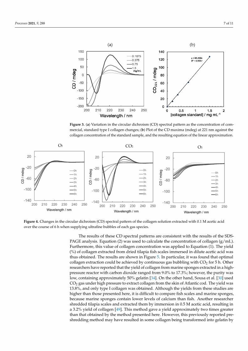
Figure 3a shows the CD spectrum of the aqueous collagen standard from the tilapiascales. The CD spectrum of type I collagen exhibits a positive maximum at 221 nm and anegative minimum at 198 nm. This CD spectral pattern was derived from the triple-helixstructure of type I collagen and other collagen types [35,48]. Collagen is denatured byheating, where the triple helix becomes unstable and changes to gelatin, which results inthe CD spectral pattern changing from that of collagen to that of gelatin. The CD values at221 nm for the collagen concentrations of the standards are plotted in Figure 3b. The CDvalue at 221 nm increased linearly with increasing collagen concentration. From the linearapproximation of Figure 3b, the concentration of triple-helical collagen was calculatedusing Equation (2).
Concentration of collagen (mg/mL) = CD221/84.468 (2)
Figure 4 shows the CD spectra of the samples extracted using 0.1 M acetic acid andeach gas type. However, the absorption wavelength of dissolved collagen could not bedetected in 0.5 M acetic acid because of the strong absorption of acetic acid. For this reason,we measured CD spectra of samples extracted only under 0.1 M acetic acid conditions.Comparing each spectral pattern, the spectrum of the collagen triple helix was most similarin the case of CO2. The spectral intensity increased with the aeration time of the gas. On theother hand, the spectrum obtained when O2 was used was weaker than that obtained withCO2. In the case of O3 gas, the spectral pattern was similar to that observed for the randomstructure of normal proteins. Thus, it is possible that collagen may have been transformedinto gelatin in the presence of O3 or fragmented into peptides.
Processes 2021, 9, 288 7 of 11
Figure 3. (a) Variation in the circular dichroism (CD) spectral pattern as the concentration of com-mercial, standard type I collagen changes; (b) Plot of the CD maxima (mdeg) at 221 nm against thecollagen concentration of the standard sample, and the resulting equation of the linear approximation.
Figure 4. Changes in the circular dichroism (CD) spectral pattern of the collagen solution extracted with 0.1 M acetic acidover the course of 6 h when supplying ultrafine bubbles of each gas species.
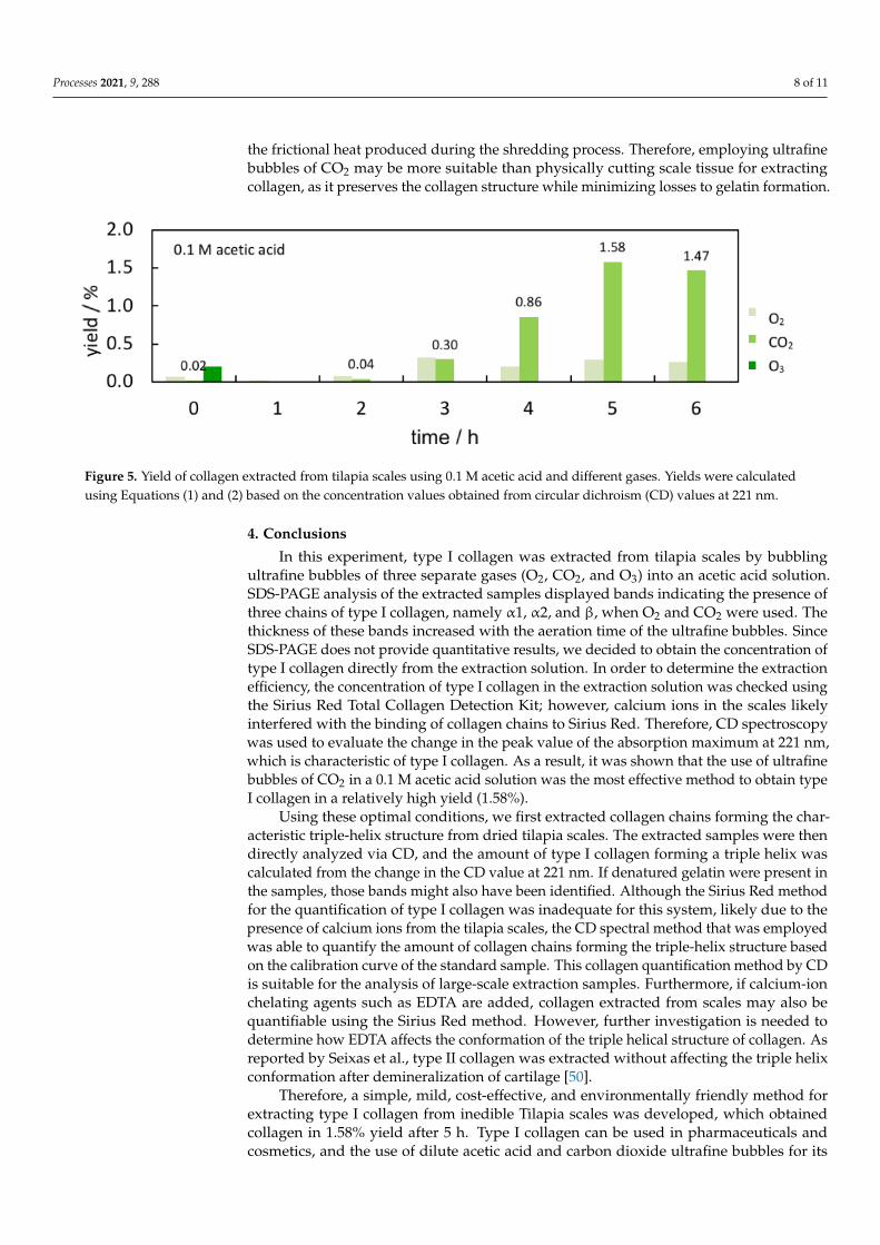
The results of these CD spectral patterns are consistent with the results of the SDS-PAGE analysis. Equation (2) was used to calculate the concentration of collagen (g/mL).Furthermore, this value of collagen concentration was applied to Equation (1). The yield(%) of collagen extracted from dried tilapia fish scales immersed in dilute acetic acid wasthus obtained. The results are shown in Figure 5. In particular, it was found that optimalcollagen extraction could be achieved by continuous gas bubbling with CO2 for 5 h. Otherresearchers have reported that the yield of collagen from marine sponges extracted in a high-pressure reactor with carbon dioxide ranged from 9.0% to 17.3%; however, the purity waslow, containing approximately 50% gelatin [34]. On the other hand, Sousa et al. [30] usedCO2 gas under high pressure to extract collagen from the skin of Atlantic cod. The yield was13.8%, and only type I collagen was obtained. Although the yields from these studies arehigher than those presented here, it is difficult to compare fish scales and marine sponges,because marine sponges contain lower levels of calcium than fish. Another researchershredded tilapia scales and extracted them by immersion in 0.5 M acetic acid, resulting ina 3.2% yield of collagen [49]. This method gave a yield approximately two times greaterthan that obtained by the method presented here. However, this previously reported pre-shredding method may have resulted in some collagen being transformed into gelatin by
Processes 2021, 9, 288 8 of 11
the frictional heat produced during the shredding process. Therefore, employing ultrafinebubbles of CO2 may be more suitable than physically cutting scale tissue for extractingcollagen, as it preserves the collagen structure while minimizing losses to gelatin formation.
Figure 5. Yield of collagen extracted from tilapia scales using 0.1 M acetic acid and different gases. Yields were calculatedusing Equations (1) and (2) based on the concentration values obtained from circular dichroism (CD) values at 221 nm.
4. Conclusions
In this experiment, type I collagen was extracted from tilapia scales by bubblingultrafine bubbles of three separate gases (O2, CO2, and O3) into an acetic acid solution.SDS-PAGE analysis of the extracted samples displayed bands indicating the presence ofthree chains of type I collagen, namely α1, α2, and β, when O2 and CO2 were used. Thethickness of these bands increased with the aeration time of the ultrafine bubbles. SinceSDS-PAGE does not provide quantitative results, we decided to obtain the concentration oftype I collagen directly from the extraction solution. In order to determine the extractionefficiency, the concentration of type I collagen in the extraction solution was checked usingthe Sirius Red Total Collagen Detection Kit; however, calcium ions in the scales likelyinterfered with the binding of collagen chains to Sirius Red. Therefore, CD spectroscopywas used to evaluate the change in the peak value of the absorption maximum at 221 nm,which is characteristic of type I collagen. As a result, it was shown that the use of ultrafinebubbles of CO2 in a 0.1 M acetic acid solution was the most effective method to obtain typeI collagen in a relatively high yield (1.58%).
Using these optimal conditions, we first extracted collagen chains forming the char-acteristic triple-helix structure from dried tilapia scales. The extracted samples were thendirectly analyzed via CD, and the amount of type I collagen forming a triple helix wascalculated from the change in the CD value at 221 nm. If denatured gelatin were present inthe samples, those bands might also have been identified. Although the Sirius Red methodfor the quantification of type I collagen was inadequate for this system, likely due to thepresence of calcium ions from the tilapia scales, the CD spectral method that was employedwas able to quantify the amount of collagen chains forming the triple-helix structure basedon the calibration curve of the standard sample. This collagen quantification method by CDis suitable for the analysis of large-scale extraction samples. Furthermore, if calcium-ionchelating agents such as EDTA are added, collagen extracted from scales may also bequantifiable using the Sirius Red method. However, further investigation is needed todetermine how EDTA affects the conformation of the triple helical structure of collagen. Asreported by Seixas et al., type II collagen was extracted without affecting the triple helixconformation after demineralization of cartilage [50].
Therefore, a simple, mild, cost-effective, and environmentally friendly method forextracting type I collagen from inedible Tilapia scales was developed, which obtainedcollagen in 1.58% yield after 5 h. Type I collagen can be used in pharmaceuticals andcosmetics, and the use of dilute acetic acid and carbon dioxide ultrafine bubbles for its
Processes 2021, 9, 288 9 of 11
extraction ensures that these collagen-containing products are free of harsh chemicals.Furthermore, the use of processed fish scales promotes resource recycling, making thisextraction method more environmentally friendly than other methods while obtaining areasonable yield of type I collagen. In the future, the author plans to investigate the effectsof the flow rate and diameter of the CO2 ultrafine bubbles on the yield of collagen, as wellas to clarify the extraction mechanism.
Funding: J.K. is grateful for KAKENHI Grant Numbers 23500922 and 15K12249 from the JapanSociety for the Promotion of Science (JSPS). This study was funded by the FS project of the FukuokaPrefecture Foundation for Industry, Science and Technology, Japan in 2015-2016.
Institutional Review Board Statement: Not applicable.
Informed Consent Statement: Not applicable.
Data Availability Statement: Data from this study can be made available upon request.
Acknowledgments: The author would like to thank Kasumi Nakata, Yumi Tsuyama, Yurika Sak-aguchi, and Kouki Mihashi of B4 for their help in conducting this experiment. The author would liketo express their deepest appreciation to Shinryo Corporation for providing the fish scales. The authorwould like to thank Editage for English language editing.
Conflicts of Interest: The author declares no conflict of interest.
References1. Walker, C.; Mojares, E.; del Río Hernández, A. Role of extracellular matrix in development and cancer progression. Int. J. Mol. Sci.
2018, 19, 3028. [CrossRef] [PubMed]2. Zdzieblik, D.; Oesser, S.; Baumstark, M.; Gollhofer, A.; König, D. Collagen peptide supplementation in combination with resistance
training improves body composition and increases muscle strength in elderly sarcopenic men: A randomised controlled trial. Br.J. Nutr. 2015, 114, 1237–1245. [CrossRef]
3. Addad, S.; Exposito, J.-Y.; Faye, C.; Ricard-Blum, S.; Lethias, C. Isolation, Characterization and Biological Evaluation of JellyfishCollagen for Use in Biomedical Applications. Mar. Drugs 2011, 9, 967–983. [CrossRef] [PubMed]
4. Gauza-Włodarczyk, M.; Kubisz, L.; Mielcarek, S.; Włodarczyk, D. Comparison of thermal properties of fish collagen and bovinecollagen in the temperature range 298–670 K. Mater. Sci. Eng. C 2017, 80, 468–471. [CrossRef] [PubMed]
5. Miranda-Nieves, D.; Chaikof, E.L. Collagen and elastin biomaterials for the fabrication of engineered living tissues. ACS Biomater.Sci. Eng. 2017, 3, 694–711. [CrossRef]
6. Parenteau-Bareil, R.; Gauvin, R.; Berthod, F. Collagen-based biomaterials for tissue engineering applications. Materials 2010, 3,1863–1887. [CrossRef]
7. Lovell, C.R.; Smolenski, K.A.; Duance, V.C.; Light, N.D.; Young, S.; Dyson, M. Type I and III collagen content and fibre distributionin normal human skin during ageing. Br. J. Dermatol. 1987, 117, 419–428. [CrossRef]
8. Fang, M.; Goldstein, E.L.; Turner, A.S.; Les, C.M.; Orr, B.G.; Fisher, G.J.; Welch, K.B.; Rothman, E.D.; Banaszak Holl, M.M. Type Icollagen D-spacing in fibril bundles of dermis, tendon, and bone: Bridging between nano- and micro-level tissue hierarchy. ACSNano 2012, 6, 9503–9514. [CrossRef]
9. Kew, S.J.; Gwynne, J.H.; Enea, D.; Abu-Rub, M.; Pandit, A.; Zeugolis, D.; Brooks, R.A.; Rushton, N.; Best, S.M.; Cameron, R.E.Regeneration and repair of tendon and ligament tissue using collagen fibre biomaterials. Acta Biomater. 2011, 7, 3237–3247.[CrossRef]
10. Tiku, M.L.; Madhan, B. Preserving the longevity of long-lived type II collagen and its implication for cartilage therapeutics.Ageing Res. Rev. 2016, 62–71. [CrossRef]
11. Alves, A.L.; Marques, A.L.P.; Martins, E.; Silva, T.H.; Reis, R.L. Cosmetic potential of marine fish skin collagen. Cosmetics 2017, 4, 39.[CrossRef]
12. Tziveleka, L.-A.; Ioannou, E.; Tsiourvas, D.; Berillis, P.; Foufa, E.; Roussis, V. Collagen from the Marine Sponges Axinellacannabina and Suberites carnosus: Isolation and Morphological, Biochemical, and Biophysical Characterization. Mar. Drugs 2017,15, 152. [CrossRef] [PubMed]
13. Berillis, P. Marine Collagen: Extraction and Applications. In Research Trends in Biochemistry, Molecular Biology and Microbiology;SM Group: Dover, DE, USA, 2015; pp. 1–13.
14. Brodsky, B.; Thiagarajan, G.; Madhan, B.; Kar, K. Triple-helical peptides: An approach to collagen conformation, stability, andself-association. Biopolymers 2008, 89, 345–353. [CrossRef] [PubMed]
15. Veit, G.; Kobbe, B.; Keene, D.R.; Paulsson, M.; Koch, M.; Wagener, R. Collagen XXVIII, a novel von Willebrand factor Adomain-containing protein with many imperfections in the collagenous domain. J. Biol. Chem. 2006, 281, 3494–3504. [CrossRef]
16. Ricard-Blum, S.; Ruggiero, F. The collagen superfamily: From the extracellular matrix to the cell membrane. Pathol. Biol. 2005, 53,430–442. [CrossRef] [PubMed]
Processes 2021, 9, 288 10 of 11
17. Jafari, H.; Lista, A.; Siekapen, M.M.; Ghaari-Bohlouli, P.; Nie, L.; Alimoradi, H.; Shavandi, A. Fish Collagen: Extraction,characterization and applications for biomaterials engineering. Polymers 2020, 12, 2230. [CrossRef] [PubMed]
18. Gurney, T.A.; Kim, D.W. Applications of porcine dermal collagen (ENDURAGen) in facial plastic surgery. Facial Plast. Surg. Clin.N. Am. 2007, 15, 113–121. [CrossRef] [PubMed]
19. Asai, T.; Takahashi, A.; Ito, K.; Uetake, T.; Matsumura, Y.; Ikeda, K.; Inagaki, N.; Nakata, M.; Imanishi, Y.; Sato, K. Amount ofcollagen in the meat contained in Japanese daily dishes and the collagen peptide content in human blood after ingestion ofcooked fish meat. J. Agric. Food Chem. 2019, 67, 2831–2838. [CrossRef] [PubMed]
20. Hoyer, B.; Bernhardt, A.; Heinemann, S.; Stachel, I.; Meyer, M.; Gelinsky, M. Biomimetically mineralized salmon collagen scaffoldsfor application in bone tissue engineering. Biomacromolecules 2012, 13, 1059–1066. [CrossRef] [PubMed]
21. Jithendra, P.; Merlin Rajam, A.; Kalaivani, T.; Baran Mandal, A.; Rose, C. Preparation and characterization of aloe vera blendedcollagen-chitosan composite scaffold for tissue engineering applications. ACS Appl. Mater. Interfaces 2013, 5, 7291–7298. [CrossRef][PubMed]
22. Sousa, R.O.; Alves, A.L.; Carvalho, D.N.; Martins, E.; Oliveira, C.; Silva, T.H.; Reis, R.L. Acid and enzymatic extraction of collagenfrom Atlantic cod (Gadus Morhua) swim bladders envisaging health-related applications. J. Biomater. Sci. Polym. Ed. 2020, 31,20–37. [CrossRef]
23. Tylingo, R.; Mania, S.; Panek, A.; PiÄtek, R.; PawÅowicz, R. Isolation and Characterization of Acid Soluble Collagen from theSkin of African Catfish (Clarias gariepinus), Salmon (Salmo salar) and Baltic Cod (Gadus morhua). J Biotechnol. Biomater. 2016, 6.[CrossRef]
24. Minh Thuy, L.T.; Okazaki, E.; Osako, K. Isolation and characterization of acid-soluble collagen from the scales of marine fishesfrom Japan and Vietnam. Food Chem. 2014, 149, 264–270. [CrossRef] [PubMed]
25. Rodrigues Menezes, M.d.L.L.; Ribeiro, H.L.; da Silva Abreu, F.d.O.M.; de Andrade Feitosa, J.P.; de Sá Moreira de, S.; Filho,M. Optimization of the collagen extraction from Nile tilapia skin (Oreochromis niloticus) and its hydrogel with hyaluronic acid.Colloids Surf. B 2020, 189, 110852. [CrossRef] [PubMed]
26. Chen, J.; Li, L.; Yi, R.; Gao, R.; He, J. Release kinetics of tilapia scale collagen I peptides during tryptic hydrolysis. Food Hydrocoll.2018, 77, 931–936. [CrossRef]
27. Liu, W.; Zhang, Y.; Cui, N.; Wang, T. Extraction and characterization of pepsin-solubilized collagen from snakehead (Channa argus)skin: Effects of hydrogen peroxide pretreatments and pepsin hydrolysis strategies. Process Biochem. 2019, 76, 194–202. [CrossRef]
28. Kim, T.-K.; Ham, Y.-K.; Shin, D.-M.; Kim, H.-W.; Jang, H.W.; Kim, Y.-B.; Choi, Y.-S. Extraction of crude gelatin from duck skin:Effects of heating methods on gelatin yield. Polut. Sci. 2020, 99, 590–596. [CrossRef] [PubMed]
29. Woo, J.-W.; Yu, S.-J.; Cho, S.-M.; Lee, Y.-B.; Kim, S.-B. Extraction optimization and properties of collagen from yellowfin tuna(Thunnus albacares) dorsal skin. Food Hydrocoll. 2008, 22, 879–887. [CrossRef]
30. Sousa, R.O.; Martins, E.; Carvalho, D.N.; Alves, A.L.; Oliveira, C.; Duarte, A.R.C.; Silva, T.H.; Reis, R.L. Collagen from Atlanticcod (Gadus morhua) skins extracted using CO2 acidified water with potential application in healthcare. J. Polym. Res. 2020, 27, 73.[CrossRef]
31. Alheshibri, M.; Qian, J.; Jehannin, M.; Craig, V.S.J. A history of nanobubbles. Langmuir 2016, 32, 11086–11100. [CrossRef]32. Luu, T.Q.; Truong, P.N.H.; Zitzmann, K.; Nguyen, K.T. Effects of ultrafine bubbles on gram-negative bacteria: Inhibition or
selection? Langmuir 2019, 35, 13761–13768. [CrossRef]33. Wang, C.; Lin, C.-Y.; Liao, G.-Y. Degradation of antibiotic tetracycline by ultrafine-bubble ozonation process. J. Water Process Eng.
2020, 37, 101463. [CrossRef]34. Paola, C.M.; Camila, A.M.; Ana, C.; Marlon, O.; Diego, S.; Robin, Z.; Beatriz, G.; Cristina, C. Functional textile finishing of type I
collagen isolated from bovine bone for potential healthtech. Heliyon 2019, 5, e01260. [CrossRef] [PubMed]35. Silva, J.C.; Barros, A.A.; Aroso, I.M.; Fassini, D.; Silva, T.H.; Reis, R.L.; Duarte, A.R.C. Extraction of collagen/gelatin from the
marine demosponge Chondrosia reniformis (Nardo, 1847) using water acidified with carbon dioxide—Process optimization. Ind.Eng. Chem. Res. 2016, 55, 6922–6930. [CrossRef]
36. Bürck, J.; Heissler, S.; Geckle, U.; Ardakani, M.F.; Schneider, R.; Ulrich, A.S.; Kazanci, M. Resemblance of electrospun collagennanofibers to their native structure. Langmuir 2013, 29, 1562–1572. [CrossRef] [PubMed]
37. Caputo, I.; Lepretti, M.; Scarabino, C.; Esposito, C.; Proto, A. An acetic acid-based extraction method to obtain high qualitycollagen from archeological bone remains. Anal. Biochem. 2012, 421, 92–96. [CrossRef]
38. Laemmli, U.K. Cleavage of structural proteins during the assembly of the head of bacteriophage T4. Nature 1970, 227, 680–685.[CrossRef]
39. Sun, L.; Hou, H.; Li, B.; Zhang, Y. Characterization of acid- and pepsin-soluble collagen extracted from the skin of Nile tilapia(Oreochromis niloticus). Int. J. Biol. Macromol. 2017, 99, 8–14. [CrossRef]
40. Liu, D.; Liang, L.; Regenstein, J.M.; Zhou, P. Extraction and characterisation of pepsin-solubilised collagen from fins, scales, skins,bones and swim bladders of bighead carp (Hypophthalmichthys nobilis). Food Chem. 2012, 133, 1441–1448. [CrossRef]
41. Duan, R.; Zhang, J.; Du, X.; Yao, X.; Konno, K. Properties of collagen from skin, scale and bone of carp (Cyprinus carpio). FoodChem. 2009, 112, 702–706. [CrossRef]
42. Gistelinck, C.; Gioia, R.; Gagliardi, A.; Tonelli, F.; Marchese, L.; Bianchi, L.; Landi, C.; Bini, L.; Huysseune, A.; Witten, P.E.; et al.Zebrafish Collagen Type I: Molecular and Biochemical Characterization of the Major Structural Protein in Bone and Skin. Sci. Rep.2016, 6, 21540. [CrossRef] [PubMed]
Processes 2021, 9, 288 11 of 11
43. Qiu, Y.-T.; Wang, Y.-M.; Yang, X.-R.; Zhao, Y.-Q.; Chi, C.-F.; Wang, B. Gelatin and Antioxidant Peptides from Gelatin Hydrolysateof Skipjack Tuna (Katsuwonus pelamis) Scales: Preparation, Identification and Activity Evaluation. Mar. Drugs 2019, 17, 565.[CrossRef] [PubMed]
44. Marotta, M.; Martino, G. Sensitive spectrophotometric method for the quantitative estimation of collagen. Anal. Biochem. 1985,150, 86–90. [CrossRef]
45. Junqueira, L.C.; Cossermelli, W.; Brentani, R. Differential staining of collagens type I, II and III by Sirius Red and polarizationmicroscopy. Arch. Histol. Jpn. 1978, 41, 267–274. [CrossRef]
46. Lattouf, R.; Younes, R.; Lutomski, D.; Naaman, N.; Godeau, G.; Senni, K.; Changotade, S. Picrosirius Red Staining: A Useful Toolto Appraise Collagen Networks in Normal and Pathological Tissues. J. Histochem. Cytochem. 2014, 62, 751–758. [CrossRef]
47. Rodríguez-Rodríguez, P.; Arribas, S.M.; de Pablo, A.L.L.; González, M.C.; Abderrahim, F.; Condezo-Hoyos, L. A simpledot-blot-Sirius red-based assay for collagen quantification. Anal. Bioanal. Chem. 2013, 405, 6863–6871. [CrossRef]
48. Ge, B.; Wang, H.; Li, J.; Liu, H.; Yin, Y.; Zhang, N.; Qin, S. Comprehensive Assessment of Nile Tilapia Skin (Oreochromis niloticus)Collagen Hydrogels for Wound Dressings. Mar. Drugs 2020, 18, 178. [CrossRef]
49. Chen, J.; Li, L.; Yi, R.; Xu, N.; Gao, R.; Hong, B. Extraction and characterization of acid-soluble collagen from scales and skin oftilapia (Oreochromis niloticus). LWT Food Sci. Technol. 2016, 66, 453–459. [CrossRef]
50. Seixas, M.J.; Martins, E.; Reis, R.L.; Silva, T.H. Extraction and Characterization of Collagen from Elasmobranch Byproducts forPotential Biomaterial Use. Mar. Drugs 2020, 18, 617. [CrossRef]
Short Biography of Author
Junko Kuwahara is a professor in the Department of Life, Environment and Applied Chemistry at the FukuokaInstitute of Technology, Japan. She received her B.E. in Applied Chemistry (1995) and her M.E. (2002) and Ph.D. (2005) inPeptide Chemistry (Prof. Norikazu Nishino) from the Kyushu Institute of Technology, Japan. She worked as a researchassistant in the laboratory of colloid and interface chemistry (Prof. Hideo Akisada) at Kyushu Kyoritsu University, Japanfrom 1996 to 2008. She joined the Fukuoka Institute of Technology in 2008 and was appointed as an Associate Professor in2012 and a Professor in 2018. Her current research interests include methods of extracting substances that can be appliedto cosmetics and medical materials, the science of emulsification, and the interaction of surfactants with proteins andpolysaccharides.


