Embed Size (px)
Citation preview
ORIGINAL RESEARCH ARTICLEpublished: 23 July 2013
doi: 10.3389/fnhum.2013.00368
Facial reactions in response to dynamic emotional stimuliin different modalities in patients suffering fromschizophrenia: a behavioral and EMG studyMariateresa Sestito 1*, Maria Alessandra Umiltà1, Giancarlo De Paola2, Renata Fortunati 2,Andrea Raballo 3, Emanuela Leuci2, Simone Maffei2, Matteo Tonna2, Mario Amore4, Carlo Maggini2
and Vittorio Gallese1*
1 Unit of Physiology, Department of Neuroscience, University of Parma, Parma, Italy2 Psychiatric Division, Department of Neuroscience, University of Parma, Parma, Italy3 Department of Mental Health, AUSL of Reggio Emilia, Reggio Emilia, Italy4 Psychiatric Division, Department of Neuroscience, University of Genova, Genova, Italy
Edited by:
Martin Klasen, RWTH AachenUniversity, Germany
Reviewed by:
Giuliana Lucci, IRCCS Santa Lucia ofRome, ItalyLindsay M. Oberman, University ofCalifornia, San Diego, USA
*Correspondence:
Mariateresa Sestito, Unit ofPhysiology, Department ofNeuroscience, University of Parma,Via Volturno 39, I-43100, Parma, Italye-mail: [email protected];Vittorio Gallese, Section ofPhysiology, Department ofNeuroscience, University of Parma,Via Volturno 39, I-43100, Parma, Italye-mail: [email protected]
Emotional facial expression is an important low-level mechanism contributing to theexperience of empathy, thereby lying at the core of social interaction. Schizophrenia isassociated with pervasive social cognitive impairments, including emotional processingof facial expressions. In this study we test a novel paradigm in order to investigate theevaluation of the emotional content of perceived emotions presented through dynamicexpressive stimuli, facial mimicry evoked by the same stimuli, and their functional relation.Fifteen healthy controls and 15 patients diagnosed with schizophrenia were presentedwith stimuli portraying positive (laugh), negative (cry) and neutral (control) emotionalstimuli in visual, auditory modalities in isolation, and congruently or incongruentlyassociated. Participants where requested to recognize and quantitatively rate theemotional value of the perceived stimuli, while electromyographic activity of Corrugatorand Zygomaticus muscles was recorded. All participants correctly judged the perceivedemotional stimuli and prioritized the visual over the auditory modality in identifying theemotion when they were incongruently associated (Audio-Visual Incongruent condition).The neutral emotional stimuli did not evoke any muscle responses and were judged byall participants as emotionally neutral. Control group responded with rapid and congruentmimicry to emotional stimuli, and in Incongruent condition muscle responses were drivenby what participants saw rather than by what they heard. Patient group showed a similarpattern only with respect to negative stimuli, whereas showed a lack of or a non-specificZygomaticus response when positive stimuli were presented. Finally, we found that onlypatients with reduced facial mimicry (Internalizers) judged both positive and negativeemotions as significantly more neutral than controls. The relevance of these findings forstudying emotional deficits in schizophrenia is discussed.
Keywords: EMG, emotions, empathy, facial mimicry, schizophrenia, simulation
INTRODUCTIONEmotional expressions are widely acknowledged as essential incommunicating internal feelings and intentions (Ekman andOster, 1979). The ability to communicate and understand theemotional states of others and their intentions is a fundamen-tal social skill. Indeed, facial expressions are among the mostcommon and significant emotion stimuli.
To this end, it is well-known that humans react to emotionalfacial expressions with specific, congruent facial muscle mimicry,which can be reliably measured by electromyography (EMG; e.g.,Dimberg, 1982, 1988). For example, pictures of sad facial expres-sions evoke increased muscle Corrugator Supercilii activity, whilepictures of happy facial expressions increase muscle ZygomaticusMajor activity and decrease muscle Corrugator Supercilii activity(Lundqvist and Dimberg, 1995; Han et al., 2012). These facial
muscular reactions appear to be spontaneous and automatic(Dimberg and Thunberg, 1998; Dimberg et al., 2000, 2002; Larsenet al., 2003). Many studies demonstrated that facial mimicry con-tributes to recognition of specific facial expressions (for a review,see Goldman and Sripada, 2005; Niedenthal et al., 2010). Indeed,blocking facial mimicry impairs recognition of facial expressionof emotions (Oberman et al., 2007). Furthermore, it has beenproposed that mimicry reflects internal embodied simulationof the perceived facial expression in order to facilitate under-standing of its emotional meaning (Gallese, 2003, 2005, 2006;Niedenthal, 2007; Halberstadt et al., 2009; Niedenthal et al., 2009)and promoting empathy by means one’s facial feedback system(for a review of the facial feedback hypothesis, see Adelmannand Zajonc, 1989). A recent EMG study (Dimberg et al., 2011)showed that high empathic people, with respect to low empathic
Frontiers in Human Neuroscience www.frontiersin.org July 2013 | Volume 7 | Article 368 | 1
HUMAN NEUROSCIENCE
Sestito et al. Facial EMG responses in patients with schizophrenia
group, are particularly sensitive in reacting with facial reactionswhen they look to emotional facial expressions. Moreover, highempathic people rated perceived facial emotional expressions asmore intense with respect to low empathic ones.
Historically, affective features of schizophrenia were con-sidered an integral part of the disorder. Bleuler (1950) con-sidered affective disturbance to be a fundamental symptomof schizophrenia, whereas hallucinations and delusions wereregarded as accessory symptoms. Studies on patients’ facialmimicry in response to emotional stimuli showed that they acti-vate the same muscle of control subjects, but such activation wasfound to be weaker in patients than in healthy controls (Earnstet al., 1996; Kring and Earnst, 1999). Another study showed, onthe other hand, that in contrast to healthy controls, patients diag-nosed with schizophrenia demonstrated atypical facial mimicry,which was not associated with any clinical feature of the dis-order. The authors of this study suggested that this evidencemight account for a low-level disruption contributing to empa-thy deficits in schizophrenia (Varcin et al., 2010). Similarly, Wolfet al. (2006) found an undecipherable and bizarre mimic patternwithin a sample of patients suffering from schizophrenia, called“mimic disintegration” (see Heimann and Spoerri, 1957). Mimicdisintegration is defined as the inability to organize specific facialmuscle movements as an integrated whole, thus making difficultfor observers to decode the emotional state and establish contactor develop a deeper relationship with the patients. Furthermore,many studies investigating everyday life of patients diagnosedwith schizophrenia documented an emotional-affective patterncharacterized by many negative and few positive experiences, thusmaking patients’ affectivity more negative. Some studies (Matteset al., 1995; Iwase et al., 1999; Wolf et al., 2004, 2006) founda minor activity of Zygomaticus muscle in response to positivestimuli, whereas another study (Sison et al., 1996) found anoverall major activation of Corrugator Supercilii muscle, inter-preted as a sign of the negative attitude showed by patients ineveryday life.
Reduced emotional expression (i.e., flat affect) is not only atypical symptom of full-blown schizophrenia (Andreasen, 1984a;Bleuler, 1950). Many findings lend support to the assump-tion that vulnerability to schizophrenia may be subtly mani-fested in emotional behavior long before the onset of clinicalsymptoms. Furthermore, after schizophrenia onset, flat affectincreases (Walker et al., 1993). Reduced emotional facial expres-sion could be a disease risk index for high-vulnerability subjects(e.g., Schizotypal Personality patients and first degree relatives)(Phillips and Seidman, 2008). Moreover, previous research on flataffect showed a disjunction between the expression and the expe-rience of emotion in schizophrenia (Bleuler, 1950; Berenbaumand Oltmanns, 1992; Kring et al., 1993; Kring and Neale, 1996;Aghevli et al., 2003; Kring and Earnst, 2003). These studiesshowed that patients with schizophrenia often reported experi-encing strong emotions, but they were significantly less expressivethan controls. Thus, observers could note no visible sign ofemotion.
The studies using EMG recording to investigate emotionalexpression in schizophrenia, used different materials and meth-ods. In particular, often non-ecological stimuli, like static images
non-facial stimuli, or fiction movies were used. Many stud-ies indeed highlighted, on the other hand, the importance ofdynamic stimuli in the evaluation of emotional expression. Arecent study on healthy individuals showed that presentation ofdynamic facial expressions evokes stronger EMG responses thanstatic ones. Moreover, participants rated dynamic expressions asmore intense that static ones (Rymarczyk et al., 2011).
Emotional facial expression communicates feelings, but is alsoan important low-level mechanism contributing to the expe-rience of empathy, thereby lying at the core of social interac-tion. Schizophrenia is associated with pervasive social cognitiveimpairments that include emotional processing of facial expres-sions. Despite such disorder might play a crucial role in empathiz-ing deficits and consequently impoverished social skills, previousresearch on facial expression of emotions in schizophrenia has notyielded unequivocal results. In particular, it remains unaddressedthe issue of patients’ facial expression as a medium of empathicresonance contributing to the recognition and evaluation of theperceived emotion expressed by others.
The aim of this study was to investigate whether subjectivefacial mimicry affects the quantitative evaluation of the emo-tional content of perceived emotions presented through dynamicexpressive stimuli, in healthy participants and in patients diag-nosed with schizophrenia. To this purpose we employed a novelparadigm by means of which emotional dynamic ecological stim-uli were presented in the visual and auditory modalities in isola-tion and congruently or incongruently associated. This approachenabled us to study the dimensional quality and possible alter-ation of the emotional responses in these two experimentalgroups.
MATERIALS AND METHODSPARTICIPANTSThirty participants took part to the experiment. Control par-ticipants (CNT; ten males, five females, mean age 35.8 yearsSE ± 2.3) were recruited by public announcement and were blindto the experimental goals. None of them reported the presence ofany neurological or psychiatric disorder. Patient group (SZP; tenmales, five females, mean age 32.8 years SE ± 1.7) were recruitedfrom the Clinical Psychiatry Institute of the University of Parma.All of them were chronic clinically stable outpatients, mainly diag-nosed with schizophrenia, paranoid subtype. Only one patientwas diagnosed with a disorganized subtype, one with an undif-ferentiated subtype and two patients with a residual subtype.Psychiatric diagnosis was established via a structured interview(Structured Clinical Interview for DSM–IV, SCID). Exclusioncriteria were the presence of neurological and vascular disor-ders, dysmetabolic syndrome, alcohol or drugs abuse and mentalretardation (Intelligence Quotient score <70). All participantshad normal or corrected to normal vision. In addition to beingclosely matched for gender, the two groups did not differ in age[t(30) = −1.06, p > 0.05]. All clinical participants (SZP) werereceiving antipsychotic medication (most of them were admin-istered new generation atypical antipsychotics). Since the age ofonset and the illness duration indicated that the clinical sam-ple was heterogeneous, for comparing dosages of different drugswe converted doses of medication to chlorpromazine equivalents.
Frontiers in Human Neuroscience www.frontiersin.org July 2013 | Volume 7 | Article 368 | 2
Sestito et al. Facial EMG responses in patients with schizophrenia
Then we multiplied these equivalents by the time an individualhad been on a given dose to obtain cumulative value measuredin dose-years. After each dose had been converted to dose-years,the results could be summed to provide a cumulative measure oflifetime exposure (Andreasen et al., 2010).
In order to describe psychopathological features related withschizophrenia, patients were administered a variety of tests: scalefor the Assessment of Negative Symptoms (SANS; Andreasen,1984a), Scale for the Assessment of Positive Symptoms (SAPS;Andreasen, 1984b), Social Anhedonia Scale (SAS; Chapman et al.,1976), Physical Anhedonia Scale (PAS; Chapman et al., 1976).Given that all patients were under medication, we also adminis-tered them the Simpson-Angus Extrapyramidal side-effects Scale(Simpson and Angus, 1970), an established, valid and reli-able instrument for assessing neuroleptic-induced parkinsonism(Janno et al., 2005). None of them were beyond cut-off value,indicating that SZP participants did not show any significantextrapyramidal side-effect related with drugs assumption. Detailsabout CNT and SZP samples are provided in Table 1. Writteninformed consent was obtained from all participants before enter-ing the study. The local Ethical Committee approved the study.
STIMULITwo professional actors (one male and one female) were usedfor stimuli preparation. Stimuli consisted of 2-s colored videoclips showing positive (laugh), negative (cry) and neutral (con-trol) emotions. The neutral video clips showed actors makingvarious faces (i.e., “making a face”) that did not imply any par-ticular emotional content, just that the actors were adoptingsome specific facial expressions. Actors when performed neutralstimuli always associated the making a face with specific vocaliza-tions. The sound of the neutral stimuli was a vocalization similarto “ahh,” “ohh,” or “eemmh.” Actors’ full face was presentedagainst a gray background. Stimuli consisted of actors’ Laugh(Positive), Cry (Negative) and Control (Neutral) accompanied
by the simultaneously produced sound of laughter, crying and anon-emotional sound, respectively. Half of the stimuli was per-formed by the male actor, whereas the other half was performedby the female actress. Stimuli were recorded using a digital cam-era (25 frames/s, 720×576 pixels), with audio digitally recorded at44.1 kHz. Stimuli were divided into four presentation modalities:Visual only, Audio only, Audio-Visual congruent and Audio-Visual incongruent. Every presentation modality was made of 60stimuli [24 Laugh (Positive) stimuli, 24 Cry (Negative) stimuliand 12 Control (Neutral) stimuli]. In the Audio modality (A),the sound of the video clips of laugh, cry and control stimuliwas extracted from the original video clips and presented alone.In the Video modality (V), only the visual component of video-clips was presented, devoid of any sound. In the Audio-VisualCongruent modality (AVC), the original video clips were pre-sented with both modalities. In the Incongruent Audio-Visualmodality (AVI), the video of a given expression was coupled andpresented with the audio pertaining to a different video clip per-formed by the same actor (e.g., audio of laugh with the videoof cry, audio of cry with the video of laughs and audio of agiven neutral sound with the video of another neutral stimulus).Consequently, in AVI Laugh participants saw an actor crying butheard laughing, in AVI Cry participants saw an actor laughing butheard crying, and in Control condition they saw an actor makingan unemotional face while hearing the sound of a different neutralstimulus.
EXPERIMENTAL PROCEDUREParticipants were individually tested in a sound attenuated lab-oratory room. They were invited to sit on a comfortable chairin front of a 19-inch computer monitor used for stimuli pre-sentation, located at a distance of 70 cm. Audio tracks werepresented at a comfortable sound level (<70 dB) through loud-speakers integrated in the computer monitor. Before starting,participants were invited to relax and refrain from moving during
Table 1 | Demographic variables and characteristics of Schizophrenia (SZP) and Control (CNT) participants.
Characteristic Patients Controls
Mean SE Range Cut-off Mean SE Range
Age (years) 32.80 1.69 25–49 35.80 2.28 25–53
SAPS 26.67 4.17 0–170SANS 48.09 4.56 0–125PAS 26.36 2.26 0–61 >18SAS 19.00 1.74 0–40 >12
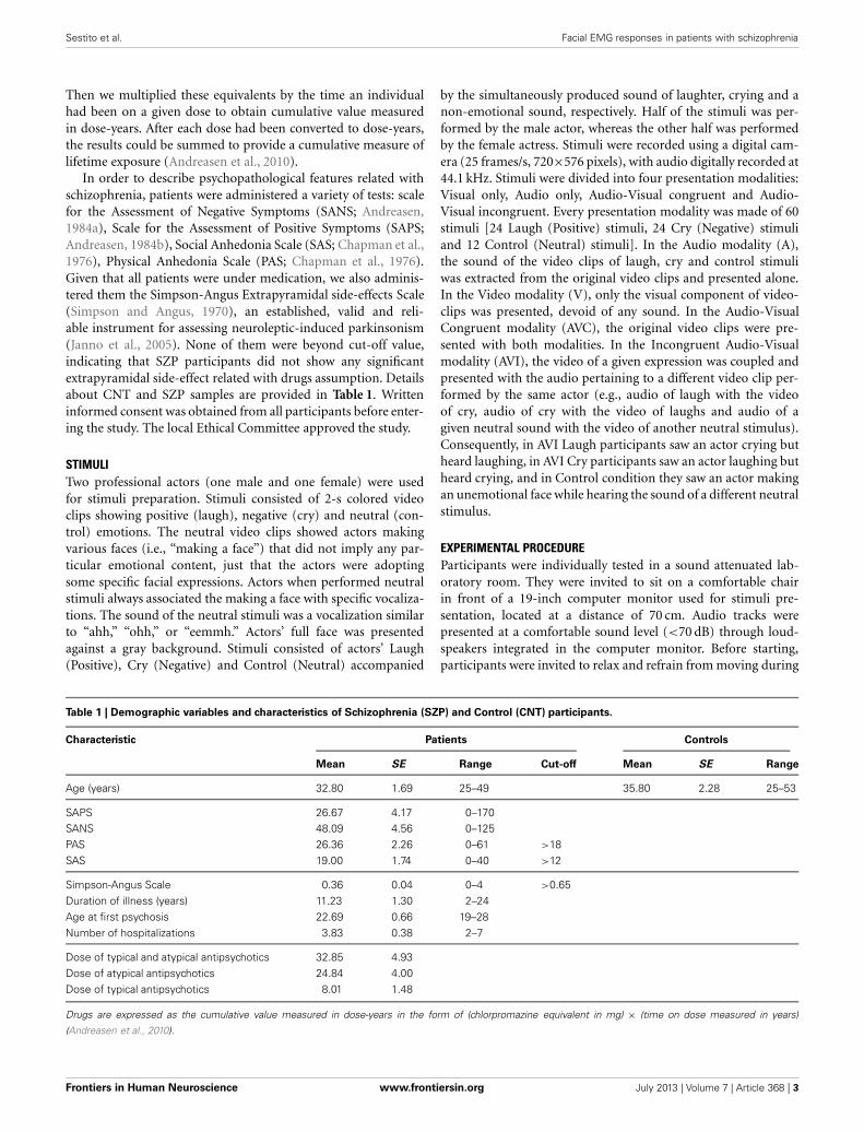
Simpson-Angus Scale 0.36 0.04 0–4 >0.65Duration of illness (years) 11.23 1.30 2–24Age at first psychosis 22.69 0.66 19–28Number of hospitalizations 3.83 0.38 2–7
Dose of typical and atypical antipsychotics 32.85 4.93Dose of atypical antipsychotics 24.84 4.00Dose of typical antipsychotics 8.01 1.48
Drugs are expressed as the cumulative value measured in dose-years in the form of (chlorpromazine equivalent in mg) × (time on dose measured in years)
(Andreasen et al., 2010).
Frontiers in Human Neuroscience www.frontiersin.org July 2013 | Volume 7 | Article 368 | 3
Sestito et al. Facial EMG responses in patients with schizophrenia
the experiment. Participants were instructed to carefully listen toand/or watch audiovisual stimuli. After exposure to each stimu-lus, participants were required to verbally rate how much positiveor negative the stimulus was perceived on a Likert scale rangingfrom −3 (very negative) to +3 (very positive), where 0 indicatedlack of perceived emotional content.
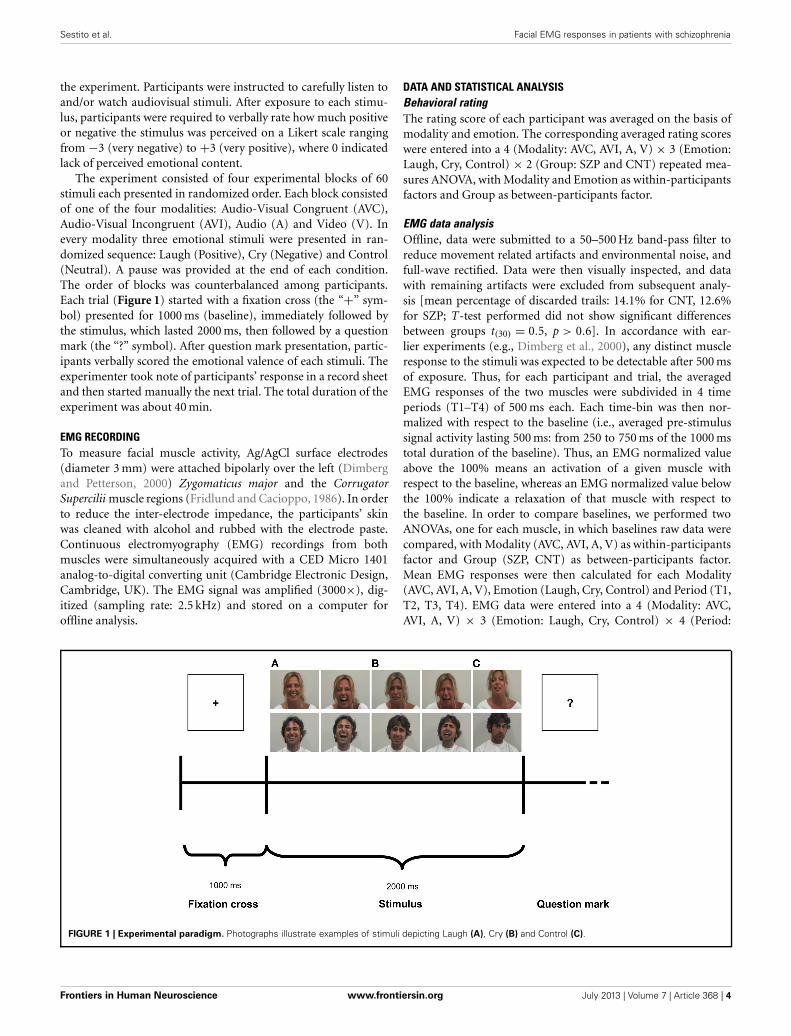
The experiment consisted of four experimental blocks of 60stimuli each presented in randomized order. Each block consistedof one of the four modalities: Audio-Visual Congruent (AVC),Audio-Visual Incongruent (AVI), Audio (A) and Video (V). Inevery modality three emotional stimuli were presented in ran-domized sequence: Laugh (Positive), Cry (Negative) and Control(Neutral). A pause was provided at the end of each condition.The order of blocks was counterbalanced among participants.Each trial (Figure 1) started with a fixation cross (the “+” sym-bol) presented for 1000 ms (baseline), immediately followed bythe stimulus, which lasted 2000 ms, then followed by a questionmark (the “?” symbol). After question mark presentation, partic-ipants verbally scored the emotional valence of each stimuli. Theexperimenter took note of participants’ response in a record sheetand then started manually the next trial. The total duration of theexperiment was about 40 min.
EMG RECORDINGTo measure facial muscle activity, Ag/AgCl surface electrodes(diameter 3 mm) were attached bipolarly over the left (Dimbergand Petterson, 2000) Zygomaticus major and the CorrugatorSupercilii muscle regions (Fridlund and Cacioppo, 1986). In orderto reduce the inter-electrode impedance, the participants’ skinwas cleaned with alcohol and rubbed with the electrode paste.Continuous electromyography (EMG) recordings from bothmuscles were simultaneously acquired with a CED Micro 1401analog-to-digital converting unit (Cambridge Electronic Design,Cambridge, UK). The EMG signal was amplified (3000×), dig-itized (sampling rate: 2.5 kHz) and stored on a computer foroffline analysis.
DATA AND STATISTICAL ANALYSISBehavioral ratingThe rating score of each participant was averaged on the basis ofmodality and emotion. The corresponding averaged rating scoreswere entered into a 4 (Modality: AVC, AVI, A, V) × 3 (Emotion:Laugh, Cry, Control) × 2 (Group: SZP and CNT) repeated mea-sures ANOVA, with Modality and Emotion as within-participantsfactors and Group as between-participants factor.
EMG data analysisOffline, data were submitted to a 50–500 Hz band-pass filter toreduce movement related artifacts and environmental noise, andfull-wave rectified. Data were then visually inspected, and datawith remaining artifacts were excluded from subsequent analy-sis [mean percentage of discarded trails: 14.1% for CNT, 12.6%for SZP; T-test performed did not show significant differencesbetween groups t(30) = 0.5, p > 0.6]. In accordance with ear-lier experiments (e.g., Dimberg et al., 2000), any distinct muscleresponse to the stimuli was expected to be detectable after 500 msof exposure. Thus, for each participant and trial, the averagedEMG responses of the two muscles were subdivided in 4 timeperiods (T1–T4) of 500 ms each. Each time-bin was then nor-malized with respect to the baseline (i.e., averaged pre-stimulussignal activity lasting 500 ms: from 250 to 750 ms of the 1000 mstotal duration of the baseline). Thus, an EMG normalized valueabove the 100% means an activation of a given muscle withrespect to the baseline, whereas an EMG normalized value belowthe 100% indicate a relaxation of that muscle with respect tothe baseline. In order to compare baselines, we performed twoANOVAs, one for each muscle, in which baselines raw data werecompared, with Modality (AVC, AVI, A, V) as within-participantsfactor and Group (SZP, CNT) as between-participants factor.Mean EMG responses were then calculated for each Modality(AVC, AVI, A, V), Emotion (Laugh, Cry, Control) and Period (T1,T2, T3, T4). EMG data were entered into a 4 (Modality: AVC,AVI, A, V) × 3 (Emotion: Laugh, Cry, Control) × 4 (Period:
FIGURE 1 | Experimental paradigm. Photographs illustrate examples of stimuli depicting Laugh (A), Cry (B) and Control (C).
Frontiers in Human Neuroscience www.frontiersin.org July 2013 | Volume 7 | Article 368 | 4
Sestito et al. Facial EMG responses in patients with schizophrenia
T1: 0–500 ms, T2: 500–1000 ms, T3: 1000–1500 ms, T4: 1500–2000 ms) repeated measures ANOVA, with Modality, Emotionand Period as the within-participants factors and Group (SZP andCNT) as between-participants factor. One separated ANOVA wasconducted for each muscle (Corrugator and Zygomaticus).
Functional relation between EMG and behavioral ratingIn order to investigate functional relations between the recordedEMG responses and behavioral rating, we calculated medianEMG responses, separately for each group and for each emo-tion (positive, negative), irrespective of modalities and periods.We excluded from this analysis Control stimuli because theydid not evoke any significant EMG response in both muscles(see Results). Regarding positive emotions, we considered forthis analysis the following modalities in which we measured (seeResults) Zygomaticus muscle activation: AVI Cry, AVC Laugh,A Laugh and V Laugh. Regarding negative emotions, we con-sidered the following modalities in which we measured (seeResults) Corrugator muscle activation: AVI Laugh, AVC Cry, ACry and V Cry.
For each participant, we calculated the median EMG responsefor each emotion (positive, negative). If this value was equal orgreater than the median value calculated separately for positiveand negative emotions for the group the participant belongedto, we classified this participant as Externalizer. If, instead, thisvalue was smaller than the median value calculated separatelyfor positive and negative emotions for the group the partici-pant belonged to, we classified this participant as Internalizer (seeKring and Gordon, 1998). Following this procedure, in the CNTgroup we obtained the median value of 95.14% (8 Externalizersand 7 Internalizers) for positive emotions and the median valueof 99.24% (8 Externalizers and 7 Internalizers) for negative emo-tions. In the SZP group we obtained the median value of 95.15%(6 Externalizers and 9 Internalizers) for positive emotions andthe median value of 100% (6 Externalizers and 9 Internalizers)for negative emotions. The corresponding averaged rating scoreswere entered into a 4 (Modality: AVC, AVI, A, V) × 2 (Group:SZP and CNT) repeated-measures ANOVAs, with Modality aswithin-participants factor and Group as between-participantsfactor. Overall, we ran totally 4 ANOVAs, two in order to analyzebehavioral data of the Externalizer cohort (one for each emotionvalence: positive, negative) and two in order to analyze behavioraldata of the Internalizer cohort (one for each emotion valence:positive, negative).
For all performed analyses, the significance level was set at p <
0.05. Post-hoc comparisons (LSD Fisher test) were applied on allsignificant main factors and interactions.
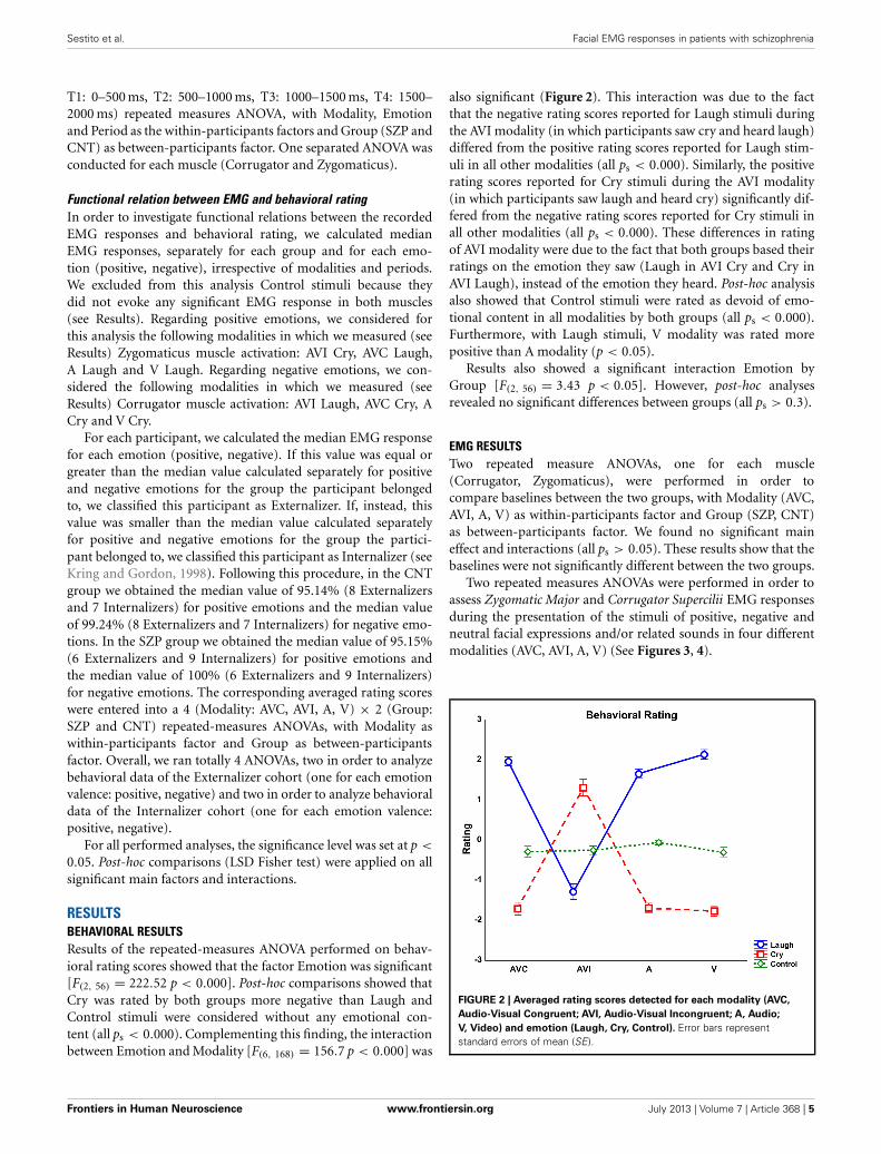
RESULTSBEHAVIORAL RESULTSResults of the repeated-measures ANOVA performed on behav-ioral rating scores showed that the factor Emotion was significant[F(2, 56) = 222.52 p < 0.000]. Post-hoc comparisons showed thatCry was rated by both groups more negative than Laugh andControl stimuli were considered without any emotional con-tent (all ps < 0.000). Complementing this finding, the interactionbetween Emotion and Modality [F(6, 168) = 156.7 p < 0.000] was
also significant (Figure 2). This interaction was due to the factthat the negative rating scores reported for Laugh stimuli duringthe AVI modality (in which participants saw cry and heard laugh)differed from the positive rating scores reported for Laugh stim-uli in all other modalities (all ps < 0.000). Similarly, the positiverating scores reported for Cry stimuli during the AVI modality(in which participants saw laugh and heard cry) significantly dif-fered from the negative rating scores reported for Cry stimuli inall other modalities (all ps < 0.000). These differences in ratingof AVI modality were due to the fact that both groups based theirratings on the emotion they saw (Laugh in AVI Cry and Cry inAVI Laugh), instead of the emotion they heard. Post-hoc analysisalso showed that Control stimuli were rated as devoid of emo-tional content in all modalities by both groups (all ps < 0.000).Furthermore, with Laugh stimuli, V modality was rated morepositive than A modality (p < 0.05).
Results also showed a significant interaction Emotion byGroup [F(2, 56) = 3.43 p < 0.05]. However, post-hoc analysesrevealed no significant differences between groups (all ps > 0.3).
EMG RESULTSTwo repeated measure ANOVAs, one for each muscle(Corrugator, Zygomaticus), were performed in order tocompare baselines between the two groups, with Modality (AVC,AVI, A, V) as within-participants factor and Group (SZP, CNT)as between-participants factor. We found no significant maineffect and interactions (all ps > 0.05). These results show that thebaselines were not significantly different between the two groups.
Two repeated measures ANOVAs were performed in order toassess Zygomatic Major and Corrugator Supercilii EMG responsesduring the presentation of the stimuli of positive, negative andneutral facial expressions and/or related sounds in four differentmodalities (AVC, AVI, A, V) (See Figures 3, 4).
FIGURE 2 | Averaged rating scores detected for each modality (AVC,
Audio-Visual Congruent; AVI, Audio-Visual Incongruent; A, Audio;
V, Video) and emotion (Laugh, Cry, Control). Error bars representstandard errors of mean (SE).
Frontiers in Human Neuroscience www.frontiersin.org July 2013 | Volume 7 | Article 368 | 5
Sestito et al. Facial EMG responses in patients with schizophrenia
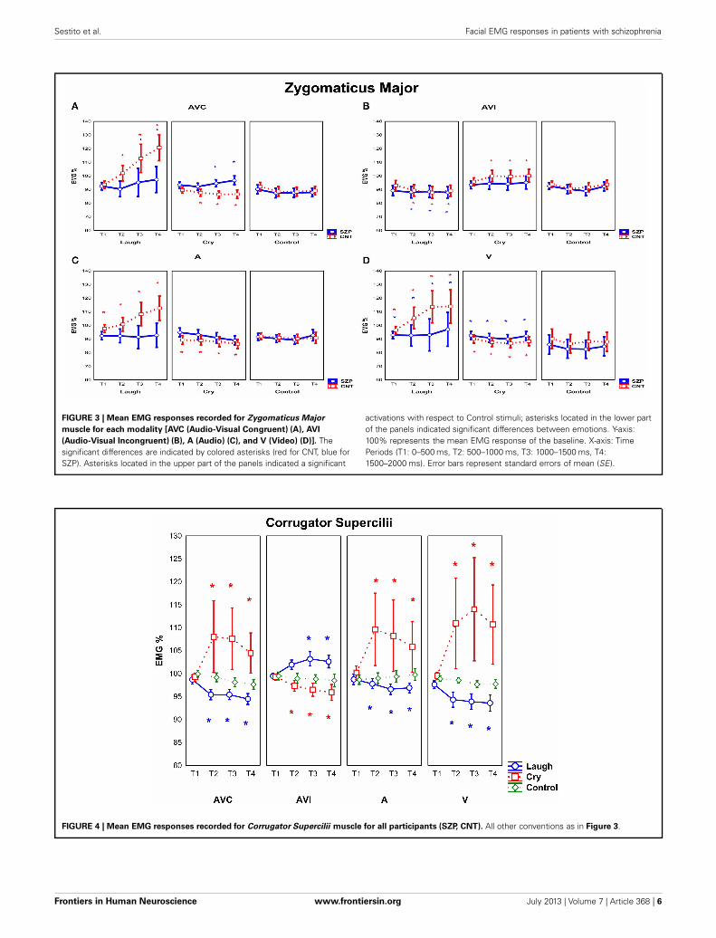
FIGURE 3 | Mean EMG responses recorded for Zygomaticus Major
muscle for each modality [AVC (Audio-Visual Congruent) (A), AVI
(Audio-Visual Incongruent) (B), A (Audio) (C), and V (Video) (D)]. Thesignificant differences are indicated by colored asterisks (red for CNT, blue forSZP). Asterisks located in the upper part of the panels indicated a significant
activations with respect to Control stimuli; asterisks located in the lower partof the panels indicated significant differences between emotions. Y-axis:100% represents the mean EMG response of the baseline. X-axis: TimePeriods (T1: 0–500 ms, T2: 500–1000 ms, T3: 1000–1500 ms, T4:1500–2000 ms). Error bars represent standard errors of mean (SE).
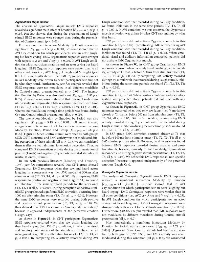
FIGURE 4 | Mean EMG responses recorded for Corrugator Supercilii muscle for all participants (SZP, CNT). All other conventions as in Figure 3.
Frontiers in Human Neuroscience www.frontiersin.org July 2013 | Volume 7 | Article 368 | 6
Sestito et al. Facial EMG responses in patients with schizophrenia
Zygomaticus Major muscleThe analysis of Zygomaticus Major muscle EMG responsesrevealed a significant main effect of Emotion [F(2, 56) = 4.20 p <
0.05]. Post-hoc showed that during the presentation of Laughstimuli EMG responses were stronger than during the presenta-tion of Control stimuli (p < 0.01).
Furthermore, the interaction Modality by Emotion was alsosignificant [F(6, 168) = 4.55 p < 0.001]. Post-hoc showed that inAVI Cry condition (in which participants saw an actor laugh-ing but heard crying) EMG Zygomaticus responses were strongerwith respect to A cry and V cry (p < 0.05). In AVI Laugh condi-tion (in which participants saw instead an actor crying but heardlaughing), EMG Zygomaticus responses were weaker than to allother conditions (i.e., AVC laugh, A laugh and V laugh) (p <
0.01). In sum, results showed that EMG Zygomaticus responsesin AVI modality were driven by what participants saw and notby what they heard. Furthermore, post-hoc analysis revealed thatEMG responses were not modulated in all different modalitiesby Control stimuli presentation (all ps > 0.05). The interac-tion Emotion by Period was also significant [F(6, 168) = 2.82 p <
0.05]. Post-hoc comparisons showed that during Laugh stim-uli presentation Zygomatic EMG responses increased with time(T1 vs. T3 p < 0.01, T1 vs. T4 p < 0.0001, T2 vs. T4 p < 0.01),whereas no modulation through time periods was found duringCry and Control stimuli presentation (all ps > 0.05).
The interaction Modality by Emotion by Period was alsosignificant [F(18, 504) = 3.19 p < 0.0001]. Of most interest,a significant interaction of all factors was observed amongModality, Emotion, Period and Group [F(18, 504) = 1.68 p <
0.05] (Figure 3). Since Control stimuli were rated by both groups(SZP, CNT) as neutral and EMG activity was not modulated dur-ing perception of these stimuli (all ps > 0.1), we can consideredthem as effective neutral stimuli for emotion perception. Thus, wecompared EMG Zygomaticus activity during the presentation ofpositive (Laugh) and negative (Cry) emotion-related stimuli withneutral (Control) stimuli.
In line with previous literature (Dimberg and Thunberg,1998), post-hoc comparisons revealed that CNT group showedZygomaticus EMG responses when they saw and heard actorslaughing in a congruent way (i.e., AVC modality) 500 ms afterstimulus onset (T2, T3, T4; all ps < 0.000). By comparing EMGresponses to positive and negative stimuli (Figure 3A), we foundan inhibition in the same temporal periods for the latter ones(T2, T3, T4; all ps < 0.000). During perception of positive stim-uli SZP group showed significant EMG activation, occurring later,1000 ms after stimulus onset (T3, T4; all ps < 0.01). However,the same EMG responses were recorded during both positiveand negative stimuli presentations (T3, T4; all ps > 0.8). Wethus defined this EMG response as “non-specific activation,”because it appeared independently of the perceived emotion(Laugh, Cry).
As shown in Figure 3B, in CNT participants ZygomaticusEMG responses occurred when they saw actors laughing butthey heard crying (i.e., AVI Cry condition, in which the visualand auditory components of the stimuli are combined in anincongruent way) 500 ms after stimulus onset (T2, T3, T4; allps < 0.05). By comparing EMG activity recorded during AVI
Laugh condition with that recorded during AVI Cry condition,we found inhibition in the same time periods (T2, T3, T4 allps < 0.001). In AVI condition, we thus observed that Zygomaticmuscle activation was driven by what CNT saw and not by whatthey heard.
SZP participants did not activate Zygomatic muscle in thiscondition (all ps > 0.05). By contrasting EMG activity during AVILaugh condition with that recorded during AVI Cry condition,inhibition was found (T2, T3, T4; all ps < 0.05). When emo-tional visual and auditory information contrasted, patients didnot activate EMG Zygomaticus muscle.
As shown in Figure 3C, in CNT group Zygomaticus EMGresponses occurred when they only heard laughing (i.e., A modal-ity) already at T1 that is, before 500 ms from stimulus onset (T1,T2, T3, T4; all ps < 0.05). By comparing EMG activity recordedduring Cry stimuli with that recorded during Laugh stimuli, inhi-bition during the same time periods was found (T1, T2, T3, T4;all ps < 0.01).
SZP participants did not activate Zygomatic muscle in thiscondition (all ps > 0.4). When positive emotional auditory infor-mation was presented alone, patients did not react with anyZygomatic EMG responses.
As shown in Figure 3D, in CNT group Zygomaticus EMGresponses occurred when they only saw laugh (i.e., V modality)already at T1 that is, before 500 ms from stimulus onset (T1, T2,T3, T4; all ps < 0.05). Still in V modality, by comparing EMGactivity recorded during Cry stimuli with that recorded duringLaugh stimuli, inhibition during the same time periods was found(T1, T2, T3, T4; all ps < 0.05).
In SZP group EMG activation occurred already at T1 thatis, before 500 ms from stimulus onset (T1, T2, T3, T4; all ps <
0.05) during positive stimuli. However, no difference was foundbetween EMG responses recorded during negative and posi-tive stimuli, because, similarly to AVC modality, Zygomaticusresponded also during negative stimuli presentation (T1, T2, T3,T4; all ps > 0.05). We define this EMG response as “non-specificactivation,” because it appeared independently of the perceivedemotion (Laugh, Cry).
Corrugator Supercilii muscleThe analysis of Corrugator Supercilii muscle EMG responsesrevealed a significant interaction Modality by Emotion[F(6, 168) = 3.11 p < 0.01]. Post-hoc showed that in AVICry condition (in which participants saw an actor laughing butheard crying) EMG Corrugator responses were weaker than inall other conditions (i.e., AVC cry, A cry and V cry) (p < 0.05).In AVI Laugh condition (in which participants saw an actorcrying but heard laughing), EMG Corrugator responses werestronger only with respect to the V laugh condition (p < 0.05).Furthermore, post-hoc analysis revealed that EMG responses werenot modulated by different modalities during Control stimulipresentation (all ps > 0.7).
Most interestingly, a significant interaction Modality byEmotion by Period was also observed [F(18, 504) = 2.78 p <
0.001] (Figure 4). Since Control stimuli had been rated neu-tral by both groups (SZP, CNT) and EMG activity was nevermodulated during this condition (all ps > 0.2), we considered
Frontiers in Human Neuroscience www.frontiersin.org July 2013 | Volume 7 | Article 368 | 7
Sestito et al. Facial EMG responses in patients with schizophrenia
Control stimuli, also for Corrugator EMG responses, as effectiveneutral stimuli for emotion perception. We performed the samecomparisons already described for Zygomaticus muscle.
Post-hoc comparisons revealed that in both groups (SZP, CNT)Corrugator EMG responses occurred when they saw and heardcry (i.e., AVC modality) 500 ms after stimulus onset (T2, T3,T4; all ps < 0.01). Inhibition occurred during positive stimulipresentation in the same time periods (T2, T3, T4; all ps < 0.000).
Both groups activated Corrugator muscle when they saw crybut they heard laugh (i.e., AVI Laugh modality) 1000 ms afterstimulus onset (T3 p < 0.05; T4 p = 0.05). By comparing EMGactivity recorded during AVI Cry with that recorded during AVILaugh, inhibition 500 ms after stimulus onset was found (T2, T3,T4 all ps < 0.05). In AVI modality, Corrugator muscle activationwas driven by what both groups of participants saw and not bywhat they heard.
Corrugator EMG responses occurred when both groups heardcry (i.e., A modality) 500 ms after stimulus onset (T2, T3, T4; allps < 0.01), as it happened in AVC modality. EMG activity wasinhibited during presentation of Laugh stimuli in the same timeperiods (T2, T3, T4; all ps < 0.0001).
Corrugator EMG responses occurred when both groups sawcry (i.e., V modality) 500 ms after stimulus onset (T2, T3, T4;all ps = 0.0000), as in AVC and A modalities. EMG activity wasinhibited during presentation of Laugh stimuli in the same timeperiods (T2, T3, T4; all ps < 0.000).
For Corrugator muscle, we found no significant main effectsand interactions of Group factor (all ps > 0.05).
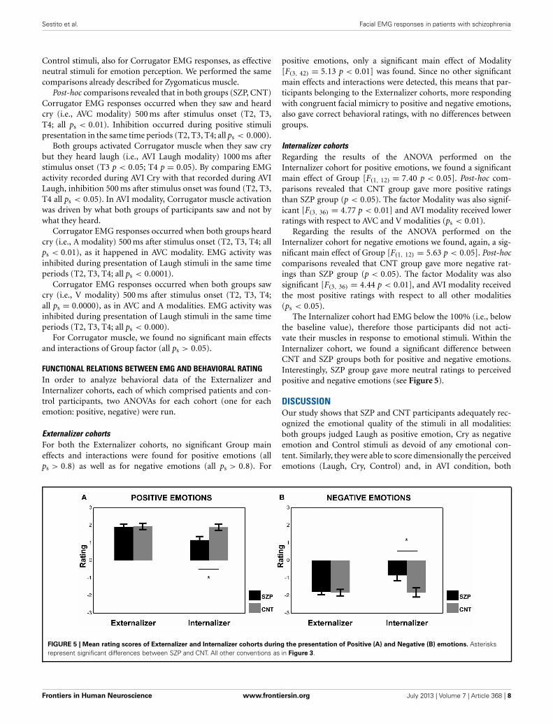
FUNCTIONAL RELATIONS BETWEEN EMG AND BEHAVIORAL RATINGIn order to analyze behavioral data of the Externalizer andInternalizer cohorts, each of which comprised patients and con-trol participants, two ANOVAs for each cohort (one for eachemotion: positive, negative) were run.
Externalizer cohortsFor both the Externalizer cohorts, no significant Group maineffects and interactions were found for positive emotions (allps > 0.8) as well as for negative emotions (all ps > 0.8). For
positive emotions, only a significant main effect of Modality[F(3, 42) = 5.13 p < 0.01] was found. Since no other significantmain effects and interactions were detected, this means that par-ticipants belonging to the Externalizer cohorts, more respondingwith congruent facial mimicry to positive and negative emotions,also gave correct behavioral ratings, with no differences betweengroups.
Internalizer cohortsRegarding the results of the ANOVA performed on theInternalizer cohort for positive emotions, we found a significantmain effect of Group [F(1, 12) = 7.40 p < 0.05]. Post-hoc com-parisons revealed that CNT group gave more positive ratingsthan SZP group (p < 0.05). The factor Modality was also signif-icant [F(3, 36) = 4.77 p < 0.01] and AVI modality received lowerratings with respect to AVC and V modalities (ps < 0.01).
Regarding the results of the ANOVA performed on theInternalizer cohort for negative emotions we found, again, a sig-nificant main effect of Group [F(1, 12) = 5.63 p < 0.05]. Post-hoccomparisons revealed that CNT group gave more negative rat-ings than SZP group (p < 0.05). The factor Modality was alsosignificant [F(3, 36) = 4.44 p < 0.01], and AVI modality receivedthe most positive ratings with respect to all other modalities(ps < 0.05).
The Internalizer cohort had EMG below the 100% (i.e., belowthe baseline value), therefore those participants did not acti-vate their muscles in response to emotional stimuli. Within theInternalizer cohort, we found a significant difference betweenCNT and SZP groups both for positive and negative emotions.Interestingly, SZP group gave more neutral ratings to perceivedpositive and negative emotions (see Figure 5).
DISCUSSIONOur study shows that SZP and CNT participants adequately rec-ognized the emotional quality of the stimuli in all modalities:both groups judged Laugh as positive emotion, Cry as negativeemotion and Control stimuli as devoid of any emotional con-tent. Similarly, they were able to score dimensionally the perceivedemotions (Laugh, Cry, Control) and, in AVI condition, both
FIGURE 5 | Mean rating scores of Externalizer and Internalizer cohorts during the presentation of Positive (A) and Negative (B) emotions. Asterisksrepresent significant differences between SZP and CNT. All other conventions as in Figure 3.
Frontiers in Human Neuroscience www.frontiersin.org July 2013 | Volume 7 | Article 368 | 8
Sestito et al. Facial EMG responses in patients with schizophrenia
groups privileged the visual over the auditory modality. Indeed,they judged the emotion they saw rather than the emotion theyheard.
With respect to EMG recordings, CNT group results are coher-ent with previous studies (Dimberg and Thunberg, 1998) thatdocumented the role of Zygomaticus muscle in response topositive emotional stimuli and that of Corrugator Supercilii inresponse to negative ones. Also the timing of the activation of themuscles was in line with previous findings (500 ms after stimu-lus onset). However, with “single modalities” (A and V) intenseEMG responses of Zygomaticus muscle were evoked by positiveemotions even before 500 ms from stimulus onset. Notably, theinclusion of Control stimuli enriched the experimental paradigmwith an effective neutral stimulus, which proved to evoke noEMG response and be judged by all participants as emotionallyneutral. This shows that facial mimicry does not occur indis-criminately whenever one looks at the moving face of someoneelse, but requires the observation of emotion-specific pattern offacial movements. The same emotional specificity of EMG activa-tion also occurred in Audio modality where only Laugh and Crysounds were able to evoke EMG activation of the Zygomaticusand Corrugator muscles, respectively.
A further innovative feature of the adopted paradigm was theIncongruent (AVI) condition. Interestingly enough, we discov-ered that in AVI condition healthy participants reacted with rapidand automatic mimicry following the visual emotional content ofthe stimuli, while disregarding the auditory expressed contrastingemotion.
Whereas CNT and SZP groups reacted to negative emotionalstimuli in the same way, SZP participants did not respond orshowed inadequate EMG reactions (a “non-specific response”)to positive emotions. Indeed, in AVI and Audio modalities noEMG activation was found, whereas in AVC and V modalities anon-specific response appeared independently of the perceivedemotion (Laugh, Cry).
Several studies investigated how emotional stimuli conveyedby visual and auditory modalities are integrated in healthy popu-lation (for a review, see Klasen et al., 2012) and in patients withschizophrenia (de Gelder et al., 2005; de Jong et al., 2009, 2010;Castagna et al., 2013). Studies conducted with healthy partici-pants demonstrated that congruent audiovisual emotions usuallyyield the highest recognition rates, followed by visual emo-tions, with the auditory emotions being most difficult to classify(Pourtois et al., 2005; Kreifelts et al., 2007; Collignon et al., 2008;Klasen et al., 2011). In our experiment, both groups prioritizedthe visual over the auditory modality in identifying the emotionwhen they were incongruently associated (i.e., AVI modality).Furthermore, only for positive emotions, Video modality wasrated by all participants as more intense with respect to Audiomodality, whereas the AVC modality was judged as equally intensewith respect to single modalities (i.e., Audio and Video). The lackof significant rate advantage of bimodality may be explained bya ceiling effect of high rates in both unimodal conditions (cf. deGelder and Vroomen, 2000).
A recent study investigating multisensory integration inschizophrenia (Castagna et al., 2013) found that patients were notimpaired in basic non-emotional and emotional prosody tasks,
whereas they showed a specific impairment in decoding emotionin a conflicting auditory condition (i.e., when the content of asentence was not congruent with the emotional tone expressedby the voice) and in a multisensory integration condition (i.e.,when complex emotional auditory and visual cues had to beassociated). Another study (de Jong et al., 2010) found that incontrast to controls, a stronger impact of facial on vocal emo-tion perception occurs in patients diagnosed with schizophre-nia. Differently from our experiment, all these studies usedcomplex tasks (e.g., cross-modal emotional recognition tasks)that required top-down attentional processes that demandedsuperior cognitive abilities (i.e., executive functions), which arenotoriously impaired in patients suffering from schizophrenia(Castagna et al., 2013). In our experiment, instead, we investi-gated automatic bottom-up responses to multimodal emotionalstimuli presentation.
In our study, we found that CNT and SZP groups similarlyreacted with EMG corrugator responses to negative emotionalstimuli. Notably, our findings regarding patients’ motor res-onance in response to negative stimuli cohere with previousqualitative and quantitative studies documenting how every-day life of patients suffering from schizophrenia is marked byselective biases toward negative emotional experiences whichamplify stress-vulnerability and are possibly fostered by perse-cutory feelings, increased impressionability and oversensitivityto perceived threats. This might be interpreted in line withKapur’s concept of aberrant salience, which posits that posi-tive symptoms of schizophrenia may arise out of “the aberrantassignment of salience to external objects and internal represen-tations” (Kapur, 2003; Van Os and Kapur, 2009). Hence patients’emotional susceptibility to negative stimuli, resulting in theirpersistent negative attitude in everyday life might act as a self-perpetuating mechanism of disturbed salience (Mattes et al.,1995; Kring and Earnst, 1999). Thus, positive daily experiencesare few and the occasions of showing congruent motor reso-nance with happy emotions could be consequently uncommonin patients suffering from schizophrenia (Kring and Earnst, 2003;Wolf et al., 2004, 2006; Trèmeau, 2006), hence the lack of spe-cific and congruent responses of Zygomaticus muscle to positivestimuli.
According to previous results (Kring and Neale, 1996; Sisonet al., 1996; Aghevli et al., 2003), patients diagnosed withschizophrenia also showed a disjunction between the expressionand the behavioral rating of emotions. In the present study wefound this dissociation only for positive emotions, where nor-mal emotional rating was not accompanied by congruent EMGresponses.
However, by dichotomizing both groups of participants intwo cohorts (Externalizers and Internalizers, Kring and Gordon,1998) according to the intensity of their EMG congruentresponses, we found that the patients’ cohort of Internalizersgave more neutral ratings with respect to control group. Thismeans that in patients facial mimicry in response to positive andnegative emotions is crucial to correctly judge from a dimen-sional point of view the perceived emotion. These data coherewith previous findings documenting empathic response deficitsin Schizophrenia (Derntl et al., 2009; Varcin et al., 2010) that
Frontiers in Human Neuroscience www.frontiersin.org July 2013 | Volume 7 | Article 368 | 9
Sestito et al. Facial EMG responses in patients with schizophrenia
may be related with abnormalities in the mirror neurons mech-anisms. According to this model, involuntary facial mimicryconstitutes an important low-level mechanism contributing tothe experience of empathy (for a review, see Singer and Lamm,2009), via processes of simulation and perception-action cou-pling subserved by activation of the mirror neurons mecha-nism. In other words, involuntary facial mimicry reflects anembodied simulation of the perceived emotion, which facili-tates its understanding (Gallese, 2003, 2005, 2006; Niedenthal,2007; Halberstadt et al., 2009; Niedenthal et al., 2009) by pro-moting primary empathic resonance on a bodily level (Gallese,2001; Preston and De Waal, 2002; Sonnby-Borgström, 2002;Sonnby-Borgström et al., 2003; Oberman et al., 2007). Hence,the disruption of this low-level mechanism may contribute tothe well-known empathy deficits in schizophrenia (Varcin et al.,2010). Along similar lines, Dimberg et al. (2011) demonstratedthat the ability to react with facial EMG activations to facialexpressions and to rate these stimuli as more intense is par-ticularly evident among people with high emotional empathy.Our findings cohere with those of Dimberg et al. (2011), sinceInternalizer patients neither react with any EMG response, norrated positive or negative emotional stimuli as significantlymore intense.
It should be added that Internalizer healthy participantscould correctly score perceived emotions despite their apparentEMG hyporeactivity. This result shows that multi-modal emotionrecognition can occur even without full-blown facial mimicry.This might be due to the recruitment of high-level cognitivemechanisms possibly fostered through coping strategies. Facialmimicry might be a necessary condition for fine-grained emo-tional evaluation only for Internalizer patients, who are impairedin correctly judging the intensity of positive and negative emo-tions.
The interpretation of the current findings, however, shouldbe tempered by some limitations. First, the relatively modestsample size reduced the statistical power. Hence possible groupdifferences, such as regarding the EMG corrugator responseto negative stimuli, might not have been detected. Second, allparticipants with schizophrenia were under antipsychotic med-ications, which might act as a confounders in EMG responses.Nonetheless, since the participants’ SAS score (a specific psy-chometric index sensitive to neuroleptic-induced parkinsonism)was below the cut-off, we are inclined to consider minimal suchpotential confounder.
It might also be worth noting, that group differences in EMGactivation could not be attributed to attentional or motivationalfactors, for three main reasons. First, all patients were clinicallystable (i.e., without hallucinations and similarly flamboyant psy-chopathology) when underwent the current experiment. Second,the ratings showed that both groups correctly scored the differ-ent emotions without significant inter-group differences. Third,the lack of responses that characterized patients was emotion spe-cific, was present only in two modalities, and it was not casuallydistributed among conditions.
In conclusion, this study provides new evidence on theemotion-specificity of facial mimicry. Further, it demonstrates
that (1) congruent facial mimicry can be evoked multi-modallyand that (2) when Video and Audio modalities are incon-gruently associated, the Video modality prevails on the Audioas a response-trigger. The paradigm also proved sensitive todetect deficits in rapid facial mimicry for positive emotionsin patients diagnosed with schizophrenia. We interpreted suchdeficits in rapid facial mimicry as indicative of a possible low-level impairment of motor resonance mechanisms, which mayexplain a portion of the empathizing deficits in schizophrenia.This coheres with our finding that the weaker facial mimicryresponse shown by patients’ Internalizer cohort is related to dif-ficulties in correctly judging the intensity of positive and negativeemotions.
In our view, these findings could lead to future studies on thenature of emotional deficits in Schizophrenia, capitalizing on theconvergence between neuroscience and psychopathology. Indeed,contemporary psychopathological research emphasizes the rel-evance of disruption of implicit bodily functioning (of whichfacial mimicry is a crucial component) for the loss of practi-cal immersion in the intersubjective world that constitutes thehallmark of schizophrenia spectrum vulnerability (see Parnaset al., 2002; Stanghellini, 2004; Fuchs, 2005; Parnas, 2011; seealso Ebisch et al., 2012; Ferri et al., 2012; Gallese and Ferri,2013). Therefore, the disturbance of motor resonance revealedin this study, might be implicated in some of the disordersof intersubjective attunement that phenomenologically-orientedpsychopathology indicates as core features of Schizophrenia(Minkowski, 1927; Blankenburg, 1971; Parnas and Bovet, 1991;Parnas et al., 2002).
The interpretation of these results grounded on the hypothe-sis of an impaired functionality of motor resonance mechanismsin patients diagnosed with schizophrenia, should be limited bythe lack in our study of direct measures of the entailed under-pinning neural mechanisms. Nevertheless, a previous fMRI studycarried out by Carr et al. (2003), demonstrated that in healthyparticipants observation and imitation of emotions activated asimilar neural network of brain areas, in which the insula actedas an interface between the premotor component of the mirrormechanism and the limbic system, thus enabling the transla-tion of an observed or imitated facial emotional expression intoits internally felt emotional significance. Such results were inter-preted by the authors as a mechanism that may mediate theunderstanding of the emotional state of others, thus contributingto empathy.
Overall, our results provide an encouraging exploratoryparadigm to investigate the nature of emotional deficits inSchizophrenia that could be fruitfully coupled with neuroimag-ing studies aimed to investigate the neural substrate underpinningthe deficits in rapid facial mimicry in patients suffering fromschizophrenia.
ACKNOWLEDGMENTSThis work was supported by the EU grants TESIS to VittorioGallese. We would like to acknowledge C. Ferrari, F. Tragni,I. Florindo, and M. Sato for their collaboration in stimulipreparation.
Frontiers in Human Neuroscience www.frontiersin.org July 2013 | Volume 7 | Article 368 | 10
Sestito et al. Facial EMG responses in patients with schizophrenia
REFERENCESAdelmann, P. K., and Zajonc, R. B.
(1989). Facial efference and experi-ence of emotion. Ann. Rev. Psychol.40, 249–280. doi: 10.1146/annurev.ps.40.020189.001341
Aghevli, M. A., Blanchard, J. J.,and Horan, W. P. (2003). Theexpression and experience ofemotion in schizophrenia: astudy of social interactions.Psychiatry Res. 119, 261–270. doi:10.1016/S0165-1781(03)00133-1
Andreasen, N. C. (1984a). Scale forthe Assessment of Negative Symptoms(SANS). Iowa City, IA: University ofIowa.
Andreasen, N. C. (1984b). Scale forthe Assessment of Positive Symptoms(SAPS). Iowa City, IA: University ofIowa.
Andreasen, N. C., Pressler, M.,Nopoulos, P., Miller, D., and Ho,B. C. (2010). Antipsychotic doseequivalents and dose-years: astandardized method for compar-ing exposure to different drugs.Biol. Psychiatry 67, 255–262. doi:10.1016/j.biopsych.2009.08.040
Berenbaum, H., and Oltmanns,T. F. (1992). Emotional expe-rience and expression inschizophrenia and depression.J. Abnorm. Psychol. 101, 37–44. doi:10.1037/0021-843X.101.1.37
Blankenburg, W. (1971). Der Verlust dernatürlichen Selbstverständlichkeit.Ein Beitrag zur Psychopathologiesymptomarmer Schizophrenien.Stuttgart: Enke.
Bleuler, E. (1950). Dementia Praecox orthe Group of Schizophrenias. Oxford,UK: International UniversitiesPress. (Original work published in1911).
Carr, L., Iacoboni, M., Dubeau, M.C., Mazziotta, J. C., and Lenzi,G. L. (2003). Neural mechanismsof empathy in humans: a relayfrom neural systems for imitationto limbic areas. Proc. Natl. Acad.Sci. U.S.A. 100, 5497–5502. doi:10.1073/pnas.0935845100
Castagna, F., Montemagni, C., Milani,A. M., Rocca,. G., Rocca, P.,Casacchia, M., et al. (2013).Prosody recognition and audio-visual emotion matching inschizophrenia: the contribution ofcognition and psychopathology.Psichiatry Res. 205, 192–198. doi:10.1016/j.psychres.2012.08.038
Chapman, L. J., Chapman, J. P., andRaulin, M. L. (1976). Scales forphysical and social anhedonia.J. Abnorm. Psychol. 85, 374–382.doi: 10.1037/0021-843X.85.4.374
Collignon, O., Girard, S., Gosselin, F.,Roy, S., Saint-Amour, D., Lassonde,
M., et al. (2008). Audio-visualintegration of emotion expression.Brain Res. 1242, 126–135. doi:10.1016/j.brainres.2008.04.023
de Gelder, B., and Vroomen, J.(2000). The perception ofemotions by ear and by eye.Cogn. Emot. 14, 289–311. doi:10.1080/026999300378824
de Gelder, B., Vroomen, J., deJong, S. J., Masthoff, E. D.,Trompenaars, F. J., and Hodiamont,P. (2005). Multisensory inte-gration of emotional facesand voices in schizophrenics.Schizophr. Res. 72, 195–203. doi:10.1016/j.schres.2004.02.013
de Jong, J. J., Hodiamont, P. P., andde Gelder, B. (2010). Modality-specific attention and multisen-sory integration of emotions inschizophrenia: reduced regulatoryeffects. Schizophr. Res. 122, 136–143.doi: 10.1016/j.schres.2010.04.010
de Jong, J. J., Hodiamont, P. P., Vanden Stock, J., and de Gelder, B.(2009). Audiovisual emotion recog-nition in schizophrenia: reducedintegration of facial and vocal affect.Schizophr. Res. 107, 286–293. doi:10.1016/j.schres.2008.10.001
Derntl, B., Finkelmeyer, A., Toygar, T.K., Hülsmann, A., Schneider, F.,Falkenberg, D. I., et al. (2009).Generalized deficit in all core com-ponents of empathy in schizophre-nia. Schizophr. Res. 108, 197–206.doi: 10.1016/j.schres.2008.11.009
Dimberg, U. (1982). Facial reactions tofacial expressions. Psychophysiology19, 643–647. doi: 10.1111/j.1469-8986.1982.tb02516.x
Dimberg, U. (1988). Facial electromyo-graphy and the experience of emo-tion. J. Psychophysiol. 2, 277–282.
Dimberg, U., Andréasson, P.,and Thumberg, M. (2011).Emotional empathy and facialreactions to facial expressions.J. Psychophysiol. 25, 26–31. doi:10.1027/0269-8803/a000029
Dimberg, U., and Petterson, M. (2000).Facial reactions to happy andangry facial expressions: evidencefor right hemisphere dominance.Psychophysiology 37, 693–696. doi:10.1111/1469-8986.3750693
Dimberg, U., and Thunberg, M.(1998). Rapid facial reactionsto emotional facial expressions.Scand. J. Psychol. 39, 39–45. doi:10.1111/1467-9450.00054
Dimberg, U., Thunberg, M., andElmehed, K. (2000). Unconsciousfacial reactions to emotional facialexpressions. Psychol. Sci. 11, 86–89.doi: 10.1111/1467-9280.00221
Dimberg, U., Thunberg, M., andGrunedal, S. (2002). Facial reactions
to emotional stimuli: automaticallycontrolled emotional responses.Cogn. Emot. 16, 449–471. doi:10.1080/02699930143000356
Earnst, K. S., Kring, A. M., Kadar,M. A., Salem, J. E., Shepard, D.A., and Loosen, P. T. (1996).Facial expression in schizophrenia.Biol. Psychiatry 40, 556–558. doi:10.1016/0006-3223(96)00171-0
Ebisch, S. J. H., Salone, A., Ferri, F., DeBerardis, D., Mantini, D., Ferro, F.M., et al. (2012). Out of touch withreality? Social perception in firstepisode schizophrenia. Soc. Cogn.Affect. Neurosci. 8, 394–403. doi:10.1093/scan/nss012
Ekman, P., and Oster, H. (1979). Facialexpressions of emotion. Annu. Rev.Psychol. 30, 527–554. doi: 10.1146/annurev.ps.30.020179.002523
Ferri, F., Frassinetti, F., Mastrangelo,F., Salone, A., Ferro, F. M., andGallese, V. (2012). Bodily selfand schizophrenia: the loss ofimplicit self-body knowledge.Conscious. Cogn. 21, 1365–1374.doi: 10.1016/j.concog.2012.05.001
Fridlund, A. J., and Cacioppo,J. T. (1986). Guidelines forhuman electromyographicresearch. Phsychophysiology 23,567–589. doi: 10.1111/j.1469-8986.1986.tb00676.x
Fuchs, T. (2005). Corporealizedand disembodied minds: a phe-nomenological view of the bodyin melancholia and schizophre-nia. Philos. Psychiat. Psychol. 12,95–107.
Gallese, V. (2001). The ‘shared mani-fold’ hypothesis: from mirror neu-rons to empathy. J. Consc Stud. 8,33–50.
Gallese, V. (2003). The roots of empa-thy: the shared manifold hypothesisand the neural basis of intersubjec-tivity. Psychopathology 36, 171–180.doi: 10.1159/000072786
Gallese, V. (2005). Embodied simula-tion: from neurons to phenomenalexperience. Phenomenol. Cogn. Sci.4, 23–48. doi: 10.1007/s11097-005-4737-z
Gallese, V. (2006). Intentional attune-ment: a neurophysiologicalperspective on social cognitionand its disruption in autism.Brain Res. 1079, 15–24. doi:10.1016/j.brainres.2006.01.054
Gallese, V., and Ferri, F. (2013). Jaspers,the body and Schizophrenia.The bodily self. Psychopathology(in press).
Goldman, A. I., and Sripada, C.S. (2005). Simulationist modelsof face-based emotion recogni-tion. Cognition 94, 193–213. doi:10.1016/j.cognition.2004.01.005
Halberstadt, J., Winkielman, P.,Niedenthal, P. M., and Dalle, N.(2009). Emotional conception:how embodied emotion conceptsguide perception and facial action.Psychol. Sci. 20, 1254–1261. doi:10.1111/j.1467-9280.2009.02432.x
Han, C., Park, G. Y., Wang, S. M.,Lee, S. Y., Lee, S. J., Bahk, W.M., et al. (2012). Can botulinumtoxin improve mood in depressedpatients? Expert Rev. Neurother.12, 1049–1051. doi: 10.1586/ern.12.92
Heimann, H., and Spoerri, T. (1957).Das Ausdruckssyndrom der mimis-chen Düsintegrierung bei chronis-chen Schizophrenen. Schweiz. Med.Wochenschr. 35/36, 1126–1132.
Iwase, M., Yamashita, K., Takahashi,K., Kajimoto, O., Shimizu, A.,Nishikawa, T., et al. (1999).Diminished facial expressiondespite the existence of pleas-ant emotional experience inschizophrenia. Methods Find. Exp.Clin. Pharmacol. 21, 189–194. doi:10.1358/mf.1999.21.3.534828
Janno, S., Holi, M. M., Tuisku, K.,and Wahlbeck, K. (2005). Validityof Simpson-Angus Scale (SAS) ina naturalistic schizophrenia pop-ulation. BMC Neurol. 5:5. doi:10.1186/1471-2377-5-5
Kapur, S. (2003). Psychosis as a stateof aberrant salience: a frameworklinking biology, phenomenology,and pharmacology in schizophre-nia. Am. J. Psychiatry 160, 13–23.doi: 10.1176/appi.ajp.160.1.13
Klasen, M., Chen, Y. H., and Mathiak,K. A. (2012). Multisensory emo-tions: perception, combinationand underlying neural processes.Rev. Neurosci. 23, 381–392. doi:10.1515/revneuro-2012-0040
Klasen, M., Kenworthy, C. A.,Mathiak, K. A., Kircher, T. T., andMathiak, K. (2011). Supramodalrepresentation of emotions.J. Neurosci. 31, 13635–13643. doi:10.1523/JNEUROSCI.2833-11.2011
Kreifelts, B., Ethofer, T., Grodd,W., Erb, M., and Wildgruber, D.(2007). Audiovisual integrationof emotional signals in voice andface: an event-related fMRI study.Neuroimage 37, 1445–1456. doi:10.1016/j.neuroimage.2007.06.020
Kring, A. M., and Earnst, K. S.(1999). Stability of emotionalresponding in schizophrenia.Behav. Ther. 30, 373–388. doi:10.1016/S0005-7894(99)80016-1
Kring, A. M., and Earnst, K. S. (2003).“Nonverbal behavior in schizophre-nia,” in Nonverbal Behavior inClinical Settings, eds P. Philippot,E. Coats, and R. S. Feldman
Frontiers in Human Neuroscience www.frontiersin.org July 2013 | Volume 7 | Article 368 | 11
Sestito et al. Facial EMG responses in patients with schizophrenia
(New York, NY: Oxford UniversityPress), 263–286.
Kring, A. M., and Gordon, A. H.(1998). Sex differences in emo-tion: expression, experience, andphysiology. J. Pers. Soc. Psychol.74, 686–703. doi: 10.1037/0022-3514.74.3.686
Kring, A. M., Kerr, S. L., Smith, D.A., and Neale, J. M. (1993). Flataffect in schizophrenia does notreflect diminished subjective experi-ence of emotion. J. Abnorm. Psychol.102, 507–517. doi: 10.1037/0021-843X.102.4.507
Kring, A. M., and Neale, J. M. (1996).Do schizophrenic patients showa disjunctive relationship amongexpressive, experiential, and psy-chophysiological components ofemotions? J. Abnorm. Psychol. 105,249–257. doi: 10.1037/0021-843X.105.2.249
Larsen, J. T., Norris, C. J., andCacioppo, J. T. (2003). Effectsof positive and negative affect onelectromyographic activity overzygomaticus major and corrugatorsupercilii. Psychophysiology 40,776–785. doi: 10.1111/1469-8986.00078
Lundqvist, L. O., and Dimberg, U.(1995). Facial expressions are conta-gious. J. Psychophysiol. 9, 203–211.
Mattes, R. M., Schneider, F., Heimann,H., and Birbaumer, N. (1995).Reduced emotional response inschizophrenic patients in remis-sion during social interaction.Schizophr. Res. 17, 249–255. doi:10.1016/0920-9964(95)00014-3
Minkowski, E. (1927). La schizophrénie.Psychopathologie des Schizoides et desSchizophrènes. Paris: Payot.
Niedenthal, P. M. (2007). Embodyingemotion. Science 316, 1002–1005.doi: 10.1126/science.1136930
Niedenthal, P. M., Mermillod, M.,Maringer, M., and Hess, U. (2010).The simulation of smiles (SIMS)model: embodied simulation and
the meaning of facial expression.Behav. Brain Sci. 33, 417–433. doi:10.1017/S0140525X10000865
Niedenthal, P. M., Winkielman, P.,Mondillon, L., and Vermeulen, N.(2009). Embodiment of emotionconcepts. J. Pers. Soc. Psychol. 96,1120–1136. doi: 10.1037/a0015574
Oberman, L. M., Winkielman, P., andRamachandran, V. S. (2007). Faceto face: blocking facial mimicrycan selectively impair recogni-tion of emotional expressions.Soc. Neurosci. 2, 167–78. doi:10.1080/17470910701391943
Parnas, J. (2011). A disappearing her-itage: the clinical core of schizophre-nia. Schizophr. Bull. 37, 1121–1130.doi: 10.1093/schbul/sbr081
Parnas, J., and Bovet, P. (1991).Autism in schizophrenia revisited.Compr. Psychiatry 32, 7–21. doi:10.1016/0010-440X(91)90065-K
Parnas, J., Bovet, P., and Zahavi, D.(2002). Schizophrenic autism:clinical phenomenology andpathogenetic implications. WorldPsychiatry 1, 131–136.
Phillips, L. K., and Seidman, L. J.(2008). Emotion Processing inPersons at Risk for Schizophrenia.Schizophr. Bull. 34, 888–903. doi:10.1093/schbul/sbn085
Pourtois, G., de Gelder, B., Bol, A.,and Crommelinck, M. (2005).Perception of facial expres-sions and voices and of theircombination in the humanbrain. Cortex 41, 49–59. doi:10.1016/S0010-9452(08)70177-1
Preston, S. D., and De Waal, F. B. M.(2002). Empathy: its ultimate andproximate bases. Behav. Brain Sci.25, 1–20.
Rymarczyk, K., Biele, C., Grabowska,A., and Majczynski, H. (2011).EMG activity in response tostatic and dynamic facial expres-sions. Int. J. Psychophysiol. 79,330–333. doi: 10.1016/j.ijpsycho.2010.11.001
Simpson, G. M., and Angus, G. W. S.(1970). A rating scale for extrapyra-midal side effects. Psychiatr. Scand.45, 11–19. doi: 10.1111/j.1600-0447.1970.tb02066.x
Singer, T., and Lamm, C.(2009). The social neuro-science of empathy. Ann. N.Y.Acad. Sci. 1156, 81–96. doi:10.1111/j.1749-6632.2009.04418.x
Sison, C. E., Alpert, M., Fudge, R.,and Stern, R. (1996). Constrictedexpressiveness and psychophysio-logical reactivity in schizophrenia.J. Nerv. Ment. Dis. 184, 589–597.doi: 10.1097/00005053-199610000-00002
Sonnby-Borgström, M. (2002).Automatic mimicry reactions asrelated to differences in emo-tional empathy. Scand. J. Psychol.43, 433–443. doi: 10.1111/1467-9450.00312
Sonnby-Borgström, M., Jönsson,P., and Svensson, O. (2003).Emotional empathy as related tomimicry reactions at differentlevels of information processing.J. Nonverbal Behav. 27, 3–23. doi:10.1023/A:1023608506243
Stanghellini, G. (2004). DisembodiedSpirits and Deanimated Bodies: ThePsychopathology of Common Sense.Oxford: Oxford University Press.doi: 10.1093/med/9780198520894.001.0001
Trèmeau, F. (2006). A review of emo-tion deficits in schizophrenia.Dialogues Clin. Neurosci. 8, 59–70.
Van Os, J., and Kapur, S. (2009).Schizophrenia. Lancet 374,635–645. doi: 10.1016/S0140-6736(09)60995-8
Varcin, K. J., Bailey, P. E., and Henry,J. D. (2010). Empathic deficits inschizophrenia: the potential roleof rapid facial mimicry. J. Int.Neuropsychol. Soc. 16, 621–629. doi:10.1017/S1355617710000329
Walker, E. F., Grimes, K. E.,Davis, D. M., and Smith, A. J.
(1993). Childhood precursors ofschizophrenia: facial expressionsof emotion. Am. J. Psychiatry 150,1654–1660.
Wolf, K., Mass, R., Kiefer, F., Eckert, K.,Weinhold, N., Wiedemann, K., et al.(2004). The influence of olanzapineon facial expression of emotions inschizophrenia - an improved facialEMG study. Germ. J. Psychiatry 7,14–19.
Wolf, K., Mass, R., Kiefer, F.,Wiedemann, K., and Naber, D.(2006). Characterization of thefacial expression of emotions inSchizophrenia Patients: preliminaryfindings with a new electromyogra-phy method. Can. J. Psychiatry. 51,335–341.
Conflict of Interest Statement: Theauthors declare that the researchwas conducted in the absence of anycommercial or financial relationshipsthat could be construed as a potentialconflict of interest.
Received: 15 April 2013; accepted: 25June 2013; published online: 23 July2013.Citation: Sestito M, Umiltà MA, DePaola G, Fortunati R, Raballo A, Leuci E,Maffei S, Tonna M, Amore M, MagginiC and Gallese V (2013) Facial reactionsin response to dynamic emotional stimuliin different modalities in patients suf-fering from schizophrenia: a behavioraland EMG study. Front. Hum. Neurosci.7:368. doi: 10.3389/fnhum.2013.00368Copyright © 2013 Sestito, Umiltà,De Paola, Fortunati, Raballo, Leuci,Maffei, Tonna, Amore, Maggini andGallese. This is an open-access article dis-tributed under the terms of the CreativeCommons Attribution License, whichpermits use, distribution and reproduc-tion in other forums, provided the origi-nal authors and source are credited andsubject to any copyright notices concern-ing any third-party graphics etc.
Frontiers in Human Neuroscience www.frontiersin.org July 2013 | Volume 7 | Article 368 | 12


