Embed Size (px)
Citation preview
Am. J. Hum. Genet. 63:1363–1375, 1998
1363
Familial Porphyria Cutanea Tarda: Characterization of Seven NovelUroporphyrinogen Decarboxylase Mutations and Frequency of CommonHemochromatosis AllelesManuel Mendez,1,2 Lonnie Sorkin,1 Maria Victoria Rossetti,1,2 Kenneth H. Astrin,1Alcira M. del C. Batlle,2 Victoria E. Parera,2 Gerardo Aizencang,1 and Robert J. Desnick1
1Department of Human Genetics, Mount Sinai School of Medicine, New York, and 2Centro de Investigaciones sobre Porfirinas y Porfirias,University of Buenos Aires, Buenos Aires
Summary
Familial porphyria cutanea tarda (f-PCT) results fromthe half-normal activity of uroporphyrinogen decarbox-ylase (URO-D). Heterozygotes for this autosomal dom-inant trait are predisposed to photosensitive cutaneouslesions by various ecogenic factors, including iron over-load and alcohol abuse. The 3.6-kb URO-D gene wascompletely sequenced, and a long-range PCR methodwas developed to amplify the entire gene for mutationanalysis. Four missense mutations (M165R, L195F,N304K, and R332H), a microinsertion (g10insA), a de-letion (g645D1053), and a novel exonic splicing defect(E314E) were identified. Expression of the L195F,N304K, and R332H polypeptides revealed significantresidual activity, whereas reverse transcription–PCR andsequencing demonstrated that the E314E lesion causedabnormal splicing and exon 9 skipping. Haplotypingindicated that three of the four families with the g10insAmutation were unrelated, indicating that these microin-sertions resulted from independent mutational events.Screening of nine f-PCT probands revealed that 44%were heterozygous or homozygous for the commonhemochromatosis mutations, which suggests that ironoverload may predispose to clinical expression. How-ever, there was no clear correlation between f-PCT dis-ease severity and the URO-D and/or hemochromatosisgenotypes. These studies doubled the number of knownf-PCT mutations, demonstrated that marked geneticheterogeneity underlies f-PCT, and permitted presymp-tomatic molecular diagnosis and counseling in thesefamilies to enable family members to avoid disease-pre-cipitating factors.
Received July 15, 1998; accepted for publication August 24, 1998;electronically published October 27, 1998.
Address for correspondence and reprints: Dr. Robert J. Desnick,Department of Human Genetics, Box 1498, Mount Sinai School ofMedicine, Fifth Avenue at 100th Street, New York, NY 10029. E-mail:[email protected]
q 1998 by The American Society of Human Genetics. All rights reserved.0002-9297/98/6305-0013$02.00
Introduction
Familial porphyria cutanea tarda (f-PCT; MIM 176100)is an autosomal dominant disorder that results from thesystemic half-normal activity of uroporphyrinogen de-carboxylase (URO-D; E.C.4.1.1.37), the fifth enzyme inthe heme biosynthetic pathway (Kushner et al. 1976; deVerneuil et al. 1978). The enzymatic defect leads to theaccumulation of uroporphyrins and predisposes genecarriers to cutaneous manifestations including photo-sensitive dermatitis, mechanical fragility of the light-exposed skin, hyperpigmentation, and hypertrichosis(Grossman et al. 1979; Kappas et al. 1995; McGovernet al. 1996; Elder 1998). Homozygosity for URO-D mu-tations results in the rarer but more severe homozygousdominant disorder hepatoerythropoietic porphyria(HEP), which has clinical onset in childhood and a phe-notype similar to that of congenital erythropoietic por-phyria (Smith 1986; Koszo et al. 1990; Kappas et al.1995). In addition, a more common acquired form, spo-radic porphria cutanea tarda (s-PCT), is characterizedby similar clinical features but is due to the isolateddeficiency of hepatic URO-D activity (de Verneuil et al.1978; Elder et al. 1978). The liver-specific enzyme de-ficiency in s-PCT does not result from mutations in theURO-D gene (Garey et al. 1993) but is induced by un-known mechanisms associated with iron overload, al-cohol abuse, estrogens, and/or hepatitis C infection(Grossman et al. 1979; Fargion et al. 1992; DeCastroet al. 1993; Kappas et al. 1995).
URO-D catalyzes the conversion of uroporphyrinogenIII to coproporphyrinogen III by the sequential removalof the four carboxylic groups of the acetic-acid side(Jackson et al. 1976; de Verneuil et al. 1980). The en-zyme has been purified to homogeneity from humanerythrocytes and has been shown to have a molecularweight of ∼46 kD (de Verneuil et al. 1983). Recently,the crystal structure of human URO-D has been deter-mined at 1.6-A resolution (Whitby et al. 1998), whichhas revealed a homodimeric structure with a single activesite. The enzyme is encoded by a single gene localized
1364 Am. J. Hum. Genet. 63:1363–1375, 1998
to chromosomal region 1p34 (de Verneuil et al. 1984;Dubart et al. 1986). The URO-D cDNA was isolated in1986 and contains an open reading frame of 1,104 nu-cleotides that encodes a 367-amino-acid polypeptide(Romeo et al. 1986). Portions of the genomic sequencehave been reported (Romana et al. 1987; McManus etal. 1994), but only recently, while this research was beingperformed, was the entire 3-kb gene sequenced (Moran-Jimenez et al. 1996; Sorkin et al. 1996). The gene con-tains 10 exons and a TATA box and a GC box in thepromoter region. There is no evidence for erythroid-spe-cific and housekeeping forms of URO-D, as have beenidentified in porphobilinogen synthase and hydroxy-methylbilane synthase (Grandchamp et al. 1987; Yoo etal. 1993; Kaya et al. 1994). The availability of the cDNAand genomic sequences encoding URO-D permits in-vestigation of the mutations causing f-PCT. Such studiesare particularly valuable for accurate presymptomaticdiagnosis and counseling of gene carriers in f-PCT fam-ilies. To date, only eight mutations, (L253Q, G281V,G281E, G318R, I334T, 828D31, 890DC, and IVS611)have been identified in unrelated families with f-PCT(Garey et al. 1989, 1990; Roberts et al. 1995; McManuset al. 1996; also see Human Gene Mutation Database).
Although iron overload has been considered an im-portant precipitating factor for s-PCT (Lundvall et al.1970; Felsher and Kushner 1977), its role in f-PCT isnot clear. The recent identification of the hemochro-matosis (HFE) gene and its common C282Y and H63Dmutations has permitted investigation of the frequencyof these lesions in PCT patients (Feder et al. 1996). Todate, the occurrence of these HFE mutations in f-PCThas not been investigated, but in the two European s-PCT populations studied to date, the frequency of eitherthe C282Y or H63D mutation was increased in Britishand Italian s-PCT patients compared with that in therespective normal populations (Fargion et al. 1996; Rob-erts et al. 1997; Santos et al. 1997; Sampietro et al.1998). The frequency of the C282Y allele in 41 Britishs-PCT patients was almost fivefold higher (30.5% vs.5.9%) than that in normal British individuals, whereasthere was no difference between the frequency of theH63D allele in the British s-PCT and that in normalBritish individuals (Roberts et al. 1997). Interestingly,the H63D allele was twofold more frequent (28.7% vs.12.6%) in the 68 Italian s-PCT patients than in normalItalian individuals, whereas there was no difference be-tween the frequency of the C282Y allele among Italians-PCT patients and normal Italian individuals (Sampie-tro et al. 1998). In sum, the occurrence of heterozygosityor homozygosity for the HFE mutations among Britishand Italian s-PCT patients was 68.3% and 52.9%, re-spectively—frequencies higher than those in the respec-tive normal populations.
In the present report, we describe an efficient long-range PCR method for amplifying the entire 3.6-kbgenomic sequence as a single amplicon for detectionof gene rearrangements and for sequencing. By thismethod, seven new URO-D mutations, including fourmissense mutations, a single base insertion, a deletion,and a novel splicing defect, were identified in 10 unre-lated f-PCT patients from Argentina. Each missense mu-tation was characterized by expression studies, and thesplice-site mutation was confirmed by reverse transcrip-tion–PCR (RT-PCR) analysis. Of interest, the microin-sertion found in four presumably unrelated f-PCT fam-ilies was shown by haplotype analysis to have arisenindependently in at least three of the families. In addi-tion, the occurrence of HFE C282Y and H63D muta-tions was determined in these probands with the inher-ited form of PCT, to assess their predisposition to ironoverload and to correlate disease severity with theirURO-D and HFE genotypes.
Material and Methods
Patient Specimens, Enzyme Assays, and DNA Isolation
Ten unrelated f-PCT patients from Argentina werestudied. Fresh 24-h urine samples and peripheral bloodsamples were collected from each f-PCT patient and fam-ily member, for diagnostic and research purposes, afterinformed consent had been obtained, and the urinaryporphyrin concentrations (Batlle 1997) and the eryth-rocyte URO-D activity of each patient were determined(Lim et al. 1983; Afonso et al. 1985). Cultured lymphoidcell lines were established and maintained as describedelsewhere (Anderson and Gusella 1984). DNA was iso-lated from either peripheral blood or cultured lympho-blasts by the Puregene DNA Isolation Kit (GentraSystems).
PCR Amplification of the Entire URO-D Gene
PCR primers for amplifying the introns of the URO-D gene were designed on the basis of the cDNA andintron-exon sequences (Romana et al. 1987; McManuset al. 1994; Moran-Jimenez et al. 1996), and the entiregenomic sequence was determined. Here, the A of theinitiation of translation ATG is numbered as 1; upstreamnucleotides are indicated by negative numbers, begin-ning with C as 21. PCR and sequencing were performedas described elsewhere (Chen et al. 1994). On the basisof the information obtained from sequencing the entiregene, primers for long-range PCR and the sequencing ofeach exon and intron/exon boundaries were designed asshown in table 1.
Mendez et al.: URO-D Gene Mutations Causing f-PCT 1365
Table 1
PCR Primers for Long-Range Amplification, Sequencing Reactions, and Site-DirectedMutagenesis of the URO-D Gene
Primer Type Sequence
Long-range PCR:Upper 5′-TATGGACCTGGCTGGATAAGACTGTTGGT-3′
Lower 5′-GGGACAATCTTTCACAAACAAAACTACAC-3′
Sequencing, for each exon:1 5′-TACAGAAAGGGGCGGAGCCTGGACTGG-3′
2 5′-GGGAGCTGGCCTGGAGGAGGTAGATAG-3′
3 5′-GAGGAGAAAAGTTTTCGAGGGGCA-3′
4 5′-CAAAAGAGGGAAAGATTTATGCCTTCA-3′
5 5′-CTGAACAGAACCTTTCCTCCTGGATTC-3′
6 5′-GGGGTAGACAAAAGGAAGGGTCAGTC-3′
7 5′-GTGGATTTTGTATGTGGGGGAAACTTC-3′
8 5′-GGGATGGGTTGAGTGAAGGTGGTCCTG-3′
9 5′-GGAGCTGCCATGTATGCAGTTACCA-3′
10 5′-ATAGGGAGGACAAAGGCTTGCTGGT-3′
Site-directed mutagenesis:R332H-S 5′-CTGTGCCTTGTATGCATCTGAGGAGGAGATCGGGCAGTTGGTGAAGC-
AGATGCTGGATGACTTTGGACCACATCACTAC-3′
R332H-AS 5′-ATCAGGCTGAAAATCTTCTCTCATCCGCCA-3′
N304K-S 5′-TGGCCAAGCAAGTGAAGGCCAGGTTGCGGGA-3′
N304K-AS 5′-TGCCCGATCTCCTCCTCAGATGCATACAAGGCACAGGGGTCCAGTTTGCC-3′
M165R-1 5′-TGTTGTACCCCAGGCACTGGGCATGGAGGT-3′
M165R-2 5′-CACCACCCTCAACCCTGTATGTCATCAGG-3′
M165R-3 5′-CCTGATGACATACAGGGTTGAGGGTGGTG-3′
M165R-4 5′-ATCAGGCTGAAAATCTTCTCTCATCCGCCA-3′
L195F-1 5′-TGTTGTACCCCAGGCACTGGGCATGGAGGT-3′
L195F-2 5′-CAGAGCATCAGTGAAGATGCGAAGCAGCT-3′
L195F-3 5′-AGCTGCTTCGCATCTTCACTGATGCTCTG-3′
L195F-4 5′-ATCAGGCTGAAAATCTTCTCTCATCCGCCA-3′
RT-PCR:RT-1 5′-GTTACAGACAGCTGACCATGGA-3′
RT-2 5′-CGATCAATCATCTGTGTTAGTGG-3′
Long-Range PCR and Cyclic Sequencing
The URO-D gene was amplified from genomic DNA(1 mg) in a single reaction using sense and antisense prim-ers (table 1) and the GeneAmp XL PCR Kit (Perkin-Elmer), according to the manufacturer’s instructions.For the first 16 cycles, annealing and extension were for3 min each at 677C, and for the last 12 cycles the timeswere increased 15 s each cycle, with a final extensioncycle of 10 min at 727C. A portion of the PCR productwas analyzed by agarose-gel electrophoresis to deter-mine that the long-range reaction was successful and toidentify any gross gene rearrangements. For sequencing,each exon and the adjacent intron/exon boundary wasPCR-cycle sequenced with the indicated primers (table1), with 1 ml of the long-range PCR product used as thetemplate and with the AmpliCycle Sequencing Kit (Per-kin-Elmer) used according to manufacturer’s instruc-tions. The PCR sequencing program consisted of 30 cy-cles of 957C for 30 s, 607C for 30 s, and 727C for 1min. The reactions were run on acrylamide sequencing
gels. The gels were dried, placed against Kodak X-OARfilm for 24 h, and then developed.
Prokaryotic Expression and Characterization of URO-DPoint Mutations
The normal and mutant URO-D alleles were ex-pressed in Escherichia coli strain lJM109, by thepKK223-3 expression system (Pharmacia Biotechology)as described elsewhere (Tsai et al. 1988; Warner et al.1992; Chen et al. 1994; Xu et al. 1995). The EcoRIfragment of the full-length URO-D cDNA was blunt-end ligated into the HindIII site of pKK223-3. This con-struct was designated “pK-UROD.” Each of the mis-sense mutations was introduced into the pK-UROD plas-mid by site-directed mutagenesis (Chen et al. 1994). Forthe R332H and N304K mutations, each expression con-struct was generated by amplification with the indicatedsense and antisense primers (table 1). To introduce mu-tations M165R and L195F into the expression vector,two amplifications were necessary, since there were no
1366 Am. J. Hum. Genet. 63:1363–1375, 1998
convenient restriction sites. First, each region containinga restriction site was amplified individually, and then thePCR products were ligated. For M165R, one set of prim-ers was pM165R-1 and pM165R-2, whereas the otherprimer set was pM165R-3 and pM165R-4. After thetwo PCR products had been ligated together, the finalconstruct was produced by primers 1 and 4. A similarprocedure was used to construct an expression vectorcontaining the L195F mutation. One primer set waspL195F-1 and pL195F-2, whereas the other set waspL195-3 and pL195-4. Primers 1 and 4 also were usedto produce the final L195F construction. Each expres-sion construct was sequenced, to ensure that only thedesired mutation had been introduced and that the re-mainder of the sequence was correct.
Assay of URO-D Activity
Transfection, bacterial growth, and isopropylthioga-lactoside (IPTG) induction were as described elsewhere(Tsai et al. 1988). The cells were washed twice with PBS,and the pellet was resuspended in 250 ml of lysis buffer(0.067 M sodium phosphate, pH 7.0, 0.1% Triton-X-100) and sonicated on ice. The extract was then centri-fuged for 1 min at 13,000 g, and the supernatant wasused as the enzyme source. The URO-D assay consistedof 0.67 M sodium phosphate, pH 7.0 (50 ml), 20 mMglutathione (50 ml), 1 mM EDTA (50 ml), uroporphyrin-ogen III (35 mg/ml; 50 ml), obtained by uroporphyrinreduction in 25 mM NaOH with 3% sodium amalgam,H2O (250 ml), and bacterial extract (50 ml). The reactionwas performed in tubes that had been filled with nitrogengas and were incubated for 30 min at 377C, with shak-ing, in the dark. The reaction was stopped by the ad-dition of 55 ml of 50% trichloroacetic acid. The porphy-rinogens were oxidized to porphyrins by exposure towhite light for 20 min. The samples were then centri-fuged for 1 min at 13,000 g, filtered through 0.2-mmWhatman filters, and analyzed by high-pressure liquidchromotagraphy (HPLC), according to the method ofLim et al. (1983). Specific activity was calculated as nmolof coproporphyrin III produced/h/mg protein. The na-nomoles of coproporphyrin III were determined by ex-trapolation from a calibration curve of nanomoles ofcoproporphyrin III versus peak area.
Thermal Stability of pK-URO-D and pK-R332H
Bacterial lysates containing either pK-UROD or pK-UROD-R332H were induced with IPTG for 3 h. Ex-tracts were diluted with 0.067 M sodium phosphate buf-fer, pH 7, to the same protein concentration (0.1–0.2mg/ml). They were preincubated for various times, from0 to 180 min, at 377C, were placed on ice, and thenwere assayed for URO-D activity. Two enzyme prepa-
rations from independent inductions were assayed, andthe results were averaged.
RT-PCR Studies of URO-D Mutation E314E
To confirm that mutation E314E was a splice-site mu-tation, RT-PCR was performed. Leukocytes were iso-lated from blood by Ficoll-Paque (Pharmacia Biotech),and RNA was isolated from the leukocytes by RNA ZolB (Tel-Test) and was reverse transcribed with M-MLVReverse Transcriptase and Oligo(dT)12–18 primers (LifeTechnologies), according to manufacturer’s instructions.The total amount of cDNA was then amplified by prim-ers RT-1 and RT-2 (table 1), and the PCR product wasanalyzed on 1.5% agarose gels. Each band was cut outof the gel separately, and the DNA was eluted by soakingthe agarose in a small amount of Tris-EDTA buffer (10mM Tris, 1 mM EDTA, pH 7.4). Each exon was thencycle sequenced by the fmol DNA cycle sequencing kit(Promega), according to manufacturer’s instructions.
Analysis of the HFE C282Y and H63D Mutations
Genomic DNAs from the f-PCT probands were an-alyzed for the common HFE mutations, C282Y andH63D. DNA was PCR amplified by the primers andconditions described by Feder et al. (1996). The PCRproducts were cut by the restriction enzyme Sau3AI(New England Biolabs), in the case of H63D, and therestriction enzyme RsaI, in the case of C282Y, and wereanalyzed by agarose-gel electrophoresis.
Haplotyping of f-PCT Probands with URO-D Mutationg10insA
To determine whether the four presumably unrelatedprobands from Argentina who had the g10insA muta-tion were related to each other, haplotyping was per-formed. Six CA-repeat markers from the chromosome1p34 region (Dib et al. 1996) were obtained from Re-search Genetics; these six markers, from centromereto telomere, were D1S193 (AFM057xf4), D1S463(AFM154xg11), D1S2713 (AFMa349yb5), D1S211(AFM122xe1), D1S2724 (AFMb015wc9), and D1S451(AFM248tf9). PCR amplification and gel analysis wereperformed as described by Gelb et al. (1995).
Results
Sequencing of the URO-D Gene
To facilitate identification of mutations in the URO-D gene, the entire genomic sequence was determined byprimers based on the published cDNA and intron/exonboundary sequences (Romeo et al. 1986; Romana etal. 1987). Our sequence (Entrez Nucleotide Sequence
Mendez et al.: URO-D Gene Mutations Causing f-PCT 1367
Table 2
Sequence Differences Identified in the URO-D Gene
Location andGenomic Numbera Differenceb
5′ UTR:2225 insG2222 delT2207 insC2213 TrG2172 insC2167 insG2158 insC
Intron 1:120 delA146 insG236 TrC237 CrT317 delT322 delG474 insCACC543 delG593 delG598 ArG
Intron 2:794 insG
Intron 6:1853 CrA1855 GrC1901 TrC
Intron 9:2768 delC2790 delC2848 CrA2849 ArC2858 CrA2941 insG2976 delT2990 delC3001 CrG3003 insC
3’ UTR:3234 CrG3235 CrT3311 DelC
a Based on sequence in present study.b Based on comparison of sequence in present study
(Entrez Nucleotide Sequence Search, Genbank acces-sion number AF047383) versus that reported byMoran-Jimenez et al. (1996).
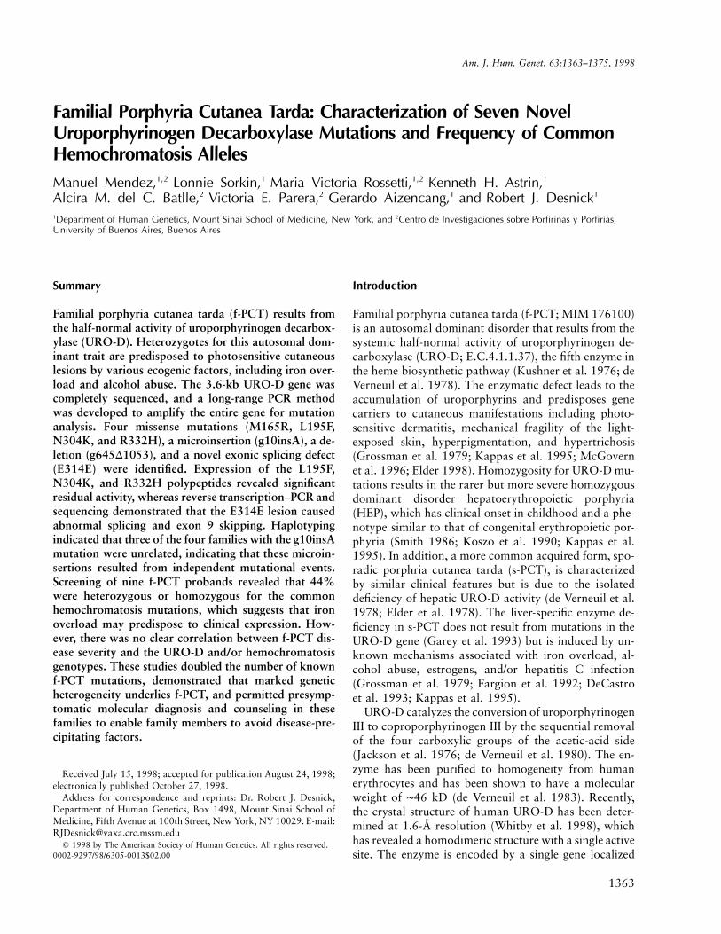
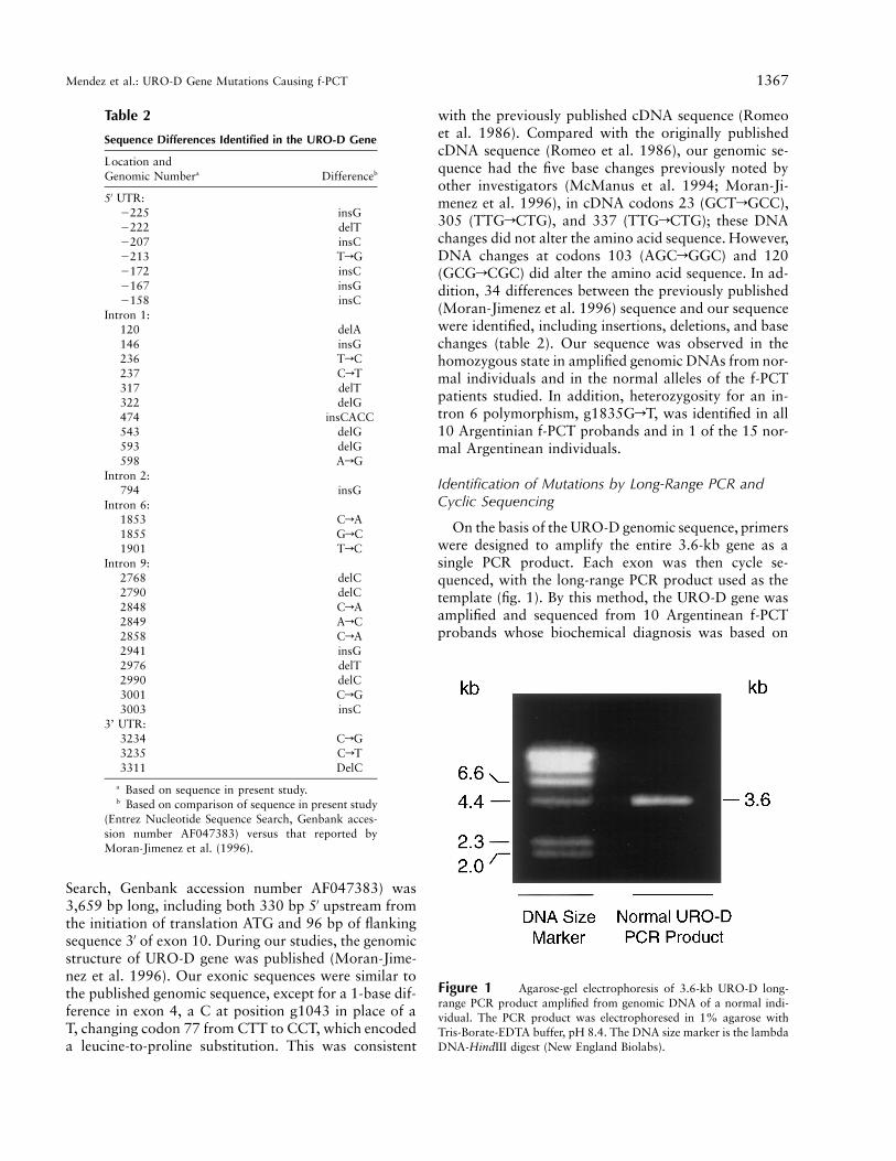
Figure 1 Agarose-gel electrophoresis of 3.6-kb URO-D long-range PCR product amplified from genomic DNA of a normal indi-vidual. The PCR product was electrophoresed in 1% agarose withTris-Borate-EDTA buffer, pH 8.4. The DNA size marker is the lambdaDNA-HindIII digest (New England Biolabs).
Search, Genbank accession number AF047383) was3,659 bp long, including both 330 bp 5′ upstream fromthe initiation of translation ATG and 96 bp of flankingsequence 3′ of exon 10. During our studies, the genomicstructure of URO-D gene was published (Moran-Jime-nez et al. 1996). Our exonic sequences were similar tothe published genomic sequence, except for a 1-base dif-ference in exon 4, a C at position g1043 in place of aT, changing codon 77 from CTT to CCT, which encodeda leucine-to-proline substitution. This was consistent
with the previously published cDNA sequence (Romeoet al. 1986). Compared with the originally publishedcDNA sequence (Romeo et al. 1986), our genomic se-quence had the five base changes previously noted byother investigators (McManus et al. 1994; Moran-Ji-menez et al. 1996), in cDNA codons 23 (GCTrGCC),305 (TTGrCTG), and 337 (TTGrCTG); these DNAchanges did not alter the amino acid sequence. However,DNA changes at codons 103 (AGCrGGC) and 120(GCGrCGC) did alter the amino acid sequence. In ad-dition, 34 differences between the previously published(Moran-Jimenez et al. 1996) sequence and our sequencewere identified, including insertions, deletions, and basechanges (table 2). Our sequence was observed in thehomozygous state in amplified genomic DNAs from nor-mal individuals and in the normal alleles of the f-PCTpatients studied. In addition, heterozygosity for an in-tron 6 polymorphism, g1835GrT, was identified in all10 Argentinian f-PCT probands and in 1 of the 15 nor-mal Argentinean individuals.
Identification of Mutations by Long-Range PCR andCyclic Sequencing
On the basis of the URO-D genomic sequence, primerswere designed to amplify the entire 3.6-kb gene as asingle PCR product. Each exon was then cycle se-quenced, with the long-range PCR product used as thetemplate (fig. 1). By this method, the URO-D gene wasamplified and sequenced from 10 Argentinean f-PCTprobands whose biochemical diagnosis was based on
1368 Am. J. Hum. Genet. 63:1363–1375, 1998
Table 3
Biochemical and Molecular Findings in Argentinean f-PCT Probands
Source (Ancestry/Sex)of Proband
Total UrinaryPorphyrins(mg/24 h)
PlasmaPorphyrin
Index
URO-DErythrocyte
Activity(% of Normala) URO-D Mutation
Normal controls !250 !1.3 100 )1 (Italian/F) 8,910 7.48 45 E314E2 (Portuguese/M) 11,600 6.00 42 R332H3 (Spanish/F) 3,040 3.10 50 L195F4 (Italian/F) 5,010 6.75 46 M165R5 (Spanish/F) 1,130 2.30 51 N304K6 (Spanish/M) 2,030 2.10 42 g645D10537 (Arabic/F) 7,530 5.87 52 g10insA8 (Spanish/M) 4,490 4.30 40 g10insA9 (Spanish/M) 3,120 3.43 44 g10insA10 (Spanish/M) 3,960 4.25 22 g10insA
a Normal mean activity of erythrocyte URO-D 5 4.2 5 0.6 nmol of coproporphyrinogen/h/ml packed erythrocytes. Data were obtained at the time of diagnosis; at present, all patientsare on therapy and in remission.
erythrocyte URO-D enzyme activity and on urinary andplasma porphyrin concentrations (table 3). Seven newmutations were identified, including four missense mu-tations: a TrG tranversion in exon 6, at genomic ntg1663 (cDNA nt 494), which predicted replacement ofa methionine by an arginine at position 165 (a changedesignated “M165R”); a CrT transition in exon 6, atgenomic nt g1752 (cDNA nt 583), which predicted sub-stitution of a leucine by a phenylalanine at residue 195(a change designated “L195F”); a CrA transversion inexon 9, at genomic nt g2711 (cDNA nt 912), whichpredicted an asparagine-to-lysine substitution of posi-tion 304 (a change designated “N304K”); and a GrAtransition (at a CpG dinucleotide) of genomic nt g3124(cDNA nt 995) in exon 10, which predicted an arginine-to-histidine substitution at residue 332 (a change des-ignated “R332H”) (table 3).
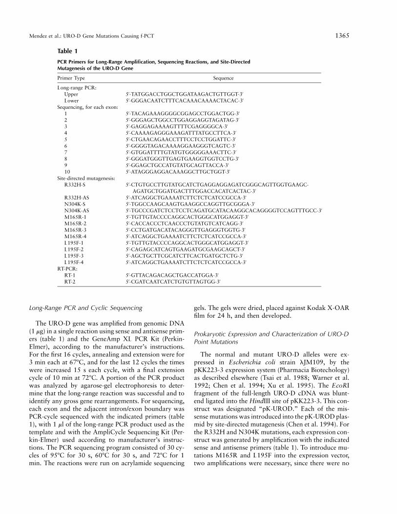
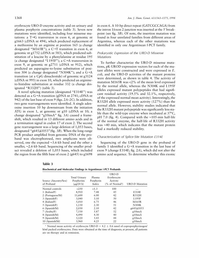
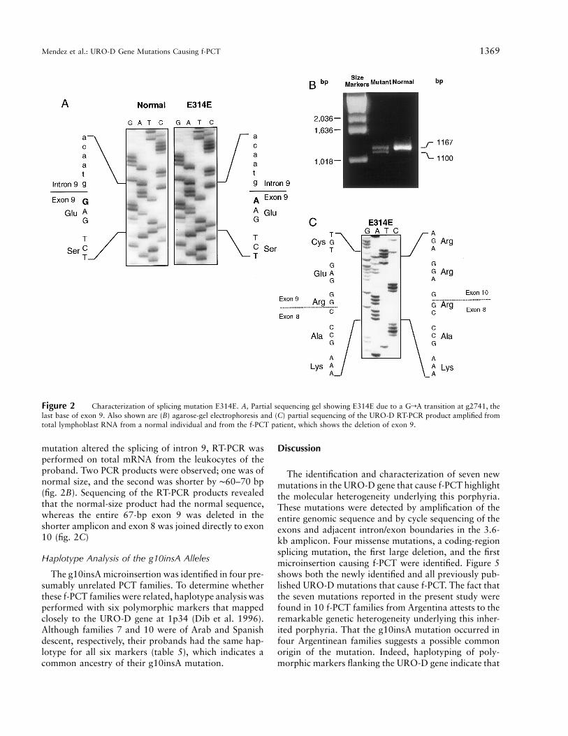
A novel splicing mutation (designated “E314E”) wasdetected as a GrA transition (gDNA nt 2741; cDNA nt942) of the last base of exon 9 (figs. 2A–2C). In addition,two gene rearrangements were identified. A single aden-osine insertion 10 bp downstream from the initiationATG in exon 1, at genomic nt g10 (cDNA nt 10; achange designated “g10insA” fig. 3A) caused a frame-shift, which resulted in 13 different amino acids and ina termination signal in codon 17 of exon 2. The secondgene rearrangement was a large deletion of 1,053 bases,designated “g645D1053”(fig. 3B). When the long-rangePCR product amplified from genomic DNA of the pro-band was electrophoresed, two amplicons were ob-served, one the expected ∼3.6-kb band and the other asmaller, ∼2.6-kb band. Sequencing of the smaller prod-uct revealed a deletion of 1,053 bases, which includedthe region from the fifth base of exon 2 (g645) to g1698
in exon 6. A 10-bp direct repeat (GATCGCCAGA) fromthe intron 1/exon 2 junction was inserted at the 5′ break-point (see fig. 3B). Of note, the insertion mutation wasfound in four unrelated families from different areas ofArgentina, whereas each of the other mutations wasidentified in only one Argentinean f-PCT family.
Prokaryotic Expression of the URO-D MissenseMutations
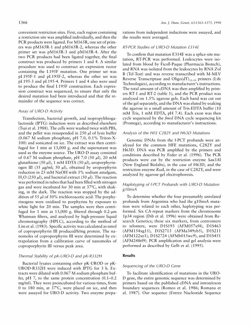
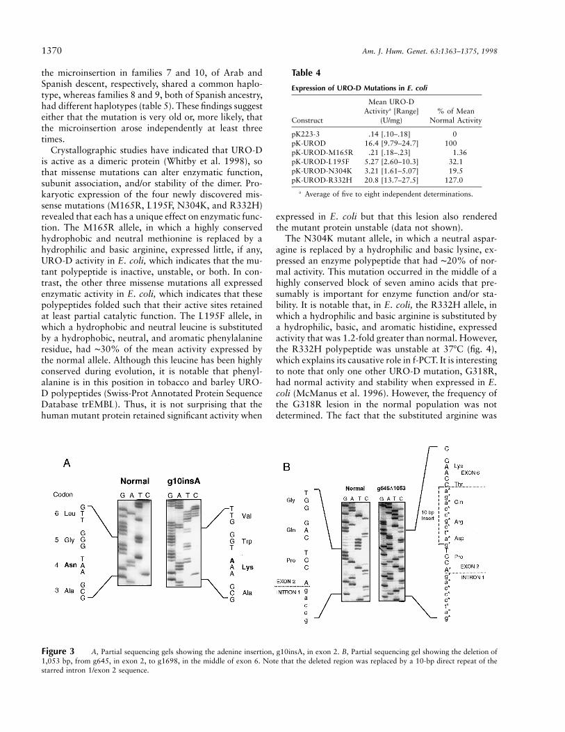
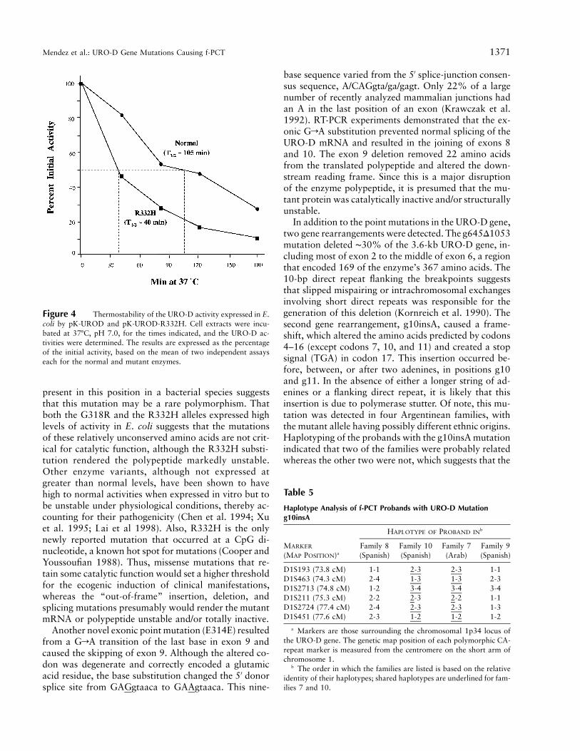
To further characterize the URO-D missense muta-tions, pK-UROD expression vectors for each of the mu-tant alleles were constructed and were expressed in E.coli, and the URO-D activities of the mutant proteinswere determined, as shown in table 4. The activity ofmutation M165R was !2% of the mean level expressedby the normal allele, whereas the N304K and L195Falleles expressed mutant polypeptides that had signifi-cant residual activity (19.5% and 32.1%, respectively,of the expressed normal mean activity). Interestingly, theR332H allele expressed more activity (127%) than thenormal allele. However, stability studies indicated thatthe R332H mutant polypeptide was significantly less sta-ble than the wild-type enzyme when incubated at 377C,pH 7.0 (fig. 4). Compared with the ∼105-min half-lifeof the normal enzyme, the half-life of R332H activitywas ∼40 min, which indicates that the mutant proteinhad a markedly reduced stability.
Characterization of Splice-Site Mutation E314E
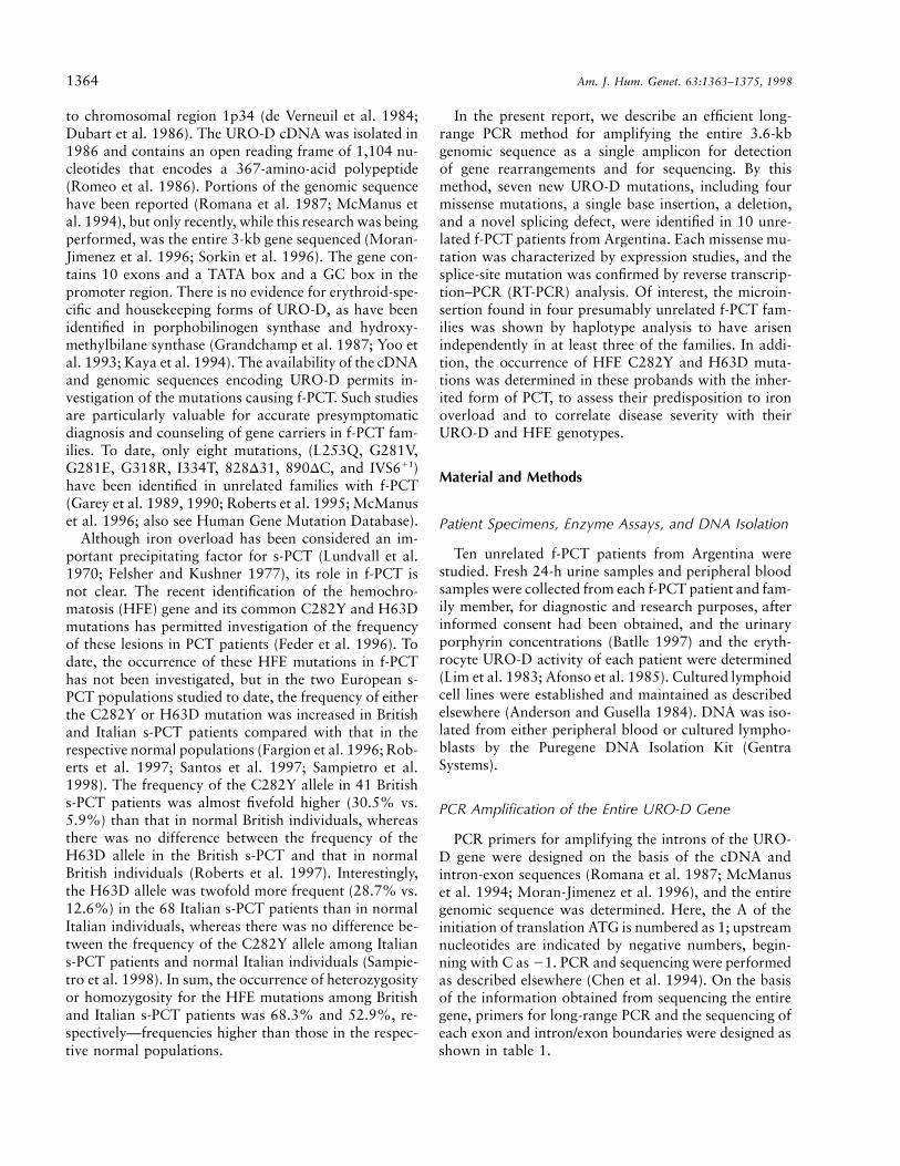
Sequencing of the URO-D gene in the proband offamily 1 identified a GrA transition in the last base ofexon 9 (change E314E; fig. 2A), which did not alter theamino acid sequence. To determine whether this exonic
Mendez et al.: URO-D Gene Mutations Causing f-PCT 1369
Figure 2 Characterization of splicing mutation E314E. A, Partial sequencing gel showing E314E due to a GrA transition at g2741, thelast base of exon 9. Also shown are (B) agarose-gel electrophoresis and (C) partial sequencing of the URO-D RT-PCR product amplified fromtotal lymphoblast RNA from a normal individual and from the f-PCT patient, which shows the deletion of exon 9.
mutation altered the splicing of intron 9, RT-PCR wasperformed on total mRNA from the leukocytes of theproband. Two PCR products were observed; one was ofnormal size, and the second was shorter by ∼60–70 bp(fig. 2B). Sequencing of the RT-PCR products revealedthat the normal-size product had the normal sequence,whereas the entire 67-bp exon 9 was deleted in theshorter amplicon and exon 8 was joined directly to exon10 (fig. 2C)
Haplotype Analysis of the g10insA Alleles
The g10insA microinsertion was identified in four pre-sumably unrelated PCT families. To determine whetherthese f-PCT families were related, haplotype analysis wasperformed with six polymorphic markers that mappedclosely to the URO-D gene at 1p34 (Dib et al. 1996).Although families 7 and 10 were of Arab and Spanishdescent, respectively, their probands had the same hap-lotype for all six markers (table 5), which indicates acommon ancestry of their g10insA mutation.
Discussion
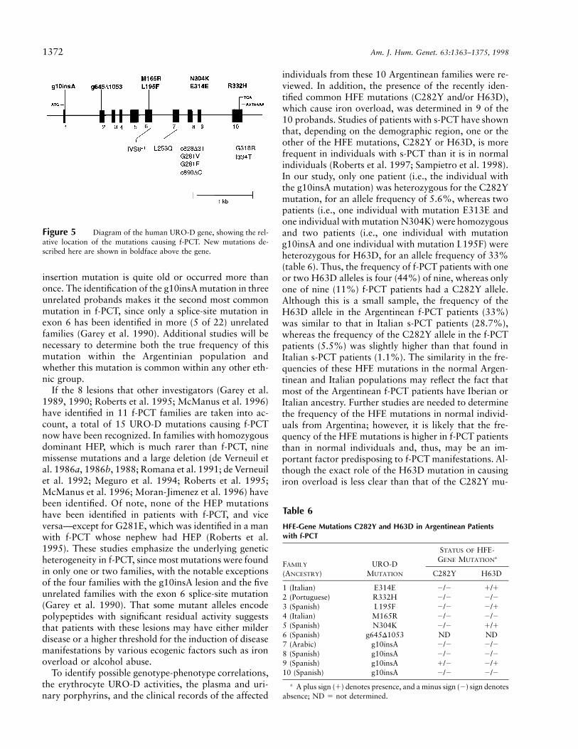
The identification and characterization of seven newmutations in the URO-D gene that cause f-PCT highlightthe molecular heterogeneity underlying this porphyria.These mutations were detected by amplification of theentire genomic sequence and by cycle sequencing of theexons and adjacent intron/exon boundaries in the 3.6-kb amplicon. Four missense mutations, a coding-regionsplicing mutation, the first large deletion, and the firstmicroinsertion causing f-PCT were identified. Figure 5shows both the newly identified and all previously pub-lished URO-D mutations that cause f-PCT. The fact thatthe seven mutations reported in the present study werefound in 10 f-PCT families from Argentina attests to theremarkable genetic heterogeneity underlying this inher-ited porphyria. That the g10insA mutation occurred infour Argentinean families suggests a possible commonorigin of the mutation. Indeed, haplotyping of poly-morphic markers flanking the URO-D gene indicate that
1370 Am. J. Hum. Genet. 63:1363–1375, 1998
Table 4
Expression of URO-D Mutations in E. coli
Construct
Mean URO-DActivitya [Range]
(U/mg)% of Mean
Normal Activity
pK223-3 .14 [.10–.18] 0pK-UROD 16.4 [9.79–24.7] 100pK-UROD-M165R .21 [.18–.23] 1.36pK-UROD-L195F 5.27 [2.60–10.3] 32.1pK-UROD-N304K 3.21 [1.61–5.07] 19.5pK-UROD-R332H 20.8 [13.7–27.5] 127.0
a Average of five to eight independent determinations.
Figure 3 A, Partial sequencing gels showing the adenine insertion, g10insA, in exon 2. B, Partial sequencing gel showing the deletion of1,053 bp, from g645, in exon 2, to g1698, in the middle of exon 6. Note that the deleted region was replaced by a 10-bp direct repeat of thestarred intron 1/exon 2 sequence.
the microinsertion in families 7 and 10, of Arab andSpanish descent, respectively, shared a common haplo-type, whereas families 8 and 9, both of Spanish ancestry,had different haplotypes (table 5). These findings suggesteither that the mutation is very old or, more likely, thatthe microinsertion arose independently at least threetimes.
Crystallographic studies have indicated that URO-Dis active as a dimeric protein (Whitby et al. 1998), sothat missense mutations can alter enzymatic function,subunit association, and/or stability of the dimer. Pro-karyotic expression of the four newly discovered mis-sense mutations (M165R, L195F, N304K, and R332H)revealed that each has a unique effect on enzymatic func-tion. The M165R allele, in which a highly conservedhydrophobic and neutral methionine is replaced by ahydrophilic and basic arginine, expressed little, if any,URO-D activity in E. coli, which indicates that the mu-tant polypeptide is inactive, unstable, or both. In con-trast, the other three missense mutations all expressedenzymatic activity in E. coli, which indicates that thesepolypeptides folded such that their active sites retainedat least partial catalytic function. The L195F allele, inwhich a hydrophobic and neutral leucine is substitutedby a hydrophobic, neutral, and aromatic phenylalanineresidue, had ∼30% of the mean activity expressed bythe normal allele. Although this leucine has been highlyconserved during evolution, it is notable that phenyl-alanine is in this position in tobacco and barley URO-D polypeptides (Swiss-Prot Annotated Protein SequenceDatabase trEMBL). Thus, it is not surprising that thehuman mutant protein retained significant activity when
expressed in E. coli but that this lesion also renderedthe mutant protein unstable (data not shown).
The N304K mutant allele, in which a neutral aspar-agine is replaced by a hydrophilic and basic lysine, ex-pressed an enzyme polypeptide that had ∼20% of nor-mal activity. This mutation occurred in the middle of ahighly conserved block of seven amino acids that pre-sumably is important for enzyme function and/or sta-bility. It is notable that, in E. coli, the R332H allele, inwhich a hydrophilic and basic arginine is substituted bya hydrophilic, basic, and aromatic histidine, expressedactivity that was 1.2-fold greater than normal. However,the R332H polypeptide was unstable at 377C (fig. 4),which explains its causative role in f-PCT. It is interestingto note that only one other URO-D mutation, G318R,had normal activity and stability when expressed in E.coli (McManus et al. 1996). However, the frequency ofthe G318R lesion in the normal population was notdetermined. The fact that the substituted arginine was
Mendez et al.: URO-D Gene Mutations Causing f-PCT 1371
Figure 4 Thermostability of the URO-D activity expressed in E.coli by pK-UROD and pK-UROD-R332H. Cell extracts were incu-bated at 377C, pH 7.0, for the times indicated, and the URO-D ac-tivities were determined. The results are expressed as the percentageof the initial activity, based on the mean of two independent assayseach for the normal and mutant enzymes.
Table 5
Haplotype Analysis of f-PCT Probands with URO-D Mutationg10insA
MARKER
(MAP POSITION)a
HAPLOTYPE OF PROBAND INb
Family 8(Spanish)
Family 10(Spanish)
Family 7(Arab)
Family 9(Spanish)
D1S193 (73.8 cM) 1-1 2-3 2-3 1-1D1S463 (74.3 cM) 2-4 1-3 1-3 2-3D1S2713 (74.8 cM) 1-2 3-4 3-4 3-4D1S211 (75.3 cM) 2-2 2-3 2-2 1-1D1S2724 (77.4 cM) 2-4 2-3 2-3 1-3D1S451 (77.6 cM) 2-3 1-2 1-2 1-2
a Markers are those surrounding the chromosomal 1p34 locus ofthe URO-D gene. The genetic map position of each polymorphic CA-repeat marker is measured from the centromere on the short arm ofchromosome 1.
b The order in which the families are listed is based on the relativeidentity of their haplotypes; shared haplotypes are underlined for fam-ilies 7 and 10.
present in this position in a bacterial species suggeststhat this mutation may be a rare polymorphism. Thatboth the G318R and the R332H alleles expressed highlevels of activity in E. coli suggests that the mutationsof these relatively unconserved amino acids are not crit-ical for catalytic function, although the R332H substi-tution rendered the polypeptide markedly unstable.Other enzyme variants, although not expressed atgreater than normal levels, have been shown to havehigh to normal activities when expressed in vitro but tobe unstable under physiological conditions, thereby ac-counting for their pathogenicity (Chen et al. 1994; Xuet al. 1995; Lai et al 1998). Also, R332H is the onlynewly reported mutation that occurred at a CpG di-nucleotide, a known hot spot for mutations (Cooper andYoussoufian 1988). Thus, missense mutations that re-tain some catalytic function would set a higher thresholdfor the ecogenic induction of clinical manifestations,whereas the “out-of-frame” insertion, deletion, andsplicing mutations presumably would render the mutantmRNA or polypeptide unstable and/or totally inactive.
Another novel exonic point mutation (E314E) resultedfrom a GrA transition of the last base in exon 9 andcaused the skipping of exon 9. Although the altered co-don was degenerate and correctly encoded a glutamicacid residue, the base substitution changed the 5′ donorsplice site from GAGgtaaca to GAAgtaaca. This nine-
base sequence varied from the 5′ splice-junction consen-sus sequence, A/CAGgta/ga/gagt. Only 22% of a largenumber of recently analyzed mammalian junctions hadan A in the last position of an exon (Krawczak et al.1992). RT-PCR experiments demonstrated that the ex-onic GrA substitution prevented normal splicing of theURO-D mRNA and resulted in the joining of exons 8and 10. The exon 9 deletion removed 22 amino acidsfrom the translated polypeptide and altered the down-stream reading frame. Since this is a major disruptionof the enzyme polypeptide, it is presumed that the mu-tant protein was catalytically inactive and/or structurallyunstable.
In addition to the point mutations in the URO-D gene,two gene rearrangements were detected. The g645D1053mutation deleted ∼30% of the 3.6-kb URO-D gene, in-cluding most of exon 2 to the middle of exon 6, a regionthat encoded 169 of the enzyme’s 367 amino acids. The10-bp direct repeat flanking the breakpoints suggeststhat slipped mispairing or intrachromosomal exchangesinvolving short direct repeats was responsible for thegeneration of this deletion (Kornreich et al. 1990). Thesecond gene rearrangement, g10insA, caused a frame-shift, which altered the amino acids predicted by codons4–16 (except codons 7, 10, and 11) and created a stopsignal (TGA) in codon 17. This insertion occurred be-fore, between, or after two adenines, in positions g10and g11. In the absence of either a longer string of ad-enines or a flanking direct repeat, it is likely that thisinsertion is due to polymerase stutter. Of note, this mu-tation was detected in four Argentinean families, withthe mutant allele having possibly different ethnic origins.Haplotyping of the probands with the g10insA mutationindicated that two of the families were probably relatedwhereas the other two were not, which suggests that the
1372 Am. J. Hum. Genet. 63:1363–1375, 1998
Figure 5 Diagram of the human URO-D gene, showing the rel-ative location of the mutations causing f-PCT. New mutations de-scribed here are shown in boldface above the gene.
Table 6
HFE-Gene Mutations C282Y and H63D in Argentinean Patientswith f-PCT
FAMILY
(ANCESTRY)URO-D
MUTATION
STATUS OF HFE-GENE MUTATIONa
C282Y H63D
1 (Italian) E314E 2/2 1/12 (Portuguese) R332H 2/2 2/23 (Spanish) L195F 2/2 2/14 (Italian) M165R 2/2 2/25 (Spanish) N304K 2/2 1/16 (Spanish) g645D1053 ND ND7 (Arabic) g10insA 2/2 2/28 (Spanish) g10insA 2/2 2/29 (Spanish) g10insA 1/2 2/110 (Spanish) g10insA 2/2 2/2
a A plus sign (1) denotes presence, and a minus sign (2) sign denotesabsence; ND 5 not determined.
insertion mutation is quite old or occurred more thanonce. The identification of the g10insA mutation in threeunrelated probands makes it the second most commonmutation in f-PCT, since only a splice-site mutation inexon 6 has been identified in more (5 of 22) unrelatedfamilies (Garey et al. 1990). Additional studies will benecessary to determine both the true frequency of thismutation within the Argentinian population andwhether this mutation is common within any other eth-nic group.
If the 8 lesions that other investigators (Garey et al.1989, 1990; Roberts et al. 1995; McManus et al. 1996)have identified in 11 f-PCT families are taken into ac-count, a total of 15 URO-D mutations causing f-PCTnow have been recognized. In families with homozygousdominant HEP, which is much rarer than f-PCT, ninemissense mutations and a large deletion (de Verneuil etal. 1986a, 1986b, 1988; Romana et al. 1991; de Verneuilet al. 1992; Meguro et al. 1994; Roberts et al. 1995;McManus et al. 1996; Moran-Jimenez et al. 1996) havebeen identified. Of note, none of the HEP mutationshave been identified in patients with f-PCT, and viceversa—except for G281E, which was identified in a manwith f-PCT whose nephew had HEP (Roberts et al.1995). These studies emphasize the underlying geneticheterogeneity in f-PCT, since most mutations were foundin only one or two families, with the notable exceptionsof the four families with the g10insA lesion and the fiveunrelated families with the exon 6 splice-site mutation(Garey et al. 1990). That some mutant alleles encodepolypeptides with significant residual activity suggeststhat patients with these lesions may have either milderdisease or a higher threshold for the induction of diseasemanifestations by various ecogenic factors such as ironoverload or alcohol abuse.
To identify possible genotype-phenotype correlations,the erythrocyte URO-D activities, the plasma and uri-nary porphyrins, and the clinical records of the affected
individuals from these 10 Argentinean families were re-viewed. In addition, the presence of the recently iden-tified common HFE mutations (C282Y and/or H63D),which cause iron overload, was determined in 9 of the10 probands. Studies of patients with s-PCT have shownthat, depending on the demographic region, one or theother of the HFE mutations, C282Y or H63D, is morefrequent in individuals with s-PCT than it is in normalindividuals (Roberts et al. 1997; Sampietro et al. 1998).In our study, only one patient (i.e., the individual withthe g10insA mutation) was heterozygous for the C282Ymutation, for an allele frequency of 5.6%, whereas twopatients (i.e., one individual with mutation E313E andone individual with mutation N304K) were homozygousand two patients (i.e., one individual with mutationg10insA and one individual with mutation L195F) wereheterozygous for H63D, for an allele frequency of 33%(table 6). Thus, the frequency of f-PCT patients with oneor two H63D alleles is four (44%) of nine, whereas onlyone of nine (11%) f-PCT patients had a C282Y allele.Although this is a small sample, the frequency of theH63D allele in the Argentinean f-PCT patients (33%)was similar to that in Italian s-PCT patients (28.7%),whereas the frequency of the C282Y allele in the f-PCTpatients (5.5%) was slightly higher than that found inItalian s-PCT patients (1.1%). The similarity in the fre-quencies of these HFE mutations in the normal Argen-tinean and Italian populations may reflect the fact thatmost of the Argentinean f-PCT patients have Iberian orItalian ancestry. Further studies are needed to determinethe frequency of the HFE mutations in normal individ-uals from Argentina; however, it is likely that the fre-quency of the HFE mutations is higher in f-PCT patientsthan in normal individuals and, thus, may be an im-portant factor predisposing to f-PCT manifestations. Al-though the exact role of the H63D mutation in causingiron overload is less clear than that of the C282Y mu-
Mendez et al.: URO-D Gene Mutations Causing f-PCT 1373
tation, the Italian study suggests that the H63D mutationmay cause a subtle iron-metabolism abnormality thatresults in hepatocellular accumulation of toxic iron spe-cies, thereby predisposing toward or precipitating theclinical manifestations of s-PCT (Sampietro et al. 1998).
It is interesting to note that the proband with thesplice-site mutation, who is homozygous for the H63Dmutation, had severe PCT clinical manifestations andthat these began at the age of 5 years (A. M. de C. Batlle,unpublished data). The other patient homozygous forH63D had a point mutation in her URO-D gene andhad milder PCT manifestations. However, overall, noclear genotype-phenotype correlations between the dif-ferent URO-D mutations, clinical symptoms, and pres-ence or absence of HFE mutations was observed in theseArgentinean patients, which suggests that other impor-tant factors contribute to the onset, frequency, and se-verity of the clinical manifestations in PCT. Moreover,once the diagnosis is established, the institution of ef-fective therapy by chronic phlebotomies and/or chlo-roquine obfuscates such correlations. In summary, theidentification and characterization of these seven newmutations in the URO-D gene that cause f-PCT highlightthe molecular heterogeneity underlying f-PCT, permitthe precise diagnosis of asymptomatic heterozygotes inthese Argentinean families, and provide the informationfor future structure-function studies of the humanenzyme.
Acknowledgments
We thank Mr. Raman Reddy for his expert technical assis-tance, and we thank Dr. H. Muramatsu, Mrs. B. Riccilo deAprea, and Lic. L. Dato for their valuable help with the pa-tients. We also thank Drs. Bruce Gelb and George Diaz fortheir advice on the haplotyping studies. This work was sup-ported, in part, by National Institutes of Health research grants5 R01 DK26824, 5 M01 RR00071 (for the Mount Sinai Gen-eral Clinical Research Center), and 5 P30 HD28822 (for theMount Sinai Child Health Research Center) (all to R.J.D.) and,in part, by Conselo Nacional de Investigaciones Cientificas yTecnicas (CONICET) grant 00509108/97, Science and Tech-nology Agency grant PMT-PICT002697, and University ofBuenos Aires grant EX032/95-97. A.B., M.V.R., and V.E.P. aresuperior, independent, and associate researchers, respectively,in the Career of Scientific Researcher program of the ArgentineNational Research Council (CONICET), and M.M. is a CON-ICET fellow. This work represents part of the doctoral thesissubmitted by M.M. to the University of Buenos Aires.
Electronic-Database Information
Accession numbers and URLs for data in this article are asfollows:
Entrez Nucleotide Sequence Search, http://www.ncbi.nlm.nih.gov/Entrez/nucleotide.html (for AF047383)
Human Gene Mutation Database, http://www.uwcm.ac.uk/uwcm/mg/hgmd0.html (for URO-D)
Online Mendelian Inheritance in Man (OMIM), http://www.ncbi.nim.nih.gov/Omim (for porphyria cutanea tarda[MIM 176100])
Swiss-Prot Annotated Protein Sequence Database trEMBL,http://www.expasy.ch/sprot/sprot-top.html (for tobacco andbarley URO-D polypeptides)
References
Afonso S, Chinarro S, Stella A, Batlle AM, Lenczner M, MaginP (1985) Uroporfirinogeno decarboxlasa eritocitaria y he-patica en porfiria cutanea tardia. Rev Arg Dermatol 66:12–24
Anderson MA, Gusella JF (1984) Use of cyclosporin A in es-tablishing Epstein-Barr virus-transformed human lympho-blastoid cell lines. In Vitro 20:856–858
Batlle A (1997) Porfirias y porfirinas-aspectos clinicos, bio-quimicos y biologia molecular. Fed Bioquim P BS AS (BuenosAires) Suppl 3:1–171
Chen CH, Astrin KH, Lee G, Anderson KE, Desnick RJ (1994)Acute intermittent porphyria: identification and expressionof exonic mutations in the hydroxymethylbilane synthasegene: an initiation codon missense mutation in the house-keeping transcript causes “variant acute intermittent por-phyria” with normal expression of the erythroid-specificenzyme. J Clin Invest 94:1927–1937
Cooper DN, Youssoufian H (1988) The CpG dinucleotide andhuman genetic disease. Hum Genet 78:151–155
DeCastro M, Sanchez J, Herrera JF, Chaves A, Duran R, Gar-cia-Buey L, Garcia-Monzon C, et al (1993) Hepatitis C virusantibodies and liver disease in patients with porphyria cu-tanea tarda. Hepatology 17:551–557
de Verneuil H, Aitken G, Nordmann Y (1978) Familial andsporadic porphyria cutanea: two different diseases. HumGenet 44:145–151
de Verneuil H, Bourgeois F, de Rooij F, Siersema PD, WilsonJH, Grandchamp B, Nordmann Y (1992) Characterizationof a new mutation (R292G) and a deletion at the humanuroporphyrinogen decarboxylase locus in two patients withhepatoerythropoietic porphyria. Hum Genet 89:548–552
de Verneuil H, Grandchamp B, Beaumont C, Picat C, Nord-mann Y (1986a) Uroporphyrinogen decarboxylase struc-tural mutant (Gly281rGlu) in a case of porphyria. Science234:732–734
de Verneuil H, Grandchamp B, Foubert C, Weil D, N’GuyenVC, Gross MS, Sassa S, et al (1984) Assignment of the genefor uroporphyrinogen decarboxylase to human chromosome1 by somatic cell hybridization and specific enzyme immu-noassay. Hum Genet 66:202–205
de Verneuil H, Grandchamp B, Nordmann Y (1980) Somekinetic properties of human red cell uroporphyrinogen de-carboxylase. Biochim Biophys Acta 611:174–186
de Verneuil H, Grandchamp B, Romeo PH, Raich N, Beau-mont C, Goossens M, Nicolas H, et al (1986b) Molecularanalysis of uroporphyrinogen decarboxylase deficiency in afamily with two cases of hepatoerythropoietic porphyria. JClin Invest 77:431–435
de Verneuil H, Hansen J, Picat C, Grandchamp B, Kushner J,Roberts A, EIder G, et al (1988) Prevalence of the 281
1374 Am. J. Hum. Genet. 63:1363–1375, 1998
(GlyrGlu) mutation in hepatoerythropoietic porphyria andporphyria cutanea tarda. Hum Genet 78:101–102
de Verneuil H, Sassa S, Kappas A (1983) Purification and prop-erties of uroporphyrinogen decarboxylase from humanerythrocytes: a single enzyme catalyzing the four sequentialdecarboxylations of uroporphyrinogens I and III. J BiolChem 258:2454–2460
Dib C, Faure S, Fizames C, Samson D, Drouot N, Vignal A,Millasseau P, et al (1996) A comprehensive genetic map ofthe human genome based on 5,264 microsatellites. Nature380:152–154
Dubart A, Mattei MG, Raich N, Beaupain D, Romeo PH,Mattei JF, Goossens M (1986) Assignment of human uro-porphyrinogen decarboxylase (URO-D) to the p34 band ofchromosome 1. Hum Genet 73:277–279
Elder GH (1998) Porphyria cutanea tarda. Semin Liver Dis18:67–75
Elder GH, Lee GB, Tovey JA (1978) Decreased activity ofhepatic uroporphyrinogen decarboxylase in sporadic por-phyria cutanea tarda. N Engl J Med 299:274–278
Fargion S, Fracanzani AL, Romano R, Cappellini MD, FareM, Mattioli M, Piperno A, et al (1996) Genetic hemochro-matosis in Italian patients with porphyria cutanea tarda:possible explanation for iron overload. J Hepatol 24:564–569
Fargion S, Piperno A, Cappellini MD, Sampietro M, Fracan-zani AL, Romano R, Caldarelli R, et al (1992) Hepatitis Cvirus and porphyria cutanea tarda: evidence of a strong as-sociation. Hepatology 16:1322–1326
Feder JN, Gnirke A, Thomas W, Tsuchihashi Z, Ruddy DA,Basava A, Dormishian F, et al (1996) A novel MHC classI–like gene is mutated in patients with hereditary haemo-chromatosis. Nat Genet 13:399–408
Felsher BF, Kushner JP (1977) Hepatic siderosis and porphyriacutanea tarda: relation of iron excess to the metabolic defect.Semin Hematol 14:243–251
Garey JR, Franklin KF, Brown DA, Harrison LM, Metcalf KM,Kushner JP (1993) Analysis of uroporphyrinogen decarbox-ylase complementary DNAs in sporadic porphyria cutaneatarda. Gastroenterology 105:165–169
Garey JR, Hansen JL, Harrison LM, Kennedy JB, Kushner JP(1989) A point mutation in the coding region of uropor-phyrinogen decarboxylase associated with familial por-phyria cutanea tarda. Blood 73:892–895
Garey JR, Harrison LM, Franklin KF, Metcalf KM, RadiskyES, Kushner JP (1990) Uroporphyrinogen decarboxylase: asplice site mutation causes the deletion of exon 6 in multiplefamilies with porphyria cutanea tarda. J Clin Invest 86:1416–1422
Gelb BD, Edelson JG, Desnick RJ (1995) Linkage of pycnodys-ostosis to chromosome 1q21 by homozygosity mapping. NatGenet 10:235–237
Grandchamp B, de Verneuil H, Beaumont C, Chretien S, Wal-ter O, Nordmann Y (1987) Tissue-specific expression of por-phobilinogen deaminase: two isoenzymes from a single gene.Eur J Biochem 162:105–110
Grossman ME, Bickers DR, Poh-Fitzpatrick MB, Deleo VA,Harber LC (1979) Porphyria cutanea tarda: clinical featuresand laboratory findings in 40 patients. Am J Med 67:277–286
Jackson AH, Sancovich HA, Ferramola AM, Evans N, GamesDE, Matlin SA, Elder GH, et al (1976) Macrocyclic inter-mediates in the biosynthesis of porphyrins. Philos Trans RSoc Lond [Biol] 273:191–206
Kappas A, Sassa S, Galbraith RA, Nordmann Y (1995) Theporphyrias. In: Scriver CR, Beaudet AL, Sly WS, Valle D(eds) Metabolic and molecular bases of inherited disease.McGraw-Hill, New York, pp 2103–2160
Kaya AH, Plewinska M, Wong DM, Desnick RJ, Wetmur JG(1994) Human delta-aminolevulinate dehydratase (ALAD)gene: structure and alternative splicing of the erythroid andhousekeeping mRNAs. Genomics 19:242–248
Kornreich R, Bishop DF, Desnick RJ (1990) Alpha-galactosi-dase A gene rearrangements causing Fabry disease: identi-fication of short direct repeats at breakpoints in an Alu-richgene. J Biol Chem 265:9319–9326
Koszo F, Elder GH, Roberts A, Simon N (1990) Uroporphyrin-ogen decarboxylase deficiency in hepatoerythropoietic por-phyria: further evidence for genetic heterogeneity. Br J Der-matol 122:365–370
Krawczak M, Reiss J, Cooper DN (1992) The mutational spec-trum of single base-pair substitutions in mRNA splice junc-tions of human genes: causes and consequences. Hum Genet90:41–54
Kushner JP, Barbuto AJ, Lee GR (1976) An inherited enzymaticdefect in porphyria cutanea tarda: decreased uroporphyrino-gen decarboxylase activity. J Clin Invest 58:1089–1097
Lai K, Langley SD, Dembure PP, Hjelm LN, Elsas LJ (1998)Duarte allele impairs biostability of galactose-1-phosphateuridyltransferase in human lymphoblasts. Hum Mutat 11:28–38
Lim CK, Rideout JM, Wright DJ (1983) Separation of por-phyrin isomers by high-performance liquid chromatography.Biochem J 211:435–438
Lundvall O, Weinfeld A, Lundin P (1970) Iron storage in por-phyria cutanea tarda. Acta Med Scand 1–2:37–53
McGovern MM, Anderson KE, Astrin KH, Desnick RJ (1996)Inherited porphyrias. In: Rimoin DL, Connor JM, PyeritzRE (eds) Emery and Rimoin’s principles and practice of med-ical genetics. Churchill Livingston, New York, pp2009–2037
McManus JF, Begley CG, Ratnaike S (1994) Complex patternof alternative splicing in the normal uroporphyrinogen de-carboxylase gene: implications for diagnosis of familial por-phyria cutanea tarda. Clin Chem 40:1884–1889
McManus JF, Begley CG, Sassa S, Ratnaike S (1996) Five newmutations in the uroporphyrinogen decarboxylase geneidentified in families with cutaneous porphyria. Blood 88:3589–3600
Meguro K, Fujita H, Ishida N, Akagi R, Kurihara T, GalbraithRA, Kappas A, et al (1994) Molecular defects of uroporphy-rinogen decarboxylase in a patient with mild hepatoery-thropoietic porphyria. J Invest Dermatol 102:681–685
Moran-Jimenez MJ, Ged C, Romana M, Enriquez De Sala-manca R, Taeb A, Topi G, D’Alessandro L, et al (1996)Uroporphyrinogen decarboxylase: complete human gene se-quence and molecular study of three families with hepa-toerythropoietic porphyria. Am J Hum Genet 58:712–721
Roberts AG, Elder GH, De Salamanca RE, Herrero C, LechaM, Mascaro JM (1995) A mutation (G281E) of the human
Mendez et al.: URO-D Gene Mutations Causing f-PCT 1375
uroporphyrinogen decarboxylase gene causes both hepa-toerythropoietic porphyria and overt familial porphyria cu-tanea tarda: biochemical and genetic studies on Spanish pa-tients. J Invest Dermatol 104:500–502
Roberts AG, Whatley SD, Morgan RR, Worwood M, ElderGH (1997) Increased frequency of the haemochromatosisCys282Tyr mutation in sporadic porphyria cutanea tarda.Lancet 349:321–323
Romana M, Dubart A, Beaupain D, Chabret C, Goossens M,Romeo PH (1987) Structure of the gene for human uropor-phyrinogen decarboxylase. Nucleic Acids Res 15:7343–7356
Romana M, Grandchamp B, Dubart A, Amselem S, ChabretC, Nordmann Y, Goossens M, et al (1991) Identification ofa new mutation responsible for hepatoerythropoietic por-phyria. Eur J Clin Invest 21:225–229
Romeo PH, Raich N, Dubart A, Beaupain D, Pryor M, Kush-ner J, Cohen-Solal M, et al (1986) Molecular cloning andnucleotide sequence of a complete human uroporphyrinogendecarboxylase cDNA. J Biol Chem 261:9825–9831
Sampietro M, Piperno A, Lupica L, Arosio C, Vergani A, Cor-betta N, Malosio I, et al (1998) High prevalence of theHis63Asp HFE mutation in Italian patients with porphyriacutanea tarda. Hepatology 27:181–184
Santos M, Clevers HC, Marx JJ (1997) Mutations of the he-reditary hemochromatosis candidate gene HLA-H in por-phyria cutanea tarda. N Engl J Med 336:1327–1328
Smith S (1986) Hepatoerythropoietic porphyria. Semin Der-matol 5:125–137
Sorkin L, Mendez M, Rossetti MV, Wu Y, Astrin KH, BatlleA, Perara V, et al (1996) Identification of six new mutationsin the uroporphyrinogen decarboxylase gene causing famil-ial porphyria cutanea tarda. Am J Hum Genet Suppl 59:A285
Tsai SF, Bishop DF, Desnick RJ (1988) Human uroporphyrino-gen III synthase: molecular cloning, nucleotide sequence, andexpression of a full-length cDNA. Proc Natl Acad Sci USA85:7049–7053
Warner CA, Yoo HW, Roberts AG, Desnick RJ (1992) Con-genital erythropoietic porphyria: identification and expres-sion of exonic mutations in the uroporphyrinogen III syn-thase gene. J Clin Invest 89:693–700
Whitby FG, Phillips JD, Kushner JP, Hill CP (1998) Crystalstructure of human uroporphyrinogen decarboxylase.EMBO J 17:2463–2471
Xu W, Warner CA, Desnick RJ (1995) Congenital erythro-poietic porphyria: identification and expression of 10 mu-tations in the uroporphyrinogen III synthase gene. J ClinInvest 95:905–912
Yoo HW, Warner CA, Chen CH, Desnick RJ (1993) Hydrox-ymethylbilane synthase: complete genomic sequence andamplifiable polymorphisms in the human gene. Genomics15:21–29


