Embed Size (px)
Citation preview

Affective startle modulation Psychopharmacological studies on the roles of CRF and serotonin
in the regulation of emotions

CIP-gegevens Koninklijke Bibliotheek, Den Haag Bijlsma, EY. Affective startle modulation: Psychopharmacological studies on the roles of CRF and serotonin in the regulation of emotions Thesis Utrecht University – with ref. – with summary in Dutch ISBN: 978-90-5335-344-8 © 2010 Elisabeth Y Bijlsma. For published articles the copyright has been transferred to the respective publisher. No part of this thesis may be reproduced, stored in a retrieval system, or transmitted in any form or by any means without permission of the author, or when appropriate, the published of the manuscript. Cover art: Wintersonne bei Maloja by Giovanni Giacometti Cover design Ridderprint, Ridderkerk Printed by Ridderprint, Ridderkerk

Affective startle modulation Psychopharmacological studies on the roles of CRF and serotonin
in the regulation of emotions
Affectieve startle modulatie Psychofarmacologische studies naar de rol van CRF en serotonine in de regulatie van emoties (met een samenvatting in het Nederlands)
Proefschrift
ter verkrijging van de graad van doctor aan de Universiteit Utrecht op gezag van de rector magnificus, prof.dr. J.C. Stoof, ingevolge het besluit van
het college voor promoties in het openbaar te verdedigen op woensdag 15 december 2010 des middags te 2.30 uur
door
Elisabeth Yvonne Bijlsma
geboren op 8 februari 1982 te Drachten

Promotor: Prof. dr. B. Olivier Co-promotor: Dr. L. Groenink

Voor mijn ouders


Outline Chapter 1 General introduction 9
Chapter 2 Fear potentiated startle and light-enhanced startle 25 models in drug discovery
Part 1: The influence of affective state on affective startle modulation Chapter 3 Fear-potentiated startle, but not light-enhanced startle, is 45
enhanced by anxiogenic drugs
Chapter 4 Disrupted startle modulation in animal models for 63 affective disorders
Chapter 5 Cocaine-induced changes in affective state modulate 81 the light-enhanced startle response
Part 2: The roles of CRF and serotonin in affect regulation Chapter 6 Local repeated CRF infusion exacerbates anxiety- and fear- 91
related behaviour: Differential involvement of the basolateral amygdala and medial prefrontal cortex
Chapter 7 Acute and chronic paroxetine affects neither fear acquisition 107 nor expression of cued fear and sustained anxiety
Chapter 8 Deletion of the serotonin transporter disrupts fear 121 acquisition and exacerbates contextual conditioned fear that can be normalized by CRF1 receptor blockade.
Chapter 9 General discussion 141
References 157
Samenvatting in het Nederlands 179
List of publications 189
Acknowledgements 193
About the author 199


1
General introduction
9

Chapter 1
10
General introduction
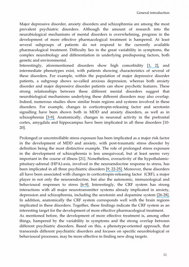
Major depressive disorder, anxiety disorders and schizophrenia are among the most prevalent psychiatric disorders. Although the amount of research into the neurobiological mechanisms of mental disorders is overwhelming, progress in the development of more effective pharmacological treatment is hampered. At least several subgroups of patients do not respond to the currently available pharmacological treatment. Difficulty lies in the great variability in symptoms, the complex neurobiology and differentiation in underlying predisposing factors, both genetic and environmental. Interestingly, aforementioned disorders show high comorbidity [1, 2], and intermediate phenotypes exist, with patients showing characteristics of several of these disorders. For example, within the population of major depressive disorder patients, a subgroup shows so-called anxious depression, whereas both anxiety disorder and major depressive disorder patients can show psychotic features. These strong relationships between these different mental disorders suggest that neurobiological mechanisms underlying these different disorders may also overlap. Indeed, numerous studies show similar brain regions and systems involved in these disorders. For example, changes in corticotropin-releasing factor and serotonin signalling have been shown both in MDD and anxiety disorders, as well as in schizophrenia [3-9]. Anatomically, changes in neuronal activity in the prefrontal cortex, amygdala and hippocampus have been implicated in all these disorders [10-20]. Prolonged or uncontrollable stress exposure has been implicated as a major risk factor in the development of MDD and anxiety, with post-traumatic stress disorder by definition being the most distinctive example. The role of prolonged stress exposure in the development of schizophrenia is less unequivocal, but at least seems very important in the course of illness [21]. Nonetheless, overactivity of the hypothalamic-pituitary-adrenal (HPA)-axis, involved in the neuroendocrine response to stress, has been implicated in all three psychiatric disorders [9, 22-25]. Moreover, these disorders all have been associated with changes in corticotropin-releasing factor (CRF), a major player in not only the neuroendocrine, but also the autonomic, immunological and behavioural responses to stress [6-9]. Interestingly, the CRF system has strong interactions with all major neurotransmitter systems already implicated in anxiety, depression and schizophrenia, including the serotonin and dopamine system [26-28]. In addition, anatomically the CRF system corresponds well with the brain regions implicated in these disorders. Together, these findings indicate the CRF system as an interesting target for the development of more effective pharmacological treatment. As mentioned before, the development of more effective treatment is, among other things, hampered by the variability in symptoms and the strong overlap between different psychiatric disorders. Based on this, a phenotype-oriented approach, that transcends different psychiatric disorders and focuses on specific neurobiological or behavioural processes, may be more effective in finding new drug targets.
11

Chapter 1
One characteristic that seems to be evident both in anxiety disorders and major depression, as well as in schizophrenia, is disturbed affect regulation [29-31]. Affect regulation refers to the process that mediates the activation of appropriate behavioural responses to emotionally relevant stimuli. Affect regulation Behaviour is driven by the motivation to avoid harmful situations and seek pleasant situations. This results in a balance between avoidance behaviour, strengthened by punishment and fear, and approach behaviour, strengthened by reward (For review, see e.g. [32]). Affect refers to the experience of the emotionally relevant stimuli that induce these fundamental processes of avoidance and approach behaviour. As mentioned above, changes in affect regulation are a common feature in several psychiatric disorders, including major depression, anxiety disorders and schizophrenia. To understand how emotional brain systems are affected in these disorders and establish whether similar behavioural changes in these disorders also result from similar underlying predisposing factors, understanding of normal functioning of the emotional brain is necessary. The regulation of the complex behavioural responses is primarily mediated by the limbic system, where emotional relevant stimuli are processed and motivational information is sent to higher brain systems. The limbic system is subdivided in at least two subsystems: 1. The limbic forebrain, including the prefrontal and cingulate cortices; the amygdala; the extended amygdala (bed nucleus of the stria terminalis) and the hippocampus. 2. The limbic midbrain, including the limbic thalamus (medial dorsal, anterior and lateral dorsal nuclei); the nucleus accumbens; the anterior hypothamalus; the ventral tegmental area and the raphe nuclei [33, 34]. Within these subsystems, responses to appetitive and aversive emotional stimuli are, at least partly, regulated by different pathways. Behavioural responses to appetitive stimuli are highly dependent on the brain reward system, including the ventral tegmental area (VTA), nucleus accumbens and prefrontal cortex [34, 35]. For example, the prefrontal cortex and nucleus accumbens are activated in response to pleasurable pictures [36, 37]. Furthermore, the nucleus accumbens has been described as an integration site for both cortical and subcortical information and seems to have a primary role in motivational circuitry by translating motivation into goal-directed behaviour [34]. Especially dopaminergic neurons within the nucleus accumbens seem to be involved in guiding behaviour to positive incentives and, during conditioned reward, mediate the incentive salience of previously neutral events [38]. Although the amygdala has primarily been implicated in the processing of aversive stimuli (see below), it is also involved in conditioned reward and reward-related arousal [39, 40]. Projections from both the amygdala and the prefrontal cortex to the nucleus accumbens are involved in the processing of rewarding stimuli and in the execution of appropriate behavioural responses [41-43].
12

General introduction
The processing of, and behavioural responses to, aversive stimuli depends strongly on several subnuclei of the amygdala, including the central amygdala and basolateral amygdala, but also on the extended amygdala (bed nucleus of the stria terminalis, BNST), the hippocampus and prefrontal cortex [35, 40, 44, 45]. In general, the basolateral amygdala is proposed to be the major input site of the amygdala, which receives sensory information from cortical areas and forms associations between specific sensory information and biologically relevant events [46]. The basolateral amygdala has direct projections to the BNST and central amygdala, of which the latter one appears to be the major output site of the amygdala. The prefrontal cortex contains reciprocal connections with the amygdala and modulates amygdala activity [47-49]. Functional MRI and PET imaging studies in humans show that the amygdala is activated in response to various fear-related stimuli, including fearful faces and aversive pictures [36, 50, 51]. In addition, lesions of the human amygdala are accompanied by disturbances in the recognition of fearful faces and fear conditioning [52]. The specific neuronal pathway involved depends, however, on the nature of the aversive stimulus. For example, differences exist in the processing of stimuli that are of conditioned versus unconditioned nature and of stimuli that reflect immediate versus potential danger [53-56]. For example, the central amygdala has been specifically implicated in behavioural responses to conditioned aversive cues, whereas the BNST appears to be involved in responses to aversive contexts (both conditioned and unconditioned) [53]. On the other hand, the basolateral amygdala, as major input site, does appear to be more generally involved in the processing of aversive stimuli (e.g. [53]). Affect regulation in psychiatric disorders As mentioned earlier, changes in affect regulation are a common feature in several psychiatric disorders, including major depression, anxiety disorders and schizophrenia. Emotional stimuli induce either a defensive or appetitive emotional state. Disturbances in affect regulation can therefore also be subdivided in these two modalities. Disturbed processing and/or experience of appetitive stimuli (i.e. anhedonia), is one of the core symptoms or major depression, but is also present in schizophrenia and PTSD patients [57, 58]. In addition, depressive and psychotic disorders have also been associated with blunted responses to aversive stimuli, resembling a form of emotional numbing [31, 59], although a bias towards identification of emotional information as negative has also been reported [31, 60]. On the other hand, anxiety disorders have been associated with exaggerated responding to aversive (e.g. fearful) stimuli. Maladaptive fear responses, as seen in anxiety disorders, can be reflected either in an exaggerated and/or prolonged response to aversive stimuli or in fear-like responses to stimuli or contexts that do not predict harmful outcome [19, 20, 61].
13

Chapter 1
Anatomical and physiological changes In psychiatric patients, various structural changes have been observed in the limbic structures regulating affect. For example, depression has been associated with decreased volume of the hippocampus, amygdala and nucleus accumbens and decreased density and number of glial cells in the amygdala, anterior cingulate cortex, orbitofrontal cortex and dorsolateral PFC [62-68]. Also in schizophrenia patients, changes in neuronal cell integrity and volume reductions have been reported for the hippocampus, amygdala and frontal cortices [69-74]. Anxiety disorders have mainly been associated with increased amygdala volume (see e.g. [75, 76]). In addition to these structural changes, functional alterations have also been reported for these brain regions. For example, various studies show increased activity in the amygdala, PFC and ACC in depressed patients, both under basal conditions and in response to emotional stimuli [10-13], although decreased PFC activation has also been reported [13]. For schizophrenia, it has been reported that patients fail to show increased activity of the nucleus accumbens and parahippocampal gyrus in response to unpleasant odours [77] and also do not show activation of the amygdala in response to fearful facial expressions [78], aversive scenes [79] and during sad mood induction [15]. Although the spectrum of anxiety disorders encompasses a set of very different disorders, they do show some overlap in functional changes. PTSD, social anxiety and specific phobia are all associated with increased amygdala activity [16]. Moreover, social phobia patients show increased amygdala activation in response to angry faces [17]. Patients with PTSD exhibit altered neural responses in both the amygdala and mPFC in response to aversive trauma-unrelated stimuli [18]. Interestingly, anxious subjects were also reported to show increased amygdala activation in response to neutral faces, which can be characterized as affectively ambiguous [19, 20]. The level of activation in response to neutral faces was positively correlated with state anxiety and, therefore, it has been hypothesized that anxious patients may infer greater threat from the ambiguous expression [19, 20, 76]. This finding further underlines the above-mentioned hypothesis that anxiety disorders entail fear-like responses to stimuli that are not harmful. Measuring affect In humans, various methods are available to objectively study emotional responses to external stimuli, independent of subjective ratings of the experience of emotional states. Regional changes in brain activity in response to emotional stimuli can be detected with functional MRI and PET imaging, but also with EEG measurements (Event Related Potentials). In addition, autonomic responses can be analyzed using measures like heart rate acceleration and skin conductance. These measures are all affected by emotional valent stimuli and some are also differently modulated by appetitive and aversive stimuli, which enables us to study specific changes in the processing of either aversive or appetitive stimuli, or both [80, 81]. However, in some
14

General introduction
of these measures the level of arousal strongly interferes with measuring the level of positive or negative affect induced by emotional stimuli [80]. In addition, these measures are hard to measure in rodents, which limits the translational value of these methods. One measure that can be used for studying the regulation of affect and does show high translational value is the acoustic startle reflex. Affective startle modulation The acoustic startle reflex is a fast defensive response to an unexpected and intense acoustic stimulus, which includes eye-lid closure and contraction of facial, neck and skeletal muscles [82]. The acoustic startle reflex has been proposed to serve as a protection against harmful stimuli and to prepare for a fight/flight response. This reflex can be established in various species, but is especially well characterized in humans and rodents. The neural pathway mediating the startle reflex consists of the auditory nerve; the ventral cochlear nucleus; the dorsal nucleus of the lateral lemniscus; the caudal pontine reticular nucleus; spinal interneurons and spinal motor neurons [83]. The startle reflex has a non-zero baseline, which can be both enhanced and attenuated by additional (emotional) manipulations. In addition, this reflex shows a high level of plasticity. These aspects make the startle reflex a very useful tool in studying various processes underlying behavioural responses to environmental stimuli. First, the acoustic startle response can be used to study affect regulation, as it can be modulated by emotional stimuli, but also by internal emotional states [84]. For example, positive emotional states elicited by e.g. viewing a funny film clip or pleasant pictures, decrease the acoustic startle reflex. On the other hand, negative emotional states, induced by e.g. aversive picture or stimuli previously associated with a negative experience, potentiate the acoustic startle reflex [85-88]. The affective state induced by emotional stimuli is thought to modulate startle responding via the so-called priming principle [89]. Emotional stimuli induce either a defensive or appetitive emotional state. The direction of startle modulation depends on the specific motivational pathway activated. As a consequence, disturbances in affective startle modulation could result from deficits in the circuits that assign emotional valence to emotional stimuli or in either one of the specific motivational pathways. Second, the startle reflex can be used to study sensorimotor gating, a filtering process needed to direct attention to relevant stimuli [90]. This so-called prepulse inhibition of the startle reflex is defined as the reduction in startle reflex magnitude when a startling stimulus is preceded by a weak pre-stimulus. Prepulse inhibition is a measure of the early pre-attentive stages of information processing and is used as an operational measure of sensorimotor gating. Prepulse inhibition is disrupted in patients with various psychiatric disorders, especially those with psychotic features [91-93].
15

Chapter 1
Affective startle modulation in humans Several forms of affective startle modulation can be distinguished, depending on the type of emotional stimulus used. In humans, unconditioned forms of affective startle modulation can be induced by the presentation of appetitive or aversive pictures or film clips [86, 89, 94]. In addition, several methods have been introduced to specifically study fear- and anxiety-related responses, of which the fear-potentiated startle paradigm is especially well described. Fear-potentiated startle is induced by a conditioned stimulus, previously associated with exposure to an aversive event (e.g. receiving an electrical shock). This paradigm is proposed to model conditioned fear [95, 96]. Additional forms of affective startle modulation measuring anxiety- and fear-related processes are dark-enhanced startle, contextual potentiated startle and startle sensitization. Dark-enhanced startle is based on the comprehension that human beings are diurnal organisms and feel less safe in a dark environment [97]. Contextual potentiated startle results from a conditioning procedure where an aversive stimulus is associated with a certain environment [98, 99]. Startle sensitization is related to this contextual potentiated startle response. Exposure to electrical shocks results in a rapid increase in startle reactivity. It is proposed to involve rapid contextual fear conditioning, reflecting the development of contextual fear to the environment where the shocks were presented [87, 100, 101]. Affective startle modulation in psychiatric patients Several clinical studies already reported on changes in affective startle modulation in psychiatric patients. Depressed patients have been reported to show a blunted or potentiated, instead of attenuated, response to pleasant stimuli and a blunted response to aversive stimuli [102-104]. The level of disruption of the affective startle response has been correlated to the severity of depression, the number of depressive episodes and the level of anhedonia [103, 105, 106]. Blunted responding to aversive stimuli is, however, not limited to depressed patients. Failure to show potentiation of the startle reflex in response to aversive stimuli has also been associated a family history of alcoholism [107], and has been reported in psychopaths [108], Parkinson’s patients and a subgroup of attention-deficit-hyperactivity-disorder (ADHD) patients [109]. Anxious traits, on the other hand, have been associated with an overall increase in startle responding, independent of stimulus valence [61, 103, 110, 111]. Post-traumatic stress disorder (PTSD), for example, has been associated with overall increased startle amplitude, but shows normal affective startle modulation, including fear-potentiated startle and dark-enhanced startle [111, 112]. Similarly, panic disorder patients show increased overall startle reactivity [113]. And, despite clear evidence of high anxiety, they fail to show clear potentiation of the startle reflex in response to fearful faces or conditioned cues [114, 115]. In addition, they show increased contextual fear, which suggests generalization of fear responses to other irrelevant neutral environmental cues. A similar form of fear generalization has also been reported in PTSD patients [116, 117]. Thus, in general, anxiety is associated with overall increases in startle
16

General introduction
responding, in the absence of increased responding to discrete aversive stimuli, and generalization of fear responses, On the other hand, depression is associated with blunted startle responding to emotional stimuli. Only a few studies exist on affective startle modulation in schizophrenia patients. Despite the changes in neuronal activation in response to emotional stimuli as discussed above, schizophrenia patients show normal affective startle modulation, despite clear signs of affective flattening in these patients [118, 119]. However, schizophrenia patients did show a delay in startle potentiation [119]. It was suggested that it takes longer for schizophrenia patients to process aversive stimuli, resulting in delayed output of the defence system [119]. Affective startle modulation in rodents Several paradigms have been developed to evaluate affective modulation of the startle reflex in rodents. Paradigms to study the response to aversive emotional stimuli have been well characterized. In 1951, the fear-potentiated startle (FPS) paradigm was introduced, which measures a conditioned aversive reaction to a cue previously paired with mild foot shock [85]. As humans, rodents show a potentiated response to a startle stimulus, when it is presented together with the conditioned cue. More recently, the light-enhanced startle (LES) paradigm was introduced, which is dependent on the natural aversion of nocturnal rodents for brightly lit environments (compare to humans: startle potentiation in dark versus light). Both paradigms are thought to represent certain forms of fear or anxiety and have been validated with various anxiolytic compounds and, in this respect, show a very similar pharmacological profile [95, 120-122]. However, FPS and LES are emotionally and neurobiologically distinct processes. LES is a long lasting process (minutes), which is accompanied by a slow return to baseline startle level after the light is turned off. On the other hand, FPS is a fast response to the conditioned cue (milliseconds) and startle level shows a fast return to baseline [123]. In addition, clear neuroanatomical differences exist between both measures. Whereas FPS specifically depends on the central amygdala [124, 125], LES depends on the bed nucleus of the stria terminalis [53]. Additional areas have been implicated in the regulation of FPS, including the basolateral amygdala, hippocampus and dorsal raphe nucleus [126-129]. Next to its role in fear-potentiated startle, the BLA is also important in the establishment of LES [53]. Additional brain regions implicated in LES are the anterior cingulate cortex, lateral septum and medial amygdala [130, 131]. Additional measures of affective startle modulation in response to aversive stimuli are contextual potentiated startle and foot shock sensitization, both measuring aspects of contextual conditioned fear [87, 101, 132, 133]. Interestingly, in line with aforementioned findings of disrupted affective startle modulation in depressed patients, a recent study showed blunted light-enhanced startle in female rats subjected to early maternal deprivation, a paradigm proposed to model early life adversity [134]. Next to paradigms measuring emotional responses to aversive stimuli, some attempts have been made to develop startle paradigms measuring the emotional response to
17

Chapter 1
pleasant stimuli, the so-called pleasure-attenuated startle paradigm [135-138]. In a paradigm developed by Schmid and Koch, rats were conditioned to associate a house light with the availability of palatable food [135, 136]. This process was shown to depend on the nucleus accumbens, but not amygdala [135]. In a recent attempt, Schneider et al. established pleasure-attenuated startle by learning rats to associate orange odour with the availability of sweet milk [138]. This process appeared to be dependent on opioid signalling. However, reports and follow up studies on the pleasure-attenuated startle paradigm are scarce and various attempts of other research groups failed to induce pleasure-attenuated startle in rodents (Bijlsma et al, unpublished data, [139]). Therefore, rodent studies on affective modulation of the startle reflex have primarily focused on the regulation of responses to aversive stimuli. Brain systems Corticotropin-releasing factor The neuropeptide CRF was characterized in 1981 by Vale and colleagues [140]. Since then, CRF has been implicated in numerous autonomic, endocrine and behavioural responses to stress. CRF is abundantly expressed in the central nervous system. The major sites of expression are the cerebral cortex, the parvocellular part of the paraventricular nucleus of the hypothalamus (PVN), the amygdala, the hippocampus and the cerebellum [141]. Centrally, CRF can bind to two different G-protein coupled receptors, the CRF1 and CRF2α receptor, which are widely expressed throughout the brain, although they differ in distribution pattern [142-146]. The CRF1 receptor shows high expression in limbic brain circuits and brain stem nuclei, including the cortex, amygdala, BNST, hippocampus and brain stem nuclei, including raphe nuclei and locus coeruleus [142, 146]. CRF2 receptor expression, on the other hand, is predominantly restricted to sites in the lateral septum, hypothalamus and raphe nuclei, although it is widely expressed in peripheral tissue, including heart, gastrointestinal tract, lung, skeletal muscle and vasculature [143-146]. Activation of these receptors can result in activation of different intracellular signalling pathways, depending on the specific G-protein coupled to the activated receptor [147-149]. Next to the different receptor subtypes, CRF can bind a soluble CRF-binding protein (CRF-BP) [147, 150]. This protein can inactivate CRF upon binding to modulate the endocrine activity of CRF [147]. CRF-binding protein has also been detected in brain areas not associated with CRF activity, suggesting that it may also have CRF-independent actions [151]. CRF receptors are not only activated by CRF, but also by the more recently identified ligands Urocortin I, Urocortin II and Urocortin III [152-154]. These neuropeptides not only differ in tissue distribution, but also in affinity for the CRF receptors (for review see [155]). The CRF system can be anatomically and functionally subdivided in a hypothalamic and an extra-hypothalamic (or central) subsystem. The hypothalamic subsystem is
18

General introduction
responsible for activation of the HPA-axis in response to stress. These CRF-containing neurons originate from the PVN and project to the media eminence. Here, CRF is released into the portal circulation, which ultimately results in binding to and activation of CRF1 receptors at the anterior pituitary. Activation of these receptors results in ACTH release from the anterior pituitary into the bloodstream. ACTH on its turn binds receptors at the adrenal cortex, which leads to the synthesis and release of corticosteroids, like cortisol (corticosterone in rodents) [156]. Extra-hypothalamic CRF acts as a neuromodulator, regulating the autonomic and behavioural responses to stress. CRF containing neurons originate mainly from the amygdala and the cerebral cortex and they project to one another, but also to several other brain areas within the mesocorticolimbic regions, including the nucleus accumbens, the dorsal raphe and locus coeruleus. In rodents, intracerebroventricular administration of CRF has behavioural and physiological effects, as well as effects on c-fos activation in the brain, that resemble changes following psychological stress [157, 158]. Behavioural effects of central CRF administration depend, however, on the emotional state of the animal [159-161]. For example, administration of CRF in non-stressed rats at low levels of arousal, leads to an activation of behaviour (i.e. enhanced locomotor activation). On the other hand, central CRF administration under more stressful conditions, results in enhanced behavioural inhibition (i.e. increased freezing behaviour) [162, 163]. Involvement of corticotropin-releasing factor in psychiatric illness As CRF is very important in the behavioural, autonomic and endocrine responses to stress, altered CRF signalling has received a lot of attention as a key player in the development of stress-related psychiatric disorders. Altered CRF signalling has been implicated both in major depressive disorder and anxiety disorder, as well as schizophrenia. Major depression has been associated with increased CRF concentrations in the cerebrospinal fluid [164, 165] and increased CRF expression in the hypothalamus and locus coeruleus [166-168]; decreased CRF receptor binding [8] and decreased CRF1 receptor mRNA levels in the cortex [9]. Moreover, altered sensitivity and reactivity of the HPA-axis has been reported in depressed patients [169-172]. Several clinical studies have also shown associations between CRF dysfunction and anxiety-related symptoms. For example, increased cerebrospinal fluid concentration of CRF has been reported in PTSD [6, 173] and increased HPA-axis reactivity has been reported in panic disorder patients [174]. In schizophrenia patients, increased cerebrospinal fluid concentrations of CRF [165] and altered sensitivity and reactivity of the HPA-axis have been found [170]. In addition, decreased CRF binding protein levels are present in the basolateral amygdala of post mortem tissue from bipolar and schizophrenic patients [7]. Next to these neurobiological changes, genetic studies also direct to an involvement of the CRF system in stress-related psychiatric disorders. More specifically, several polymorphisms in the CRF1 receptor gene have been associated with depression [175] and responsiveness to antidepressant treatment [176, 177], and with the development of depression following early life trauma [178, 179]. In addition, polymorphisms in
19

Chapter 1
both the CRF and CRF1 receptor genes have been associated with traits predictive for panic disorder [180-182]. However, negative findings have also been reported [183, 184]. Role of corticotropin-releasing factor in affect regulation An enormous amount of preclinical data exists on the role of CRF in anxiety- and depression-like behaviour both under baseline conditions and in rodent models for stress-related psychopathology [For review, see e.g. [147, 155, 185]. When considering the proposed dual motivational system of approach and avoidance, CRF can actually be linked to the regulation of behavioural responses to both appetitive and aversive stimuli. On basis of above mentioned extensive research, it has been proposed that CRF promotes avoidance of negatively valenced stimuli. On the other hand, CRF is involved in several aspects of drug addiction, including drug withdrawal, relapse and the development of drug addiction following stressful life events [186, 187], which suggest that CRF is also involved in the processing of appetitive stimuli. Several preclinical studies also direct to a role for CRF in affective startle modulation. For example, light-enhanced startle and context-potentiated startle are decreased by CRF receptor antagonists [123, 188]. In addition, contextual fear during foot shock sensitization is dependent on the CRF1 receptor [133] and central CRF administration results in a relatively longlasting elevation in startle reactivity, so-called CRF-enhanced startle [189, 190]. Interestingly, both CRF-enhanced startle and light-enhanced startle depend on CRF1 receptor activation [123, 188, 191-193] and are proposed to be regulated by projections from the BLA to the BNST [194]. On the other hand, the involvement of CRF in fear-potentiated startle is less clear. Whereas CRF receptor antagonists were shown to either block FPS [189, 195] or have no effect [123, 133, 188], CRF receptor agonists did not affect fear-potentiated startle [196, 197]. Furthermore, both CRF1 receptor knock out and CRF2 receptor knock out mice show normal acquisition and expression of fear-potentiated startle [133]. The serotonin system Serotonin is a monoamine neurotransmitter that is synthesized from L-tryptophan, a process that is controlled by the rate-limiting enzyme tryptophan hydroxylase type 2 [198-200]. The primary sources of serotonin producing cells in the central nervous system are the dorsal and median raphe nuclei [201]. The serotonergic neurons originating from these nuclei extensively innervate most of the brain including the key corticolimbic structures involved in the regulation of affective responses, such as the medial prefrontal cortex, septum, amygdala, BNST and hippocampus [202, 203]. After release, serotonin can bind 14 different receptors. From this family of receptors, 5-HT1A, 5-HT1B, 5-HT2A and 5-HT2C receptors have been most studied in relation to anxiety (For review, see e.g. [204]). Serotonin released from the presynaptic neuron is removed from the extracellular space by the high-affinity serotonin transporter (SERT) and therefore SERT has an important role in determining the magnitude and duration of serotonin’s activity on its presynaptic and postsynaptic receptors [205]. Other
20

General introduction
determinants of serotonin activity are serotonergic autoreceptors, located either on the cell bodies (5-HT1A) or on presynaptic nerve endings (5-HT1B), responsible for inhibiting serotonin release, and monoamine oxidase A (MAOA) [206], responsible for degradation of serotonin after clearance from the extracellular space. Acute stress increases the activity of dorsal raphe neurons [207, 208] and serotonin release is increased both in the vicinity of the dorsal raphe, as well as in corticolimbic projection areas, including the medial prefrontal cortex, amygdala and hippocampus [209-212]. In relation to stress, the serotonin 1A receptor has two very important roles. By acting as an autoreceptor, it regulates dorsal raphe neuronal firing and consequently serotonin release in corticolimbic areas [213, 214]. In addition, the serotonin 1A receptor acts as a postsynaptic receptor mediating the serotonin-induced actions on corticolimbic regions. Both fear-potentiated startle and light-enhanced startle are blocked by serotonin 1A receptor agonists [120, 122, 215]. In addition, the level of serotonin 1B receptor expression was found to be a determinant of the level of fear-potentiated startle [129]. Involvement of serotonin in psychiatric illness Dysfunctioning of the serotonergic system has been reported in both major depressive disorder and anxiety disorder patients [5, 216]. An association has been found of a functional 5-HT1A receptor polymorphism with anxiety disorder and anxiety-related traits [217-219], as well as depressive disorder and depressive-like traits [217, 220]. Although absence of such associations has also been reported [221, 222]. Findings from imaging studies suggest that both pre- and postsynaptic 5-HT1A receptor binding may be reduced in patients with anxiety disorders [223-227], whereas depression has been associated with both increased [228, 229] and decreased 5-HT1A receptor binding [230, 231]. Numerous studies also direct to a specific involvement of the serotonin transporter (SERT) in anxiety disorders and major depression. Selective serotonin re-uptake inhibitors (SSRIs), acting on the SERT, are medication of choice for both anxiety disorders and major depression [232]. How the SERT mediates the effects of SSRI treatment is currently not clear. However, the delayed onset of action demonstrates that SERT blockade is in itself not sufficient to alleviate anxiety- and depressive-like symptoms [233]. In contrast, initiation of SSRI treatment has even been associated with increased anxiety-like responses. There is evidence of decreased SERT binding in the PFC and amygdala of depressed patients [234, 235]. In addition, a common polymorphism in the promoter region of the SERT gene (SLC6A4), which results in decreased SERT availability [236] (but also see [237, 238]), has been associated with increased risk for the development of anxiety disorders and major depression [219, 239, 240], anxiety-related traits [240] and increased amygdala activity in response to threatening faces [241]. Furthermore, an interaction was found between polymorphisms in the SERT and 5-HT1A genes in the risk for depression following early life trauma [219]. Preclinically, decreased SERT mRNA levels have been reported in chronically stressed rats [242]. Also, rodents lacking the SERT show
21

Chapter 1
increased anxiety- and depression-like behaviour in a wide variety of behavioural tests [243-245]. In humans, chronic treatment with the SSRI citalopram was shown to attenuate the level of sustained anxiety, but not cued fear, in an adapted fear-potentiated startle procedure [99]. Affective startle modulation in rodents is not affected by acute SSRI treatment [120, 246]. No reports are currently available on the effects of chronic SSRI treatment on affective startle modulation in rodents. This is quite surprising considering that affective startle modulation paradigms are often used as a tool to study anxiolytic potency of new compounds and chronic SSRI treatment is currently the most effective pharmacological treatment for both anxiety disorders and depression. Interactions between corticotropin-releasing factor and serotonin Several preclinical studies suggest direct interactions between the CRF and 5-HT systems. For example, CRF and its receptors are expressed in the raphe nuclei, the origin of the major serotonergic pathways in the brain, and central administration of CRF alters 5-HT neuronal activity in the dorsal raphe nucleus (DR) and 5-HT release in the forebrain in a dose-related manner [247-250]. In addition, CRF-overexpressing mice show increased CRF2 receptor expression in the dorsal raphe, suggesting that local interactions between CRF and serotonin within the dorsal raphe have changed in CRF-overexpressing mice [251]. Furthermore, several studies suggest the involvement of serotonin in CRF-induced changes in locomotion and sexual receptivity [252]. In addition, a recent genetic study has shown an interaction between a CRF1 receptor polymorphism, a 5-HTTLPR polymorphism and early life stress in the risk for developing psychiatric illness [179]. Thus, CRF-induced changes in serotonin signalling may mediate the development of anxiety disorders and depression following prolonged stress exposure. And, therefore, it would be interesting to study how changes within these two brain systems and their interactions affect processing of and responding to emotional stimuli.
22
General introduction
Aim and scope of this thesis 1. To evaluate how changes in affective state influence affective startle modulation
in rodents. 2. To further study the involvement of CRF, serotonin and their interactions in affect
regulation. Chapter 2 provides a point-to-point guideline for the implementation of the fear-potentiated startle and light-enhanced startle paradigms in drug research. In order to evaluate how affective startle modulation is affected by changes in affective state, several approaches were used. Chapter 3 describes the ability of putative anxiogenic drugs to affect fear-potentiated and light-enhanced startle in rats. In chapter 4, several rat models were used to study how affective startle modulation and sensory information processing are influenced by paradigms modelling affective disorders. Chapter 5 describes the effects of cocaine-induced positive and negative affect on affective startle modulation in rats. To further study the involvement of CRF and serotonin in affect regulation, the effects of specific alterations in these systems were studied. In chapter 6, the involvement of the basolateral amygdala and medial prefrontal cortex in CRF-induced effects on affective startle modulation and sensory information processing were studied in rats. In chapter 7 and 8 the role of the serotonin transporter in affect regulation was studied. Two different approaches were used. On the one hand, the effects of chronic treatment with a selective serotonin-reuptake inhibitor on affective startle modulation were investigated in rats (chapter 7). On the other hand, baseline changes in affective startle modulation in rats lacking the serotonin transporter are described (chapter 8). In addition, chapter 8 describes the potential involvement of the CRF1 receptor in preventing the development of maladaptive fear in these serotonin transporter knock out rats. Finally, in chapter 9 a general discussion is held on the findings and an overall interpretation of the results is provided.
23

24

2
Fear potentiated startle and light-enhanced startle models in drug discovery
Current Protocols in Pharmacology 2008; 41:5.48.1-5.48.15
Lucianne Groenink Elisabeth Y. Bijlsma
Berend Olivier
25

Chapter 2
Abstract Described in this unit are the fear-potentiated startle (FPS) and light-enhanced startle (LES) tests. These protocols have proven reliable in detecting the anxiolytic properties of test compounds. The principle of these tests is that the magnitude of the acoustic startle reflex is an index of anxiety. The FPS test includes two training sessions in which an intrinsically aversive foot shock is paired with a neutral cue light. In the test session presentation of this cue light is subsequently used to elicit startle potentiation. In the LES test startle reactivity is increased by presentation of bright light. Because LES is based on the innate aversion of rodents for bright light it does not require training sessions. Although LES has been used less frequently than FPS for screening compounds, it has an advantage in that drug effects on startle potentiation are independent of memory retrieval. Further, the contextual anxiety measured in the LES test could be more relevant for pathological anxiety than the conditioned fear associated with the FPS test.
26

Startle models in drug discovery
Introduction Described in this unit are two procedures that use enhancement of the acoustic startle reflex to detect anxiolytic properties of drugs, namely the fear-potentiated startle test (Basic Protocol 1) and the light-enhanced startle test (Basic Protocol 2). Both tests are based on the fact that in mammals the whole-body startle response is augmented during threat. In the fear-potentiated startle test startle reactivity is increased by presentation of a conditioned aversive stimulus, typically a shock-paired cue light. In the light-enhanced startle test startle reactivity is increased by presentation of bright illumination, which is an unconditioned and more ethologically threatening stimulus. Administration of anxiolytic compounds before the test session reduces startle potentiation. These startle response tests are a valuable addition to approach-avoidance-based tests of anxiety as the startle response paradigms assess passive reflex reactivity and do not involve measures of exploratory behaviour or conflict situations. Moreover, as startle reactivity is a cross-species defensive behaviour it has translational value for investigating neuronal mechanisms and circuits involved in anxiety. Furthermore, comparable procedures can be used in humans during clinical development of novel anxiolytics. Included in this unit are descriptions of equipment and procedures needed to reliably elicit and measure the fear-potentiated and light-enhanced startle response in rats and to assess anxiolytic activity of test compounds. NOTE: All protocols using live animals must first be reviewed and approved by an Institutional Animal Care and Use Committee (IACUC) or must conform to governmental regulations regarding the care and use of laboratory animals. Strategic planning: Experimental materials Measurement of the acoustic startle response requires specialized equipment and careful attention to several parameters. Commercial systems are available from San Diego Instruments, MED Associates, Coulborn Instruments and TSE systems. The key elements of a startle apparatus are as follows (adapted from [253]): 1. A startle device (test unit), which is placed in a ventilated sound-attenuated cubicle (see Fig. 1). The startle device consists of an animal enclosure, which is placed on a base, and a transduction system that is attached to the base (see point 2 below). Make sure the sound-attenuated cubicles are placed on a stable base so that measurements within a unit are not influenced by vibrations in the neighbourhood (including, e.g., other test units, doors and local trains). 2. A transduction system that allows for linear, graded measurement of the animal’s startle response, which is produced by the startle-eliciting stimulus and transmitted through the startle device. The transducer output is given in arbitrary units. The literature describes the use of startle device-mounted transducers such as
27

Chapter 2
accelerometers, piezoelectric discs, or load-cell platforms. These devices can all be employed successfully, provided they are calibrated to respond in a graded, linear fashion to startle responses of varying magnitudes. 3. An amplifier/signal processor for the signal generated by the transducer. 4. A stimulus-control system for delivering acoustic stimuli. The system should be capable of generating background noise and delivering acoustic stimuli (pure tone or white noise) of different intensities (70 to 120 dB) and durations (10 to 500 msec) with a sufficiently rapid rise time (≤8 msec) to reliably elicit a startle response in a rat. 5. A data-collection system. The system should be able to sample the analog output from the transducer (up to 65-200 msec after the stimulus onset), digitizing the signal, and extracting appropriate parameters (i.e., peak amplitude). Basic protocol 1 Fear-potentiated startle (FPS) in rats The fear-potentiated startle test (FPS) uses classical conditioning to induce fear. The FPS protocol typically includes two training sessions in which rats learn to associate an intrinsically aversive stimulus, such as a foot shock, with a previously neutral stimulus, such as a cue light. As a result of the conditioning, the cue light becomes associated with and predictive of the shock and can thus be used to induce fear on its own. Thus, fear can be induced in the absence of any intrinsically aversive stimuli. Subsequently, during testing rats are presented with startle-eliciting stimuli in the presence (fear-potentiated startle or ‘light-noise’) and absence (baseline startle or ‘noise alone’) of the now-aversive cue light. As a result of the fear conditioning, the magnitude of the startle response will be increased in the presence of the cue light. Test compounds with anxiolytic properties specifically reduce the fear-potentiated startle response with no or smaller effects on baseline startle response (see also commentary and figures 2 and 3). Materials • Adult, male rats (Wistar, in the present protocol), 175-225g on delivery, 10-15 rats per dose group. • Apparatus to measure the acoustic startle response in rats (see Strategic Planning), including programmable shockers and shock grids for each test unit. The voltage applied should be from a constant current source. This means the circuit will adjust the voltage to that needed to deliver a specific and constant current. The stimulus-control system should be able to control onset and duration of the cue light as well as onset, duration and intensity of the shock. • A device for calibrating the test unit. Cage calibrators are commercially available from San Diego Instruments, MED Associates, Coulborn Instruments and TSE systems. • Sound level meter. Accurate speaker calibration requires a sound-level measuring device capable of impact sound measurement as well as measurement of steady-state
28

Startle models in drug discovery
ambient noise. A high-quality instrument is recommended to ensure accurate measurements of sound levels (e.g., Bruel and Kjaer, Rion). Perception of sound levels differs between humans and rodents. Especially at the lower frequencies these differences become apparent. Humans hear low frequencies (<500 Hz) as softer (lower decibel) than rodents. Measuring sound intensities for humans is normally performed with an A-weighing, in which a filter is used to mimic the human ear. Values are then expressed as dB(A).For rodents it would be more appropriate to use a C-weighing, as this best mimics their hearing. However, the speakers used in startle equipment generally have a range from 1000- 15.000 Hz. At these frequencies differences between humans and rodents are small, making the choice which weighing to use less delicate*. For adequate calibration of your system the most important thing is to standardize the weighing you use and to standardize the position of the microphone. * In the authors’ experience, sound intensity measurements with either A- or C-weighing yield more or less similar decibels in their startle equipment. • Test compound solutions • Data processing system (e.g., Excel, SPSS)
1. Order rats from the supplier so that they are delivered to the laboratory at least one week before starting the matching session (see step 9 below). Group-house the rats and provide free access to standard rodent diet and tap water. Ideally, the number of rats per home cage should equal the number of test units (or multiple cages provide the required number of rats). Maintain the animal on a non-reversed light/dark cycle, as they are tested in their inactive period. Handle, tail mark and weigh animals at least one or two times before running the matching session. 2. Calibrate the speakers while all equipment in the room is turned on. Use a sound-level meter to calibrate the speakers that generate background noise and startle-eliciting stimuli. Connect the microphone with a microphone extension cable to the sound-level meter. To calibrate the speaker, fix the microphone on top of the startle device using a holder. In this way standardized measurements can be recorded over time and between test units. Place the sound level meter outside the box, close the cubicle and then sample background noise levels and generated acoustic stimuli. Speaker calibration is easiest if using a program in which the analogue levels used to generate acoustic stimuli are presented in ascending order separated by short intervals (10 sec). Start at a level well below background noise to assess the background noise level. Write the measured decibel (dB) level associated with the analogue level on a pre-printed checklist. Reset the sound level meter after each stimulus. Use the analogue values that generate the required intensities in the test program (see step 4, below).
29

Chapter 2
3. Check the shockers and cue light of each test unit. Run a short program in which the cue light and shockers are turned on and off to check their functionality. Touch a finger to the shock grid to check if the shocker is working or place a wet tissue on the shock grid and check in the display of the shocker unit if the programmed current is achieved. Use the same specifications for cue light and shock onset and offset that will be used in the training program (see step 5, below). 4. Put the shock grids in the animal enclosures and use a cage calibrator to calibrate the response of each test unit. Screw the calibrator on the enclosure and run a program in which transducer output is measured 1000 times, for 65 msec, once every second. Adjust the amplification settings if necessary, according to the system specifications. Make sure that after calibration of the test units, the use of a particular animal enclosure and shock grid is confined to a particular test unit. Calibration is important to ensure comparison of startle magnitudes across different test units throughout the study.
Steps 2 to 4 are performed before starting a study. Importantly, the sensitivity of a startle device, which is set during calibration should not be changed during a study. Only perform equipment checks if there is suspicion that something is wrong with the equipment or a particular unit.
5. Design the program for the training session. Define the characteristics and duration of the various stimuli used for fear conditioning. As an example:
b. Background noise 70dB, (that is 2dB above environmental background noise). c. Cue light (e.g. 24 V, 50 mA, AC), duration 3700 msec. d. Shock intensity 0.6 mA, shock duration 500 msec, presented during the last 500 msec of
cue light presentation e. Time between shock-light pairings is 4 min on average (range 3-5 min). f. Total number of shocks in one training session is 10.
Make sure the program starts with 5 min acclimation and that the background noise is on during the whole session. Design a trial with the following specifics: turn cue light on, start shock 3200 msec later. At time 3700 msec both cue light and shock are turned off. Repeat the same trial 9 times within one session with a defined variable interval (average interval between trials is 4 min (range 3-5 min).
You may want to record shock reactivity during training. For standard compound screening this is not necessary, but you can easily include it in the training program (add: data recording at time 3200 msec). Shock reactivity during training may give additional information in pre-treated or chronically stressed rats.
6. Design the program for the test session. First define the stimulus parameters and time frames for producing and measuring fear-potentiated startle. As an example: • Background noise 70dB, (that is 2dB above environmental background noise).
30

Startle models in drug discovery
• Cue light (same as during training), duration 3250 msec. • Startle stimuli: 100, 105, 115 dB white noise bursts of 50 msec duration, 10 each (35,
40 and 50dB above background). A range of noise intensities is to be preferred in order to capture individual differences in startle reactivity (Davis, 1979). These intensities cover the middle portion of the typical adult rat dynamic startle range.
• A fixed inter-stimulus interval of 30 sec. • Data sampling over 65 msec, beginning at the onset of the startle stimulus, 1000
samples/sec. 7. Now write the six different trial types:
• For baseline startle response, three noise-alone (NA) trials in absence of the conditioned stimulus (CS-), to establish the baseline response to startle stimuli at different levels of the measurement scale, e.g.: 1. 100-dB white noise burst, 50 msec (NA100) 2. 105-dB white noise burst, 50 msec (NA105) 3. 115-dB white noise burst, 50 msec (NA115)
Figure 1. A test unit, consisting of an animal enclosure and transduction system. The test unit is placed in a ventilated sound-attenuated cubicle. The animal enclosure (Plexiglas cylinder) is fixed on a Plexiglas base. A piezoelectric film is attached to the Plexiglas base and measures cage movements, which in turn generates a voltage that is transmitted via the output cable for further signal processing. The cue light and speaker are situated above the enclosure. A shock grid is placed in the animal enclosure and connected to a shocker which is placed on top of the sound attenuated cubicle. On the back wall a fluorescent light bulb is mounted to elicit light-enhanced startle.
31

Chapter 2
For fear-potentiated startle response, three light-noise (LN) trials in presence of the conditioned stimulus (CS+)), to establish fear potentiation startle response to startle stimuli at different levels of the measurement scale (noise intensities similar to NA trials): a) Cue light on; 3200 msec later 100 dB white noise burst (50 msec); cue light off (LN100). b) Cue light on; 3200 msec later 105 dB white noise burst (50 msec); cue light off (LN105). c) Cue light on, 3200 msec later 115 dB white noise burst (50 msec); cue light off (LN115). Noise-alone and light-noise trials are compared to calculate the amount of fear potentiation. Make sure the program starts with 5 min acclimation and that the background noise is on during the whole session. Subsequently, present 10 startle stimuli of 105 dB white noise with a 30-sec inter stimulus interval. These startle stimuli are to minimize the influence of the initial rapid-phase habituation to the startle stimuli. Typically the first few startle stimuli induce very high startle responses. Startle responses then habituate quickly to a certain stable level. Habituation measurements are not used in the data analysis. 8. Now present the six different trial types (described in step 7) in a balanced irregular order. During the test, present each trial type five times. To easily create such a balanced irregular order, divide the 30 trials over 5 blocks. One block consists of 6 trials, containing one of each trial type. Now make sure that the order in which the six trial types are presented within a block are different for each block. Use a 30-sec interval between each trial. Run the new programs and check for bugs and typographical errors. 9. Run a matching session before conducting the first training session. During this matching session rats are pre-exposed to a number of startle stimuli in the test cage. Do not put the shock grids in the enclosures as pre-exposure to the grids may weaken the fear conditioning during training. Place the rats in the startle enclosures and start the session (background noise on, 5-min acclimatization period, followed by presentation of 30 startle stimuli of 50 msec duration (10 white noise bursts at each of three different intensities (100, 105, and 115 dB) with a 30-sec inter stimulus interval). NOTE: During all startle sessions, background noise is on and background illumination (‘house light’) is off. Always carefully clean the animal enclosures and shock grids in between rats. Do not wet transduction system, cables or plugs. Thoroughly dry the enclosure and shock grid before reusing. 10. Use the individual mean startle amplitude collected in the matching session to compose experimental groups with equal mean baseline startle responses.
32

Startle models in drug discovery
11. Conduct the first training session 1-3 days after the matching session. Make sure all equipment is turned on. Place the rats in the startle enclosures and start the session. Ensure that from now on a particular rat is tested in the same test unit, and preferentially around the same time of day. 12. Conduct a second training session 24 hr later. Make sure rats are trained in the same test unit as in the first session. 13. 24 hr after the second of the two conditioning sessions, evaluate the test compound in a test session. Prepare test compound solutions such that all substances are administered at 2 ml/kg. Use a coding system (A, B, C, etc) so that treatments can be administered blindly. Ensure an even distribution of the different treatments over the test units and over time. 14. Weigh animals to determine the appropriate injection volume and inject the rats with vehicle or one of the compound doses. Standard injection-test interval is 30 min, but the appropriate interval may vary with different routes of administration and compound characteristics. During testing, rats are presented with startle stimuli in the presence and absence of the aversive cue light as detailed in step 6. Make sure the shock grids are in the animal enclosures. Although no shocks are given during testing, the presence of the grids is essential for fear memory retrieval. 15. Capture, summarize and backup the data for subsequent analysis. Peak amplitude is quantified over a 65 msec sampling window as measured from the onset of the eliciting stimulus. These extracted data are stored and converted to ASCII files for import into standard data-analysis packages for data summarization, statistical analyses, and graphical presentation. Quantification of the analog startle signal may also include measurement of peak-to-peak amplitude (or peak positive amplitude in the case of a positively rectified signal) or average amplitude of the rectified or unrectified signal over the chosen sampling period. 16. Use standard software packages to perform data reduction, graphical presentation (e.g., Excel or Sigmaplot) and statistical analyses (e.g., SAS or SPSS). 17. First inspect the raw data set for extremely low or high startle response values, or repetition of (nearly) identical values in a row. Also check that startle input (voltage recorded in the first millisecond of the response window) is not too high. Such values would suggest technical problems (see Commentary). Then calculate the mean startle amplitude for each trial type for each individual rat (in Excel or using the aggregate function in a statistical package). 18. Perform repeated measures ANOVA with startle intensity (3 levels) and condition (2 levels) as within factors and treatment as between factor. Subsequently, use a post-
33
Chapter 2
hoc analysis (paired students t-test corrected for multiple testing) to determine if a test compound blocks fear potentiation (that is, at a certain dose, light-noise and noise-alone trials do not differ significantly) or significantly reduces fear potentiation (interaction effect of condition × treatment and significant difference in absolute fear potentiation startle values between vehicle treated rats and (a) certain dose(s) of the test compound). Basic protocol 2 Light-enhanced startle (LES) in rats The light-enhanced startle test is based on the innate aversion of nocturnal animals to bright light. Rats show an increase in baseline acoustic startle response when exposed to bright light for at least 3 min [123, 254]. Therefore no training is needed in this procedure. A complete LES test consists of two separate sessions, and each session consists of two phases. In one session (dark-dark session) rats are tested in the dark twice, to measure drug effects on baseline startle response over time. In the other session (dark-light session) rats are tested in the dark first, which is followed by a light phase to measure the startle-enhancing effects of the light. The order of sessions is counterbalanced across animals within treatment groups. That is, half of the rats start the study with a dark-dark session, and the other half begins with a dark-light session. Materials • Adult, male rats (Wistar, in the present protocol), 175-225 g on delivery, group-
housed. 16-18 rats per dose group. • Apparatus to measure acoustic startle response in rats (see Strategic Planning),
including a fluorescent lamp for bright illumination to elicit light-enhanced startle. The lamp should not produce heat.
• Cage calibrator (see Basic Protocol 1). • Sound level meter (see Basic Protocol 1). • Light intensity meter (lux meter) (e.g., Gossen luxmeter, MAVOLUX, Minolta). • Test compound solutions • Data processing system (e.g., Excel, SPSS) 1. Order rats, house and handle them as described in Basic Protocol 1, step 1. 2. Calibrate the speakers (see Basic Protocol 1, step 2). 3. Determine illumination level within the animal enclosure. If possible, close the enclosure and cubicle while measuring, as reflections highly influence the illumination level. In the literature, light-enhanced startle is evoked with fluorescent light bulbs inducing illumination levels ranging from 1100 lux (similar to 700 foot lamberts; [53, 120, 121, 123, 254] to 3000 lux [255]. Light intensity needed to elicit strong startle potentiation seems independent of housing conditions, as all rats were
34

Startle models in drug discovery
housed at 120-200 lux in the studies mentioned above (personal communication V Risbrough). Illumination from energy efficient light bulbs however, may induce stronger LES 4. Calibrate each test unit (see Basic Protocol 1, step 4), except do not install the shock grids). Steps 2 to 4 are performed before starting a study. Importantly, the response of a startle cage, which is set during cage calibration should not be changed during a study. 5. Design the test program. First, define the stimulus parameters for measuring light-enhanced startle. For example: • Background noise 70 dB, (that is 2 dB above environmental background noise). • Startle stimuli: 100, 105, 115 dB white noise bursts of 50 msec duration, 10 each in each
phase (35, 40 and 50 dB above background). • A range of noise intensities is to be preferred to capture individual differences in startle
reactivity [256]. These intensities cover the middle portion of the typical adult rat dynamic startle range.
• A fixed inter stimulus interval of 30 sec. • Data sampling during 65 msec, beginning at the onset of the startle stimulus, 1000
samples/sec. 6. Now write the three different trial types, that is:
a) 100 dB white noise burst, 50 msec b) 105 dB white noise burst, 50 msec c) 115 dB white noise burst, 50 msec
The purpose of these trials is to establish baseline response to startle stimuli at different levels of the measurement scale. Make sure the program starts with 5 min acclimation and that the background noise is on and background illumination is off during the whole session. Subsequently, present the three different trial types (100, 105 and 115 db, 10 each), with a 30 sec inter-stimulus interval, in a balanced irregular order (see Basic Protocol 1, step 6, but divide the startle stimuli over 10 blocks where one block consists of 3 different trial types). These 30 stimuli constitute phase 1. Program a 5 min stimulus-free period. Then repeat the presentation of trials exactly as in phase 1. Turn on the fluorescent lights in the appropriate cages directly after the last stimulus of phase 1. Dark-dark and dark- light phase trials are compared to calculate the amount of light enhanced startle. 7. Run the new program and check for bugs and typographical errors. 8. Run a matching session 1 to 3 days before conducting the first test session (see Basic Protocol 1, step 10) and use the individual mean startle amplitude collected in
35

Chapter 2
the matching session to compose experimental groups with equal mean baseline startle responses. 9. Evaluate the test compound in the test session. Conduct the first test session 1-3
days after the matching session. Use a run sheet specifying the test unit in which a particular rat is tested and specifying in which test units light has to be switched on for the second phase of the test. Prepare the test solutions and inject the rats with vehicle or one of the dose levels as described (see Basic Protocol steps 14 and 15).
10. Place the rats in the startle enclosures and start the session. Turn on the bright
illumination at the appropriate time. Turn off the light immediately after the magnitude of the last startle response is sampled and return the rats to their home cages. Ensure that from now on a particular rat is tested in the same test unit, preferentially around the same time of day.
11. Run the second test session 2 to 3 days later. Follow the description of test session
1 (steps 9 and 10). Make sure each rat is tested in the same test unit as in the first session, is treated with the same drug condition and is now tested in the alternate lighting condition.
12. Capture, summarize and backup the data for subsequent analysis as described in
Basic Protocol 1 step 16. 13. For data reduction and statistical analysis, see Basic Protocol 1, steps 17 to 19. Reagents and solutions Test compound solution Dissolve soluble substances in distilled water (p.o.) or physiological saline (0.9% NaCl; i.p. or s.c.). Disperse insoluble substances in gelatin mannitol or 0.2% (w/v) hydroxyl-propyl-methylcellulose in distilled water (p.o.) or physiological saline (i.p. or s.c.). All substances are administered in a volume of 2 ml/kg (or 5 ml/kg p.o.). Commentary Background information. The acoustic startle response can be increased by presenting the startle-eliciting noise in the presence of a cue which has been previously paired with foot shock. This fear-potentiated startle (FPS) paradigm was first described by Brown et al. (1951), but further developed and extensively applied by Davis and colleagues to increase our understanding of the neural and pharmacological mechanisms involved in conditioned fear (for review, see [95] or [82]). This paradigm has substantial predictive validity for screening anxiolytic drugs [122, 246, 256, 257], is broadly used and
36
Startle models in drug discovery
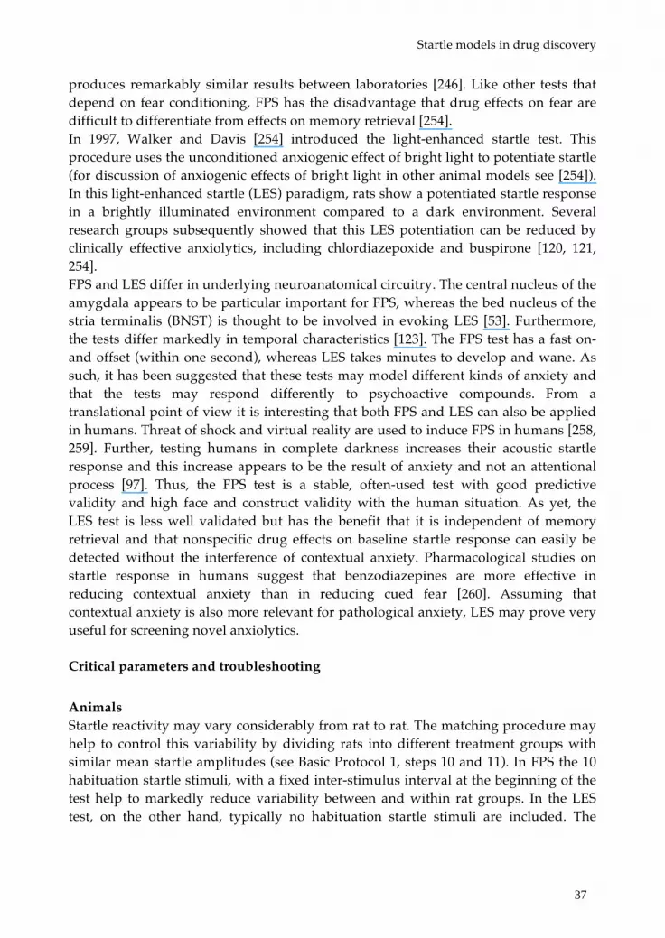
produces remarkably similar results between laboratories [246]. Like other tests that depend on fear conditioning, FPS has the disadvantage that drug effects on fear are difficult to differentiate from effects on memory retrieval [254]. In 1997, Walker and Davis [254] introduced the light-enhanced startle test. This procedure uses the unconditioned anxiogenic effect of bright light to potentiate startle (for discussion of anxiogenic effects of bright light in other animal models see [254]). In this light-enhanced startle (LES) paradigm, rats show a potentiated startle response in a brightly illuminated environment compared to a dark environment. Several research groups subsequently showed that this LES potentiation can be reduced by clinically effective anxiolytics, including chlordiazepoxide and buspirone [120, 121, 254]. FPS and LES differ in underlying neuroanatomical circuitry. The central nucleus of the amygdala appears to be particular important for FPS, whereas the bed nucleus of the stria terminalis (BNST) is thought to be involved in evoking LES [53]. Furthermore, the tests differ markedly in temporal characteristics [123]. The FPS test has a fast on- and offset (within one second), whereas LES takes minutes to develop and wane. As such, it has been suggested that these tests may model different kinds of anxiety and that the tests may respond differently to psychoactive compounds. From a translational point of view it is interesting that both FPS and LES can also be applied in humans. Threat of shock and virtual reality are used to induce FPS in humans [258, 259]. Further, testing humans in complete darkness increases their acoustic startle response and this increase appears to be the result of anxiety and not an attentional process [97]. Thus, the FPS test is a stable, often-used test with good predictive validity and high face and construct validity with the human situation. As yet, the LES test is less well validated but has the benefit that it is independent of memory retrieval and that nonspecific drug effects on baseline startle response can easily be detected without the interference of contextual anxiety. Pharmacological studies on startle response in humans suggest that benzodiazepines are more effective in reducing contextual anxiety than in reducing cued fear [260]. Assuming that contextual anxiety is also more relevant for pathological anxiety, LES may prove very useful for screening novel anxiolytics. Critical parameters and troubleshooting Animals Startle reactivity may vary considerably from rat to rat. The matching procedure may help to control this variability by dividing rats into different treatment groups with similar mean startle amplitudes (see Basic Protocol 1, steps 10 and 11). In FPS the 10 habituation startle stimuli, with a fixed inter-stimulus interval at the beginning of the test help to markedly reduce variability between and within rat groups. In the LES test, on the other hand, typically no habituation startle stimuli are included. The
37

Chapter 2
authors’ unpublished results suggest that habituation trials in the LES session may actually reduce the level of startle potentiation. Both Wistar and Sprague Dawley rats are frequently used in pharmacological FPS and LES studies and results appear to be interchangeable. In general, results are more stable if constant background ‘noise’ is provided in the animal facilities. House and test rats in rooms where the radio is turned on. Apart from this, try to keep the animal facilities as quiet as possible. Equipment The equipment for these startle test is extremely sensitive, hence parts must always be handled carefully. Calibrate speakers and startle cages between studies, while taking into account the particular specifics of the study. For instance, the animal’s body weight has a direct effect on startle response magnitude [134]. Consider such aspects while setting the sensitivity of the transducer during calibration. If results deviate significantly from what was expected, first check if the equipment is functioning properly. Sometimes experimental failure is due to simple electronic problems such loose cables or poor grounding. For instance, if the output of a particular cage (raw data set) during a session is a more or less constant at a low value, there is probably a defective or loose cable. Constant high values, on the other hand, may suggest poor grounding. A multimeter can be used to assess these situations. Experimental conditions The level of fear conditioning is determined by many factors, including shock intensity, time between and number of light-shock pairings, and length of the cue light (unconditioned stimulus; UCS). Apart from some specific parametric studies on FPS, there is extensive literature on factors relevant for classical fear conditioning that also applies to FPS. A few examples are described here. There is an inverted U-shaped relationship between shock intensity and fear potentiation: both low (<0.3 mA) and high shock intensities (>1 mA) will result in poorer fear conditioning [261]. The interval between light-shock pairings should not be too short as shortening the interval may reduce the conditioning [262]. Conversely, presenting the 20 light-shock pairings divided over two days instead of all on one day strengthens the fear conditioning in FPS. If poor startle potentiation is observed and equipment problems have been ruled out, it is important to exclude the possibility of a scaling problem before varying other experimental conditions. Make sure that baseline startle response occurs near the dynamic middle of the measurement scale. If baseline startle responses are too high this could result in a ceiling effect (‘fear’ cannot further enhance the already high startle response). If so, reduce the intensity of the startle stimuli or decrease sensitivity of the transducer. Similarly, if baseline startle responses are too low fear will not readily potentiate such a small response (floor effect). If this is the case, increase the intensity of the startle stimuli or increase the sensitivity of the transducer.
38

Startle models in drug discovery
Finally, this unit describes a set of parameters that have been determined to be optimal under the described experimental conditions, although different parameters may be optimal in other laboratories. This for instance seems to be the case with the fluorescent lamps in the LES test (see Basic Protocol 2 introduction). Thus, pilot studies may be necessary to optimize parameters in a particular laboratory. Study design The protocols described in this unit are for a between-subjects design, that is, the different treatments are tested in different groups of animals. However, both the FPS and LES tests are ideally suited to run a within-subject design [120, 122, 263]. For a within-subject design, with rats being tested once a week, match the rats (Basic Protocol 1, step 10) and use a Latin square design to evenly distribute four different treatments over 4 test days. For FPS the training sessions and tests are performed as described in the first week. In the subsequent weeks, give the rats only one training session and test them 24 hr later. For LES match the rats, use a Latin square treatment design and repeat the normal protocol, with two LES sessions per week (dark-dark, and dark-light) for 3 weeks.
Figure 2. Mean startle response magnitude (±SEM) of rats measured during matching and in reaction to baseline trials (noise-alone) and fear potentiation trials (light-noise) in the FPS test measured 5 days later. * significantly different from match session (p <0.05), ˆ significantly different from baseline in FPS test (p <0.05).
100 105 1150
500
1000
1500
2000Baseline matching'Baseline' in FPS testFear potentiated startle
Startle reactivitybefore and after FPS training
*
*
**
**^
^
^
Startle stimulus intensity (dB)
Mea
n st
artle
mag
nitu
de
39

Chapter 2
Anticipated Results FPS As described in Basic Protocol 1, in an FPS test session startle stimuli are presented under two conditions: in the absence and presence of the cue light (noise-alone, and light-noise trials, respectively). The noise-alone trials, in which only the startle stimuli are presented (CS-), yield data on baseline startle response. In the light-noise trials startle stimuli are presented in the presence of the fear-conditioned stimulus light (CS+). These trials yield data on the FPS response. Thus, successful fear conditioning will result in a significant ‘condition’ effect (or an interaction effect of ‘condition’ with drug treatment). Figure 2 shows the mean baseline startle response of rats measured in the matching session and their mean startle responses on the baseline (noise-alone) and fear potentiation (light-noise) trials in the FPS test measured 5 days later. Note that the ‘baseline’ values measured in the FPS test are markedly higher than during matching. This elevation of baseline startle is a result of contextual conditioning. After the training sessions, rats associate the test unit with shock. Being returned to the test unit will induce anxiety and thus raise the startle response. Comparing the matching and baseline startle response data of the vehicle control group provides a check of whether the training sessions (fear conditioning) were successful. This context conditioning effect may also explain why many anxiolytics somewhat reduce baseline startle response in the FPS test (for discussion see [120], but also [264]). Ideally, an anxiolytic compound will have a strong effect on FPS (light-noise) with little or no effect on baseline startle (noise-alone). Statistically this will result in an interaction effect of light condition with drug treatment. It is important to separately evaluate drug effects on baseline startle response. If a drug strongly reduces baseline startle response this suggests sedative or motor relaxant effects (see Figure 3). Smaller drug effects on baseline startle may also be ascribed to anxiolytic properties, as discussed above. FPS results can also be expressed as percent potentiation (100 × (LN-NA)/NA)) and statistics applied on this parameter. However, interpretation is less unequivocal as alterations in percentage potentiation (and absence thereof) can result from changes in FPS response, but also from changes in baseline startle response. In the data presented in Figure 3, chlordiazepoxide treatment resulted in a significant interaction between treatment × light condition. At the highest dose, chlordiazepoxide blocked FPS (no significant difference between light-noise and noise-alone trials), indicating anxiolytic properties. However, due to the marked effect of chlordiazepoxide on baseline startle no significant treatment effect is found on percent fear potentiation. For a discussion on use of percent potentiation see Walker and Davis [196].
40

Startle models in drug discovery
Figure 3. The effect of chlordiazepoxide on FPS as absolute values of the mean startle response magnitude (mean ± SEM) on baseline and fear potentiation trials. Inset: the same data depicted as percent fear potentiation. * significantly different from vehicle control (p <0.05), ˆ significantly different from baseline startle response. LES A typical LES study consists of two sessions (dark-dark and dark-light), and data of both sessions are shown (see Figure 4). In the dark-dark session normal habituation is expected. Thus, the second dark phase of a dark-dark session should yield lower mean startle magnitude than the first dark phase. This session is used to control for nonspecific drug effects on baseline startle response. In the dark-light session a significant increase in startle amplitude is expected in the second phase. Ideally, an anxiolytic compound will have a strong effect on the light phase of the light-dark session, with no or limited effect on the dark phase. The effect of chlordiazepoxide on LES is shown in Figure 4. At higher doses chlordiazepoxide significantly reduced baseline startle magnitude (dark-dark session). This effect is most likely due to sedative effects, and also occurs in FPS (see also Figure 3). In the dark-light session, chlordiazepoxide significantly reduced LES relative to the dark phase 1 (phase × treatment interaction). It could be argued that this reduction in LES is confounded by the nonspecific startle-reducing effects of chlordiazepoxide. This seems unlikely however, as in the dark-dark session chlodiazepoxide reduced startle amplitude similarly in phase 1 and 2, causing the difference scores to remain constant (Figure 4A). Thus, lower startle response amplitudes do not necessarily result in lower
^
0
500
1000
1500
2000
2500
*
** ^
^
0 2.5 5 10
**
Chlordiazepoxide (mg/kg)
Mea
n st
artle
mag
nitu
de
0
50
100
150
0 2.5 5 10
perc
ent p
oten
tiatio
n
Chlordiazepoxide in fear-potentiated startle
41

Chapter 2
Figure 4. The effect of chlordiazepoxide on the absolute values of the mean startle response magnitude (group mean ± SEM) during (A) a dark-dark session (bars represent phases 1 and 2, both ‘dark’), and (B) a dark-light session (black bar represents phase 1 ‘dark’, white bar represents phase 2 ‘light’). (C) Difference scores for dark-dark and dark-light sessions (black and gray bars, respectively). * Significantly different from corresponding vehicle (p < 0.05). ˆ Significantly different from baseline startle response (phase 1 dark).
0
500
1000
1500
2000
dark 2
* *
dark
0 2.5 5 10
Chlordiazepoxide (mg/kg i.p.)
Mea
n st
artle
mag
nitu
de
0
500
1000
1500
2000darklight
* *
*
^^
Chlordiazepoxide (mg/kg i.p.)
Mea
n st
artle
mag
nitu
de
0 2.5 5 10
-500
-250
0
250
500
* *
0 2.5 5 10
Chlordiazepoxide (mg/kg i.p.)
Mea
n st
artle
mag
nitu
de
A
C
B
42

Startle models in drug discovery
difference scores. A data set is best reflected in bar graphs, but may also be shown and analyzed as difference score or percentage (see also Anticipated Results for FPS). Time considerations. Rats have to be well adjusted to the laboratory before starting a study, and this will take at least a week. Duration of sessions: The durations of the respective sessions are ~20 min for a matching session, ~45 min for an FPS training session, ~25 min for a FPS test and ~45 min for an LES session. These time estimates do not include the time needed for cleaning equipment and transferring and injecting rats. The total amount of time needed to complete an FPS or LES test for all experimental groups depends on how many rats can be tested at one time. Both FPS and LES can be tested throughout the day, but only the light phase of the light/dark cycle should be used (that is, reported pharmacological studies have mostly been performed during the light phase), test each rat at approximately the same time of day and evenly distribute the different treatments over the day. Time between sessions: Timing between matching and a subsequent startle session is not too critical. However, if rats are regrouped after matching, a minimum interval of 3 days between matching and testing is advised. Also, start the next session within a week, because matching not only serves to equalize experimental groups, but also contributes to the familiarization to the startle stimuli. In pharmacological studies time between the two FPS training sessions, and between the last FPS training and FPS test, is typically 24 hrs. For LES, the time interval between the two sessions should be at least 48 hrs because LES is more stable if the interval between test sessions is longer and this interval length limits potential carryover effects of test compounds. Key References Davis, M., Falls, W. A., Campeau, S. and Kim, M. Fear-potentiated startle: a neural and pharmacological analysis. Behav Brain Res 1993:58:175-198. Reviews the underlying mechanism of FPS and pharmacological sensitivity. Hijzen, T.H., Houtzager, S.W., Joordens, R.J., Olivier, B. and Slangen, J.L. Predictive validity of the potentiated startle response as a behavioural model for anxiolytic drugs. Psychopharmacology 1995:118:150-154. Thorough investigation into the predictive validity of the FPS test using a wide range of compounds. The importance of adequate control groups is also discussed.
43

44

3
Fear-potentiated startle, but not light-enhanced startle, is enhanced by anxiogenic drugs
Pharmacology, Biochemistry and Behavior 2010; 96(1):24-31
Elisabeth Y. Bijlsma Reinoud de Jongh
Berend Olivier Lucianne Groenink
45

Chapter 3
Abstract The light-enhanced startle paradigm (LES) is suggested to model anxiety, because of the non-specific cue and the long-term effect. In contrast, the fear-potentiated startle (FPS) is suggested to model conditioned fear. However, the pharmacological profiles of these two paradigms are very similar. The present study investigated the effects of putative anxiogenic drugs on LES and FPS and aimed at determining the sensitivity of LES for anxiogenic drugs and to potentially showing a pharmacological differentiation between these two paradigms. Male Wistar rats received each dose of either the α2-adrenoceptor antagonist yohimbine (0.25-1.0 mg/kg), the 5-HT2C receptor agonist m-chlorophenylpiperazine (mCPP, 0.5-2.0 mg/kg) or the GABAA inverse receptor agonist pentylenetetrazole (PTZ, 3-30 mg/kg) and were subsequently tested in either LES or FPS. None of the drugs enhanced LES, whereas mCPP increased percentage FPS and yohimbine increased absolute FPS values. Furthermore, yohimbine increased baseline startle amplitude in the LES, while mCPP suppressed baseline startle in both the LES and FPS and PTZ suppressed baseline startle in the FPS. In contrast to findings in the FPS paradigm, none of the drugs were able to exacerbate the LES response. Thus, a clear pharmacological differentiation was found between LES and FPS.
46

Anxiogenic drugs
Introduction The acoustic startle response can be increased by presenting the startle-eliciting noise in the presence of a cue previously paired with footshock. This fear-potentiated startle (FPS) paradigm was first described in 1951 [85] and has since then greatly increased our understanding of the neural and pharmacological mechanisms that underlie conditioned fear (for review see [95] or [82]. FPS is pharmacologically well characterized, with selective sensitivity for anxiolytic drugs [122, 246, 256] and not other classes of psychoactive drugs [122, 265]. In addition, FPS can be enhanced by anxiogenic drugs [266]. More recently, it was shown that the startle response can also be increased by bright light, which in rats proves to be an unconditioned anxiogenic stimulus [254]. In this procedure, which has been termed light-enhanced startle (LES), rats show a potentiated startle response in a brightly illuminated environment, compared to a dark environment. Interestingly, Grillon et al. (1997) showed that in humans, startle is increased when tested in the dark and that this increase appears to be the result of fear or anxiety and not an attentional process [97]. It is suggested that the increase of startle in rats tested in bright light and in humans tested in the dark has an evolutionary basis, that is, rats are nocturnal and are more vulnerable in the light, whereas humans are diurnal and more vulnerable in the dark [97, 254].The LES paradigm has been pharmacologically characterized to some extent. It was shown to be sensitive to various anxiolytic drugs, namely the GABAA receptor agonist chlordiazepoxide [120, 121], the partial 5-HT1A receptor agonist buspirone [254] and the full 5-HT1A receptor agonist flesinoxan [120]. Sensitivity to anxiogenic drug effects, however, has not been studied in the LES paradigm. The enhancement of the startle response in the FPS and LES appears to be mediated by different brain regions. Infusions of the AMPA receptor antagonist NBQX into the central nucleus of the amygdala blocked FPS, but not LES, whereas infusion into the bed nucleus of the stria terminalis (BNST) blocked LES and sustained fear responses, but not FPS [194, 267, 268]. In addition, a recent study suggested an important role for the anterior cingulate cortex and septo-hippocampal system in the LES response, but not FPS [130]. In addition to this regional differentiation between LES and FPS, a pharmacological differentiation was observed after blockade of CRF receptors. Both the nonspecific CRF antagonist α-helical CRF and the specific CRF1 receptors antagonist GSK876008 blocked LES but not FPS [123, 188]. The aim of the present study was to investigate whether anxiogenic drug effects enhance LES and to investigate whether LES and FPS can be pharmacologically differentiated on basis of anxiogenic drug effects. Therefore, we studied the effects of three putative anxiogenic drugs acting on different neurotransmitter systems on both LES and FPS, namely the α2-adrenoceptor agonist yohimbine, the 5-HT2C receptor agonist m-chlorophenylpiperazine (mCPP) and the GABAA receptor inverse agonist pentylenetetrazole (PTZ). In addition to their effect on FPS, these drugs were already
47

Chapter 3
shown to have anxiogenic effects in several rodent [266, 269-272] and human studies [273-276]. In the present experiment, the potentially conditioned fear and anxiety-enhancing drug effects were assessed by comparing changes in startle amplitude under several conditions (see also table 1). Firstly, the enhancement of baseline startle amplitude, as measured during the dark-dark session in the LES paradigm. Secondly, enhancement of startle responding during the light phase in the LES paradigm and during Noise Alone trials following FPS conditioning, which both reflect exacerbation of sustained anxiety [277]. Thirdly, enhancement of startle responding to Light Noise trials in the FPS paradigm, which reflects potentiation of conditioned fear. The drug effects on startle responding in these different conditions were all studied to differentiate between drug effects on conditioned fear and anxiety. Materials and methods Subjects Male Wistar rats (Harlan, Horst, The Netherlands), weighing 300-350 g at the beginning of the experiments, were housed in groups of four in a temperature (21°C ± 2), humidity (55% ± 5), and light controlled environment (lights on from 6 AM to 6 PM). Food and water were freely available in the home cages. The experiments were carried out during the light phase of the day-night cycle between 9 AM and 2:30 PM. The study was approved by the ethical committee of the Faculties of Pharmaceutical Sciences, Chemistry and Biology, Utrecht University, The Netherlands. Apparatus Four startle devices were used simultaneously (SR-lab, San Diego instruments, San Diego CA, USA). The startle devices consisted of a Plexiglas cylinder (8.8 cm in diameter and 20.3 cm in length) with a stainless steel grid floor placed on a Plexiglas base. Each startle device was placed in a ventilated sound attenuated cubicle. Cage movements were measured with a piezoelectric film attached to the Plexiglas base of the startle device. A calibration system (San Diego Instruments) was used to ensure comparable startle magnitudes across the four devices throughout the experiment. Startle stimuli, consisting of 50 ms white-noise bursts, were presented through a piezoelectric tweeter situated 15.2 cm from the top of the cylinder. Background noise was 55 dB. Sound intensities were measured using a microphone, which was placed on top of the Plexiglas cylinder and fitted to a Bruel and Kjaer sound level meter (Type 2226). Startle amplitudes were sampled each ms during a period of 65 ms beginning at the onset of the startle stimulus. Each startle device was equipped with a white fluorescent bulb (9 W) on the back wall of the sound attenuated cubicle and a stimulus light in the ceiling situated 15.2 cm from the top of the cylinder. The fluorescent bulb produced an illumination level of approximately 900 lux and the stimulus light an illumination level of approximately 180 lux, both measured from
48

Anxiogenic drugs
inside the Plexiglas cylinder using a Gossen luxmeter (MAVOLUX 5032C). There was no background illumination in any of the experiments. Procedure Light-enhanced startle Light-enhanced startle was performed as previously described [120, 278]. In short, animals were placed in the startle chamber and, after a 5 min acclimation period, presented with 30 startle stimuli, 10 each at 90, 95 and 105 dB, with an inter stimulus interval of 30 sec. Within every block of three stimuli, the three intensities were presented in a random order, with each intensity being presented only once. These 30 stimuli constituted phase 1. Then, the procedure, including the acclimation period, was repeated. This second set of 30 stimuli constituted phase 2. Depending on the experimental condition, the level of illumination was changed between phase 1 and phase 2. Animals were tested twice a week for four successive weeks, with test days separated by a minimum of 72 hrs. On one of these test days, the light remained off during both phases (dark-dark). On the other day, a light that produced an illumination level of approximately 900 lux was on during phase 2 (dark-light). Half of the rats started the experiment with the dark-dark session type; the other half began with the dark-light session type. The session type was alternated throughout the experiment. Each drug was tested in a separate group of animals, carried out using the same procedure. In each of the three groups, 4 dosages of the drug (including vehicle) were administered according to a balanced within-subjects design. That is, each rat received each dose of the drug in both session types. Fear-potentiated startle Fear-potentiated startle was performed as previously described [122, 278]. In short, three separate groups of rats were trained once a day for 2 consecutive days. During each training session, rats were presented with 10 light-shock pairings at an average interval of 4 min (range: 3-5 min). A 0.6 mA foot shock was presented during the last 500 ms of the 3700 ms light period. Shock reactivity, registered by measuring cage movements, was sampled each ms during a period of 200 ms beginning at the onset of foot shock. Each drug was tested in a separate group of animals, carried out using the same procedure. One day after the last training session, the animals in each group received one of 4 dosages of the drug (including vehicle) according to a balanced within-subjects design. After an acclimation period of 5 min, 10 startle stimuli of 105 dB were presented (ISI 30 sec), followed by 30 startle stimuli at an ISI of 30 sec, 10 each at 90, 95 and 105 dB. Half of the 30 startle stimuli were presented during the last 50 ms of a 3250 ms light period; the other half were delivered in darkness. The six different trial types were presented in a balanced, irregular order across the test session. During the following three weeks, the training and test procedures were repeated 3 times, separated by one week. During these weeks, rats were only trained once a week, followed by a test session the next day.
49

Chapter 3
Drugs Yohimbine HCl (0, 0.25, 0.5 and 1.0 mg/kg), m-chlorophenylpiperazine HCl (mCPP; 0, 0.5, 1.0, 2.0 mg/kg) and pentylenetetrazole (PTZ; 0, 3, 10, 30 mg/kg) were dissolved in 0.9% saline (vehicle) and administered intraperitoneally. mCPP was administered 25 min before testing. Yohimbine and PTZ were administered 10 min before testing. All drugs were given in a volume of 2 ml/kg. Drug and vehicle solutions were freshly prepared each morning. Statistics Light-enhanced startle Repeated measures ANOVAs were used to analyze overall startle reactivity. Condition (two levels: dark-dark or dark-light session type), phase (two levels: phase 1 and phase 2) and dose (four levels) as within-subject factors. Additionally, to determine drug effects on baseline startle en sustained anxiety, repeated measures ANOVAs were used to analyze the mean startle amplitudes in the dark-dark and dark-light session types separately. Phase (two levels: phase 1 and phase 2) and dose (four levels) were used as within-subjects factors. Comparisons between different drug doses were made by simple contrasts. The percentage change [(phase 2–phase 1)/phase 1] was also calculated for each rat and the mean percentages were subsequently analyzed by repeated measures ANOVA with session type (two levels: dark-dark and dark-light) and dose (four levels) as within-subjects factors. The significance level for all analyses was 5%. Rats that did not show light potentiation in the dark-light session type (percentage increase during light phase vs dark phase <0%) under vehicle conditions and statistical outliers, with startle responses more than two standard deviations away from the mean, were excluded from data analysis (1 for yohimbine, 4 for mCPP and 3 for PTZ). In addition, in the PTZ group, two rats were excluded from the analysis because the highest dose of PTZ resulted in convulsions. Fear-potentiated startle A repeated measures ANOVA was used to analyze the mean startle amplitudes on the Noise Alone and Light Noise trials. Trial type (two levels: Noise Alone and Light Noise) and dose (four levels) were used as within-subjects factors. Comparisons between different drug doses were made by simple contrasts. The percentage change [(Light Noise – Noise Alone)/Noise Alone] was also calculated for each rat and the mean percentages were subsequently analyzed by a repeated measures ANOVA with dose (four levels) as within-subjects factor. The significance level for all analyses was 5%.
50

Anxiogenic drugs
Results Light-enhanced startle The mean startle amplitudes for the dark-dark (DD) session type (left panels) and for the dark-light (DL) session type (right panels) for the three drugs are depicted in figure 1. In all three groups tested significant light-enhanced startle was induced during the DL session (session x phase interactions: Yohimbine [F(1, 10) = 29.5; p<0.001], mCPP [F(1, 7) = 26.2; p < 0.001] and PTZ [F(1, 8) = 17.9; p < 0.01]). Specific analyses of behavioural responses under vehicle conditions revealed significant light-enhanced startle also under control conditions specifically (phase 2 vs phase 1 during DL session: Yohimbine [T(1, 10) = -3,795, p < 0.01], mCPP [T(1, 7) = -4.619, p < 0.01] and PTZ [T(1, 7) = -2.632, p < 0.05]). In the yohimbine group, analyses of DD and DL session types separately showed that yohimbine did not influence the light-enhanced startle response. However, yohimbine increased overall startle amplitude in the DD session (DD session: main effect dose [F(3, 30) = 3.1; p < 0.05]), but not in the DL session. Simple contrasts revealed that this effect of yohimbine in the DD session type was mediated by the 1.0 mg/kg dose [F(1, 10) = 10.7; p < 0.01]. In the mCPP group, analyses of the DD and DL session types separately showed that mCPP had no effect on the light-enhanced startle response. However, mCPP decreased overall startle amplitude in both session types (DD session type: [dose F(3, 5) = 3.4; p < 0.05]; DL session type: [dose F(3, 5) = 8.4; p < 0.001]). Simple contrasts revealed that, in both session types, overall startle amplitude was decreased after 1.0 mg/kg and 2.0 mg/kg mCPP (1.0 mg/kg: DD session type [F(1, 7) = 29.2; p < 0.001]; DL session type [F(1, 7) = 16.7; p < 0.01] and 2.0 mg/kg: DD session type [F(1, 7) = 7.4; p < 0.05]; DL session type: [F(1, 7) = 14.5; p < 0.01]). In the PTZ group, analyses of the two session types separately showed that PTZ had no effect on absolute light-enhanced startle responding. In addition, PTZ did not affect overall startle amplitude in the DL session types, although there was a trend towards an effect on overall startle amplitude in the DD session type [DD session type: [F(3, 24) = 2.507, p < 0.1]. This trend in the DD session type could not be specifically ascribed to a specific dose. Figure 2 depicts the mean percentage change in startle potentiation in the DD session type and in the DL session type for the three drugs. In all three groups significant light-enhanced startle was induced (main effect session type: yohimbine [F(1, 10) = 28.4; p < 0.001, mCPP [F(1, 7) = 21.8; p < 0.01] and PTZ [F(1, 8) = 22.9; p < 0.001]). However, none of the drugs affected percentage light-enhanced startle.
51

Chapter 3
Figure 1. The effects of yohimbine (n=11), mCPP (n=8) and PTZ (n=9) on light-enhanced startle. The figure shows mean startle amplitudes during dark and dark 2 for the dark-dark session type (left panels) and during dark and light for the dark-light session type (right panels) of animals treated with yohimbine (A), mCPP (B) and PTZ (C). * indicates overall significant difference compared to 0 mg/kg for absolute mean startle amplitudes.
Yohimbine
0
200
400
600
800
*
dose (mg/kg)
Sta
rtle
am
plitu
de
0.0 0.25 0.5 1.0
mCPP
0
100
200
300
400
500
* *
dose (mg/kg)
Sta
rtle
am
plitu
de
0.0 0.5 1.0 2.0
PTZ
0
100
200
300
400
500
dose (mg/kg)
Sta
rtle
am
plitu
de
0 3 10 30
Yohimbine
0
200
400
600
800
dose (mg/kg)
Sta
rtle
am
plitu
de
0.0 0.25 0.5 1.0
mCPP
0
100
200
300
400
500
* *
dose (mg/kg)
Sta
rtle
am
plitu
de
0.0 0.5 1.0 2.0
PTZ
0
100
200
300
400
500
dose (mg/kg)
Sta
rtle
am
plitu
de
0 3 10 30
Dark - Dark session Dark - Light session
1A
1B
1C
dark dark2 dark light
52

Anxiogenic drugs
Figure 2. The effects of yohimbine (n=11), mCPP (n=8) and PTZ (n=9) on light-enhanced startle. The figure shows the mean percentage decrease in the dark-dark session type and the mean percentage increase in the dark-light session type for yohimbine (A) mCPP (B) and PTZ (C).
Yohimbine
-50
0
50
100
150
200
dose (mg/kg)
perc
enta
ge c
hang
e
0.0 0.25 0.5 1.0
mCPP
-50
0
50
100
150
200
0.0 0.5 1.0 2.0dose (mg/kg)
Per
cent
age
chan
ge
PTZ
-50
0
50
100
150
200
dose (mg/kg)
Per
cent
age
chan
ge
0 3 10 30
Dark - Dark
2A
2B
2C
Dark - Light
53
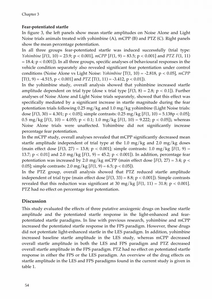
Chapter 3
Fear-potentiated startle In figure 3, the left panels show mean startle amplitudes on Noise Alone and Light Noise trials animals treated with yohimbine (A), mCPP (B) and PTZ (C). Right panels show the mean percentage potentiation. In all three groups fear-potentiated startle was induced successfully (trial type: Yohimbine [F(1, 10) = 23.9; p < 0.001], mCPP [F(1, 9) = 83.5; p < 0.001] and PTZ F(1, 11) = 18.4; p < 0.001]). In all three groups, specific analyses of behavioural responses in the vehicle condition separately also revealed significant fear potentiation under control conditions (Noise Alone vs Light Noise: Yohimbine [T(1, 10) = -2.818, p < 0.05], mCPP [T(1, 9) = -4.515, p < 0.001] and PTZ [T(1, 11) = -3.412, p < 0.01]). In the yohimbine study, overall analysis showed that yohimbine increased startle amplitude dependent on trial type (dose x trial type [F(3, 8) = 2.8; p < 0.1]). Further analyses of Noise Alone and Light Noise trials separately, showed that this effect was specifically mediated by a significant increase in startle magnitude during the fear potentiation trials following 0.25 mg/kg and 1.0 mg/kg yohimbine (Light Noise trials: dose [F(3, 30) = 4.301; p < 0.05]; simple contrasts: 0.25 mg/kg [F(1, 10) = 5.138p < 0.05]; 0.5 mg/kg [F(1, 10) = 4.055; p < 0.1; 1.0 mg/kg [F(1, 10) = 9.222; p < 0.05]), whereas Noise Alone trials were unaffected. Yohimbine did not significantly increase percentage fear potentiation. In the mCPP study, overall analyses revealed that mCPP significantly decreased mean startle amplitude independent of trial type at the 1.0 mg/kg and 2.0 mg/kg doses (main effect dose [F(3, 27) = 13.8; p < 0.001]; simple contrasts: 1.0 mg/kg [F(1, 9) = 13.7; p < 0.01] and 2.0 mg/kg [F(1, 9) = 45.2; p < 0.001]). In addition, percentage fear potentiation was increased by 2.0 mg/kg mCPP (main effect dose [F(3, 27) = 3.4; p < 0.05]; simple contrasts: 2.0 mg/kg [F(1, 9) = 6.5; p < 0.05]). In the PTZ group, overall analysis showed that PTZ reduced startle amplitude independent of trial type (main effect dose [F(3, 33) = 8.8; p < 0.001]). Simple contrasts revealed that this reduction was significant at 30 mg/kg [F(1, 11) = 31.8; p < 0.001]. PTZ had no effect on percentage fear potentiation. Discussion This study evaluated the effects of three putative anxiogenic drugs on baseline startle amplitude and the potentiated startle response in the light-enhanced and fear-potentiated startle paradigms. In line with previous research, yohimbine and mCPP increased the potentiated startle response in the FPS paradigm. However, these drugs did not potentiate light-enhanced startle in the LES paradigm. In addition, yohimbine increased baseline startle amplitude in the LES study, whereas mCPP decreased overall startle amplitude in both the LES and FPS paradigm and PTZ decreased overall startle amplitude in the FPS paradigm. PTZ had no effect on potentiated startle response in either the FPS or the LES paradigm. An overview of the drug effects on startle amplitude in the LES and FPS paradigms found in the current study is given in table 1.
54
Anxiogenic drugs
Figure 3. The effects of yohimbine (n = 11), mCPP (n=10) and PTZ (n=12) on fear-potentiated startle. Left panels show mean startle amplitudes on Noise Alone and Light-Noise trials and right panels show the mean percentage potentiation of animals treated with yohimbine (A), mCPP (B) and PTZ (C). # Indicates overall significant effect compared to 0 mg/kg during fear potentiation trials (left panel). * indicates significant difference compared to 0 mg/kg for mean percentage potentiation (right panel).
Yohimbine
0
500
1000
1500
2000
2500
3000
3500 #
#
dose (mg/kg)
Sta
rtle
am
plitu
de
0.0 0.25 0.5 1.0
mCPP
0
500
1000
1500
2000
2500
* *
dose (mg/kg)
Sta
rtle
am
plitu
de
0.0 0.5 1.0 2.0
PTZ
0
500
1000
1500
2000
2500
*
dose (mg/kg)
Sta
rtle
am
plitu
de
0 3 10 30
Yohimbine
0.00 0.25 0.50 1.000
20
40
60
80
100
dose (mg/kg)
perc
enta
ge p
oten
tiatio
nmCPP
0.0 0.5 1.0 2.00
50
100
150
200
250
300
350
400
*
dose (mg/kg)
Per
cent
age
pote
ntia
tion
PTZ
0 3 10 300
20
40
60
80
100
120
140
dose (mg/kg)
Per
cent
age
pote
ntia
tion
Startle amplitudes Potentiation
3A
3B
3C
NA LN
55

Chapter 3
Effect of yohimbine on baseline startle amplitude and startle potentiation Yohimbine increased fear potentiation in the FPS paradigm when displayed as absolute startle potentiation. Startle potentiation in the LES paradigm, however, was not affected by yohimbine in the dose range tested, showing a pharmacological differentiation between FPS and LES. The lack of effect of yohimbine on LES was somewhat surprising. Systemic administration of yohimbine results in activation of the BNST, lateral septum and cingulate cortex, all brain areas implicated in LES [130, 279, 280]. In addition, decreasing noradrenergic signalling in the BNST via local administration of clonidine inhibits LES [281]. A possible explanation for the lack of effect of yohimbine on LES could be the specific anxiety-like state that is induced or exacerbated by yohimbine. It has been proposed that yohimbine potentiates neural mechanisms mediating flight. Flight, freezing and defensive threat/attack are the responses to immediate threat and constitute the fear/defence pattern [282]. A potential threat elicits the anxiety/defence pattern, with risk assessment as the dominant response [282]. As FPS is proposed to measure short duration conditioned fear and LES is proposed to measure sustained anxiety responses to a potentially threatening context, this differential influence of yohimbine on specific anxiety-like states may explain the differential sensitivity of the FPS and LES paradigm to detect yohimbine-induced effects. Interestingly, studies in patient groups underline these specific effects of yohimbine: While panic disorder patients show increased reports of arousal and anxiety compared to healthy subjects following yohimbine administration [273], generalized anxiety disorder patients are not more responsive than healthy subjects to yohimbine-induced arousal and anxiety [283]. The finding of yohimbine-induced increases in baseline startle response in the LES fits a previous study in rats [284]. A similar pattern has been detected in humans, although this was primarily found in psychiatric patients suffering from anxiety disorders [275]. The question remains, however, what this increase in baseline startle responding reflects. It might reflect general arousal induced by autonomic activation. In post-traumatic stress disorder patients, yohimbine increased baseline startle amplitude, without altering the level of self reported anxiety [285]. The yohimbine-induced increase in baseline startle may even simply reflect excitation of spiny motor neurons, as a study by Kehne and Davis (1985) indicated that the yohimbine-induced effect on baseline startle was specifically mediated by norepinephrine release within the spinal cord [284]. Both possibilities would indicate that yohimbine-induced effects on baseline startle are independent of anxiety state. On the other hand, it has also been shown that local infusion of yohimbine into the central amygdala increases the startle response similar to shock-induced sensitization of the startle response. This shock-induced sensitization of the startle response could be blocked by local infusion of the α2-adrenergic agonist ST-91 [286]. These findings suggest that yohimbine is able to induce a negative affective state (anxiety state) similar to that induced during shock-induced sensitization. It may be that this kind of anxiety-inducing effect is involved in
56

Anxiogenic drugs
the increased startle response found in the current study. Further research should look into the specific yohimbine-induced state that is reflected by increased baseline startle. In summary, based on the current findings yohimbine seems to specifically alter startle responding related to fear-defense patterns, resulting in increased FPS. In addition, baseline startle is increased following yohimbine administration. Yohimbine, however, does not exacerbate sustained anxiety, as measured during LES and Noise Alone trials in the FPS paradigm. Table 1. Overview of anxiogenic effects of yohimbine, mCPP and PTZ. Indicated are anxiogenic drug effects on startle magnitude as measured during dark-dark (DD) session (baseline startle), light-enhanced startle and Noise Alone trials following fear-potentiated startle conditioning (sustained anxiety) and Light Noise trials during fear-potentiated startle (conditioned fear).
Baseline startle
Sustained anxiety Conditioned fear
DD session NA trials LES FPS
Yohimbine ↑ X X ↑
mCPP ↓ ↓ X ↑
PTZ X ↓ X X
Effect of mCPP on baseline startle amplitude and startle potentiation mCPP increased percentage potentiation in the FPS paradigm, but did not affect startle potentiation in the LES paradigm. This would suggest that mCPP specifically influences conditioned fear responses and not the type of anxiety measured in the LES paradigm. Interestingly, a study by Mora et al (1997) showed that mCPP specifically enhances fear-like responding, but not anxiety-like responding, in the elevated T-maze. A similar differentiation was found in a study with healthy subjects, wherein mCPP markedly enhanced fear in a conditioned fear paradigm, but did not influence a more generalized form of anxiety during a public speaking task [216]. However, this line of evidence contrasts the effects of mCPP in unconditioned anxiety-like paradigms, like the elevated plus maze and social interaction test, and it brain activation pattern [130, 271, 280, 287]. Thus, alternatively, it might be that the anxiogenic response to bright light just cannot be exacerbated by pharmacological treatment. In contrast to the current study, Mansbach and colleagues (1988) were unable to detect an effect on fear potentiation. This contrast is most likely explained by the dose range tested. The increment in FPS in the current study was found only at
57

Chapter 3
the highest dose (2.0 mg/kg), while the highest dose tested by Mansbach and colleagues was 1.0 mg/kg [257]. The suppressing effect of mCPP on overall startle responding in both FPS and LES is consistent with a previously reported reduction in baseline startle in the FPS paradigm [257]. It has been suggested that baseline startle responding in the FPS paradigm (Noise Alone trials) might be sensitive to effects of anxiolytic drugs as a result of the induction of contextual fear during FPS training [277]. The reduced baseline startle amplitude in the FPS following mCPP administration, however, is unlikely due to blockade of contextual fear, as baseline startle responding in the LES paradigm, which does not involve conditioned contextual fear, is also decreased following mCPP administration. The mCPP-induced effects on baseline startle are probably best explained by locomotor suppression [271, 287, 288] and overall behavioural suppression [289]. Unfortunately, considering the finding that mCPP did have an anxiogenic effect on Noise Alone trials in mice that were insensitive to the locomotor suppressing effects of mCPP [197], it can not be excluded that the overall mCPP-mediated behavioural suppression may have prevented the detection of possible anxiogenic effects of mCPP within the current study. In addition, the profound effect of mCPP on overall startle responding results in a discrepancy between absolute and proportional difference scores, that could possibly lead to misinterpretation of the FPS data. In summary, although it can not be excluded that certain drug effects were concealed by behavioural suppression, the current results suggest that mCPP specifically increases conditioned fear, as measured with FPS, and does not influence sustained anxiety, as measured with LES and during Noise Alone trials following fear conditioning. Effect of PTZ on baseline startle amplitude and startle potentiation PTZ had no effect on startle potentiation in neither the LES nor the FPS paradigm. The lack of effect on startle potentiation was unexpected, as PTZ was already found to be anxiogenic in various measures of anxiety-like behaviour [270, 272] and GABAA
receptor activation with for example the anxiolytic chlordiazepoxide inhibits both LES and FPS [120, 122]. Several studies, however, failed to detect an anxiogenic effect of PTZ [290-292]. In addition, previous studies in the FPS paradigm with other ligands of the GABAA receptor complex have also shown variable effects of GABAA receptor blockade. For example, the full GABAA inverse receptor agonist DMCM (methyl-6,7-dimethoxy-4-ethyl-beta-carboline-3-carboxylate) and lindane, a neurotoxin that interferes with the chloride channel of the GABAA receptor, increased FPS [263], whereas the partial GABAA inverse receptor agonist FG-7142 decreased the FPS response [293]. The present findings suggest that, although stimulation of GABAA receptors with for example the anxiolytic chlordiazepoxide (CDP) has an anxiolytic profile in both the FPS and LES paradigm [120, 122, 277], GABAA receptor inactivation with PTZ has no readily detectable effect on fear- and anxiety-related startle responding.
58

Anxiogenic drugs
PTZ suppressed the overall startle response in both the FPS and LES at the 30 mg/kg dose. PTZ has been reported to suppress locomotion [289] and in mice, administration of 30 mg/kg PTZ severely disrupted behaviour, indicative of a preconvulsant state [291]. In the present experiment, two rats had to be excluded from the study, because the highest dose of PTZ caused convulsions in these rats. As with mCPP, it is likely that PTZ-induced motor suppression interfered with the execution of the startle response at the highest dose tested. A possible limitation of this study is that no positive control was used in the LES paradigm. However, this was also not possible due to the limited pharmacological validation of the LES paradigm. To the best of our knowledge, no reports have been made on drugs that are able to enhance the LES response. Another possible limitation of this study is the use of a within-subject design. It could be argued that this has prevented the detection of drug effects on LES and FPS, as exposure to an anxiogenic drug might sensitize behavioural responses in subsequent tests and these sensitized responses might interfere with possible drug effects. Analysis of time effects on both baseline startle responding and potentiated startle responding, however, did not reveal any sensitization in response to the drugs tested. Therefore, it seems unlikely that the design of the current study has prevented the detection of possible additional drug effects. The more as a highly significant effect of experimental manipulation was found under vehicle conditions. In addition, it might be possible that the exclusion of non-responders from the LES studies has interfered with the detection of anxiogenic drug effects, because of a ceiling effect. Excluding non-responders from analysis could have resulted in the selection of animals that already show relatively high anxiety levels (startle magnitudes) under control conditions, and therefore the range left to further enhance the startle response in response to an anxiogenic drug may be relatively small as compared to animals that show low anxiety levels under control conditions. A median split analysis of the LES data on basis of percentage LES under vehicle conditions (resulting in a low LES-reactive and a high LES-reactive group) did not reveal any differential drug effects in the low and high reactive group nor did it reveal drug x group interactions for any of the drugs, excluding a possible ceiling effect. It must be mentioned, though, that group size in this analysis was relatively small (4-6 animals per group). Another argument against absence of effects in LES due to ceiling effects is that the drugs did not only fail to enhance LES, they also did not increase responding to Noise Alone trials, a measure of sustained anxiety related to the LES response. To the best of our knowledge, this is the first study on the effect of anxiogenic drugs on LES. Although the FPS paradigm has been extensively validated with different classes of psychoactive drugs, the sensitivity of the LES paradigm for detecting drug effects has hardly been investigated. So far, it has been shown that the LES response can be attenuated by several anxiolytic drugs [120, 121] and that CRF is specifically involved in the LES response, as it is blocked by CRF antagonists [123, 188]. In the
59
Chapter 3
current study, none of the drugs influenced LES. Therefore, it could be concluded that the LES paradigm is not able to detect the specific anxiety states induced by yohimbine, mCPP and PTZ. On the other hand, one could argue that the anxiety state induced in the LES paradigm can not be exacerbated by an additional anxiogenic manipulation. This hypothesis may be true for pharmacological manipulations. However, it has already been shown that LES can be enhanced by behavioural stress manipulations [255, 294], although opposite effects of stress manipulations on LES have also been reported [134, 295]. Altogether, current findings show that the putative anxiogenic drugs yohimbine and mCPP potentiate FPS, but not LES. PTZ, however, had no effect on the potentiated startle response in either FPS or LES. The differential influence of yohimbine and mCPP on LES and FPS suggests that these drugs specifically enhance conditioned fear responses, as measured during FPS, and not sustained anxiety, as measured during LES. Acknowledgements The authors would like to thank Jocelien Olivier and Marijke de Graaff for their excellent technical assistance.
60

61

62

4
Disrupted startle modulation in animal models for affective disorders
Behavioural Brain Research 2010; 208(2): 383-390.
Elisabeth Y. Bijlsma Ronald S. Oosting
Berend Olivier Lucianne Groenink
63

Chapter 4
Abstract Affective startle modulation is used to study emotional reactivity in humans, and blunted affective startle modulation has been reported in depressed patients. To determine whether blunted affective startle modulation is also a common feature in animal models for affective disorders, light-enhanced startle was studied in three models: Inescapable foot shock (IFS), repeated restraint stress (RRS) and olfactory bulbectomy (OBX). In addition, prepulse inhibition was studied in these models. Light-enhanced startle was blunted following IFS and OBX and RRS decreased overall startle responding. Prepulse inhibition, however, was unaffected. These findings indicate that induction models for affective disorders may be associated with long- term effects on affective startle modulation. The lack of changes in sensorimotor gating suggests that these changes can be ascribed to alterations in emotional reactivity. In conclusion, our results indicate that the blunted affective startle modulation seen in animal models for affective disorders may be used to examine the mechanisms underlying altered emotional reactivity.
64

Blunted affective startle modulation in animal models for affective disorders
Introduction Affective disorders, such as major depression, are among the most prevalent psychiatric disorders with a life time prevalence around 15% [296, 297]. A major aspect of affective disorders is altered emotional reactivity, which is thought to entail either specific sensitized responding to aversive stimuli [298] or blunted responding to both aversive and pleasurable stimuli [299, 300]. Mechanisms underlying these alterations in emotional reactivity are still fairly unknown, which hampers development of effective treatment. A useful tool for studying emotional reactivity in both humans and (non-human) animals is the acoustic startle response. The acoustic startle response is a fast reflexive response to a strong unexpected acoustic stimulus [82]. This reflex is a non-baseline response, which can be both enhanced in the presence of an aversive cue and attenuated in the presence of a rewarding or pleasant cue [301]. This cross-species phenomenon is referred to as affective startle modulation and is used as a measure of emotional reactivity. Affective startle modulation was shown to be blunted in patients suffering from affective disorders [103, 105, 106]. This disruption was found in response to both aversive and pleasant contexts and seemed to be correlated to either the level of anhedonia [103] or the number of depressive episodes [106]. Because of its potential translational value, affective startle modulation may be a very useful read out to examine the mechanisms underlying altered emotional reactivity. In rodent models for affective disorders, however, alterations in affective startle modulation have hardly been investigated. Several paradigms are available to study affective startle modulation in rats. Fear-potentiated startle and light-enhanced startle (LES) both measure startle potentiation in the presence of an aversive context, whereas pleasure-attenuated startle measures startle attenuation in the presence of a rewarding cue [85, 136, 254]. LES is based on the fact that nocturnal animals, like rodents, have a natural aversion to brightly lit environments. The LES paradigm is especially interesting for research on emotional reactivity, because it is based on an innate aversion and therefore does not depend on cognitive functioning, a feature that seems to be altered in affective disorders [302]. The LES paradigm has primarily been used in anxiety research, and has been validated using anxiolytic drugs [120, 121]. However, recently a first study was published that showed a blunted LES in female, but not male, rats subjected to early maternal deprivation [134]. Considering that a lack of startle modulation is associated with depression in humans, it is interesting to determine whether blunted affective startle modulation is a common feature in diverse animal models for affective disorders. Therefore, in the current study, LES was studied in three different animal paradigms modelling various aspects of affective disorders, which are described in short below. The inescapable foot shock model, also referred to as the stress sensitization model, is a well-established animal model for stress-related disorders [303, 304]. Animals are exposed to short severe stress, which results in long term alterations in behavioural responses. For example, animals show diminished exploratory behaviour and
65
Chapter 4
increased freezing in an open field, up to ten weeks after stress exposure [304, 305]. Startle responding following stress sensitization has shown variable effects, showing both diminished and exaggerated startle responding [306-308]. Repeated restraint is proposed to model sustained stress exposure and is used to study the effects of chronic physical and emotional stress on affective responses [309]. It is a non-painful manipulation sufficient to produce clear changes in behavioural responses [310-313] and stress-induced HPA-axis activation [314]. Repeated restraint has been shown to alter baseline startle amplitude and habituation [315]. The olfactory bulbectomy model is a pharmacologically validated model for depression. Removal of the olfactory bulbs results in several behavioural and neurochemical alterations similar to those found in depressed patients [316-318]. In addition, several of these changes are sensitive to chronic, but not acute, pharmacological treatment acting on various neurotransmitter systems [319-322] and these normalizing effects can persist for up to ten weeks after cessation of treatment [322, 323]. Olfactory bulbectomy also seems to alter reward-related behaviour, a phenomenon related to hedonic state in humans [324, 325]. Startle responding has hardly been studied in the OBX model. McNish and Davis reported on increased shock-induced sensitization of the startle response following olfactory bulbectomy, whereas fear-potentiated startle responding was normal [326]. Prepulse inhibition of the acoustic startle response (PPI), a different form of startle modulation, is used as a measure of sensory motor gating, a neural filtering process that allows attention to be directed to the most relevant stimuli in the environment [90, 327]. The acoustic startle response is inhibited when a non-startling stimulus (prepulse) is presented 30-300ms before the startling stimulus [328]. As stress may affect the processing of external stimuli and abnormalities in PPI have been associated stress-related psychiatric disorders [113, 329], it is interesting to study possible alterations in prepulse inhibition in parallel to alterations in affective startle modulation. In summary, the aim of the present study was to study startle modulation in response to aversive context versus startle modulation following presentation of a prepulse in three animal paradigms modelling affective disorders, which are based on markedly different induction methods: Single severe stress, repeated stress and brain lesion. Materials and Methods Subjects For both the inescapable foot shock experiment and the olfactory bulbectomy experiment, male Sprague Dawley rats (Harlan, Horst, The Netherlands) were used that weighed 200-225g at the beginning of the experiment. Animals were housed in groups of four. For repeated restraint, male Wistar (Harlan, Horst, The Netherlands) were used, that weighed 200-230g at the beginning of the experiment. Restraint and control rats were individually housed in separate adjacent animal rooms. All animals were housed in a temperature (21°C ± 2), humidity (55% ± 5), and light controlled
66

Blunted affective startle modulation in animal models for affective disorders
environment (lights on from 6 AM to 6 PM). Food and water were freely available in the home cages. The experiments were carried out during the light phase of the day-night cycle between 9 AM and 4 PM. All studies were approved by the ethical committee of the Academic Biomedical Center (DEC-ABC, Utrecht University, The Netherlands). Acoustic startle response. Apparatus Eight startle devices were used simultaneously (SR-lab, San Diego instruments, San Diego CA, USA). The startle devices consisted of a Plexiglas cylinder (9 cm in diameter and 20 cm in length) with a stainless steel grid floor placed on a Plexiglas base. Each startle device was placed in a ventilated sound attenuated cubicle. Cage movements were measured with a piezoelectric film attached to the Plexiglas base of the startle device. A calibration system (San Diego Instruments) was used to ensure comparable startle magnitudes across the eight devices throughout the experiment. Startle stimuli, consisting of 50 ms white-noise bursts, were presented through a piezoelectric tweeter situated 15 cm from the top of the cylinder. Background noise was 70 dB. Sound intensities were measured using a microphone which was placed on top of the Plexiglas cylinder and fitted to a Bruel and Kjaer sound level meter (Type 2226). Startle amplitudes were sampled each ms during a period of 65 ms beginning at the onset of the startle stimulus. There was no background illumination in any of the experiments. Light-enhanced startle For light-enhanced startle measurements, each startle device was equipped with a white fluorescent bulb on the back wall of the sound attenuated cubicle, which produced an illumination level of approximately 2000 lux measured from inside the Plexiglas cylinder using a Gossen luxmeter (MAVOLUX 5032C). Animals were placed in the startle chamber and, after a 5 min acclimation period, presented with 30 startle stimuli (10x 100, 105 and 115 dB), with an inter stimulus interval of 30s. Within every block of three stimuli each intensity was presented only once, in random order. These 30 stimuli constituted phase 1. After phase 1, the light was switched on and the procedure, including the acclimation period, was repeated. This second set of 30 stimuli constituted phase 2. Prepulse inhibition Animals were placed in the startle chamber and, after a 5 min acclimation period, presented with startle stimuli (115dB, 50ms) that were presented alone or preceded by noise prepulses (20ms), with 100ms between onsets of the prepulse and startle stimulus. Throughout the session background noise was set at 70dB. The prepulse intensities tested in Experiment 1 were 2, 4, 8 and 16 dB above background, whereas prepulse intensities of 3, 6 and 12dB above background were used in Experiments 2 and 3. In addition, no-stimulus trials were included as a measure of general activity.
67
Chapter 4
The test session was build up in 4 blocks. Block 1 and block 4 consisted of 5 startle stimulus trials. Block 2 and 3 both contained 5 startle stimulus trials, 5 prepulse + startle stimulus trials of each prepulse intensity and 5 no-stimulus trials. Trials were presented in a pseudorandom order and the inter trial intervals ranged from 20 to 30 seconds. For percentage prepulse inhibition, startle stimulus trials and prepulse trials from block 2 and 3 were taken together. Percentage prepulse inhibition was calculated for each prepulse intensity as percentage of the response to startle stimulus trials during block 2 and 3. Habituation was studied by looking at the response to startle stimulus trials during block 1 to 4. Locomotor activity Locomotor activity was assessed during a 15 minutes open field test under controlled light intensity of 420lux. This open field consisted of a 70x70 cm grey box (height = 45cm), which in case of Experiment 1 was situated in the same animal room as where the rats had been exposed to the inescapable foot shock procedure. Four animals were tested simultaneously without background noise. The boxes were cleaned with ethanol (70%) between sessions. Animals were tracked with Noldus EthoVision (Noldus Information Technology, Leesburg, Virginia). Experimental set up Experiment 1: Inescapable foot shock Groups (control/IFS) were matched on basis of pre-IFS locomotor activity, prepulse inhibition and light-enhanced startle. Animals were placed in a shock apparatus with grid floor situated in a sound-attenuated chamber. IFS animals were exposed to ten inescapable foot shocks (1mA, 6 seconds in duration) over the course of 15 minutes, with a variable inter-shock interval. No-shock controls were placed in the same shock apparatus, but did not receive any shocks. Four animals were exposed to the IFS or control procedure at the same time and the apparatus was cleaned thoroughly between sessions with a 70% ethanol solution. After exposure to the IFS or control procedure, animals were left undisturbed in their homecage for 1 week. Subsequently, animals were studied for locomotor activity, prepulse inhibition (2 weeks post-exposure) and light-enhanced startle (three weeks post-exposure). These timepoints were chosen because behavioural and physiological changes were shown to be fully expressed during this timeframe [303]. Locomotor activity was assessed to check whether the IFS procedure had been successful (animals exposed to IFS show hypo-locomotion in an open field for up to ten weeks after the IFS procedure [304]). Experiment 2: Restraint stress Animals were restrained by being placed in a small perspex restrainer for 2 hrs on each of 4 consecutive days starting around 9 AM. Animals were weighed before being placed in the restrainer. The control group remained undisturbed for this period of time except for being weighted around the same time as restraint animals. Bodyweight gain was monitored as an indication of successful application of the
68

Blunted affective startle modulation in animal models for affective disorders
stress model. Animals were tested for light-enhanced startle 1 week after the last restraint episode. Prepulse inhibition was measured 3 weeks after repeated restraint exposure. Experiment 3: Olfactory bulbectomy Animals were assigned to sham or obx surgery on basis of pre-surgical locomotor activity and light-enhanced startle. Animals were anesthetized with isoflurane gas anaesthetic (2%-3%), mixed with oxygen and nitrous oxide and placed in a stereotact (Kopf). Before starting the procedure, animals received eye ointment on both eyes and the skin was disinfected with ethanol (70%). An incision was made and the wound was locally anesthetized with Lidocaine (5%). Next, holes were drilled bilaterally (2mm in diameter, 8 mm anterior to bregma and 2 mm from the midline of the frontal bone overlying the olfactory bulbs. In sham-operated animals, after the burr holes were drilled, the dura mater was damaged with a small needle. In the bulbectomized animals, olfactory bulb tissue was removed with a blunt hypodermic needle and a vacuum pump. Afterwards the burr holes were packed with hemostatic sponge to prevent blood loss. The incision was closed with 5-0 vicryl suture material (serobable). Animals received carprofen (5mg/kg, subcutaneously) at the end of surgery and two times a day for two days post-surgery to reduce pain. Animals were allowed to recover for two weeks. Olfactory bulbectomy results in stable behavioural changes that can be measured throughout the rest of the animal’s life. After recovery animals were first tested for locomotor activity, as measured in an open field, to check whether OBX surgery resulted in hyperactivity. Subsequently, the animals were exposed to light-enhanced startle three weeks post-surgery. The effect of olfactory bulbectomy on prepulse inhibition was studied in a separate group of animals. Animals were tested for prepulse inhibition 8 weeks post-surgery. At the end of both experiments, animals were sacrificed to check for proper removal of the olfactory bulbs. Statistics Light-enhanced startle Repeated measures ANOVAs were used to analyze light-enhanced startle with experimental group as between-subject factor and phase (two levels: phase 1 and phase 2) and intensity (100dB, 105dB and 115dB) as within-subject factors. Baseline startle (as measured during phase 1) was analyzed with the use of repeated measures ANOVAs with experimental group as between-subject factor and intensity (100dB, 105dB and 115dB) as within-subject factor. Prepulse inhibition Percentage prepulse inhibition was analyzed with the use of repeated measures ANOVAs with experimental group as between-subject factor and prepulse intensity (Experiment 1: four levels: 2, 4, 8 and 16dB; Experiments 2 and 3: three levels: 3, 6 and 12dB) as within subject factor. Habituation was analyzed with the use of repeated
69

Chapter 4
measures ANOVAs with experimental group as between-subject factor and block (4 levels) as within-subject factor. Locomotor activity Oneway ANOVA was used to analyze total distance travelled, with experimental group as between-subject factor. Bodyweight Repeated measures ANOVA was used to analyse bodyweight change over time with experimental group as between-subject factor and day as within subject factor. Further analyses of the days separately were done with the use of independent-samples T-Tests. The significance level for all analyzes was 5%. Results Experiment 1: Inescapable foot shock Inescapable foot shock significantly decreased locomotor activity in the IFS group relative to controls [Main effect shock F(1, 31) = 7.434; p = 0.01] (mean activity levels ±SEM: 5959±273 and 4139±609 for control and IFS rats respectively). Figure 1A shows mean absolute startle levels during light-enhanced startle in the control and IFS group 3 weeks post-IFS. Light-enhanced startle was significantly affected by the IFS procedure, displayed as a phase x condition interaction [F(1, 30) = 5.064, p < 0.05], whereas baseline startle responding was unaffected (main effect condition [F(1, 30) = 0.047, NS]. Further analysis showed that while control animals showed significant light-enhanced startle [Main effect phase F(1, 15) = 11.242, p < 0.01], animals exposed to IFS did not show this light-enhanced startle response [main effect phase F(1, 15) = 0.366, NS]. Figure 1B shows the mean percentage prepulse inhibition in the control and IFS group. There was a significant effect of prepulse intensity [F(3, 30) = 155.511, p < 0.001]. However, IFS did not significantly affect percentage prepulse inhibition (main effect condition [F(1, 30) = 1.984, NS]; prepulse x condition interaction [F(1, 30) = 2.579, NS]). Habituation of the startle response as measured during the prepulse inhibition paradigm was not affected by IFS (Main effect condition [F(1, 30) = 0.244, NS]; condition x block interaction [F(1, 30) = 0.378, NS]). Experiment 2: Restraint stress Repeated restraint stress decreased body weight gain over time [condition x time interaction F(4, 72) = 35.008, p < 0.001]. Further analyses of the days of restraint separately showed that the effect of RRS on body weight became significantly different from control at day 4 of restraint [T(1, 18) = 2.776, p = 0.01] (mean bodyweight at day 4 of restraint: 233.5±3.0 and 222.5±2.5 for control and RRS rats respectively).
70

Blunted affective startle modulation in animal models for affective disorders
Figure 1. The effect of inescapable foot shock on light-enhanced startle, prepulse inhibition and habituation. A: The figure shows mean (±SEM) startle amplitudes during phase 1 and phase 2 for light-enhanced startle in control and IFS animals. B: The figure shows mean (±SEM) percentage inhibition in response to prepulse intensities of 2, 4, 8 and 16dB above background noise in control and IFS animals. C: The figure shows mean (±SEM) startle amplitudes for block 1 to 4 in control and IFS animals. * indicates significant difference between mean startle amplitude during phase 1 and phase 2.
Light-enhanced startle
control IFS0
500
1000
1500
2000
2500 dark
light*
condition
star
tle a
mpl
itude
Prepulse inhibition
2 4 8 160
20
40
60
80
100controlIFS
prepulse intensity(dB above background)
perc
ent i
nhib
ition
1A
1B
Habituation
1 2 3 40
500
1000
1500
2000
2500 control
IFS
block
star
tle a
mpl
itude
1C
71

Chapter 4
Figure 2. The effect of repeated restraint stress on light-enhanced startle and prepulse inhibition. A: The figure shows mean (±SEM) startle amplitudes during phase 1 and phase 2 for light-enhanced startle in control and RRS animals. B: The figure shows mean (±SEM) percentage inhibition in response to prepulse intensities of 3, 6 and 12dB above background noise in control and RRS animals. C: The figure shows mean (±SEM) startle amplitudes for block 1 to 4 in control and RRS animals. * indicates significant overall difference in startle responding.
Light-enhanced startle
control RRS0
200
400
600 darklight
*
#
condition
star
tle a
mpl
itude
Prepulse inhibition
3 6 120
25
50
75
100controlRRS
prepulse intensity(dB above background)
perc
ent i
nhib
ition
2A
2B
Habituation
1 2 3 40
200
400
600
800
1000 controlRRS
block
star
tle a
mpl
itude
2C
72

Blunted affective startle modulation in animal models for affective disorders
Figure 2A shows light-enhanced startle in the control and RRS group 1 week after the last restraint session. Overall startle responding was decreased following RRS [main effect condition F(1, 18) = 5.766, p < 0.05]. However, RRS did not affect light-enhanced startle specifically [phase x condition interaction F(1, 18) = 0.505, NS]. Figure 2B shows the mean percent prepulse inhibition in the control and RRS group. The level of inhibition was dependent on prepulse intensity [F(2, 36) = 16.677, p < 0.001]. RRS did not significantly affect percent prepulse inhibition (main effect of condition [F(1, 17) = 1.909, p = NS]; prepulse x condition interaction [F(2, 34) = 0.833, NS]). Habituation of the startle response as measured during the prepulse inhibition paradigm was not affected by RRS (main effect condition [F(1, 18) = 0.422, NS]; condition x block interaction [F(3, 54) = 0.613, NS]). Experiment 3: Olfactory bulbectomy Olfactory bulbectomy significantly induced hyperactivity in obx compared to sham-operated animals [main effect surgery F(1, 61) = 19.573, p < 0.001] (Mean activity levels ±SEM: 5250±219 and 7086±349 for sham and obx animals respectively). Figure 3A shows mean absolute startle levels during light-enhanced startle in the sham-operated and bulbectomized group 3 weeks post-surgery. Olfactory bulbectomy significantly affected light-enhanced startle, displayed as a phase x surgery interaction [F(1, 61) = 4.577, p < 0.05], without affecting baseline startle responding [main effect surgery F(1, 61) = 0.740, NS]. Further analyses showed that while sham-operated animals showed significant light-enhanced startle, obx animals did not show light-enhanced startle [main effect phase: sham F(1, 30) = 8.417, p < 0.01; obx F(1, 31) = 1.209, NS]. Figure 3B shows the mean percentage prepulse inhibition in the sham-operated and obx group. The level of inhibition was dependent on prepulse intensity [F(2, 42) = 37.859, p < 0.001]. Olfactory bulbectomy did not affect percentage prepulse inhibition (main effect surgery [F(1,21) = 0.654, p = 0.428]; prepulse x surgery interaction [F(1, 21) = 0.692, NS]). Habituation of the startle response as measured during the prepulse inhibition paradigm was not significantly affected by OBX (main effect surgery [F (1, 21) = 0.720, NS]; block x surgery interaction [F(3, 63) = 2.294, NS]).
Discussion The effects of three different paradigms modelling affective disorders, namely inescapable foot shock, repeated restraint stress and olfactory bulbectomy on light-enhanced startle and prepulse inhibition of the acoustic startle response were studied. This study shows blunted LES in the inescapable foot shock and olfactory bulbectomy paradigms, whereas PPI was unaltered. In addition, repeated restraint stress resulted in an overall decrease in startle responding, without affecting PPI.
73

Chapter 4
Figure 3. The effect of olfactory bulbectomy on light-enhanced startle, prepulse inhibition and startle habituation. A: The figure shows mean (±SEM) startle amplitudes during dark en light condition in light-enhanced startle in sham-operated and bulbectomized animals. B: The figure shows mean (±SEM) percentage inhibition in response to prepulse intensities of 3, 6 and 12dB above background noise in sham-operated and bulbectomized animals. C: The figure shows mean (±SEM) startle amplitudes for block 1 to 4 in sham-operated and bulbectomized animals. * indicates significant difference between mean startle amplitude during dark and light condition.
Light-enhanced startle
sham obx0
200
400
600
800
1000
light* dark
surgery
star
tle a
mpl
itude
Prepulse inhibition
3 6 120
20
40
60
80
100 shamobx
prepulse intensity(dB above background)
perc
ent i
nhib
ition
3A
3B
Habituation
1 2 3 40
1000
2000
3000 shamOBX
block
star
tle a
mpl
itude
3C
74

Blunted affective startle modulation in animal models for affective disorders
Light-enhanced startle One single session of inescapable foot shocks (IFS) resulted in blunted LES, whereas baseline startle responding was not altered. IFS has been shown to enhance behavioural responses to aversive manipulations. For example, animals exposed to IFS showed increased freezing behaviour in response to a noise off procedure [304]. Interestingly, in the current study animals exposed to the IFS procedure also showed increased immobility in the open field, but they did not show an enhanced response in the LES paradigm, rather a blunted response. The finding that startle habituation, as measured in the PPI paradigm, was unaffected by IFS, suggests that the effect of IFS on LES is not due to effects on startle sensitization or habituation over time, rather a specific effect of IFS on the enhancement of startle by bright light. Until now, only baseline startle responding was studied following IFS. In contrast to the present study, enhanced startle responding has been reported both 10 days [330] and 19 days [307] following stress exposure. In general, post-traumatic stress disorder (PTSD) in humans is associated with a sensitized startle response [111, 331, 332], but also see [333, 334]. However, dark-enhanced startle, the human equivalent of the LES response measured in rats, is not specifically sensitized in PTSD patients. Rather, sensitized dark-enhanced startle was found to be specific for combat experience, not PTSD [111]. In contrast to IFS, repeated restraint stress (RRS) resulted in long term overall attenuation of startle responding. This finding is in agreement with a study by Conti et al (2003), who reported that 24 hrs after the last exposure, 5 days of restraint resulted in diminished startle responding. In the current study, we add to this finding by showing that this effect of RRS remains apparent for at least one week following the last stress exposure. In addition, the current study shows that this effect on startle responding is not due to increased habituation over time, as habituation of the startle response, as measured in the PPI paradigm, was not affected by RRS. Decreased startle responding following chronic physical stress and abuse has also been reported in PTSD patients [335]. However, the current findings are in contrast with some other studies on restraint stress that showed increased startle responding following RRS [309, 336, 337]. It must be mentioned, though, that in those studies startle measures were taken immediately after the restraint procedure, whereas in the current study and that by Conti et al [315] startle measures were taken at least 24 hrs after the last restraint session. Acutely, restraint increases stress levels and therefore this difference in approach may well explain the differences in startle responding found. Another factor that might explain these contrasting results is the rat strain used. RRS-induced changes in startle responding in the studies by Faraday et al were found in Sprague Dawley rats [309, 337], whereas Wistar rats were used in the current study. Light-enhanced startle was not specifically affected by RRS. However, it must be mentioned, that the control animals in this experiment did not show a very strong LES response: Overall, startle responding was significantly increased during the light phase [p = 0.025]. However, further analyses of the LES response in control and RRS animals separately showed that there was only a trend towards LES in control animals [p =
75

Chapter 4
0.061]. This lack of a clear LES response in control animals may have prevented the detection of any specific effects of RRS on LES. Olfactory bulbectomy (OBX) resulted in a blunted LES response, whereas baseline startle responding was unaffected. The finding that startle habituation, as measured in the PPI paradigm, was unaffected OBX animals, suggests that the OBX-induced disruption of LES is not due to effects on startle sensitization or habituation over time, rather a specific effect of IFS on the enhancement of startle by bright light. This is the first study on LES in the OBX paradigm. An earlier study revealed that bulbectomized rats show shock sensitization in response to a level of foot shocks that does not induce shock sensitization in sham-operated rats [326]. Fear-potentiated startle, another form of startle potentiation in response to an aversive stimulus, was unaffected [326]. The response to emotional stimuli is regulated by mesolimbic and mesocortical pathways, in which the amygdala, prefrontal cortex and the ventral striatum seem to be key regulators [338]. Within these pathways, emotional valence is ascribed to sensory input and attention is driven to these stimuli. Subsequently, appropriate response mechanisms are activated, being either behavioural responses of approach or withdrawal (for review, see e.g. [338]). The emotional state induced by affective stimuli is thought to modulate startle responding via the so called startle priming principle [89]. Emotional stimuli induce either a defensive emotional state (in response to aversive stimuli) or an appetitive state (in response to pleasant stimuli). The direction of startle modulation depends on the specific motivation pathway activated. As a consequence, disturbances in affective startle modulation could result from deficits in the circuits that assign emotional valence to emotional stimuli or in either one of the specific motivational pathways. To the best of our knowledge, this is the first report on LES in all three animal models tested. Until now, LES has hardly been studied in rodent models for affective disorders. Therefore, mechanisms underlying blunted affective startle modulation are still unknown. The current study does not give direct evidence for potential mechanisms underlying altered affective startle modulation. However, on basis of previous studies on physiological alterations found in these paradigms modelling affective disorders some potential candidate systems can be put forward. First, glutamatergic neurotransmission within the amygdala, especially the BLA, might be involved. Glutamate release within the BLA seems important in normal expression of the LES response, as shown by a study by Walker & Davis [53], where glutamatergic lesions of the BLA resulted in disruption of the LES response. In the BLA a sensitive balance exists between excitatory glutamatergic and inhibitory GABAergic neurotransmission, with GABAergic neurons being important in preventing glutamate hyper-release [339]. A shift in this balance towards decreased excitatory output might play a role in the disruption of the LES response. Both in the IFS paradigm and the OBX paradigm, changes have been found that direct towards a mechanism that prevents glutamate hyper-release. IFS was found to increase mRNA expression of (presynaptic) group II metabotropic glutamate receptors within the BLA [340], whereas OBX resulted in an increase in inhibitory neurotransmission within the
76

Blunted affective startle modulation in animal models for affective disorders
amygdala [316]. Further (pharmacological) research should be performed to elucidate whether an altered balance between glutamatergic and GABAergic signalling within the amygdala is responsible for the alterations in startle modulation found in these paradigms. The current study shows clear difference in the effects of repeated stress versus one time stress exposure on LES, as measured following RRS and IFS respectively. RRS, in contrast to IFS, seems to shift the balance between glutamatergic and GABAergic neurotransmission within the BLA towards increased excitatory neurotransmission [341-343], which might explain why RRS did not result in a blunted LES response. However, because only a few, different, physiological and anatomical parameters have been studied in these different stress paradigms, it is hard to say what parameters are responsible for the differential effects found. In addition, it can not be excluded that other factors play a role. First of all, it might be that strain differences were responsible for the differential effects found. In the current study, the effects of RRS were studied in Wistar rats, whereas the effects of IFS were studied in Spraque Dawley rats. Wistar and Sprague Dawley rats are known to differ in stress sensitivity. For example, Sprague Dawley rats were found to be sensitive to stress-induced alterations in startle responding, displayed as a failure to habituate [344]. This difference in stress sensitivity between rat strains might explain the differential effects found in the two stress models. Although it must be mentioned, that in the current study, habituation of the startle response was unaffected by stress exposure in both Wistar and Sprague Dawley rats. Secondly, not only the duration of the stressor, but also the type of stressor might be important in the differential behavioural responses induced by the paradigms. Another possible mediator in the blunted LES response is the catecholamine dopamine. As dopamine signalling within the mesolimbic and mesocortical pathways is very important in the regulation of affect, alterations in dopamine signalling might alter affective modulation of the startle response. A recent study by Prins et al. shows that OBX animals have altered dopaminergic signalling within the mPFC [345]. OBX animals show diminished DA release within the mPFC in response to the triple (dopamine, norepinephrine and serotonin) re-uptake inhibitor DOV216,303. As dopaminergic signalling within the PFC is important in emotional responses and because activation of the anterior cingulate cortex (ACC), a subregion of the PFC, has been associated with the expression of the LES response [130], this altered DA signalling following OBX might explain the blunted LES response. Although in animal paradigms modelling affective disorders only indirect evidence exists for a role of DA in changes in affective startle modulation, the possible involvement of DA is more strongly supported by clinical studies. It has recently been shown that disrupted responding to emotional stimuli is not constricted to psychiatric disorders with an obvious affective component, like major depression. Disrupted affective startle modulation has also been reported in Parkinson’s patients [109] and in specific
77

Chapter 4
subgroups of Attention Deficit Hyperactivity Disorder patients, especially those with the hyperactive-impulsive type [346]. In both disorders dopamine plays an important role in the aetiology of the disease. Moreover, disrupted affective startle modulation in ADHD patients was normalized by treatment with methylphenidate, a dopamine reuptake inhibitor. The possible relationship between anhedonia and affective startle modulation seen in the report by Kaviani et al. [103], also strengthens the proposed role for dopamine in this process. The role of DA in affective startle modulation could be further studied by investigating whether the lack of startle modulation following OBX and IFS can be normalized by treatment with dopaminergic agents, like buproprion and methylphenidate. Prepulse inhibition IFS did not result in significantly altered PPI. Earlier studies have shown both decreased [347] and increased PPI following foot shock stress [308]. However, both studies used different and more chronic like exposure to inescapable foot shock. In addition, the current study shows no alterations in PPI in RRS-exposed animals. In literature, also RRS-induced effects on PPI are variable [336, 337, 348]. It must be mentioned, though, that in these studies effects on PPI were only evaluated at days that animals were also exposed to restraint stress. It seems likely that acute stress exposure alters processing of external stimuli and thereby influences the effectiveness of a prepulse to inhibit startle responding. This idea is supported by a study in healthy subjects, where acute fear was shown to increase PPI [349]. The current study, on the other hand, studied the effects of both IFS and RRS on sensory motor gating under “baseline” conditions one and three weeks following stress-exposure respectively. The current data show that, in contrast to their effects on startle modulation, neither IFS nor RRS have long lasting effects on PPI. Not only in the IFS and RRS paradigms, but also in other stress paradigms stress-induced effects on PPI are variable, as seen for example in studies on maternal deprivation [350, 351]. PPI is regulated by an extensive network of different brain regions and several neurotransmitter pathways (For review, see e.g. [90]), of which at least some are affected by IFS or RRS [343, 352]. Still, these paradigms do not consistently affect PPI. Also in clinical literature the relationship between stress exposure and PPI is not straightforward. Studies on PPI in stress-related psychiatric disorders report both decreased [353] and normal PPI [354]. It might be that the physiological changes induced by either single or repeated stress exposure are not that strong that they can not be compensated for within the extensive network that regulates PPI. Overall, the current findings suggest that neither exposure to a single stressful event nor exposure to repeated stress, as measured following IFS and RRS respectively, result in long term deficits in sensory information processing. Also olfactory bulbectomy did not affect PPI. Deficits in PPI are associated with alterations in the mesolimbic dopamine system [90]. OBX is known to result in alterations in this system, although these findings are variable. For example, both decreased and increased DA turnover in the striatum have been reported following
78

Blunted affective startle modulation in animal models for affective disorders
OBX [355, 356]. To the best of our knowledge this is the first study on PPI following OBX. The current findings indicate that, although obx-induced changes have been reported in the brain systems involved in the regulation of PPI, including dopaminergic systems, this response is not affected by olfactory bulbectomy. The current study provides interesting data on altered affective startle modulation similar to the alterations found in several psychiatric disorders, especially affective disorders [102-104]. Moreover, the lack of deficits in sensory motor gating suggests that the alterations seen in startle modulation specifically result from changes in the processing of emotional stimuli. Therefore, our results further substantiate the translational value of affective startle modulation as a measure for emotional reactivity and indicate that this measure may be suitable for further research into the mechanisms underlying altered emotional reactivity in psychiatric disorders.
79

80

5
Cocaine-induced changes in affective state modulate the light- enhanced startle response
Behavioural Brain Research 2010; 213(1): 117-120
Elisabeth Y. Bijlsma Berend Olivier
Lucianne Groenink
81

Chapter 5
Abstract In order to evaluate the influence of changes in affective state on light-enhanced startle, the effects of positive affect, induced by acute cocaine administration, and the effect of negative affect, induced by spontaneous cocaine withdrawal-induced anxiety, were studied. Acute cocaine administration decreased LES, whereas withdrawal from chronic cocaine administration exacerbated LES 24 hrs after withdrawal, an effect indicative of increased anxiety. This exacerbated LES was reduced, but not back to normal, 4 days after withdrawal. The finding that both cocaine-induced positive and negative affect can be detected in LES, suggests that this may be a valuable tool in studying affect regulation in rodents.
82

Cocaine-induced changes in affective state
Introduction The acoustic startle response is a fast defensive reflexive reaction to an unexpected and intense acoustic stimulus. This response is well studied in both humans and rodents. The acoustic startle response has a non-zero baseline, which can be both enhanced, in the presence of an aversive cue, and attenuated, in the presence of a rewarding or pleasant cue [82, 85, 89, 95, 102, 103, 138]. This phenomenon, referred to as affective startle modulation, is used as a tool to study affect regulation. However, little is known about how behaviourally and pharmacologically induced changes in affective state are reflected in affective startle paradigms in rodents. The light-enhanced startle paradigm, which is based on the comprehension that rodents have a natural aversion for brightly lit environments, appears to be a valuable measure in studying affective startle modulation in rodents [120, 123, 254]. Recently, it has been reported that affective startle modulation, as measured with the light-enhanced startle paradigm, is disturbed following both repeated maternal separation and inescapable foot shock, two paradigms modelling affective disorders [134, 357]. In addition, it has been shown that nicotine withdrawal-induced anxiety exacerbates light-enhanced startle [358]. These findings indicate that alterations in affect regulation in rodents can be detected in the light-enhanced startle paradigm. In order to further evaluate the effects of changes in affective state on light-enhanced startle, the effects of cocaine-induced positive affect and cocaine withdrawal-induced anxiety on light-enhanced startle were studied. Cocaine is a psycho-stimulant drug regularly (ab)used for its strong positive valence. The rewarding effects of cocaine are mediated by monoaminergic (dopamine, serotonin and norepinephrine) neurotransmission within the limbic system, including the ventral tegmental area, the nucleus accumbens and the amygdala, all areas implicated in the regulation of affective state [338, 359]. Chronic use of cocaine often results in a strong association between drug-paired cues and the rewarding effects of the drug. For instance, rodents develop strong preference for an environment where they have received cocaine [360, 361]. On the other hand, cocaine withdrawal can lead to various psychological and physical symptoms, including alterations in affective state [362, 363]. These withdrawal symptoms after repeated cocaine already occur within 24 hours after the last administration and are at first primarily characterized by increased anxiety [364, 365]. In order to evaluate the influence of cocaine-induced positive affect, the acute rewarding effects of cocaine on light-enhanced startle were studied. In order to evaluate the influence of cocaine-induced negative affect, the effects of cocaine withdrawal-induced anxiety on light-enhanced startle were studied.
83

Chapter 5
Methods Subjects 38 male Wistar rats (Harlan, Zeist, The Netherlands), weighing around 350g at the beginning of the experiment, were group-housed in a temperature (21 ± 2 °C), humidity (55 ± 5 %) and light controlled environment (lights on from 6 AM till 6 PM). Food and water were freely available in the home cages. This study was approved by the ethical committee of the Faculties of Pharmaceutical Sciences, Chemistry and Biology (DEC/FSB), Utrecht University, The Netherlands. Apparatus Eight startle devices were used simultaneously (SR-lab, San Diego instruments, San Diego CA, USA). The startle devices consisted of a Plexiglas cylinder (9cm diameter, 20cm length). Each startle device was placed in a separate sound attenuated, ventilated cubicle. Startle reflexes were measured with a piezoelectric film attached to the Plexiglas base of the startle device. To ensure comparable measurements throughout the experiment and between the different startle devices, a calibration system (San Diego instruments) was used. Startle stimuli were presented through a piezoelectric tweeter situated 15cm above the top of the startle device. Sound intensities were checked with a microphone, placed in the Plexiglas cylinder and fitted to a Bruel and Kjaer sound level meter (type 2226). Startle amplitudes were sampled each ms during a period of 65 ms beginning at the onset of the startle stimulus. There was no background illumination in any of the experiments. For light-enhanced startle (LES) measurements, each startle device was equipped with a white fluorescent bulb on the back wall of the cubicle, which produced an illumination level of approximately 1100 lux, as measured inside the Plexiglas cylinder using a Gossen luxmeter (Mavolux 5032C). Animals were placed in the startle chamber and after an acclimation period presented with 30 startle stimuli (10x 100, 105 and 115dB) under dark control conditions (that is baseline startle) and with 30 startle stimuli (10x 100, 105 and 115dB) under brightly lit conditions (that is light-enhanced startle). The inter stimulus interval was set at 30s. Procedure After baseline LES was assayed for all animals, two experiments were performed: 1. The acute effects of cocaine on LES. 2. The effects of spontaneous cocaine withdrawal from chronic cocaine treatment. The effects of withdrawal were measured at 24hrs (before drug administration on day 7 of chronic treatment) and 4 days of withdrawal. For experiment 1, 24 animals were tested. Animals were assigned to either control (saline, n = 12) or cocaine (20mg/kg, n = 12) treatment on basis of their baseline LES response. Animals were injected in their home cage environment and placed in the startle box immediately after drug administration. For experiment 2, a week later 26 animals were assigned to either cocaine or saline treatment, based on their baseline LES response. 12 out of 26 animals had previously been used for experiment 1. These
84
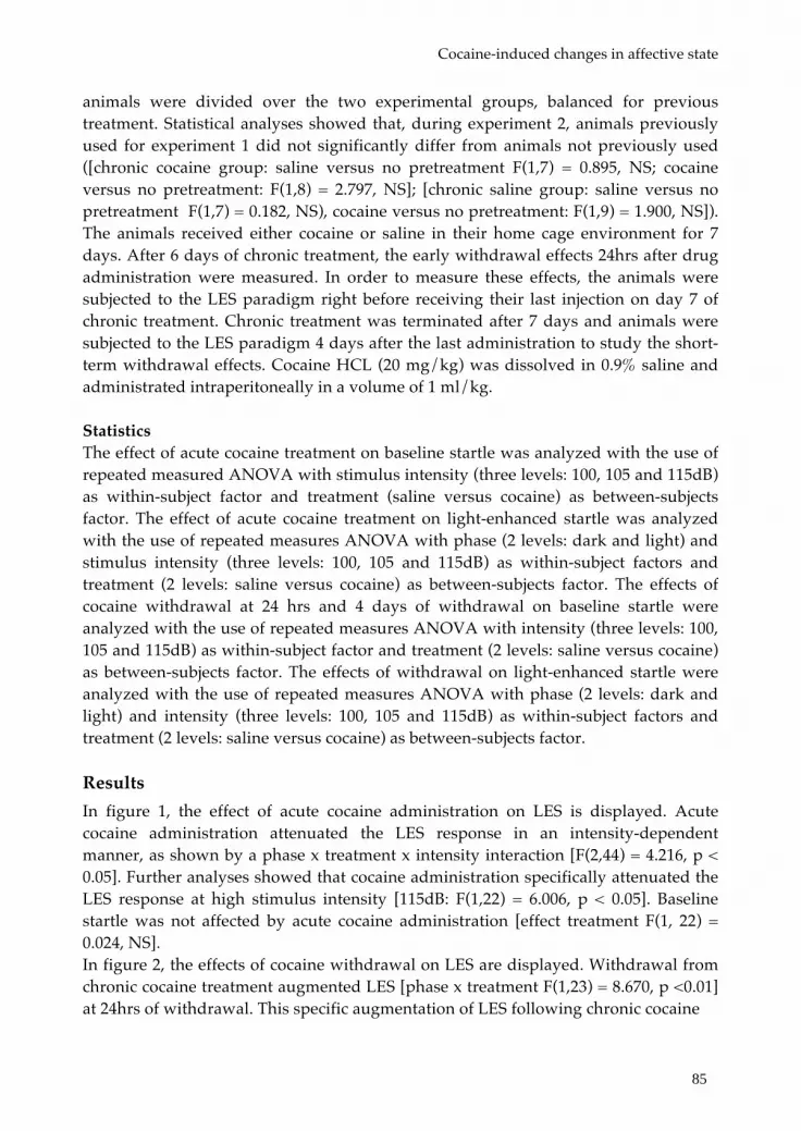
Cocaine-induced changes in affective state
animals were divided over the two experimental groups, balanced for previous treatment. Statistical analyses showed that, during experiment 2, animals previously used for experiment 1 did not significantly differ from animals not previously used ([chronic cocaine group: saline versus no pretreatment F(1,7) = 0.895, NS; cocaine versus no pretreatment: F(1,8) = 2.797, NS]; [chronic saline group: saline versus no pretreatment F(1,7) = 0.182, NS), cocaine versus no pretreatment: F(1,9) = 1.900, NS]). The animals received either cocaine or saline in their home cage environment for 7 days. After 6 days of chronic treatment, the early withdrawal effects 24hrs after drug administration were measured. In order to measure these effects, the animals were subjected to the LES paradigm right before receiving their last injection on day 7 of chronic treatment. Chronic treatment was terminated after 7 days and animals were subjected to the LES paradigm 4 days after the last administration to study the short-term withdrawal effects. Cocaine HCL (20 mg/kg) was dissolved in 0.9% saline and administrated intraperitoneally in a volume of 1 ml/kg. Statistics The effect of acute cocaine treatment on baseline startle was analyzed with the use of repeated measured ANOVA with stimulus intensity (three levels: 100, 105 and 115dB) as within-subject factor and treatment (saline versus cocaine) as between-subjects factor. The effect of acute cocaine treatment on light-enhanced startle was analyzed with the use of repeated measures ANOVA with phase (2 levels: dark and light) and stimulus intensity (three levels: 100, 105 and 115dB) as within-subject factors and treatment (2 levels: saline versus cocaine) as between-subjects factor. The effects of cocaine withdrawal at 24 hrs and 4 days of withdrawal on baseline startle were analyzed with the use of repeated measures ANOVA with intensity (three levels: 100, 105 and 115dB) as within-subject factor and treatment (2 levels: saline versus cocaine) as between-subjects factor. The effects of withdrawal on light-enhanced startle were analyzed with the use of repeated measures ANOVA with phase (2 levels: dark and light) and intensity (three levels: 100, 105 and 115dB) as within-subject factors and treatment (2 levels: saline versus cocaine) as between-subjects factor. Results In figure 1, the effect of acute cocaine administration on LES is displayed. Acute cocaine administration attenuated the LES response in an intensity-dependent manner, as shown by a phase x treatment x intensity interaction [F(2,44) = 4.216, p < 0.05]. Further analyses showed that cocaine administration specifically attenuated the LES response at high stimulus intensity [115dB: F(1,22) = 6.006, p < 0.05]. Baseline startle was not affected by acute cocaine administration [effect treatment F(1, 22) = 0.024, NS]. In figure 2, the effects of cocaine withdrawal on LES are displayed. Withdrawal from chronic cocaine treatment augmented LES [phase x treatment F(1,23) = 8.670, p <0.01] at 24hrs of withdrawal. This specific augmentation of LES following chronic cocaine
85

Chapter 5
Figure 1. The effect of acute cocaine (n=12) versus saline (n=12) administration on light-enhanced startle. The figure shows mean startle amplitudes during the dark and light phase in response to 100 (A), 105 (B) and 115dB (C) stimulus trials. * Difference between dark and light phase significantly decreased as compared to vehicle control.
100dB
saline cocaine0
250
500
750
1000
treatment
Star
tle a
mpl
itude
105dB
saline cocaine0
500
1000
1500
treatment
Star
tle a
mpl
itude
115dB
saline cocaine0
500
1000
1500
2000
2500
*
treatment
Star
tle a
mpl
itude
darklight
1A
1B
1C
86

Cocaine-induced changes in affective state
treatment was no longer significant at 4 days of withdrawal [phase x treatment interaction F(1,23) = 3.000, p < 0.1]. Cocaine withdrawal did not affect baseline startle, neither at 24hrs [F(1,23) = 0.260, NS] nor at 4 days of withdrawal [F(1,23) = 0.437, NS]. Discussion The attenuated light-enhanced startle found following acute cocaine administration suggests that an anxiety-related response to an aversive context can be counteracted by the induction of a positive affective state. An fMRI study in humans showed that acute cocaine administration increased activity in the cingulate cortex, whereas it decreased activity in the amygdala [366]. As anxiety has been associated with decreased cortical inhibition of subcortical systems and increased amygdalar activity [29] and both brain areas have been implicated in LES [130, 267], this cocaine-induced activation pattern may explain the
Figure 2. The effects of cocaine withdrawal on light-enhanced startle. The figure shows mean startle amplitudes during the dark and light phase. A: 24 hrs after cocaine administration. * Difference between dark and light phase significantly increased (p<0.05) compared to vehicle control. B: 4 days after the last administration. # Trend (P<0.1) towards increased difference between dark and light phase compared to vehicle control.
24 hrs of withdrawal
saline cocaine0
500
1000
1500 darklight *
treatment
Sta
rtle
am
plitu
de
4 days of withdrawal
saline cocaine0
500
1000
1500 darklight
#
treatment
Sta
rtle
am
plitu
de
2A
2B
87

Chapter 5
acute effects found. Similar to current findings, Davis et al. also did not find an effect of a 20mg/kg dose on baseline startle reactivity following acute cocaine administration [367]. Together these findings direct towards a very specific cocaine-induced attenuation of anxiety, as measured during light-enhanced startle. The augmented light-enhanced startle found 24hrs and, to a lesser extent, 4 days after termination of chronic cocaine treatment, shows that cocaine withdrawal-induced anxiety exacerbates light-enhanced startle. Our findings are in line with the findings from Jonkman et al (2007), who showed spontaneous withdrawal from chronic nicotine administration also results in augmented LES [358]. The current data adds to previous findings of increased anxiety-like responding following cocaine withdrawal in other paradigms, like the elevated plus maze [368] and ultrasonic vocalization [369]. At first glance, the current findings do not seem in line with previous studies on startle responding following withdrawal from chronic cocaine administration. For example, it was shown that 3 and 7 days of withdrawal from chronic cocaine treatment in the home cage environment did not alter fear-potentiated startle (FPS), an affective startle measure dependent on pavlovian fear conditioning [370-372]. However, LES and FPS are proposed to measure different types of anxiety-like responses that are, at least partially, mediated by different brain areas [130, 279]. Another big difference between the LES and FPS paradigms is the need of fear learning in order to show FPS. As conditioned and unconditioned anxiety-like responses, and thereby FPS and LES, are differently regulated in the brain, withdrawal may differently affect both paradigms. Mechanistically, another differentiation can be made between LES and FPS, as several studies suggest a specific sensitivity of LES, but not FPS, for changes in corticotropin-releasing factor (CRF) signalling. LES, but not FPS, could be attenuated by both the nonspecific CRF receptor antagonist alpha-helical CRF and the CRF1 receptor antagonist GSK876008 [123, 373]. Interestingly, CRF is strongly implicated in the neurobiology of cocaine addiction and in the aversive effect of cocaine withdrawal [187, 374]. In rodent models of cocaine withdrawal increased CRF mRNA levels were found in the amygdala, hypothalamus and basal forebrain after 2 days of cocaine withdrawal, a physiological effect accompanied by increased anxiety-like behaviour in the elevated plus maze [368, 375]. These findings may explain how cocaine-induced withdrawal resulted in augmented LES. In addition, they may explain the differential cocaine-induced effects found in FPS and LES paradigms. In conclusion, the current study showed specific cocaine-induced changes in affective startle modulation, as measured with light-enhanced startle. These findings suggest that not only negative affect, like an anxious state, but also positive affect can be detected in the light-enhanced startle paradigm. Therefore, light-enhanced startle may be a valuable tool in studying affect regulation in rodents.
88

89

90

6
Local repeated CRF infusion exacerbates anxiety- and fear-related behaviour: Differential
involvement of the basolateral amygdala and medial prefrontal cortex
Submitted for publication
Elisabeth Y. Bijlsma
Marianne L. F. van Leeuwen Koen G. C. Westphal
Berend Olivier Lucianne Groenink
91

Chapter 6
Abstract Increased central corticotropin-releasing factor (CRF) signalling has been associated with various psychiatric symptoms, including anxiety, depression and psychosis. CRF signalling in both the basolateral amygdala (BLA) and medial prefrontal cortex (mPFC) has been implicated in anxiety-like behaviour. In addition, repeated activation of CRF receptors within the BLA induces a chronic anxious state. Here we studied the effects of local repeated CRF infusion in the BLA and mPFC on different forms of anxiety, as assessed during light-enhanced startle (LES, general anxiety) and acquisition of fear-potentiated startle (FPS, cue-conditioned fear). In addition, as CRF has been implicated in sensorimotor gating, prepulse inhibition (PPI) was assessed to determine if local CRF infusion within the BLA or mPFC would interfere with the processing of sensory information. To this end, canulas were placed bilaterally in either the BLA or mPFC of Wistar rats. After recovery, animals were infused with h/rCRF (200ng/side) or vehicle for 5 consecutive days. Long-term effects of local CRF infusion on LES and acquisition of FPS were measured 4 and 10 days post-treatment, respectively. In addition, the acute (day 1), sub-chronic (day 5) and long-term (7 days post treatment) effects on PPI were measured in the same animals. A clear regional differentiation was found on the long lasting effect of CRF on anxiety-like behaviour: infusion into the BLA only enhanced acquisition of FPS, whereas infusion into the mPFC only enhanced LES. Sub-chronic CRF infusion into the BLA, but not the mPFC, disrupted PPI. This disturbed PPI was normalized 7 days post-treatment. Together, the current study shows that local repeated CRF receptor activation in the BLA and mPFC is differentially involved in anxiety- and fear-related behaviour. In addition, the BLA may be involved in CRF-induced sensorimotor gating deficits. The absence of a long-term effect on these deficits suggests that lasting activation of CRF receptors is a prerequisite for CRF-mediated effects on sensorimotor gating. The long-term effects of repeated CRF infusion on LES and acquisition of FPS on the other hand, show that in case of anxiety-related processes repeated CRF infusion may have lasting effects.
92

Repeated local CRF infusion exacerbates fear and anxiety
Introduction Corticotropin-releasing factor (CRF), a 41 amino acid neuropeptide, is involved in a large variety of physiological processes [140, 376-379]. CRF plays an important role in the activation of the hypothalamic-pituitary-adrenal (HPA-) axis in response to stress. In addition, CRF acts as a neuromodulator in extra-hypothalamic regions of the brain, where it regulates behavioural responses to stress (for review, see e.g. [252, 380]. Centrally, CRF can bind to two different receptor types, the CRF1 receptor and the CRF2α receptor which are widely expressed throughout the brain, although they differ in distribution pattern [146]. Altered CRF signalling has been implicated in various psychiatric disorders, including schizophrenia, anxiety and depression [9, 173, 381-383]. For example, several studies have shown increased cerebrospinal fluid levels of CRF and decreased central CRF receptor levels in psychiatric patients [6, 8, 9, 164, 165, 384]. As stress activates the CRF system, local CRF hyperactivity may be responsible for the development of anxiety- and depression-like symptoms following prolonged stress. The CRF1 receptor is highly expressed in mesocorticolimbic areas implicated in the processing of emotionally relevant stimuli, including the cortex, striatum, hippocampus and specific subareas of the amygdala [146, 338]. CRF signalling in both the basolateral amygdala (BLA) and media prefrontal cortex have been implicated in anxiety-like behaviour [385-387]. Interestingly, repeated activation of CRF1 receptors in the BLA induces long term neuronal changes that results in a chronic anxious state, as shown by decreased social interaction and increase anxiety-like behaviour in the elevated plus maze [385]. In addition, CRF1 receptor signalling in the BLA is involved in the consolidation of conditioned fear [388-390]. Although CRF receptors in the PFC have been implicated in anxiety and depression [8, 9, 387], the effects of repeated local CRF infusion in the prefrontal cortex on anxiety-like behaviour have not yet been studied. The aim of this study was to further investigate the effects of local repeated CRF infusion in the basolateral amygdala and prefrontal cortex on different forms of anxiety. These effects were assessed using affective modulation of the acoustic startle reflex. The startle reflex is a fast, involuntary contraction of facial and body muscles evoked by a sudden and intense acoustic stimulus [82]. The startle reflex is a cross-species phenomenon that shows high plasticity, which makes the startle reflex a very useful tool in studying various processes underlying behavioural responses to environmental stimuli. The acoustic startle reflex is modulated by emotionally valent stimuli (i.e. affective startle modulation [82, 89]. In relation to anxiety-like processes, the startle reflex can be potentiated by a cue previously coupled with an aversive event (e.g. receiving a foot shock: fear-potentiated startle) or by an aversive context (e.g. bright light: light-enhanced startle). Affective startle modulation is affected in several psychiatric disorders [103, 105, 106, 391, 392] and preclinical studies implicate CRF in affective startle modulation. For example, light-enhanced startle and potentiated startle induced by conditioned contextual fear are blocked by CRF receptor antagonists [123, 393]. However, the involvement of CRF in fear-potentiated startle is less clear. CRF
93

Chapter 6
receptor antagonists were shown to either block fear-potentiated startle [189, 195] or have no effect [123, 393]. In the current study, the effects of repeated CRF infusion in the BLA and mPFC on anxiety- and fear-related behaviour were measured in the light-enhanced startle test and in the acquisition of fear-potentiated startle, respectively. The behavioural response to emotional stimuli may depend on the ability to direct attention to relevant stimuli and on adequate processing of sensory information. Sensorimotor gating, the filtering process needed to direct attention to relevant stimuli, can easily be studied using the acoustic startle reflex [90]. This so-called prepulse inhibition of the startle reflex (PPI) is defined as the reduction in startle reflex magnitude when a startling stimulus is preceded by a weak pre-stimulus. PPI is disrupted in patients with various psychiatric disorders, especially those with psychotic features [91, 92]. Preclinical studies have already shown that the BLA and mPFC are involved in PPI [394-398]. In addition, numerous studies suggest a role for CRF in PPI. PPI is disrupted following acute intracerebroventricular (ICV) administration of CRF [192, 399]. In addition, transgenic mice that centrally over-express CRF show disrupted PPI [400, 401]. CRF-induced disruption of PPI seems to be mediated by the CRF1 receptor [192, 401]. Thus, the aim of this study was to assess the long-term effects of local repeated CRF infusion in the BLA and mPFC on different forms of anxiety. Therefore, we evaluated the long-term effects of repeated local CRF infusion in the BLA and mPFC on light-enhanced startle and the acquisition of fear-potentiated startle. In addition, prepulse inhibition was studied as a measure of sensory information processing, which was assessed at day 1 and 5 of treatment and 7 days post-treatment. Methods Subjects Male Wistar rats (Harlan, Horst, The Netherlands) were used that weighed 250-300g at the beginning of the experiment. Animals were housed in groups of four in a temperature (21°C ± 2), humidity (55% ± 5), and light controlled environment (lights on from 6 AM to 6 PM). Food and water were freely available in the home cages. The experiments were carried out during the light phase of the day-night cycle between 9 AM and 2 PM. All studies were approved by the ethical committee of the Academic Biomedical Center (DEC-ABC), Utrecht University, The Netherlands. Surgery Two weeks after arrival, animals were anesthetized with isoflurane gas anesthetic (2%-3%), mixed with oxygen and nitrous oxide and placed in a stereotact with blunted ear bars (Kopf instruments, Tujunga, USA). Before starting the procedure, animals received eye ointment on both eyes and the skin was disinfected with ethanol (70%). An incision was made and the wound was locally anesthetized with Lidocaine (5%). Guide canulas (Plastics One) were placed bilaterally in either the basolateral
94

Repeated local CRF infusion exacerbates fear and anxiety
amygdala (-2.1 AP, 5.0 ML, -7.5 DV from bregma) or medial prefrontal cortex (3.2 AP, 0.75 ML, -3.6 DV from bregma). For both locations the tooth bar was set at -3.3 mm. The guide canulas were secured in place with dental cement and three anchor screws in the skull. Guide canulas were inserted with a dummy canula and covered with dust caps to prevent clotting of and damage to the guides. Animals received 5 ml/kg of saline (subcutaneously) and Rimadyl (5mg/kg, subcutaneously) at the end of surgery and two times a day for two days post-surgery to reduce pain. Animals were allowed to recover for one week. Drugs Human/rat corticotropin-releasing factor (h/rCRF, Bachem, Weil am Rein, Germany) was dissolved in 0.05 M acetic acid and stored in aliquots at -80°C until use. Each infusion day, infusion samples were freshly prepared. Peptide was thawed and diluted 1:10 in sterile artificial cerebrospinal fluid (aCSF: 125mM NaCl, 2.5mM KCl, 1mM MgCl2, 1.25mM NaH2PO4, 2mM CaCl2.2H2O, 25mM NaHCO3, 25mM glucose and 0.5% albumin). Vehicle consisted of 10% 0.05 M acetic acid in aCSF. h/rCRF (200ng/side) or vehicle was infused bilaterally in a volume of 1µL. Infusion procedure Drug or vehicle was infused bilaterally with the use of an infusion canula that extended 1.0mm beyond the tip of the guide canula. The infusion canulas were connected to Hamilton syringes, placed in a syringe pump (KdScientific 220 series, USA), via polyethylene tubing. Rats were lightly anesthetized with isoflurane gas anesthetic (2%-3%), mixed with oxygen and nitrous oxide, for the duration of the infusion procedure (3 minutes). Before infusion, dummy canulas were removed and infusion canulas were inserted. h/rCRF or vehicle was infused directly after insertion. Solutions were infused with a rate of 1 µl/min and after infusion, infusion canulas were kept in place for an additional minute to allow adequate diffusion of the solution. After infusion, dummy canulas were inserted and the rats were put back in their home cage. Apparatus Eight startle devices were used simultaneously (SR-lab, San Diego instruments, San Diego CA, USA). The startle devices consisted of a Plexiglas cylinder (8.8 cm in diameter and 20.3 cm in length) with a stainless steel grid floor placed on a Plexiglas base. Each startle device was placed in a ventilated sound attenuated cubicle. Cage movements were measured with a piezoelectric film attached to the Plexiglas base of the startle device. A calibration system (San Diego Instruments) was used to ensure comparable startle magnitudes across the eight devices throughout the experiment. Startle stimuli, consisting of 50 ms white-noise bursts, were presented through a piezoelectric tweeter situated 15.2 cm from the top of the cylinder. Sound intensities were measured using a microphone, which was placed on top of the Plexiglas
95

Chapter 6
cylinder and fitted to a Bruel and Kjaer sound level meter (Type 2226). Startle amplitudes were sampled each ms during a period of 65 ms beginning at the onset of the startle stimulus. Each startle device was equipped with a white fluorescent bulb (9 W) on the back wall of the sound attenuated cubicle and a stimulus light in the ceiling situated 15.2 cm from the top of the cylinder. The fluorescent bulb produced an illumination level of approximately 2000lx and the stimulus light an illumination level of approximately 180lx, both measured from inside the Plexiglas cylinder using a Gossen luxmeter (MAVOLUX 5032C). There was no background illumination in any of the experiments. Light-enhanced startle Animals were placed in the startle chamber and, after a 5 min acclimation period, presented with 30 startle stimuli, 10 each at 100, 105 and 115db. These 30 stimuli constituted phase 1 (=baseline startle). Then, the fluorescent light bulb (2000lx) was turned on and the procedure, including the acclimatization period, was repeated. These 30 startle stimuli constituted phase 2 (=light-enhanced startle). A third phase, in which 30 startle stimuli were presented, followed directly after the light was turned off. The inter stimulus interval was set at 30s. Acquisition of fear-potentiated startle The acquisition of fear-potentiated startle was measured during fear-potentiated startle training. After a 5 minute acclimatization period, rats were first presented with 10 startle stimuli (115dB) to habituate the animals. After habituation, rats were presented with 10 light-shock pairings at an average interval of 4 min (range: 3-5 min). A 0.6 mA foot shock was presented during the last 500 ms of the 3700 ms light period. Shock reactivity was assessed by measuring the startle reflex in response to the foot shock. To measure the level of acquisition, animals were presented with 20 startle stimuli of 115dB over the course of the training session. More specifically, before the first light-shock pairing and in between the other light-shock pairings animals were exposed to two startle stimuli (115dB), one delivered in darkness (NA), the other one during the last 50ms of a 3250 ms light period (LN). Prepulse inhibition Animals were placed in the startle chamber and, after a 5 min acclimation period, presented with startle stimuli (115dB, 50ms) that were presented alone or preceded by noise prepulses (20ms), with 100ms between onsets of the prepulse and startle stimulus. Throughout the session background noise was set at 70dB. The prepulse intensities tested were 2, 4, 8 and 16 dB above background noise. In addition, no-stimulus trials were included as a measure of general activity. The test session was build up in 4 blocks. Block 1 and block 4 consisted of 5 startle stimulus trials. Block 2 and 3 both contained 5 startle stimulus trials, 5 prepulse + startle stimulus trials of each prepulse intensity and 5 no-stimulus trials. Trials were presented in a pseudorandom order and an average inter trial interval of 15s (range 10 to 20s).
96

Repeated local CRF infusion exacerbates fear and anxiety
Figure 1. Study design. Arrows represent repeated CRF infusions. PPI: prepulse inhibition test; LES: light-enhanced startle test; ACQ: acquisition of fear-potentiated startle during training. Experimental set-up The time line of the experiment is depicted in figure 1. For the effects of repeated CRF infusion into the BLA on PPI and LES, two cohorts of animals were combined, whereas acquisition of FPS was assessed in only 1 of the two cohorts (n = 6). One week after recovery from surgery, animals were exposed to a baseline startle test to habituate them to the experimental set up. After a 5 min acclimation period, 10 blocks of 3 startle stimuli of different intensities (100, 105 and 115dB) were presented in pseudorandom order within each block (background noise was 70dB and the inter stimulus interval was set at 30 sec). Four days later animals were tested for baseline prepulse inhibition and these data were used to match animals on basis of percentage prepulse inhibition and basal startle response to stimulus alone trials. Within each home cage, half of the animals received h/rCRF whereas the other half received vehicle. Animals were infused for 5 consecutive days and after cessation of CRF treatment, the long-term effects of repeated CRF infusion were assessed on light-enhanced startle (post-CRF day 4) and acquisition of fear-potentiated startle (post-CRF day 10). In addition, the acute (day 1), sub-chronic (day 5) and long-term (post-CRF day 7) effects on prepulse inhibition were studied. On testing days 1 and 5, animals were returned to their home cage after infusion and were tested 60 minutes later. Canula localization At the end of the experiment animals were sacrificed and brains were collected to determine the location of the canulas. Brains were cut into 60µm sections. Relevant sections were collected on alcohol-gelatin coated glass slides and stained with cresyl fast violet staining. The location of the damage made by the tip of the guide was determined and rats were removed from the dataset when guides were misplaced. Statistics Light-enhanced startle For light-enhanced startle data for both absolute startle values and percent light-enhanced startle (mean startle amplitude light – mean startle amplitude phase 2/ mean startle amplitude phase 1 * 100%) were analyzed. Analysis of absolute startle values was performed with repeated measures ANOVA with treatment as between-subjects factor and phase (3 levels) and stimulus intensity (3 levels) as within-subject
97

Chapter 6
factors. Additional repeated measures ANOVAs were done to analyze treatment effects on the different phases. In addition, independent samples t-tests were done to compare drug effects on light-enhanced startle during phase 2 at the separate stimulus intensities. Percentage light-enhanced startle was analyzed with repeated measured ANOVA with treatment as between-subjects factor and intensity (3 levels) as within-subject factor. Acquisition of fear-potentiated startle For the acquisition of fear-potentiated startle both absolute startle values and percent fear potentiation (Light Noise – Noise Alone / Noise Alone * 100%) were analyzed. For analysis of absolute startle values, repeated measures ANOVA was used with treatment as between-subjects factor and trial type (2 levels: NA vs LN) as within-subject factor. Percent fear-potentiated startle was analysed with one way ANOVA, with percent potentiation as dependent variable and treatment as fixed factor. Prepulse inhibition Percent prepulse inhibition was calculated as the mean startle magnitude to startle stimulus trials, minus the mean startle magnitude to prepulse-stimulus trials, all divided by the mean startle stimulus, and multiplied by 100. Repeated measures ANOVAs were used to analyze percent PPI, with treatment as between-subjects factor and prepulse intensity (four levels: 2, 4, 8 and 16dB) as within-subject factor. Post hoc analyses of treatment effects on the different prepulse intensities were performed with t-tests, corrected for multiple testing with a Bonferroni correction for α. Results Light-enhanced startle Figure 2 shows the long-term effect of h/rCRF treatment on light-enhanced startle. In the BLA group, significant light-enhanced startle was induced in both vehicle and CRF treated rats (Figure 3A, effect phase: vehicle [F(2,18) = 15.199, p < 0.001]; CRF [F(2,22) = 10.771, p = 0.001]). CRF infusion into the BLA did not affect light-enhanced startle (phase x treatment [F(2, 40) < 1]; phase x intensity x treatment [F(4,80) < 1]). Percentage light-enhanced startle was also not affected by repeated local CRF infusion into the BLA. Prior repeated CRF infusion into the mPFC resulted in a significantly stronger increase in light-enhanced startle than in vehicle-treated rats, which was intensity-dependent [phase x intensity x treatment: F(4,48) = 3.145, p <0.05]. Further analyses showed that this effect was due to a specific increase in startle responding to the high intensity probe (115dB) during the light phase (startle magnitude at 115 dB: vehicle group 1013±154, CRF-treated group 2192±423: phase x treatment interaction [F(2,24) = 3.351, p = 0.05]; effects during phase 2: intensity x treatment interaction [F (2,24) = 5.094, p = 0.01] and effect treatment at 115dB [F(1,13) = 6.084, p <0.05]), whereas baseline startle during phase 1 was unaffected (vehicle group 427±75; CRF group
98

Repeated local CRF infusion exacerbates fear and anxiety
611±108) Separate analysis showed that significant light-enhanced startle was induced in both vehicle and CRF treated rats (Figure 3B,]; CRF [F(2,12) = 11.098, p < 0.01]). Although the effect of prior repeated CRF infusion in the mPFC was reflected in the percentage light-enhanced startle data, the stronger increase in percentage light-enhanced startle did not reach significance (treatment x intensity [F(2,24) = 1.965, NS]; main effect treatment [F(1,12) < 1]). Acquisition of fear-potentiated startle Figure 3 shows the long-term effect of CRF treatment on absolute startle values (A) and percent potentiation (B) during acquisition of fear-potentiated startle. Prior repeated CRF infusion into the BLA increased the level of fear acquisition 10 days
Figure 2. The long-term effects of repeated CRF infusion in the basolateral amygdala or medial prefrontal cortex on light-enhanced startle. A: The figure shows mean (±SEM) startle amplitudes during phase 1 (dark), phase 2 (light) and phase 3 (dark 2) in vehicle (aCSF) and CRF treated animals after prior repeated local infusion in the basolateral amygdala (vehicle: n = 10, CRF: n = 12) or medial prefrontal cortex (vehicle: n = 7; CRF: n = 7). B: The figure shows percentage light-enhanced startle in vehicle and CRF treated animals after prior repeated local infusion in the basolateral amygdala or medial prefrontal cortex. * indicates significant effect of light condition during light-enhanced startle. # indicates significant treatment x intensity interaction during light condition.
mPFC
aCSF CRF0
50
100
150
200
treatment
perc
ent p
oten
tiatio
nBLA
aCSF CRF0
250
500
750
1000
1250
1500
**
treatment
star
tle a
mpl
itude
BLA
aCSF CRF0
50
100
150
200
treatment
perc
ent p
oten
tiatio
n
2A
2B
Absolute startle values
Percent potentiation
dark light dark2
mPFC
0
250
500
750
1000
1250
1500*
*
#
aCSF CRF
treatment
Sta
rtle
am
plitu
de
99

Chapter 6
post-treatment, as shown by a significant increase in percentage potentiation [treatment F(1,10) = 5.865, p = 0.036]. Analysis of absolute startle values reflected this difference in fear acquisition [trial x treatment F(1,10) = 4.270, p = 0.066]. Separate analysis showed that significant fear-potentiated startle was induced in both control and CRF treated rats as assessed during the acquisition session (effect trial [F(1, 5) = 6.918, p <0.05] and [F(1, 5) = 12.270, p < 0.02] for control and CRF treated rats, respectively). Shock reactivity did not differ between vehicle and CRF treated rats (mean shock reactivity ±SEM: 106±19 and 120±23 for vehicle and CRF treated rats, respectively).
Figure 3. The long-term effects of repeated CRF infusion in the basolateral amygdala or medial prefrontal cortex on the acquisition of fear-potentiated startle. A: The figures show mean (±SEM) startle amplitudes in response to Noise Alone (NA, black bars) and Light Noise (LN, light gray bars) trials and the absolute difference score between both trial types (dark gray bars) in vehicle (aCSF, BLA: n= 6; mPFC: n = 7) and CRF (BLA: n = 6; mPFC: n = 7) treated animals after prior repeated local infusion in the basolateral amygdala or medial prefrontal cortex. * indicates significant difference between startle reactivity in response to NA and LN trials. B: The figures show percentage startle potentiation during fear acquisition in vehicle and CRF treated rats after prior repeated local infusion in the basolateral amygdala or medial prefrontal cortex. * indicates significant increase as compared to vehicle treated animals.
BLA
aCSF CRF0
500
1000
1500
2000
2500
3000
**
treatment
Sta
rtle
am
plitu
de
BLA
aCSF CRF0
20
40
60
80
100
*
treatment
perc
ent p
oten
tiatio
n
mPFC
aCSF CRF0
500
1000
1500
2000
2500
3000
*
*
treatment
star
tle a
mpl
itude
Absolute startle values
Percentage fear potentiation
3A
3B
NA LN difference
mPFC
aCSF CRF0
25
50
75
100
treatment
perc
ent p
oten
tiatio
n
100
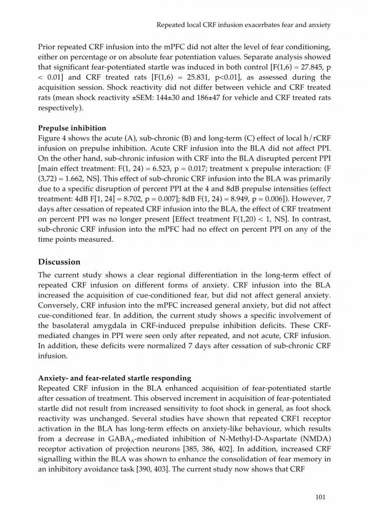
Repeated local CRF infusion exacerbates fear and anxiety
Prior repeated CRF infusion into the mPFC did not alter the level of fear conditioning, either on percentage or on absolute fear potentiation values. Separate analysis showed that significant fear-potentiated startle was induced in both control [F(1,6) = 27.845, p < 0.01] and CRF treated rats [F(1,6) = 25.831, p<0.01], as assessed during the acquisition session. Shock reactivity did not differ between vehicle and CRF treated rats (mean shock reactivity ±SEM: 144±30 and 186±47 for vehicle and CRF treated rats respectively). Prepulse inhibition Figure 4 shows the acute (A), sub-chronic (B) and long-term (C) effect of local h/rCRF infusion on prepulse inhibition. Acute CRF infusion into the BLA did not affect PPI. On the other hand, sub-chronic infusion with CRF into the BLA disrupted percent PPI [main effect treatment: F(1, 24) = 6.523, p = 0.017; treatment x prepulse interaction: (F (3,72) = 1.662, NS]. This effect of sub-chronic CRF infusion into the BLA was primarily due to a specific disruption of percent PPI at the 4 and 8dB prepulse intensities (effect treatment: 4dB F[1, 24] = 8.702, p = 0.007]; 8dB F(1, 24) = 8.949, p = 0.006]). However, 7 days after cessation of repeated CRF infusion into the BLA, the effect of CRF treatment on percent PPI was no longer present [Effect treatment F(1,20) < 1, NS]. In contrast, sub-chronic CRF infusion into the mPFC had no effect on percent PPI on any of the time points measured. Discussion The current study shows a clear regional differentiation in the long-term effect of repeated CRF infusion on different forms of anxiety. CRF infusion into the BLA increased the acquisition of cue-conditioned fear, but did not affect general anxiety. Conversely, CRF infusion into the mPFC increased general anxiety, but did not affect cue-conditioned fear. In addition, the current study shows a specific involvement of the basolateral amygdala in CRF-induced prepulse inhibition deficits. These CRF-mediated changes in PPI were seen only after repeated, and not acute, CRF infusion. In addition, these deficits were normalized 7 days after cessation of sub-chronic CRF infusion. Anxiety- and fear-related startle responding Repeated CRF infusion in the BLA enhanced acquisition of fear-potentiated startle after cessation of treatment. This observed increment in acquisition of fear-potentiated startle did not result from increased sensitivity to foot shock in general, as foot shock reactivity was unchanged. Several studies have shown that repeated CRF1 receptor activation in the BLA has long-term effects on anxiety-like behaviour, which results from a decrease in GABAA-mediated inhibition of N-Methyl-D-Aspartate (NMDA) receptor activation of projection neurons [385, 386, 402]. In addition, increased CRF signalling within the BLA was shown to enhance the consolidation of fear memory in an inhibitory avoidance task [390, 403]. The current study now shows that CRF
101

Chapter 6
Figure 4. The acute (A), sub-chronic (B) and long-term (C) effects of CRF infusion in the basolateral amygdala (left panel) or medial prefrontal cortex (right panel) on prepulse inhibition. The figure shows mean (±SEM) percentage inhibition in response to prepulse intensities of 2, 4, 8 and 16dB above background noise in vehicle (aCSF, light gray bars) and CRF (dark gray bars) treated animals. Number of subjects for each test: Acute (A): BLA [vehicle: n = 17; CRF: n = 19]; mPFC [vehicle: n = 7; CRF: n = 8], sub-chronic (B): BLA [vehicle: n = 12; CRF: n = 14]; mPFC [vehicle: n = 7; CRF: n = 7], long-term (C): BLA [vehicle: n = 10; CRF: n = 12]; mPFC [vehicle: n = 7; CRF: n = 7]. * indicates a significant decrease as compared to vehicle-treated animals.
BLA
2 4 8 160
20
40
60
80
100 aCSFCRF
prepulse intensity (dB above background)
perc
ent i
nhib
ition
mPFC
2 4 8 160
20
40
60
80
100aCSFCRF
prepulse intensity(dB above background)
perc
ent i
nhib
ition
BLA
2 4 8 160
20
40
60
80
100aCSFCRF
**
prepulse intensity(dB above background)
perc
ent i
nhib
ition
mPFC
2 4 8 160
20
40
60
80
100 aCSFCRF
prepulse intensity(dB above background)
perc
ent i
nhib
ition
BLA
2 4 8 160
20
40
60
80
100aCSFCRF
prepulse intensity(dB above background)
perc
ent i
nhib
ition
mPFC
2 4 8 160
20
40
60
80
100aCSFCRF
prepulse intensity(dB above background)
perc
ent i
nhib
ition
Acute effect of CRF on prepulse inhibition
Subchronic effect of CRF on prepulse inhibition
Long term effect of CRF on prepulse inhibition
4A
4B
4C
102

Repeated local CRF infusion exacerbates fear and anxiety
signalling within the BLA is also important at an earlier stage of fear conditioning, i.e. the initial acquisition phase. As long-term effects of CRF receptor activation within the BLA are due to increased NMDA receptor-mediated activation [385, 386, 402] and glutamate transmission in the BLA is required for adequate acquisition of FPS [404, 405], altered NMDA-mediated glutamate signalling may be responsible for the increase in acquisition of fear-potentiated startle found in the current study. Light-enhanced startle was not affected by repeated CRF infusion into the BLA. The BLA is important for light-enhanced startle, as it was shown that inactivation of non-NMDA receptors in the BLA blocks light-enhanced startle [53]. Current findings suggest that repeated CRF receptor activation does not affect non-NMDA-mediated glutamatergic signalling within the BLA. Interestingly, in another test measuring responses to potential threat, the elevated plus maze, repeated CRF infusion in the BLA also did not result in increased anxiety-like behaviour [406]. Together, these findings may suggest a specific involvement of CRF in the BLA in conditioned fear, but not anxiety responses to potential threat. A possible limitation of this study may be the relatively low acquisition of fear-potentiated startle in the BLA control group. Effects of BLA and mPFC infusions were studied in separate groups and individual differences may result in differences in the level of acquisition in separate experimental groups, especially in case of small experimental groups as in the current study. An additional factor influencing the level of acquisition is the number of cue-shock pairings. In the standardized fear-potentiated startle paradigm, the fear-potentiated startle test is preceded by 20 cue-shock pairings (2 training sessions) to establish reliable fear-potentiated startle in all control animals [95, 278]. In the current study, the effect of CRF treatment was measured during the acquisition session, which consisted of 10 cue-shock pairings. Therefore the level of acquisition was still sub-maximal. Light-enhanced startle was exacerbated following repeated CRF infusion in the mPFC. To the best of our knowledge, this is the first study showing behavioural effects of repeated CRF receptor activation in the mPFC. This observed increase in LES following repeated CRF infusion into the mPFC shows that repeated local CRF infusion may have long-term effects on neuronal excitability within the mPFC as well. Earlier studies already showed that light-enhanced startle can be decreased by CRF receptor blockade [123, 393]. Recently, the anterior cingulate cortex, a sub-region of the mPFC, was also specifically implicated in light-enhanced startle, but not fear-potentiated startle [130], which is in accordance with the current findings. We now show that the CRF-induced effects on light-enhanced startle are, at least partly, mediated by the mPFC. Together with a recent report that local acute CRF infusion into the mPFC exacerbates anxiety-like responding in the elevated plus maze [407], the current findings could indicate that CRF receptor activation in the mPFC exacerbates unconditioned generalized anxiety.
103

Chapter 6
Together, current findings confirm that repeated CRF receptor activation within the BLA exacerbates anxiety-related behaviour. We further show that this exacerbation is specific for the acquisition of cue-conditioned fear, but not generalized anxiety. On the other hand, repeated CRF receptor activation within the mPFC exacerbates generalized anxiety, but not the acquisition of cue-conditioned fear. It is highly unlikely that long-term effects of CRF are due to a direct effect of CRF, as CRF is no longer on board at the time of testing. CRF probably induces long term changes in neuronal excitability as already shown in the BLA following repeated CRF1 receptor activation [385]. The observed increase in LES following repeated CRF infusion into the mPFC shows that repeated local CRF infusion may have long-term effects on neuronal excitability within the mPFC as well. Prepulse inhibition Both the mPFC and BLA have been implicated in the regulation of PPI [394-398]. Interestingly, the current study shows that only the BLA is specifically involved in the effect of CRF on PPI. The effects of CRF in the BLA became apparent after repeated infusion and had normalized 7 days post-treatment. The finding that CRF-induced PPI deficits were normalized after cessation of treatment could mean that continued CRF receptor activation is needed for the disruption of PPI. This idea is supported by data from repeated ICV administration of CRF. Conti and co-workers showed that PPI was only disrupted following repeated ICV CRF treatment (0.3ug) in rats that also received CRF on the testing day, whereas PPI was normal in saline treated rats previously exposed to repeated CRF treatment [408]. Thus, although decreased GABAergic inhibition in the BLA has been implicated in sensorimotor gating deficits [409], long-term alterations in neuronal excitability within the BLA did not affect sensorimotor gating in the current study. In transgenic mice with central CRF excess, the PPI deficits can be normalized by acute administration of a CRF1 receptor antagonist [401], strengthening the hypothesis that PPI deficits are dependent on the level of CRF receptor activation and not on secondary changes in neuronal activity or plasticity. Together, these findings suggest that the mechanisms underlying CRF-induced sensorimotor gating deficits are different from the mechanism underlying CRF-induced effects on anxiety reported earlier [385, 386, 402]. In contrast to acute ICV administration [192, 399], acute infusion of CRF into the BLA did not affect PPI. This suggests that the acute effect of CRF on PPI following ICV administration is not solely mediated by activation of CRF receptors within the BLA, which are mainly CRF1 receptors. ICV CRF administration activates several brain areas in a pattern resembling CRF1 receptor distribution [141]. CRF1 receptors are expressed in the cortex, striatum, hippocampus, nucleus accumbens and the basolateral amygdala, all areas implicated in the control of PPI [410-413]. The acute effect of ICV CRF administration on PPI may be a summation of effects, where activation of CRF1 receptor in a range of brain areas all add up to show an acute effect
104

Repeated local CRF infusion exacerbates fear and anxiety
on PPI. However, it cannot be excluded that the dose tested in the current study was just not sufficient to induce acute effects on PPI, although this dose was shown to be sufficient to affect feeding behaviour [414]. CRF infusion into the mPFC did not affect PPI at any time point measured. Research into the role of mPFC in the regulation of PPI has primarily focused on dopaminergic signalling. In general, PPI deficits have been associated with decreased dopaminergic tone in the mPFC [90, 397, 398]. Little is known about the effects of local CRF receptor activation and its interactions with other neurotransmitter systems within the mPFC, although some studies indicate that CRF and dopamine may interact at the level of the mPFC [415, 416]. The absence of effect of CRF infusion into the mPFC on PPI in the present study, however, shows that such interactions do not affect the circuitry involved in PPI. To the best of our knowledge, this is the first study showing specific involvement of CRF signalling within the BLA in PPI. Overall, the current findings suggest that the BLA is an important structure in CRF-induced deficits in sensorimotor gating. Deficits in sensorimotor gating have also been demonstrated in patients with psychotic symptoms [91, 92, 417]. As schizophrenic patients show altered CRF-binding protein expression levels in the BLA [7], the current findings may suggest an important role for CRF signalling in the BLA in sensorimotor gating processes related to psychosis. In conclusion, the current study shows that the long-term effects of repeated CRF infusion on anxiety- and fear related behaviour depend on the brain region that is targeted. This indicates a region-specific role for CRF in the regulation of different forms of anxiety. In addition, the BLA appears to be important in sensorimotor gating deficits induced by repeated CRF infusion. A strong link has been proposed between chronic stress exposure and the development of several psychiatric disorders. As chronic stress may lead to increased activity of CRF systems, the present study suggests that regional alterations in CRF signalling may be an important factor in the development of specific psychiatric symptoms following chronic stress exposure. Individual differences in sensitivity of certain brain areas or systems may eventually determine which systems are affected by chronic stress and, consequently, which psychiatric symptoms may develop.
105

106
7
Acute and chronic paroxetine treatment affects neither fear acquisition nor
expression of cued fear and sustained anxiety
Elisabeth Y. Bijlsma Saskia Oosterbroek
Berend Olivier Lucianne Groenink
107

Chapter 7
Abstract Various studies indicate a specific involvement of the serotonin transporter in anxiety disorders. Selective serotonin re-uptake inhibitors (SSRIs) are medication of choice for anxiety disorders. However, SSRI treatment has also been associated with an initial anxiogenic effect. The exact role for SERT functioning in the development of anxiety disorders and the mechanism underlying the effects of acute and chronic SSRI treatment on anxiety-like responses are still unclear. In the current study, the acute and chronic effects of the SSRI paroxetine were studied on both the acquisition and expression of cued fear in the fear-potentiated startle paradigm. In addition, the effects of both acute and chronic paroxetine on unconditioned anxiety were assessed in the light-enhanced startle paradigm. Both acute and chronic paroxetine treatment did not affect the expression of fear-potentiated startle and light-enhanced startle. However, acute paroxetine treatment did result in overall increase in startle responding when administered during acquisition of fear-potentiated startle, suggesting increased contextual conditioning. Current findings suggest that, although paroxetine is indicated as treatment for several different anxiety disorders, paroxetine does not affect cued fear and unconditioned anxiety, as measured in the fear-potentiated startle and light-enhanced startle paradigm, respectively.
108

Paroxetine does not influence affective startle modulation
Introduction Serotonin plays an important role in emotional responses and changes in serotonin functioning have been implicated in various psychiatric disorders, including anxiety disorders and depression [5]. Various studies suggest a specific involvement of the serotonin transporter, responsible for re-uptake of serotonin from the synaptic cleft, in anxiety disorders. A common polymorphism in the promoter region of the SERT gene (5-HTTLPR) has been associated with anxiety-related traits [240], increased amygdala activity in response to threatening faces [241] and increased risk for the development of anxiety disorders and depression [239, 240], although a large meta-analysis study did not confirm this association [418]. In addition, selective serotonin re-uptake inhibitors (SSRIs) are medication of choice for anxiety disorders [232, 419]. However, SSRI treatment has also been associated with an initial anxiogenic effect in both patients [420] and healthy volunteers [421]. The exact role for SERT functioning in the development of anxiety disorders and the mechanism underlying the effects of acute and chronic SSRI treatment on anxiety-like responses are still unclear. In animals, acute SSRI treatment can increase [422, 423] or decrease [424, 425] anxiety-like responses, depending on the model used. It has been hypothesized that different animal paradigms represent qualitatively different types of fear or anxiety which are differently affected by serotonin, as serotonin has effects on multiple brain structures that mediate anxiety via multiple pathways and receptors [216, 421]. The acoustic startle response is a fast reflexive response to a sudden unexpected noise. The acoustic startle response can be modulated by salient stimuli, including conditioned fear cues (i.e. fear-potentiated startle [85]) and contexts [393, 421], as well as sustained exposure to bright light (i.e. light-enhanced startle [120, 254]). This makes the acoustic startle response a very useful tool to measure different anxiety-related processes. Studies in healthy volunteers have already shown that fear-potentiated startle is differently affected by acute and chronic citalopram treatment: acute citalopram exacerbated cued fear potentiation, whereas chronic citalopram did not affect cued fear potentiation [99, 421]. Interestingly, sustained anxiety was decreased by chronic citalopram treatment in healthy volunteers [99]. In rodents, acute treatment with citalopram increases, whereas chronic treatment with citalopram decreases acquisition of cue-conditioned fear, as measured with a conditioned freezing response [426]. The effects of chronic SSRI treatment have not yet been studied in rodent startle paradigms measuring anxiety and fear-related responses. An important difference between human and rodent fear-potentiated startle paradigms is that in human studies the level of fear-potentiated startle is measured in the same session as acquisition takes place, whereas in most rodent studies the level of fear-potentiated startle is determined in a separate session a day after fear acquisition. This makes direct comparison of drug effects on fear-potentiated startle in human and rodent studies difficult. Therefore, in the current study, we studied the acute and chronic effects of the SSRI paroxetine on both the acquisition and expression of fear-
109
Chapter 7
potentiated startle. In addition, the effects of both acute and chronic paroxetine on unconditioned anxiety were assessed in the light-enhanced startle paradigm. Materials and methods Subjects Each experiment was performed in a separate experimental group. Male Wistar rats were housed in groups of four in a temperature (21°C ± 2), humidity (55% ± 5), and light controlled environment (lights on from 6 AM to 6 PM). Food and water were freely available in the home cages. The experiments were carried out during the light phase of the day-night cycle between 8 AM and 4 PM. The study was approved by the ethical committee of the Academic Biomedical Center (DEC-ABC), Utrecht University, The Netherlands. Separate experimental groups were used for each experiment. Drugs Paroxetine hydrochloride hemi-hydrate (10 and 20mg/kg, pharmacy Mediq, Bergen op Zoom) was dissolved in water and administered daily in a volume of 2ml/kg (p.o.). On testing days, paroxetine was administered 1 hour before the test. Startle apparatus Eight startle devices were used simultaneously (SR-lab, San Diego instruments, San Diego CA, USA). The startle devices consisted of a Plexiglas cylinder (9 cm diameter and 20 cm length) placed on a Plexiglas base. During the fear-potentiated startle experiments this startle device was equipped with a stainless steel grid floor. Each startle device was placed in a ventilated sound attenuated cubicle. Cage movements were measured with a piezoelectric film attached to the Plexiglas base of the startle device. A calibration system (San Diego Instruments) was used to ensure comparable startle magnitudes across the eight devices throughout the experiment. Startle stimuli, consisting of 50 ms white-noise bursts, were presented through a piezoelectric tweeter situated 15 cm from the top of the cylinder. Background noise was set at 70 dB. Sound intensities were measured using a microphone which was placed on top of the Plexiglas cylinder and fitted to a Bruel and Kjaer sound level meter (Type 2226). Startle amplitudes were sampled each ms during a period of 65 ms beginning at the onset of the startle stimulus. Each startle device was equipped with a stimulus light in the ceiling situated 15 cm from the top of the cylinder for fear-potentiated startle conditioning and a white fluorescent bulb (9 W) on the back wall of the sound attenuated cubicle for light-enhanced startle measurements. The fluorescent bulb produced an illumination level of approximately 2000 lux and the stimulus light an illumination level of approximately 180 lux, both measured from inside the Plexiglas cylinder using a Gossen luxmeter (MAVOLUX 5032C). There was no background illumination in any of the experiments.
110

Paroxetine does not influence affective startle modulation
Experimental procedure Experiment 1: Fear-potentiated startle Fear-potentiated startle was performed as previously described [278]. In short, 4 days before the start of the experiment, animals (n = 11-12 per group) were acclimatized to the startle procedure during a habituation session (10x 100, 105 and 115dB presented in a pseudorandom order, background noise was 70dB and ISI was set at 30 sec). Then, rats were trained once a day for 2 consecutive days. During each training session, rats were presented with 10 light-shock pairings at an average interval of 4 min (range: 3-5 min). A 0.6 mA foot shock was presented during the last 500 ms of the 3700 ms light period. Shock reactivity, registered by measuring cage movements, was sampled each ms during a period of 200 ms beginning at the onset of foot shock. 24hrs after the second training, animals were put in the startle box to assess basal fear-potentiated startle values, which were used to assign rats to the different treatment groups. A week later, animals were exposed to an additional training and 24hrs later paroxetine treatment started. Animals were treated for 21 days and the acute (day1) sub-chronic (day 7) and chronic (day14 and day21) effects of paroxetine on FPS were studied. Animals did not receive additional training during the treatment period, to prevent possible interference of treatment with further acquisition. On testing days, animals were put in the startle box. After an acclimation period of 5 min, 10 startle stimuli of 105 dB were presented (ISI 30 sec), followed by 60 startle stimuli at an ISI of 30 sec, 20 each at 95, 100 and 105 dB. Half of the 60 startle stimuli were presented during the last 50 ms of a 3250 ms light period; the other half were delivered in darkness. The six different trial types were presented in a balanced, irregular order across the test session. Experiment 2: Fear acquisition The effect of acute en chronic paroxetine treatment on acquisition of fear-potentiated startle was assessed in different groups, as the level of acquisition can only be assessed in naïve rats. Before the start of the experiment, animals were acclimatized during a habituation session (set up as previously described) and were assigned to treatment groups (n = 11-12 per group) on basis of their baseline startle reactivity during this habituation session. For the acute experiment, one week after acclimatization rats received a dose of paroxetine or vehicle and they were trained as previously described. 24hrs later, animals were tested, to assess the level of fear-potentiated startle. For the chronic experiment, paroxetine treatment started one week after the habituation session and lasted for 21 days. On day 21, animals were trained as previously described and 24hrs later, animals were tested to assess the level of fear-potentiated startle. In both experiments, animals were not administered with a dose of paroxetine before the fear-potentiated startle test. Experiment 3: Light-enhanced startle Basal light-enhanced startle values were assessed to assign animals to the different treatment groups (n = 15-16 per group). A week later, paroxetine treatment started.
111

Chapter 7
Animals were treated for 21 days and the acute (day1) sub-chronic (day 7) and chronic (day14 and day21) effects of paroxetine on LES were studied. On testing days, the animals were placed in the startle chamber and, after a 5 min acclimation period, presented with 30 startle stimuli (10x 100, 105 and 115 dB) under dark control conditions and with 30 startle stimuli (10x 100, 105 and 115 dB) under brightly lit conditions. The inter stimulus interval was set at 30 sec. Statistics Fear-potentiated startle To analyze the specific effect of acute, sub-chronic and chronic paroxetine treatment, separate analyses were done for each test day. A repeated measures ANOVA was used to analyze the effect of paroxetine on mean startle amplitudes during Noise Alone and Light Noise trials. Trial type (two levels: Noise Alone and Light Noise) and stimulus intensity (three levels: 95dB, 100dB and 105dB) were used as within-subject factors and dose was used as between-subjects factor. The significance level for all analyses was 5%. Fear acquisition The effect of paroxetine on the level of fear acquisition was analyzed on basis of the level of fear-potentiated startle, as measured the day after fear acquisition. A repeated measures ANOVA was used to analyze the effect of paroxetine on mean startle amplitudes during Noise Alone and Light Noise trials. Trial type (two levels: Noise Alone and Light Noise) and stimulus intensity (three levels: 95dB, 100dB and 105dB) were used as within-subject factors and dose was used as between subjects factor. The significance level for analyses was 5% Light-enhanced startle Repeated measures ANOVAs were used to analyze overall startle reactivity. phase (two levels: dark and light) and stimulus intensity (three levels: 100dB, 105dB and 115dB) were used as within-subject factors and dose was used as between-subject factor. The significance level for analyses was 5%. Results Fear potentiated startle Figure 1 shows the effect of paroxetine on fear-potentiated startle. Fear-potentiated startle was not affected by acute paroxetine treatment (trial x dose interaction [F(2,30) < 1]; main effect dose [F(2,30) = 1.426, NS]). Sub-chronic paroxetine treatment also did not affect fear-potentiated startle (trial x dose interaction [F(2,30) < 1]; main effect dose [F(2,30) < 1]). Chronic treatment with paroxetine did not affect fear-potentiated startle, neither at 14 days (trial x dose interaction [F(2,30) < 1]; main effect dose [F(2,30) < 1]) nor at 21 days (trial x dose interaction [F(2,30) < 1]; main effect dose [F(2,30) < 1]). Significant fear-potentiated startle was established at all time points measured (effect
112
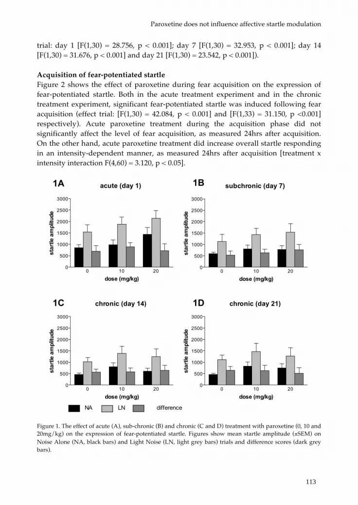
Paroxetine does not influence affective startle modulation
trial: day 1 [F(1,30) = 28.756, p < 0.001]; day 7 [F(1,30) = 32.953, p < 0.001]; day 14 [F(1,30) = 31.676, p < 0.001] and day 21 [F(1,30) = 23.542, p < 0.001]). Acquisition of fear-potentiated startle Figure 2 shows the effect of paroxetine during fear acquisition on the expression of fear-potentiated startle. Both in the acute treatment experiment and in the chronic treatment experiment, significant fear-potentiated startle was induced following fear acquisition (effect trial: [F(1,30) = 42.084, p < 0.001] and [F(1,33) = 31.150, p <0.001] respectively). Acute paroxetine treatment during the acquisition phase did not significantly affect the level of fear acquisition, as measured 24hrs after acquisition. On the other hand, acute paroxetine treatment did increase overall startle responding in an intensity-dependent manner, as measured 24hrs after acquisition [treatment x intensity interaction F(4,60) = 3.120, p < 0.05].
Figure 1. The effect of acute (A), sub-chronic (B) and chronic (C and D) treatment with paroxetine (0, 10 and 20mg/kg) on the expression of fear-potentiated startle. Figures show mean startle amplitude (±SEM) on Noise Alone (NA, black bars) and Light Noise (LN, light grey bars) trials and difference scores (dark grey bars).
subchronic (day 7)
0 10 200
500
1000
1500
2000
2500
3000
dose (mg/kg)
star
tle a
mpl
itude
chronic (day 21)
0 10 200
500
1000
1500
2000
2500
3000
dose (mg/kg)
star
tle a
mpl
itude
chronic (day 14)
0 10 200
500
1000
1500
2000
2500
3000
dose (mg/kg)
star
tle a
mpl
itude
acute (day 1)
0 10 200
500
1000
1500
2000
2500
3000
dose (mg/kg)
star
tle a
mpl
itude
1A 1B
1C 1D
NA LN difference
113

Chapter 7
This increase was primarily due to an increased overall response to the highest intensity following treatment with the 10mg/kg dose (105dB: effect treatment [F(1,30) = 3.804, p < 0.05], 10mg/kg versus vehicle: p < 0.1). Chronic paroxetine treatment did not affect the level of fear acquisition, as measured 24hrs later (trail x dose [F(2, 32) < 1], main effect dose [F(1,32) < 1]).
Figure 2. The effect of acute (A, B) and chronic (C) treatment with paroxetine (0, 10 and 20mg/kg) on the level of fear acquisition, as assessed in a drug free state 24hrs following acquisition. Figure 2A en 2B show mean startle amplitude (±SEM) on Noise Alone (NA, black bars) and Light Noise (LN, light grey bars) trials and difference scores (dark grey bars). The insert in figure 2A shows mean startle amplitudes (NA and LN trials collapsed) in response to 95dB, 100dB and 105dB stimulus trials.
acute
0 10 200
250
500
750
1000
1250
1500
1750
2000
dose (mg/kg)
star
tle a
mpl
itude
2A
chronic
0 10 200
250
500
750
1000
1250
1500
1750
2000
dose (mg/kg)
star
tle a
mpl
itude
2B
NA LN difference
overall startle responding
0 10 200
500
1000
1500
200095dB100dB105dB
dose (mg/kg)
star
tle a
mp
litu
de
114

Paroxetine does not influence affective startle modulation
Light-enhanced startle Figure 3 shows the effect of paroxetine on light-enhanced startle. Significant light-enhanced startle was induced at all time points measured (effect phase: acute [F(1,45) = 31.015, p < 0.001], sub-chronic [F(1,43) = 59.807, p < 0.001], chronic day 14 [F(1,43) = 72.680, p < 0.001], chronic day 21 [F(1,41) = 28.041, p < 0.001]). Light-enhanced startle was not affected by acute [F(2,45) < 1], sub-chronic [F(2,43) < 1] or chronic paroxetine treatment (day 14 [F(2,43) < 1]; day 21 [F(1,41) = 1.073, NS]). Discussion The current study shows that acute and chronic paroxetine treatment has no effect on either acquisition or expression of fear-potentiated startle in rats. In addition, light-enhanced startle was not affected by acute and chronic paroxetine treatment. Acute paroxetine treatment during acquisition training did increase overall startle responding as measured 24hrs later.
Figure 3. The effect of acute (A), sub-chronic (B) and chronic (C and D) treatment with paroxetine (0, 10 and 20mg/kg) on light-enhanced startle. Figures show mean startle amplitude (±SEM) during dark (black bars) and brightly lit (light grey bars) conditions and difference scores (dark grey bars).
acute (day 1)
0 10 200
100
200
300
400
500
600
700
dose (mg/kg)
star
tle a
mpl
itude
chronic (day 14)
0 10 200
500
1000
1500
2000
2500
3000
dose (mg/kg)
star
tle a
mpl
itude
subchronic (day 7)
0 10 200
100
200
300
400
500
600
700
dose (mg/kg)
star
tle a
mpl
itude
chronic (day 21)
0 10 200
500
1000
1500
2000
2500
3000
dose (mg/kg)
star
tle a
mpl
itude
3A 3B
3C 3D
dark light difference
115

Chapter 7
Neither acute nor chronic treatment with paroxetine affected the expression of fear-potentiated startle. To the best of our knowledge, this is the first report on the effect of chronic paroxetine treatment on the expression of fear-potentiated startle in rodents. The findings are in line with the one human study that also showed no effect of chronic SSRI treatment on cued fear, as measured with fear-potentiated startle [99]. It has been proposed that SSRIs reduce overgeneralization of fear, preventing contextual anxiety. As discrete conditioned fear cues remain relevant aversive stimuli, this may explain why SSRIs do not affect potentiated startle response to discrete cues [99]. The idea that SSRIs are not involved in cued fear is further supported by a clinical study that showed that SSRI treatment does not affect fear-related responding in simple phobia patients [427]. In mice, however, cue-conditioned freezing was decreased following chronic SSRI treatment [426]. Which dissimilarities in experimental set up or read out measure are responsible for these differential findings is currently unknown. However, a common feature in studies showing discrepancies in pharmacological effects on fear-related responding is the use of the startle response versus freezing behaviour as a read out. Specific comparison of the effect of contextual conditioned fear on the freezing response and startle, have shown differences in how both measures are affected by the intensity of contextual fear and in their sensitivity to pharmacological treatment [428, 429]. Thus, as freezing behaviour and the startle reflex depend on different neural substrates, they can be differentially affected by the level of fear and its modulation by pharmacological manipulations. Acute administration of paroxetine during fear acquisition increased overall startle responding, as measured 24 hrs after acquisition. Serotonin transporter knock out rats have increased extracellular serotonin levels in the hippocampus, an area implicated in contextual fear conditioning [54, 430, 431]. Interestingly, these rats also show exacerbated contextual conditioned fear following acquisition of fear- potentiated startle [432]. Thus, increases in extracellular serotonin levels following acute paroxetine may have exacerbated the development of contextual conditioned fear, resulting in an increase in overall startle responding. The finding that both acute and chronic administration of paroxetine did not affect the acquisition of fear-potentiated startle is in contrast with earlier reports that showed increased cue-conditioned fear following acute SSRI treatment in both humans and mice [421, 426]. Differences in the SSRIs tested may account for some of the discrepancies between the current findings and other studies. In aforementioned studies, citalopram was used, whereas paroxetine was used in the current study. Paroxetine and citalopram differ in affinity and selectivity for the serotonin transporter [433, 434]. Moreover, they differ in the level of activation of receptor subtypes [422, 435, 436, 215], all factors that may influence the net effect of an SSRI on fear-related behaviour. Whether these differences resemble differential involvement of serotonergic receptors in cue-conditioned fear in general, or resemble differences in sensitivity of different read outs for activation of specific receptors, remains to be elucidated.
116

Paroxetine does not influence affective startle modulation
Light-enhanced startle was not affected by chronic paroxetine treatment. A previous study in our laboratory had already shown that light-enhanced startle is not affected by acute fluvoxamine treatment [120]. The current study adds to this finding by showing that both acute and chronic paroxetine treatment also do not affect light-enhanced startle. This finding was quite surprising, as paroxetine is indicated for the treatment of panic disorder and generalized anxiety disorder [232], disorders that are associated with high levels of generalized anxiety. Light-enhanced startle is proposed to be mediated by CRF receptor activation in the BNST [53, 123, 393]. There is some evidence that SSRIs can affect CRF expression [437]. However, it has also been reported that chronic treatment with the SSRI citalopram does not affect stress-induced increases in CRF expression [438]. It could be that SSRIs do not affect CRF signalling in the brain regions involved in light-enhanced startle. Why light-enhanced startle is not affected by chronic paroxetine treatment, although this is suggested to be a measured of generalized anxiety, is subject for further investigation. It may be that the type of anxiety measured with the light-enhanced startle paradigm is different from the anxiety-like symptoms that can be alleviated by chronic SSRIs treatment. Although paroxetine is frequently used as a pharmacological treatment for various anxiety disorders [439], the current study shows no effect of chronic paroxetine treatment on affective startle paradigms measuring fear and anxiety. Does this mean that these affective startle paradigms are not a valid measure of fear or anxiety? This is not necessarily the case. First of all, in the current study the effect of chronic paroxetine treatment was studied in healthy rats. In contrast, symptom alleviation is observed in anxiety disorder patients that have a diseased brain. The anxiety- or fear-like responses seen in rodent affective startle paradigms represent a healthy and adequate response to immediate or potential threat. This may explain why this behaviour is insensitive to SSRI treatment. In line with this, insensitivity to chronic SSRI treatment has been reported in several rodent paradigms measuring anxiety, like the elevated plus maze [440, 441] and the stress-induced hyperthermia response [442]. In addition, anxiolytic effects of chronic SSRI treatment in healthy rodents may be restricted to highly anxious strains [443]. Second, the specific characteristics of the type of fear or anxiety investigated may be more important than previously acknowledged. It may be that the type of anxiety and fear measured in the light-enhanced startle and fear-potentiated startle paradigms, respectively, are different from the type of anxiety experienced by anxiety disorder patients. Moreover, affective startle paradigms in rodents differ in set up from those used in human studies. For example, differences exist in the need for conditioning and the level of awareness. Therefore, the exact type of anxiety measured in rodent startle paradigms, and its underlying neural substrate, may differ from the type of anxiety measured in human startle paradigms. This hypothesis is supported by the finding that benzodiazepines are not able to decrease the level of anxiety in human affective startle paradigms [444], whereas they do block the fear- or anxiety-like response in rodent affective startle paradigms [120, 121, 256]. Further development of affective
117

Chapter 7
startle paradigms in both humans and rodents that results in stronger similarity would increase the translational value of these paradigms and would help to more specifically characterize different forms of fear or anxiety and their underlying neural substrates. In conclusion, the current findings show that both acute and chronic paroxetine treatment does not affect the acquisition and expression of cued fear. In addition, paroxetine does not affect light-enhanced startle, a rodent read-out for generalized anxiety. Thus, although paroxetine is indicated for the treatment of panic disorder, obsessive compulsive disorders, social phobia and generalized anxiety disorder, paroxetine does not have an anxiolytic profile in rat startle paradigms measuring cued fear and generalized anxiety.
Acknowledgements The authors would like to thank Koen Westphal for his practical assistance.
118

119

120

8
Deletion of the serotonin transporter disrupts fear acquisition and exacerbates
contextual conditioned fear that can be normalized by CRF1 receptor blockade
Elisabeth Y. Bijlsma
Mark J. Millan Johnny S. Chan Edwin Cuppen Berend Olivier
Lucianne Groenink
121

Chapter 8
Abstract Various studies implicate the serotonin transporter (SERT) in anxiety disorders, although its exact role is still unclear. To further study the involvement of the SERT in fear and anxiety, different forms of affective startle modulation were assessed in the serotonin transporter knock out rat. fear-potentiated startle (FPS, cue-conditioned fear), light-enhanced startle (LES, unconditioned contextual fear), foot shock sensitization (FSS, contextual conditioned fear) and prepulse inhibition (PPI, sensory motor gating) were assessed in SERT-/-, SERT+/- and SERT+/+ rats. Changes in cue-conditioned fear were further studied during fear acquisition. In addition, the effect of disturbed fear acquisition on the level of contextual conditioned fear was studied. Furthermore, the putative normalizing effects of the CRF1 receptor antagonist CP-154,526 were studied on both the acquisition and expression of FPS. In the FPS test, SERT-/- showed blunted cue-conditioned fear as well as a deficit in fear acquisition of cue-conditioned fear. Furthermore, contextual conditioned fear in the FPS test was exacerbated in SERT-/-. Administration of CP-154,526 during acquisition, but not expression, of FPS, normalized both cue-conditioned fear and contextual conditioned fear in SERT-/- rats. Unconditioned contextual fear (LES) and PPI were normal in SERT-/-. The disturbances found in SERT-/- rats show that the SERT is important for normal fear acquisition. In addition, the absence of the SERT results in exacerbated contextual conditioned fear, resembling a pattern of fear generalization seen in panic disorder patients. Furthermore, the finding that CP-154,526 was able to normalize the acquisition of FPS and prevent exacerbation of contextual conditioned fear suggests a role for central CRF signalling in fear generalization.
122

SERT deletion results in disturbed fear acquisition
Introduction Serotonin plays an important role in emotional regulation and cognition and changes in serotonergic functioning have been implicated in various psychiatric disorders [4, 5, 216]. Various studies suggest a specific involvement of the serotonin transporter, responsible for re-uptake of serotonin from the synaptic cleft, in anxiety disorders. A common polymorphism in the promoter region of the SERT gene (SLC6A4) has been associated with anxiety-related traits [240], increased amygdala activity in response to threatening faces [241] and increased risk for the development of anxiety disorders and depression [239, 240], although a large meta-analysis study did not confirm this association [418]. In addition, selective serotonin re-uptake inhibitors (SSRIs) are medication of choice for anxiety disorders [232]. Among other things, chronic SSRI treatment alleviates symptoms of anticipatory anxiety in panic disorder patients [419]. However, SSRI treatment has also been associated with an initial anxiogenic effect in both patients [420] and healthy volunteers [421]. The exact role for SERT functioning in the development of anxiety disorders is still unclear. In animals, acute SSRI treatment can increase [422, 423] or decrease [424, 425] anxiety-like responses, depending on the paradigm used. It has been hypothesized that different animal paradigms represent qualitatively different types of fear or anxiety which are differentially affected by serotonin, as serotonin has effects on multiple brain structures that mediate anxiety via multiple pathways and receptors [216, 421]. Recently, a SERT knock out model was created in rats by N-ethyl-N-nitrosurea (ENU)-driven target-selected mutagenesis resulting in a premature stop codon [445]. This premature stop codon results in a complete ablation of SERT in the SERT-/- [431]. This SERT-/- rat shows selective disturbances in 5-HT homeostasis [431]. They show nine-fold higher extracellular 5-HT levels in the hippocampus, decreased intracellular availability of 5-HT [431] and desensitization of the serotonin 1A receptor [446-448]. Behaviourally, the SERT-/- rat shows increased anxiety-like behaviour in exploration-driven paradigms [243], changes in learning and memory processes [449] and in adaptive behaviour [450-452]. In order to further study the involvement of the serotonin transporter (SERT) in specific forms of fear and anxiety, different forms of affective startle modulation were assessed in the SERT-/- rat. The acoustic startle response is a fast reflexive response to a sudden unexpected noise. The acoustic startle response can be modulated by salient stimuli, including conditioned fear cues (i.e. fear-potentiated startle, [85]) and contexts [133, 393, 444], as well as sustained exposure to bright light (light-enhanced startle [120, 254]. This makes the acoustic startle response a very useful tool to measure different anxiety-related processes. Moreover, humans carriers of the short allele of the HTTLPR showed increased startle responding [453]. In the current study, fear-potentiated startle and light-enhanced startle were assessed in homozygous serotonin transporter knock out rats and their heterozygous and wildtype littermates. Prepulse inhibition was studied to control for possible general deficits in sensory motor gating that may interfere with processing of the emotional
123

Chapter 8
stimuli presented in this study. Results showed that SERT-/- rats had a blunted fear-potentiated startle response, which was accompanied by increased contextual conditioned fear. This blunted FPS response could be ascribed to a deficit in the acquisition of cue-conditioned fear. As SERT-/- rats were shown to be more anxious [243], which may interfere with fear acquisition, we hypothesized that blockade of general anxiety during acquisition could normalize the disturbances in fear acquisition. To this end, we tested whether the CRF1 receptor antagonist CP-154,526, known to specifically block contextual fear [454], was able to normalize the disturbances in fear acquisition. Materials and methods Subjects Male SERT knockout rats (Slc6a4 [1Hubr]) on a Wistar rat genetic background were generated by ENU-driven mutagenesis [445]. All males were derived from crossings between heterozygous rats and genotyped as described previously [36]. In all experiments, SERT+/+, SERT+/- and SERT-/- rats were compared. Animals were housed in groups of four, with mixed genotype, and housed in a temperature (21°C ± 2), humidity (55% ± 5), and light controlled environment (lights on from 6 AM to 6 PM). Food and water were freely available in the home cages. The experiments were carried out during the light phase of the day-night cycle between 8 AM and 4 PM. The study was approved by the ethical committee of the Academic Biomedical Center (DEC-ABC), Utrecht University, The Netherlands. Experimental procedure All the 5 experiments were carried out in separate experimental groups. Four days before the start of an experiment, animals were habituated to the startle set up during a habituation session. During this session animals were exposed to 30 startle stimuli (10 each of 100, 105 and 115dB), presented in a pseudorandom order. For experiment 4 and 5, animals were assigned to experimental groups on basis of mean startle amplitude during this habituation session. In experiment 1 baseline levels of fear-potentiated startle, light-enhanced startle and prepulse inhibition were assessed in separate groups of SERT+/+, SERT+/- and SERT-/- rats. In Experiment 2 the level of fear acquisition was assessed over the course of the first FPS training and, in experiment 3, changes in contextual conditioned fear were assessed during both foot shock sensitization of the startle response and contextual conditioned fear 24hrs post-shock. We then studied whether disturbed fear acquisition resulted in exacerbated contextual conditioned fear in the FPS paradigm (experiment 4A) and whether the blunted FPS in SERT-/- rats could be normalized by blocking general anxiety during fear acquisition (experiment 4B). To this end, the effect of the CRF1 receptor antagonist CP-154,526 on fear acquisition was tested. CP-154,526 was administered once, 30 minutes before fear acquisition and effects were analyzed during FPS expression 24hrs later. To determine the level of contextual
124

SERT deletion results in disturbed fear acquisition
conditioned fear in the experimental groups (experiment 4A), a cue-no-shock vehicle control group was added for each genotype. Finally, the effect of CP-154,526 on FPS post-training was studied in experiment 5. The effect of CP-154,526 (10mg/kg versus vehicle) post-training was tested in a within-subject design. Animals were tested twice, with a one-week interval. Animals were trained and 24hrs later, administered with CP-154,526 or vehicle and tested for FPS. Treatment was counterbalanced over the two test sessions and assignment of animals was balanced for baseline FPS values. Drug CP-154,526 (butyl-ethyl-(2,5-dimethyl-7-(2,4,6-trimethylphenyl)-7H-pyrrolo(2.3-d) pyrimidin-4-yl)amine)-HCl; gift from Servier, Croissy/Seine France) was suspended in 2% Tween 80 in distilled water and administered intraperitoneally in volume of 2 ml/kg 30 minutes before the fear-potentiated startle training (fear acquisition, 10 and 30 mg/kg) or test (10 mg/kg). Startle apparatus Eight startle devices were used simultaneously (SR-lab, San Diego instruments, San Diego CA, USA). The startle devices consisted of a Plexiglas cylinder (9 cm diameter and 20 cm length) placed on a Plexiglas base. During the FPS experiments this startle device was equipped with a stainless steel grid floor. Each startle device was placed in a ventilated sound-attenuated cubicle. Cage movements were measured with a piezoelectric film attached to the Plexiglas base of the startle device. A calibration system (San Diego Instruments) was used to ensure comparable startle magnitudes across the eight devices throughout the experiment. Startle stimuli (50 ms white-noise bursts) were presented through a piezoelectric tweeter situated 15 cm from the top of the cylinder. Background noise was set at 70 dB. Sound intensities were measured using a microphone, which was placed on top of the Plexiglas cylinder and fitted to a Bruel and Kjaer sound level meter (Type 2226). Startle amplitudes were sampled each ms during a period of 65 ms beginning at the onset of the startle stimulus. Each startle device was equipped with a stimulus light in the ceiling situated 15 cm from the top of the cylinder for FPS conditioning and a white fluorescent bulb (9 W) on the back wall of the sound attenuated cubicle for light-enhanced startle measurements. The fluorescent bulb produced an illumination level of approximately 2000 lux and the stimulus light an illumination level of approximately 180 lux, both measured from inside the Plexiglas cylinder using a Gossen luxmeter (MAVOLUX 5032C). There was no background illumination in any of the experiments. Fear-potentiated startle Fear-potentiated startle was performed as previously described [122, 278]. In short, rats were trained once a day for 2 consecutive days. During each training session, rats were presented with 10 light-shock pairings at an average interval of 4 min (range: 3-5 min). A 0.6 mA foot shock was presented during the last 500 ms of the 3700 ms light period. Shock reactivity, registered by measuring cage movements, was sampled each
125

Chapter 8
ms during a period of 200 ms beginning at the onset of foot shock. After an acclimation period of 5 min, 10 startle stimuli of 115 dB were presented (ISI 30 sec), followed by 60 startle stimuli at an ISI of 30 sec, 20 each at 100, 105 and 115 dB. Half of the 60 startle stimuli were presented during the last 50 ms of a 3250 ms light period (Light Noise, LN); the other half were delivered in darkness (Noise Alone, NA). The six different trial types were presented in a balanced, irregular order across the test session. Light-enhanced startle Animals were placed in the startle chamber and, after a 5 min acclimation period, presented with 30 startle stimuli, 10 each at 100, 105 and 115 dB, with an inter stimulus interval of 30 sec. Within every block of three stimuli, the three intensities were presented in a random order, with each intensity being presented only once. After the 30th stimulus the level of illumination was changed and then the procedure, including the acclimation period, was repeated. This second set of 30 stimuli constituted phase 2. Prepulse inhibition Animals were placed in the startle chamber and, after a 5 min acclimation period, presented with startle stimuli (115dB, 50ms) that were presented alone or preceded by noise prepulses (20ms), with 100ms between onsets of the prepulse and startle stimulus. The test session was build up in 4 blocks. Block 1 and block 4 consisted of 5 startle stimulus trials (115dB). Block 2 and 3 both consisted of 5 presentations of 115dB startle stimulus trials and 5 presentations each of 3 different prepulse trials (4, 8 and 16 above background preceding a 115dB pulse). In addition, no-stimulus trials were included as a measure of general activity. All trials were presented in a pseudorandom order and the inter trial intervals ranged from 25 to 35 seconds. Within-session habituation was analysed from the 115dB stimulus trials during block 1 to 4. Percentage prepulse inhibition was calculated for each prepulse intensity as percent change compared to the mean startle reflex in response to the 115dB startle stimulus. Fear acquisition Acquisition of FPS was assessed during an adjusted training session. The acquisition was similar to the normal training session. However, before the first light-shock pairing and in between the other light-shock pairings animals were exposed to two startle stimuli (105dB), one delivered in darkness (NA), the other one during the last 50ms of a 3250 ms light period (LN). Foot shock sensitization After a 5 min acclimation period, animals were presented 40 startle stimuli (115dB). After this first set of stimuli, animals received 10 foot shocks (0.6mA, 500ms in duration with an interval of 1sec). Directly after the last foot shock, animals were
126

SERT deletion results in disturbed fear acquisition
presented with another set of 40 startle stimuli (115dB). The inter-stimulus interval was set at 30sec. 24hrs later, after a 5 min acclimation period, animals were again presented with a set of 40 startle stimuli (115dB) to measure contextual conditioned fear. Statistics Baseline startle reactivity (habituation session) was analyzed with repeated measures ANOVA with stimulus intensity (three levels: 100dB, 105dB and 115dB) as within-subjects factor and genotype as between-subjects factor. In experiment 1, for fear-potentiated startle data, both the mean startle amplitudes on the Noise Alone (NA) and Light Noise (LN) trials, as well as percentage fear potentiation were analyzed. Repeated measures ANOVAs were used to analyze the mean startle amplitudes with trial type (two levels: NA and LN) and stimulus intensity (three levels: 100dB, 105dB and 115dB) as within-subjects factor and genotype (three levels) as between subjects factor. Percentage fear potentiation [(LN – NA)/NA *100%] was analyzed by oneway ANOVA with genotype as between-subjects factor. For light-enhanced startle, both the mean startle amplitudes during phase 1 and phase 2, as well as percent potentiation were analyzed. Repeated measures ANOVA was used to analyze mean startle amplitude with phase (two levels: phase 1 and phase 2) and stimulus intensity (three levels: 100dB, 105dB and 115dB) as within-subject factors and genotype as between-subjects factor. The percentage potentiation [(phase 2–phase 1)/phase 1 *100%] was analyzed by oneway ANOVA with genotype as between subject factor. Percent prepulse inhibition (mean startle amplitude to stimulus-alone minus mean startle amplitude to stimulus + prepulse, divided by mean startle amplitude to stimulus-alone, and multiplied by 100) and startle habituation (mean startle amplitude to stimulus trials during block 1 to 4) were analyzed with repeated measures ANOVAs with prepulse intensity (PPI, 3 levels: 4dB, 8dB and 16dB above background) or block (habituation, 4 levels) as within-subject factor and genotype as between-subjects factor. In experiment 2, fear acquisition data was collapsed into three phases, namely ‘initial’, representing the response to the NA and LN trials before the first cue-shock pairing, ‘1st phase’ representing the mean response to NA and LN trials presented in between the first set of 5 cue-shock pairings, and ‘2nd phase’, representing the response to NA and LN trials presented in between the second set of 5 cue-shock pairings. Repeated measured ANOVAs were used to analyze the level of acquisition with phase (three levels: initial, phase 1 and phase 2) and trial type (NA and LN) as within-subject factors and genotype as between-subject factor. Further analyses of the level of differentiation between NA en LN trials in the different genotypes, was done with repeated measured ANOVAs for each genotype separately, with phase and trial as within-subject factors. Post Hoc analyses of trial effects at the different time points were done with paired samples t-tests. In experiment 3, foot shock sensitization data from 10 trials were collapsed, resulting in 4 pre- and 4 post-shock mean startle amplitudes (blocks). Overall analysis was done
127

Chapter 8
by repeated measures ANOVA with block (8 levels) as within-subject factor and genotype as between subject factor. Subsequently, block effects for each genotype were analyzed with repeated measures ANOVAs with block as within-subject factor. Post hoc analyses were done with simple contrasts. To determine the degree of contextual conditioned fear 24 hrs post-shock, data of 40 trials were collapsed resulting in a pre-shock, and 24 hrs post shock mean startle amplitude. Subsequently a repeated measures ANOVA was performed with time as within-subject factor (2 levels: pre and 24 hrs post shock), and genotype as between-subjects factors. In experiment 4A, the level of contextual conditioned fear and the effectiveness of cue-shock association were analyzed by repeated measures ANOVA with trial (NA and LN) and intensity (2 levels: 100dB and 105dB) as within-subject factors and genotype (3 levels) and condition (2 levels: cue-no-shock vehicle versus cue-shock vehicle) as between-subjects factors. Further analyses of NA and LN trials separately was done using repeated measures ANOVAs with intensity and conditioned as within-subject factors and genotype and condition as between subjects-factors. For the analyses of the effects of CP-154,526 in experiment 4B, the effects were analyzed for each genotype separately. Repeated measures ANOVA was used with trial (NA and LN) and intensity (2 levels: 100dB and 105dB) as within-subject factors and drug (3 levels: cue-shock vehicle, cue-shock CP10 and cue-shock CP30) as between-subjects factor. In experiment 5, the effect of CP-154,526 on the expression of fear-potentiated startle was analyzed by repeated measures ANOVA with trial (2 levels: NA and LN), intensity (2 levels: 100dB and 105dB) and drug (10 mg/kg and vehicle) as within-subject factors and genotype and condition (cue-no-shock and cue-shock) as between-subjects factors. Results Experiment 1: SERT-/- show disturbed fear-potentiated startle, but normal light-enhanced startle and PPI Fear potentiated startle Figure 1 (A, B) shows absolute startle values (1A) and percent potentiation (1B) for fear-potentiated startle in SERT+/+, SERT+/- and SERT-/- rats. The genotypes did not differ in baseline startle reactivity during the habituation session (main effect genotype [F(2,81) < 1]; genotype x intensity interaction [F(4, 162) = 1.768, NS], data not shown). Overall analysis revealed a strong interference of stimulus intensity with the effect of trial type (trial x intensity interaction [F(2,66) = 10.953, p < 0.001]). Therefore, further analyses were done on startle reactivity in response to 105dB stimulus trials. Fear-potentiated startle was affected by genotype, as shown by a trial x genotype interaction [F(2,33) = 3.840, p = 0.032]. Analyses of the three genotypes separately showed that SERT-/- rats did not show fear-potentiated startle (Effect of trial: [F(1,11) < 1]), whereas SERT+/+ and SERT+/- showed significant fear-potentiated startle (effect of trial: [F(1,11) = 27.896, p < 0.001] and [F (1,11) = 11.515, p = 0.006] in SERT+/+ and SERT+/-, respectively). SERT+/- showed an intermediate phenotype, although
128

SERT deletion results in disturbed fear acquisition
Figure 1. Fear-potentiated startle, light-enhanced startle, prepulse inhibition and startle habituation in SERT+/+, SERT+/- and SERT-/- rats. 1A shows mean startle amplitude (±SEM) to Noise Alone (NA, black bars) and Light Noise (LN, gray bars) trials during fear-potentiated startle (n= 12 per genotype). 1B shows percent fear-potentiated startle. 1C shows mean startle amplitude during dark (black bars) and light (grey bars) conditions during light-enhanced startle (n = 16 per genotype). 1D shows percent light-enhanced startle. 1E shows percent prepulse inhibition in response to prepulses of 4, 8 and 16dB above background noise in SERT+/+ (black bars, n = 13), SERT+/- (dark grey bars, n = 11) and SERT-/- (light grey bars, n = 8). 1F shows startle habituation during prepulse inhibition in SERT+/+ (black line, n = 13), SERT+/- (dark grey line, n = 11) and SERT-/- (light grey line, n = 8). * Significant (P < 0.05) startle potentiation. # P < 0.05 compared to SERT+/+
Startle habituation
1 2 3 40
250
500
750
1000
1250
1500
SERT-/-SERT+/-SERT+/+
block
star
tle a
mpl
itude
Prepulse inhibition
4 8 160
20
40
60
80SERT+/+
SERT+/-
SERT-/-
prepulse intensity (dB above background)
perc
ent i
nhib
ition
Percent fear potentiation
SERT+/+ SERT+/- SERT-/-0
50
100
150
genotype
perc
ent p
oten
tiatio
n
Percent light-enhanced startle
SERT+/+ SERT+/- SERT-/-0
50
100
150
genotype
perc
ent p
oten
tiatio
n
Light-enhanced startle
SERT+/+ SERT+/- SERT-/-0
250
500
750
1000
1250
1500
* * *
dark light
genotype
star
tle a
mpl
itude
Fear-potentiated startle
SERT+/+ SERT+/- SERT-/-0
500
1000
1500
2000
2500
NA LN
* *
genotype
Sta
rtle
am
plitu
de1A 1B
1C 1D
1E 1F
129

Chapter 8
Figure 2. Acquisition of cue-conditioned fear during fear-potentiated startle training in SERT+/+ (2A, n = 19), SERT+/- (2B, n = 20) and SERT-/- (2C, n = 17) rats. ‘Initial’ represents startle amplitude in response to NA (black lines) and LN trials (grey lines) before the first cue-shock pairing. ‘1st phase’ represents mean startle amplitude in response to NA and LN trials presented in between the first set of 5 cue-shock pairings. ‘2nd phase’ represents mean startle amplitude in response to NA and LN trials presented in between the second set of 5 cue-shock pairings. * Represents significant difference between NA and LN trials.
SERT+/+
0
500
1000
1500
2000
2500
3000NALN
**
initial 1st phase 2nd phase
Star
tle a
mpl
itude
SERT+/-
0
500
1000
1500
2000
2500
3000NALN
*
initial 1st phase 2nd phase
star
tle a
mpl
itude
SERT-/-
0
500
1000
1500
2000
2500
3000NALN
initial 1st phase 2nd phase
star
tle a
mpl
itude
2A
2B
2C
130

SERT deletion results in disturbed fear acquisition
statistically, these SERT+/- could not be differentiated from the other genotypes. Analyses of percentage potentiation showed a similar pattern, as there was a trend towards a significant effect of genotype [F(2,33) = 2.517, p < 0.1]. Light-enhanced startle Figure 1 shows absolute startle values (1C) and percent potentiation (1B) for light-enhanced startle in SERT+/+, SERT+/- and SERT-/- rats. LES did not differ between the genotypes (genotype x phase interaction [F(2,45) < 1]; main effect genotype [F(2,45) < 1]. This was confirmed by the analysis of percentage LES potentiation (main effect genotype [F(2,45) < 1]). Prepulse inhibition Figure 1 (E, F) shows percent prepulse inhibition (1E) and startle habituation (1F) in SERT+/+, SERT+/- and SERT-/- rats. Both PPI and startle habituation were not different between the genotypes (PPI: main effect genotype [F(2,29) < 1], genotype x prepulse interaction [F(4, 58) < 1]; startle habituation: genotype x block interaction [F(6,87) < 1]). Experiment 2: Blunted fear-potentiated startle due to disturbed fear acquisition Figure 2 shows the level of fear acquisition during FPS training for SERT+/+ (2A), SERT+/- (2B) and SERT-/- (2B) rats. Overall analyses showed that acquisition of FPS was significantly different between genotypes (trial x genotype [F (2, 50) = 3.338, p = 0.044]. Further analyses for each genotype separately showed that SERT+/+ and SERT+/- showed significant acquisition of the cue-shock association (effect trial for SERT+/+ [F(1, 17) = 11.523, P < 0.01] and SERT+/- [F(1, 18) = 4.964, p < 0.05]), whereas SERT-/- did not differentiate between LN and NA trials [Effect trial F(1,15) < 1]. Further analyses showed that SERT+/+ showed a significant effect of trial in both first and second phase of the acquisition session (effect trial: 1st phase [t (1, 18) = -2.587, P = 0.019], 2nd phase [t (1,18) = -2.998, p = 0.008]), whereas in SERT+/- rats the effect of trial was only significant during the first phase (effect trials: 1st phase [t (1,18) = -2.946, p = 0.008], 2nd phase [t (1,19) = -1.685, p = 0.108]). Experiment 3: SERT-/- show increased contextual conditioned fear Foot shock sensitization of the startle response Figure 3A shows foot shock sensitization of the startle response in SERT+/+, SERT+/- and SERT-/- rats. The level of foot shock sensitization of the startle response differed between genotypes (block x genotype interaction [F(8,172) = 2.421, p = 0.017]). Further analyses showed that SERT+/+ and SERT+/- did not show a significant increase in startle responding after foot shock. SERT-/- on the other hand showed a marked increase in startle magnitude during blocks 6, 7 and 8 relative to pre-shock levels [main effect block [F(4,52) = 13.913, p < 0.001]; simple contrasts: block 6 [p = 0.007], block 7 [p = 0.004], block 8 [p = 0.019]. Besides, all genotypes showed a significant
131

Chapter 8
decrease in startle responding during the first block post-shock, as compared to pre-shock levels (simple contrasts: SERT+/+ [p = 0.021]; SERT+/- [p = 0.036]; SERT-/- [p = 0.005]). Contextual conditioned fear As shown in figure 3B, the alteration in startle response 24 hrs post shock relative to the response before foot shock was dependent on genotype (day x genotype interaction [F(2,43) = 4.092, p = 0.024]), SERT+/+ and SERT+/- showed a significant decrease in startle reactivity compared to pre-shock levels on day 1 (main effect day: SERT+/+ [F(1,15) = 18.305, p = 0.001], SERT+/- [F(1,15) = 6.154, p = 0.025]. In SERT-/- however, the startle response 24 hrs post shock did not differ from pre-shock levels [F(1,13) < 1]. This differential response of the genotypes was also reflected in a near significant absolute difference in startle response between genotypes on day 2 (main effect genotype: [F(2,43) = 2.520, P < 0.1]).
Figure 3. Contextual conditioned fear during foot shock sensitization of the startle response (3A) and 24hrs post-shock (3B) in SERT+/+ (black line, n = 16), SERT+/- (dark grey line, n = 16) and SERT-/- (light grey line, n = 14). * P < 0.05 compared to pre-shock, block 4, in SERT-/-. & decrease in all genotypes compared to pre-shock, block 4. Experiment 4A: SERT-/- rats show exacerbated contextual conditioned fear in FPS Figures 4A, B and C show the baseline differences in fear expression in SERT+/+, SERT+/- and SERT-/- rats, respectively. To adequately assess potential differences in conditioned contextual fear between genotypes, responses of the vehicle control group (cue-shock) were compared with those of a vehicle control group that was not shocked during training (cue-no-shock group). Overall analyses showed that the effect of cue-shock association on the response to NA and LN trials differed between genotypes (trial x condition x genotype interaction F(2,66) = 3.662, p = 0.030]). Further analyses of NA trials separately showed that the level of contextual conditioned fear during NA trials differed between genotypes (condition x genotype [F(2,66) = 5.064, p = 0.009]). SERT-/- and SERT+/- rats in the cue-shock condition showed a significant
Foot shock sensitization
40
500
1000
1500
2000
2500
3000
5 6 7 8
**
&
foot shock
*
SERT+/+SERT+/-
SERT-/-
block
Sta
rtle
am
plitu
de
Contextual conditioned fear
SERT+/+ SERT+/- SERT-/-0
500
1000
1500
2000
2500
3000 pre-shockpost-shock
# #
genotype
star
tle a
mpl
itude
3A 3B
132

SERT deletion results in disturbed fear acquisition
Figure 4. Effect of CP-154,526 during acquisition on expression of fear-potentiated startle in SERT+/+, SERT+/- and SERT-/- rats (n = 11-15 per experimental group). Data is presented in absolute startle values during NA trials (black bars) and LN trials (grey bars). Figures 4A, B and C represent the effect of fear acquisition on expression of cued and contextual conditioned fear in the fear-potentiated startle paradigm in the cue-shock condition compared to the cue-no-shock controls in SERT+/+, SERT+/- and SERT-/-, respectively. Figures 4D, E and F represent the effect of CP-154,526 during acquisition on the expression of fear-potentiated startle in SERT+/+, SERT+/- and SERT-/- rats, respectively. * LN versus NA trials p < 0.05. # p < 0.05 overall increase compared to cue-no-shock vehicle control. & p < 0.05 increase in response to NA trials compared to cue-no-shock vehicle control.
SERT+/+
cue-no-shock cue-shock0
500
1000
1500
2000
*
star
tle a
mpl
itude
SERT+/-
cue-no-shock cue-shock0
500
1000
1500
2000
*
star
tle a
mpl
titud
e
SERT -/-
cue-no-shock cue-shock0
500
1000
1500
2000 #&
star
tle a
mpl
itude
SERT+/+
vehicle 10mg/kg 30mg/kg0
500
1000
1500
2000
Star
tle a
mpl
itude
SERT+/-
vehicle 10mg/kg 30mg/kg0
500
1000
1500
2000
star
tle a
mpl
itude
SERT-/-
vehicle 10mg/kg 30mg/kg0
500
1000
1500
2000
#
*
Star
tle a
mpl
itude
4A 4D
4B
4C
4E
4F
133

Chapter 8
increase in response to NA trials compared to their cue-no-shock controls (effect condition: SERT-/- [F(1,21) = 12.912, p = 0.002]; SERT+/- [F(1,23) = 7.694, p = 0.011]), whereas SERT+/+ did not (effect condition: SERT+/+ [F(1,21) = 1.519, NS]). Direct comparison of the three genotypes in the cue-shock condition confirmed that SERT-/- rats showed exacerbated contextual conditioned fear during NA trials compared to SERT+/+, whereas SERT+/- did not (main effect genotype [F(2,33) = 10.474, p < 0.001]; Post hoc analyses: SERT-/- versus SERT+/+ [p = 0.001]; SERT+/- versus SERT+/+ [p = 0.953]). Furthermore, overall analyses of the potentiated startle response in all genotypes confirmed the previously found disturbance in cue-conditioned fear in SERT-/- rats. Both SERT+/+ and SERT+/- in the cue-shock condition showed a significant increase in response to LN trials relative to cue-no-shock animals, reflecting normal FPS (SERT+/+: shock x trial interaction [F(1,22) = 7.794, p = 0.011]; SERT+/-: shock x trial interaction [F(1,23) = 9.666, p = 0.005]). On the other hand, in SERT-/- rats in the cue-shock condition the increment in responding was similar for NA and LN trials compared to the cue-no-shock condition (main effect shock [F(1,21) = 13.500, p = 0.001]; shock x trial interaction [F(1,21) < 1]; main effect trial [F(1,21) < 1]). Experiment 4B: CP-154,526 during fear acquisition normalizes fear-potentiated startle and exacerbated contextual conditioned fear in SERT-/-
Figures 4D, 4E and 4F show the effect of CP-154,526 administration during acquisition on the expression of FPS in SERT+/+ (4D), SERT+/- (4E) and SERT-/- (4F) rats. Baseline differences in fear acquisition were shown to depend strongly on genotype. Consequently, drug effects could differ strongly between genotypes. Therefore, all effects in this experiment were analyzed separately for each genotype. In SERT+/+ and SERT+/-, administration of CP-154,526 during acquisition had no effect on the expression of FPS, nor on overall startle responding (SERT+/+: trial x dose interaction [F(2,32) < 1]; main effect dose [F(2,32) < 1]; SERT+/-: trial x dose interaction [F(2,37) < 1]; main effect dose [F(2,37) = 1.140, NS]). On the other hand, in SERT-/- CP-154,526 treatment during acquisition had a differential effect on NA and LN trials, as measured during the FPS test (trial X dose interaction [F(2, 33) = 3.756, p= 0.034]). Figure 4F shows that administration of 10mg/kg CP-154,526 during acquisition resulted in a significant fear potentiated startle response (LN versus NA [t (11) = -3.666, p = 0.004]), whereas no difference was observed between the responses to NA and LN trials in the 0mg/kg and 30mg/kg condition (LN versus NA: 0mg/kg [t (10) = 0.270, NS]; 30mg/kg [t (12) = -0.168, NS]). In SERT-/-, CP-154,526 also decreased overall startle responding measured 24 hrs later, an effect that was primarily due to the 30mg/kg dose (main effect dose [F(2,38) = 4.599, p = 0.016]; Post hoc: 30mg/kg [p = 0.008], 10mg/kg [p = 0.178]).
134

SERT deletion results in disturbed fear acquisition
Figure 5. Effect of CP-154,526 on expression of fear-potentiated startle in pre-trained SERT+/+ (5A), SERT+/- (5B) and SERT-/- (5C) rats (n = 7-9 for cue-no-shock groups and n = 9-10 for cue-shock groups). Data is presented in absolute startle values during NA trials (black bars) and LN trials (grey bars). * LN versus NA trials p < 0.05. Effect of CP-154,526 on overall startle reactivity is not depicted in figures 5A-C.
SERT-/-
vehicle 10 m/kg vehicle 10 mg/kg0
500
1000
1500
2000
2500
3000
3500NALN
cue-no-shock cue-shock
star
tle a
mpl
itude
SERT+/+
vehicle 10 m/kg vehicle 10 mg/kg0
500
1000
1500
2000
2500
3000
3500 NALN
cue-no-shock cue-shock
**
star
tle a
mpl
itude
5B
5A
5C
SERT+/-
vehicle 10 m/kg vehicle 10 mg/kg0
500
1000
1500
2000
2500
3000
3500NALN
cue-no-shock cue-shock
*
*
star
tle a
mpl
itude
135
Chapter 8
Experiment 5: CP-154,526 does not affect expression of fear-potentiated startle Figure 5 (A, B, C) shows the effect of CP-154,526 on the expression of fear-potentiated startle in pre-trained SERT+/+ (5A), SERT+/- (5B) and SERT-/- (5C) rats. The genotype differences in expression of fear-potentiated startle, as detected earlier, were confirmed by a significant shock x genotype x trial interaction [F(2,47) = 4.275, p = 0.020]. In the cue-shock condition, significant fear-potentiated startle was induced in SERT+/+ and SERT+/-, but not in SERT-/- rats (trial x genotype interaction [F(2,25) = 10.021, p = 0.001]; effect trial: SERT+/+ [F(1,9) = 18.200, p = 0.002], SERT+/- [F(1,8) = 19.033, p = 0.002] and SERT-/- [F(1,8) = 1.363, NS]). In the cue-no-shock condition, on the other hand, no fear-potentiated startle was present (main effect trial [F(1,22) < 1]) and genotypes did not differ in this condition (main effect genotype [F(2,22) < 1]). CP-154,526 had no specific effect on FPS (dose x trial interaction [F(1,47) < 1]), rather it significantly decreased overall startle responding. This effect tended to be stronger in the cue-shock condition, compared to cue-no-shock condition (dose x shock interaction [F(1,47) = 3.860, p = 0.055]; cue-no-shock: effect treatment [F(1,22) = 10.093, p = 0.004]; cue-shock: effect treatment [F(1,25) = 14.667, p = 0.001]). Discussion This study evaluated different forms of affective startle modulation in the SERT knock out rat. We found a clear disturbance in cue-conditioned fear in SERT-/- rats, as measured with the fear-potentiated startle. This disturbance was accompanied by the development of increased contextual conditioned fear, as measured in response to Noise Alone trials during FPS. Exacerbated contextual conditioned fear in SERT-/- rats was confirmed in a separate paradigm showing both increased foot shock sensitization of the startle response and increased contextual conditioned fear 24hrs post shock. In contrast, unconditioned anxiety (light-enhanced startle) and sensory information processing (prepulse inhibition) were unaffected. The blunted FPS in SERT-/- could be specifically ascribed to a deficit in acquisition of the contingency between cue and shock. Moreover, both the deficit in acquisition of cue-conditioned fear and the development of exacerbated contextual conditioned fear in SERT-/- rats could be normalized by treatment with the CRF1 receptor antagonist CP-154,526. Interestingly, the blunted FPS response was accompanied by increased contextual conditioned fear. In humans, failure to learn the cue-shock contingency has already been shown to induce sustained contextual fear, a pattern associated with fear generalization [114]. In addition, this unawareness of the cue-shock contingency has been associated with increased trait anxiety [98]. From the current data we can not explicitly state that the increased contextual fear resulted from the inability to associate shock with the predictive cue (conditioned stimulus, CS), or that these deficits developed concurrently. However, the current findings fit well with aforementioned human data and suggest that these deficits in SERT-/- rats may resemble fear generalization [114]. Interestingly, a similar pattern in fear responding
136

SERT deletion results in disturbed fear acquisition
has been reported in panic disorder (PD) patients: PD patients show deficits in acquisition of cued fear and minimal startle potentiation during fear imagery [115]. The lack of differentiation between CS+ and CS- was primarily due to an increase in responding to the CS- [115], which resembles the increased response of SERT-/- to Noise Alone trials during FPS. This deficit in PD patients was proposed to reflect overgeneralization of fear [114, 115]. Moreover, PD patients showed normal potentiation of the startle response in unconditioned measures of anxiety [455]. Thus, the current data in SERT-/- rats resembles both fear- and anxiety-related potentiated startle data in PD patients. Interestingly, panic disorder has been associated with a polymorphism in the SERT gene and PD patients show decreased serotonin transporter binding in the raphe nuclei, temporal lobes and thalamus [456, 457], further strengthening the link between SERT availability and overgeneralization of fear responses. Findings from the present study indicate that the effects of CP-154,526 are specific for fear acquisition. CP-154,526 given during acquisition reinstated the FPS response and blocked the development of contextual conditioned fear in SERT-/- rats, whereas CP-154,526 given during testing did neither normalize the expression of FPS nor blocked contextual fear in SERT-/- rats . In addition, the absence of effects of CP-154,526 on FPS in pre-trained SERT+/+ rats is consistent with accumulating evidence that CRF1 receptor antagonists do not affect the expression of FPS [123, 188]. The finding that CP-154,526 normalized the acquisition of cue-conditioned fear at one specific dose, whereas it prevented the development of contextual conditioned fear at both doses, may suggest that these effects depend on different CRF1 receptor mediated mechanisms. Both the basolateral amygdala (BLA) and hippocampus are likely to be involved in the exacerbation of contextual conditioned fear in SERT-/- rats and its normalization by CP-154,536. Both the BLA and hippocampus have been implicated in contextual fear conditioning [54, 430, 458] and receive strong serotonergic input from the DR [459]. SERT-/- rats show increased activation of the BLA [452] and SERT-/- mice show both increased foot shock-induced contextual conditioned fear as well as morphological changes in the BLA [244, 245]. Moreover, CRF1 receptor blockade in the BLA was already shown to block the development of contextual conditioned fear [388-390]. And, hippocampal infusion of the CRF1 receptor antagonist CP-154,526 blocks stress-induced increases in contextual fear conditioning [460], whereas CRF1 receptor activation within the hippocampus enhances fear learning [148, 461] How CP-154,526 facilitated the acquisition of cue-conditioned fear in SERT-/- rats is unknown. It may be that CP-154,526 decreased BLA excitability and thereby specifically prevented generalization of fear responses to other environmental cues [339, 385, 389], enabling the SERT-/- rats to focus on the single relevant cue that predicted shock presentation. Another putative locus for both the basal deficits in FPS as well as its normalization by CP-154,526 may be the serotonergic projections from the dorsal raphe (DR) to the central amygdala (CeA). The serotonergic system in SERT-/- rats is proposed to lack flexibility, showing a continuous high serotonergic
137

Chapter 8
tone in projection areas, including the hippocampus [431], and desensitization of several post-synaptic serotonin receptors, including the serotonin 1A receptor [446-448]. Both the DR and CeA have been implicated in the expression of FPS [53, 129]. Although no direct evidence exists for the involvement of these brain areas in the acquisition of FPS, changes in these projections may have prevented normal acquisition of the cue-shock association in SERT-/- rats. Moreover, the activity of these serotonergic projections can be modulated by CRF1 receptors, as they are expressed on serotonergic cell bodies within the DR [141, 146, 247, 462, 463]. This would suggest a role for these projections in the normalizing effect of CP-154,526 on the acquisition of cue-conditioned fear in SERT-/- rats. How CRF1 receptor blockade affects serotonergic signalling, and consequently the establishment of cue-conditioned fear, in the SERT-/- brain is subject to further investigation. The question remains whether CRF is also involved in the changes in fear acquisition persé. To the best of our knowledge no studies have been published showing that central CRF signalling is affected in SERT-/- rodents. One study showed an exacerbated neuroendocrine response following a CRF challenge in SERT-/- mice [464]. Overactivity of the CRF system could explain some of the changes found, like increased contextual conditioned fear and foot shock sensitization [133, 393]. However, as light-enhanced startle is also highly dependent on CRF signalling [123, 393], one would have expected altered responding of SERT-/- rats in this test as well. SERT+/- showed an intermediate level of FPS in experiment 1 and 3, but normal FPS in experiment 4 and 5. In addition, SERT+/- showed normal contextual conditioned fear in the foot shock sensitization paradigm. Apparently, high redundancy exists in the involvement of SERT in normal fear acquisition, as availability of only 60% of normal SERT levels [431] is still enough to show relatively normal fear acquisition of cue- and contextual conditioned fear. The current data provides important additional information on the exacerbated anxiety-responses in SERT-/- rats observed in other tests. Previous studies have shown that SERT-/- rats show increased anxiety in exploration-based paradigms, like an open field, elevated plus maze and novelty-suppressed feeding [243]. The current data show that, although SERT-/- consistently show increased anxiety-like behaviour in exploration-based paradigms, disturbances are restricted to conditioned fear responses in startle paradigms. Differences in read out may well explain these differences. Exploratory and freezing behaviours depend on other neuronal substrates than the startle reflex [465]. Therefore, these responses may also be differently affected by functional changes in higher modulatory brain regions, like the amygdala and prefrontal cortex. The disturbances in fear acquisition in SERT-/- are very consistent, as it was replicated in all separate experimental groups throughout the study. In addition, the finding that cue-conditioned fear was not normalized by 30mg/kg CP-154,526 (figure 4C), although this dose did decrease overall startle responding and blocked the development of contextual conditioned fear, shows that the absence of FPS in SERT-/-
138

SERT deletion results in disturbed fear acquisition
rats was not due to a ceiling effect. The finding that prepulse inhibition, a measure of sensory motor gating, is intact in SERT-/- rats shows that the deficits found are not due to general deficits in information processing, rather they are specific for the processing of emotional stimuli. A possible limitation of this study may be that we did not find an absolute increase in startle responding in SERT-/- rats following contextual fear conditioning in the foot shock sensitization paradigm. However, SERT-/- rats showed normal startle habituation in the prepulse inhibition paradigm. Therefore, we can conclude that the lack of decrease in startle responding following foot shock sensitization in SERT-/- rats reflects an increase in contextual conditioned fear compared to SERT+/+ and SERT+/- rats. In conclusion, we showed that deletion of the SERT results in clear disturbances in the acquisition of conditioned fear, resembling fear generalization. These changes show strong similarity to the altered fear responding reported in panic disorder patients. These findings further implicate the serotonin transporter in fear generalization processes in panic disorder patients. The finding that CRF1 receptor blockade with CP-154,526 during fear acquisition could normalize both the acquisition of FPS and prevent the development of exacerbated contextual fear, suggests a specific role for central CRF signalling, and CRF-serotonin interactions, in fear generalization. Furthermore, current findings suggest that the CRF1 receptor may be an interesting pharmacological target to prevent fear generalization and subsequent symptom exacerbation in panic disorder patients. Acknowledgments The authors would like to thank Monika Verdouw, Wendy Timmermans, Saskia Oosterbroek and Louise Refsgaard for their practical assistance. This study was financially supported by Neuroscience & Cognition Utrecht (the Netherlands).
139

140

9
General discussion
141

Chapter 9
142

General discussion
Major depression, anxiety disorders and schizophrenia are the most prevalent psychiatric disorders. Interestingly, high comorbidity and strong overlap in symptoms exists between these disorders [1, 2], which suggests that neurobiological mechanisms may also overlap and, consequently, these disorders may have a similar therapeutic target. Focusing on specific symptoms or behavioural changes, which transcend different psychiatric disorders (phenotype-oriented approach), may therefore be more effective for the detection of new therapeutic targets. One characteristic that seems evident in all aforementioned psychiatric disorders is disturbed affect regulation, a process that refers to the process that mediates the behavioural response to emotional stimuli. The startle reflex is widely used as a tool to study affect regulation: The startle reflex can be modulated by emotional stimuli, so-called affective startle modulation. Changes in affective startle modulation have been reported in several psychiatric disorders [102, 103, 114, 115]. However, how affective startle modulation is affected by changes in affective state in rodents has hardly been studied. Because affective startle modulation is a cross-species phenomenon, which enables direct comparison between clinical and preclinical findings, this could be a very valuable tool in studying neurobiological mechanisms underlying changes in affect. This thesis aimed at: - Evaluating how changes in affective state influence affective startle modulation in
rodents. - Further studying the involvement of, and putative interaction between, CRF and
serotonin in affect regulation. 9.1 Value of rat affective startle modulation paradigms for studying affect regulation. As described in chapter 2, light-enhanced startle and fear-potentiated startle are commonly used for the detection of anxiolytic properties of pharmaceuticals. However, De Jongh and co-workers (2005) have provided the first evidence that, next to different forms of anxiety, also other changes in affect regulation may be detected in these paradigms [134]. The first part of this thesis aimed at providing further insight in how changes in affective state are reflected in different forms of affective startle modulation in rats. In chapter 3, affective state was modulated by psychoactive compounds. It was shown that fear-potentiated startle, but not light-enhanced startle, could be exacerbated by the putative anxiogenic compounds yohimbine and m-chlorophenylpiperazine (mCPP), an α2-adrenoceptor antagonist and a serotonin 2C receptor agonist, respectively. These findings indicate that anxiogenic compounds do not necessarily increase the overall level of activation of the defensive system, which would result in an overall negative affective state. Rather, they seem to affect specific forms of anxiety, namely the type of conditioned fear measured during fear-potentiated startle. This finding is in line with clinical studies that have reported that
143

Chapter 9
the sensitivity to putative anxiogenic compounds depends on the type of underlying anxious state. For example, while panic patients report increased levels of arousal and anxiety following yohimbine administration, generalized anxiety disorder patients do not [274, 283].The lack of effect on light-enhanced startle is not due to an overall insensitivity of light-enhanced startle for anxiogenic manipulations. This is clearly shown by the findings described in chapter 5. In this study it was shown that negative affect induced by cocaine withdrawal (anxiety) does exacerbate the light-enhanced startle response. Several reports suggest that the responses measured during fear-potentiated startle and light-enhanced startle differ in their time course, pharmacology and underlying neuroanatomy. Anatomically, the central amygdala has been implicated in fear-potentiated startle, whereas the bed nucleus of the stria terminalis has been implicated in light-enhanced startle [53, 124, 125]. Pharmacologically, the fear-potentiated startle and light-enhanced startle paradigms were proposed to differ in sensitivity to compounds acting on the corticotropin-releasing factor system [123, 188, 268]. The differential effects of pharmacological and neuroanatomical manipulations in fear-potentiated startle and light-enhanced startle paradigms shown throughout this thesis (see paragraph 9.2 for thorough discussion on differentiation in sensitivity for manipulations of the serotonin and CRF system), suggest that especially the combined use of these paradigms in studying the underlying mechanisms of negative affect could provide more insight in which systems and brain areas are specifically involved in different subtypes of negative affect (e.g. involvement of the CRF system in different forms of fear and anxiety). A possible drawback for the translational value of affective startle modulation as a measure of negative affect may be that anxiety disorders in humans are generally characterized by overall increases in startle responding, rather than exacerbation of fear-potentiated startle [111, 112, 114-117]. Nonetheless, the affective startle paradigms available in rodents provide very valuable information on the regulation of normal fear- and anxiety-like responses, which will eventually help to find better drug targets for the treatment of anxiety disorders. Another interesting finding from the cocaine study (chapter 5) was that positive affect, induced by acute cocaine administration, decreased light-enhanced startle. This suggests that the induction of a positive affective state can not only decrease the startle response per se [136, 138], but can also counteract the response to an aversive context. This finding provides new insight in how activation of the appetitive and defensive motivational system influence priming of the startle response [89]. The startle reflex is not only primed by activation of either the defensive or the appetitive motivational system, but the actual magnitude of the startle response depends on the level of activation of both these motivational systems. At what level interference of a positive affective state with the response to aversive stimuli takes place is subject for further investigation. It could be that aversive priming of the startle reflex is prevented because activation of the defensive system is counteracted or overruled by activation of the appetitive motivational state. Alternatively, it could be that activation
144

General discussion
of the appetitive motivational system increases the threshold for activation of the defence system. The study described in chapter 4 shows that not only positive and negative affect, but also emotional numbing, can be detected with affective startle modulation paradigms in rats. Blunted affective startle modulation, as measured with light-enhanced startle, was found in two paradigms modelling affective disorders, namely inescapable foot shock (IFS), and olfactory bulbectomy (OBX). Moreover, sensorimotor gating was unaffected, which suggests that these changes in affective startle modulation were a direct result of blunted responding to emotional stimuli. Overall, this study shows that blunted affective startle modulation is not restricted to the maternal deprivation model [134], but can be found in several paradigms modelling affective disorders. Moreover, the finding that the rats exposed to these paradigms show a response reminiscent to that seen in patients with affective disorders, suggest that the light-enhanced startle provides high translational value and, therefore, may be a very interesting measure to study neurobiological mechanisms underlying disturbances in affect regulation. However, because in the fear-potentiated and light-enhanced startle paradigms emotional numbing can be easily misinterpreted as decreased anxiety, further development of affective startle paradigms measuring positive affect would have high additional value. Decreased anxiety would only be reflected as a decreased potentiated startle response under aversive conditions, whereas a lack of affect would be detected as blunted startle modulation both under aversive and appetitive conditions. Altogether the aforementioned studies show that emotional stimuli do not only modulate the startle reflex per se, but the level of this affective startle response can be influenced by additional changes in affective state; a negative affective state can further exacerbate the aversively primed startle reflex. Positive affect, on the other hand, can attenuate the aversively primed startle reflex. In addition, emotional numbing is reflected in blunted affective startle modulation. Some neurobiological explanations for the level of modulation will be provided later in this chapter (see paragraphs 9.3 and 9.4). 9.2 Implications for neurotransmitter involvement in affect regulation 9.2.1 Corticotropin-releasing factor Chronic stress is associated with elevated levels of CRF and changes in CRF signalling have been found in several psychiatric disorders [6-9, 173]. However, how the effects of CRF are set about is fairly unknown. To get more insight in the locus of CRF-induced effects on affect regulation, local CRF hyperactivity was modelled by repeated local infusion of CRF into the BLA or medial prefrontal cortex (chapter 6). Repeated CRF infusion into the BLA, but not medial prefrontal cortex, increased the acquisition of cue-conditioned fear, as assessed in the fear-potentiated startle paradigm. On the other hand, repeated CRF infusion into the medial prefrontal cortex,
145

Chapter 9
but not the BLA, exacerbated sustained anxiety, as assessed in the light-enhanced startle paradigm. These findings support the role of the medial prefrontal cortex in light-enhanced startle, as proposed in a recent neuronal activation study [130]. In addition, this study shows that the involvement of CRF in sustained anxiety is not restricted to the BNST [268]. In various studies it has been proposed that CRF signalling is specifically involved in sustained anxiety, but not cued fear [123, 188], resulting in a pharmacological differentiation between light-enhanced startle and fear-potentiated startle. As this study clearly shows that CRF signalling within the BLA is involved in the acquisition of fear-potentiated startle, we can conclude that the pharmacological differentiation, as described by Walker and co-workers [268], is not that straight forward. Furthermore, increased fear acquisition following local repeated CRF receptor activation can have important implications for the development of maladaptive affective responding following chronic stress. Overall, these findings show that, depending on the brain area targeted, repeated activation of the CRF system induces different types of negative affect (i.e. fear or anxiety). Therefore, individual differences in sensitivity of certain brain areas or neurotransmitter systems to the effects of CRF may eventually determine which systems are affected by chronic stress and, consequently, which type of psychiatric symptoms develop. 9.2.2 Serotonin The serotonin transporter has been implicated in a wide variety of psychiatric conditions, including anxiety disorders and depression. Moreover, selective serotonin re-uptake inhibitors, that mediate their actions through blockade of the serotonin transporter, are currently pharmacological treatment of choice for both anxiety disorders and depression. However, it is still unclear what the exact involvement of the serotonin transporter is and how SSRI treatment results in symptom alleviation. In addition, although SSRI treatment seems to be especially effective in specific types of anxiety disorders [232], it is still unclear how this clinical profile relates to the involvement of the serotonin transporter in different forms of anxiety. In chapter 7 and 8 two different strategies were used to assess the involvement of the serotonin transporter in different forms of affective startle modulation, namely chronic treatment with the selective serotonin reuptake inhibitor paroxetine (chapter 7) and the serotonin transporter knock out rat (chapter 8). We tested the serotonin transporter knock out (SERT-/-) rats and their heterozygous and wildtype littermates in several affective startle modulation paradigms and clearly showed that SERT deletion results in maladaptive fear-like responding. SERT-/- rats showed blunted cue-conditioned fear, as measured with the fear-potentiated startle paradigm, which was accompanied by increased contextual conditioned fear. On the other hand, SERT-/- rats showed normal light-enhanced startle, which suggests that unconditioned generalized forms of anxiety are not affected. The changes in cued and contextual conditioned fear induced by deletion of the SERT were specifically due to disturbances in fear acquisition. SERT-/- rats were unable to learn the cue-shock
146

General discussion
association, resulting in a failure to differentiate between CS+ and CS- trials (i.e. blunted FPS) and overgeneralization of their fear responses (i.e. increased contextual conditioned fear). In contrast to life long absence of the serotonin transporter, chronic blockade of this transporter with the SSRI paroxetine had no effect on the different forms of anxiety measured in both FPS and LES. In addition, neither acute nor chronic treatment with paroxetine affected fear acquisition. This finding was highly surprising. SSRIs are known to be quite effective in alleviating feelings of anxiety in patients [232] and chronic treatment with the SSRI citalopram decreased sustained anxiety, but not cue-conditioned fear, in humans, as measured with an adapted fear-potentiated startle procedure [99]. What causes this discrepancy between chronic effects of SSRIs in rodent and human startle paradigms is currently unknown. What this discrepancy can tell us about the translational value of anxiety-related startle paradigms in the validation of new anxiolytic compounds is discussed later in this chapter (see paragraph 9.6). SERT knock out rat as a model for maladaptive fear Interestingly, the inability to learn a CS-US contingency has also been shown to induce sustained contextual fear in humans. In addition, it has been associated with trait anxiety [98]. Moreover, the alterations in fear responding in SERT-/- rats show strong similarity to those seen in panic disorder patients. Panic disorder patients show deficits in the acquisition of cue-conditioned fear and minimal startle potentiation during fear imagery [115]. The lack of CS+/CS- differentiation was primarily due to an increase in responding to the CS-, which resembles the increased response of SERT-/- rats to Noise Alone trials during FPS. The changes found in panic disorder patients have been explained by the fear generalization concept [31, 114]. This concept describes the generalization of anxiety-like responding to previously neutral environmental cues that resemble or co-occurred with the actual aversive cue. This process seems very evident in panic disorder patients. In these patients the experience of panic attacks is at first constraint to a specific stressful situation, which they associate with an aversive experience (e.g. public speaking after a previous bad experience during presenting). However, this process can transfer to similar situations (e.g. being social in a group setting or listening to someone else’s presentation). This process results in proliferation of cues that can trigger anxiety, i.e. overgeneralization of fear. In the fear-potentiated startle paradigm, one could imagine that the inability to learn the cue-shock association, and therefore the inability to differentiate between relevant and irrelevant cues, could result in a similar process of generalization, which would explain the exacerbated response to Noise Alone trials in SERT-/- rats. From the data presented in chapter 8, however, we cannot definitely conclude that the increased contextual fear resulted from the inability to associate shock with the predictive cue, or that these deficits developed concurrently. However, the strong similarities with human data support an involvement of the serotonin transporter in these fear generalization processes. In addition, based on the behavioural pattern
147

Chapter 9
shown in SERT-/- rat, it can be hypothesized that the SERT-/- rat would be a very useful model for the development of maladaptive fear in panic disorder. SERT knock out rat as a model for chronic SSRI treatment It has been proposed that the SERT-/- rat is a model for chronic SSRI treatment. Several studies have shown similar behavioural changes and physiological adaptations in SERT-/- rats as seen following chronic SSRI treatment. For example, male sexual behaviour is similarly affected [466, 467] and SERT-/- rats show similar changes in serotonin 1A receptor sensitivity as reported following chronic SSRI treatment [448, 466, 467]. However, the chronic SSRI data presented in this thesis (chapter 7) show that, regarding affective startle modulation, the SERT-/- rat is definitely not a model for chronic SSRI treatment. Several factors may explain these differences. First of all, it has to be taken in consideration that the effects of chronic SSRI treatment were studied in healthy subjects. The anxiety- or fear-like responses seen in rodent affective startle paradigms represent a healthy and adequate response to immediate or potential threat. This response may be hardwired and therefore not sensitive to relatively small, pharmacologically induced, changes in sensitivity and functionality of the serotonergic system [468]. This idea is supported by the data presented in chapter 8: Heterozygous SERT knock out rats, which express around 60% of normal SERT levels [431], show relatively normal behaviour in affective startle paradigms. A second factor that may explain the differential effects on fear-like behaviour between SERT deletion and chronic SSRI treatment is the fact that SERT-/- rats lack the serotonin transporter already from early development. It is possible that adaptive changes have occurred that do not resemble the adaptive changes that occur following chronic SSRI treatment in a fully developed adult brain. Moreover, several studies indicate that the neural systems regulating anxiety-like behaviour are especially sensitive to changes in serotonergic functioning during early development. In mice, manipulating serotonin signalling during early development was shown to disturb the normal expression of anxiety-like behaviour in adulthood [469, 470]. Moreover, a recent genetic study showed that a functional polymorphism in the serotonin transporter is associated with increased risk of developing psychiatric illness following early life stress in humans [179]. CRF-serotonin interactions in affect regulation The finding that CRF1 receptor blockade prevented the development of maladaptive fear responses in the SERT-/- rat (chapter 8) indicates a strong interaction between serotonin and CRF in the regulation of affect. We showed that blockade of the CRF1 receptor with CP-154,526 during fear acquisition normalized the acquisition of cue-conditioned fear and prevented the development of contextual conditioned fear in SERT-/- rats. Apparently, life long absence of the serotonin transporter results in physiological changes that can be, at least partly, counteracted by CRF1 receptor blockade, resulting in normalization of fear acquisition. Interestingly, CP-154,526 was only capable of normalizing fear-like responding during fear acquisition. When
148
General discussion
administered in pre-trained SERT-/- rats that already show maladaptive fear behaviour, CP-154,526 was not able to normalize behaviour. This finding further underlines the specific involvement of CRF in the acquisition, but not expression, of cued and contextual conditioned fear. In addition, it has strong implications for clinical effectiveness of CP-154,526 in the treatment of anxiety disorders, as discussed under ‘clinical implications’. In this regard, a study from Bradley and co-workers is especially interesting [178]. They showed that a specific polymorphism in the CRF1 receptor gene protects against the development of affective disorders following early life trauma. Moreover, in line with the proposed interactions between serotonin and CRF in affect regulation, a clinical study also showed a clear interaction between polymorphisms in the genes coding for the CRF1 receptor and the serotonin transporter in the vulnerability to develop psychiatric disorders following early life trauma [179]. Altogether, interactions between CRF and serotonin may play a crucial role in the transition from normal to maladaptive affective responding following prolonged stress exposure and blockade of the CRF1 receptor early in this process may have beneficial effects in anxiety disorder patients. 9.3 Implications for psychiatry Interestingly, the finding that the CRF1 receptor antagonist CP-154,526 was able to prevent fear generalization in the SERT-/- rat may have important implications for the use of CRF1 receptor antagonist in the treatment of several anxiety disorders. As mentioned earlier, the data obtained in SERT-/- rats resemble behavioural changes observed in panic disorder patients. Treatment with a CRF1 receptor antagonist may prevent the generalization of anxiety-like responding to previously neutral environmental cues and thereby prevent exacerbation of anxiety-like symptoms. Also in post-traumatic stress disorder, this approach may be very effective, as treatment with a CRF1 receptor antagonist may be able to prevent the exacerbation of anxiety-like symptoms outside of the context of the aversive event that initially triggered the development of this disorder. However, the time point of treatment may be very crucial for the effectiveness of treatment. As also shown in chapter 8, treatment with CP-154,526 was not effective in normalizing cue-conditioned fear and blocking contextual fear once maladaptive fear responses were established. This suggests that CRF1 receptor antagonists may no longer be able to alleviate the anxiety-like symptoms, once the maladaptive fear regulation has been established. Early detection of anxiety-like symptoms and initiation of pharmacological treatment before exacerbation of symptoms may therefore be crucial for CRF1 receptor antagonists to be effective. 9.4 Excitability of the basolateral amygdala The BLA has been described as an integration side of sensory information, linking this information to biologically relevant events [46]. The BLA also receives information from the hippocampus about the context related to the event. The BLA has direct
149

Chapter 9
projections to the central amygdala, implicated in cue-conditioned fear, and to the BNST, implicated in generalized anxiety. In addition, amygdala activity is regulated by inputs from the prefrontal cortex. Anxiety disorders have been associated with increased amygdala volume [75, 76], whereas major depression has been associated with decreased amygdala volume [62-68]. Because imaging techniques in humans lack spatial resolution, no definite conclusions can be drawn from these human studies about the involvement of specific subnuclei of the amygdala. However, preclinical research, including findings described throughout this thesis (chapters 4, 6, and 8) suggests that the BLA may be especially involved in the transition from normal to maladaptive affect regulation. Here I will describe why the BLA, and especially CRF1 receptor signalling within this subnucleus, may be especially involved in altered affective responding and how the level of excitability of the BLA may determine the direction of these alterations. BLA hyperexcitability has been associated with increased anxiety-like behaviour [339, 385, 386]. Repeated activation of CRF receptors within the BLA results in NMDA receptor-mediated hyperexcitability [339, 385] and increased fear acquisition (chapter 6). On the other hand, both NMDA receptor antagonist-induced BLA inactivation and local CRF1 receptor blockade disrupt the acquisition of conditioned fear [388, 389, 404]. Thus, the level of BLA activation determines the level of fear acquisition. Blunted affective startle modulation, as found following IFS and OBX (chapter 4) may also be ascribed to changes in BLA excitability. Blockade of glutamate signalling within the BLA was shown to block light-enhanced startle [53] and changes in glutamate signalling have been found both following IFS and OBX, suggesting a compensatory mechanism preventing glutamate hyperrelease [316, 340]. This decrease in glutamate-mediated excitability may have resulted in decreased (or blunted) glutamatergic activation in response to emotional stimuli. Thus, the level of excitability of the BLA seems to determine the level of affect regulation: hyperexcitability of the BLA increases, whereas reduced excitability decreases the strength of the associations formed between sensory information and environmental cues, received from the sensory cortices and hippocampus, respectively. Moreover, the level of BLA excitability and the strength of the associations formed can be modulated by the level of CRF activity. At first sight, the hypothesis that BLA hyperexcitability exacerbates the acquisition of conditioned fear is only partly supported by the findings in the SERT-/- rats. BLA activity has been associated with the acquisition of both cued and contextual conditioned fear. Although, SERT-/- rats show increased excitability of the BLA [452], they also show blunted acquisition of cue-conditioned fear. What underlies this discrepancy is unknown. However, as physiological changes have been observed in various other brain regions of the SERT-/-, including the prefrontal cortex and hippocampus [431, 452], input of information to the BLA may also be hampered. It may be that information about the predictive cue is less well processed. If BLA excitability in the SERT-/- indeed leads to an increased ability to form associations between environmental cues and sensory information, disturbed processing of the
150

General discussion
predictive cue on a different level, may lead to increased coupling of sensory information with other (previously irrelevant) environmental cues. This process could possibly explain the increased contextual conditioned fear and fear generalization detected in these animals. Interestingly, the finding that CP-154,526 could block this fear generalization process may further underline the important role of CRF in the processing of affective stimuli. Altogether, aforementioned findings suggest that the level of BLA excitability may be an important determinant in disturbed affect regulation, with increased excitability contributing to increased negative affect (anxiety-like symptoms) and decreased excitability in emotional numbing (depressive-like symptoms). In line with this hypothesis, one could also speculate that time-dependent changes in affect regulation occur. It may be that prolonged stress, at first, leads to increased excitability of the BLA (and anxiety-like symptoms). As time goes by, the brain may develop compensatory mechanisms to counteract the glutamate-mediated hyperactive state of the BLA, which may ultimately lead to emotional numbing (i.e. depressive-like state). The BLA excitability hypothesis, of course, represents a very simplified model. The BLA is innervated by and provides input to various brain regions, that all have a function in determining how affect regulation is modulated. 9.5 Sensory information processing Whether emotional valence is effectively ascribed to environmental stimuli also depends on other aspects of information processing: is attention effectively driven to relevant stimuli? And, is sensory information adequately processed? Prepulse inhibition is a measure of the early pre-attentive stages of information processing and is used as an operational measure of sensorimotor gating, a filtering process needed to direct attention to relevant stimuli [90]. In several of the studies described in this thesis (chapter 4, 6 and 8), prepulse inhibition was included to control for above mentioned additional aspects determining the level of affect regulation. This seemed especially important because stress in general, and CRF in particular, have been associated with disturbed sensory information processing [192, 399-401]. Within this thesis, we have confirmed the importance of CRF in sensory information processing, as repeated activation of CRF receptors within the BLA disrupted prepulse inhibition (chapter 6). This disruption was probably not mediated the CRF-induced BLA hyperexcitability mechanism described before, as continued CRF receptor activation was needed to show this disruption. Prepulse inhibition was not affected by any of the paradigms modelling affective disorders, namely IFS, OBX and repeated restraint stress (chapter 4) and also SERT-/- rats did not show any abnormalities on this measure (chapter 8), whereas alterations in affective startle modulation were observed in these studies. This shows that the manipulations described in this thesis all induced specific changes in the emotional domain, without affecting general processing of information. It could be expected that disturbances in sensory information processing interfere with the formation of associations between sensory information and
151
Chapter 9
environmental cues. However, it is currently unknown whether disturbed prepulse inhibition would lead to disturbances affective startle modulation. 9.6 Methodological considerations and recommendations Fear-potentiated startle, but also light-enhanced startle, is used as a tool to detect anxiolytic properties of (new) psychoactive compounds. Advantages of this use are that the startle reflex has high translational value, as it is a cross-species phenomenon. In addition, fear-potentiated startle and light-enhanced startle do not involve an active behavioural response, like measures of exploratory behaviour. However, the pharmacological data presented in this thesis (chapters 3 and 7) do not consistently confirm the proposed anxiety-modulating properties of the psychoactive compounds tested. For example, the study described in chapter 7 shows no effect of chronic paroxetine treatment on affective startle paradigms measuring fear and anxiety, although paroxetine is frequently used as a pharmacological treatment for various anxiety disorders [439]. Does this mean that these affective startle paradigms are not a valid measure of fear or anxiety? This is not necessarily the case. First of all, in aforementioned study the effects of chronic paroxetine treatment were studied in healthy rats. In contrast, symptom alleviation is observed in anxiety disorder patients that have a diseased brain. The anxiety- or fear-like responses seen in rodent affective startle paradigms represent a healthy and adequate response to immediate or potential threat. This may explain why this behaviour is insensitive to SSRI treatment. In line with this, insensitivity to chronic SSRI treatment has been reported in several rodent paradigms measuring anxiety, like the elevated plus maze [440, 441] and the stress-induced hyperthermia response [442]. In addition, anxiolytic effects of chronic SSRI treatment in healthy rodents may be restricted to highly anxious strains [443]. Second, the specific characteristics of the type of fear or anxiety investigated may be more important than previously acknowledged. It may be that the type of anxiety and fear measured in the light-enhanced startle and fear-potentiated startle paradigms, respectively, are different from the type of anxiety experienced by anxiety disorder patients. Third, affective startle paradigms in rodents differ in set up from those used in human studies. For example, differences exist in the need for conditioning and the level of awareness and predictability. Therefore, the exact type of anxiety measured in rodent startle paradigms, and its underlying neural substrate, may differ from the type of anxiety measured in human startle paradigms. This hypothesis is supported by the finding that benzodiazepines are not able to decrease the level of anxiety in human affective startle paradigms [444], whereas they do block the fear- or anxiety-like response in rodent affective startle paradigms [120, 121, 256]. Further development of affective startle paradigms in both humans and rodents that results in stronger similarity would increase the translational value of these paradigms and would help to more specifically characterize different forms of fear or anxiety and their underlying neural substrates.
152

General discussion
9.7 Concluding remarks The data presented in this thesis show that rat affective startle modulation paradigms can be used to study the brain systems involved in the regulation of affective state. It was shown that a negative affective state results in exacerbation of the potentiated startle response, whereas positive affect results in attenuation of the potentiated startle response. In addition, emotional numbing is reflected in blunted affective startle modulation. It would be interesting to further study how affective state influences the affective startle paradigms. For example, future studies could investigate what pharmacological manipulations are able to normalize the blunted affective startle modulation seen following IFS and OBX. In addition, it would be interesting to study what neurobiological changes are induced by IFS and OBX (e.g. changes receptor expression levels or neurotransmitter levels in specific neural systems) that correlate with the blunted affective startle response. In addition, the data presented in this thesis show that CRF and serotonin play an important role in affect regulation. SERT deletion was shown to disrupt fear acquisition and exacerbate contextual conditioned fear, resembling the behavioural pattern seen in panic disorder patients. Further studying the mechanism underlying disturbed fear acquisition in SERT-/- rats may provide new insight in how panic disorder develops. CRF1 receptor blockade was found to normalize the disturbed fear-like behaviour seen in SERT-/- rats, but only when the CRF1 receptor was blocked during fear acquisition. This may have strong implications for the use of CRF1 receptor antagonists in the treatment of panic disorder specifically, or anxiety disorders in general. It seems that CRF1 receptor blockade specifically prevents the development of fear generalization. This suggests that CRF1 receptor antagonists may be especially effective early on in disease development. Further validation of CRF1 receptor antagonists as a treatment for panic disorder is desirable. The effectiveness of CRF1 receptor antagonists should be tested in other animal paradigms that can measure the effects of treatment both throughout the development and after the establishment of panic-like behaviour. Moreover, it would be very interesting to study whether the formation of the generalized fear responses in panic disorder patients, as in the fear-potentiated startle paradigm [115], can be prevented by treatment with a CRF1 receptor antagonists. Next to this potential therapeutic effect of CRF1 receptor antagonists, it was shown that CRF itself also modulates affective state. Depending on the brain area targeted, repeated local CRF infusion induces different types of negative affect (i.e. fear or anxiety). Together these findings suggest an important role for CRF in affect regulation and possibly also in the development of psychiatric disorders following chronic stress. Individual differences in sensitivity of certain brain areas or neurotransmitter systems to the effects of CRF may eventually determine which systems are affected by chronic stress and, consequently, which type of psychiatric symptoms develop. Considering the strong interaction found between CRF and serotonin in chapter 8 and the recent report on interactions between functional
153
Chapter 9
polymorphisms in the SERT and CRF1 receptor genes in the risk of developing psychiatric disorder following early life trauma [179], I think that especially the influence of CRF on the serotoninergic system can be considered an important target for future research. In conclusion, rat affective startle modulation paradigms are a useful tool to study the neurobiological aspects of affect regulation. In addition, Both CRF and serotonin play an important role in the regulation of emotions. Especially studying the interactions between these two neuromodulators may give more insight in how increased negative affect develops. Moreover, this thesis provides promising data on the use of CRF1 antagonists to prevent the development of maladaptive fear in anxiety disorders.
154

155

156

References
157

158

References
[1] Ramsay R, Gorst-Unsworth C, Turner S. Psychiatric morbidity in survivors of organised state violence including torture. A retrospective series. Br J Psychiatry. 1993 Jan;162:55-9. [2] Kessler RC, Chiu WT, Demler O, Merikangas KR, Walters EE. Prevalence, severity, and comorbidity of 12-month DSM-IV disorders in the National Comorbidity Survey Replication. Arch Gen Psychiatry. 2005 Jun;62(6):617-27. [3] Murphy DL, Lerner A, Rudnick G, Lesch KP. Serotonin transporter: gene, genetic disorders, and pharmacogenetics. Molecular interventions. 2004 Apr;4(2):109-23. [4] Geyer M, Vollenweider FX. serotonin research: contributions to understanding psychosis. Trends Pharmacol Sci. 2008;29(9):445-53. [5] Deakin JF. The role of serotonin in panic, anxiety and depression. Int Clin Psychopharmacol. 1998 Apr;13 Suppl 4:S1-5. [6] Bremner JD, Licinio J, Darnell A, Krystal JH, Owens MJ, Southwick SM, et al. Elevated CSF corticotropin-releasing factor concentrations in posttraumatic stress disorder. Am J Psychiatry. 1997 May;154(5):624-9. [7] Herringa RJ, Roseboom PH, Kalin NH. Decreased amygdala CRF-binding protein mRNA in post-mortem tissue from male but not female bipolar and schizophrenic subjects. Neuropsychopharmacology. 2006 Aug;31(8):1822-31. [8] Nemeroff CB, Owens MJ, Bissette G, Andorn AC, Stanley M. Reduced Corticotropin Releasing Factor Binding Sites in the Frontal Cortex of Suicide Victims. Arch Gen Psychiatry. 1988 June 1, 1988;45(6):577-9. [9] Merali Z, Du L, Hrdina P, Palkovits M, Faludi G, Poulter MO, et al. Dysregulation in the suicide brain: mRNA expression of corticotropin-releasing hormone receptors and GABA(A) receptor subunits in frontal cortical brain region. J Neurosci. 2004 Feb 11;24(6):1478-85. [10] Drevets WC, Videen TO, Price JL, Preskorn SH, Carmichael ST, Raichle ME. A functional anatomical study of unipolar depression. J Neurosci. 1992 Sep;12(9):3628-41. [11] Abercrombie HC, Schaefer SM, Larson CL, Oakes TR, Lindgren KA, Holden JE, et al. Metabolic rate in the right amygdala predicts negative affect in depressed patients. Neuroreport. 1998 Oct 5;9(14):3301-7. [12] Mayberg HS, Liotti M, Brannan SK, McGinnis S, Mahurin RK, Jerabek PA, et al. Reciprocal limbic-cortical function and negative mood: converging PET findings in depression and normal sadness. Am J Psychiatry. 1999 May;156(5):675-82. [13] Videbech P. PET measurements of brain glucose metabolism and blood flow in major depressive disorder: a critical review. Acta Psychiatr Scand. 2000 Jan;101(1):11-20. [14] Sheline YI, Barch DM, Donnelly JM, Ollinger JM, Snyder AZ, Mintun MA. Increased amygdala response to masked emotional faces in depressed subjects resolves with antidepressant treatment: an fMRI study. Biol Psychiatry. 2001 Nov 1;50(9):651-8. [15] Schneider F, Weiss U, Kessler C, Salloum JB, Posse S, Grodd W, et al. Differential amygdala activation in schizophrenia during sadness. Schizophr Res. 1998 Nov 30;34(3):133-42. [16] Etkin A, Wager TD. Functional neuroimaging of anxiety: a meta-analysis of emotional processing in PTSD, social anxiety disorder, and specific phobia. Am J Psychiatry. 2007 Oct;164(10):1476-88. [17] Stein MB, Goldin PR, Sareen J, Zorrilla LT, Brown GG. Increased amygdala activation to angry and contemptuous faces in generalized social phobia. Arch Gen Psychiatry. 2002 Nov;59(11):1027-34. [18] Phan KL, Britton JC, Taylor SF, Fig LM, Liberzon I. Corticolimbic blood flow during nontraumatic emotional processing in posttraumatic stress disorder. Arch Gen Psychiatry. 2006 Feb;63(2):184-92. [19] Birbaumer N, Grodd W, Diedrich O, Klose U, Erb M, Lotze M, et al. fMRI reveals amygdala activation to human faces in social phobics. Neuroreport. 1998 Apr 20;9(6):1223-6. [20] Cooney RE, Atlas LY, Joormann J, Eugene F, Gotlib IH. Amygdala activation in the processing of neutral faces in social anxiety disorder: is neutral really neutral? Psychiatry Res. 2006 Nov 22;148(1):55-9. [21] Walker E, Mittal V, Tessner K. Stress and the hypothalamic pituitary adrenal axis in the developmental course of schizophrenia. Annual review of clinical psychology. 2008;4:189-216. [22] Burke HM, Davis MC, Otte C, Mohr DC. Depression and cortisol responses to psychological stress: a meta-analysis. Psychoneuroendocrinology. 2005 Oct;30(9):846-56. [23] Carroll BJ, Cassidy F, Naftolowitz D, Tatham NE, Wilson WH, Iranmanesh A, et al. Pathophysiology of hypercortisolism in depression. Acta Psychiatr Scand Suppl. 2007(433):90-103. [24] de Kloet CS, Vermetten E, Geuze E, Kavelaars A, Heijnen CJ, Westenberg HG. Assessment of HPA-axis function in posttraumatic stress disorder: pharmacological and non-pharmacological challenge tests, a review. J Psychiatr Res. 2006 Sep;40(6):550-67. [25] Altamura AC, Boin F, Maes M. HPA axis and cytokines dysregulation in schizophrenia: potential implications for the antipsychotic treatment. Eur Neuropsychopharmacol. 1999 Dec;10(1):1-4. [26] Linthorst AC, Penalva RG, Flachskamm C, Holsboer F, Reul JM. Forced swim stress activates rat hippocampal serotonergic neurotransmission involving a corticotropin-releasing hormone receptor-dependent mechanism. Eur J Neurosci. 2002 Dec;16(12):2441-52. [27] Meloni EG, Gerety LP, Knoll AT, Cohen BM, Carlezon WA, Jr. Behavioral and Anatomical Interactions between Dopamine and Corticotropin-Releasing Factor in the Rat. J Neurosci. 2006 April 5, 2006;26(14):3855-63.
159

[28] Meloni EG, Reedy CL, Cohen BM, Carlezon Jr WA. Activation of Raphe Efferents to the Medial Prefrontal Cortex by Corticotropin-Releasing Factor: Correlation with Anxiety-Like Behavior. Biological Psychiatry. 2008;63(9):832-9. [29] Davidson RJ. Anxiety and affective style: role of prefrontal cortex and amygdala. Biol Psychiatry. 2002 Jan 1;51(1):68-80. [30] Davidson RJ, Abercrombie H, Nitschke JB, Putnam K. Regional brain function, emotion and disorders of emotion. Curr Opin Neurobiol. 1999 Apr;9(2):228-34. [31] Phillips ML, Drevets WC, Rauch SL, Lane R. Neurobiology of emotion perception II: Implications for major psychiatric disorders. Biol Psychiatry. 2003 Sep 1;54(5):515-28. [32] Lang PJ, Bradley MM, Cuthbert BN. Emotion and motivation: measuring affective perception. J Clin Neurophysiol. 1998 Sep;15(5):397-408. [33] Morgane PJ, Galler JR, Mokler DJ. A review of systems and networks of the limbic forebrain/limbic midbrain. Prog Neurobiol. 2005 Feb;75(2):143-60. [34] Kalivas PW, Nakamura M. Neural systems for behavioral activation and reward. Curr Opin Neurobiol. 1999 Apr;9(2):223-7. [35] Sutton SK, Davidson RJ, Donzella B, Irwin W, Dottl DA. Manipulating affective state using extended picture presentations. Psychophysiology. 1997 Mar;34(2):217-26. [36] Paradiso S, Johnson DL, Andreasen NC, O'Leary DS, Watkins GL, Ponto LL, et al. Cerebral blood flow changes associated with attribution of emotional valence to pleasant, unpleasant, and neutral visual stimuli in a PET study of normal subjects. Am J Psychiatry. 1999 Oct;156(10):1618-29. [37] Sabatinelli D, Lang PJ, Keil A, Bradley MM. Emotional perception: correlation of functional MRI and event-related potentials. Cereb Cortex. 2007 May;17(5):1085-91. [38] Berridge KC, Robinson TE. What is the role of dopamine in reward: hedonic impact, reward learning, or incentive salience? Brain Res Brain Res Rev. 1998 Dec;28(3):309-69. [39] Hitchcott PK, Phillips GD. Amygdala and hippocampus control dissociable aspects of drug-associated conditioned rewards. Psychopharmacology (Berl). 1997 May;131(2):187-95. [40] Lane RD, Reiman EM, Bradley MM, Lang PJ, Ahern GL, Davidson RJ, et al. Neuroanatomical correlates of pleasant and unpleasant emotion. Neuropsychologia. 1997 Nov;35(11):1437-44. [41] DeCoteau WE, Kesner RP, Williams JM. Short-term memory for food reward magnitude: the role of the prefrontal cortex. Behav Brain Res. 1997 Nov;88(2):239-49. [42] LeDoux J. The emotional brain: The mysterious underspinnings of emotional life. New York, NY 1996. [43] Meil WM, See RE. Lesions of the basolateral amygdala abolish the ability of drug associated cues to reinstate responding during withdrawal from self-administered cocaine. Behav Brain Res. 1997 Sep;87(2):139-48. [44] Cahill L, McGaugh JL. Mechanisms of emotional arousal and lasting declarative memory. Trends Neurosci. 1998 Jul;21(7):294-9. [45] Scott SK, Young AW, Calder AJ, Hellawell DJ, Aggleton JP, Johnson M. Impaired auditory recognition of fear and anger following bilateral amygdala lesions. Nature. 1997 Jan 16;385(6613):254-7. [46] LeDoux JE. Emotion circuits in the brain. Annu Rev Neurosci. 2000;23:155-84. [47] Carmichael ST, Price JL. Limbic connections of the orbital and medial prefrontal cortex in macaque monkeys. J Comp Neurol. 1995 Dec 25;363(4):615-41. [48] Sarter M, Markowitsch HJ. Collateral innervation of the medial and lateral prefrontal cortex by amygdaloid, thalamic, and brain-stem neurons. J Comp Neurol. 1984 Apr 10;224(3):445-60. [49] Timms RJ. Cortical inhibition and facilitation of the defence reaction [proceedings]. J Physiol. 1977 Mar;266(1):98P-9P. [50] Breiter HC, Etcoff NL, Whalen PJ, Kennedy WA, Rauch SL, Buckner RL, et al. Response and habituation of the human amygdala during visual processing of facial expression. Neuron. 1996 Nov;17(5):875-87. [51] Morris JS, Frith CD, Perrett DI, Rowland D, Young AW, Calder AJ, et al. A differential neural response in the human amygdala to fearful and happy facial expressions. Nature. 1996 Oct 31;383(6603):812-5. [52] Adolphs R, Tranel D, Hamann S, Young AW, Calder AJ, Phelps EA, et al. Recognition of facial emotion in nine individuals with bilateral amygdala damage. Neuropsychologia. 1999 Sep;37(10):1111-7. [53] Walker DL, Davis M. Double dissociation between the involvement of the bed nucleus of the stria terminalis and the central nucleus of the amygdala in startle increases produced by conditioned versus unconditioned fear. J Neurosci. 1997 Dec 1;17(23):9375-83. [54] Phillips RG, LeDoux JE. Differential contribution of amygdala and hippocampus to cues and contextual fear conditioning. Behav Neurosci. 1992;106(2):274-85. [55] LeDoux JE. Brain mechanisms of emotion and emotional learning. Curr Opin Neurobiol. 1992 Apr;2(2):191-7. [56] Kim JJ, Fanselow MS, DeCola JP, Landeira-Fernandez J. Selective impairment of long-term but not short-term conditional fear by the N-methyl-D-aspartate antagonist APV. Behav Neurosci. 1992 Aug;106(4):591-6. [57] Horan WP, Blanchard JJ, Clark LA, Green MF. Affective Traits in Schizophrenia and Schizotypy. 2008:856-74.
160

References
[58] Amdur RL, Larsen R, Liberzon I. Emotional processing in combat-related posttraumatic stress disorder: a comparison with traumatized and normal controls. Journal of anxiety disorders. 2000 May-Jun;14(3):219-38. [59] Bell M, Bryson G, Lysaker P. Positive and negative affect recognition in schizophrenia: a comparison with substance abuse and normal control subjects. Psychiatry Res. 1997 Nov 14;73(1-2):73-82. [60] Phillips ML, Senior C, David AS. Perception of threat in schizophrenics with persecutory delusions: an investigation using visual scan paths. Psychol Med. 2000 Jan;30(1):157-67. [61] Grillon C, Baas J. A review of the modulation of the startle reflex by affective states and its application in psychiatry. Clin Neurophysiol. 2003 Sep;114(9):1557-79. [62] Cotter D, Mackay D, Landau S, Kerwin R, Everall I. Reduced glial cell density and neuronal size in the anterior cingulate cortex in major depressive disorder. Arch Gen Psychiatry. 2001 Jun;58(6):545-53. [63] Coffey CE, Wilkinson WE, Weiner RD, Parashos IA, Djang WT, Webb MC, et al. Quantitative cerebral anatomy in depression. A controlled magnetic resonance imaging study. Arch Gen Psychiatry. 1993 Jan;50(1):7-16. [64] Goodwin GM. Neuropsychological and neuroimaging evidence for the involvement of the frontal lobes in depression. J Psychopharmacol. 1997;11(2):115-22. [65] Rajkowska G, Miguel-Hidalgo JJ, Wei J, Dilley G, Pittman SD, Meltzer HY, et al. Morphometric evidence for neuronal and glial prefrontal cell pathology in major depression. Biol Psychiatry. 1999 May 1;45(9):1085-98. [66] Rajkowska G, Halaris A, Selemon LD. Reductions in neuronal and glial density characterize the dorsolateral prefrontal cortex in bipolar disorder. Biol Psychiatry. 2001 May 1;49(9):741-52. [67] Bremner JD, Narayan M, Anderson ER, Staib LH, Miller HL, Charney DS. Hippocampal volume reduction in major depression. Am J Psychiatry. 2000 Jan;157(1):115-8. [68] MacQueen GM, Campbell S, McEwen BS, Macdonald K, Amano S, Joffe RT, et al. Course of illness, hippocampal function, and hippocampal volume in major depression. Proc Natl Acad Sci U S A. 2003 Feb 4;100(3):1387-92. [69] Benes FM. Evidence for neurodevelopment disturbances in anterior cingulate cortex of post-mortem schizophrenic brain. Schizophr Res. 1991 Oct;5(3):187-8. [70] Benes FM, Sorensen I, Bird ED. Reduced neuronal size in posterior hippocampus of schizophrenic patients. Schizophrenia bulletin. 1991;17(4):597-608. [71] Rajkowska G, Selemon LD, Goldman-Rakic PS. Neuronal and glial somal size in the prefrontal cortex: a postmortem morphometric study of schizophrenia and Huntington disease. Arch Gen Psychiatry. 1998 Mar;55(3):215-24. [72] Nasrallah HA, Skinner TE, Schmalbrock P, Robitaille PM. Proton magnetic resonance spectroscopy (1H MRS) of the hippocampal formation in schizophrenia: a pilot study. Br J Psychiatry. 1994 Oct;165(4):481-5. [73] Wright IC, Rabe-Hesketh S, Woodruff PW, David AS, Murray RM, Bullmore ET. Meta-analysis of regional brain volumes in schizophrenia. Am J Psychiatry. 2000 Jan;157(1):16-25. [74] Altshuler LL, Bartzokis G, Grieder T, Curran J, Jimenez T, Leight K, et al. An MRI study of temporal lobe structures in men with bipolar disorder or schizophrenia. Biol Psychiatry. 2000 Jul 15;48(2):147-62. [75] De Bellis MD, Casey BJ, Dahl RE, Birmaher B, Williamson DE, Thomas KM. A pilot study of amygdala volumes in pediatric generalized anxiety disorder. Biological Psychiatry. 2000;48: 51–7. [76] Monk CS. The development of emotion-related neural circuitry in health and psychopathology. Dev Psychopathol. 2008 Fall;20(4):1231-50. [77] Crespo-Facorro B, Paradiso S, Andreasen NC, O'Leary DS, Watkins GL, Ponto LL, et al. Neural mechanisms of anhedonia in schizophrenia: a PET study of response to unpleasant and pleasant odors. Jama. 2001 Jul 25;286(4):427-35. [78] Phillips ML, Williams L, Senior C, Bullmore ET, Brammer MJ, Andrew C, et al. A differential neural response to threatening and non-threatening negative facial expressions in paranoid and non-paranoid schizophrenics. Psychiatry Res. 1999 Nov 8;92(1):11-31. [79] Taylor SF, Liberzon I, Decker LR, Koeppe RA. A functional anatomic study of emotion in schizophrenia. Schizophr Res. 2002 Dec 1;58(2-3):159-72. [80] Briggs KE, Martin FH. Affective picture processing and motivational relevance: arousal and valence effects on ERPs in an oddball task. Int J Psychophysiol. 2009 Jun;72(3):299-306. [81] Williams LM, Gatt JM, Schofield PR, Olivieri G, Peduto A, Gordon E. `Negativity bias' in risk for depression and anxiety: Brain-body fear circuitry correlates, 5-HTT-LPR and early life stress. NeuroImage. 2009;47(3):804-14. [82] Koch M. The neurobiology of startle. Prog Neurobiol. 1999 Oct;59(2):107-28. [83] Davis M, Gendelman DS, Tischler MD, Gendelman PM. A primary acoustic startle circuit: lesion and stimulation studies. J Neurosci. 1982 Jun;2(6):791-805. [84] Bradley MM, Lang PJ, Cuthbert BN. Emotion, novelty, and the startle reflex: habituation in humans. Behav Neurosci. 1993 Dec;107(6):970-80. [85] Brown JS, Kalish HI, Farber IE. Conditioned fear as revealed by magnitude of startle response to an auditory stimulus. J Exp Psychol. 1951 May;41(5):317-28.
161

[86] Vrana SR, Spence EL, Lang PJ. The startle probe response: a new measure of emotion? J Abnorm Psychol. 1988 Nov;97(4):487-91. [87] Greenwald MK, Bradley MM, Cuthbert BN, Lang PJ. Startle potentiation: shock sensitization, aversive learning, and affective picture modulation. Behav Neurosci. 1998 Oct;112(5):1069-79. [88] Skolnick AI, Davidson RI. Affective modulation of eyeblink startle with reward and threat. Psychophysiology. 2002 Nov;39(6):835-50. [89] Lang PJ, Bradley MM, Cuthbert BN. Emotion, attention, and the startle reflex. Psychol Rev. 1990 Jul;97(3):377-95. [90] Swerdlow NR, Geyer MA, Braff DL. Neural circuit regulation of prepulse inhibition of startle in the rat: current knowledge and future challenges. Psychopharmacology (Berl). 2001 Jul;156(2-3):194-215. [91] Braff D, Stone C, Callaway E, Geyer M, Glick I, Bali L. Prestimulus effects on human startle reflex in normals and schizophrenics. Psychophysiology. 1978 Jul;15(4):339-43. [92] Grillon C, Ameli R, Charney DS, Krystal J, Braff D. Startle gating deficits occur across prepulse intensities in schizophrenic patients. Biol Psychiatry. 1992 Nov 15;32(10):939-43. [93] Swerdlow NR, Paulsen J, Braff DL, Butters N, Geyer MA, Swenson MR. Impaired prepulse inhibition of acoustic and tactile startle response in patients with Huntington's disease. Journal of neurology, neurosurgery, and psychiatry. 1995 Feb;58(2):192-200. [94] Kaviani H, Gray JA, Checkley SA, Veena K, Wilson GD. Modulation of the acoustic startle reflex by emotionally-toned film-clips. International Journal of Psychophysiology. 1999;32(1):47-54. [95] Davis M, Falls WA, Campeau S, Kim M. Fear-potentiated startle: a neural and pharmacological analysis. Behav Brain Res. 1993 Dec 20;58(1-2):175-98. [96] Grillon C. Models and mechanisms of anxiety: evidence from startle studies. Psychopharmacology (Berl). 2008 Aug;199(3):421-37. [97] Grillon C, Pellowski M, Merikangas KR, Davis M. Darkness facilitates the acoustic startle reflex in humans. Biol Psychiatry. 1997 Sep 15;42(6):453-60. [98] Baas JM, van Ooijen L, Goudriaan A, Kenemans JL. Failure to condition to a cue is associated with sustained contextual fear. Acta Psychol (Amst). 2008 Mar;127(3):581-92. [99] Grillon C, Chavis C, Covington MF, Pine DS. Two-week treatment with the selective serotonin reuptake inhibitor citalopram reduces contextual anxiety but not cued fear in healthy volunteers: a fear-potentiated startle study. Neuropsychopharmacology. 2009 Mar;34(4):964-71. [100] Davis M. Sensitization of the acoustic startle reflex by footshock. Behav Neurosci. 1989 Jun;103(3):495-503. [101] Richardson R, Elsayed H. Shock sensitization of startle in rats: the role of contextual conditioning. Behav Neurosci. 1998 Oct;112(5):1136-41. [102] Allen NB, Trinder J, Brennan C. Affective startle modulation in clinical depression: preliminary findings. Biol Psychiatry. 1999 Aug 15;46(4):542-50. [103] Kaviani H, Gray JA, Checkley SA, Raven PW, Wilson GD, Kumari V. Affective modulation of the startle response in depression: influence of the severity of depression, anhedonia, and anxiety. J Affect Disord. 2004 Nov 15;83(1):21-31. [104] Mneimne M, McDermut W, Powers AS. Affective Ratings and Startle Modulation in People With Nonclinical Depression. Emotion. 2008;8(4):552-9. [105] Dichter GS, Tomarken AJ, Shelton RC, Sutton SK. Early- and late-onset startle modulation in unipolar depression. Psychophysiology. 2004;41(3):433-40. [106] Forbes EE, Miller A, Cohn JF, Fox NA, Kovacs M. Affect-modulated startle in adults with childhood-onset depression: relations to bipolar course and number of lifetime depressive episodes. Psychiatry Res. 2005 Mar 30;134(1):11-25. [107] Miranda R, Meyerson LA, Buchanan TW, Lovallo WR. Altered Emotion-Modulated Startle in Young Adults With a Family History of Alcoholism. 2002:441-8. [108] Justus AF, PR. Startle modulation in non-incarcerated men and women with psychophathic traits. Pers Individ Dif. 2007;43(8):2057-71. [109] Bowers D, Miller K, Mikos A, Kirsch-Darrow L, Springer U, Fernandez H, et al. Startling facts about emotion in Parkinson's disease: blunted reactivity to aversive stimuli. Brain. 2006 December 1, 2006;129(12):3356-65. [110] Waters AM, Neumann DL, Henry J, Craske MG, Ornitz EM. Baseline and Affective Startle Modulation by Angry and Neutral Faces in 4-8 Year Old Anxious and Non-Anxious Children. Biological Psychology. 2008;78(1):10-9. [111] Grillon C, Morgan CA, 3rd, Davis M, Southwick SM. Effect of darkness on acoustic startle in Vietnam veterans with PTSD. Am J Psychiatry. 1998 Jun;155(6):812-7. [112] Morgan CA, 3rd, Grillon C, Southwick SM, Davis M, Charney DS. Fear-potentiated startle in posttraumatic stress disorder. Biol Psychiatry. 1995 Sep 15;38(6):378-85. [113] Ludewig S, Geyer MA, Ramseier M, Vollenweider FX, Rechsteiner E, Cattapan-Ludewig K. Information-processing deficits and cognitive dysfunction in panic disorder. J Psychiatry Neurosci. 2005 Jan;30(1):37-43.
162

References
[114] Lissek S, Rabin S, Heller RE, Lukenbaugh D, Geraci M, Pine DS, et al. Overgeneralization of conditioned fear as a pathogenic marker of panic disorder. Am J Psychiatry. 2010 Jan;167(1):47-55. [115] Lissek S, Rabin SJ, McDowell DJ, Dvir S, Bradford DE, Geraci M, et al. Impaired discriminative fear-conditioning resulting from elevated fear responding to learned safety cues among individuals with panic disorder. Behav Res Ther. 2009 Feb;47(2):111-8. [116] Grillon C, Morgan CA, 3rd. Fear-potentiated startle conditioning to explicit and contextual cues in Gulf War veterans with posttraumatic stress disorder. J Abnorm Psychol. 1999 Feb;108(1):134-42. [117] Wessa M, Flor H. Failure of extinction of fear responses in posttraumatic stress disorder: evidence from second-order conditioning. Am J Psychiatry. 2007 Nov;164(11):1684-92. [118] Curtis CE, Lebow B, Lake DS, Katsanis J, Iacono WG. Acoustic startle reflex in schizophrenia patients and their first-degree relatives: evidence of normal emotional modulation. Psychophysiology. 1999 Jul;36(4):469-75. [119] Volz M, Hamm AO, Kirsch P, Rey ER. Temporal course of emotional startle modulation in schizophrenia patients. Int J Psychophysiol. 2003 Aug;49(2):123-37. [120] de Jongh R, Groenink L, van Der Gugten J, Olivier B. The light-enhanced startle paradigm as a putative animal model for anxiety: effects of chlordiazepoxide, flesinoxan and fluvoxamine. Psychopharmacology (Berl). 2002 Jan;159(2):176-80. [121] Walker DL, Davis M. Light-enhanced startle: further pharmacological and behavioral characterization. Psychopharmacology (Berl). 2002 Jan;159(3):304-10. [122] Hijzen TH, Houtzager SW, Joordens RJ, Olivier B, Slangen JL. Predictive validity of the potentiated startle response as a behavioral model for anxiolytic drugs. Psychopharmacology (Berl). 1995 Mar;118(2):150-4. [123] de Jongh R, Groenink L, van der Gugten J, Olivier B. Light-enhanced and fear-potentiated startle: temporal characteristics and effects of alpha-helical corticotropin-releasing hormone. Biol Psychiatry. 2003 Nov 15;54(10):1041-8. [124] Hitchcock JM, Davis M. Efferent pathway of the amygdala involved in conditioned fear as measured with the fear-potentiated startle paradigm. Behav Neurosci. 1991 Dec;105(6):826-42. [125] Rosen JB, Hitchcock JM, Sananes CB, Miserendino MJ, Davis M. A direct projection from the central nucleus of the amygdala to the acoustic startle pathway: anterograde and retrograde tracing studies. Behav Neurosci. 1991 Dec;105(6):817-25. [126] Miserendino MJ, Sananes CB, Melia KR, Davis M. Blocking of acquisition but not expression of conditioned fear-potentiated startle by NMDA antagonists in the amygdala. Nature. 1990 Jun 21;345(6277):716-8. [127] Campeau S, Miserendino MJ, Davis M. Intra-amygdala infusion of the N-methyl-D-aspartate receptor antagonist AP5 blocks acquisition but not expression of fear-potentiated startle to an auditory conditioned stimulus. Behav Neurosci. 1992 Jun;106(3):569-74. [128] Lehmann H, Sparks FT, O'Brien J, McDonald RJ, Sutherland RJ. Retrograde amnesia for fear-potentiated startle in rats after complete, but not partial, hippocampal damage. Neuroscience.167(4):974-84. [129] Clark MS, Vincow ES, Sexton TJ, Neumaier JF. Increased expression of 5-HT1B receptor in dorsal raphe nucleus decreases fear-potentiated startle in a stress dependent manner. Brain Research. 2004;1007(1-2):86-97. [130] Veening JG, Bocker KB, Verdouw PM, Olivier B, De Jongh R, Groenink L. Activation of the septohippocampal system differentiates anxiety from fear in startle paradigms. Neuroscience. 2009 Nov 10;163(4):1046-60. [131] Vinkers CH, Bijlsma EY, Houtepen LC, Westphal KG, Veening JG, Groenink L, et al. Medial amygdala lesions differentially influence stress responsivity and sensorimotor gating in rats. Physiol Behav. 2009 Dec 13. [132] Hitchcock JM, Sananes CB, Davis M. Sensitization of the startle reflex by footshock: blockade by lesions of the central nucleus of the amygdala or its efferent pathway to the brainstem. Behav Neurosci. 1989 Jun;103(3):509-18. [133] Risbrough VB, Geyer MA, Hauger RL, Coste S, Stenzel-Poore M, Wurst W, et al. CRF1 and CRF2 receptors are required for potentiated startle to contextual but not discrete cues. Neuropsychopharmacology. 2009 May;34(6):1494-503. [134] de Jongh R, Geyer MA, Olivier B, Groenink L. The effects of sex and neonatal maternal separation on fear-potentiated and light-enhanced startle. Behav Brain Res. 2005 Jun 20;161(2):190-6. [135] Koch M, Schmid A, Schnitzler HU. Pleasure-attenuation of startle is disrupted by lesions of the nucleus accumbens. Neuroreport. 1996 May 31;7(8):1442-6. [136] Schmid A, Koch M, Schnitzler HU. Conditioned pleasure attenuates the startle response in rats. Neurobiol Learn Mem. 1995 Jul;64(1):1-3. [137] Steidl S, Li L, Yeomans JS. Conditioned brain-stimulation reward attenuates the acoustic startle reflex in rats. Behav Neurosci. 2001 Jun;115(3):710-7. [138] Schneider M, Spanagel R. Appetitive odor-cue conditioning attenuates the acoustic startle response in rats. Behav Brain Res. 2008 May 16;189(1):226-30.
163
[139] Cousens G, Blumenthal A, Boska CR, Greger-Moser M, Hull-Rawson LH, Miele F. Modulation of acoustic startle by appetitive and aversive conditioned olfactory cues. Neuroscience Washington, DC 2008. [140] Vale W, Spiess J, Rivier C, Rivier J. Characterization of a 41-residue ovine hypothalamic peptide that stimulates secretion of corticotropin and beta-endorphin. Science. 1981 Sep 18;213(4514):1394-7. [141] Bittencourt JC, Sawchenko PE. Do Centrally Administered Neuropeptides Access Cognate Receptors?: An Analysis in the Central Corticotropin-Releasing Factor System. J Neurosci. 2000 February 1, 2000;20(3):1142-56. [142] Potter E, Behan DP, Fischer WH, Linton EA, Lowry PJ, Vale WW. Cloning and characterization of the cDNAs for human and rat corticotropin releasing factor-binding proteins. Nature. 1991 Jan 31;349(6308):423-6. [143] Lovenberg TW, Chalmers DT, Liu C, De Souza EB. CRF2 alpha and CRF2 beta receptor mRNAs are differentially distributed between the rat central nervous system and peripheral tissues. Endocrinology. 1995 Sep;136(9):4139-42. [144] Lovenberg TW, Liaw CW, Grigoriadis DE, Clevenger W, Chalmers DT, De Souza EB, et al. Cloning and characterization of a functionally distinct corticotropin-releasing factor receptor subtype from rat brain. Proc Natl Acad Sci U S A. 1995 Jan 31;92(3):836-40. [145] Perrin MH, Vale WW. Corticotropin releasing factor receptors and their ligand family. Ann N Y Acad Sci. 1999 Oct 20;885:312-28. [146] Van Pett K, Viau V, Bittencourt JC, Chan RK, Li HY, Arias C, et al. Distribution of mRNAs encoding CRF receptors in brain and pituitary of rat and mouse. J Comp Neurol. 2000 Dec 11;428(2):191-212. [147] Bale TL, Vale WW. CRF and CRF receptors: role in stress responsivity and other behaviors. Annu Rev Pharmacol Toxicol. 2004;44:525-57. [148] Blank T, Nijholt I, Grammatopoulos DK, Randeva HS, Hillhouse EW, Spiess J. Corticotropin-releasing factor receptors couple to multiple G-proteins to activate diverse intracellular signaling pathways in mouse hippocampus: role in neuronal excitability and associative learning. J Neurosci. 2003 Jan 15;23(2):700-7. [149] Grammatopoulos DK, Randeva HS, Levine MA, Kanellopoulou KA, Hillhouse EW. Rat cerebral cortex corticotropin-releasing hormone receptors: evidence for receptor coupling to multiple G-proteins. J Neurochem. 2001 Jan;76(2):509-19. [150] Lowry CA, Moore FL. Regulation of behavioral responses by corticotropin-releasing factor. Gen Comp Endocrinol. 2006 Mar;146(1):19-27. [151] Potter E, Sutton S, Donaldson C, Chen R, Perrin M, Lewis K, et al. Distribution of corticotropin-releasing factor receptor mRNA expression in the rat brain and pituitary. Proc Natl Acad Sci U S A. 1994 Sep 13;91(19):8777-81. [152] Donaldson CJ, Sutton SW, Perrin MH, Corrigan AZ, Lewis KA, Rivier JE, et al. Cloning and characterization of human urocortin [published erratum appears in Endocrinoogy 1996 Sep;137(9):3896]. Endocrinology. 1996 May 1, 1996;137(5):2167-70. [153] Reyes TM, Lewis K, Perrin MH, Kunitake KS, Vaughan J, Arias CA, et al. Urocortin II: A member of the corticotropin-releasing factor (CRF) neuropeptide family that is selectively bound by type 2 CRF receptors. PNAS. 2001 February 27, 2001;98(5):2843-8. [154] Lewis K, Li C, Perrin MH, Blount A, Kunitake K, Donaldson C, et al. Identification of urocortin III, an additional member of the corticotropin-releasing factor (CRF) family with high affinity for the CRF2 receptor. PNAS. 2001 June 19, 2001;98(13):7570-5. [155] Reul JM, Holsboer F. Corticotropin-releasing factor receptors 1 and 2 in anxiety and depression. Curr Opin Pharmacol. 2002 Feb;2(1):23-33. [156] Tsatsanis C, Dermitzaki E, Venihaki M, Chatzaki E, Minas V, Gravanis A, et al. The corticotropin-releasing factor (CRF) family of peptides as local modulators of adrenal function. Cellular and Molecular Life Sciences (CMLS). [157] Arnold FJ, De Lucas Bueno M, Shiers H, Hancock DC, Evan GI, Herbert J. Expression of c-fos in regions of the basal limbic forebrain following intracerebroventricular corticotropin-releasing factor in unstressed or stressed male rats. Neuroscience. 1992 Nov;51(2):377-90. [158] Imaki T, Shibasaki T, Hotta M, Demura H. Intracerebroventricular administration of corticotropin-releasing factor induces c-fos mRNA expression in brain regions related to stress responses: comparison with pattern of c-fos mRNA induction after stress. Brain Res. 1993 Jul 9;616(1-2):114-25. [159] Hennessy MB, O'Neil DR, Becker LA, Jenkins R, Williams MT, Davis HN. Effects of centrally administered corticotropin-releasing factor (CRF) and alpha-helical CRF on the vocalizations of isolated guinea pig pups. Pharmacol Biochem Behav. 1992 Sep;43(1):37-43. [160] Morimoto A, Nakamori T, Morimoto K, Tan N, Murakami N. The central role of corticotrophin-releasing factor (CRF-41) in psychological stress in rats. J Physiol. 1993 Jan;460:221-9. [161] Dirks A, Fish EW, Kikusui T, van der Gugten J, Groenink L, Olivier B, et al. Effects of corticotropin-releasing hormone on distress vocalizations and locomotion in maternally separated mouse pups. Pharmacol Biochem Behav. 2002 Jul;72(4):993-9. [162] Heinrichs SC, Lapsansky J, Lovenberg TW, De Souza EB, Chalmers DT. Corticotropin-releasing factor CRF1, but not CRF2, receptors mediate anxiogenic-like behavior. Regul Pept. 1997 Jul 23;71(1):15-21.
164

References
[163] Koob GF, Heinrichs SC, Pich EM, Menzaghi F, Baldwin H, Miczek K, et al. The role of corticotropin-releasing factor in behavioural responses to stress. Ciba Foundation symposium. 1993;172:277-89; discussion 90-5. [164] Nemeroff CB, Widerlov E, Bissette G, Walleus H, Karlsson I, Eklund K, et al. Elevated concentrations of CSF corticotropin-releasing factor-like immunoreactivity in depressed patients. Science. 1984 Dec 14;226(4680):1342-4. [165] Banki CM, Bissette G, Arato M, O'Connor L, Nemeroff CB. CSF corticotropin-releasing factor-like immunoreactivity in depression and schizophrenia. Am J Psychiatry. 1987 Jul;144(7):873-7. [166] Purba JS, Raadsheer FC, Hofman MA, Ravid R, Polman CH, Kamphorst W, et al. Increased number of corticotropin-releasing hormone expressing neurons in the hypothalamic paraventricular nucleus of patients with multiple sclerosis. Neuroendocrinology. 1995 Jul;62(1):62-70. [167] Raadsheer FC, Hoogendijk WJ, Stam FC, Tilders FJ, Swaab DF. Increased numbers of corticotropin-releasing hormone expressing neurons in the hypothalamic paraventricular nucleus of depressed patients. Neuroendocrinology. 1994 Oct;60(4):436-44. [168] Bissette G, Klimek V, Pan J, Stockmeier C, Ordway G. Elevated concentrations of CRF in the locus coeruleus of depressed subjects. Neuropsychopharmacology. 2003 Jul;28(7):1328-35. [169] Slotkin TA, Hays JC, Nemeroff CB, Carroll BJ. Dexamethasone suppression test identifies a subset of elderly depressed patients with reduced platelet serotonin transport and resistance to imipramine inhibition of transport. Depress Anxiety. 1997;6(1):19-25. [170] Duval F, Mokrani M-C, Monreal-Ortiz JA, Fattah S, Champeval C, Schulz P, et al. Cortisol hypersecretion in unipolar major depression with melancholic and psychotic features: Dopaminergic, noradrenergic and thyroid correlates. Psychoneuroendocrinology. 2006;31(7):876. [171] Appelhof BC, Huyser J, Verweij M, Brouwer JP, van Dyck R, Fliers E, et al. Glucocorticoids and relapse of major depression (dexamethasone/corticotropin-releasing hormone test in relation to relapse of major depression). Biol Psychiatry. 2006 Apr 15;59(8):696-701. [172] Pintor L, Torres X, Navarro V, Martinez de Osaba MA, Matrai S, Gasto C. Corticotropin-releasing factor test in melancholic patients in depressed state versus recovery: a comparative study. Prog Neuropsychopharmacol Biol Psychiatry. 2007 Jun 30;31(5):1027-33. [173] Sautter FJ, Bissette G, Wiley J, Manguno-Mire G, Schoenbachler B, Myers L, et al. Corticotropin-releasing factor in posttraumatic stress disorder (PTSD) with secondary psychotic symptoms, nonpsychotic PTSD, and healthy control subjects. Biol Psychiatry. 2003 Dec 15;54(12):1382-8. [174] Strohle A, Holsboer F. Stress responsive neurohormones in depression and anxiety. Pharmacopsychiatry. 2003 Nov;36 Suppl 3:S207-14. [175] Liu Z, Zhu F, Wang G, Xiao Z, Wang H, Tang J, et al. Association of corticotropin-releasing hormone receptor1 gene SNP and haplotype with major depression. Neurosci Lett. 2006 Sep 1;404(3):358-62. [176] Liu Z, Zhu F, Wang G, Xiao Z, Tang J, Liu W, et al. Association study of corticotropin-releasing hormone receptor1 gene polymorphisms and antidepressant response in major depressive disorders. Neuroscience Letters. 2007;414(2):155-8. [177] Papiol S, Arias B, Gasto C, Gutierrez B, Catalan R, Fananas L. Genetic variability at HPA axis in major depression and clinical response to antidepressant treatment. Journal of Affective Disorders. 2007;104(1-3):83-90. [178] Bradley RG, Binder EB, Epstein MP, Tang Y, Nair HP, Liu W, et al. Influence of child abuse on adult depression: moderation by the corticotropin-releasing hormone receptor gene. Arch Gen Psychiatry. 2008 Feb;65(2):190-200. [179] Ressler KJ, Bradley B, Mercer KB, Deveau TC, Smith AK, Gillespie CF, et al. Polymorphisms in CRHR1 and the serotonin transporter loci: gene × gene × environment interactions on depressive symptoms. 2010;153B(3):812-24. [180] Smoller JW, Rosenbaum JF, Biederman J, Kennedy J, Dai D, Racette SR, et al. Association of a genetic marker at the corticotropin-releasing hormone locus with behavioral inhibition. Biol Psychiatry. 2003 Dec 15;54(12):1376-81. [181] Smoller JW, Yamaki LH, Fagerness JA, Biederman J, Racette S, Laird NM, et al. The corticotropin-releasing hormone gene and behavioral inhibition in children at risk for panic disorder. Biol Psychiatry. 2005 Jun 15;57(12):1485-92. [182] Keck ME, Kern N, Erhardt A, Unschuld PG, Ising M, Salyakina D, et al. Combined effects of exonic polymorphisms in CRHR1 and AVPR1B genes in a case/control study for panic disorder. Am J Med Genet B Neuropsychiatr Genet. 2008 Oct 5;147B(7):1196-204. [183] Van Den Eede F, Venken T, Del-Favero J, Norrback K-F, Souery D, Nilsson LG, et al. Single nucleotide polymorphism analysis of corticotropin-releasing factor-binding protein gene in recurrent major depressive disorder. Psychiatry Research. 2007;153(1):17-25. [184] Tharmalingam S, King N, De Luca V, Rothe C, Koszycki D, Bradwejn J, et al. Lack of association between the corticotrophin-releasing hormone receptor 2 gene and panic disorder. Psychiatric genetics. 2006 Jun;16(3):93-7. [185] Bale TL. Sensitivity to stress: dysregulation of CRF pathways and disease development. Horm Behav. 2005 Jun;48(1):1-10.
165

[186] Koob GF. Stress, corticotropin-releasing factor, and drug addiction. Ann N Y Acad Sci. 1999;897:27-45. [187] Koob GF. The role of CRF and CRF-related peptides in the dark side of addiction. Brain Res. 2009 Nov 11. [188] Walker D, Yang Y, Ratti E, Corsi M, Trist D, Davis M. Differential Effects of the CRF-R1 Antagonist GSK876008 on Fear-Potentiated, Light- and CRF-Enhanced Startle Suggest Preferential Involvement in Sustained vs Phasic Threat Responses. Neuropsychopharmacology. 2009;34(6):1533-42. [189] Swerdlow NR, Britton KT, Koob GF. Potentiation of acoustic startle by corticotropin-releasing factor (CRF) and by fear are both reversed by alpha-helical CRF (9-41). Neuropsychopharmacology. 1989 Dec;2(4):285-92. [190] Swerdlow NR, Geyer MA, Vale WW, Koob GF. Corticotropin-releasing factor potentiates acoustic startle in rats: blockade by chlordiazepoxide. Psychopharmacology (Berl). 1986;88(2):147-52. [191] Risbrough VB, Hauger RL, Pelleymounter MA, Geyer MA. Role of corticotropin releasing factor (CRF) receptors 1 and 2 in CRF-potentiated acoustic startle in mice. Psychopharmacology (Berl). 2003 Nov;170(2):178-87. [192] Risbrough VB, Hauger RL, Roberts AL, Vale WW, Geyer MA. Corticotropin-Releasing Factor Receptors CRF1 and CRF2 Exert Both Additive and Opposing Influences on Defensive Startle Behavior. J Neurosci. 2004 July 21, 2004;24(29):6545-52. [193] Schulz DW, Mansbach RS, Sprouse J, Braselton JP, Collins J, Corman M, et al. CP-154,526: a potent and selective nonpeptide antagonist of corticotropin releasing factor receptors. Proc Natl Acad Sci U S A. 1996 Sep 17;93(19):10477-82. [194] Walker DL, Davis M. Role of the extended amygdala in short-duration versus sustained fear: a tribute to Dr. Lennart Heimer. Brain structure & function. 2008 Sep;213(1-2):29-42. [195] Fendt M, Koch M, Schnitzler HU. Corticotropin-releasing factor in the caudal pontine reticular nucleus mediates the expression of fear-potentiated startle in the rat. Eur J Neurosci. 1997 Feb;9(2):299-305. [196] Walker DL, Davis M. Quantifying fear potentiated startle using absolute versus proportional increase scoring methods: implications for the neurocircuitry of fear and anxiety. Psychopharmacology (Berl). 2002 Nov;164(3):318-28. [197] Risbrough VB, Geyer MA. Anxiogenic treatments do not increase fear-potentiated startle in mice. Biol Psychiatry. 2005 Jan 1;57(1):33-43. [198] McGeer PL, McGeer EG. Neurotransmitter synthetic enzymes. Prog Neurobiol. 1973;2(1):69-117. [199] Fratta W, Biggio G, Mercurio G, Di Vittorio P, Tagliamonte A, Gessa GL. Letter: The effect of D- and L-p-chlorophenylalanine on the metabolism of 5-hydroxytryptamine in brain. The Journal of pharmacy and pharmacology. 1973 Nov;25(11):908-9. [200] Walther DJ, Bader M. A unique central tryptophan hydroxylase isoform. Biochemical pharmacology. 2003 Nov 1;66(9):1673-80. [201] Dahlstrom A, Fuxe K. Localization of monoamines in the lower brain stem. Experientia. 1964 Jul 15;20(7):398-9. [202] Ma QP, Yin GF, Ai MK, Han JS. Serotonergic projections from the nucleus raphe dorsalis to the amygdala in the rat. Neurosci Lett. 1991 Dec 16;134(1):21-4. [203] Steinbusch HMW, Nieuwenhuys R. The raphe nuclei of the rat brainstem: A cytoarchitectonic and immunohistochemical study. New York: Raven Press 1983. [204] Kent JM, Mathew SJ, Gorman JM. Molecular targets in the treatment of anxiety. Biol Psychiatry. 2002 Nov 15;52(10):1008-30. [205] Blakely RD, Berson HE, Fremeau RT, Jr., Caron MG, Peek MM, Prince HK, et al. Cloning and expression of a functional serotonin transporter from rat brain. Nature. 1991 Nov 7;354(6348):66-70. [206] Shih JC, Chen K, Ridd MJ. Monoamine oxidase: from genes to behavior. Annu Rev Neurosci. 1999;22:197-217. [207] Amat J, Baratta MV, Paul E, Bland ST, Watkins LR, Maier SF. Medial prefrontal cortex determines how stressor controllability affects behavior and dorsal raphe nucleus. Nat Neurosci. 2005 Mar;8(3):365-71. [208] Takase LF, Nogueira MI, Baratta M, Bland ST, Watkins LR, Maier SF, et al. Inescapable shock activates serotonergic neurons in all raphe nuclei of rat. Behav Brain Res. 2004 Aug 12;153(1):233-9. [209] Keck ME, Sartori SB, Welt T, Muller MB, Ohl F, Holsboer F, et al. Differences in serotonergic neurotransmission between rats displaying high or low anxiety/depression-like behaviour: effects of chronic paroxetine treatment. J Neurochem. 2005 Mar;92(5):1170-9. [210] Kirby LG, Allen AR, Lucki I. Regional differences in the effects of forced swimming on extracellular levels of 5-hydroxytryptamine and 5-hydroxyindoleacetic acid. Brain Res. 1995 Jun 5;682(1-2):189-96. [211] Kirby LG, Lucki I. The effect of repeated exposure to forced swimming on extracellular levels of 5-hydroxytryptamine in the rat. Stress. 1998 Dec;2(4):251-63. [212] Price ML, Kirby LG, Valentino RJ, Lucki I. Evidence for corticotropin-releasing factor regulation of serotonin in the lateral septum during acute swim stress: adaptation produced by repeated swimming. Psychopharmacology (Berl). 2002 Aug;162(4):406-14.
166

References
[213] Sotelo C, Cholley B, El Mestikawy S, Gozlan H, Hamon M. Direct Immunohistochemical Evidence of the Existence of 5-HT1A Autoreceptors on Serotoninergic Neurons in the Midbrain Raphe Nuclei. Eur J Neurosci. 1990;2(12):1144-54. [214] Verge D, Daval G, Marcinkiewicz M, Patey A, el Mestikawy S, Gozlan H, et al. Quantitative autoradiography of multiple 5-HT1 receptor subtypes in the brain of control or 5,7-dihydroxytryptamine-treated rats. J Neurosci. 1986 Dec;6(12):3474-82. [215] Joordens RJ, Hijzen TH, Olivier B. The effects of 5-HT1A receptor agonists, 5-HT1A receptor antagonists and their interaction on the fear-potentiated startle response. Psychopharmacology (Berl). 1998 Oct;139(4):383-90. [216] Graeff FG, Guimaraes FS, De Andrade TG, Deakin JF. Role of 5-HT in stress, anxiety, and depression. Pharmacol Biochem Behav. 1996 May;54(1):129-41. [217] Strobel A, Gutknecht L, Rothe C, Reif A, Mossner R, Zeng Y, et al. Allelic variation in 5-HT1A receptor expression is associated with anxiety- and depression-related personality traits. J Neural Transm. 2003 Dec;110(12):1445-53. [218] Rothe C, Gutknecht L, Freitag C, Tauber R, Mossner R, Franke P, et al. Association of a functional 1019C>G 5-HT1A receptor gene polymorphism with panic disorder with agoraphobia. Int J Neuropsychopharmacol. 2004 Jun;7(2):189-92. [219] Zhang K, Xu Q, Xu Y, Yang H, Luo J, Sun Y, et al. The combined effects of the 5-HTTLPR and 5-HTR1A genes modulates the relationship between negative life events and major depressive disorder in a Chinese population. J Affect Disord. 2009 Apr;114(1-3):224-31. [220] Le Francois B, Czesak M, Steubl D, Albert PR. Transcriptional regulation at a HTR1A polymorphism associated with mental illness. Neuropharmacology. 2008 Nov;55(6):977-85. [221] Chipman P, Jorm AF, Tan XY, Easteal S. No association between the serotonin-1A receptor gene single nucleotide polymorphism rs6295C/G and symptoms of anxiety or depression, and no interaction between the polymorphism and environmental stressors of childhood anxiety or recent stressful life events on anxiety or depression. Psychiatric genetics. 2010 Feb;20(1):8-13. [222] Hettema JM, An SS, van den Oord EJ, Neale MC, Kendler KS, Chen X. Association study between the serotonin 1A receptor (HTR1A) gene and neuroticism, major depression, and anxiety disorders. Am J Med Genet B Neuropsychiatr Genet. 2008 Jul 5;147B(5):661-6. [223] Lanzenberger RR, Mitterhauser M, Spindelegger C, Wadsak W, Klein N, Mien LK, et al. Reduced serotonin-1A receptor binding in social anxiety disorder. Biol Psychiatry. 2007 May 1;61(9):1081-9. [224] Nash JR, Sargent PA, Rabiner EA, Hood SD, Argyropoulos SV, Potokar JP, et al. Serotonin 5-HT1A receptor binding in people with panic disorder: positron emission tomography study. Br J Psychiatry. 2008 Sep;193(3):229-34. [225] Neumeister A, Bain E, Nugent AC, Carson RE, Bonne O, Luckenbaugh DA, et al. Reduced serotonin type 1A receptor binding in panic disorder. J Neurosci. 2004 Jan 21;24(3):589-91. [226] Drevets WC, Thase ME, Moses-Kolko EL, Price J, Frank E, Kupfer DJ, et al. Serotonin-1A receptor imaging in recurrent depression: replication and literature review. Nucl Med Biol. 2007 Oct;34(7):865-77. [227] Sullivan GM, Oquendo MA, Simpson N, Van Heertum RL, Mann JJ, Parsey RV. Brain serotonin1A receptor binding in major depression is related to psychic and somatic anxiety. Biol Psychiatry. 2005 Dec 15;58(12):947-54. [228] Miller JM, Brennan KG, Ogden TR, Oquendo MA, Sullivan GM, Mann JJ, et al. Elevated Serotonin 1A Binding in Remitted Major Depressive Disorder: Evidence for a Trait Biological Abnormality. Neuropsychopharmacology. 2009;34(10):2275-84. [229] Parsey RV, Oquendo MA, Ogden RT, Olvet DM, Simpson N, Huang Y-y, et al. Altered Serotonin 1A Binding in Major Depression: A [carbonyl-C-11]WAY100635 Positron Emission Tomography Study. Biological Psychiatry. 2006;59(2):106-13. [230] Drevets WC, Frank E, Price JC, Kupfer DJ, Holt D, Greer PJ, et al. PET imaging of serotonin 1A receptor binding in depression. Biol Psychiatry. 1999 Nov 15;46(10):1375-87. [231] Sargent PA, Kjaer KH, Bench CJ, Rabiner EA, Messa C, Meyer J, et al. Brain serotonin1A receptor binding measured by positron emission tomography with [11C]WAY-100635: effects of depression and antidepressant treatment. Arch Gen Psychiatry. 2000 Feb;57(2):174-80. [232] Andrews G, Hunt C. Treatments that work in anxiety disorders. The Medical journal of Australia. 1998 Jun 15;168(12):628-34. [233] Blier P, Bouchard C. Modulation of 5-HT release in the guinea-pig brain following long-term administration of antidepressant drugs. Br J Pharmacol. 1994 Oct;113(2):485-95. [234] Oquendo MA, Hastings RS, Huang YY, Simpson N, Ogden RT, Hu XZ, et al. Brain serotonin transporter binding in depressed patients with bipolar disorder using positron emission tomography. Arch Gen Psychiatry. 2007 Feb;64(2):201-8. [235] Parsey RV, Kent JM, Oquendo MA, Richards MC, Pratap M, Cooper TB, et al. Acute occupancy of brain serotonin transporter by sertraline as measured by [11C]DASB and positron emission tomography. Biol Psychiatry. 2006 May 1;59(9):821-8.
167

[236] Reimold M, Smolka MN, Schumann G, Zimmer A, Wrase J, Mann K, et al. Midbrain serotonin transporter binding potential measured with [<sup>11</sup>C]DASB is affected by serotonin transporter genotype. Journal of neural transmission. 2007;114(5):635-9. [237] Murthy NV, Selvaraj S, Cowen PJ, Bhagwagar Z, Riedel WJ, Peers P, et al. Serotonin transporter polymorphisms (SLC6A4 insertion/deletion and rs25531) do not affect the availability of 5-HTT to [11c] DAB binding in the living human brain. Neuroimage. 2010;52(1):50-4. [238] Shioe K, Ichimiya T, Suhara T, Takano A, Sudo Y, Yasuno F, et al. no association between genotype of the promotor region of the serotonin transporter gene and serotonin transporter binding in human brain measured by PET. Synapse. 2003;48(4):184-8. [239] Collier DA, Stober G, Li T, Heils A, Catalano M, Di Bella D, et al. A novel functional polymorphism within the promoter of the serotonin transporter gene: possible role in susceptibility to affective disorders. Mol Psychiatry. 1996 Dec;1(6):453-60. [240] Lesch KP, Bengel D, Heils A, Sabol SZ, Greenberg BD, Petri S, et al. Association of anxiety-related traits with a polymorphism in the serotonin transporter gene regulatory region. Science. 1996 Nov 29;274(5292):1527-31. [241] Lau J, Goldman D, Buzas B, Fromm SJ, Guyer AE, Hodgkinson C, et al. Amygdala function and 5-HTT gene variants in adolescent anxiety and major depressive disorder. Biol Psychiatry. 2009;65(4):349-55. [242] McKittrick CR, Magarinos AM, Blanchard DC, Blanchard RJ, McEwen BS, Sakai RR. Chronic social stress reduces dendritic arbors in CA3 of hippocampus and decreases binding to serotonin transporter sites. Synapse. 2000 May;36(2):85-94. [243] Olivier JD, Van Der Hart MG, Van Swelm RP, Dederen PJ, Homberg JR, Cremers T, et al. A study in male and female 5-HT transporter knockout rats: an animal model for anxiety and depression disorders. Neuroscience. 2008 Mar 27;152(3):573-84. [244] Kalueff AV, Olivier JD, Nonkes LJ, Homberg JR. Conserved role for the serotonin transporter gene in rat and mouse neurobehavioral endophenotypes. Neurosci Biobehav Rev. 2010 Mar;34(3):373-86. [245] Wellman CL, Izquierdo A, Garrett JE, Martin KP, Carroll J, Millstein R, et al. Impaired stress-coping and fear extinction and abnormal corticolimbic morphology in serotonin transporter knock-out mice. J Neurosci. 2007 Jan 17;27(3):684-91. [246] Joordens RJ, Hijzen TH, Peeters BW, Olivier B. Fear-potentiated startle response is remarkably similar in two laboratories. Psychopharmacology (Berl). 1996 Jul;126(2):104-9. [247] De Souza EB. Corticotropin-releasing factor receptors: physiology, pharmacology, biochemistry and role in central nervous system and immune disorders. Psychoneuroendocrinology. 1995;20(8):789-819. [248] Price ML, Curtis AL, Kirby LG, Valentino RJ, Lucki I. Effects of corticotropin-releasing factor on brain serotonergic activity. Neuropsychopharmacology. 1998 Jun;18(6):492-502. [249] Kirby LG, Rice KC, Valentino RJ. Effects of corticotropin-releasing factor on neuronal activity in the serotonergic dorsal raphe nucleus. Neuropsychopharmacology. 2000 Feb;22(2):148-62. [250] Price ML, Lucki I. Regulation of serotonin release in the lateral septum and striatum by corticotropin-releasing factor. J Neurosci. 2001 Apr 15;21(8):2833-41. [251] Korosi A, Veening JG, Kozicz T, Henckens M, Dederen J, Groenink L, et al. Distribution and expression of CRF receptor 1 and 2 mRNAs in the CRF over-expressing mouse brain. Brain Res. 2006 Feb 9;1072(1):46-54. [252] Lowry CA, Moore FL. Regulation of behavioral responses by corticotropin-releasing factor. General and Comparative Endocrinology. 2006;146(1):19-27. [253] Kehne JH, D.C. H, Cassella JV. Models of schizophrenia: Phencyclidine disruption of prepulse inhibition (PPI) of startle in rats. Current protocols in Pharmacology. 1998:5.15.1-5..2. [254] Walker DL, Davis M. Anxiogenic effects of high illumination levels assessed with the acoustic startle response in rats. Biol Psychiatry. 1997 Sep 15;42(6):461-71. [255] Jonkman S, Risbrough VB, Geyer MA, Markou A. Spontaneous Nicotine Withdrawal Potentiates the Effects of Stress in Rats. Neuropsychopharmacology. 2007;33(9):2131-8. [256] Davis M. Diazepam and flurazepam: effects on conditioned fear as measured with the potentiated startle paradigm. Psychopharmacology (Berl). 1979 Mar 29;62(1):1-7. [257] Mansbach RS, Geyer MA. Blockade of potentiated startle responding in rats by 5-hydroxytryptamine1A receptor ligands. Eur J Pharmacol. 1988 Nov 8;156(3):375-83. [258] Grillon C, Ameli R, Foot M, Davis M. Fear-potentiated startle: relationship to the level of state/trait anxiety in healthy subjects. Biol Psychiatry. 1993;33:566-74. [259] Baas JM, Nugent M, Lissek S, Pine DS, Grillon C. Fear conditioning in virtual reality contexts: a new tool for the study of anxiety. Biol Psychiatry. 2004;55(1056-1060). [260] Grillon C, Baas JM, Pine DS, Lissek S, Lawley M, Ellis V, et al. The benzodiazepine alprazolam dissociates contextual fear from cued fear in humans as assessed by fear-potentiated startle. Biol Psychiatry. 2006;60(760-766). [261] Davis M, Astrachan DI. Conditioned fear and startle magnitude: effects of different footshock or backshock intensities used in training. J Exp Psychol Anim Behav Process. 1978;4:95-103. [262] Josselyn SA, Shi C, Carlezon Jr WA, Neve RL, Nestler EJ, Davis M. Long-term memory is facilitated by cAMP response element-binding protein overexpression in the amygdala. J Neurosci. 2001;21:2404-12.
168

References
[263] Hijzen TH, Slangen JL. Effects of midazolam, DMCM and lindane on potentiated startle in the rat. Psychopharmacology. 1989;99(3):362-5. [264] Joordens RJ, Hijzen TH, Peeters BW, Olivier B. Control conditions in the fear-potentiated startle response paradigm. Neuroreport. 1997;8(1031-1034). [265] Cassella JV, Davis M. Fear-enhanced acoustic startle is not attenuated by acute or chronic imipramine treatment in rats. Psychopharmacology (Berl). 1985;87(3):278-82. [266] Davis M, Redmond DE, Jr., Baraban JM. Noradrenergic agonists and antagonists: effects on conditioned fear as measured by the potentiated startle paradigm. Psychopharmacology (Berl). 1979 Oct;65(2):111-8. [267] Davis M, Walker DL, Lee Y. Roles of the amygdala and bed nucleus of the stria terminalis in fear and anxiety measured with the acoustic startle reflex. Possible relevance to PTSD. Ann N Y Acad Sci. 1997 Jun 21;821:305-31. [268] Walker DL, Miles LA, Davis M. Selective participation of the bed nucleus of the stria terminalis and CRF in sustained anxiety-like versus phasic fear-like responses. Prog Neuropsychopharmacol Biol Psychiatry. 2009 Jul 10. [269] Cole BJ, Hillmann M, Seidelmann D, Klewer M, Jones GH. Effects of benzodiazepine receptor partial inverse agonists in the elevated plus maze test of anxiety in the rat. Psychopharmacology (Berl). 1995 Sep;121(1):118-26. [270] Johnston AL, File SE. Sodium phenobarbitone reverses the anxiogenic effects of compounds acting at three different central sites. Neuropharmacology. 1989 Jan;28(1):83-8. [271] Kennett GA, Whitton P, Shah K, Curzon G. Anxiogenic-like effects of mCPP and TFMPP in animal models are opposed by 5-HT1C receptor antagonists. Eur J Pharmacol. 1989 May 30;164(3):445-54. [272] Ramos A, Pereira E, Martins GC, Wehrmeister TD, Izidio GS. Integrating the open field, elevated plus maze and light/dark box to assess different types of emotional behaviors in one single trial. Behav Brain Res. 2008 Nov 21;193(2):277-88. [273] Charney DS, Woods SW, Goodman WK, Heninger GR. Neurobiological mechanisms of panic anxiety: biochemical and behavioral correlates of yohimbine-induced panic attacks. Am J Psychiatry. 1987 Aug;144(8):1030-6. [274] Charney DS, Woods SW, Goodman WK, Heninger GR. Serotonin function in anxiety. II. Effects of the serotonin agonist MCPP in panic disorder patients and healthy subjects. Psychopharmacology (Berl). 1987;92(1):14-24. [275] Morgan CA, 3rd, Southwick SM, Grillon C, Davis M, Krystal JH, Charney DS. Yohimbine-facilitated acoustic startle reflex in humans. Psychopharmacology (Berl). 1993;110(3):342-6. [276] Rodin EA, Calhoun HD. Metrazol tolerance in a "normal" volunteer population. A ten year follow-up report. J Nerv Ment Dis. 1970 Jun;150(6):438-43. [277] Guscott MR, Cook GP, Bristow LJ. Contextual fear conditioning and baseline startle responses in the rat fear-potentiated startle test: a comparison of benzodiazepine/gamma-aminobutyric acid-A receptor agonists. Behav Pharmacol. 2000 Sep;11(6):495-504. [278] Groenink L, Bijlsma EY, Olivier B. Fear-potentiated startle and light-enhanced startle models in drug discovery. Current Protocols in Pharmacology. 2008;5.48(Supplement 41):1-15. [279] Davis M, Shi C. The extended amygdala: are the central nucleus of the amygdala and the bed nucleus of the stria terminalis differentially involved in fear versus anxiety? Ann N Y Acad Sci. 1999 Jun 29;877:281-91. [280] Singewald N, Salchner P, Sharp T. Induction of c-Fos expression in specific areas of the fear circuitry in rat forebrain by anxiogenic drugs. Biological Psychiatry. 2003;53(4):275-83. [281] Schweimer J, Fendt M, Schnitzler HU. Effects of clonidine injections into the bed nucleus of the stria terminalis on fear and anxiety behavior in rats. Eur J Pharmacol. 2005 Jan 10;507(1-3):117-24. [282] Blanchard RJ, Blanchard DC. Attack and defense in rodents as ethoexperimental models for the study of emotion. Prog Neuropsychopharmacol Biol Psychiatry. 1989;13 Suppl:S3-14. [283] Charney DS, Woods SW, Heninger GR. Noradrenergic function in generalized anxiety disorder: effects of yohimbine in healthy subjects and patients with generalized anxiety disorder. Psychiatry Res. 1989 Feb;27(2):173-82. [284] Kehne JH, Davis M. Central noradrenergic involvement in yohimbine excitation of acoustic startle: effects of DSP4 and 6-OHDA. Brain Res. 1985 Mar 18;330(1):31-41. [285] Morgan CA, 3rd, Grillon C, Southwick SM, Nagy LM, Davis M, Krystal JH, et al. Yohimbine facilitated acoustic startle in combat veterans with post-traumatic stress disorder. Psychopharmacology (Berl). 1995 Feb;117(4):466-71. [286] Fendt M, Koch M, Schnitzler H-U. Amygdaloid noradrenaline is involved in the sensitization of the acoustic startle response in rats. Pharmacology Biochemistry and Behavior. 1994;48(2):307-14. [287] Fone KC, Shalders K, Fox ZD, Arthur R, Marsden CA. Increased 5-HT2C receptor responsiveness occurs on rearing rats in social isolation. Psychopharmacology (Berl). 1996 Feb;123(4):346-52. [288] Kennedy AJ, Gibson EL, O'Connell MT, Curzon G. Effects of housing, restraint and chronic treatments with mCPP and sertraline on behavioural responses to mCPP. Psychopharmacology (Berl). 1993;113(2):262-8.
169

[289] Jones N, Duxon MS, King SM. Ethopharmacological analysis of the unstable elevated exposed plus maze, a novel model of extreme anxiety: predictive validity and sensitivity to anxiogenic agents. Psychopharmacology (Berl). 2002 May;161(3):314-23. [290] De Vry J, Benz U, Schreiber R, Traber J. Shock-induced ultrasonic vocalization in young adult rats: a model for testing putative anti-anxiety drugs. Eur J Pharmacol. 1993 Nov 16;249(3):331-9. [291] Rodgers RJ, Cole JC, Aboualfa K, Stephenson LH. Ethopharmacological analysis of the effects of putative 'anxiogenic' agents in the mouse elevated plus-maze. Pharmacol Biochem Behav. 1995 Dec;52(4):805-13. [292] Treit D. Ro 15-1788, CGS 8216, picrotoxin, and pentylenetetrazol: do they antagonize anxiolytic drug effects through an anxiogenic action? Brain Res Bull. 1987 Oct;19(4):401-5. [293] Hart S, Sarter M, Berntson GG. Cardiovascular and somatic startle and defense: Concordant and discordant actions of benzodiazepine receptor agonists and inverse agonists. Behavioural Brain Research. 1998;90(2):175-86. [294] Tazumi T, Hori E, Uwano T, Umeno K, Tanebe K, Tabuchi E, et al. Effects of prenatal maternal stress by repeated cold environment on behavioral and emotional development in the rat offspring. Behav Brain Res. 2005 Jul 1;162(1):153-60. [295] Bijlsma EY, Oosting RS, Olivier B, Groenink L. Disrupted startle modulation in animal models for affective disorders. Behav Brain Res. 2010 Dec 16;208:383-90. [296] Alonso J, Angermeyer MC, Bernert S, Bruffaerts R, Brugha TS, Bryson H, et al. Prevalence of mental disorders in Europe: results from the European Study of the Epidemiology of Mental Disorders (ESEMeD) project. Acta Psychiatr Scand Suppl. 2004(420):21-7. [297] Kessler RC, Berglund P, Demler O, Jin R, Koretz D, Merikangas KR, et al. The epidemiology of major depressive disorder: results from the National Comorbidity Survey Replication (NCS-R). Jama. 2003 Jun 18;289(23):3095-105. [298] Clark LA, Watson D. Tripartite model of anxiety and depression: psychometric evidence and taxonomic implications. J Abnorm Psychol. 1991 Aug;100(3):316-36. [299] Gehricke J, Shapiro D. Reduced facial expression and social context in major depression: discrepancies between facial muscle activity and self-reported emotion. Psychiatry Res. 2000 Aug 21;95(2):157-67. [300] Lawrence NS, Williams AM, Surguladze S, Giampietro V, Brammer MJ, Andrew C, et al. Subcortical and ventral prefrontal cortical neural responses to facial expressions distinguish patients with bipolar disorder and major depression. Biol Psychiatry. 2004 Mar 15;55(6):578-87. [301] Lang PJ. The emotion probe. Studies of motivation and attention. Am Psychol. 1995 May;50(5):372-85. [302] Cassens G, Wolfe L, Zola M. The neuropsychology of depressions. J Neuropsychiatry Clin Neurosci. 1990 Spring;2(2):202-13. [303] Stam R. PTSD and stress sensitisation: a tale of brain and body Part 2: animal models. Neurosci Biobehav Rev. 2007;31(4):558-84. [304] Van Dijken HH, Van der Heyden JA, Mos J, Tilders FJ. Inescapable footshocks induce progressive and long-lasting behavioural changes in male rats. Physiol Behav. 1992 Apr;51(4):787-94. [305] Van Dijken HH, Tilders FJ, Olivier B, Mos J. Effects of anxiolytic and antidepressant drugs on long-lasting behavioural deficits resulting from one short stress experience in male rats. Psychopharmacology (Berl). 1992;109(4):395-402. [306] Chalmers DV, Hohf JC, Levine S. The effects of prior aversive stimulation on the behavioral and physiological responses to intense acoustic stimuli in the rat. Physiology & Behavior. 1974;12(5):711-7. [307] Milde AM, Sundberg Hk, RØseth AG, Murison R. Proactive Sensitizing Effects of Acute Stress on Acoustic Startle Responses and Experimentally Induced Colitis in Rats: Relationship to Corticosterone. Informa Healthcare 2003:49 - 57. [308] Pijlman FT, Herremans AH, van de Kieft J, Kruse CG, van Ree JM. Behavioural changes after different stress paradigms: prepulse inhibition increased after physical, but not emotional stress. Eur Neuropsychopharmacol. 2003 Oct;13(5):369-80. [309] Faraday MM. Rat sex and strain differences in responses to stress. Physiol Behav. 2002 Apr 1;75(4):507-22. [310] Albonetti ME, Farabollini F. Effects of single and repeated restraint on the social behavior of male rats. Physiol Behav. 1993 May;53(5):937-42. [311] Luine V, Villegas M, Martinez C, McEwen BS. Repeated stress causes reversible impairments of spatial memory performance. Brain Res. 1994 Mar 7;639(1):167-70. [312] Beck KD, Luine VN. Sex differences in behavioral and neurochemical profiles after chronic stress: role of housing conditions. Physiol Behav. 2002 Apr 15;75(5):661-73. [313] Kleen JK, Sitomer MT, Killeen PR, Conrad CD. Chronic stress impairs spatial memory and motivation for reward without disrupting motor ability and motivation to explore. Behav Neurosci. 2006 Aug;120(4):842-51. [314] Chen J, Young S, Subburaju S, Sheppard J, Kiss A, Atkinson H, et al. Vasopressin Does Not Mediate Hypersensitivity of the Hypothalamic Pituitary Adrenal Axis during Chronic Stress. 2008:349-59.
170

References
[315] Conti LH, Printz MP. Rat strain-dependent effects of repeated stress on the acoustic startle response. Behavioural Brain Research. 2003;144(1-2):11-8. [316] Jancsar SM, Leonard BE. Changes in neurotransmitter metabolism following olfactory bulbectomy in the rat. Prog Neuropsychopharmacol Biol Psychiatry. 1984;8(2):263-9. [317] Kelly JP, Wrynn AS, Leonard BE. The olfactory bulbectomized rat as a model of depression: an update. Pharmacology & therapeutics. 1997;74(3):299-316. [318] van der Stelt HM, Breuer ME, Olivier B, Westenberg HG. Permanent deficits in serotonergic functioning of olfactory bulbectomized rats: an in vivo microdialysis study. Biol Psychiatry. 2005 May 1;57(9):1061-7. [319] Chaki S, Nakazato A, Kennis L, Nakamura M, Mackie C, Sugiura M, et al. Anxiolytic- and antidepressant-like profile of a new CRF1 receptor antagonist, R278995/CRA0450. European Journal of Pharmacology. 2004;485(1-3):145-58. [320] Iijima M, Chaki S. An arginine vasopressin V1b antagonist, SSR149415 elicits antidepressant-like effects in an olfactory bulbectomy model. Prog Neuropsychopharmacol Biol Psychiatry. 2007 Apr 13;31(3):622-7. [321] Breuer ME, Chan JS, Oosting RS, Groenink L, Korte SM, Campbell U, et al. The triple monoaminergic reuptake inhibitor DOV 216,303 has antidepressant effects in the rat olfactory bulbectomy model and lacks sexual side effects. Eur Neuropsychopharmacol. 2008 Dec;18(12):908-16. [322] Breuer ME, Groenink L, Oosting RS, Westenberg HG, Olivier B. Long-term behavioral changes after cessation of chronic antidepressant treatment in olfactory bulbectomized rats. Biol Psychiatry. 2007 Apr 15;61(8):990-5. [323] Breuer ME, van Gaalen MM, Wernet W, Claessens SE, Oosting RS, Behl B, et al. SSR149415, a non-peptide vasopressin V1b receptor antagonist, has long-lasting antidepressant effects in the olfactory bulbectomy-induced hyperactivity depression model. Naunyn-Schmiedeberg's archives of pharmacology. 2009 Jan;379(1):101-6. [324] Calcagnetti DJ, Quatrella LA, Schechter MD. Olfactory bulbectomy disrupts the expression of cocaine-induced conditioned place preference. Physiol Behav. 1996 Apr-May;59(4-5):597-604. [325] Slattery DA, Markou A, Cryan JF. Evaluation of reward processes in an animal model of depression. Psychopharmacology (Berl). 2007 Mar;190(4):555-68. [326] McNish KA, Davis M. Olfactory bulbectomy enhances sensitization of the acoustic startle reflex produced by acute or repeated stress. Behav Neurosci. 1997 Feb;111(1):80-91. [327] Geyer MA, Krebs-Thomson K, Braff DL, Swerdlow NR. Pharmacological studies of prepulse inhibition models of sensorimotor gating deficits in schizophrenia: a decade in review. Psychopharmacology (Berl). 2001 Jul;156(2-3):117-54. [328] Graham FK. Presidential Address, 1974. The more or less startling effects of weak prestimulation. Psychophysiology. 1975 May;12(3):238-48. [329] Perry W, Minassian A, Feifel D, Braff DL. Sensorimotor gating deficits in bipolar disorder patients with acute psychotic mania. Biol Psychiatry. 2001 Sep 15;50(6):418-24. [330] Servatius RJ, Ottenweller JE, Natelson BH. Delayed startle sensitization distinguishes rats exposed to one or three stress sessions: Further evidence toward an animal model of PTSD. Biological Psychiatry. 1995;38(8):539-46. [331] Morgan CA, 3rd, Grillon C, Lubin H, Southwick SM. Startle reflex abnormalities in women with sexual assault-related posttraumatic stress disorder. Am J Psychiatry. 1997 Aug;154(8):1076-80. [332] Morgan CA, 3rd, Grillon C, Southwick SM, Davis M, Charney DS. Exaggerated acoustic startle reflex in Gulf War veterans with posttraumatic stress disorder. Am J Psychiatry. 1996 Jan;153(1):64-8. [333] Grillon C, Morgan CA, Southwick SM, Davis M, Charney DS. Baseline startle amplitude and prepulse inhibition in Vietnam veterans with posttraumatic stress disorder. Psychiatry Res. 1996 Oct 16;64(3):169-78. [334] Lipschitz DS, Mayes LM, Rasmusson AM, Anyan W, Billingslea E, Gueorguieva R, et al. Baseline and modulated acoustic startle responses in adolescent girls with posttraumatic stress disorder. Journal of the American Academy of Child and Adolescent Psychiatry. 2005 Aug;44(8):807-14. [335] Medina AM, Mejia VY, Schell AM, Dawson ME, Margolin G. Startle reactivity and PTSD symptoms in a community sample of women. Psychiatry Res. 2001 Mar 25;101(2):157-69. [336] Acri JB. Nicotine modulates effects of stress on acoustic startle reflexes in rats: dependence on dose, stressor and initial reactivity. Psychopharmacology (Berl). 1994 Nov;116(3):255-65. [337] Faraday MM, O'Donoghue VA, Grunberg NE. Effects of nicotine and stress on startle amplitude and sensory gating depend on rat strain and sex. Pharmacol Biochem Behav. 1999 Feb;62(2):273-84. [338] Davidson RJ, Irwin W. The functional neuroanatomy of emotion and affective style. Trends in cognitive sciences. 1999 Jan;3(1):11-21. [339] Shekhar A, Truitt W, Rainnie D, Sajdyk T. Role of stress, corticotrophin releasing factor (CRF) and amygdala plasticity in chronic anxiety. Stress. 2005 Dec;8(4):209-19. [340] Stam R, de Lange R, Graveland H, Verhave P, Wiegant V. Involvement of group II metabotropic glutamate receptors in stress-induced behavioural sensitization. Psychopharmacology. 2007;191(2):365-75.
171

[341] Vyas A, Pillai AG, Chattarji S. Recovery after chronic stress fails to reverse amygdaloid neuronal hypertrophy and enhanced anxiety-like behavior. Neuroscience. 2004;128(4):667-73. [342] Reznikov LR, Reagan LP, Fadel JR. Activation of phenotypically distinct neuronal subpopulations in the anterior subdivision of the rat basolateral amygdala following acute and repeated stress. J Comp Neurol. 2008 May 20;508(3):458-72. [343] Reznikov LR, Reagan LP, Fadel JR. Effects of acute and repeated restraint stress on GABA efflux in the rat basolateral and central amygdala. Brain Res. 2009 Feb 23;1256:61-8. [344] Garrick T, Morrow N, Shalev AY, Eth S. Stress-induced enhancement of auditory startle: an animal model of posttraumatic stress disorder. Psychiatry. 2001 Winter;64(4):346-54. [345] Prins J, Denys DA, Westphal KG, Korte-Bouws GA, Quinton MS, Schreiber R, et al. The putative antidepressant DOV 216,303, a triple reuptake inhibitor, increases monoamine release in the prefrontal cortex of olfactory bulbectomized rats. Eur J Pharmacol. May 10;633(1-3):55-61. [346] Conzelmann A, Mucha RF, Jacob CP, Weyers P, Romanos J, Gerdes ABM, et al. Abnormal Affective Responsiveness in Attention-Deficit/Hyperactivity Disorder: Subtype Differences. Biological Psychiatry. 2009;65(7):578-85. [347] Sipos ML, Bauman RA, Widholm JJ, Kant GJ. Behavioral Effects Of 8-OH-DPAT in Chronically Stressed Male and Female Rats. Pharmacology Biochemistry and Behavior. 2000;66(2):403-11. [348] Sutherland JC, L.H. The effects of selective corticotropin-releasing factor receptor antagonists on the restraint stress-induced decrease in prepulse inhibition. Society for Neuroscience. Washington DC 2008. [349] Grillon C, Davis M. Effects of stress and shock anticipation on prepulse inhibition of the startle reflex. Psychophysiology. 1997 Sep;34(5):511-7. [350] Ellenbroek BA, Cools AR. The long-term effects of maternal deprivation depend on the genetic background. Neuropsychopharmacology. 2000 Jul;23(1):99-106. [351] Ellenbroek BA, van den Kroonenberg PT, Cools AR. The effects of an early stressful life event on sensorimotor gating in adult rats. Schizophr Res. 1998 Apr 10;30(3):251-60. [352] Bruijnzeel AW, Stam R, Compaan JC, Croiset G, Akkermans LMA, Olivier B, et al. Long-term sensitization of Fos-responsivity in the rat central nervous system after a single stressful experience. Brain Research. 1999;819(1-2):15-22. [353] Ornitz EM, Pynoos RS. Startle modulation in children with posttraumatic stress disorder. Am J Psychiatry. 1989 Jul;146(7):866-70. [354] Grillon C, MorganIii CA, Davis M, Southwick SM. Effects of experimental context and explicit threat cues on acoustic startle in vietnam veterans with posttraumatic stress disorder. Biological Psychiatry. 1998;44(10):1027-36. [355] Lumia AR, Teicher MH, Salchli F, Ayers E, Possidente B. Olfactory bulbectomy as a model for agitated hyposerotonergic depression. Brain Res. 1992 Aug 7;587(2):181-5. [356] Masini CV, Holmes PV, Freeman KG, Maki AC, Edwards GL. Dopamine overflow is increased in olfactory bulbectomized rats: an in vivo microdialysis study. Physiol Behav. 2004 Mar;81(1):111-9. [357] Bijlsma EY, Oosting RS, Olivier B, Groenink L. Disrupted startle modulation in animal models for affective disorders. Behav Brain Res. 2009 Dec 16. [358] Jonkman S, Risbrough VB, Geyer MA, Markou A. Spontaneous Nicotine Withdrawal Potentiates the Effects of Stress in Rats. Neuropsychopharmacology. 2007. [359] Markou A, Kosten TR, Koob GF. Neurobiological similarities in depression and drug dependence: a self-medication hypothesis. Neuropsychopharmacology. 1998 Mar;18(3):135-74. [360] Bardo MT, Bevins RA. Conditioned place preference: what does it add to our preclinical understanding of drug reward? Psychopharmacology (Berl). 2000 Dec;153(1):31-43. [361] Russo SJ, Jenab S, Fabian SJ, Festa ED, Kemen LM, Quinones-Jenab V. Sex differences in the conditioned rewarding effects of cocaine. Brain Res. 2003 Apr 25;970(1-2):214-20. [362] Barr AM, Markou A. Psychostimulant withdrawal as an inducing condition in animal models of depression. Neurosci Biobehav Rev. 2005;29(4-5):675-706. [363] Gawin FH, Kleber HD. Abstinence symptomatology and psychiatric diagnosis in cocaine abusers. Clinical observations. Arch Gen Psychiatry. 1986 Feb;43(2):107-13. [364] Gawin FH. Cocaine addiction: psychology and neurophysiology. Science. 1991 Mar 29;251(5001):1580-6. [365] Weddington WW, Brown BS, Haertzen CA, Cone EJ, Dax EM, Herning RI, et al. Changes in mood, craving, and sleep during short-term abstinence reported by male cocaine addicts. A controlled, residential study. Arch Gen Psychiatry. 1990 Sep;47(9):861-8. [366] Breiter HC, Gollub RL, Weisskoff RM, Kennedy DN, Makris N, Berke JD, et al. Acute effects of cocaine on human brain activity and emotion. Neuron. 1997 Sep;19(3):591-611. [367] Davis M. Cocaine: excitatory effects on sensorimotor reactivity measured with acoustic startle. Psychopharmacology (Berl). 1985;86(1-2):31-6. [368] Sarnyai Z, Biro E, Gardi J, Vecsernyes M, Julesz J, Telegdy G. Brain corticotropin-releasing factor mediates 'anxiety-like' behavior induced by cocaine withdrawal in rats. Brain Res. 1995 Mar 27;675(1-2):89-97.
172

References
[369] Mutschler NH, Miczek KA. Withdrawal from i.v. cocaine "binges" in rats: ultrasonic distress calls and startle. Psychopharmacology (Berl). 1998 Jan;135(2):161-8. [370] Borowski TB, Kokkinidis L. Cocaine preexposure sensitizes conditioned fear in a potentiated acoustic startle paradigm. Pharmacol Biochem Behav. 1994 Dec;49(4):935-42. [371] Gordon MK, Rosen JB. Lasting effect of repeated cocaine administration on acoustic and fear-potentiated startle in rats. Psychopharmacology (Berl). 1999 May;144(1):1-7. [372] Willick ML, Kokkinidis L. Cocaine enhances the expression of fear-potentiated startle: evaluation of state-dependent extinction and the shock-sensitization of acoustic startle. Behav Neurosci. 1995 Oct;109(5):929-38. [373] Walker D, Yang Y, Ratti E, Corsi M, Trist D, Davis M. Differential Effects of the CRF-R1 Antagonist GSK876008 on Fear-Potentiated, Light- and CRF-Enhanced Startle Suggest Preferential Involvement in Sustained vs Phasic Threat Responses. Neuropsychopharmacology. 2008. [374] Sarnyai Z, Shaham Y, Heinrichs SC. The role of corticotropin-releasing factor in drug addiction. Pharmacological reviews. 2001 Jun;53(2):209-43. [375] Zhou Y, Spangler R, Ho A, Kreek MJ. Increased CRH mRNA levels in the rat amygdala during short-term withdrawal from chronic 'binge' cocaine. Brain Res Mol Brain Res. 2003 May 26;114(1):73-9. [376] Bale TL, Giordano FJ, Hickey RP, Huang Y, Nath AK, Peterson KL, et al. Corticotropin-releasing factor receptor 2 is a tonic suppressor of vascularization. Proc Natl Acad Sci U S A. 2002 May 28;99(11):7734-9. [377] Contarino A, Gold LH. Targeted mutations of the corticotropin-releasing factor system: effects on physiology and behavior. Neuropeptides. 2002;36(2-3):103-16. [378] Maillot C, Wang L, Million M, Tache Y. Intraperitoneal corticotropin-releasing factor and urocortin induce Fos expression in brain and spinal autonomic nuclei and long lasting stimulation of colonic motility in rats. Brain Res. 2003 Jun 6;974(1-2):70-81. [379] Rivier C, Vale W. Influence of corticotropin-releasing factor on reproductive functions in the rat. Endocrinology. 1984 Mar;114(3):914-21. [380] Koob GF, Heinrichs SC. A role for corticotropin releasing factor and urocortin in behavioral responses to stressors. Brain Res. 1999 Nov 27;848(1-2):141-52. [381] Austin MC, Janosky JE, Murphy HA. Increased corticotropin-releasing hormone immunoreactivity in monoamine-containing pontine nuclei of depressed suicide men. Mol Psychiatry. 2003 Mar;8(3):324-32. [382] Merali Z, Kent P, Du L, Hrdina P, Palkovits M, Faludi G, et al. Corticotropin-releasing hormone, arginine vasopressin, gastrin-releasing peptide, and neuromedin B alterations in stress-relevant brain regions of suicides and control subjects. Biol Psychiatry. 2006 Apr 1;59(7):594-602. [383] Catalan R, Gallart JM, Castellanos JM, Galard R. Plasma corticotropin-releasing factor in depressive disorders. Biol Psychiatry. 1998 Jul 1;44(1):15-20. [384] Lee R, Geracioti TD, Jr., Kasckow JW, Coccaro EF. Childhood trauma and personality disorder: positive correlation with adult CSF corticotropin-releasing factor concentrations. Am J Psychiatry. 2005 May;162(5):995-7. [385] Rainnie DG, Bergeron R, Sajdyk TJ, Patil M, Gehlert DR, Shekhar A. Corticotrophin Releasing Factor-Induced Synaptic Plasticity in the Amygdala Translates Stress into Emotional Disorders. J Neurosci. 2004 April 7, 2004;24(14):3471-9. [386] Sajdyk TJ, Schober DA, Gehlert DR, Shekhar A. Role of corticotropin-releasing factor and urocortin within the basolateral amygdala of rats in anxiety and panic responses. Behav Brain Res. 1999 Apr;100(1-2):207-15. [387] Magalhaes AC, Holmes KD, Dale LB, Comps-Agrar L, Lee D, Yadav PN, et al. CRF receptor 1 regulates anxiety behavior via sensitization of 5-HT2 receptor signaling. Nat Neurosci. 2010;13(5):622-9. [388] Hubbard DT, Nakashima BR, Lee I, Takahashi LK. Activation of basolateral amygdala corticotropin-releasing factor 1 receptors modulates the consolidation of contextual fear. Neuroscience. 2007 Dec 19;150(4):818-28. [389] Roozendaal B, Brunson KL, Holloway BL, McGaugh JL, Baram TZ. Involvement of stress-released corticotropin-releasing hormone in the basolateral amygdala in regulating memory consolidation. Proc Natl Acad Sci U S A. 2002 Oct 15;99(21):13908-13. [390] Roozendaal B, Schelling G, McGaugh JL. Corticotropin-Releasing Factor in the Basolateral Amygdala Enhances Memory Consolidation via an Interaction with the {beta}-Adrenoceptor-cAMP Pathway: Dependence on Glucocorticoid Receptor Activation. J Neurosci. 2008 June 25, 2008;28(26):6642-51. [391] Corr PJ, Kumari V, Wilson GD, Checkley S, Gray JA. Harm avoidance and affective modulation of the startle reflex: A replication. Personality and Individual Differences. 1997;22(4):591-3. [392] Cook EW, 3rd, Hawk LW, Jr., Davis TL, Stevenson VE. Affective individual differences and startle reflex modulation. J Abnorm Psychol. 1991 Feb;100(1):5-13. [393] Walker D, Yang Y, Ratti E, Corsi M, Trist D, Davis M. Differential Effects of the CRF-R1 Antagonist GSK876008 on Fear-Potentiated, Light- and CRF-Enhanced Startle Suggest Preferential Involvement in Sustained vs Phasic Threat Responses. Neuropsychopharmacology. 2008;34(6):1533-42. [394] Wan FJ, Swerdlow NR. The basolateral amygdala regulates sensorimotor gating of acoustic startle in the rat. Neuroscience. 1996;76(3):715-24.
173

[395] Shoemaker JM, Pitcher L, Noh HR, Swerdlow NR. Quetiapine produces a prolonged reversal of the sensorimotor gating-disruptive effects of basolateral amygdala lesions in rats. Behav Neurosci. 2003 Feb;117(1):136-43. [396] Decker MW, Curzon P, Brioni JD. Influence of separate and combined septal and amygdala lesions on memory, acoustic startle, anxiety, and locomotor activity in rats. Neurobiol Learn Mem. 1995 Sep;64(2):156-68. [397] Ellenbroek BA, Budde S, Cools AR. Prepulse inhibition and latent inhibition: the role of dopamine in the medial prefrontal cortex. Neuroscience. 1996 Nov;75(2):535-42. [398] Bubser M, Koch M. Prepulse inhibition of the acoustic startle response of rats is reduced by 6-hydroxydopamine lesions of the medial prefrontal cortex. Psychopharmacology (Berl). 1994 Jan;113(3-4):487-92. [399] Conti L, Murry J, Ruiz M, Printz M. Effects of corticotropin-releasing factor on prepulse inhibition of the acoustic startle response in two rat strains. Psychopharmacology. 2002;161(3):296-303. [400] Dirks A, Groenink L, Schipholt MI, van der Gugten J, Hijzen TH, Geyer MA, et al. Reduced startle reactivity and plasticity in transgenic mice overexpressing corticotropin-releasing hormone. Biol Psychiatry. 2002 Apr 1;51(7):583-90. [401] Groenink L, Dirks A, Verdouw PM, de Graaff M, Peeters BW, Millan MJ, et al. CRF1 not glucocorticoid receptors mediate prepulse inhibition deficits in mice overexpressing CRF. Biol Psychiatry. 2008 Feb 15;63(4):360-8. [402] Sajdyk TJ, Gehlert DR. Astressin, a corticotropin releasing factor antagonist, reverses the anxiogenic effects of urocortin when administered into the basolateral amygdala. Brain Res. 2000 Sep 22;877(2):226-34. [403] Liang KC, Lee EH. Intra-amygdala injections of corticotropin releasing factor facilitate inhibitory avoidance learning and reduce exploratory behavior in rats. Psychopharmacology (Berl). 1988;96(2):232-6. [404] Campeau S, Davis M. Involvement of the central nucleus and basolateral complex of the amygdala in fear conditioning measured with fear-potentiated startle in rats trained concurrently with auditory and visual conditioned stimuli. J Neurosci. 1995 Mar;15(3 Pt 2):2301-11. [405] Walker DL, Rattiner LM, Davis M. Group II metabotropic glutamate receptors within the amygdala regulate fear as assessed with potentiated startle in rats. Behav Neurosci. 2002 Dec;116(6):1075-83. [406] Daniels WM, Richter L, Stein DJ. The effects of repeated intra-amygdala CRF injections on rat behavior and HPA axis function after stress. Metab Brain Dis. 2004 Jun;19(1-2):15-23. [407] Jaferi A, Bhatnagar S. Corticotropin-releasing hormone receptors in the medial prefrontal cortex regulate hypothalamic-pituitary-adrenal activity and anxiety-related behavior regardless of prior stress experience. Brain Research. 2007;1186:212-23. [408] Conti LH, Berridge CW, Tayler JE. Both corticotropin-releasing factor and apomorphine reduce prepulse inhibition following repeated central infusion of corticotropin-releasing factor. Pharmacology Biochemistry and Behavior. 2006;85(1):261-72. [409] Fendt M, Koch M, Schnitzler HU. Sensorimotor gating deficit after lesions of the superior colliculus. Neuroreport. 1994 Sep 8;5(14):1725-8. [410] Chalmers DT, Lovenberg TW, De Souza EB. Localization of novel corticotropin-releasing factor receptor (CRF2) mRNA expression to specific subcortical nuclei in rat brain: comparison with CRF1 receptor mRNA expression. J Neurosci. 1995 Oct;15(10):6340-50. [411] De Souza EB. Corticotropin-releasing factor receptors in the rat central nervous system: characterization and regional distribution. J Neurosci. 1987 Jan;7(1):88-100. [412] Bakshi VP, Geyer MA. Alpha-1-adrenergic receptors mediate sensorimotor gating deficits produced by intracerebral dizocilpine administration in rats. Neuroscience. 1999;92(1):113-21. [413] Swerdlow NR, Braff DL, Taaid N, Geyer MA. Assessing the validity of an animal model of deficient sensorimotor gating in schizophrenic patients. Arch Gen Psychiatry. 1994 Feb;51(2):139-54. [414] Jochman KA, Newman SM, Kalin NH, Bakshi VP. Corticotropin-releasing factor-1 receptors in the basolateral amygdala mediate stress-induced anorexia. Behav Neurosci. 2005 Dec;119(6):1448-58. [415] Dunn AJ, Berridge CW. Corticotropin-releasing factor administration elicits a stress-like activation of cerebral catecholaminergic systems. Pharmacol Biochem Behav. 1987 Aug;27(4):685-91. [416] Lavicky J, Dunn AJ. Corticotropin-releasing factor stimulates catecholamine release in hypothalamus and prefrontal cortex in freely moving rats as assessed by microdialysis. J Neurochem. 1993 Feb;60(2):602-12. [417] Braff DL, Grillon C, Geyer MA. Gating and habituation of the startle reflex in schizophrenic patients. Arch Gen Psychiatry. 1992 Mar;49(3):206-15. [418] Risch N, Herrell R, Lehner T, Liang K-Y, Eaves L, Hoh J, et al. Interaction Between the Serotonin Transporter Gene (5-HTTLPR), Stressful Life Events, and Risk of Depression: A Meta-analysis. 2009:2462-71. [419] Romano P, van Beek N, Cucchi M, Biffi S, Perna G. Anxiety sensitivity and modulation of the serotonergic system in patients with PD. Journal of anxiety disorders. 2004;18(3):423-31. [420] Gorman JM, Liebowitz MR, Fyer AJ, Goetz D, Campeas RB, Fyer MR, et al. An open trial of fluoxetine in the treatment of panic attacks. Journal of clinical psychopharmacology. 1987 Oct;7(5):329-32.
174

References
[421] Grillon C, Levenson J, Pine DS. A single dose of the selective serotonin reuptake inhibitor citalopram exacerbates anxiety in humans: a fear-potentiated startle study. Neuropsychopharmacology. 2007 Jan;32(1):225-31. [422] Bagdy G, Graf M, Anheuer ZE, Modos EA, Kantor S. Anxiety-like effects induced by acute fluoxetine, sertraline or m-CPP treatment are reversed by pretreatment with the 5-HT2C receptor antagonist SB-242084 but not the 5-HT1A receptor antagonist WAY-100635. Cambridge Journals Online 2001:399-408. [423] Griebel G, Moreau JL, Jenck F, Misslin R, Martin JR. Acute and chronic treatment with 5-HT reuptake inhibitors differentially modulate emotional responses in anxiety models in rodents. Psychopharmacology (Berl). 1994 Jan;113(3-4):463-70. [424] Inoue T, Hashimoto S, Tsuchiya K, Izumi T, Ohmori T, Koyama T. Effect of citalopram, a selective serotonin reuptake inhibitor, on the acquisition of conditioned freezing. Eur J Pharmacol. 1996 Sep 5;311(1):1-6. [425] Sanchez C, Meier E. Behavioral profiles of SSRIs in animal models of depression, anxiety and aggression. Are they all alike? Psychopharmacology (Berl). 1997 Feb;129(3):197-205. [426] Burghardt NS, Sullivan GM, McEwen BS, Gorman JM, LeDoux JE. The selective serotonin reuptake inhibitor citalopram increases fear after acute treatment but reduces fear with chronic treatment: a comparison with tianeptine. Biol Psychiatry. 2004 Jun 15;55(12):1171-8. [427] Baldwin DS, Anderson IM, Nutt DJ, Bandelow B, Bond A, Davidson JR, et al. Evidence-based guidelines for the pharmacological treatment of anxiety disorders: recommendations from the British Association for Psychopharmacology. J Psychopharmacol. 2005 Nov;19(6):567-96. [428] Santos JM, Gargaro AC, Oliveira AR, Masson S, Brandao ML. Pharmacological dissociation of moderate and high contextual fear as assessed by freezing behavior and fear-potentiated startle. Eur Neuropsychopharmacol. 2005 Mar;15(2):239-46. [429] Santos JM, Martinez RCR, Brandão ML. Effects of acute and subchronic treatments with fluoxetine and desipramine on the memory of fear in moderate and high-intensity contextual conditioning. European Journal of Pharmacology. 2006;542(1-3):121-8. [430] Malin EL, McGaugh JL. Differential involvement of the hippocampus, anterior cingulate cortex, and basolateral amygdala in memory for context and footshock. 2006:1959-63. [431] Homberg JR, Olivier JD, Smits BM, Mul JD, Mudde J, Verheul M, et al. Characterization of the serotonin transporter knockout rat: a selective change in the functioning of the serotonergic system. Neuroscience. 2007 Jun 8;146(4):1662-76. [432] Bijlsma EY, Millan MJ, Chan J, Cuppen E, Olivier B, Groenink L. Deletion of the serotonin transporter disrupts fear acquisition and exacerbates contextual conditioned fear that can be normalized by CRF1 receptor blockade. submitted. [433] Preskorn SH. Debate resolved: there are differential effects of serotonin selective reuptake inhibitors on cytochrome P450 enzymes. J Psychopharmacol. 1998;12(3 Suppl B):S89-97. [434] Richelson E. Synaptic effects of antidepressants. Journal of clinical psychopharmacology. 1996 Jun;16(3 Suppl 2):1S-7S; discussion S-9S. [435] Burghardt NS, Bush DE, McEwen BS, LeDoux JE. Acute selective serotonin reuptake inhibitors increase conditioned fear expression: blockade with a 5-HT(2C) receptor antagonist. Biol Psychiatry. 2007 Nov 15;62(10):1111-8. [436] El Mansari M, Blier P. Responsiveness of 5-HT(1A) and 5-HT2 receptors in the rat orbitofrontal cortex after long-term serotonin reuptake inhibition. J Psychiatry Neurosci. 2005 Jul;30(4):268-74. [437] De Bellis MD, Gold PW, Geracioti TD, Jr., Listwak SJ, Kling MA. Association of fluoxetine treatment with reductions in CSF concentrations of corticotropin-releasing hormone and arginine vasopressin in patients with major depression. Am J Psychiatry. 1993 Apr;150(4):656-7. [438] Hesketh S, Jessop DS, Hogg S, Harbuz MS. Differential actions of acute and chronic citalopram on the rodent hypothalamic-pituitary-adrenal axis response to acute restraint stress. J Endocrinol. 2005 Jun;185(3):373-82. [439] Tang SW, Helmeste D. Paroxetine. 2008:787-94. [440] Griebel G, Cohen C, Perrault G, Sanger DJ. Behavioral Effects of Acute and Chronic Fluoxetine in Wistar-Kyoto Rats. Physiology & Behavior. 1999;67(3):315-20. [441] Silva RCB, Brandão ML. Acute and Chronic Effects of Gepirone and Fluoxetine in Rats Tested in the Elevated Plus-maze: An Ethological Analysis. Pharmacology Biochemistry and Behavior. 2000;65(2):209-16. [442] Roche M, Harkin A, Kelly JP. Chronic Fluoxetine Treatment Attenuates Stressor-Induced Changes in Temperature, Heart Rate, and Neuronal Activation in the Olfactory Bulbectomized Rat. Neuropsychopharmacology. 2006;32(6):1312-20. [443] Dulawa SC, Holick KA, Gundersen B, Hen R. Effects of Chronic Fluoxetine in Animal Models of Anxiety and Depression. Neuropsychopharmacology. 2004;29(7):1321-30. [444] Baas JM, Mol N, Kenemans JL, Prinssen EP, Niklson I, Xia-Chen C, et al. Validating a human model for anxiety using startle potentiated by cue and context: the effects of alprazolam, pregabalin, and diphenhydramine. Psychopharmacology (Berl). 2009 Jul;205(1):73-84.
175

[445] Smits BM, Mudde JB, van de Belt J, Verheul M, Olivier J, Homberg J, et al. Generation of gene knockouts and mutant models in the laboratory rat by ENU-driven target-selected mutagenesis. Pharmacogenetics and genomics. 2006 Mar;16(3):159-69. [446] Homberg JR, De Boer SF, Raaso HS, Olivier JD, Verheul M, Ronken E, et al. Adaptations in pre- and postsynaptic 5-HT1A receptor function and cocaine supersensitivity in serotonin transporter knockout rats. Psychopharmacology (Berl). 2008 Oct;200(3):367-80. [447] Olivier JD, Cools AR, Olivier B, Homberg JR, Cuppen E, Ellenbroek BA. Stress-induced hyperthermia and basal body temperature are mediated by different 5-HT(1A) receptor populations: a study in SERT knockout rats. Eur J Pharmacol. 2008 Aug 20;590(1-3):190-7. [448] Snoeren E, Chan J, Bovens A, Cuppen E, Waldinger M, Olivier B, et al. Serotonin Transporter Null Mutation and Sexual Behavior in Female Rats: 5-HT1A Receptor Desensitization. Journal of sexual medicine. 2010. [449] Olivier JD, Jans LA, Blokland A, Broers NJ, Homberg JR, Ellenbroek BA, et al. Serotonin transporter deficiency in rats contributes to impaired object memory. Genes Brain Behav. 2009 Nov;8(8):829-34. [450] Homberg JR, Pattij T, Janssen MC, Ronken E, De Boer SF, Schoffelmeer AN, et al. Serotonin transporter deficiency in rats improves inhibitory control but not behavioural flexibility. Eur J Neurosci. 2007 Oct;26(7):2066-73. [451] Homberg JR, van den Bos R, den Heijer E, Suer R, Cuppen E. Serotonin transporter dosage modulates long-term decision-making in rat and human. Neuropharmacology. 2008 Jul;55(1):80-4. [452] Nonkes LJ, Tomson K, Maertin A, Dederen J, Roald Maes JH, Homberg J. Orbitofrontal cortex and amygdalar over-activity is associated with an inability to use the value of expected outcomes to guide behaviour in serotonin transporter knockout rats. Neurobiol Learn Mem. 2010 Apr 11. [453] Armbruster D, Moser DA, Strobel A, Hensch T, Kirschbaum C, Lesch K-P, et al. Serotonin transporter gene variation and stressful life events impact processing of fear and anxiety. Int J Neuropsychopharmacol. 2009;12(03):393-401. [454] Hikichi T, Akiyoshi J, yamamoto Y, Tsutsumi T, Isogawa K, Nagayama H. Suppression of conditioned fear by administration of CRF receptor antagonist CP-154,526. Pharmacopsychiatry. 2000;33(5):189-93. [455] Melzig CA, Weike AI, Zimmermann J, Hamm AO. Startle reflex modulation and autonomic responding during anxious apprehension in panic disorder patients. Psychophysiology. 2007 Nov;44(6):846-54. [456] Strug LJ, Suresh R, Fyer AJ, Talati A, Adams PB, Li W, et al. Panic disorder is associated with the serotonin transporter gene (SLC6A4) but not the promoter region (5-HTTLPR). Mol Psychiatry. 2010 Feb;15(2):166-76. [457] Maron E, Shlik J. Serotonin function in panic disorder: important, but why? Neuropsychopharmacology. 2006 Jan;31(1):1-11. [458] Cousens G, Otto T. Both pre- and posttraining excitotoxic lesions of the basolateral amygdala abolish the expression of olfactory and contextual fear conditioning. Behav Neurosci. 1998;112(5):1092-103. [459] Hensler JG. Serotonergic modulation of the limbic system. Neurosci Biobehav Rev. 2006;30(2):203-14. [460] Blank T, Nijholt I, Vollstaedt S, Spiess J. The corticotropin-releasing factor receptor 1 antagonist CP-154,526 reverses stress-induced learning deficits in mice. Behav Brain Res. 2003 Jan 22;138(2):207-13. [461] Radulovic J, Ruhmann A, Liepold T, Spiess J. Modulation of learning and anxiety by corticotropin-releasing factor (CRF) and stress: differential roles of CRF receptors 1 and 2. J Neurosci. 1999 Jun 15;19(12):5016-25. [462] Swanson LW, Sawchenko PE, Rivier J, Vale WW. Organization of ovine corticotropin-releasing factor immunoreactive cells and fibers in the rat brain: an immunohistochemical study. Neuroendocrinology. 1983;36(3):165-86. [463] Sakanaka M, Shibasaki T, Lederis K. Corticotropin releasing factor-like immunoreactivity in the rat brain as revealed by a modified cobalt-glucose oxidase-diaminobenzidine method. J Comp Neurol. 1987 Jun 8;260(2):256-98. [464] Jiang X, Wang J, Luo T, Li Q. Impaired hypothalamic-pituitary-adrenal axis and its feedback regulation in serotonin transporter knockout mice. Psychoneuroendocrinology. 2009;34(3):317-31. [465] McNish KA, Gewirtz JC, Davis M. Evidence of contextual fear after lesions of the hippocampus: a disruption of freezing but not fear-potentiated startle. J Neurosci. 1997;17(23):9353-60. [466] Chan JSW, Snoeren EMS, Cuppen E, Waldinger MD, Olivier B, Oosting RS. The serotonin transporter plays an important role in male sexual behavior: a study in serotonin transporter knockout rats. J Sexual Medicine. 2010;in press. [467] de Jong TR, Pattij T, Veening JG, Waldinger MD, Cools AR, Olivier B. Effects of chronic selective serotonin reuptake inhibitors on 8-OH-DPAT-induced facilitation of ejaculation in rats: comparison of fluvoxamine and paroxetine. Psychopharmacology (Berl). 2005 May;179(2):509-15. [468] Troelsen K, Nielsen E, Mirza N. Chronic treatment with duloxetine is necessary for an anxiolytic-like response in the mouse zero maze: the role of the serotonin transporter. Psychopharmacology. 2005;181(4):741-50.
176

References
[469] Gross C, Zhuang X, Stark K, Ramboz S, Oosting R, Kirby L, et al. Serotonin1A receptor acts during development to establish normal anxiety-like behaviour in the adult. Nature. 2002 Mar 28;416(6879):396-400. [470] Vinkers CH, Oosting RS, van Bogaert MJV, Olivier B, Groenink L. Early-Life Blockade of 5-HT1A Receptors Alters Adult Anxiety Behavior and Benzodiazepine Sensitivity. Biological Psychiatry. 2010;67(4):309-16.
177

178

Samenvatting in het Nederlands
179

180

Samenvatting in het Nederlands
Introductie Depressie en angststoornissen behoren tot de meest voorkomende psychiatrische stoornissen. De sterke gelijkenis in symptomen tussen deze stoornissen wijst op een overlap in neurobiologische oorzaken. Dit idee wordt ondersteund door vele studies die overeenkomstige veranderingen vinden in gevoeligheid van neurotransmitter systemen, zoals het corticotropin-releasing factor (CRF) en serotonine systeem, en hersengebieden, zoals de amygdala en de prefrontale cortex. Ondanks dat er veel onderzoek wordt gedaan naar de neurobiologische oorzaak van deze psychiatrische stoornissen, laat de ontwikkeling van effectievere farmacologische behandeling nog op zich wachten. Eén van de overeenkomstige kenmerken van depressie en angststoornissen die meer inzicht zou kunnen geven in de neurobiologische achtergrond van deze aandoeningen, is een verstoorde emotionele regulatie. Emotionele regulatie staat voor de selectie en executie van een gepaste gedragsrespons in reactie op relevante emotionele stimuli, zowel positief als negatief. Positieve en negatieve emotionele stimuli activeren, respectievelijk, het beloningsysteem danwel defensieve emotionele systeem. In dit proefschrift is emotionele regulatie bestudeerd met behulp van de akoestische startle respons. De akoestische startle respons is een defensieve reflex in reactie op een plotseling hard geluid (startle stimulus). Deze startle respons wordt gemoduleerd wordt door het tegelijkertijd aanbieden van een emotioneel geladen stimulus of context, zogenaamde affectieve startle modulatie: positieve emotionele stimuli verlagen de startle respons, terwijl aversieve stimuli hem verhogen. De mate van modulatie wordt bepaald door de mate van activatie van het beloningssysteem of het defensieve emotionele systeem. In verscheidene psychiatrische stoornissen, waaronder depressie en angststoornissen, zijn veranderingen gevonden in affectieve startle modulatie. Omdat affectieve startle modulatie in zowel mens als dier gemeten kan worden en preklinische en klinische bevindingen op dit gebied dus direct met elkaar vergeleken kunnen worden, lijkt dit een waardevolle maat om de regulatie van emoties te onderzoeken. Echter, hoe affectieve startle modulatie wordt beïnvloed door emotionele staat in knaagdieren is nog vrijwel niet onderzocht. Het doel van dit proefschrift was: - Te bepalen hoe emotionele staat affectieve startle modulatie beinvloedt in ratten. - Te bestuderen welke rol CRF, serotonine en hun mogelijke interacties spelen in de
regulatie van emoties. De invloed van emotionele staat op affectieve startle modulatie Er is al veel bekend over het gebruik van het ‘fear-potentiated startle’ en ‘light-enhanced startle’ paradigma voor de detectie van anxiolytische activiteit van farmaca (zie ook hoofdstuk 2). Fear-potentiated startle is een maat voor cue-geïnduceerde vrees, een geconditioneerde kortdurende vorm van angst. De ratten wordt geleerd dat een 3 seconden durend licht signaal een milde voetschok voorspelt. Hierna zullen de
181

ratten een hogere startle respons laten zien wanneer de startle stimulus tegelijkertijd met het licht signaal wordt aangeboden dan wanneer de startle stimulus onder controle condities wordt aangeboden. Light-enhanced startle, daarentegen, is gebaseerd op de natuurlijk aversieve voor knaagdieren voor een fel verlichte omgeving en is een maat voor een ongeconditioneerde, langdurige vorm van angst. In dit proefschrift is verder onderzocht hoe veranderingen in emotionele staat de affectieve startle modulatie in ratten beïnvloeden. Allereerst is in hoofdstuk 3 de emotionele staat farmacologisch beïnvloed. Deze studie toonde aan dat fear-potentiated startle, maar niet light-enhanced startle, versterkt wordt door anxiogene farmaca. Dit laat zien dat anxiogene farmaca niet een algehele activatie van het defensieve emotionele systeem induceren, maar leiden tot een selectieve verhoging van cue-geïnduceerde vrees. De ongevoeligheid van het light-enhanced startle paradigma voor anxiogene farmaca betekent niet dat light-enhanced startle ongevoelig is voor alle anxiogene manipulaties, zoals aangetoond is in hoofdstuk 5. Deze studie laat namelijk zien dat een negatieve emotionele staat, geïnduceerd door cocaïne onthouding, wel leidt tot een versterkte light-enhanced startle respons. Verschillende studies lijken aan te tonen dat de fear-potentiated startle en light-enhanced startle respons verschillen in duur, farmacologie en anatomie. Dit proefschrift laat ook differentiële effecten van farmacologische en anatomische manipulaties op fear-potentiated startle en light-enhanced startle zien. Daarom lijkt juist de gecombineerde toepassing van deze verschillende maten waardevol in het bestuderen van de onderliggende neurobiologie van verschillende vormen van emotionele regulatie. Een inconsistentie in de translationele waarde van affectieve startle modulatie is de bevinding dat patienten met een angststoornis over het algemeen een algehele verhoging van de startle respons laten zien in plaats van een specifieke verhoging in reactie op een aversieve stimulus. Desalniettemin zijn deze paradigma’s erg waardevol in het onderzoeken van de neurobiologische achtergrond van normaal angstgedrag in ratten, wat uiteindelijk nieuw inzicht zal geven wat gebruikt kan worden voor het vinden van nieuwe aangrijpingspunten voor behandeling. In hoofdstuk 5 is ook gevonden dat, naast een negatieve emotionele staat, ook een positieve emotionele staat invloed heeft op de mate van affectieve startle modulatie in reactie op een aversieve stimulus. In deze studie werd namelijk gevonden dat een positieve emotionele staat, geïnduceerd door acute cocaïne toediening, de light-enhanced startle respons verlaagt. Dit laat zien dat een positieve emotionele staat niet alleen de startle respons op zich kan verlagen, maar ook de gepotentieerde reactie op een aversieve omgeving kan verminderen. Daaropvolgend lijkt deze studie aan te geven dat de startle respons niet alleen gemoduleerd wordt door de mate van activatie van óf het beloningssysteem óf het defensieve emotionele systeem, maar dat het uiteindelijke niveau van affectieve modulatie afhankelijk is van de mate van activatie van beide systemen. Op welk niveau een positieve emotionele staat interfereert met de executie van aversieve startle modulatie moet verder worden onderzocht.
182

Samenvatting in het Nederlands
Recent hebben De Jongh et al. laten zien dat, naast verschillende vormen van angst, ook een gebrek aan emotionele regulatie (emotionele afstomping) gedetecteerd kan worden in deze paradigma’s. Hij vond een verstoorde light-enhanced startle respons in ratten die blootgesteld waren aan maternale separatie, een diermodel voor jeugdtrauma-geinduceerde affectieve stoornissen. Deze bevinding wordt in dit proefschrift verder uitgebouwd. In hoofdstuk 4 wordt namelijk verstoorde light-enhanced startle laten zien in nog twee andere diermodellen voor affectieve stoornissen, namelijk inescapable foot shock en olfactoire bulbectomie. De bevinding dat ratten die blootgesteld worden aan deze modellen, net als depressieve patiënten, een verstoorde affectieve startle modulatie laten zien, geeft aan dat het light-enhanced startle paradigma een hoge translationele waarde heeft en daarom een interessante maat is om de onderliggende neurobiologie van deze emotionele afstomping verder te bestuderen. Om meer te weten te komen over het mechanisme dat ten grondslag ligt aan verstoorde affectieve startle modulatie, zou vervolgonderzoek gericht kunnen zijn op het bestuderen van de neurobiologische veranderingen die ten grondslag liggen aan verstoorde affectieve startle modulatie in IFS en OBX dieren en met welke (farmacologische) behandeling deze verstoring genormaliseerd kan worden. De rol van CRF en serotonine in de regulatie van emoties In dit proefschrift is gekeken naar de rol van CRF en serotonine in de regulatie van emoties. Verschillende eerdere bevindingen duidden al op een belangrijke rol voor deze neuromodulatoren in emotionele regulatie. In dit proefschrift is de rol van deze neuromodulatoren verder bestudeerd. Corticotropin-releasing factor Verscheidene psychiatrische stoornissen laten veranderingen zien in het CRF systeem. Echter, hoe CRF de emotionele regulatie precies beinvloedt, is nog relatief onbekend. Wanneer specifiek gekeken wordt naar affectieve startle modulatie, lijkt voorgaand farmacologisch onderzoek uit te wijzen dat CRF betrokken is bij langdurige angst processen (light-enhanced startle en context-gepotentieerde startle), maar niet bij cue-geinduceerde vrees (fear-potentiated startle). In hoofdstuk 6 wordt echter aangetoond dat dit niet zo is. In deze studie kregen ratten een infusie canule in de basolaterala amygdala (BLA) of de mediale prefrontale cortex (mPFC) en werden zij 5 dagen lokaal geinfuseerd met CRF. De ratten die CRF hadden gekregen in de BLA lieten een versterkte acquisitie van de fear-potentiated startle, maar normale light-enhanced startle zien. De ratten die CRF hadden gekregen in de mPFC lieten daarentegen een versterkte light-enhanced startle, maar een normale acquisitie van de fear-potentiated startle zien. Deze bevinding toont aan dat CRF wel degelijk betrokken is bij de totstandkoming van de cue-geinduceerde vrees. De bevinding dat herhaalde CRF receptor activatie de acquisitie van cue-geinduceerde vrees versterkt suggereert dat CRF een belangrijke rol zou kunnen spelen bij de ontwikkeling van maladaptieve emotionele reacties na blootstelling aan chronische stress. Daarnaast laat deze studie
183

zien dat herhaalde activatie van het CRF systeem verschillende vormen van angst kan induceren, afhankelijk van welk hersengebied getriggerd wordt. Individuele verschillen in gevoeligheid van bepaalde hersengebieden of neurotransmitter systemen voor de effecten van CRF zouden dus uiteindelijk kunnen bepalen welke systemen worden aangetast door chronische stress, en zodoende, welke psychiatrische symptomen worden ontwikkeld. Serotonine Selectieve serotonine heropname remmers (SSRIs) blokkeren de heropname van serotonine via de serotonine transporter (SERT). Zij zijn op dit moment de meest gebruikte farmacologische behandeling voor zowel depressie als angtstoornissen. Echter, hoe de SERT betrokken is bij deze psychiatrische stoornissen en hoe SSRIs zorgen voor vermindering van de symptomen is op dit moment nog niet duidelijk. Om hier meer inzicht in te krijgen, is in hoofdstuk 7 en 8 gekeken hoe de SERT betrokken is bij affectieve startle modulatie. De studie naar affectieve startle modulatie in de SERT knock out rat (SERT-/-, hoofstuk 8), heeft laten zien dat deletie van de SERT resulteert in verstoorde cue-geinduceerde vrees en exacerbatie van context-geinduceerde vrees, wat veroorzaakt wordt door een verstoring in de acquisitie. SERT-/- ratten konden de cue-schok associatie niet leren, wat resulteerde in verstoorde fear-potentiated startle en versterkte reactie op controle trials. Deze verandering in SERT-/- ratten lijkt sprekend op de veranderde angstrespons die patienten met een paniekstoornis laten zien in het fear-potentiated startle paradigma. Genetische studies hebben laten zien dat een functioneel polymorfisme in de SERT geassocieerd is met de ontwikkeling van paniekstoornissen. Dit zou dus kunnen betekenen dat de SERT een belangrijke rol speelt in de ontwikkeling van een paniekstoornis. Het bestuderen van de mechanismen die betrokken zijn bij de verstoorde angst conditionering in SERT-/- zou daarom meer inzicht kunnen geven in hoe een paniekstoornis ontwikkelt. Uit hoofdstuk 7 is gebleken dat acute en chronische behandeling met de SSRI paroxetine in volwassen dieren, daarentegen, geen effect heeft op zowel acquisitie van fear-potentiated startle als expressie van fear-potentiated startle en light-enhanced startle. In verschillende studies wordt gesteld dat de SERT-/- rat een model is voor chronische SSRI behandeling. Dit wordt door de bevindingen in dit proefschrift, gekeken naar affectieve startle modulatie, niet bevestigd. Verschillende factoren kunnen hieraan ten grondslag liggen. Allereerst zijn de effecten van chronische SSRI behandeling bestudeerd in gezonde dieren. De angstresponsen die gemeten worden in de fear-potentiated startle en light-enhanced startle paradigma’s zijn gezonde en adequate reacties op direct of mogelijk gevaar. Deze reacties zijn zo fundamenteel dat ze wellicht niet gevoelig zijn voor relatief kleine veranderingen in gevoeligheid van het serotonerge systeem. Dit idee wordt ook bevestigd door het vrijwel normale angstgedrag van heterozygote SERT knock out ratten (hoofdstuk 8), welke nog ongeveer 60% van de normale SERT levels tot expressie brengen. Een tweede factor die van belang kan zijn is dat SERT knock out ratten de SERT al missen tijdens de vroege ontwikkeling. De adaptaties die plaatsgevonden hebben kunnen verschillen
184

Samenvatting in het Nederlands
van de adaptaties die plaatsvinden bij chronische SERT blokkade op volwassen leeftijd. Verscheidene studies in muizen lieten al zien dat farmacologische manipulatie van het serotonerge systeem tijdens de ontwikkeling grote effecten heeft op angst gedrag op volwassen leeftijd. CRF-serotonine interacties In hoofdstuk 8 is laten zien dat blokkade van de CRF1 receptor met CP-154,526 tijdens acquisitie de ontwikkeling van verstoorde cue- en context-geinduceerde vrees in SERT-/- voorkomt. Ook is laten zien dat CP-154,526 het maladaptieve angstgedrag niet kan normaliseren in dieren die de maladaptieve respons reeds ontwikkeld hebben. Deze bevinding onderschrijft de belangrijke rol van CRF in de acquisitie, maar niet expressie, van cue-geïnduceerde vrees. Daarnaast laat het zien dat er een sterke interactie is tussen CRF en serotonine in de regulatie van emoties: SERT deletie leidt tot fysiologische veranderingen die, in ieder geval gedeeltelijk, kunnen worden genormaliseerd door blokkade van de CRF1 receptor. Deze bevinding sluit goed aan bij een genetische studie die aantoonde dat er een interactie bestaat tussen polymorfismen in de genen die coderen voor de CRF1 receptor en de SERT in het vergroten van het risico op een affectieve stoornis na jeugdtrauma. Interacties tussen CRF en serotonine zouden dus een cruciale rol kunnen spelen in de overgang van normaal naar maladaptieve emotionele responsen. De bevinding dat CP-154,526 exacerbatie van angst in de SERT-/- kan voorkomen heeft ook belangrijke implicaties voor de klinische toepassing van CP-154,526 als behandeling voor angststoornissen. Patiënten met een paniekstoornis laten generalisatie van de angstrespons zien, dat wil zeggen, zij ontwikkelen angst reacties op neutrale irrelevante cues. Behandeling met de CRF1 receptor antagonist CP-154,526 zou deze generalisatie kunnen tegengaan en daarmee de verergering van angst symptomen kunnen voorkomen. Het zou daarom zinvol zijn om de effectiviteit van CRF1 receptor antagonisten in de behandeling van paniekstoornis verder te bestuderen. Hiervoor zouden deze farmaca allereerst ook verder getest moeten worden in andere paradigma’s die paniekgedrag in dieren meten. Daarnaast zou het interessant zijn om te kijken of een CRF1 receptor antagonist ook de ontwikkeling van een gegeneraliseerde angstrespons in het fear-potentiated startle paradigma van patiënten met een paniekstoornis kan voorkomen. Echter, omdat CP-154,526 vooral effectief lijkt in het voorkomen van angst generalisatie en niet in het normaliseren van reeds ontwikkelde angst generalisatie, zal behandeling wellicht het meest effectief zijn wanneer hij vroeg in het ziekteproces wordt toegepast. Methodologische overwegingen Chronische behandeling met paroxetine had geen effect op de acquisitie van FPS en de expressie van fear-potentiated startle en light-enhanced startle. Deze bevinding was geheel tegen de verwachting, daar SSRIs heel effectief zijn in het verminderen van angstgevoelens van angst in patiënten en eerder onderzoek heeft aangetoond dat chronische behandeling met de SSRI citalopram context-gerelateerde angst verlaagt in
185

een FPS paradigma in gezonde mensen. Betekent deze discrepantie dat affectieve startle paradigma’s in ratten geen hoge translationele waarde hebben? En, dat zij dus geen goede maat zijn voor angst en vrees in mensen? Nee, niet per se. Allereerst moet in acht genomen worden dat de effecten van SSRIs in hoofdstuk 7 bestudeerd zijn in gezonde dieren. Zoals al eerder vermeld, zijn de angstresponsen die gemeten worden met fear-potentiated startle en light-enhanced startle gezonde en adequate reacties op direct of mogelijk gevaar, wat mogelijk verklaart waarom ze ongevoelig zijn voor SSRIs. Verschillende andere angsttesten in knaagdieren zijn ook ongevoelig gebleken voor SSRIs, zoals de ‘elevated plus maze’ en stress-geïnduceerde hyperthermie. Daarnaast zijn de affectieve startle paradigma’s in ratten ook niet geheel gelijk aan die in mensen. Zij verschillen bijvoorbeeld in de afhankelijkheid van conditionering, de voorspelbaarheid en de mate waarin het proefdier of de proefpersoon zicht bewust is van de emotionele stimulus. De precieze vorm van angst die gemeten wordt kan hierdoor verschillen. Dit idee wordt ondersteunt door de bevinding dat benzodiazepinen niet in staat zijn angst te verlagen in humane affectieve startle paradigma’s, terwijl ze dat wel doen in startle paradigma’s in ratten. Het vergroten van de gelijkenis tussen humane en ratten startle paradigma’s zou de translationele waarde van startle paradigma’s verder verhogen en bijdragen aan de verdere specificatie van verschillende typen angst and hun onderliggende neurobiologische substraten. Conclusie De resultaten van dit proefschrift laten zien dat verscheidene farmacologisch en gedragsmatig geïnduceerde veranderingen in emotionele staat, zowel positief als negatief, kunnen worden gedetecteerd in affectieve startle paradigma’s in ratten. Een negatieve emotionele staat induceert, afhankelijke van de manipulatie, een selectieve verhoging van aversieve modulatie of een gegeneraliseerde angstrespons. Een positieve emotionele staat verlaagt de mate van aversieve modulatie. Emotionele afstomping wordt gemeten als een gebrek aan affectieve startle modulatie. Overall toont het effect van de verschillende manipulaties op de mate van affectieve startle modulatie duidelijke gelijkenis met veranderde modulatie in psychiatrisch patiënten. Verder is in dit proefschrift aangetoond dat CRF en serotonine een belangrijke rol spelen in de regulatie van emoties. SERT deletie leidde tot een gegeneraliseerde angst respons die lijkt op de veranderde angst respons van patiënten met een paniekstoornis. Concluderend kan gesteld worden dat de resultaten in dit proefschrift laten zien dat affectieve startle modulatie een waardevolle maat is voor het bestuderen van de neurobiologie van emoties in ratten.
186

187

188

List of publications
189

190

List of publications
Elisabeth Y. Bijlsma, Ronald S. Oosting, Berend Olivier, Lucianne Groenink (2010). Disrupted startle modulation in animal models for affective disorders. Behavioural Brain Research 208(2):383-390. Elisabeth Y. Bijlsma, Berend Olivier, Lucianne Groenink (2010). Cocaine-induced changes in affective state modulate the light-enhanced startle response. Behavioural Brain Research 213(1):117-120 Elisabeth Y. Bijlsma, Reinoud de Jongh, Berend Olivier, Lucianne Groenink (2010). Fear-potentiated startle, but not light-enhanced startle, is enhanced by anxiogenic drugs. Pharmacology, Biochemistry and Behavior 96(1):24-31 Lucianne Groenink, Elisabeth Y Bijlsma, Meg J van Bogaert, Ronald S Oosting, Berend Olivier (2010). Serotonin(1A) receptor deletion does not interact with maternal separation-induced increases in startle reactivity and prepulse inhibition deficits. Psychopharmacology (Berl.) in press Christiaan H. Vinkers, Elisabeth Y. Bijlsma, Lotte C. Houtepen, Koen G.C. Westphal, Jan G. Veening, Lucianne Groenink, Berend Olivier (2010). Medial amygdala lesions differentially influence stress responsivity and sensorimotor gating in rats. Physiology & Behavior 99(3):295-401 Lucianne Groenink, Elisabeth Y. Bijlsma, Berend Olivier (2008). Fear-potentiated startle and light-enhanced startle models in drug discovery. Current Protocols in Pharmacology 41:5.48.1-5.48.15. Elisabeth Y. Bijlsma, Marianne van Leeuwen, Koen G.C. Westphal, Berend Olivier, Lucianne Groenink. Local repeated CRF infusion exacerbates anxiety- and fear-related behavior: Differential involvement of the basolateral amygdala and medial prefrontal cortex Submitted for publication Elisabeth Y. Bijlsma, Mark J. Millan, Johnny Chan, Edwin Cuppen, Berend Olivier, Lucianne Groenink. Genetic deletion of the serotonin transporter in rats disrupts fear acquisition and exacerbates contextual conditioned fear: prevention by blockade of CRF1 receptors. Submitted for publication
191

192

Acknowledgements
193

194

198

About the author
199

200

About the author
Elisabeth Bijlsma was born on February 8th, 1982 in Drachten. In 2000, she passed here Gymnasium exam at ‘Het Drachtster Lyceum’ in Drachten. She studied Biomedical Sciences and graduated Cum Laude for the masterprogram Experimental and Clinical Neuroscience at Utrecht University. During her study she did a research project at the department of Pharmacology and Anatomy of the Rudolf Magnus Institute of Neuroscience at UMC Utrecht, the Netherlands (supervised by Dr. Hillebrand and Prof. Dr. Adan). A second research project was done at the department of Psychopharmacology of the Utrecht Institute for Pharmaceutical Sciences, the Netherlands (supervised by Dr. Groenink and Prof. Dr. Olivier). After graduation in 2006, she worked as a junior researcher at the department of Psychopharmacoloy, and in September of 2006, she started her PhD project at this same department under supervision of Dr. Groenink and Prof. Dr. Olivier, of which the research is described in this thesis.
201


