Embed Size (px)
Citation preview

BioMed CentralBMC Neuroscience
ss
Open AcceResearch articleElectrocortical effects of MDMA are potentiated by acoustic stimulation in ratsMichelangelo Iannone*1, Stefania Bulotta2, Donatella Paolino2, Maria Cristina Zito2, Santo Gratteri2, Francesco S Costanzo†3 and Domenicantonio Rotiroti†1,2Address: 1CNR – Institute of Neurological Science, Section of Pharmacology, Catanzaro, 88021, Roccelletta di Borgia, Catanzaro, Italy, 2Faculty of Pharmacy, University "Magna Græcia" of Catanzaro, Catanzaro, 88021, Roccelletta di Borgia (CZ) Catanzaro, Italy and 3Faculty of Medicine and Surgery, University "Magna Græcia" of Catanzaro, Viale Europa, Località Germaneto, Catanzaro, Italy
Email: Michelangelo Iannone* - [email protected]; Stefania Bulotta - [email protected]; Donatella Paolino - [email protected]; Maria Cristina Zito - [email protected]; Santo Gratteri - [email protected]; Francesco S Costanzo - [email protected]; Domenicantonio Rotiroti - [email protected]
* Corresponding author †Equal contributors
AbstractBackground: 3,4-Methylenedioxymethamphetamine (MDMA; ecstasy) is known for itstoxicological, psychopathological and abuse potential. Some environmental conditions, e.g. acousticstimulation typical of the "rave scene" can influence the toxicity of this drug.
Results: We investigated the effects of low doses of MDMA in vivo using Wistar rats in theabsence of acoustic stimulation (white noise; 95 Db) demonstrating that ecstasy is able to induce asignificant activation (reduction of Electrocortical total power) of the telencephalic cortex thatspontaneously reverts in the absence of sensorial stimuli, whereas it persists for several days if, inaddition to MDMA, the animals are exposed to acoustic stimulation.
Conclusion: Our data demonstrate that low doses of MDMA are able to reduce electrocorticaltotal power, and that this effect is potentiated by sensorial stimuli commonly present in certainenvironments, such as rave parties.
BackgroundThe use of illicit drugs such as 3,4-Methylenedioxymeth-amphetamine (MDMA; ecstasy) has increased amongyoung people in Europe and North America [1,2] over thepast years.
Concern has been expressed about the increasing popular-ity of this stimulant drug and its association with certainyouth subcultures, in particular the dance music scene [3].
The widespread use of ecstasy is due to its ability to pro-duce feelings of euphoria and energy and a desire tosocialize. In addition to these positive effects, MDMA isrelatively inexpensive to produce and purchase and hasthe reputation of being safer than other recreationaldrugs.
Yet there is mounting evidence that ecstasy does notdeserve this rosy reputation. In fact, evidence has beenaccumulated, both in human and animal studies, that
Published: 16 February 2006
BMC Neuroscience 2006, 7:13 doi:10.1186/1471-2202-7-13
Received: 28 July 2005Accepted: 16 February 2006
This article is available from: http://www.biomedcentral.com/1471-2202/7/13
© 2006 Iannone et al; licensee BioMed Central Ltd. This is an Open Access article distributed under the terms of the Creative Commons Attribution License (http://creativecommons.org/licenses/by/2.0), which permits unrestricted use, distribution, and reproduction in any medium, provided the original work is properly cited.
Page 1 of 6(page number not for citation purposes)

BMC Neuroscience 2006, 7:13 http://www.biomedcentral.com/1471-2202/7/13
Page 2 of 6(page number not for citation purposes)
The effects of MDMA and acoustic stimulation on ECoG power spectrumFigure 1The effects of MDMA and acoustic stimulation on ECoG power spectrum. Effects of MDMA administration in the presence or absence of sound stimulation on Electrocortical (ECoG) power spectrum power in rats at various times after administration. (A) short term (180 min) and (B) long term (5 days) evaluation of ECoG power changes.
A
0
10
20
30
40
50
60
70
80
90
100
110
120
30 60 90 180
Time (minutes) from administration
% c
hang
es o
f ECo
G sp
ectr
um p
ower
(V)
saline
saline + sound
MDMA 3 mg/Kg
MDMA 3 mg/Kg + sound
MDMA 6 mg/Kg
MDMA 6 mg/Kg + sound
*p<0,001 vs. saline, saline+sound, MDMA 3 mg/Kg, MDMA 6 mg/Kg
**p<0.001 vs. saline, saline+sound, MDMA 3 mg/Kg
***p<0.001 vs. saline, saline+sound, MDMA 3 mg/Kg, MDMA 3 mg/Kg+sound, MDMA 6mg/Kg
**
*
*
*
*
***
***
******
**
**
**
% c
hang
es o
f ECo
G sp
ectru
m p
ower
( V)
*
**
130
120
110
100
90
80
70
60
50
40
30
20
10
0
B
24 hours 3 days 5 days
Time from administration
*p<0.001vs. Saline, saline+sound, MDMA 3 mg/Kg, MDMA 3 mg/Kg+sound, MDMA 6 mg/Kg

BMC Neuroscience 2006, 7:13 http://www.biomedcentral.com/1471-2202/7/13
shows the possible risks engendered by the consumptionof MDMA [4]. In many reviews these risks are extensivelydiscussed in terms of toxicity, psychopathology and abusepotential associated with acute and chronic use [5].
It is also clear that some environmental conditions caninfluence the toxicity of this drug in humans. For example,one of the consequences of the use of ecstasy at "raves" isthe increase in body temperature that is due to a directaction of the drug on the thermoregulatory system, to theintense muscular activity and to elevated environmentaltemperatures. In addition, evidence from research donewith an assortement of animal species from rodents tonon-human primates, has shown that ecstasy is neuro-toxic [5]. In fact, it has been shown that ecstasy is able tocause serotonergic [6,7] and dopaminergic [8] neuronaltoxicity in every animal species tested and short-termchanges in the noradrenergic system [5].
It has also been repeatedly demonstrated that electroen-cephalography may be a cheap and effective tool forexamining neurotoxic effects of MDMA in humans whereecstasy use is positively correlated with absolute powerchanges in some frequency bands [4,9].
One of the questions which need addressing by research ishow other factors typical of the "rave scene", such as sen-sorial auditory (techno music) stimuli, can affect higherneural functions and in particular electrocortical activity[10].
Based on these evidences, we investigated whether soundstimulation affects electrocorticographic changes of totalspectrum power induced by simultaneous administrationof low doses of MDMA in rats.
ResultsShort term evaluationIn the short term evaluation set of experiments, theadministration of saline did not induce any modificationin ECoG total spectrum power values in rats exposed toacoustic stimulation with respect to non-stimulated ani-mals (Fig 1A).
The systemic administration of MDMA (3 mg/kg) was notable to modify ECoG total spectrum power values withrespect to saline treated animals. On the contrary, inMDMA-treated (3 mg/Kg) animals, acoustic stimulationinduced a significant reduction in ECoG total spectrumpower with respect to saline -sound off (P < 0.001; F =0.92), saline -sound on (P < 0.001; F = 0.94) and MDMA-sound off (P < 0.001; F = 1.2) treated animals (Fig. 1A).
MDMA administered at the dose of 6 mg/kg -sound offinduced a marked decrease of ECoG total spectrum power
in comparison to control (saline-treated sound off; P <0.001; F = 0.89) and MDMA (3 mg/Kg) -sound off (P <0.001; F = 0.78) group. Sensorial stimulation enhancedECoG activation with respect to the control (saline-treated-sound on; P < 0.001; F = 0.96), MDMA (3 mg/Kg) -soundoff/sound on (P < 0.001; F = 0.95/P < 0.001; F = 0.82) andMDMA 6 mg/Kg -sound off (P < 0.001; F = 0.97) (Fig. 1A).
In all the experiments the effects of MDMA became evi-dent within 1–3 min after the treatment.
Long term evaluationIn the long term evaluation set of experiments, animalstreated with saline-sound off did not show any change ofECoG total spectrum power values with respect to ratstreated with saline-sound on, MDMA 3 mg/Kg -sound off,6 mg/Kg -sound off and MDMA 3 mg/kg -sound on. Theseeffects lasted 120–180 min after administration and theevaluation of ECoG total spectrum power 24 h, 3 and 5days after treatment, did not evidence any difference withrespect to the control (saline-treated) group (Fig. 1B).
On the contrary, the long term evaluation of ECoGparameters in animals treated with the higher dose ofMDMA (6 mg/kg) -sound on, evidenced a significantdecrease of total spectrum power values 24 h, 3 and 5 daysafter treatment with respect to the control (saline-soundoff; P < 0.001; F = 0.90), saline-sound on (P < 0.001; F =0.88), MDMA (3 mg/Kg) -sound off (P < 0.001; F = 0.87)/-sound on (P < 0.001; F = 0.95) and to MDMA (6 mg/Kg)-sound off (P < 0.001; F = 0.94) treated animals.
DiscussionThe most relevant finding in these experiments is that ratsexposed to an acoustic stimulation (95 Db) that per sedoes not modify the electrocortical parameters evaluated,show, after the administration of MDMA, a markedincrease in electrocortical activity with respect to animalstreated with the same dose of drug but in absence of sen-sorial stimulation.
In particular, the lower dose of MDMA used (3 mg/Kg)was not able to modify electrocortical parameters consid-ered only in absence of sound stimulation when, in thesame treatment group, the administration of sound signif-icantly reduced the ECoG power. In addition, the admin-istration of a single dose of 6 mg/kg of MDMA, induced(in the presence of acoustic stimulation), significant stim-ulation of the electrical activity of the brain cortex lastingfor five days after the administration of the drug.
The mechanisms underlying these differences in the dura-tion of effects of similar treatments (MDMA 3 or 6 mg/kg-sound on) remain obscure; however, one might speculatethat the higher (6 mg/kg) dosage of MDMA used in the
Page 3 of 6(page number not for citation purposes)
BMC Neuroscience 2006, 7:13 http://www.biomedcentral.com/1471-2202/7/13
present study might have produced a comparable cross-sensitisation of the animals to react with higher (and longlasting) electrocortical activity to acoustic stimuli.
Also the neurochemical basis of the synergism betweennoise exposure and MDMA call for more in-depth studiesaimed at disclosing the fine mechanisms underlying thisenhancement. In fact it has been well demonstrated thatexposure to MDMA produces in mice long-lasting EEGchanges and latent brain hyperexcitability, as shown bypersistent changes in baseline and activated EEG, seizurefacilitation and latent metabolic hyperactivity and thatthese effects are concomitant with monoamine depletionwithin limbic regions and basal ganglia [8], and studiesfocused on the basal ganglia circuitry [11], evidentiatethat neurotoxicity affect either serotonin (5-HT) or/anddopamine (DA) nerve endings.
Indeed, the data available in literature mainly relate totests on animals, in which the short- and long-term neu-rotoxic effects of MDMA are evaluated following adminis-tration of high doses (ranging from 10 to 20 mg/kg)which in some studies are repeated for as long as sevenconsecutive days [1,5,8,10].
It has been also well demonstrated that acoustic stimula-tion combined with ecstasy produces a selective enhance-ment of neurotoxicity (nigrostriatal damage) [8] andcardiotoxicity [12] in the mouse.
Despite the increasimg number of evidences demonstrat-ing the synergism between noise and MDMA in inducingtoxical effects, it is very difficult to indicate the mechanismunderliyng these effects. The persistence of the electrocor-tical effects need in-depht studies aimed at elucidating thelink between serotonergic and acoustic systems and thebiochemical changes induced by MDMA and noise –treatment.
Our experiments evaluated the effect on animals of lowdoses of MDMA associated with sensorial (acoustic stim-uli) comparable to those occurring in human life withinyoung people's social gatherings of the "rave" or "techno"type, whose habitués are known to regularly take this typeof drug, especially during parties chiefly characterised bystrong sensorial stimulations.
ConclusionTaken together, our data demonstrate that MDMA, eventaken in low doses, is capable of reducing the total powerof the electrocorticographic spectrum, a parameter for theevaluation of the activation of the telencephalic cortex, inrats.
In our experimental conditions this activation spontane-ously reverts in the absence of sensorial stimuli, whereasit persists for several days if, in addition to MDMA, theanimals are exposed to acoustic stimulation.
We can therefore state that the effects of this drug could bepotentiated by relatively common environmental factorsand stress the potential danger for man of substances thathave been so "popularly" accepted as relatively "safe"owing to their "short term" effects.
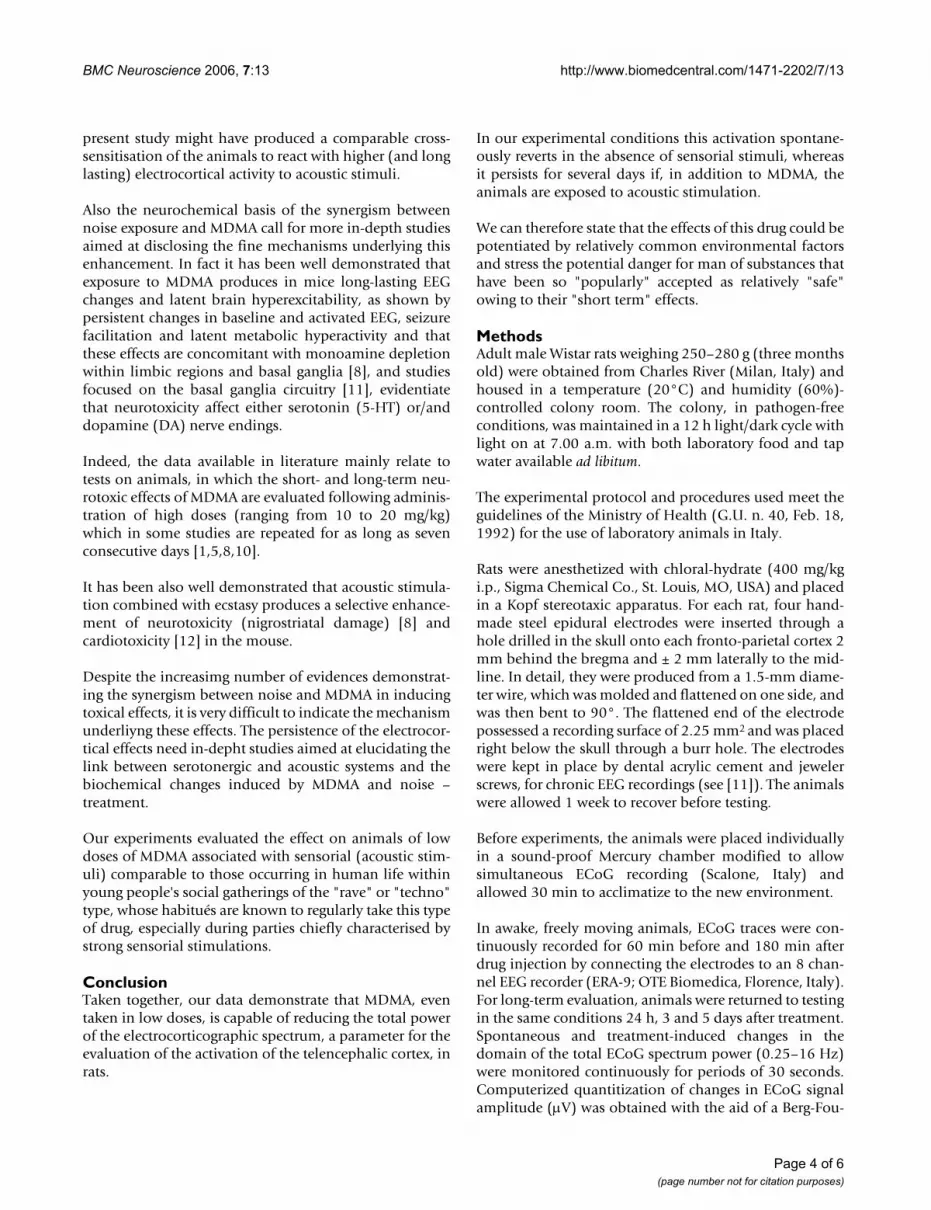
MethodsAdult male Wistar rats weighing 250–280 g (three monthsold) were obtained from Charles River (Milan, Italy) andhoused in a temperature (20°C) and humidity (60%)-controlled colony room. The colony, in pathogen-freeconditions, was maintained in a 12 h light/dark cycle withlight on at 7.00 a.m. with both laboratory food and tapwater available ad libitum.
The experimental protocol and procedures used meet theguidelines of the Ministry of Health (G.U. n. 40, Feb. 18,1992) for the use of laboratory animals in Italy.
Rats were anesthetized with chloral-hydrate (400 mg/kgi.p., Sigma Chemical Co., St. Louis, MO, USA) and placedin a Kopf stereotaxic apparatus. For each rat, four hand-made steel epidural electrodes were inserted through ahole drilled in the skull onto each fronto-parietal cortex 2mm behind the bregma and ± 2 mm laterally to the mid-line. In detail, they were produced from a 1.5-mm diame-ter wire, which was molded and flattened on one side, andwas then bent to 90°. The flattened end of the electrodepossessed a recording surface of 2.25 mm2 and was placedright below the skull through a burr hole. The electrodeswere kept in place by dental acrylic cement and jewelerscrews, for chronic EEG recordings (see [11]). The animalswere allowed 1 week to recover before testing.
Before experiments, the animals were placed individuallyin a sound-proof Mercury chamber modified to allowsimultaneous ECoG recording (Scalone, Italy) andallowed 30 min to acclimatize to the new environment.
In awake, freely moving animals, ECoG traces were con-tinuously recorded for 60 min before and 180 min afterdrug injection by connecting the electrodes to an 8 chan-nel EEG recorder (ERA-9; OTE Biomedica, Florence, Italy).For long-term evaluation, animals were returned to testingin the same conditions 24 h, 3 and 5 days after treatment.Spontaneous and treatment-induced changes in thedomain of the total ECoG spectrum power (0.25–16 Hz)were monitored continuously for periods of 30 seconds.Computerized quantitization of changes in ECoG signalamplitude (µV) was obtained with the aid of a Berg-Fou-
Page 4 of 6(page number not for citation purposes)

BMC Neuroscience 2006, 7:13 http://www.biomedcentral.com/1471-2202/7/13
rier analyser (OTE Biomedica, Florence, Italy). For statisti-cal purposes, ECoG signal amplitude was expressed asmean ± s.e. mean percentage changes from control ampli-tude. The resulting means from control and test experi-
ments were evaluated statistically for differences by priortwo way ANOVA followed by Tukey Test. The parametersevaluated were: stimulation (sound on vs sound off) andtreatment (saline vs dose of MDMA used). ECoG activity
Electrocorticographic activity changes in dependence of sensorial stimuli in MDMA – treated ratsFigure 2Electrocorticographic activity changes in dependence of sensorial stimuli in MDMA – treated rats. Sequential spectral analysis illustrating the effects of (A) MDMA (6 mg/kg; i.p.) and (B) sound (95 dB) + MDMA (6 mg/Kg; i.p.) on electro-corticographic activity in rats. The ECoG activity, evaluated at various times after treatment shows a marked decrease in total power after simultaneous administration of sound (A VS B).
A-sound off B-sound on 180
120
90
60
30
0 0 4 8 12 16 0 4 8 12 16
= MDMA administration
= Sound on
Tim
e(m
inut
es)
Frequency bands (Hz)
Page 5 of 6(page number not for citation purposes)
BMC Neuroscience 2006, 7:13 http://www.biomedcentral.com/1471-2202/7/13
Publish with BioMed Central and every scientist can read your work free of charge
"BioMed Central will be the most significant development for disseminating the results of biomedical research in our lifetime."
Sir Paul Nurse, Cancer Research UK
Your research papers will be:
available free of charge to the entire biomedical community
peer reviewed and published immediately upon acceptance
cited in PubMed and archived on PubMed Central
yours — you keep the copyright
Submit your manuscript here:http://www.biomedcentral.com/info/publishing_adv.asp
BioMedcentral
was monitorized 24 h, 3 and 5 days after by repeatingrecording in the same conditions but omitting pharmaco-logical treatment or acoustic stimulation.
For the sensorial stimulation rats were exposed for all theduration of the first electrocortical recording (60 minbefore and 180 min after administration; in no case ani-mals were exposed to another session of acoustic stimula-tion) to continuous white noise produced by twoloudspeakers (set at 95 dB) driven by a white-noise gener-ator (0–26 kHz), which was installed 30 cm apart fromthe cage. The sound level was monitored by a sound levelmeter (Quest electronics, 215) and it was uniformthroughout tha cage. The level of loud noise was selectedin order to mimic the same intensity to which humans areexposed in the discoteques (95 dB is the maximum inten-sity permitted from the Italian law).
3,4-Methylenedioxymethamphetamine, purchased fromSALARS (Como, Italy), was dissolved in normal saline.Rats (n = 5 for each group) were randomly assigned to oneof the regimens, each receiving one intraperitoneal injec-tion of either normal saline (0.5 ml) or MDMA (3 or 6mg/kg; 0.5 ml) with sound (95 Db) on or off.
Thus, the treatment regimens were as follows: Sound off +saline; Sound on + saline; Sound off + MDMA; Sound on+ MDMA. The administration of sound was started 60min before the injection of saline or MDMA.
Authors' contributionsMI coinceived and coordinated the study and performedelectrocortical analisys. SB, DP and MCZ carried the studyand performed the statistical analysis. SG participated indesign and coordination of the study and helped to draftthe manuscript. FSC and DR supervised the study. Allauthors read and approved the final manuscript.
AcknowledgementsOur thanks go to Mr Frustaci S., Mr Macrì A. and Mr Saturnino D. for excel-lent technical assistance, to Mr Apuzzo D. for administrative assistance and for english revision of the manuscript to Mrs Lynn Ann Whitted. To mr. Benito Rocco Scalone (Girifalco, Catanzaro, Italy) go our particular thanks for the realization of the technical apparatus. This work was economically supported from the Presidence of Calabria Region, Italy.
References1. Baumgarten HG, Lachenmayer L: Serotonin neurotoxins – past
and present. Neurotox Res 2004, 6:589-614.2. Peroutka SJ: Incidence of recreational use of 3,4-methylened-
imethoxymethamphetamine (MDMA, "ecstasy") on anundergraduate campus. N Engl J Med 1987, 317:1542-1543.
3. Randall T: 'Rave' scene, ecstasy use, leap atlantic. JAMA 1992,268:1506.
4. Gamma A, Frei E, Lehmann D, Pascual-Marqui RD, Hell D, Vollenwei-der F: Mood state and brain electric activity in Ecstasy users.Neuroreport 2000, 11:157-162.
5. Kalant H: The pharmacology and toxicology of "ecstasy"(MDMA) and related drugs. CMAJ 2001, 165:917-928.
6. O'Hearn E, Battaglia G, De Souza EB, Kuhar MJ, Molliver ME: Meth-ylendioxyamphetamine (MDA) and methylendioxymetham-phetamine (MDMA) cause selective ablation ofserotoninergic axon terminal in forebrain: immunocyto-chemical evidence for neurotoxicity. J Neurosci 1988,8:2788-2803.
7. Ricaurte GA, Yuan J, McCann UD: 3,4-Methylene-dioxymetham-phetamine ('ecstasy') – induced serotonin neurotoxicity:studies in animals. Neuropsycobiology 2000, 42:5-10.
8. Gesi M, Ferrucci M, Giusiani M, Lenzi P, Lazzeri G, Alessandri MG, Sal-vadorini A, Fulceri F, Pellegrini A, Fornai F, Paparelli A: Loud noiseenhances nigrostriatal dopamine toxicity induced by MDMAin mice. Microsc Res Tech 2004, 64:297-303.
9. Dafters RI, Duffy F, O'Donnell PJ, Bouquet C: Level of use of 3,4-methylenedioxymethamphetamine (MDMA or Ecstasy) inhumans correlates with EEG power and coherence. Psychop-harmacology 1999, 145:82-89.
10. Morton A, Jennifer CA, Hickey MA, Dean LC: Methamphetaminetoxicity in mice is potentiated by exposure to loud music.Neuroreport 2001, 12:3277-3281.
11. Giorgi FS, Pizzanelli C, Ferrucci M, Lazzeri G, Faetti M, Giusiani M,Pontarelli F, Busceti L, Murrib L, Fornaia F: Previous exposure to3,4-methylenedioxymethamphetamine produces long-last-ing alteration in limbic brain excitability measured by elec-troencephalogram spectrum analysis, brain metabolism andseizure susceptibility. Neuroscience 2005, 136:43-53.
12. Gesi M, Soldani P, Lenzi P, Ferrucci M, Giusiani A, Fornai F, PaparelliA: Ecstasy during loud noise exposure induces dramaticultrastructural changes in the heart. Pharmacol Toxicol 2002,91:29-33.
Page 6 of 6(page number not for citation purposes)


