Embed Size (px)
Citation preview
�����������������
Citation: Surma, S.; Banach, M.
Fibrinogen and Atherosclerotic
Cardiovascular Diseases—Review of
the Literature and Clinical Studies.
Int. J. Mol. Sci. 2022, 23, 193. https://
doi.org/10.3390/ijms23010193
Academic Editor:
Anastasios Lymperopoulos
Received: 22 November 2021
Accepted: 21 December 2021
Published: 24 December 2021
Publisher’s Note: MDPI stays neutral
with regard to jurisdictional claims in
published maps and institutional affil-
iations.
Copyright: © 2021 by the authors.
Licensee MDPI, Basel, Switzerland.
This article is an open access article
distributed under the terms and
conditions of the Creative Commons
Attribution (CC BY) license (https://
creativecommons.org/licenses/by/
4.0/).
International Journal of
Molecular Sciences
Review
Fibrinogen and Atherosclerotic CardiovascularDiseases—Review of the Literature and Clinical Studies
Stanisław Surma 1,2 and Maciej Banach 3,4,5,*
1 Faculty of Medical Sciences in Katowice, Medical University of Silesia in Katowice, 40-752 Katowice, Poland;[email protected]
2 Club of Young Hypertensiologists, Polish Society of Hypertension, 80-952 Gdansk, Poland3 Department of Preventive Cardiology and Lipidology, Medical University of Lodz, 93-338 Lodz, Poland4 Cardiovascular Research Centre, University of Zielona Gora, 65-417 Zielona Gora, Poland5 Department of Cardiology and Adult Congenital Heart Diseases, Polish Mother’s Memorial Hospital
Research Institute (PMMHRI), 93-338 Lodz, Poland* Correspondence: [email protected]; Tel.: +48-422-711-124
Abstract: Atherosclerotic cardiovascular diseases (ASCVD), including coronary artery disease, cere-brovascular disease, and peripheral arterial disease, represent a significant cause of premature deathworldwide. Biomarkers, the evaluation of which would allow the detection of ASCVD at the earlieststage of development, are intensively sought. Moreover, from a clinical point of view, a valuablebiomarker should also enable the assessment of the patient’s prognosis. It has been known for manyyears that the concentration of fibrinogen in plasma increases, inter alia, in patients with ASCVD. Onthe one hand, an increased plasma fibrinogen concentration may be the cause of the development ofatherosclerotic lesions (increased risk of atherothrombosis); on the other hand, it may be a biomarkerof ASCVD, as it is an acute phase protein. In addition, a number of genetic polymorphisms andpost-translational modifications of fibrinogen were demonstrated that may contribute to the risk ofASCVD. This review summarizes the current data on the importance of fibrinogen as a biomarker ofASCVD, and also presents the relationship between molecular modifications of this protein in thecontext of ASCVD.
Keywords: fibrinogen; atherosclerosis; atherosclerotic cardiovascular disease
1. Introduction
Atherosclerotic changes appear from childhood [1]. A number of risk factors, such ashyperlipidemia, hyperhomocysteinemia, arterial hypertension, hyperuricemia, smoking,metabolic syndrome, hypertriglyceridemia and diabetes, accelerate the progression ofatherosclerotic lesions, leading to the development of atherosclerotic cardiovascular disease(ASCVD) [2]. ASCVD is defined as a coronary artery disease (CAD), cerebrovasculardisease, or peripheral arterial disease (PAD) of atherosclerotic origin. ASCVD representsthe number one cause of morbidity and mortality worldwide [3]. In 2019, the number ofpatients with cardiovascular diseases worldwide was 523 million, while the number ofdeaths due to these diseases reached 18.6 million [4]. In 2017, the number of patients withCAD worldwide reached 126 million (1.72% of the world population), and it is estimatedto increase every year. Worldwide, CAD caused nine million deaths in 2017, making thedisease the leading cause of death [5]. The incidence of stroke is also a significant problem.In 2019, the number of patients with stroke worldwide was 101 million, while the numberof deaths due to stroke was 6.55 million [6]. PAD is also a widespread disease. In 2019,the number of patients with PAD worldwide was 113 million, and the disease caused74.1 thousand deaths [4].
The huge prevalence of ASCVD diseases worldwide means that factors that causetheir occurrence are searched for in order to develop appropriate methods of preventionand therapy. One such factor is the plasma fibrinogen concentration.
Int. J. Mol. Sci. 2022, 23, 193. https://doi.org/10.3390/ijms23010193 https://www.mdpi.com/journal/ijms
Int. J. Mol. Sci. 2022, 23, 193 2 of 20
Already in the 1950s, it was found that plasma fibrinogen concentration is associatedwith a risk of CVD [7]. Many years ago, in the Framingham study involving 1315 healthypeople, a relationship was found between plasma fibrinogen concentration and the inci-dence of CVD, including CAD and stroke, over a 12-year follow-up. People whose fibrino-gen plasma concentration was in the second and third terciles (concentrations: 2.7–3.1 g/Land 3.1–7.0 g/L, respectively) were characterized by a significantly higher frequency ofCVD than those in the first tercile (1.3–2.7 g/L). A higher plasma fibrinogen concentration,as a separate variable, had a similar effect on cardiovascular risk as well-known risk factorssuch as smoking cigarette, obesity, arterial hypertension, and diabetes [8].
This literature review summarizes the pathophysiological and clinical understandingof fibrinogen and its relationship with the risk of ASCVD.
2. Fibrinogen—Physiological and Pathophysiological Aspects
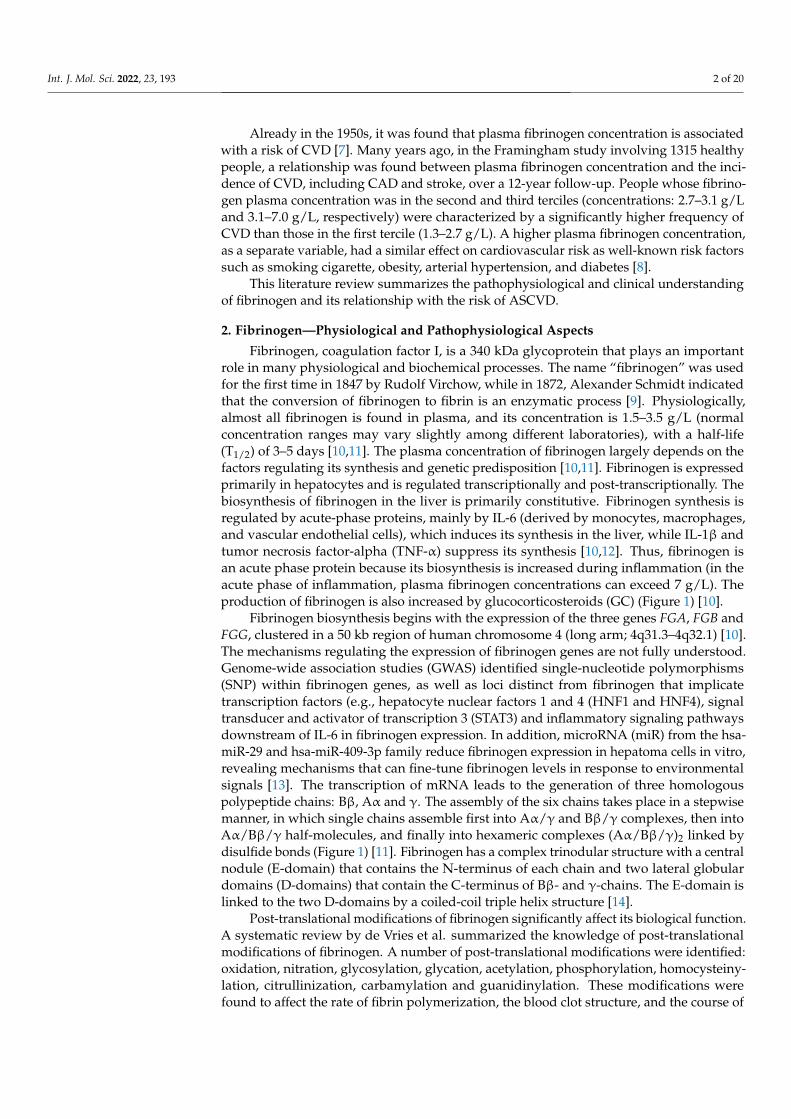
Fibrinogen, coagulation factor I, is a 340 kDa glycoprotein that plays an importantrole in many physiological and biochemical processes. The name “fibrinogen” was usedfor the first time in 1847 by Rudolf Virchow, while in 1872, Alexander Schmidt indicatedthat the conversion of fibrinogen to fibrin is an enzymatic process [9]. Physiologically,almost all fibrinogen is found in plasma, and its concentration is 1.5–3.5 g/L (normalconcentration ranges may vary slightly among different laboratories), with a half-life(T1/2) of 3–5 days [10,11]. The plasma concentration of fibrinogen largely depends on thefactors regulating its synthesis and genetic predisposition [10,11]. Fibrinogen is expressedprimarily in hepatocytes and is regulated transcriptionally and post-transcriptionally. Thebiosynthesis of fibrinogen in the liver is primarily constitutive. Fibrinogen synthesis isregulated by acute-phase proteins, mainly by IL-6 (derived by monocytes, macrophages,and vascular endothelial cells), which induces its synthesis in the liver, while IL-1β andtumor necrosis factor-alpha (TNF-α) suppress its synthesis [10,12]. Thus, fibrinogen isan acute phase protein because its biosynthesis is increased during inflammation (in theacute phase of inflammation, plasma fibrinogen concentrations can exceed 7 g/L). Theproduction of fibrinogen is also increased by glucocorticosteroids (GC) (Figure 1) [10].
Fibrinogen biosynthesis begins with the expression of the three genes FGA, FGB andFGG, clustered in a 50 kb region of human chromosome 4 (long arm; 4q31.3–4q32.1) [10].The mechanisms regulating the expression of fibrinogen genes are not fully understood.Genome-wide association studies (GWAS) identified single-nucleotide polymorphisms(SNP) within fibrinogen genes, as well as loci distinct from fibrinogen that implicatetranscription factors (e.g., hepatocyte nuclear factors 1 and 4 (HNF1 and HNF4), signaltransducer and activator of transcription 3 (STAT3) and inflammatory signaling pathwaysdownstream of IL-6 in fibrinogen expression. In addition, microRNA (miR) from the hsa-miR-29 and hsa-miR-409-3p family reduce fibrinogen expression in hepatoma cells in vitro,revealing mechanisms that can fine-tune fibrinogen levels in response to environmentalsignals [13]. The transcription of mRNA leads to the generation of three homologouspolypeptide chains: Bβ, Aα and γ. The assembly of the six chains takes place in a stepwisemanner, in which single chains assemble first into Aα/γ and Bβ/γ complexes, then intoAα/Bβ/γ half-molecules, and finally into hexameric complexes (Aα/Bβ/γ)2 linked bydisulfide bonds (Figure 1) [11]. Fibrinogen has a complex trinodular structure with a centralnodule (E-domain) that contains the N-terminus of each chain and two lateral globulardomains (D-domains) that contain the C-terminus of Bβ- and γ-chains. The E-domain islinked to the two D-domains by a coiled-coil triple helix structure [14].
Post-translational modifications of fibrinogen significantly affect its biological function.A systematic review by de Vries et al. summarized the knowledge of post-translationalmodifications of fibrinogen. A number of post-translational modifications were identified:oxidation, nitration, glycosylation, glycation, acetylation, phosphorylation, homocysteiny-lation, citrullinization, carbamylation and guanidinylation. These modifications werefound to affect the rate of fibrin polymerization, the blood clot structure, and the course of
Int. J. Mol. Sci. 2022, 23, 193 3 of 20
fibrinolysis. Thus, post-translational modifications of fibrinogen may play an importantrole in the physiology and pathophysiology of blood coagulation [15].
Figure 1. Fibrinogen biosynthesis in liver hepatocytes [9–11]. DNA—deoxyribonucleic acid; mRNA—Messenger ribonucleic acid; FGA-FGB-FGG—Fibrinogen chain genes; miRNA—Micro RNA; HNF4α—Hepatocyte Nuclear Factor 4 Alpha; HNF1—Hepatocyte nuclear factor 1; C/EBP—CCAAT enhancerbinding proteins; IL-6RE—IL-6 response element; SOCS3—Suppressor of cytokine signaling 3; miR-18a—microRNA-18a; NFκB—Nuclear factor kappa-light-chain-enhancer of activated B cells; PIAS3—Protein inhibitor of activated STAT 3; STAT3—Signal transducer and activator of transcription 3;HNF3—Hepatocyte nuclear factor 3; IL-1β—Interleukin 1β, IL-6—Interleukin 6; GR—Glucocorticoidreceptor; GC—Glucocorticosteroids; Bβ, Aα and γ—Fibrinogen polypeptide chains.
Moreover, genetic polymorphisms, which may influence the risk of various diseases,play an important role in the properties of fibrinogen [16].
Many factors and/or conditions were shown to increase the plasma concentrationof fibrinogen. These include: female gender, Black ethnicity, age, diabetes, smoking andalcohol consumption, arterial hypertension, obesity, lipid disorders, metabolic syndrome,menopause, oral contraceptives, microalbuminuria, lower socioeconomic status and pre-mature family history of CVD. The association with body mass index (BMI) was twice asstrong in women as in men. However, the association with smoking cigarettes was muchstronger in men. Interestingly, plasma fibrinogen concentration is inversely related to serumHDL cholesterol (high-density lipoprotein cholesterol) concentration [17–19]. Interestingly,the renin-angiotensin-aldosterone system (RAAS) plays an important role in the regulationof fibrinogen plasma concentration. Therefore, as pointed out by Kryczka et al., differencesin the regulation of RAAS in women (including the effect of estrogens) and in men mayaffect the fibrinogen plasma concentration and observed clinical effect [20]. The basic
Int. J. Mol. Sci. 2022, 23, 193 4 of 20
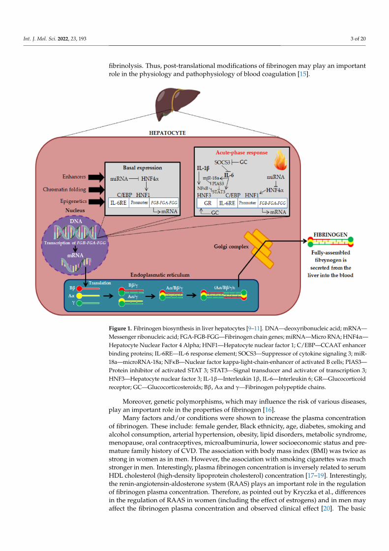
physiological function of fibrinogen is participation in the final stage of the clotting processand transformation into a fibrillar protein—fibrin, which forms a blood clot (Figure 2) [11].
Figure 2. The role of fibrinogen in blood coagulation [21]. TF—Tissue factor, GP IIb/IIIa—Integrin receptors.
Moreover, fibrinogen is involved in the matrix physiology (by interaction with plas-minogen, FXIII, vitronectin and fibronectin), regulation of the inflammatory process, infec-tion, wound healing, intercellular interaction, cell migration, tumor growth, angiogenesis,and metastasis [11].
The increased fibrinogen plasma concentration directly activates many mechanisms,which, consequently, may intensify the progression of atherosclerosis [20]. The pro-atherogenic mechanisms of action of fibrinogen are summarized in Table 1.
Table 1. The pro-atherogenic mechanisms of action of fibrinogen [20]. IL-1—Interleukin 1; TNF-α—Tumor necrosis factor α; IL-8—Interleukin 8; MCP-1—Monocyte chemoattractant protein-1; GPIIb/IIIa—Integrin receptors; IL-1β—Interleukin aβ; ICAM-1—Intercellular adhesion molecule 1;EC—Endothelial cells; LDL—Low-density lipoprotein; SMC—Smooth muscle cell.
Main Pro-Atherogenic Properties of Fibrinogen
3 ↑ Severity of inflammation: promotes an inflammatory response by inducing the exposition of proinflammatory cytokines onmonocytes (IL-1 and TNF-α) as well as chemokines, such as IL-8 and MCP-1, on endothelium and fibroblasts, which promoteatherosclerotic plaque formation;
3 Activation of platelets (via GP IIb/IIIa receptors) leading to the production of the pro-inflammatory cytokines, CD40 ligandand IL-1β, which promote atherosclerotic plaque formation;
3 ↑ Expression of adhesion molecules (ICAM-1) on vascular EC leading to the adhesion of leukocytes, macrophages and platelets;3 ↑ Production of vasoactive factors by EC leading to an increase in its permeability and impairing its vasorelaxant properties;3 Accumulation of fibrinogen in the vessel wall enhances the infiltration of macrophages, which are precursors of foam cells;3 The accumulated fibrinogen deposits in the vessel wall absorb LDL cholesterol, which leads to the formation and growth of
atherosclerotic plaque;3 Increasing the adhesion of neutrophils to activated platelets attached to the injured arterial wall, which promotes the
formation of atherosclerotic plaque;3 ↑ SMC migration and proliferation, as well as stimulation of angiogenesis.
In summary, fibrinogen has many important functions in human physiology, but alsomany unfavorable pathophysiological pathways induced by increased fibrinogen plasmaconcentration were described, which aggravate the atherosclerotic process.
3. Fibrinogen and Cardiovascular Risk
The results of numerous epidemiological studies indicate that increased plasma fib-rinogen concentrations are a risk factor for ASCVD. Importantly, the results of some studieseven indicate that increased plasma fibrinogen concentrations more adversely affect the riskof CVD than only increased serum cholesterol concentrations. In the European Concerted
Int. J. Mol. Sci. 2022, 23, 193 5 of 20
Action on Thrombosis and Disabilities Angina Pectoris Study (ETAT), which enrolled over3000 patients with angiographically documented CAD, cholesterol was not found to bean independent risk factor for coronary events. Its mean concentration was 257 mg/dLin the group with adverse events in comparison to 246 mg/dL in the group of peoplewithout complications. However, a strong relationship was demonstrated in relation tofibrinogen. The risk of coronary events, including sudden cardiac death, increased withfibrinogen plasma concentrations, which remained low in the case of high cholesterol, aslong as the fibrinogen concentration remained low [22]. Moreover, a prospective study byMa et al., including 14,916 men in the Physicians’ Health Study, aged 40–84 years, assessedthe relationship between the plasma fibrinogen concentration and the risk of a myocardialinfraction (MI). In addition, subjects were randomized to take aspirin (325 mg every otherday) or placebo for 5 years. It was shown that people who experienced an MI had ahigher plasma fibrinogen concentration (p = 0.02). A high fibrinogen plasma concentration(≥343 mg/dL) had a twofold increase in MI risk (RR = 2.09; 95% CI: 1.15–3.78) comparedwith those with fibrinogen below 343 mg/dL. There was no interaction between the fib-rinogen plasma concentration and aspirin treatment. Thus, it was found that fibrinogenwas associated with an increased risk of future MI independent of other CVD risk factors,atherogenic factors such as lipids, and antithrombotics such as aspirin [23].
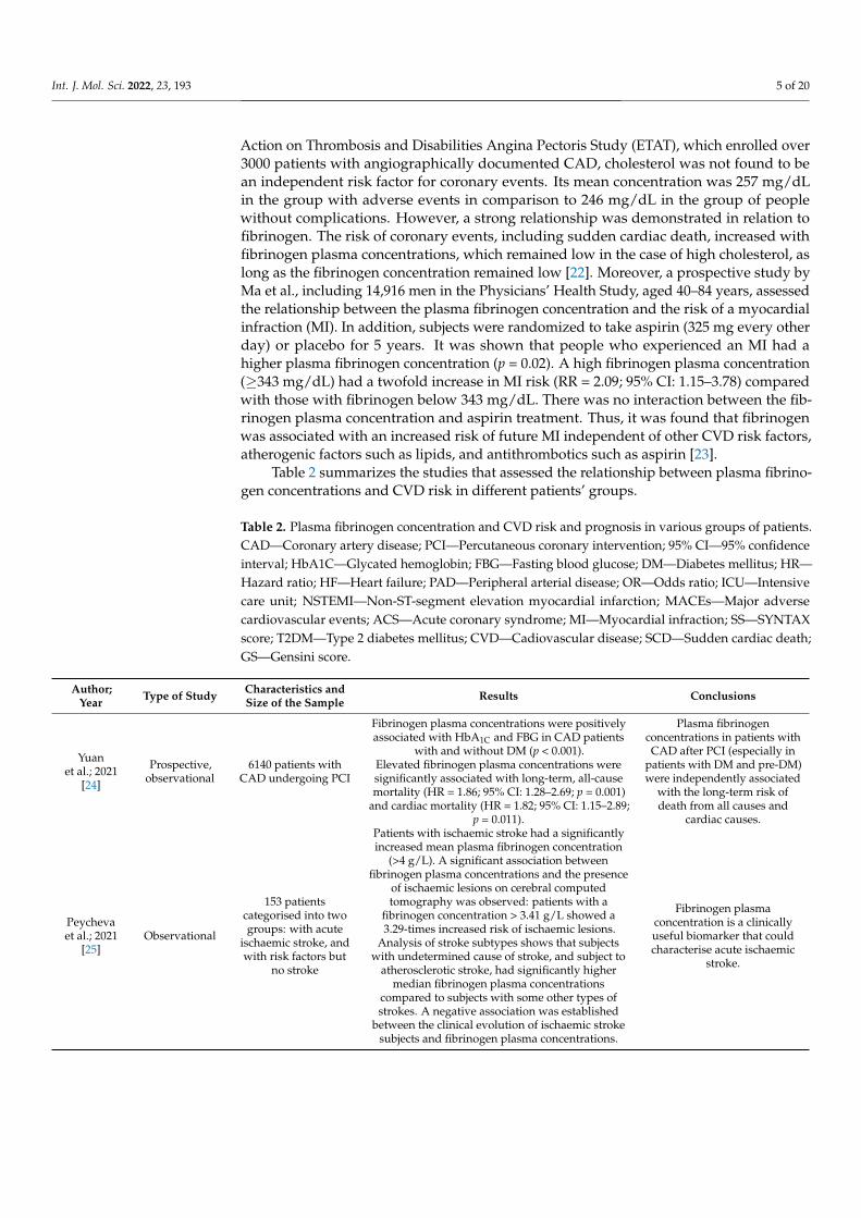
Table 2 summarizes the studies that assessed the relationship between plasma fibrino-gen concentrations and CVD risk in different patients’ groups.
Table 2. Plasma fibrinogen concentration and CVD risk and prognosis in various groups of patients.CAD—Coronary artery disease; PCI—Percutaneous coronary intervention; 95% CI—95% confidenceinterval; HbA1C—Glycated hemoglobin; FBG—Fasting blood glucose; DM—Diabetes mellitus; HR—Hazard ratio; HF—Heart failure; PAD—Peripheral arterial disease; OR—Odds ratio; ICU—Intensivecare unit; NSTEMI—Non-ST-segment elevation myocardial infarction; MACEs—Major adversecardiovascular events; ACS—Acute coronary syndrome; MI—Myocardial infraction; SS—SYNTAXscore; T2DM—Type 2 diabetes mellitus; CVD—Cadiovascular disease; SCD—Sudden cardiac death;GS—Gensini score.
Author;Year Type of Study Characteristics and
Size of the Sample Results Conclusions
Yuanet al.; 2021
[24]
Prospective,observational
6140 patients withCAD undergoing PCI
Fibrinogen plasma concentrations were positivelyassociated with HbA1C and FBG in CAD patients
with and without DM (p < 0.001).Elevated fibrinogen plasma concentrations weresignificantly associated with long-term, all-causemortality (HR = 1.86; 95% CI: 1.28–2.69; p = 0.001)
and cardiac mortality (HR = 1.82; 95% CI: 1.15–2.89;p = 0.011).
Plasma fibrinogenconcentrations in patients withCAD after PCI (especially in
patients with DM and pre-DM)were independently associated
with the long-term risk ofdeath from all causes and
cardiac causes.
Peychevaet al.; 2021
[25]Observational
153 patientscategorised into twogroups: with acute
ischaemic stroke, andwith risk factors but
no stroke
Patients with ischaemic stroke had a significantlyincreased mean plasma fibrinogen concentration
(>4 g/L). A significant association betweenfibrinogen plasma concentrations and the presence
of ischaemic lesions on cerebral computedtomography was observed: patients with a
fibrinogen concentration > 3.41 g/L showed a3.29-times increased risk of ischaemic lesions.
Analysis of stroke subtypes shows that subjectswith undetermined cause of stroke, and subject to
atherosclerotic stroke, had significantly highermedian fibrinogen plasma concentrations
compared to subjects with some other types ofstrokes. A negative association was established
between the clinical evolution of ischaemic strokesubjects and fibrinogen plasma concentrations.
Fibrinogen plasmaconcentration is a clinicallyuseful biomarker that couldcharacterise acute ischaemic
stroke.
Int. J. Mol. Sci. 2022, 23, 193 6 of 20
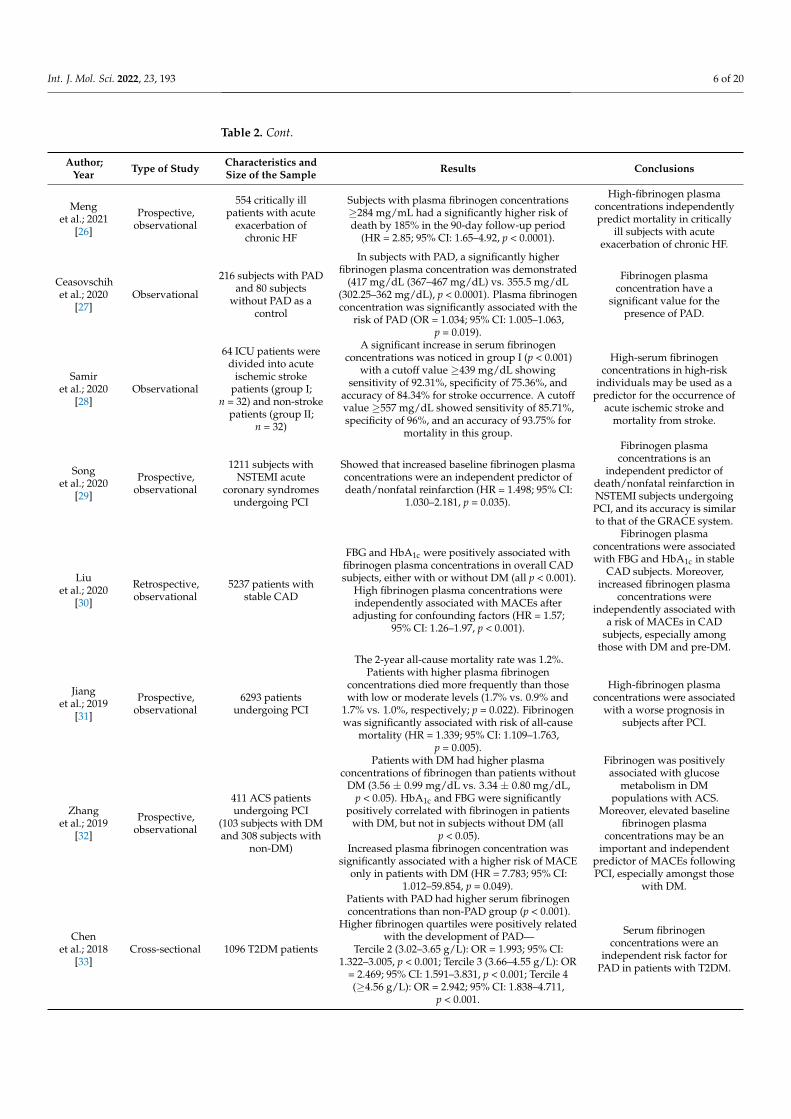
Table 2. Cont.
Author;Year Type of Study Characteristics and
Size of the Sample Results Conclusions
Menget al.; 2021
[26]
Prospective,observational
554 critically illpatients with acute
exacerbation ofchronic HF
Subjects with plasma fibrinogen concentrations≥284 mg/mL had a significantly higher risk ofdeath by 185% in the 90-day follow-up period
(HR = 2.85; 95% CI: 1.65–4.92, p < 0.0001).
High-fibrinogen plasmaconcentrations independentlypredict mortality in critically
ill subjects with acuteexacerbation of chronic HF.
Ceasovschihet al.; 2020
[27]Observational
216 subjects with PADand 80 subjects
without PAD as acontrol
In subjects with PAD, a significantly higherfibrinogen plasma concentration was demonstrated
(417 mg/dL (367–467 mg/dL) vs. 355.5 mg/dL(302.25–362 mg/dL), p < 0.0001). Plasma fibrinogenconcentration was significantly associated with the
risk of PAD (OR = 1.034; 95% CI: 1.005–1.063,p = 0.019).
Fibrinogen plasmaconcentration have a
significant value for thepresence of PAD.
Samiret al.; 2020
[28]Observational
64 ICU patients weredivided into acute
ischemic strokepatients (group I;
n = 32) and non-strokepatients (group II;
n = 32)
A significant increase in serum fibrinogenconcentrations was noticed in group I (p < 0.001)
with a cutoff value ≥439 mg/dL showingsensitivity of 92.31%, specificity of 75.36%, and
accuracy of 84.34% for stroke occurrence. A cutoffvalue ≥557 mg/dL showed sensitivity of 85.71%,specificity of 96%, and an accuracy of 93.75% for
mortality in this group.
High-serum fibrinogenconcentrations in high-risk
individuals may be used as apredictor for the occurrence of
acute ischemic stroke andmortality from stroke.
Songet al.; 2020
[29]
Prospective,observational
1211 subjects withNSTEMI acute
coronary syndromesundergoing PCI
Showed that increased baseline fibrinogen plasmaconcentrations were an independent predictor ofdeath/nonfatal reinfarction (HR = 1.498; 95% CI:
1.030–2.181, p = 0.035).
Fibrinogen plasmaconcentrations is an
independent predictor ofdeath/nonfatal reinfarction inNSTEMI subjects undergoingPCI, and its accuracy is similarto that of the GRACE system.
Liuet al.; 2020
[30]
Retrospective,observational
5237 patients withstable CAD
FBG and HbA1c were positively associated withfibrinogen plasma concentrations in overall CADsubjects, either with or without DM (all p < 0.001).
High fibrinogen plasma concentrations wereindependently associated with MACEs afteradjusting for confounding factors (HR = 1.57;
95% CI: 1.26–1.97, p < 0.001).
Fibrinogen plasmaconcentrations were associatedwith FBG and HbA1c in stable
CAD subjects. Moreover,increased fibrinogen plasma
concentrations wereindependently associated with
a risk of MACEs in CADsubjects, especially among
those with DM and pre-DM.
Jianget al.; 2019
[31]
Prospective,observational
6293 patientsundergoing PCI
The 2-year all-cause mortality rate was 1.2%.Patients with higher plasma fibrinogen
concentrations died more frequently than thosewith low or moderate levels (1.7% vs. 0.9% and
1.7% vs. 1.0%, respectively; p = 0.022). Fibrinogenwas significantly associated with risk of all-cause
mortality (HR = 1.339; 95% CI: 1.109–1.763,p = 0.005).
High-fibrinogen plasmaconcentrations were associated
with a worse prognosis insubjects after PCI.
Zhanget al.; 2019
[32]
Prospective,observational
411 ACS patientsundergoing PCI
(103 subjects with DMand 308 subjects with
non-DM)
Patients with DM had higher plasmaconcentrations of fibrinogen than patients without
DM (3.56 ± 0.99 mg/dL vs. 3.34 ± 0.80 mg/dL,p < 0.05). HbA1c and FBG were significantly
positively correlated with fibrinogen in patientswith DM, but not in subjects without DM (all
p < 0.05).Increased plasma fibrinogen concentration was
significantly associated with a higher risk of MACEonly in patients with DM (HR = 7.783; 95% CI:
1.012–59.854, p = 0.049).
Fibrinogen was positivelyassociated with glucose
metabolism in DMpopulations with ACS.
Moreover, elevated baselinefibrinogen plasma
concentrations may be animportant and independent
predictor of MACEs followingPCI, especially amongst those
with DM.
Chenet al.; 2018
[33]Cross-sectional 1096 T2DM patients
Patients with PAD had higher serum fibrinogenconcentrations than non-PAD group (p < 0.001).
Higher fibrinogen quartiles were positively relatedwith the development of PAD—
Tercile 2 (3.02–3.65 g/L): OR = 1.993; 95% CI:1.322–3.005, p < 0.001; Tercile 3 (3.66–4.55 g/L): OR
= 2.469; 95% CI: 1.591–3.831, p < 0.001; Tercile 4(≥4.56 g/L): OR = 2.942; 95% CI: 1.838–4.711,
p < 0.001.
Serum fibrinogenconcentrations were an
independent risk factor forPAD in patients with T2DM.
Int. J. Mol. Sci. 2022, 23, 193 7 of 20
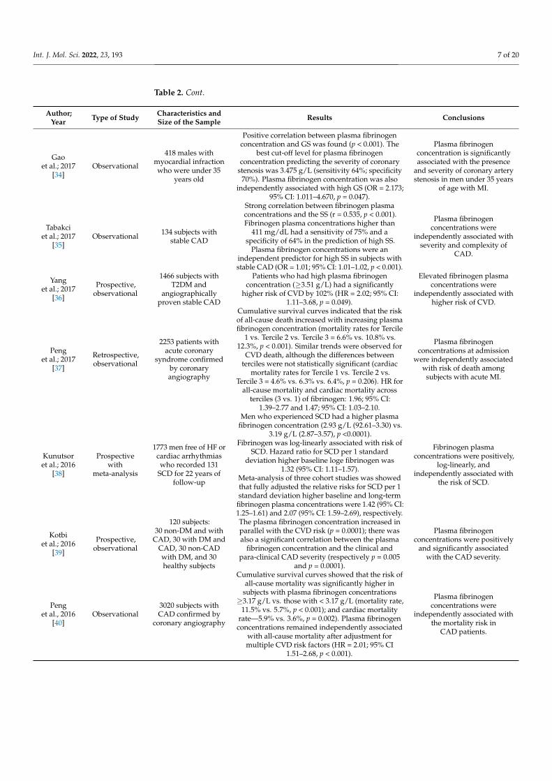
Table 2. Cont.
Author;Year Type of Study Characteristics and
Size of the Sample Results Conclusions
Gaoet al.; 2017
[34]Observational
418 males withmyocardial infractionwho were under 35
years old
Positive correlation between plasma fibrinogenconcentration and GS was found (p < 0.001). The
best cut-off level for plasma fibrinogenconcentration predicting the severity of coronary
stenosis was 3.475 g/L (sensitivity 64%; specificity70%). Plasma fibrinogen concentration was also
independently associated with high GS (OR = 2.173;95% CI: 1.011–4.670, p = 0.047).
Plasma fibrinogenconcentration is significantlyassociated with the presence
and severity of coronary arterystenosis in men under 35 years
of age with MI.
Tabakciet al.; 2017
[35]Observational 134 subjects with
stable CAD
Strong correlation between fibrinogen plasmaconcentrations and the SS (r = 0.535, p < 0.001).Fibrinogen plasma concentrations higher than
411 mg/dL had a sensitivity of 75% and aspecificity of 64% in the prediction of high SS.
Plasma fibrinogen concentrations were anindependent predictor for high SS in subjects withstable CAD (OR = 1.01; 95% CI: 1.01–1.02, p < 0.001).
Plasma fibrinogenconcentrations were
independently associated withseverity and complexity of
CAD.
Yanget al.; 2017
[36]
Prospective,observational
1466 subjects withT2DM and
angiographicallyproven stable CAD
Patients who had high plasma fibrinogenconcentration (≥3.51 g/L) had a significantly
higher risk of CVD by 102% (HR = 2.02; 95% CI:1.11–3.68, p = 0.049).
Elevated fibrinogen plasmaconcentrations were
independently associated withhigher risk of CVD.
Penget al.; 2017
[37]
Retrospective,observational
2253 patients withacute coronary
syndrome confirmedby coronaryangiography
Cumulative survival curves indicated that the riskof all-cause death increased with increasing plasmafibrinogen concentration (mortality rates for Tercile
1 vs. Tercile 2 vs. Tercile 3 = 6.6% vs. 10.8% vs.12.3%, p < 0.001). Similar trends were observed for
CVD death, although the differences betweenterciles were not statistically significant (cardiac
mortality rates for Tercile 1 vs. Tercile 2 vs.Tercile 3 = 4.6% vs. 6.3% vs. 6.4%, p = 0.206). HR for
all-cause mortality and cardiac mortality acrossterciles (3 vs. 1) of fibrinogen: 1.96; 95% CI:
1.39–2.77 and 1.47; 95% CI: 1.03–2.10.
Plasma fibrinogenconcentrations at admission
were independently associatedwith risk of death among
subjects with acute MI.
Kunutsoret al.; 2016
[38]
Prospectivewith
meta-analysis
1773 men free of HF orcardiac arrhythmiaswho recorded 131
SCD for 22 years offollow-up
Men who experienced SCD had a higher plasmafibrinogen concentration (2.93 g/L (92.61–3.30) vs.
3.19 g/L (2.87–3.57), p <0.0001).Fibrinogen was log-linearly associated with risk of
SCD. Hazard ratio for SCD per 1 standarddeviation higher baseline loge fibrinogen was
1.32 (95% CI: 1.11–1.57).Meta-analysis of three cohort studies was showedthat fully adjusted the relative risks for SCD per 1standard deviation higher baseline and long-term
fibrinogen plasma concentrations were 1.42 (95% CI:1.25–1.61) and 2.07 (95% CI: 1.59–2.69), respectively.
Fibrinogen plasmaconcentrations were positively,
log-linearly, andindependently associated with
the risk of SCD.
Kotbiet al.; 2016
[39]
Prospective,observational
120 subjects:30 non-DM and with
CAD, 30 with DM andCAD, 30 non-CADwith DM, and 30healthy subjects
The plasma fibrinogen concentration increased inparallel with the CVD risk (p = 0.0001); there wasalso a significant correlation between the plasma
fibrinogen concentration and the clinical andpara-clinical CAD severity (respectively p = 0.005
and p = 0.0001).
Plasma fibrinogenconcentrations were positively
and significantly associatedwith the CAD severity.
Penget al., 2016
[40]Observational
3020 subjects withCAD confirmed by
coronary angiography
Cumulative survival curves showed that the risk ofall-cause mortality was significantly higher in
subjects with plasma fibrinogen concentrations≥3.17 g/L vs. those with < 3.17 g/L (mortality rate,
11.5% vs. 5.7%, p < 0.001); and cardiac mortalityrate—5.9% vs. 3.6%, p = 0.002). Plasma fibrinogenconcentrations remained independently associated
with all-cause mortality after adjustment formultiple CVD risk factors (HR = 2.01; 95% CI
1.51–2.68, p < 0.001).
Plasma fibrinogenconcentrations were
independently associated withthe mortality risk in
CAD patients.
Int. J. Mol. Sci. 2022, 23, 193 8 of 20
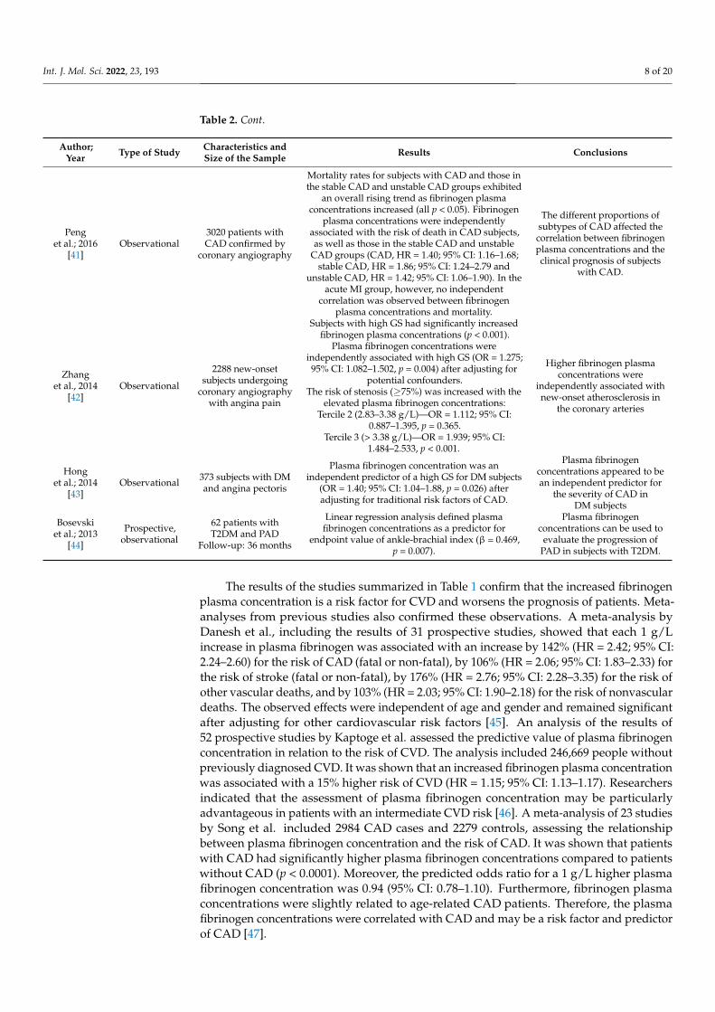
Table 2. Cont.
Author;Year Type of Study Characteristics and
Size of the Sample Results Conclusions
Penget al.; 2016
[41]Observational
3020 patients withCAD confirmed by
coronary angiography
Mortality rates for subjects with CAD and those inthe stable CAD and unstable CAD groups exhibited
an overall rising trend as fibrinogen plasmaconcentrations increased (all p < 0.05). Fibrinogen
plasma concentrations were independentlyassociated with the risk of death in CAD subjects,as well as those in the stable CAD and unstable
CAD groups (CAD, HR = 1.40; 95% CI: 1.16–1.68;stable CAD, HR = 1.86; 95% CI: 1.24–2.79 and
unstable CAD, HR = 1.42; 95% CI: 1.06–1.90). In theacute MI group, however, no independent
correlation was observed between fibrinogenplasma concentrations and mortality.
The different proportions ofsubtypes of CAD affected thecorrelation between fibrinogenplasma concentrations and theclinical prognosis of subjects
with CAD.
Zhanget al., 2014
[42]Observational
2288 new-onsetsubjects undergoing
coronary angiographywith angina pain
Subjects with high GS had significantly increasedfibrinogen plasma concentrations (p < 0.001).
Plasma fibrinogen concentrations wereindependently associated with high GS (OR = 1.275;95% CI: 1.082–1.502, p = 0.004) after adjusting for
potential confounders.The risk of stenosis (≥75%) was increased with the
elevated plasma fibrinogen concentrations:Tercile 2 (2.83–3.38 g/L)—OR = 1.112; 95% CI:
0.887–1.395, p = 0.365.Tercile 3 (> 3.38 g/L)—OR = 1.939; 95% CI:
1.484–2.533, p < 0.001.
Higher fibrinogen plasmaconcentrations were
independently associated withnew-onset atherosclerosis in
the coronary arteries
Honget al.; 2014
[43]Observational 373 subjects with DM
and angina pectoris
Plasma fibrinogen concentration was anindependent predictor of a high GS for DM subjects
(OR = 1.40; 95% CI: 1.04–1.88, p = 0.026) afteradjusting for traditional risk factors of CAD.
Plasma fibrinogenconcentrations appeared to bean independent predictor for
the severity of CAD inDM subjects
Bosevskiet al.; 2013
[44]
Prospective,observational
62 patients withT2DM and PAD
Follow-up: 36 months
Linear regression analysis defined plasmafibrinogen concentrations as a predictor for
endpoint value of ankle-brachial index (β = 0.469,p = 0.007).
Plasma fibrinogenconcentrations can be used to
evaluate the progression ofPAD in subjects with T2DM.
The results of the studies summarized in Table 1 confirm that the increased fibrinogenplasma concentration is a risk factor for CVD and worsens the prognosis of patients. Meta-analyses from previous studies also confirmed these observations. A meta-analysis byDanesh et al., including the results of 31 prospective studies, showed that each 1 g/Lincrease in plasma fibrinogen was associated with an increase by 142% (HR = 2.42; 95% CI:2.24–2.60) for the risk of CAD (fatal or non-fatal), by 106% (HR = 2.06; 95% CI: 1.83–2.33) forthe risk of stroke (fatal or non-fatal), by 176% (HR = 2.76; 95% CI: 2.28–3.35) for the risk ofother vascular deaths, and by 103% (HR = 2.03; 95% CI: 1.90–2.18) for the risk of nonvasculardeaths. The observed effects were independent of age and gender and remained significantafter adjusting for other cardiovascular risk factors [45]. An analysis of the results of52 prospective studies by Kaptoge et al. assessed the predictive value of plasma fibrinogenconcentration in relation to the risk of CVD. The analysis included 246,669 people withoutpreviously diagnosed CVD. It was shown that an increased fibrinogen plasma concentrationwas associated with a 15% higher risk of CVD (HR = 1.15; 95% CI: 1.13–1.17). Researchersindicated that the assessment of plasma fibrinogen concentration may be particularlyadvantageous in patients with an intermediate CVD risk [46]. A meta-analysis of 23 studiesby Song et al. included 2984 CAD cases and 2279 controls, assessing the relationshipbetween plasma fibrinogen concentration and the risk of CAD. It was shown that patientswith CAD had significantly higher plasma fibrinogen concentrations compared to patientswithout CAD (p < 0.0001). Moreover, the predicted odds ratio for a 1 g/L higher plasmafibrinogen concentration was 0.94 (95% CI: 0.78–1.10). Furthermore, fibrinogen plasmaconcentrations were slightly related to age-related CAD patients. Therefore, the plasmafibrinogen concentrations were correlated with CAD and may be a risk factor and predictorof CAD [47].
Int. J. Mol. Sci. 2022, 23, 193 9 of 20
Additionally, the data suggesting that plasma fibrinogen concentration are of clinicalinterest and may be a useful biomarker for subclinical atherosclerosis.
In a study of 652 men aged 40–60 years old (asymptomatic and never treated from CVDcauses) with at least one of the following CVD risk factors: total cholesterol >6.2 mmol/Land/or systolic blood pressure ≥160 mmHg and/or diastolic blood pressure ≥95 mmHg,and/or smoking cigarettes, the relationship between plasma fibrinogen concentration andthe presence of atherosclerotic plaques was assessed. The independent associations be-tween fibrinogen and the presence and extent of atherosclerosis were indicated. Plaqueprevalence was significantly more pronounced with the increasing tercile of fibrinogenplasma concentrations. The odds ratio of the upper to lower fibrinogen terciles for thepresence of plaque was 1.6 (95% CI: 1.4–1.8) and 1.4 (95% CI: 1.2–1.7) for its extent. Theadjustment for other risk factors slightly reduced the association between fibrinogen andatherosclerosis without changing the direction of the associations [48]. In the newer CAR-DIA study (Coronary Artery Risk Development in Young Adults) by Green et al. alsoassessed the possibility of using fibrinogen as a biomarker of subclinical atherosclerosis.The study included 1396 participants aged 25–37 who were assessed for coronary arterycalcification (CAC) and carotid intimal/medial thickness (CIMT) 13 years after enrolmentin the study. It was shown that the prevalence values of CAC with increasing quartiles offibrinogen were 14.4%, 15.2%, 20.0%, and 29.1% (p < 0.001). This finding was still significantafter adjusting for a number of risk factors for CVD. A similar trend was observed forCIMT (p = 0.014). Interestingly, the prevalence of CAC was not associated with increasingquartiles of FVII, FVIII, or the von Willebrand factor, suggesting they may be less involvedin plaque progression than fibrinogen. The researchers concluded that the elevated fib-rinogen plasma concentration in persons aged 25–37 was independently associated withsubclinical cardiovascular disease in the subsequent decade [49]. Other analyses of the13-year CARDIA study also found that higher fibrinogen plasma concentrations duringyoung adulthood were positively associated with the incidence of CAC and increasedCIMT in middle age [50]. Moreover, in a study by Menti et al., involving 74 people withexcess weight and mild, untreated dyslipidemia, the possibility of using fibrinogen as abiomarker of a vascular endothelial function using brachial artery flow-mediated dilation(BAFMD) was assessed. Higher serum concentrations of fibrinogen were shown to besignificantly and independently associated with a BAFMD below 8% (p = 0.02) [51].
The research results also provided interesting data indicating the possibility of usingfibrinogen as a biomarker for atherosclerotic plaque composition. Patients with a non-calcified plaque (NCP) or mix plaque (MP) are known to have a higher risk of poorCVD outcomes. In a study by Li et al. involving 329 people, the relationship betweenplasma fibrinogen concentration and NCP and MP was assessed. It was shown thatfemale patients with NCP/MP had significantly higher fibrinogen plasma concentrationscompared to male patients. A multiple logistic regression analysis showed that higherfibrinogen plasma concentrations were an independent risk factor for the presence ofNCP/MP (OR = 3.677, 95% CI: 1.539–8.785, p = 0.003) in females (optimal plasma fibrinogenconcentrations cut-off value for the NCP/MP prediction was 3.41 g/L) [52]. However, astudy by Wang et al., involving 154 patients with CAD, and using intravascular opticalcoherence tomography, showed that serum fibrinogen concentration was not associatedwith coronary atherosclerotic plaque vulnerability [53].
More and more data appear in the literature indicating the great usefulness of theplasma fibrinogen concentration test in combination with other biomarkers (D-dimer andalbumin) for the assessment of CVD risk.
A very interesting study by Bai et al. assessed the clinical usefulness of the D-dimerto fibrinogen ratio (DFR) in patients with CAD after percutaneous coronary intervention(PCI). The patients were divided into two groups according to DFR values: the lower group(DFR < 0.52, n = 2123) and the higher group (DFR ≥ 0.52, n = 1073). The follow-up timewas 37.59 ± 22.24 months, and the primary endpoints were all-cause mortality (ACM) andcardiac mortality (CM). Significant differences between the two groups in terms of ACM
Int. J. Mol. Sci. 2022, 23, 193 10 of 20
(2.4% vs. 6.6%, p < 0.001) and CM (1.5% vs. 4.0%, p < 0.001) were shown. DFR was foundto be an independent predictor of ACM (HR = 1.743; 95% CI: 1.187–2.559, p = 0.005) andCM (HR = 1.695; 95% CI: 1.033–2.781, p = 0.037) in long-term follow-up for patients withCAD after PCI [54].
A number of studies indicate a high diagnostic potential for the determination of thefibrinogen to albumin ratio (FAR). In the study by Zhang et al., including 5829 patientswith CAD after PCI, the clinical value of FAR in predicting CVD events was assessed.Patients were divided according to FAR values (FAR < 0.095, n = 3811), and a high group(FAR ≥ 0.095, n = 2018) were followed for 35.9 ± 22.6 months. FAR was shown to be inde-pendently correlated with all-cause mortality (HR = 1.432; 95% CI: 1.134–1.808, p = 0.003),cardiac mortality (HR = 1.579; 95% CI: 1.218–2.047, p = 0.001), major adverse cardiac andcerebrovascular events (HR = 1.296; 95% CI: 1.125–1.494, p < 0.001), major adverse cardiacevents (HR = 1.357; 95% CI: 1.170–1.572, p < 0.001), and heart failure (HR = 1.540; 95%CI: 1.135–2.091, p = 0.006) in long-term follow-up for patients with CAD after PCI [55].Importantly, the high clinical usefulness of the FAR assessment was also demonstrated inthe study by Roth et al., which included 344 patients with cardiogenic shock refractory whounderwent veno-arterial extracorporeal membrane oxygenation (VA-ECMO). It was shownthat a higher FAR was significantly associated with the risk of in-hospital thromboemboliccomplications (OR = 3.72; 95% CI: 2.26–6.14) [56]. The FAR assessment may also be usefulin assessing the severity of CAD. A study by Karahan et al., involving 278 patients withSTEMI, assessed the relationship between FAR and extent and severity of CAD evaluatedby TAXUS Drug-Eluting Stent Versus Coronary Artery Bypass Surgery for the Treatmentof Narrowed Arteries (SYNTAX) Score (SS). A significant association was demonstratedbetween FAR and SS (r = 0.458, p < 0.001), stating that FAR was significantly related to SSin predicting the severity of CAD in patients with STEMI [57]. The study by Celebi et al.also showed that FAR was significantly associated with the severity of CAD in patientswith stable CAD assessed based on the SS scale [58]. The study by Zhao et al. assessedthe clinical usefulness of FAR at admission for predicting the spontaneous recanalizationof the infarct-related artery (IRA) in 255 patients with STEMI. FAR was shown to be sig-nificantly lower in the spontaneous recanalization group than in the non-spontaneousrecanalization group (p < 0.001). FAR was negatively correlated with the spontaneousrecanalization of the infarct-related artery in patients with acute STEMI (OR = 0.492; 95% CI:0.354–0.686, p < 0.001) [59]. A study by Erdogan et al. assessed the clinical usefulness ofFAR in predicting the SYNTAX score in 330 patients with NSTEMI. FAR was shown to bean independent predictor of the intermediate-high SYNTAX scores (OR = 1.478; 95% CI:1.089–2.133, p = 0.002) [60]. The study by Li et al. also analysed the usefulness of FAR in theassessment of the long-term prognosis in 1138 NSTEMI patients first implanted with drug-eluting stent (DES). The severity of CAD was evaluated using the Gensini score (GS). Theendpoints were major adverse cardiovascular events (MACE), including all-cause mortality,myocardial reinfarction, and target vessel revascularization (TVR). It was shown that FARwas an independent predictor of severe CAD (OR = 1.060; 95% CI: 1.005–1.118, p < 0.05)and that FAR was an independent prognostic factor for MACE at 30 days, 6 months, and1 year after DES implantation (HR = 1.095; 95% CI: 1.011–1.186, p = 0.025; HR = 1.076; 95%CI: 1.009–1.147, p = 0.026; HR = 1.080; 95% CI: 1.022–1.141, p = 0.006) [61]. From a clinicalpoint of view, the results of the study by Wang et al. are also important, showing that FARwas independently associated with the occurrence of post-contrast acute kidney injury(PC-AKI) and could significantly improve PC-AKI prediction over the Mehran risk score inpatients undergoing elective PCI [62].
The potential diagnostic role of the determination of plasma fibrinogen concentrationalong with hsCRP or haemoglobin is also indicated [63,64].
It should be emphasized that in the coagulation, platelet is activated, and fibrino-gen in plasma is clotting. Therefore, the fibrinogen concentrations in plasma and serummight have different clinical implication for diagnosis, however data on this is still incon-sistent. Therefore, when using the determination of fibrinogen as an indicator of CVD
Int. J. Mol. Sci. 2022, 23, 193 11 of 20
risk, one should pay attention to the method of measurement. Plasma level is the pre-ferred method for the determination of fibrinogen concentration. In summary, an increasedplasma fibrinogen concentration has, for many years, been considered a biomarker ofcardiovascular risk—both in people with and without CVD, as indicated by a numberof observational studies and their meta-analyses. Moreover, the assessment of plasmafibrinogen concentration may be a biomarker of subclinical atherosclerosis. Currently, moreand more studies indicate an even greater diagnostic importance of the assessment ofDFR and FAR. However, it should be remembered that the results of observational studiescan only help to formulate a research hypothesis but cannot confirm a cause-and-effectrelationship. Some discrepancies between studies may result from several factors affectingplasma fibrinogen concentration as well as directly on the risk of CVD, such as smoking [65],insertion/deletion polymorphism of the angiotensin converting enzyme gene [66] andpolymorphisms of the fibrinogen gene [16].
4. Fibrinogen Molecular Modifications and Cardiovascular Risk
A number of molecular modifications, such as gene polymorphisms, alternative splic-ing and post-translational modifications, may influence the plasma concentration of fib-rinogen and its biochemical properties, and consequently the risk of CVD. As mentionedearlier, fibrinogen biosynthesis begins with the expression of the three genes FGA, FGB andFGG. Many studies indicate the significant role of modifications at the stage of fibrinogenbiosynthesis in shaping the risk of CVD.
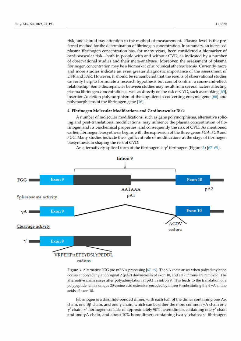
An alternatively-spliced form of the fibrinogen is γ′ fibrinogen (Figure 3) [67–69].
Figure 3. Alternative FGG pre-mRNA processing [67–69]. The γA chain arises when polyadenylationoccurs at polyadenylation signal 2 (pA2) downstream of exon 10, and all 9 introns are removed. Thealternative chain arises after polyadenylation at pA1 in intron 9. This leads to the translation of apolypeptide with a unique 20-amino acid extension encoded by intron 9, substituting the 4 γA aminoacids of exon 10.
Fibrinogen is a disulfide-bonded dimer, with each half of the dimer containing one Aα
chain, one Bβ chain, and one γ chain, which can be either the more common γA chain or aγ′ chain. γ′ fibrinogen consists of approximately 90% heterodimers containing one γ′ chainand one γA chain, and about 10% homodimers containing two γ′ chains; γ′ fibrinogen
Int. J. Mol. Sci. 2022, 23, 193 12 of 20
constitutes about 3–40% of total fibrinogen plasma concentrations [70,71]. In particular,compared to the total fibrinogen, γ′ fibrinogen forms fibrin blood clots that show differencesin clot architecture, are mechanically stiffer, and are very resistant to fibrinolysis [67]. Thereview of the literature by de Willige et al. indicates a number of different roles of thefibrinogen γ′ chain in haemostasis [68].
The results of previous studies suggested that higher plasma concentrations of γ′
fibrinogen led to the formation of blood clots that are very resistant to fibrinolysis, whichincreased the risk of CVD [72,73]. In a study by Mannila et al., including 387 postinfarctionpatients and 387 healthy individuals, it was shown that elevated plasma γ′ fibrinogenconcentration was an independent predictor of myocardial infraction (OR = 1.24; 95% CI:1.01–1.52) [74]. Other studies, such as the study by Lovely et al. of 3042 participantsfrom the Framingham Heart Study Offspring Cohort, found that γ′ fibrinogen plasmaconcentration was associated with prevalent CVD (p = 0.02), although the top 2 SNPs(rs7681423 and rs1049636) associated with γ′ fibrinogen plasma concentration were notassociated with a risk of CVD [67]. There are also newer studies, the results of whichindicate a significant role of the FGG polymorphism in the risk of CVD. In a study byDrizlionok et al. A/A and G/A genotype carriers of an SNP in rs2066865 in the FGG had ahigher plasma fibrinogen concentration, and this might be associated with an increasedrisk of microvascular thrombosis [75].
In a study by Appiah et al. covering adults ≥65 years (n = 3219) enrolled in theCardiovascular Health Study, the association of plasma γ′ fibrinogen concentration withthe incidence of CVD, independent of established CVD risk factors and total fibrinogen, wasassessed. Hazard ratio per 1 standard deviation (10.7 mg/dL) increment of γ′ fibrinogenwas 1.02 (95% CI: 0.95–1.10) for CAD; 0.88 (0.77–1.00) for ischemic stroke; 1.07 (0.87–1.32)for PAD; 1.00 (0.92–1.08) for heart failure and 1.01 (0.92–1.10) for CVD mortality [76].Nonetheless, other results were obtained in the Mendelian randomization study by Manerset al., which assessed γ′ and the total fibrinogen plasma concentration in relation to therisk of venous thromboembolism and ischemic stroke. It was shown that estimates basedon a combination of 16 genetic instruments for γ′ fibrinogen and 75 genetic instrumentsfor total fibrinogen indicated a protective effect of higher γ′ fibrinogen and higher totalfibrinogen on venous thromboembolism risk. There was also a protective effect of higherγ′ fibrinogen plasma concentration on cardioembolic and large artery stroke risk [77].
Therefore, whether or not γ′ fibrinogen is simply a marker of CVD or a prospectivelydefined risk factor for CVD remains controversial. It is known that other factors alsoinfluence the structure of a blood clot. Clot structure may contribute to an increased CVDrisk in vivo through associations with other CVD risk factors (age, metabolic syndrome,CRP, high density lipoprotein HDL cholesterol and homocysteine) independent fromtotal or γ ‘fibrinogen plasma concentrationa [78]. Similar conclusions were reached byPieters et al., who stated that CVD risk factors (excluding fibrinogen) explained 20%and 3%, respectively, of the variance in fibrinogen γ′ and the γ/total fibrinogen ratio,with C-reactive protein making the biggest contribution. More than 50% of the variance infibrinogen γ′ and γ′/total fibrinogen ratio is explained by factors other than total fibrinogenor other traditional CVD risk factors [79]. Recently, Rautenbach et al. found that the ironmetabolism activity may affect the plasma fibrinogen concentration and the percentage ofγ′ fibrinogen [80]. Moreover, another study by Rautenbach et al. also found that alcoholintake influenced the percentage of γ′ fibrinogen, as well as modulated the influenceof fibrinogen SNPs on total fibrinogen plasma concentrations [81]. The AtherosclerosisRisk in Communities (ARIC) study by Appiah et al. found that γ′ fibrinogen plasmaconcentrations seemed to reflect the general inflammation that accompanies and maycontribute to ASCVD, instead of γ′ fibrinogen being a causal risk factor [70]. The protectiveeffect of γ′ fibrinogen described in some studies may be due to the different roles offibrinogen, aside from the formation of fibrin clots, in thrombotic diseases of variousaetiologies [77]. The results of in vivo studies indicate that the elevated levels of the γA/γAfibrinogen isoform promote arterial thrombosis in vivo, whereas the γA/γ′ isoform does
Int. J. Mol. Sci. 2022, 23, 193 13 of 20
not [82]. Moreover, a significant influence on the relationship between γ′ fibrinogen andthe risk of CVD may depend on the presence of various polymorphisms, such as FGG9340T and FGA 2224G, for example [74]. In conclusion, the question of the influence of theisoform of γ′ fibrinogen on the risk of CVD requires further research.
The results of studies assessing the impact of FGB polymorphisms are inconsistent [83].In a meta-analysis by Li et al., including the results of 18 case–control studies (3033 ve-nous thromboembolism cases and 4547 healthy subjects), the relationship between theβ-fibrinogen polymorphisms, -455 G/A (rs1800790) and -148 C/T, and venous throm-boembolism risk was assessed. FGB -455 G/A and -148 C/T polymorphisms were notsignificantly associated with the susceptibility to venous thromboembolism in overallpopulations. Moreover, the -455 G/A polymorphism was associated with a decreasedrisk of venous thromboembolism, found among the Caucasian population [84]. In turn,the meta-analysis by Luo et al., including the results of 49 clinical trials, assessed therelationship between the same FGB polymorphisms with the risk of ischemic stroke. The-148 C/T polymorphism was significantly correlated with the risk of ischemic stroke inboth Asians and Caucasians, while the -455 G/A polymorphism was only significantlycorrelated with the risk of ischemic stroke in Asians. Moreover, it was found that both FGB-148 C/T and -455 G/A polymorphisms were significantly correlated with the risk of cere-bral infarction [85]. In a study by Canseco-Avila et al., covering 118 subjects with unstableand stable CAD, it was shown that fibrinogen plasma concentrations (>465 mg/dL) weresignificant in patients with CAD. These fibrinogen plasma concentrations were associatedwith CVD mortality during the follow-up analysis of the unstable coronary disease group(p = 0.04). It was also shown that the allelic loads of -455 G/A and -148 C/T were associatedwith plasma fibrinogen concentrations >450 mg/dL (p < 0.003 and p = 0.03) and with arisk of CAD (p = 0.016 and p < 0.006). Moreover, the follow-up of posterior events after anacute coronary event showed that the genetic load of the -148 C/T allele was associatedwith major adverse cardiovascular events (RR = 1.8; 95% CI: 1.01–3.35, p = 0.04) [86]. Ameta-analysis of 26 clinical trials, by Gu et al., also assessed the association between the-455 G/A polymorphism and the risk of ischemic stroke. It was shown that this polymor-phism was associated with the risk of ischemic stroke in overall, Asian, and adult analyses,but statistical association was not observed for Caucasians and children [87]. An interestingmeta-analysis of the results of 45 clinical trials, by Gu et al., attempted to summarize thedata on the influence of the -455 G/A polymorphism on the risk of ischemic stroke andCAD. It was shown that the -455 G/A polymorphism was associated with the risk ofischemic stroke when compared with the dominant model (OR = 1.518; 95% CI: 1.279–1.802for AA + GA vs. GG). In the subgroup analysis by ethnicity, significantly elevated riskswere associated with the A allele in Asians (OR = 1.700; 95% CI: 1.417–2.040) but not inCaucasians (OR = 0.942; 95% CI: 0.813–1.091). Moreover, it was found that the -455G/Apolymorphism was associated with CAD (OR = 1.802; 95% CI: 1.445–2.246) [88]. In thestudy by Hu et al., the impact of the -455 G/A polymorphism on the risk of cardioembolicstroke (CES) in patients with atrial fibrillation (AF) and a low CHA2DS2-VaSc score wasassessed. The study included 479 AF patients with CES and 580 AF patients without CES. Itwas shown that patients with the -455 G/A polymorphism had a higher plasma fibrinogenconcentration (3.29 ± 0.38 mg/dL vs. 2.87 ± 0.18 mg/dL, p < 0.001). Moreover, it wasfound that -455 G/A was independently associated with an increased risk of CES in AFpatients and the significance remained after the Bonferroni correction in the additive (AAvs. GA vs. GG), dominant (AA + GA vs. GG), and recessive models (AA vs. GA + GG),with ORs of 1.548 (95% CI: 1.251–1.915, p = 0.001), 1.588 (95% CI: (1.226–2.057, p = 0.003),and 2.394 (95% CI: 1.357–4.223, p = 0.015) [89]. In the study by Golenia et al., which included426 Polish patients with ischemic stroke, it was shown that the β-fibrinogen -455 G/Agene polymorphism was not a risk factor for this disease [90]. Reviewing the results ofother studies, one can find evidence of a different effect of the presence of the -455 G/Apolymorphism on the risk of CAD (increased risk, no effect and protective effect) [91–93].In a study by Martiskainen et al., covering 486 stroke patients (55–85 years), who were
Int. J. Mol. Sci. 2022, 23, 193 14 of 20
subjected to clinical and MRI examinations and followed over 12.5 years, the impact of the-455 G/A polymorphism on the prognosis after stroke was assessed. It was shown thatwomen aged 55–71 years who carried the FGB -455 A-allele had worse survival regardlessof smoking status compared to non-smoking FGB -455 GG homozygotes (non-smokers:HR = 5.21; 95% CI: 1.38–19.7; smokers: HR = 7.03; 95% CI: 1.81–27.3). Such a relationshipwas not demonstrated for women in the oldest age-group, nor among men [94].
The issue of the influence of the FGB polymorphism on the risk of lower extremitydeep venous thrombosis (LEDVT) is also interesting. In a study by Han et al. involving120 LEDVT patients and 120 healthy people, the relationship between six SNPs in theFGB promoter was assessed: -148 C/T, -249 C/T, -455 G/A, -854 G/A, -993 C/T and-1420 G/A and the risk of LEDVT. A higher fibrinogen plasma concentration was shownto increase the risk of LEDVT. The risk of LEDVT increased by 4.579 times for every unitincrease in fibrinogen plasma concentration. It was found that polymorphisms such as-1420 (AG + AA) and -148 (CT + TT) were associated with a higher risk of LEDVT [95].
It is worth mentioning that congenital fibrinogen deficiencies are rare bleeding dis-orders characterized by extensive genetic heterogeneity in all the three genes: FGA, FGB,and FGG (encoding the Aα, Bβ and γ chain, respectively). Depending on the type andsite of mutations, congenital defects of fibrinogen can result in variable clinical manifesta-tions, which range from asymptomatic conditions to the life-threatening bleeding or eventhromboembolic events [96–98].
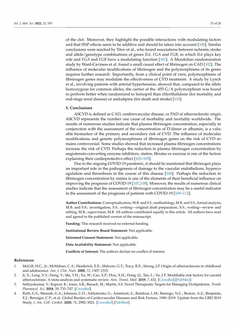
The influence of the more important mutations and polymorphisms of fibrinogengenes on the risk of CVD is summarized in Table 3.
Table 3. Fibrinogen molecular modifications and cardiovascular risk. CVD - cardiovascular disease;CAD—Coronary artery disease; MACE—major adverse cardiovascular events.
Gene Polymorphism/Mutation Effect on Cardiovascular Risk Bibliography
FGG
γ′ fibrinogen↑myocardial infraction
↑/↔ CVD↓ venous thromboembolism and ischemic stroke
[67,74,76,77]
rs7681423 and rs1049636 ↔ CVD [67]
rs2066865 ↑microvascular thrombosis [75]
FGB
-455 G/A
↔ venous thromboembolism↓ venous thromboembolism (Caucasians)
↑ ischemic stroke (Asian)↔ ischemic stroke (Caucasians and children)
↑ cerebral infarction↑ CAD
↑ cardioembolic stroke
[84–89]
-148 C/T
↔ venous thromboembolism↑ ischemic stroke (Asians and Caucasians)
↑ cerebral infarction↑ CAD↑MACE
[84–86]
-1420 (AG + AA) and -148 (CT + TT) ↑ lower extremity deep venous thrombosis [95]
In conclusion, the influence of molecular modifications of fibrinogen, especially thepolymorphisms of its genes, on the risk of CVD remains inconsistent. One possible ex-planation for the discrepancy in test results may be the complexity of the regulation offibrinogen phenotype formation. A study by Cronjé et al. found that, apart from SNPs inthe fibrinogen (FGA, FGB and FGG) genes, the fibrinogen phenotypes were also associatedwith SNPs in genes playing a role in lipid homeostasis (LDL-R, PCSK-9), together with CBSand CRP polymorphisms (particularly, CRP-rs3093068) [99]. Cronjé et al. also indicatedthe significant effect of IL-6 on the plasma fibrinogen concentration and the properties
Int. J. Mol. Sci. 2022, 23, 193 15 of 20
of the clot. Moreover, they highlight the possible interactions with modulating factorsand that SNP effects seem to be additive and should be taken into account [100]. Similarconclusions were reached by Titov et al., who found associations between ischemic strokeand allele/genotype combinations of genes IL6, FGA and FGB, in which IL6 plays keyrole and FGA and FGB have a modulating function [101]. A Mendelian randomizationstudy by Ward-Caviness et al. found a small causal effect of fibrinogen on CAD [102]. Theinfluence of molecular modifications of fibrinogen and the polymorphisms of its genesrequires further research. Importantly, from a clinical point of view, polymorphisms offibrinogen genes may modulate the effectiveness of CVD treatment. A study by Lynchet al., involving patients with arterial hypertension, showed that, compared to the allelehomozygous for common alleles, the carrier of the -455 G/A polymorphism was foundto perform better when randomized to lisinopril than chlorthalidone (for mortality andend-stage renal disease) or amlodipine (for death and stroke) [103].
5. Conclusions
ASCVD is defined as CAD, cerebrovascular disease, or PAD of atherosclerotic origin.ASCVD represents the number one cause of morbidity and mortality worldwide. Theresults of numerous studies indicate that plasma fibrinogen concentration, especially inconjunction with the assessment of the concentration of D’dimer or albumin, is a valu-able biomarker of the primary and secondary risk of CVD. The influence of molecularmodifications and genetic polymorphisms of fibrinogen genes on the risk of CVD re-mains controversial. Some studies showed that increased plasma fibrinogen concentrationsincrease the risk of CVD. Perhaps the reduction in plasma fibrinogen concentration byangiotensin-converting enzyme inhibitors, statins, fibrates or exercise is one of the factorsexplaining their cardioprotective effect [103–105].
Due to the ongoing COVID-19 pandemic, it should be mentioned that fibrinogen playsan important role in the pathogenesis of damage to the vascular endothelium, hyperco-agulation and thrombosis in the course of this disease [106]. Perhaps the reduction infibrinogen concentration by statins is one of the elements of their beneficial influence onimproving the prognosis of COVID-19 [107,108]. Moreover, the results of numerous clinicalstudies indicate that the assessment of fibrinogen concentration may be a useful indicatorin the assessment of the prognosis of patients with COVID-19 [109–112].
Author Contributions: Conceptualization, M.B. and S.S.; methodology, M.B. and S.S.; formal analysis,M.B. and S.S.; investigation, S.S.; writing—original draft preparation, S.S.; writing—review andediting, M.B.; supervision, M.B. All authors contributed equally to this article. All authors have readand agreed to the published version of the manuscript.
Funding: This research received no external funding.
Institutional Review Board Statement: Not applicable.
Informed Consent Statement: Not applicable.
Data Availability Statement: Not applicable.
Conflicts of Interest: The authors declare no conflict of interest.
References1. McGill, H.C., Jr.; McMahan, C.A.; Herderick, E.E.; Malcom, G.T.; Tracy, R.E.; Strong, J.P. Origin of atherosclerosis in childhood
and adolescence. Am. J. Clin. Nutr. 2000, 72, 1307–1315.2. Ji, X.; Leng, X.Y.; Dong, Y.; Ma, Y.H.; Xu, W.; Cao, X.P.; Hou, X.H.; Dong, Q.; Tan, L.; Yu, J.T. Modifiable risk factors for carotid
atherosclerosis: A meta-analysis and systematic review. Ann. Transl. Med. 2019, 7, 632. [CrossRef] [PubMed]3. Sathiyakumar, V.; Kapoor, K.; Jones, S.R.; Banach, M.; Martin, S.S. Novel Therapeutic Targets for Managing Dyslipidemia. Trends
Pharmacol. Sci. 2018, 39, 733–747. [CrossRef]4. Roth, G.S.; Mensah, G.A.; Johnson, C.O.; Addolorato, G.; Ammirati, E.; Baddour, L.M.; Barengo, N.C.; Beaton, A.Z.; Benjamin,
E.J.; Benziger, C.P.; et al. Global Burden of Cardiovascular Diseases and Risk Factors, 1990–2019: Update from the GBD 2019Study. J. Am. Coll. Cardiol. 2020, 76, 2982–3021. [CrossRef] [PubMed]
Int. J. Mol. Sci. 2022, 23, 193 16 of 20
5. Ab Khan, M.; Hashim, M.J.; Mustafa, H.; Baniyas, M.Y.; Al Suwaidi, S.K.B.M.; AIKatheeri, R.; Alblooshi, F.M.K.; Almatrooshi,M.E.A.H.; Alzaabi, M.E.H.; Al Darmaki, R.S.; et al. Global Epidemiology of Ischemic Heart Disease: Results from the GlobalBurden of Disease Study. Cureus 2020, 12, e9349.
6. Feigin, V.L.; Stark, B.A.; Johnson, C.O.; Roth, G.A.; Bisignano, C.; Abady, G.G.; Abbasifard, M.; Abbasi-Kangevari, M.; Abd-Allah,F.; Abdi, V.; et al. Global, regional, and national burden of stroke and its risk factors, 1990–2019: A systematic analysis for theGlobal Burden of Disease Study 2019. Lancet Neurol. 2021, 20, 795–820. [CrossRef]
7. McDonald, L.; Edgill, M. Coagulability of the blood in ischaemic heart disease. Lancet 1957, 2, 457–460. [CrossRef]8. Kannel, W.B.; Wolf, P.A.; Castelli, W.P.; D’Agostino, R.B. Fibrinogen and risk of cardiovascular disease. The Framingham Study.
JAMA 1987, 258, 1183–1186. [CrossRef]9. Pieters, M.; Wolberg, A.S. Fibrinogen and fibrin: An illustrated review. Res. Pract. Thromb. Haemost. 2019, 3, 161–172. [CrossRef]10. Vilar, R.; Fish, R.J.; Casini, A.; Neerman-Arbez, M. Fibrin(ogen) in human disease: Both friend and foe. Haematologica 2020, 105,
284–296. [CrossRef]11. Neerman-Arbez, M.; Casini, A. Clinical Consequences and Molecular Bases of Low Fibrinogen Levels. Int. J. Mol. Sci. 2018,
19, 192. [CrossRef] [PubMed]12. Murdaca, G.; Spanò, F.; Cagnati, P.; Puppo, F. Free radicals and endothelial dysfunction: Potential positive effects of TNF-α
inhibitors. Redox Rep. 2013, 18, 95–99. [CrossRef] [PubMed]13. Kattula, S.; Byrnes, J.R.; Wolberg, A.S. Fibrinogen and Fibrin in Hemostasis and Thrombosis. Arter. Thromb. Vasc. Biol. 2017, 37,
13–21. [CrossRef]14. Simurda, T.; Snahnicanova, Z.; Loderer, D.; Sokol, J.; Stasko, J.; Lasabova, Z.; Kubisz, P. Fibrinogen martin: A novel mutation in
FGB (Gln180Stop) causing congenital afibrinogenemia. Semin. Thromb. Hemost. 2016, 42, 455–458. [PubMed]15. De Vries, J.J.; Snoek, C.J.M.; Rijken, D.C.; de Maat, M.P.M. Effects of Post-Translational Modifications of Fibrinogen on Clot
Formation, Clot Structure, and Fibrinolysis: A Systematic Review. Arter. Thromb. Vasc. Biol. 2020, 40, 554–569. [CrossRef][PubMed]
16. Siegerink, B.; Rosendaal, F.R.; Algra, A. Genetic variation in fibrinogen; its relationship to fibrinogen levels and the risk ofmyocardial infarction and ischemic stroke. J. Thromb. Haemost. 2009, 7, 385–390. [CrossRef] [PubMed]
17. Lee, A.J.; Lowe, G.D.; Woodward, M.; Tunstall-Pedoe, H. Fibrinogen in relation to personal history of prevalent hypertension,diabetes, stroke, intermittent claudication, coronary heart disease, and family history: The Scottish Heart Health Study. Br. HeartJ. 1993, 69, 338–342. [CrossRef]
18. Kannel, W.B.; D’Agostino, R.B.; Belanger, A.J. Update on fibrinogen as a cardiovascular risk factor. Ann. Epidemiol. 1992, 2,457–466. [CrossRef]
19. Kaptoge, S.; White, I.R.; Thompson, S.G.; Wood, A.M.; Lewington, S.; Lowe, G.D.O.; Danesh, J. Associations of plasma fibrinogenlevels with established cardiovascular disease risk factors, inflammatory markers, and other characteristics: Individual participantmeta-analysis of 154,211 adults in 31 prospective studies: The fibrinogen studies collaboration. Am. J. Epidemiol. 2007, 166,867–879.
20. Kryczka, K.E.; Kruk, M.; Demkow, M.; Lubiszewska, B. Fibrinogen and a triad of thrombosis, inflammation, and the renin-angiotensin system in premature coronary artery disease in women: A new insight into sex-related differences in the pathogenesisof the disease. Biomolecules 2021, 11, 1036. [CrossRef]
21. Lowe, G.D.O.; Rumley, A.; Mackie, I.J. Plasma fibrinogen. Ann. Clin. Biochem. 2004, 41, 430–440. [CrossRef]22. Thompson, S.G.; Kienast, J.; Pyke, S.D.; Haverkate, F.; van de Loo, J.C. Hemostatic factors and the risk of myocardial infarction
or sudden death in patients with angina pectoris. European Concerted Action on Thrombosis and Disabilities Angina PectorisStudy Group. N. Engl. J. Med. 1995, 332, 635–641. [CrossRef] [PubMed]
23. Ma, J.; Hannekens, C.H.; Ridker, P.M.; Stampfer, M.J. A prospective study of fibrinogen and risk of myocardial infarction in thePhysicians’ Health Study. J. Am. Coll. Cardiol. 1999, 33, 1347–1352. [CrossRef]
24. Yuan, D.; Jiang, P.; Zhu, P.; Jia, S.; Zhang, C.; Liu, Y.; Liu, R.; Xu, J.; Tang, X.; Zhao, X.; et al. Prognostic value of fibrinogen inpatients with coronary artery disease and prediabetes or diabetes following percutaneous coronary intervention: 5-year findingsfrom a large cohort study. Cardiovasc. Diabetol. 2021, 20, 143. [CrossRef] [PubMed]
25. Peycheva, M.; Deneva, T.; Zahariev, Z. The role of fibrinogen in acute ischaemic stroke. Neurol. Neurochir. Pol. 2021, 55, 74–80.[CrossRef] [PubMed]
26. Meng, Z.; Zhao, Y.; He, Y. Fibrinogen level predicts outcomes in critically ill patients with acute exacerbation of chronic heartfailure. Dis. Markers 2021, 2021, 6639393. [CrossRef]
27. Ceasovschih, A.; Sorodoc, V.; Onofrei Aursulesei, V.; Tesloianu, D.; Tuchilus, C.; Anisie, E.; Petris, A.; Statescu, C.; Jaba, E.; Stoica,A.; et al. Biomarker utility for peripheral artery disease diagnosis in real clinical practice: A prospective study. Diagnostics 2020,10, 723. [CrossRef]
28. Samir, G.M.; Khalil, O.A.; Fawzy, M.S.; Sadek, A.M.E.M. Study of fibrinogen level in acute ischemic stroke patients in medicalintensive care unit. Egypt. J. Crit. Care Med. 2020, 7, 51–56.
29. Song, J.; Yu, T.; Sun, Z.; Li, Z.; He, D.; Sun, Z. Comparison of prognostic significance between serum fibrinogen and GlobalRegistry of Acute Coronary Events score for prognosis of patients with non-ST-elevation acute coronary syndromes undergoingpercutaneous coronary intervention. Coron. Artery Dis. 2020, 31, 124–129. [CrossRef]
Int. J. Mol. Sci. 2022, 23, 193 17 of 20
30. Liu, S.-L.; Wu, N.-Q.; Shi, H.-W.; Dong, Q.; Dong, Q.-T.; Gao, Y.; Guo, Y.-L.; Li, J.-J. Fibrinogen is associated with glucosemetabolism and cardiovascular outcomes in patients with coronary artery disease. Cardiovasc. Diabetol. 2020, 19, 36. [CrossRef][PubMed]
31. Jiang, P.; Gao, Z.; Zhao, W.; Song, Y.; Tang, X.-F.; Xu, J.-J.; Wang, H.-H.; Jiang, L.; Chen, J.; Qiao, S.-B.; et al. Relationship betweenfibrinogen levels and cardiovascular events in patients receiving percutaneous coronary intervention: A large single-center study.Chin. Med. J. 2019, 132, 914–921. [CrossRef] [PubMed]
32. Zhang, L.; Xu, C.; Liu, J.; Bai, X.; Li, R.; Wang, L.; Zhou, J.; Wu, Y.; Yuan, Z. Baseline plasma fibrinogen is associated withhaemoglobin A1c and 2-year major adverse cardiovascular events following percutaneous coronary intervention in patients withacute coronary syndrome: A single-centre, prospective cohort study. Cardiovasc. Diabetol. 2019, 18, 52. [CrossRef]
33. Chen, Q.-F.; Cao, D.; Ye, T.-T.; Deng, H.-H.; Zhu, H. Peripheral arterial disease in type 2 diabetes is associated with an increase infibrinogen levels. Int. J. Endocrinol. 2018, 2018, 3709534. [CrossRef]
34. Gao, X.-Y.; Zhou, B.-Y.; Zhang, M.-Z.; Zhao, X.; Qing, P.; Zhu, C.-G.; Wu, N.-Q.; Guo, Y.-L.; Gao, Y.; Li, X.-L.; et al. Associationbetween fibrinogen level and the severity of coronary stenosis in 418 male patients with myocardial infarction younger than 35years old. Oncotarget 2017, 8, 81361–81368. [CrossRef] [PubMed]
35. Tabakci, M.M.; Gerin, F.; Sunbul, M.; Toprak, C.; Durmus, H.I.; Demir, S.; Arslantas, U.; Cersit, S.; Batgerel, U.; Kargın, R. Relationof plasma fibrinogen level with the presence, severity, and complexity of coronary artery disease. Clin. Appl. Thromb. 2017, 23,638–644. [CrossRef] [PubMed]
36. Yang, S.-H.; Du, Y.; Zhang, Y.; Li, X.-L.; Li, S.; Xu, R.-X.; Zhu, C.-G.; Guo, Y.-L.; Wu, N.-Q.; Qing, P.; et al. Serum fibrinogen andcardiovascular events in Chinese patients with type 2 diabetes and stable coronary artery disease: A prospective observationalstudy. BMJ Open 2017, 7, e015041. [CrossRef] [PubMed]
37. Peng, Y.; Xia, T.-L.; Li, Y.-M.; Huang, F.-Y.; Chai, H.; Wang, P.-J.; Liu, W.; Zhang, C.; Pu, X.-B.; Chen, S.-J.; et al. Fibrinogen isrelated to long-term mortality in Chinese patients with acute coronary syndrome but failed to enhance the prognostic value of theGRACE score. Oncotarget 2017, 8, 20622–20629. [CrossRef] [PubMed]
38. Kunusor, S.K.; Kurl, S.; Zaccardi, F.; Laukkanen, J.A. Baseline and long-term fibrinogen levels and risk of sudden cardiac death: Anew prospective study and meta-analysis. Atherosclerosis 2016, 245, 171–180. [CrossRef] [PubMed]
39. Kotbi, S.; Mjabber, A.; Chadli, A.; El Hammiri, A.; El Aziz, S.; Oukkache, B.; Mifdal, H.; Nourichafi, N.; Kamal, N.; Habbal, R.;et al. Correlation between the plasma fibrinogen concentration and coronary heart disease severity in Moroccan patients withtype 2 diabetes. Prospective study. Ann. Endocrinol. 2016, 77, 606–614. [CrossRef] [PubMed]
40. Peng, Y.; Wang, H.; Li, Y.-M.; Huang, B.-T.; Huang, F.-Y.; Xia, T.-L.; Chai, H.; Wang, P.-J.; Liu, W.; Zhang, C.; et al. Relation betweenadmission plasma fibrinogen levels and mortality in Chinese patients with coronary artery disease. Sci. Rep. 2016, 6, 30506.[CrossRef]
41. Peng, Y.; Li, Y.-M.; Chai, H.; Zuo, Z.-L.; Wang, P.-J.; Gui, Y.-Y.; Huang, B.-T.; Liao, Y.-B.; Xia, T.-L.; Huang, F.-Y.; et al. Understandingthe controversy surrounding the correlation between fibrinogen level and prognosis of coronary artery disease-the role of thesubtypes of coronary artery disease. Int. J. Cardiol. 2016, 222, 968–972. [CrossRef] [PubMed]
42. Zhang, Y.; Zhu, C.-G.; Guo, Y.-L.; Xu, R.-X.; Li, S.; Dong, Q.; Li, J.-J. Higher fibrinogen level is independently linked withthe presence and severity of new-onset coronary atherosclerosis among Han Chinese population. PLoS ONE 2014, 9, e113460.[CrossRef] [PubMed]
43. Hong, L.-F.; Li, X.-L.; Luo, S.-H.; Guo, Y.-L.; Zhu, C.-G.; Qing, P.; Wu, N.-Q.; Li, J.-J. Association of fibrinogen with severity ofstable coronary artery disease in patients with type 2 diabetic mellitus. Dis. Markers 2014, 2014, 485687. [CrossRef] [PubMed]
44. Bosevski, M.; Bosevska, G.; Stojanovska, L. Influence of fibrinogen and C-RP on progression of peripheral arterial disease in type2 diabetes: A preliminary report. Cardiovasc. Diabetol. 2013, 12, 29. [CrossRef] [PubMed]
45. Danesh, J.; Lewington, S.; Thompson, S.G.; Lowe, G.D.O.; Collins, R.; Kostis, J.B.; Wilson, A.C.; Folsom, A.R.; Wu, K.; Benderly, M.;et al. Plasma fibrinogen level and the risk of major cardiovascular diseases and nonvascular mortality: An individual participantmeta-analysis. JAMA 2005, 294, 1799–1809. [PubMed]
46. Kaptoge, S.; Di Agelantonio, E.; Pennells, L.; Wood, A.M.; White, I.R.; Gao, P.; Walker, M.; Thompson, A.; Sarwar, N.; Caslake, M.;et al. C-reactive protein, fibrinogen, and cardiovascular disease prediction. N. Engl. J. Med. 2012, 367, 1310–1320.
47. Song, B.; Shu, Y.; Xu, Y.N.; Fu, P. Plasma fibrinogen lever and risk of coronary heart disease among Chinese population: Asystematic review and meta-analysis. Int. J. Clin. Exp. Med. 2015, 8, 13195–13202.
48. Levenson, J.; Giral, P.; Razavian, M.; Gariepy, J.; Simon, A. Fibrinogen and silent atherosclerosis in subjects with cardiovascularrisk factors. Arter. Thromb. Vasc. Biol. 1995, 15, 1263–1268. [CrossRef]
49. Green, D.; Foiles, N.; Chan, C.; Schreiner, P.J.; Liu, K. Elevated fibrinogen levels and subsequent subclinical atherosclerosis: TheCARDIA Study. Atherosclerosis 2009, 202, 623–631. [CrossRef] [PubMed]
50. Green, D.; Chan, C.; Kang, J.; Liu, K.; Schreiner, P.J.; Jenny, N.S.; Tracy, R.P. Longitudinal assessment of fibrinogen in relation tosubclinical cardiovascular disease: The CARDIA study. J. Thromb. Haemost. 2010, 8, 489–495. [CrossRef] [PubMed]
51. Menti, E.; Zaffari, D.; Galarraga, T.; da Conceição E Lessa, J.R.; Pontin, B.; Pellanda, L.C.; Portal, V.L. Early markers ofatherosclerotic disease in individuals with excess weight and dyslipidemia. Arq. Bras. Cardiol. 2016, 106, 457–463. [CrossRef][PubMed]
52. Li, T.; Wang, F.; Peng, R.; Pei, S.; Hou, Z.; Lu, B.; Cong, X.; Chen, X. Sex-related differences in the association between plasmafibrinogen and non-calcified or mixed coronary atherosclerotic plaques. Biol. Sex Differ. 2018, 9, 51. [CrossRef] [PubMed]
Int. J. Mol. Sci. 2022, 23, 193 18 of 20
53. Wang, J.; Jia, L.; Li, X.; Jin, S.; Li, X.; Liu, F.; Shan, C.; Zhang, Y.; Yang, Y. New insights into the association between fibrinogenand coronary atherosclerotic plaque vulnerability: An intravascular optical coherence tomography study. Cardiovasc. Ther. 2019,2019, 8563717. [CrossRef]
54. Bai, Y.; Zheng, Y.-Y.; Tang, J.-N.; Yang, Y.-M.; Guo, Q.-Q.; Zhang, J.-C.; Cheng, M.-D.; Song, F.-H.; Wang, K.; Zhang, Z.-L.; et al.D-dimer to fibrinogen ratio as a novel prognostic marker in patients after undergoing percutaneous coronary intervention:A retrospective cohort study. Clin. Appl. Thromb. Hemost. 2020, 26, 1076029620948586. [CrossRef] [PubMed]
55. Zhang, D.-P.; Mao, X.-F.; Wu, T.-T.; Chen, Y.; Hou, X.-G.; Yang, Y.; Ma, X.; Zhang, J.-Y.; Ma, Y.-T.; Xie, X.; et al. The fibrinogen-to-albumin ratio is associated with outcomes in patients with coronary artery disease who underwent percutaneous coronaryintervention. Clin. Appl. Thromb. Hemost. 2020, 26, 1076029620933008. [CrossRef] [PubMed]
56. Roth, S.; Jansen, C.; M’Pembele, R.; Stroda, A.; Boeken, U.; Akhyari, P.; Lichtenberg, A.; Hollmann, M.W.; Huhn, R.; Buse, G.L.;et al. Fibrinogen-albumin-ratio is an independent predictor of thromboembolic complications in patients undergoing VA-ECMO.Sci. Rep. 2021, 11, 16648. [CrossRef] [PubMed]
57. Karahan, O.; Acet, H.; Ertas, F.; Tezcan, O.; Çaliskan, A.; Demir, M.; Kaya, A.F.; Demirtas, S.; Çevik, M.U.; Yavuz, C. The relation-ship between fibrinogen to albumin ratio and severity of coronary artery disease in patients with STEMI. Am. J. Emerg. Med. 2016,34, 1037–1042. [CrossRef]
58. Celebi, S.; Celebi, O.O.; Berkalp, B.; Amasyali, B. The association between the fibrinogen-to-albumin ratio and coronary arterydisease severity in patients with stable coronary artery disease. Coron. Artery Dis. 2020, 31, 512–517. [CrossRef]
59. Zhao, Y.P.; Ji, Y.Y.; Wang, F.Y.; Wang, S.L.; Lai, G.K.; Wang, T.; Tang, J.M. Value of fibrinogen to albumin ratio on predictingspontaneous recanalization of infarct-related artery in patients with acute ST-segment elevation myocardial infarction. ZhonghuaXin Xue Guan Bing Za Zhi 2019, 47, 123–128, (Article in Chinese, Only Abstract in English).
60. Erdogan, G.; Arslan, U.; Yenercag, M.; Durmus, G.; Tugrul, S.; Sahin, I. Relationship between the fibrinogen-to-albumin ratio andSYNTAX score in patients with non-st-elevation myocardial infarction. Rev. Investig. Clin. 2021, 73, 182–189. [CrossRef]
61. Li, M.; Tang, C.; Luo, E.; Qin, Y.; Wang, D.; Yan, G. Relation of fibrinogen-to-albumin ratio to severity of coronary artery diseaseand long-term prognosis in patients with non-ST elevation acute coronary syndrome. BioMed Res. Int. 2020, 2020, 1860268.[CrossRef] [PubMed]
62. Wang, C.; Li, G.; Liang, X.; Qin, C.; Luo, Q.; Song, R.; Chen, W. Predictive value of fibrinogen-to-albumin ratio for post-contrastacute kidney injury in patients undergoing elective percutaneous coronary intervention. Med. Sci. Monit. 2020, 26, e924498.[CrossRef]
63. Ishihara, K.K.; Kokubo, Y.; Yokota, C.; Hida, E.; Miyata, T.; Toyoda, K.; Matsumoto, M.; Minematsu, K.; Miyamoto, Y. Effect ofplasma fibrinogen, high-sensitive C-reactive protein, and cigarette smoking on carotid atherosclerosis: The Suita study. J. StrokeCerebrovasc. Dis. 2015, 24, 2385–2389. [CrossRef] [PubMed]
64. Lassé, M.; Pilbrow, A.P.; Kleffmann, T.; Andersson Överström, E.; von Zychlinski, A.; Frampton, C.M.A.; Poppe, K.K.; Troughton,R.W.; Lewis, L.K.; Prickett, T.C.R.; et al. Fibrinogen and hemoglobin predict near future cardiovascular events in asymptomaticindividuals. Sci. Rep. 2021, 11, 4605. [CrossRef]
65. Cho, H.M.; Kang, D.R.; Kim, H.C.; Oh, S.M.; Kim, B.K.; Suh, I. Association between fibrinogen and carotid atherosclerosisaccording to smoking status in a Korean male population. Yonsei Med. J. 2015, 56, 921–927. [CrossRef]
66. Kryczka, K.E.; Płoski, R.; Ksiezycka, E.; Kruk, M.; Kostrzewa, G.; Kowalik, I.; Demkow, M.; Lubiszewska, B. The associationbetween the insertion/deletion polymorphism of the angiotensin-converting enzyme gene and the plasma fibrinogen level inwomen and men with premature coronary artery atherosclerosis. Pol. Arch. Intern. Med. 2020, 130, 748–756. [CrossRef] [PubMed]
67. Lovely, R.S.; Yang, Q.; Massaro, J.M.; Wang, J.; D’Agostino Sr, R.B.; O’Donnell, C.J.; Shannon, J.; Farrell, D.H. Assessment ofgenetic determinants of the association of γ’ fibrinogen in relation to cardiovascular disease. Arter. Thromb. Vasc. Biol. 2011, 31,2345–2352. [CrossRef]
68. De Willige, S.U.; Standeven, K.F.; Philippou, H.; Ariëns, R.A.S. The pleiotropic role of the fibrinogen gamma’ chain in hemostasis.Blood 2009, 114, 3994–4001. [CrossRef] [PubMed]
69. Farrell, D.H. Primetime for γ′. Blood 2014, 124, 1389–1390. [CrossRef] [PubMed]70. Appiah, D.; Schreiner, J.P.; MacLehose, R.F.; Folsom, A.R. Association of plasma γ’ fibrinogen with incident cardiovascular
disease: The atherosclerosis risk in communities (ARIC) study. Arter. Thromb. Vasc. Biol. 2015, 35, 2700–2706. [CrossRef] [PubMed]71. Macrae, F.L.; Swieringa, F.; Heemskerk, J.W.M.; Ariëns, R.A.S. High fibrinogen γ’ levels in patient plasma increase clot formation
at arterial and venous shear. Blood Adv. 2021, 5, 3468–3477. [CrossRef]72. Lovely, R.S.; Kazmierczak, S.C.; Massaro, J.M.; D’Agostino, R.B., Sr.; O’Donnell, C.J.; Farrell, D.H. Gamma’ fibrinogen: Evaluation
of a new assay for study of associations with cardiovascular disease. Clin. Chem. 2010, 56, 781–788. [CrossRef] [PubMed]73. Farrell, D.H. γ’ fibrinogen as a novel marker of thrombotic disease. Clin. Chem. Lab. Med. 2012, 50, 1903–1909. [CrossRef]
[PubMed]74. Mannila, M.N.; Lovely, R.S.; Kazmierczak, S.C.; Eriksson, P.; Samnegård, A.; Farrell, D.H.; Hamsten, A.; Silveira, A. Elevated
plasma fibrinogen gamma’ concentration is associated with myocardial infarction: Effects of variation in fibrinogen genes andenvironmental factors. J. Thromb. Haemost. 2007, 5, 766–773. [CrossRef]
75. Drizlionoka, K.; Zarin, š, J.; Ozolina, A.; N, ikitina-Zak, e, L.; Mamaja, B. Polymorphism rs2066865 in the fibrinogen gamma chain (FGG)gene increases plasma fibrinogen concentration and is associated with an increased microvascular thrombosis rate. Medicina 2019,55, 563. [CrossRef] [PubMed]
Int. J. Mol. Sci. 2022, 23, 193 19 of 20
76. Appiah, D.; Heckbert, S.R.; Cushman, M.; Psaty, B.M.; Folsom, A.R. Lack of association of plasma gamma prime (γ’) fibrinogenwith incident cardiovascular disease. Thromb. Res. 2016, 143, 50–52. [CrossRef]
77. Maners, J.; Gill, D.; Pankratz, N.; Laffan, M.A.; Wolberg, A.S.; de Maat, M.P.M.; Ligthart, S.; Tang, W.; Ward-Caviness, C.K.;Fornage, M.; et al. A Mendelian randomization of γ’ and total fibrinogen levels in relation to venous thromboembolism andischemic stroke. Blood 2020, 136, 3062–3069. [CrossRef]
78. Kotzé, R.C.M.; Ariëns, R.A.S.; de Lange, Z.; Pieters, M. CVD risk factors are related to plasma fibrin clot properties independentof total and or γ’ fibrinogen concentration. Thromb. Res. 2014, 134, 963–969. [CrossRef]
79. Pieters, M.; Kotze, R.C.; Jerling, J.C.; Kruger, A.; Ariëns, R.A.S. Evidence that fibrinogen γ’ regulates plasma clot structure andlysis and relationship to cardiovascular risk factors in black Africans. Blood 2013, 121, 3254–3260. [CrossRef] [PubMed]
80. Rautenbach, P.H.; Nienaber-Rousseau, C.; de Lange-Loots, Z.; Pieters, M. Certain associations between iron biomarkers and totaland γ’ fibrinogen and plasma clot properties are mediated by fibrinogen genotypes. Front. Nutr. 2021, 8, 720048. [CrossRef]
81. Rautenbach, P.H.; Nienaber-Rousseau, C.; Pieters, M. The association of alcohol with circulating total fibrinogen and plasma clotdensity is mediated by fibrinogen and FXIII genotypes. Thromb. J. 2020, 18, 35. [CrossRef]
82. Walton, B.L.; Getz, T.M.; Bergmeier, W.; de Willige, S.U.; Wolberg, A.S. Gamma prime fibrinogen does not cause arterial thrombosis.Blood 2013, 122, 1092. [CrossRef]
83. Papageorgiou, N.; Tousoulis, D.; Siasos, G.; Stefanadis, C. Is fibrinogen a marker of inflammation in coronary artery disease?Hellenic J. Cardiol. 2010, 51, 1–9. [PubMed]
84. Li, D.; Zhang, X.; Huang, H.; Zhang, H. Association of β-fibrinogen polymorphisms and venous thromboembolism risk:A PRISMA-compliant meta-analysis. Medicine 2019, 98, e18204. [CrossRef] [PubMed]
85. Luo, H.; Li, X.; Jiang, A.; Zhang, B.; Bi, P.; Dong, Y.; Guo, Y. Associations of β-fibrinogen polymorphisms with the risk of ischemicstroke: A meta-analysis. J. Stroke Cerebrovasc. Dis. 2019, 28, 243–250. [CrossRef] [PubMed]
86. Canseco-Avila, L.M.; Lopez-Roblero, A.; Serrano-Guzman, E.; Aguilar-Fuentes, J.; Jerjes-Sanchez, C.; Rojas-Martinez, A.; Ortiz-Lopez, R. Polymorphisms -455G/A and -148C/T and fibrinogen plasmatic level as risk markers of coronary disease and majoradverse cardiovascular events. Dis. Markers 2019, 2019, 5769514. [CrossRef]
87. Gu, L.; Wu, G.; Su, L.; Yan, Y.; Long, J.; Tan, J.; Liang, B.; Guo, X.; Huang, G. Genetic polymorphism of β-fibrinogen gene-455G/Acan contribute to the risk of ischemic stroke. Neurol. Sci. 2014, 35, 151–161. [CrossRef] [PubMed]
88. Gu, L.; Liu, W.; Yan, Y.; Su, L.; Wu, G.; Liang, B.; Tan, J.; Huang, G. Influence of the β-fibrinogen-455G/A polymorphism ondevelopment of ischemic stroke and coronary heart disease. Thromb. Res. 2014, 133, 993–1005. [CrossRef] [PubMed]
89. Hu, X.; Wang, J.; Li, Y.; Wu, J.; Qiao, S.; Xu, S.; Huang, J.; Chen, L. The β-fibrinogen gene 455G/A polymorphism associated withcardioembolic stroke in atrial fibrillation with low CHA2DS2-VaSc score. Sci. Rep. 2017, 7, 17517. [CrossRef]
90. Golenia, A.; Chrzanowska-Wasko, J.; Jagiella, J.; Wnuk, M.; Ferens, A.; Klimkowicz-Mrowiec, A.; Adamski, M.; Ciecko-Michalska,I.; Słowik, A. The β-fibrinogen -455G/A gene polymorphism and the risk of ischaemic stroke in a Polish population. Neurol.Neurochir. Pol. 2013, 47, 152–156. [CrossRef]
91. Durmus, G.; Karakus, N.; Yuksel, S.; Kara, N. Analysis of twelve cardiovascular disease related gene mutations among Turkishpatients with coronary artery disease. Int. J. Blood Res. Disord. 2020, 7, 047.
92. Alkhiary, W.; Azzam, H.; Yossof, M.M.A.; Aref, S.; Othman, M.; El-Sharawy, S. Association of hemostatic gene polymorphismswith early-onset ischemic heart disease in Egyptian patients. Clin. Appl. Thromb. Hemost. 2016, 22, 535–542. [CrossRef]
93. Sewelam, M.I.; Ahmed, A.A.; Alwakeel, H.A.; Khaled, M.Y. Fibrinogen -455G/A promoter polymorphism in acute ST elevationmyocardial infarction in Egyptian patients. Egypt J. Haematol. 2014, 39, 98–102.
94. Martiskainen, M.; Oksala, N.; Pohjasvaara, T.; Kaste, M.; Oksala, A.; Karhunen, P.J.; Erkinjuntti, T. Beta-fibrinogen gene promoterA -455 allele associated with poor longterm survival among 55–71 years old Caucasian women in Finnish stroke cohort. BMCNeurol. 2014, 14, 137. [CrossRef]
95. Han, S.; Yang, B.; Feng, Y.; Zhao, L.; Feng, Q.; Guan, H.; Song, D.; Yin, F.; Zhuang, L. The correlation between FGB promoterpolymorphism and clotting function in patients with idiopathic lower extremity deep venous thrombosis. Clin. Appl. Thromb.Hemost. 2021, 27, 1076029620967108. [CrossRef] [PubMed]
96. Simurda, T.; Brunclikova, M.; Asselta, R.; Caccia, S.; Zolkova, J.; Kolkova, Z.; Loderer, D.; Skornova, I.; Hudecek, J.; Lasabova,Z.; et al. Genetic variants in the FGB and FGG genes mapping in the beta and gamma nodules of the fibrinogen molecule incongenital quantitative fibrinogen disorders associated with a thrombotic phenotype. Int. J. Mol. Sci. 2020, 21, 4616. [CrossRef][PubMed]
97. Simurda, T.; Caccia, S.; Asselta, R.; Zolkova, J.; Stasko, J.; Skornova, I.; Snahnicanova, Z.; Loderer, D.; Lasabova, Z.; Kubisz, P.Congenital hypofibrinogenemia associated with a novel heterozygous nonsense mutation in the globular C-terminal domain ofthe γ-chain (p.Glu275Stop). J. Thromb. Thromb. 2020, 50, 233–236. [CrossRef]
98. Simurda, T.; Casini, A.; Stasko, J.; Hudecek, J.; Skornova, I.; Vilar, R.; Neerman-Arbez, M.; Kubisz, P. Perioperative managementof a severe congenital hypofibrinogenemia with thrombotic phenotype. Thromb. Res. 2020, 188, 1–4. [CrossRef]
99. Cronjé, H.T.; Nienaber-Rousseau, C.; Zandberg, L.; Chikowore, T.; de Lange, Z.; van Zyl, T.; Pieters, M. Candidate gene analysis ofthe fibrinogen phenotype reveals the importance of polygenic co-regulation. Matrix Biol. 2017, 60–61, 16–26. [CrossRef] [PubMed]
100. Cronjé, H.T.; Nienaber-Rousseau, C.; Zandberg, L.; de Lange, Z.; Green, F.R.; Pieters, M. Fibrinogen and clot-related phenotypesdetermined by fibrinogen polymorphisms: Independent and IL-6-interactive associations. PLoS ONE 2017, 12, e0187712.
Int. J. Mol. Sci. 2022, 23, 193 20 of 20
101. Titov, B.V.; Barsova, R.M.; Martynov, M.Y.; Nikonova, A.A.; Favorov, A.V.; Gusev, E.I.; Favorova, O.O. Polymorphic variants ofgenes encoding interleukin-6 and fibrinogen, the risk of ischemic stroke and fibrinogen levels. Mol. Biol. 2012, 46, 93–102, (Articlein Russian, Only Abstract in English). [CrossRef]
102. Ward-Caviness, C.K.; de Vries, P.S.; Wiggins, K.L.; Huffman, J.E.; Yanek, L.R.; Bielak, L.F.; Franco, G.; Xiuqing, G.; Marcus, E.K.;Tim, K.; et al. Mendelian randomization evaluation of causal effects of fibrinogen on incident coronary heart disease. PLoS ONE2019, 14, e0216222. [CrossRef] [PubMed]
103. Lynch, A.I.; Boerwinkle, E.; Davis, B.R.; Ford, C.E.; Eckfeldt, J.H.; Leiendecker-Foster, C.; Arnett, D.K. Antihypertensivepharmacogenetic effect of fibrinogen-beta variant -455G>A on cardiovascular disease, end-stage renal disease, and mortality: TheGenHAT study. Pharm. Genom. 2009, 19, 415–421. [CrossRef] [PubMed]
104. Sahebkar, A.; Serban, M.-C.; Mikhailidis, D.P.; Toth, P.P.; Muntner, P.; Ursoniu, S.; Mosterou, S.; Glasser, S.; Martin, S.S.; Jones, S.R.;et al. Head-to-head comparison of statins versus fibrates in reducing plasma fibrinogen concentrations: A systematic review andmeta-analysis. Pharmacol. Res. 2016, 103, 236–252. [CrossRef]
105. Lovely, R.; Hossain, J.; Ramsey, J.P.; Komakula, V.; George, D.; Farrell, D.H.; Balagopal, P.B. Obesity-related increased γ’ fibrinogenconcentration in children and its reduction by a physical activity-based lifestyle intervention: A randomized controlled study. J.Pediatr. 2013, 163, 333–338. [CrossRef]
106. Grobler, C.; Maphumulo, S.C.; Grobbelaar, L.M.; Bredenkamp, J.C.; Laubscher, G.J.; Lourens, P.J.; Steenkamp, J.; Kell, D.B.;Pretorius, E. Covid-19: The rollercoaster of gibrin(ogen), D-dimer, von Willebrand factor, P-selectin and their interactions withendothelial cells, platelets and erythrocytes. Int. J. Mol. Sci. 2020, 21, 5168. [CrossRef]
107. Leibovitz, E.; Hazanov, N.; Frieman, A.; Elly, I.; Gavish, D. Atorvastatin reduces fibrinogen levels in patients with severehypercholesterolemia: Additional evidence to support the anti-inflammatory effects of statins. Isr. Med. Assoc. J. 2004, 6, 456–459.
108. Surma, S.; Banach, M.; Lewek, J. COVID-19 and lipids. The role of lipid disorders and statin use in the prognosis of patients withSARS-CoV-2 infection. Lipids Health Dis. 2021, 20, 141. [CrossRef]
109. Bi, X.; Su, Z.; Yan, H.; Du, J.; Wang, J.; Chen, L.; Peng, M.; Chen, S.; Shen, B.; Li, J. Prediction of severe illness due to COVID-19based on an analysis of initial Fibrinogen to Albumin Ratio and Platelet count. Platelets 2020, 31, 674–679. [CrossRef]
110. Di Micco, P.; Russo, V.; Carannante, N.; Imparato, M.; Cardillo, G.; Lodigiani, C. Prognostic value of fibrinogen among COVID-19patients admitted to an emergency department: An Italian cohort study. J. Clin. Med. 2020, 9, 4134. [CrossRef]
111. Küçükceran, K.; Ayranci, M.K.; Girisgin, A.S.; Koçak, S. Predictive value of D-dimer/albumin ratio and fibrinogen/albumin ratiofor in-hospital mortality in patients with COVID-19. Int. J. Clin. Pract. 2021, 75, e14263. [CrossRef] [PubMed]
112. Sui, J.; Noubouossie, D.F.; Gandotra, S.; Cao, L. Elevated plasma fibrinogen is associated with excessive inflammation and diseaseseverity in COVID-19 patients. Front. Cell. Infect. Microbiol. 2021, 11, 734005. [CrossRef] [PubMed]


