Embed Size (px)
Citation preview

Five new species of the genus Papuadytes Balke, 1998 fromNew Guinea (Coleoptera: Dytiscidae)
HELENA V. SHAVERDO1, KATAYO SAGATA2 & MICHAEL BALKE3
1Naturhistorisches Museum, Vienna, Austria, 2Wildlife Conservation Society, Goroka, EHP, Papua New
Guinea, and 3Department of Entomology, The Natural History Museum, London, UK and Zoologische
Staatssammlung, Munich, Germany
AbstractFive new species of New Guinean Papuadytes Balke are described. Based on distally fused and modifiedventral sclerites of the median lobe of the aedeagus, the P. broschii species group is suggested for P.broschii Balke, 1998, P. marinae sp. nov., and P. hintelmannae sp. nov. This group is only known fromPapua New Guinea where the species occur allopatrically in different mountain ranges. The other newspecies are P. atowaso sp. nov., P. munaso sp. nov., and P. vladimiri sp. nov.
Keywords: Papuadytes, new species, Copelatinae, Dytiscidae, New Guinea
Introduction
Described as a subgenus of Copelatus, Papuadytes Balke, 1998 was recently assigned
generic status following an analysis of copelatine phylogeny based on mitochondrial
DNA sequence data (Balke et al. 2004). Papuadytes was suggested to be the sister-group
of all other Copelatinae. It is delimited to the Australo-Pacific region, with the exception
of one Chinese species (Balke & Bergsten 2003). Papuadytes is the most species rich diving
beetle group in New Guinean running water habitats, especially low-order streams
and habitats associated with wider mountain streams (i.e. backflows, interstitial and small
water holes on river banks). To date, 35 species have been described from New Guinea,
18 of which occur in Papua New Guinea (PNG) (Balke 1998, 1999, 2001; Nilsson 2001).
Local species endemism is pronounced as confirmed by recent fieldwork conducted by
K. Sagata in the course of the Water Beetles of PNG project, launched in 2003. These
samples contained more than 10 hitherto undescribed species. Here, we describe three
characteristic ones of them, along with two other rather conspicuous species from a
museum collection.
Our recent efforts in PNG underpin the need for a nationwide survey of running water
beetles, or invertebrates in general, which are remarkably diverse yet almost unknown to
science. Such a survey will certainly form the basis for an improved understanding of
freshwater diversity from which sound awareness-rising as well as management strategies can
be expected to emerge.
Correspondence: Helena Shaverdo, Naturhistorisches Museum, Burgring 7, A-1010 Vienna, Austria. E-mail: [email protected]
Aquatic Insects
December 2005; 27(4): 269 – 280
ISSN 0165-0424 print/ISSN 1744-4152 online ª 2005 Taylor & Francis
DOI: 10.1080/01650420500290169
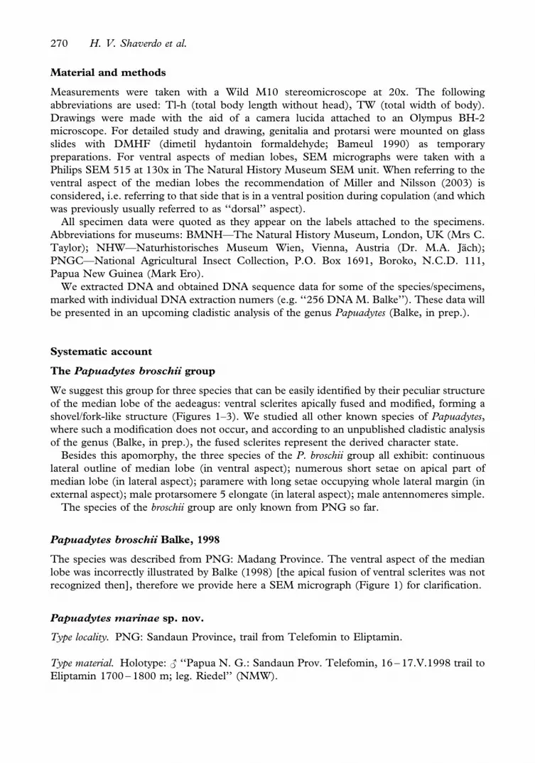
Material and methods
Measurements were taken with a Wild M10 stereomicroscope at 20x. The following
abbreviations are used: Tl-h (total body length without head), TW (total width of body).
Drawings were made with the aid of a camera lucida attached to an Olympus BH-2
microscope. For detailed study and drawing, genitalia and protarsi were mounted on glass
slides with DMHF (dimetil hydantoin formaldehyde; Bameul 1990) as temporary
preparations. For ventral aspects of median lobes, SEM micrographs were taken with a
Philips SEM 515 at 130x in The Natural History Museum SEM unit. When referring to the
ventral aspect of the median lobes the recommendation of Miller and Nilsson (2003) is
considered, i.e. referring to that side that is in a ventral position during copulation (and which
was previously usually referred to as ‘‘dorsal’’ aspect).
All specimen data were quoted as they appear on the labels attached to the specimens.
Abbreviations for museums: BMNH—The Natural History Museum, London, UK (Mrs C.
Taylor); NHW—Naturhistorisches Museum Wien, Vienna, Austria (Dr. M.A. Jach);
PNGC—National Agricultural Insect Collection, P.O. Box 1691, Boroko, N.C.D. 111,
Papua New Guinea (Mark Ero).
We extracted DNA and obtained DNA sequence data for some of the species/specimens,
marked with individual DNA extraction numers (e.g. ‘‘256 DNA M. Balke’’). These data will
be presented in an upcoming cladistic analysis of the genus Papuadytes (Balke, in prep.).
Systematic account
The Papuadytes broschii group
We suggest this group for three species that can be easily identified by their peculiar structure
of the median lobe of the aedeagus: ventral sclerites apically fused and modified, forming a
shovel/fork-like structure (Figures 1–3). We studied all other known species of Papuadytes,
where such a modification does not occur, and according to an unpublished cladistic analysis
of the genus (Balke, in prep.), the fused sclerites represent the derived character state.
Besides this apomorphy, the three species of the P. broschii group all exhibit: continuous
lateral outline of median lobe (in ventral aspect); numerous short setae on apical part of
median lobe (in lateral aspect); paramere with long setae occupying whole lateral margin (in
external aspect); male protarsomere 5 elongate (in lateral aspect); male antennomeres simple.
The species of the broschii group are only known from PNG so far.
Papuadytes broschii Balke, 1998
The species was described from PNG: Madang Province. The ventral aspect of the median
lobe was incorrectly illustrated by Balke (1998) [the apical fusion of ventral sclerites was not
recognized then], therefore we provide here a SEM micrograph (Figure 1) for clarification.
Papuadytes marinae sp. nov.
Type locality. PNG: Sandaun Province, trail from Telefomin to Eliptamin.
Type material. Holotype: < ‘‘Papua N. G.: Sandaun Prov. Telefomin, 16 – 17.V.1998 trail to
Eliptamin 1700 – 1800 m; leg. Riedel’’ (NMW).
270 H. V. Shaverdo et al.

Diagnosis. The species can be distinguished from other members of the broschii group by the
less concolorous and rather dull dorsal surface of the body, with strongly impressed
microreticulation and dense coarse punctation, as well as the shape of the median lobe and
paramere.
Figures 1 – 6. Median lobe of aedeagus, ventral aspect, SEM (at 130x): (1) Papuadytes broschii, paratype; (2) P.
marinae, holotype; (3) P. hintelmannae, holotype; (4) P. atowaso, holotype; (5) P. munaso, holotype; (6) P. vladimiri,
holotype.
Papuadytes (Coleoptera: Dytiscidae) from New Guinea 271

Description
Size and shape. Beetle small (Tl-h 3.6 mm, TW 1.9 mm), with elongate habitus, broadest at
elytral base.
Coloration. Head reddish in anterior half (especially pale on clypeus) and brownish black in
posterior part; pronotum brownish black, with reddish lateral margins (especially pale at
anterolateral angles) and very narrowly reddish at anterior and posterior margins; elytron
brownish black with narrow reddish band along suture.
Surface sculpture. Head with dense and coarse punctation, finer anteriorly; diameter of
punctures equal to or slightly smaller than diameter of cells of microreticulation. Pronotum
and elytra with distinct coarse punctation that is slightly denser on pronotum (spaces between
punctures 1 – 5 times the size of punctures); diameter of punctures equal to or slightly smaller
than the diameter of cells of microreticulation. Head, pronotum, and elytra with strong
microreticulation, dorsal surface thus not obviously shiny, rather matt. Metasternum and
metacoxa distinctly microreticulate, metacoxal plates with longitudinal strioles and transverse
wrinkles. Abdominal sternites with distinct microreticulation, strioles, and fine sparse
punctation, coarser and denser on two last abdominal sternites.
Structures. Pronotum with distinct lateral bead. Prosternum with distinct but not sharp
ridge, no lateral extensions visible anteriorly. Prosternal process lanceolate, less narrow, with
very slight longitudinal convexity, almost flat, with distinct bead, and with few setae;
prosternal ridge and prosternal process convexity more or less evenly joint. Sternite 7 slightly
truncate apically.
Male. Protarsomeres 1 – 3 (ProT 1 – 3) not expanded laterally. Protarsomere 4 (ProT 4)
cylindrical, narrow, with large anterolateral hook. Protarsomere 5 (ProT 5) simple, long and
narrow, without expansion and concavity, ventrally with anterior row of 14 short sparse setae
and posterior row of five smaller setae (Figure 7). Anterior protarsal claw simple, slightly
longer than posterior. Antenna simple. Sternite 7 with 9 – 10 lateral striae. Median lobe as in
Figures 2, 12a, in lateral aspect with apex curved and broadly pointed, in ventral aspect with
continuous lateral outline, apex almost rounded, ventral groove deep, and ventral sclerites
fused. Paramere shape as in Figure 12b.
Female. Unknown.
Distribution and habitat. The species is known only from the type locality; the habitat was
running water associated but no specific information is available.
Etymology. The species is named after Marina Iosifovna Shaverdo, the mother of the first
author, who was often upset with her daughter roaming about the treacherous bogs and
swamps.
Papuadytes hintelmannae sp. nov.
Type locality. PNG: border Simbu – Eastern Highlands Provinces: Crater Mountain, between
Wara Sera Station and Herowana Village, River (¼Wara) Hulene.
Type material. Holotype: < ‘‘PNG Simbu / EHPr. Crater Mountain, Sera-Herowana, Wara
Hulene, 1000 m, 16.IX.2002 Sagata (PNG 17)’’, ‘‘264 DNA M. Balke’’ [green label]
(BMNH). Paratypes: < ‘‘PNG Simbu/EHPr. Crater Mountain, Sera-Herowana, upper Oh
River, 1200 m, 15.IX.2002 Sagata (PNG 12)’’, ‘‘260 DNA M. Balke’’ [green label] (NMW).
272 H. V. Shaverdo et al.

< ‘‘Papua New Guinea Simbu/EHPr. Crater Mountain, Wara Sera Station, 800 m,
14.IX.2002 Sagata (PNG 10)’’, ‘‘256 DNA M. Balke’’ [green label] (PNGC).
Diagnosis. The species is similar to P. broschii sharing a shiny dorsal surface of the body, with
weak microreticulation and inconspicuous punctation but can be distinguished from it by its
larger size (Tl-h 3.9 – 4.2 mm), slightly truncate apical sternite 7, in male with 10 – 11 lateral
Figures 7 – 11. Protarsomeres 4 and 5, ventral and anterior aspect, long dorsal setae are not illustrated on Figures 8 –
11: (7) Papuadytes marinae, holotype; (8) P. hintelmannae, paratype; (9) P. atowaso, holotype; (10) P. munaso,
paratype; (11) P. vladimiri, paratype.
Papuadytes (Coleoptera: Dytiscidae) from New Guinea 273

striae (in P. broschii: Tl-h 3.4 – 3.6 mm, sternite 7 gently rounded and in male with 3 – 5 lateral
striae, as well as by the shape of the median lobe and paramere).
Description
Size. Beetle medium sized (Tl-h 3.9 – 4.2 mm, TW 2.0 – 2.2 mm).
Figures 12 – 14. Median lobe of aedeagus, lateral aspect, (a) and paramere, external aspect, (b): (12) Papuadytes
marinae, holotype; (13) P. hintelmannae, paratype; (14) P. atowaso, holotype.
274 H. V. Shaverdo et al.

Coloration. Head dark reddish brown or black (piceous), with anterior margin reddish;
pronotum piceous, with reddish lateral margin; elytra concolorous piceous.
Surface sculpture. Head with dense and fine punctation, finer anteriorly; diameter of
punctures smaller than diameter of cells of microreticulation. Pronotum and elytra with very
fine and sparse punctation that is denser than that on the pronotum; diameter of punctures
smaller than the diameter of the cells of the microreticulation. Head, pronotum, and elytra
with weekly impressed microreticulation that is stronger on the head; the dorsal surface is thus
obviously shiny. Metasternum and metacoxa distinctly microreticulate, metacoxal plates with
longitudinal strioles and transverse wrinkles. Abdominal sternites with distinct microreticula-
tion, strioles, and very fine, inconspicuous, sparse punctation, more conspicuous on two last
abdominal sternes.
Structures. Pronotum with distinct lateral bead. Prosternum with distinct but not sharp
ridge, no lateral extensions visible anteriorly. Prosternal process lanceolate, rather narrow,
with longitudinal convexity, distinct bead, and with few very fine setae; prosternal ridge and
prosternal process convexity evenly joint. Sternite 7 slightly truncate apically.
Male. ProT 1–3 not expanded laterally. ProT 4 cylindrical, narrow, with large anterolateral
hook. ProT 5 simple, long and narrow, without expansion and concavity, ventrally with
anterior row of 18 short sparse setae and posterior row of seven shorter setae (Figure 8).
Anterior protarsal claw simple, slightly longer than posterior. Antenna simple. Sternite 7 with
10 – 11 lateral striae. Median lobe as in Figures 3 and 13a, in lateral aspect with apex strongly
curved and broadly pointed, in ventral aspect with continuous lateral outline, apex truncate
and slightly concave, ventral groove deep, and ventral sclerites fused. Paramere shape as in
Figure 13b.
Female. Unknown.
Distribution and habitat. The species is known only from Crater Mountain. It was collected
from water holes in rocky stream margins, or water holes on large boulders, or water holes
with a gravelly/stony bottom besides the river which is ca. 10 m wide.
Etymology. To Elisabeth Hintelmann to acknowledge the R. Hintelmann award for systematic
biologists, which helped many colleagues during their early careers.
Other species
Papuadytes atowaso sp. nov.
Type locality. PNG: Madang Province, river below Bundi.
Type material. Holotype: < ‘‘Papua New Guinea Madang Pr. below Bundi, 500 m,
26.IX.2002 Sagata (PNG 23)’’, ‘‘267 DNA M. Balke’’ [green label] (BMNH). Paratype: <same data but without DNA extraction number (PNGC).
Diagnosis. The species can be distinguished from all other Papuadytes species by the shape of
the median lobe of the aedeagus: in ventral aspect there is a discontinuous lateral outline,
which is broadly triangular at the base and narrow, parallel-sided towards the apex with a
triangular extension on both sides almost halfway to the apex, few setae apicolaterally, as well
as a shiny dorsal surface of the body, with weak microreticulation and fine sparse punctation
with the male antennomeres simple.
Papuadytes (Coleoptera: Dytiscidae) from New Guinea 275
Description
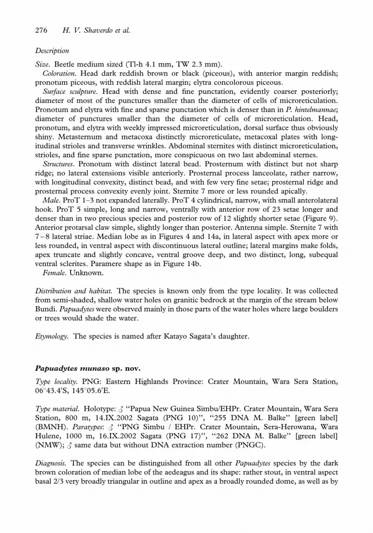
Size. Beetle medium sized (Tl-h 4.1 mm, TW 2.3 mm).
Coloration. Head dark reddish brown or black (piceous), with anterior margin reddish;
pronotum piceous, with reddish lateral margin; elytra concolorous piceous.
Surface sculpture. Head with dense and fine punctation, evidently coarser posteriorly;
diameter of most of the punctures smaller than the diameter of cells of microreticulation.
Pronotum and elytra with fine and sparse punctation which is denser than in P. hintelmannae;
diameter of punctures smaller than the diameter of cells of microreticulation. Head,
pronotum, and elytra with weekly impressed microreticulation, dorsal surface thus obviously
shiny. Metasternum and metacoxa distinctly microreticulate, metacoxal plates with long-
itudinal strioles and transverse wrinkles. Abdominal sternites with distinct microreticulation,
strioles, and fine sparse punctation, more conspicuous on two last abdominal sternes.
Structures. Pronotum with distinct lateral bead. Prosternum with distinct but not sharp
ridge; no lateral extensions visible anteriorly. Prosternal process lanceolate, rather narrow,
with longitudinal convexity, distinct bead, and with few very fine setae; prosternal ridge and
prosternal process convexity evenly joint. Sternite 7 more or less rounded apically.
Male. ProT 1–3 not expanded laterally. ProT 4 cylindrical, narrow, with small anterolateral
hook. ProT 5 simple, long and narrow, ventrally with anterior row of 23 setae longer and
denser than in two precious species and posterior row of 12 slightly shorter setae (Figure 9).
Anterior protarsal claw simple, slightly longer than posterior. Antenna simple. Sternite 7 with
7 – 8 lateral striae. Median lobe as in Figures 4 and 14a, in lateral aspect with apex more or
less rounded, in ventral aspect with discontinuous lateral outline; lateral margins make folds,
apex truncate and slightly concave, ventral groove deep, and two distinct, long, subequal
ventral sclerites. Paramere shape as in Figure 14b.
Female. Unknown.
Distribution and habitat. The species is known only from the type locality. It was collected
from semi-shaded, shallow water holes on granitic bedrock at the margin of the stream below
Bundi. Papuadytes were observed mainly in those parts of the water holes where large boulders
or trees would shade the water.
Etymology. The species is named after Katayo Sagata’s daughter.
Papuadytes munaso sp. nov.
Type locality. PNG: Eastern Highlands Province: Crater Mountain, Wara Sera Station,
06843.40S, 145805.60E.
Type material. Holotype: < ‘‘Papua New Guinea Simbu/EHPr. Crater Mountain, Wara Sera
Station, 800 m, 14.IX.2002 Sagata (PNG 10)’’, ‘‘255 DNA M. Balke’’ [green label]
(BMNH). Paratypes: < ‘‘PNG Simbu / EHPr. Crater Mountain, Sera-Herowana, Wara
Hulene, 1000 m, 16.IX.2002 Sagata (PNG 17)’’, ‘‘262 DNA M. Balke’’ [green label]
(NMW); < same data but without DNA extraction number (PNGC).
Diagnosis. The species can be distinguished from all other Papuadytes species by the dark
brown coloration of median lobe of the aedeagus and its shape: rather stout, in ventral aspect
basal 2/3 very broadly triangular in outline and apex as a broadly rounded dome, as well as by
276 H. V. Shaverdo et al.

dull dorsal surface of the body, with more strongly impressed microreticulation and dense
coarse punctation and male antennomeres simple.
Description
Size. Beetle large (Tl-h 4.8 – 5 mm, TW 2.6 mm).
Coloration. Head, pronotum, and elytra concolorous piceous, pronotum slightly reddish at
anterior part of lateral margin.
Surface sculpture. Head with dense (some punctures conjoint or spaces between punctures
1–5 times size of punctures) and coarse punctation, finer anteriorly; diameter of punctures
equal or smaller than diameter of cells of microreticulation. Pronotum and elytra with distinct
coarse punctation that is slightly denser on pronotum (spaces between punctures 1 – 5 times
the size of punctures); diameter of punctures equal to the diameter of cells of
microreticulation. Head, pronotum, and elytra with rather strong microreticulation, dorsal
surface thus not obviously shiny. Metasternum and metacoxa distinctly microreticulate,
metacoxal plates with longitudinal strioles and transverse wrinkles. Abdominal sternites with
distinct microreticulation, strioles, and fine sparse punctation which are coarse and denser on
the two last abdominal sternes.
Structures. Pronotum with distinct lateral bead. Prosternum with distinct but not sharp
ridge, no lateral extensions visible anteriorly. Prosternal process lanceolate, rather narrow,
with longitudinal convexity, distinct bead, and with few very fine setae; prosternal ridge and
prosternal process convexity evenly joint. Sternite 7 slightly truncate apically.
Male. ProT 1 – 3 not expanded laterally. ProT 4 cylindrical, narrow, with large anterolateral
hook. ProT 5 simple, long and narrow, without expansion and concavity, ventrally with
anterior row of 33 setae longer and denser than in the two first species and posterior row of six
shorter setae (Figure 10). Anterior protarsal claw simple, slightly longer than posterior.
Antenna simple. Sternite 7 with 13 – 21 lateral striae. Median lobe as in Figures 5 and 15a, in
lateral aspect with apex strongly curved and pointed, in ventral aspect with discontinuous
lateral outline; lateral margins make folds, apex rounded, ventral groove deep, and two
distinct, long, subequal ventral sclerites. Median lobe mostly of dark brown color as opposed
to all other species we have seen where the genital is ferruginous. Paramere shape as in Figure
15b.
Female. Unknown.
Distribution. The species is known only from Crater Mountain.
Etymology. Selected by Katayo Sagata: ‘‘When I was first going into Crater Mountain a friend
of mine requested that, if I find new species, I should name it after her. Munaso Zaemo is her
name and this species is named after her’’.
Papuadytes vladimiri sp. nov.
Type locality. West Papua: Yapen Island, Mantembu.
Type material. Holotype: < ‘‘Irian Jaya: Japen Isl. Mantembu 150 – 450 m, 18.II.1999 leg.
Riedel’’ (NMW). Paratypes: 2 <, 1 , the same label data as in holotype (NMW).
Diagnosis. The species can be distinguished from all other Papuadtyes species by the shape of
the median lobe of the aedeagus as depicted in Figures 6 and 16a, dull dorsal surface of the
Papuadytes (Coleoptera: Dytiscidae) from New Guinea 277

body, with strongly impressed microreticulation and dense coarse punctation, absence of
lateral bead of the pronotum, and male antennomeres simple.
Description
Size and shape. Beetle small, with broadly oval habitus (Tl-h 3.6 – 3.7 mm, TW 2.1 – 2.2 mm).
Coloration. Head yellowish red to reddish brown; disc of pronotum and elytra pale reddish
brown to dark brown, lateral sides of pronotum and sometimes base of elytra paler.
Figures 15 – 17. Median lobe of aedeagus, lateral aspect, (a) and paramere, external aspect, (b): (15) Papuadytes
munaso, paratype; (16) P. vladimiri, paratype; (17) P. vladimiri, paratype: gonocoxosternum (a) and gonocoxa (b),
ventral aspect.
278 H. V. Shaverdo et al.

Surface sculpture. Dorsal surface with microreticulation and punctation similar to P.
marinae. Head and pronotum with dense punctation (spaces between punctures 1 – 5 times
size of punctures), evidently finer than in P. marinae, diameter of most punctures distinctly
smaller than diameter of cells of microreticulation. Elytra with punctation dense and distinctly
coarser than on head and pronotum; diameter of punctures of equal diameter to cells of
microreticulation. Head, pronotum, and elytra with strong microreticulation, dorsal surface
thus not obviously shiny, rather matt. Metasternum and metacoxa distinctly microreticulate,
metacoxal plates with longitudinal strioles and transverse wrinkles. Abdominal sternites with
distinct microreticulation, strioles, and fine sparse punctation, coarser and denser on two last
abdominal sternes.
Structures. Pronotum without lateral bead. Prosternum with distinct but not sharp ridge, no
lateral extensions visible anteriorly. Prosternal process lanceolate, rather narrow, with
longitudinal convexity, distinct bead, and with few very fine setae; prosternal ridge and
prosternal process convexity evenly joint. Sternite 7 slightly truncate or rounded apically.
Male. ProT 1 – 3 not expanded laterally. ProT 4 cylindrical, narrow, with distinct
anterolateral hook. ProT 5 simple, long and narrow, without expansion and concavity,
ventrally with anterior row of ca. 40 setae longer and denser than in two first species and
posterior row of seven shorter setae (Figure 11). Anterior protarsal claw simple, slightly longer
than posterior. Antenna simple, with antennomeres slightly broader than in female. Sternite 7
with 12 – 14 lateral striae. Median lobe as in Figures 6 and 16a, in lateral aspect with apex
more or less truncate, in ventral aspect with continuous lateral outline and lateral margins
sinuate, apex truncate and concave, ventral groove deep, and with two distinct, long, subequal
ventral sclerites. Paramere shape as in Figure 16b.
Female. Antennomeres slender. Dorsal surface with punctation and microreticulation
coarser. Gonocoxa and gonocoxosternum as in Figure 17a, b.
Distribution and habitat. The species is known only from the type locality where it was
collected from a small forest stream.
Etymology. The species is named after Vladimir Vladimirovich Shaverdo, the father of the first
author, who was probably also not very happy about the unusual activities of his daughter but
still kept on making aquatic nets for her.
Acknowledgements
We thank the Department of Environment and Conservation (DEC) of PNG for granting
research permission and the people of PNG for their kind support of this fieldwork. This is the
first result of the Water Beetles of PNG survey, organized by Katayo Sagata and the Wildlife
Conservation PNG Program. WCS and RCF (Goroka) are both thanked for their constant
support on the ground. This work was supported by: Deutsche Forschungsgemeinschaft (BA
2152/1-2), The Linnean Society of London, The UK DARWIN Initiative Pre-Project
Funding and a EU Marie Curie Postdoctoral Fellowship. Our thanks also go to Dr. H.
Schillhammer (Vienna, Austria) for his comments on the manuscript and help with digital
image processing, as well as an anonymous referee for useful hints.
References
Balke M. 1998. Revision of New Guinea Copelatus Erichson, 1832 (Insecta: Coleoptera: Dytiscidae): The running
water species, Part I. Annalen des Naturhistorischen Museum Wien 100B:301 – 341.
Papuadytes (Coleoptera: Dytiscidae) from New Guinea 279

Balke M. 1999. Two new species of the genus Copelatus Erichson, 1832, subgenus Papuadytes Balke, 1998, from
Papua New Guinea (Insecta: Coleoptera: Dytiscidae). Annalen des Naturhistorischen Museum Wien
101B:273 – 276.
Balke M. 2001. Die Schwimmkafer Neu Guineas. Artenreichtum, Phylogenie, Biogeographie und Lebensweise
(Coleoptera: Dytiscidae) [dissertation]. Berlin: Freie Universitat. 167 p. þ 56 plates. Avaliable from: http://
dissertation.de (ISBN 3-89825-231-0).
Balke M, Bergsten J. 2003. Dytiscidae: Papuadytes shizong sp. nov. from Yunnan (China), the first member of
Papuadytes Balke found west of the Wallace Line (Coleoptera). In: Jach MA, Ji L, editors. Water beetles of
China. Vol. III. Wien: Zoologisch-Botanische Gesellschaft in Osterreich and Wiener Coleopterologenverein. p
89 – 94.
Balke M, Ribera I, Vogler AP. 2004. MtDNA phylogeny and biogeography of Copelatinae, a highly diverse group of
tropical diving beetles (Dytiscidae). Molecular Phylogenetics and Evolution 32:866 – 880.
Bameul F. 1990. Le DMHF: un excellent milieu de montage en entomologie. L’Entomologiste 46(5):233 – 239.
Miller KB, Nilsson AN. 2003. Homology and terminology: communicating information about rotated structures in
water beetles. Latissimus 17:1 – 4.
Nilsson AN. 2001. Dytiscidae. World catalogue of Insects 3:1 – 395.
280 H. V. Shaverdo et al.


