Embed Size (px)
Citation preview

Acute Myeloid Leukemia research paper
416 haematologica/journal of hematology vol. 88(04):april 2003
Flt3-mediated signaling in human acute myelogenous leukemia (AML) blasts: afunctional characterization of the effects of Flt3-ligand in AML cell populationswith and without genetic Flt3 abnormalities ØYSTEIN BRUSERUD, RANDI HOVLAND, LINE WERGELAND, TIEN-SHENG HUANG, BJØRN TORE GJERTSEN
Background and Objectives. Intracellular signaling ini-tiated via Flt3 seems important in both leukemogenesisand chemosensitivity in acute myelogenous leukemia(AML). Flt3 is activated by binding of its natural Flt3-lig-and (Flt3-L), but Flt3 genes with internal tandem dupli-cations (Flt3-ITD) or Asp(D)-835 point mutations encodemolecules with constitutive activation. The aim of thisstudy was to compare functional effects of exogenousFlt3-L on AML blast populations with and without genet-ic Flt3 abnormalities.
Design and Methods. Native AML blasts were derivedfrom 64 consecutive patients with high blast counts inperipheral blood, and in vitro models were used to char-acterize the Flt3-L effects.
Results. The Flt3 protein levels showed a similar widevariation between AML blast populations with and with-out genetic Flt3 abnormalities. Flt3-L was an autocrinegrowth factor only for 2 patients. Flt3-ITD+ AML cells hadlower responsiveness to exogenous cytokines than cellpopulations without Flt3 abnormalities, but exogenousFlt3-L increased blast proliferation both for patients with-out Flt3 abnormalities and patients with Flt3-ITD as wellas D835 mutations. This enhancement was observed evenin the presence of other exogenous cytokines and includ-ed clonogenic AML progenitors. Flt3-L inhibited prolifer-ation only for 1 patient, but had divergent effects on AMLblast cytokine release. Flt3-L affected AML blast differ-entiation (inhibition of erythroid colonies, increased neu-trophil granulation) only in a minority of patients, where-as it had an anti-apoptotic effect for a larger subset ofpatients.
Interpretation and Conclusions. Intracellular signalinginitiated by Flt3 ligation modulates the functional phe-notype for native human AML blasts both with and with-out genetic Flt3 abnormalities.
Key words: acute myelogenous leukemia, Flt3 internaltandem duplications, Flt3-D835 mutations, Flt3 ligand,cytokines, in vitro effects.
Haematologica 2003;88:416-428http://www.haematologica.org/2003_04/88416.htm
©2003, Ferrata Storti Foundation
Acute myelogenous leukemia (AML) is characterizedby clonal proliferation of immature myeloid cellsand has an overall disease-free survival after inten-
sive chemotherapy of less than 50%.1-4 The most impor-tant cause of death is AML relapse,1-4 and the two mostimportant predictors of relapse have been cytogeneticabnormalities and response to induction therapy.2 How-ever, recent studies have demonstrated that abnormali-ties of the Flt3 gene (a membrane-anchored receptortyrosine kinase) are also associated with an increasedrelapse risk.3-12 At diagnosis nearly 30% of AML patientshave genetic Flt3 abnormalities,10-12 and new abnormal-ities can develop later in leukemia relapse.13,14 The adverseprognostic impact of Flt3 internal tandem duplications(Flt3-ITD) is now well established,3,4 and recent evidencesuggests that Asp(D)-835 point mutations have a similarprognostic effect.12
The Flt3 gene encodes for a tyrosine kinase with anextracellular ligand-binding part and an intracellular cat-alytic unit.6-10 Ligation of the Flt3 molecule induces acti-vation of the tyrosine kinase through ligand-inducedreceptor oligomerization and autophosphorylation withsubsequent phosphorylation of cytoplasmic substrates.5-9
However, genetic Flt3 abnormalities result in the expres-sion of a tyrosine kinase with constitutive activity in theAML blasts.8,9,15,16 Previous experimental evidence sug-gests that the constitutive activation is implicated inleukemogenesis, and recent clinical studies have demon-strated that constitutive kinase activation is also impor-tant for chemosensitivity in AML.3,4,12
The clinical and experimental studies discussed abovesuggest that intracellular signaling events initiated byFlt3 activation are important for the regulation of func-tional characteristics of AML blasts.3,4,8-10 However, it isnot known whether the functional effects of Flt3-ligationby the natural Flt3-L differ between AML blasts withoutgenetic Flt3 abnormalities and leukemia cells that expressabnormal Flt3 molecules with constitutive activity. Theaim of the present study was, therefore, to characterizethe functional effects of natural Flt3 ligation in detail ina large group of consecutive patients, and to comparethese effects in native AML blasts with and withoutgenetic Flt3 abnormalities (Flt3-ITD or D835 mutations).
Design and Methods
PatientsThe study was approved by the local Ethics Committee
and samples were collected after informed consent had
From the Division of Hematology, Department of Medicine, MedicalDepartment (OB, LW, BTG), Department of Medical Genetics (RH) andDepartment of Molecular Biology (T-SH), Haukeland University Hospital,University of Bergen, Bergen, Norway.
Correspondence: Dr. Øystein Bruserud, MD, Department of Medicine,Haukeland University Hospital, N-5021 Bergen, Norway. E-mail: [email protected]

haematologica/journal of hematology vol. 88(04):april 2003 417
been provided. During the period 1991-2001 AMLblasts were taken from 64 consecutive patientswith high peripheral blood blast counts. Thepatients were classified as having AML-M0/M1 (21patients), AML-M2 (20 patients) and AML-M4/M5(23 patients). Forty-six patients had newly diag-nosed de novo AML, 6 patients had AML relapse,and 12 patients had AML secondary to chemother-
apy (4 patients), chronic myeloproliferative disor-ders (2 patients) or primary myelodysplastic syn-dromes (6 patients). Leukemic cells from the last 20patients were used in most experiments, and thecharacteristics of these patients are presented inTable 1.
Cytogenetic analyses were performed for the last48 patients included in our study; of these, 28
In vitro effects of Flt3-L on human AML blasts
Table 1. Clinical and biological characteristics of AML patients.
Membrane molecule expression1
Pat. Sex Age Previous malignant FAB CD13 CD14 CD15 CD33 CD34 Cytogenetic FLT3- WBCor pre malignant classification analysis abnormality2 counts3
disease
1. M 44 AML-M5 + − + + + inv(16) PM 351
2. F 36 Neurofibromatosis, AML-M5 + − + + − t(9;11) − 37.6malignant Schwannoma
3. M 49 AML-M4 − − + + − nt3 ITD 78
4. M 69 AML-M2 + − nt + − inv(16) − 89
5. F 87 AML-M1 − − nt + − nt − 51.2
6. M 83 AML-M1 − − − + + nt − 80
7. M 72 AML-M4 + + + + − +11 ITD 290
8. M 49 AML-M5 + + nt + − Normal ITD 63.5
9. F 58 AML-M2 + − + + − Normal ITD, wt− 40.7
10. F 56 AML-M2 + − − + − +21 − 69.2
11. F 38 AML-M5 + + + + + Normal nt 182
12. F 55 AML-M0 + − − − + Normal ITD 43.6
13. M 51 AML-M4 + + + + − Normal − 31.4
14. F 49 AML-M1 − − − + + +21 − 121
15. M 65 AML-M1 + − − + − Normal ITD, wt− 166
16. M 64 AML-M2 + − + + + nt − 23.5
17. F 63 AML-M5 − + + + − t(2;3), (q37;q21), − 57.8(q13;q21;q21) der (11q),
19q+
18. F 36 AML-M5 + + + + − Normal nt 88.6
19. F 82 AML-M2 + − − + + nt ITD 49
20. F 63 AML-M4 − − − + − nt nt 126
Patients were regarded as positive when more than 20% of blast cells stained positive judged by flow cytometric analysis. All AMLpopulations were negative for T-lymphocyte (CD2, CD3) and B-lymphocyte (CD19, CD20) markers; Flt3- abnormalities were internaltandem duplications (ITD), Asp-D835 point mutations (PM) and loss of wild type (wt−), nt, not tested; white blood cell (WBC)counts in peripheral blood are expressed as ×109/L (normal range 3.5-10.5×10⁹/L ).The WBC included at least 80% leukemia blasts.

418 haematologica/journal of hematology vol. 88(04):april 2003
patients had a normal karyotype, 3 patients had afavorable karyotype (all inv(16)) and 5 had an unfa-vorable karyotype according to the definitions usedby Wheatley et al.2 and Kottaridis et al.3 A total of98 patients with AML were admitted to our insti-tution during the same period. The distribution ofkaryotypes in the patients selected for our presentstudy did not differ significantly from that in thewhole series of patients seen during the same peri-od. The relatively low frequency of patients (3/48)with favorable karyotypes is different from thatreported in other studies2,3 and was also observedfor the whole series of patients from the same peri-od (6/98).
Preparation of AML blastsNative AML blasts. Leukemic peripheral blood
mononuclear cells (PBMC) were isolated by densi-ty gradient separation (Ficoll-Hypaque; NyCoMed,Oslo, Norway; specific density 1.077) from theperipheral blood of patients with a high percent-age of AML blasts among blood leukocytes (Table1). Cells were stored frozen in liquid nitrogen.23 Thepercentage of blasts among leukemic PBMCexceeded 95% for all patients,24-26 the contami-nating cells being small lymphocytes.
Enriched AML blasts. Immunomagnetic beadscoated with anti-CD2 and anti-CD19 specific mon-oclonal antibodies (Dynabeads; Dynal, Oslo, Nor-way) were used for depletion of CD2+ and CD19+
cells, respectively.24 Depletion was performed intwo separate steps before adherent cells wereremoved, and the enriched populations contained<1% of CD2+ T-cells and CD19+ B-cells.24
Analysis of Flt3 abnormalities inAML blasts
Our method for analysis of Flt3-ITD has recentlybeen described in detail.14 Analysis of D835 pointmutations (PM) was performed using the restric-tion fragment gene length polymorphism at codon835/836 as described previously.27 Briefly, afteramplification of a 111 bp fragment from exon 20using genomic DNA with primer 20F-5’-CCGCCA-GGAACGTGCTTG-3’ and 20R-5’-GCCTCACATTGCC-CCTGA-3’, polymerase chain reaction (PCR) prod-ucts were digested by EcoRV and the fluorescence(6-FAM, 6-carboxylfluorescein)-labeled productanalyzed.14 Undigested products served as templatefor a new polymerase chain reaction with the sameprimer combination, and the products were there-after cleaned using ExoSAP-IT before being direct-ly sequenced using an ABI BigDyeTM TerminatorCycle Sequencing Kit (Applied Biosystems, FosterCity, CA, USA).
Analysis of Flt3 expressionCell suspensions were lysed at 4°C in 60-100 µL
of lysis buffer (10 mM trishydroxymethyl-amino-
methane (Tris) with pH 7.5, 400 mM NaCl, 10%glycerol, 0.5% detergent Nonidet P-40 (AmershamBiosciences, Uppsala, Sweden), 5 mM NaF, 0.5 mMNa-orthovanadate, 1 mM dithiotreitol (DTT), andprotease inhibitor cocktail Complete (Roche, Basel,Switzerland). Samples were kept at 4°C, homoge-nized and centrifuged (14,000g, 15 minutes). 3Xsodium dodecyl sulphate (SDS) loading buffer (0.5M Tris pH 6.8, 2 M β-mercaptoethanol, 12% SDS,30% glycerol, bromphenol blue) was added tosupernatant aliquots containing 40 µg of protein,and thereafter boiled for 7 minutes before separa-tion in SDS-polyacrylamide gel electrophoresis(PAGE) minigels with 7.5% polyacrylamide. Afterelectroblotting to polyvinylidene difluoride (PVDF)membranes (Amersham Biosciences) and blockingfor 1 h in phosphate-buffered saline with 0.5%Tween (PBS-T), the filters were incubated with pri-mary anti-Flt3 antibody (the rabbit polyclonal S18and C20 antibodies diluted 1:250 in PBS-T; SantaCruz, CA, USA) for 1 h (room temperature) orovernight (4°C) before washing for 1 hour in PBS-T. Both antibodies recognized the p160 and p130isoforms of Flt3. The C-20 antibody reacts with aC-terminal epitope whereas S-18 reacts with thekinase insert region. The PVDF membranes werethereafter washed (1 hour in PBS-T), incubated for1 hour with a secondary anti-rabbit antibody con-jugated to alkaline phosphatase (the antibody dis-solved in PBS-T), washed (1 hour in PBS-T), andfinally incubated with CDP-Star ChemiluminisenceSubstrate (Applied Biosystems, Foster City, CA,USA). The membranes were then exposed to KodakX-ray films which were scanned for densiometricanalysis of the 130 plus 160 kDa bands (MicrotekScanmaker 5700, NIH Image ver. 1.60 for AppleMacintosh). The intensity for each AML sample wasnormalized to the lower 70 kDa anti-Flt3-reactiveband in Jurkat control extracts which were includ-ed in each gel (equal intensity defined as 1.0). Equalprotein loading was confirmed by staining theminigels with Coomassie blue. Actin could not beused as the loading control because of differencesin the molecular weight of the immunoreactivebands between patients.
Reagents for tissue cultureCytokines. Recombinant human Flt3-L (Pepro-
tech; Rocky Hill, NJ, USA) was used at a concen-tration of 20 (only 3H-thymidine incorporation) or50 ng/mL; this was based on previous studies of invitro cultured AML blasts showing that Flt3-Leffects reach a plateau at concentrations ≥10ng/mL.8,18,21 Other recombinant human cytokineswere used at the following concentrations: inter-leukin 1β (IL1β, Peprotech) 50 ng/mL; IL3 (Pepro-tech) 20 ng/mL, stem cell factor (SCF; Peprotech)20 ng/mL, thrombopoietin (TPO; Peprotech) 50ng/mL, vascular endothelial growth factor (VEGF;
O. Bruserud et al.

In vitro effects of Flt3-L on human AML blasts
haematologica/journal of hematology vol. 88(04):april 2003 419
Peprotech) 50 ng/mL, macrophage colony-stimu-lating factor (M-CSF, Peprotech) 50 ng/mL, granu-locyte-macrophage colony-stimulating factor(GM-CSF; Sandoz, Basel, Switzerland) 100 ng/mL,G-CSF (Roche) 100 ng/mL.
Culture media. Unless otherwise stated, the cul-ture medium was RPMI 1640 with HEPES and glu-tamine (BioWhitacker; Walkersville, MA, USA) andsupplemented with 10% inactivated fetal calfserum (FCS; BioWhitacker).26 The serum-free mediaX-vivo 10®, X-vivo 15® (BioWhitacker) and Stem-Span SFEM™ (referred to as StemSpan; Stem CellTechnologies, Vancouver, BC, Canada) were used incertain experiments.26 All the media contained 100µg/mL of gentamicin.
Antibodies. The monoclonal Flt3-L specific neu-tralizing antibody (clone 40416.111; R&D Systems,Abingdon, UK) was always tested in parallel with anisotypic control antibody; 0.02-0.06 µg/mL of thisanti-Flt3-L antibody will neutralize 50% of the bio-activity of 5 ng/mL of recombinant human Flt3-L(manufacturer’s information).
Assays for AML blast proliferation Suspension cultures. As described previously,24-26
5×10⁴ cells/well were cultured in 150 µL mediumin flat-bottomed microtiter plates (Costar 3796;Cambridge, MA, USA). Cultures were incubated at37°C in a humidified atmosphere of 5% CO2. Aftersix days 3H-thymidine (37 kBq/well; TRA 310,Amersham International, Amersham, UK) wasadded in 20 µL of 0.9% NaCl solution and nuclearradioactivity assayed 18 hours later by liquid scin-tillation counting.
Colony formation assays. AML blasts were cul-tured in different methylcellulose-based media: (i)medium alone (MethoCult H4230; Stem Cell Tech-nologies, referred to as spontaneous colony for-mation) or the same medium supplemented withGM-CSF (GM-CSF-dependent colony formation);(ii) medium with erythropoietin plus phytohemag-glutinin-leukocyte conditioned medium (Metho-Cult H4433; Stem Cells Technologies). Cells werecultured in 24 well tissue culture plates (Costar3524) with 105 cells in 0.5 mL medium per well.Cultures were incubated for 14 days before thenumber of colonies containing at least 20 cells wasdetermined by light microscopy (duplicate analy-sis). The colonies were classified as erythroid (redcolor in the whole or a part of the colony) and non-erythroid.
Cytokine analysisAnalysis of AML cell cytokine secretion. As
described previously,25 1×10⁶ AML blasts/mL werecultured in 24-well tissue culture plates (Costar3524; 2 mL medium/well) for 48 hours beforesupernatants were harvested. ELISA analyses were
used to determine levels of IL1β, IL6, tumor necro-sis factor (TNF) α (Pelikine compact ELISA kits; Cen-tral Laboratory of the Netherlands’ Red Cross BloodTransfusion Services, Amsterdam, The Netherlands),Flt3-L, G-CSF and GM-CSF (Quantikine ELISA kits;R&D Systems) in the supernatants. The minimaldetectable levels were IL1β 0.8 pg/mL, IL6 0.8pg/mL, TNFα 1.0 pg/mL, Flt3-L 7 pg/mL, GM-CSF 3pg/mL and G-CSF 8 pg/mL.
Cytokine-specific RNA levels. AML blasts (2×10⁶
cells in 2 mL FCS-containing medium per well;Costar 3524 culture plates) were cultured for 48hours before cells were harvested and washed inphosphate-buffered saline. The cell pellets werestored frozen at -70°C until total RNA was isolat-ed.29 For quantification of IL1β- and IL6-specificRNA the samples and calibrators were hybridized inmicrowells with gene-specific biotin-labeled cap-ture oligonucleotide probes and digoxigenin-labeled detection probes, and cytokine-specificRNA levels then determined in a calorimetricmicroplate assay (Quantikine RNA assay, R&D Sys-tems). The results are expressed as concentrationsof IL1β- and IL6-specific RNA when testing totalRNA at the concentration of 2.5 µg/mL.
Studies of apoptotic cell deathEstimation of the number of apoptotic cells. AML
blasts were incubated for 24 and 48 hours beforecell death was analyzed as described in detail pre-viously.24,25,30-32 Firstly, AML blasts were stained withDNA-specific bisbenzimide H33258 (Hoechst;Basel, Switzerland) or daunorubicin (Pharmacia),and the percentage of cells showing chromatin dis-tribution consistent with apoptosis was determinedby fluorescence microscopy.25,30 Secondly, detec-tion of phosphoserine exposure on the cell surfacewas used as a marker for apoptosis; flow cytomet-ric analysis was then performed on FITC-annexin Vstained cells as described previously.31 Thirdly, JC-1 staining (Molecular Probes) was used to deter-mine the mitochondrial status, and the number ofcells with depolarized mitochondria consistentwith apoptosis was determined by flow cytometricanalysis.31,32
Caspase-3 activity in AML cells. Caspase-3 activ-ity was measured in cell extracts using a specificcaspase-3 cellular activity assay kit (Calbiochem;La Jolla, CA, USA). Briefly, 1×10⁶/mL AML blasts (2mL of medium per well, 24-well Costar 3524 cul-ture plates) were cultured for 48 hours before cellswere harvested, washed twice, and cell concentra-tion adjusted to 1×10⁶ cells/mL in lysis buffer. Thesecell extracts were stored at -70°C until enzymaticactivity was assayed according to the manufactur-er’s instructions. The results are presented aspmol/min for 80 µL of sample volume.
O. Bruserud et al.
420 haematologica/journal of hematology vol. 88(04):april 2003
Presentation of the data3H-thymidine incorporation was assayed in trip-
licate and the mean counts per minute (cpm) usedfor all calculations. The incremental response wasdefined as the cpm for cultures with AML blastsminus cpm for negative controls, and significantblast proliferation was defined as an incrementalresponse exceeding 1,000 cpm. A significant alter-ation of proliferation was defined as a difference inincremental responses (i) exceeding 2000 cpm, and(ii) the difference in cpm being >20% of the controlresponse. For cytokine combinations an additiveenhancing or inhibitory effect was defined as a pro-liferative response exceeding the highest/lowest ofthe two single responses by at least 2000 cpm and20%; smaller differences are referred to as inter-mediate. A significant alteration of AML blast colonyformation was defined as a difference correspond-ing to >20% of the control response and with anabsolute value >10 per 105 seeded cells. Cytokineconcentrations were transformed to logarithmic val-ues that were used for statistical comparisons. TheSign test, χ2 test and Wilcoxon's test for paired sam-ples were used for statistical analysis, and differ-ences were regarded as significant when p<0.05.
Results
Flt3 protein expression in native AMLblasts
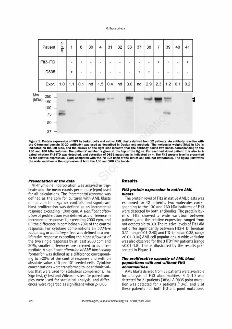
The protein level of Flt3 in native AML blasts wasexamined for 42 patients. Two molecules corre-sponding to the 130 and 160 kDa isoforms of Flt3were detected by both antibodies. The protein lev-el of Flt3 showed a wide variation betweenpatients, and the relative expression ranged fromnot detectable to 3.0. The relative levels of Flt3 didnot differ significantly between Flt3-ITD+ (median0.31, range 0.01-2.40) and ITD– (median 0.38, range<0.01-3.00) AML cell populations. A wide variationwas also observed for the 3 ITD−PM+ patients (range<0.01-1.5). This is illustrated by the results pre-sented in Figure 1.
The proliferative capacity of AML blastpopulations with and without Flt3abnormalities
AML blasts derived from 55 patients were availablefor analysis of Flt3 abnormalities. Flt3-ITD wasdetected for 21 patients (38%). A D835 point muta-tion was detected for 7 patients (13%), and 3 ofthese patients had both ITD and point mutations.
Figure 1. Protein expression of Flt3 by Jurkat cells and native AML blasts derived from 12 patients. An antibody reactive withthe C-terminal domain (C-20 antibody) was used as described in Design and methods. The molecular weight (Mw) in kDa isindicated on the left side, and the arrows on the right side indicate that the antibody bound two bands corresponding to the130 and 160 kDa isoforms. The patients’ number is given at the top of the figure. For each individual patient it is also indi-cated whether Flt3-ITD was detected, and detection of D835 mutations is indicated by +. The Flt3 protein level is presentedas the relative expression (Expr) compared with the 70 kDa band of the Jurkat cell (nd, not detectable). The figure illustratesthe wide variation in the expression of both the 130 and 160 kDa bands.

In vitro effects of Flt3-L on human AML blasts
haematologica/journal of hematology vol. 88(04):april 2003 421
The presence of Flt3-ITD showed no association withFAB classification, the expression of the stem cellmarker CD34 or the ability of autocrine proliferation(data not shown). We also investigated the prolifer-ative capacity in the presence of exogenouscytokines (IL1β, IL3, G-CSF, M-CSF, GM-CSF, SCF,Flt3-L, TPO, VEGF). This wide range of cytokines wasused because the growth factor responsiveness ofAML populations is heterogeneous. A total of 495patient/cytokine combinations were thus examined.The frequency of combinations with undetectable3H-thymidine incorporation (<1000 cpm) was sig-nificantly higher for Flt3-ITD+ patients (n=21, 90 outof 189 combinations) than for Flt3−ITD− patients(n=34, 89 out of 306 combinations, χ2 test, p<0.001).This difference was also statistically significant whenFlt3-PM+ patients were excluded from the controlgroup. Undetectable 3H-thymidine incorporation inthe presence of all nine cytokines was most commonamong patients with Flt3 abnormalities (Flt3-ITD+ orPM+ 6/21, no Flt3 abnormalities 4/34), but this dif-ference did not reach statistical significance.
Effects of exogenous Flt3-L on proliferationof AML blasts in suspension cultures
The proliferation of native AML blasts derivedfrom 64 consecutive patients was assayed whencells were cultured with and without exogenousFlt3-L 20 ng/mL in FCS-containing medium aloneand medium with various exogenous cytokines.AML blasts from 10 patients did not proliferate invitro either in medium alone or in the presence ofany exogenous cytokine. The overall results for theother 54 patients with detectable 3H-thymidineincorporation (corresponding to >1000 cpm) arepresented in Figure 2 and summarized in the upperpart of Table 2. When the statistical analysis onlyincluded those patients with detectable prolifera-tion (>1000 cpm), (i) Flt3-L increased AML blastproliferation significantly both for cells cultured inmedium alone and cells cultured with exogenousIL1β, IL1RA, IL3, G-CSF, M-CSF, TPO, and VEGF (Signtest, p<0.002 for each), and (ii) the enhancementreached a significant level (for definitions seeDesign and methods, presentation of the data) for
Figure 2. The effect of Flt3-L on AML blast proliferation. Leukemia blasts were derived from 64 consecutive patients, but thefigure presents the results only for those patients showing detectable 3H-thymidine incorporation for cultures without (−−) or with(+) Flt3-L 20 ng/mL. AML blasts were cultured either in medium alone (Sp, 40 out of 64 patients showing detectable prolifer-ation) or in the presence of IL1ββ (49/64), IL1RA (37/64), IL3 (49/64), G-CSF (52/64), M-CSF (44/64), TPO (41/64) or VEGF(38/64). All these exogenous cytokines were tested at 50 ng/mL except G-CSF that was tested at 100 ng/mL. The resultsfor each patient are presented as the mean cpm of triplicate determinations, and undetectable proliferation is indicated in thefigure (o). The median 3H-thymidine incorporation for all these patients is also indicated in the figure (___).

O. Bruserud et al.
422 haematologica/journal of hematology vol. 88(04):april 2003
a large group of patients both when cells were cul-tured in medium alone and when they were cul-tured with medium and exogenous cytokines (Table2, upper part). These differences were also statis-tically significant when all patients were includedin the analysis (data not shown). Flt3-L and theother cytokines had additive effects only for a sub-set of the patients (Table 2, middle part). Whencomparing the overall results for the 54 patientswith detectable proliferation, Flt3-L had signifi-cant effects on blast proliferation in medium aloneor in the presence of at least one exogenouscytokine for 50 of these patients. However, theFlt3-L effect showed no correlation with the Flt3protein levels (data not shown), and the growth-enhancing effect was detected for blast popula-tions both with and without Flt3 abnormalities(Table 2, lower part).
AML blast expression of wild-type Flt3 could notbe detected for 3 ITD+ patients. However, exoge-nous Flt3−L could modulate AML blast prolifera-tion even for these patients (Table 3).
The effects on spontaneous and cytokine-depen-dent blast proliferation were also compared forFlt3-L, GM-CSF and SCF (Figure 3). All threecytokines caused strong enhancement of the pro-liferation for a majority of patients both when cells
were cultured in medium alone and when the cellswere cultured with medium in the presence ofexogenous cytokines (Figure 3). Flt3-L (Table 2),GM-CSF and SCF (data not shown) had additivegrowth-enhancing effects with other cytokinesonly for a subset of patients.
Enriched AML blasts were prepared for 5 patients(Table 1, patients #7, 8, 11, 14 and 20), and Flt3-Lincreased the proliferation of enriched cells for allthese patients (data not shown).
Flt3-L as an autocrine growth factor fornative AML blasts
AML blasts derived from 64 patients were cul-tured for 48 hours before concentrations of Flt3-Lwere determined in the supernatants. Flt3-L didnot reach detectable levels (<7 pg/mL) for anypatient. Leukemia cells derived from the 9 patientswith the highest spontaneous in vitro proliferationwere also cultured with Flt3-L specific monoclon-al antibody and isotypic control antibodies. Anti-Flt3-L caused a dose-dependent inhibition of spon-taneous blast proliferation only for patient 8 (Flt3-ITD+PM−) and 10 (Flt3-ITD−PM−), but anti-Flt3-L didnot have an antiproliferative effect for any patientin the presence of exogenous GM-CSF (Figure 4).
Table 2. Effects of Flt3-L on in vitro proliferation of native AML blasts; studies of 3H-thymidine incorporation of AML blasts derivedfrom 64 patients.
Exogenous cytokine added alone and together with Flt3-L1
Comparison (patient number) Effect (number of patients)
None IL1 IL1RA IL3 G-CSF M-CSF TPO VEGF
Cultures with exogenous Flt3-L Increase2 34 26 29 29 31 23 26 28compared with correspondingcontrols ( n=64)
Intermediate2 5 21 7 17 15 20 14 8Decrease2 1 1 1 3 6 1 1 2No detectable proliferation2 24 16 27 15 12 20 23 26
Cultures with exogenous Flt3-L Additive growth-enhancing effects3 − 15 0 27 28 16 14 2compared with correspondingcontrols (n=64)
Flt3-ITD+ AML blasts (n=21)4 Flt3-L induced increase of proliferation2 9 8 7 9 13 7 7 6
Flt3-PM+ AML blasts (n=4)4 Flt3-L induced increase of proliferation 2 2 3 2 1 1 1 2
Flt3-ITD− AML blasts (n=30)4 Flt3-L induced increase of proliferation 19 13 16 12 15 12 14 15
The results for 64 patients are presented.1 For each cytokine the effects were classified (see below) and the number of patients showing enhanced/indifferent/decreased/nosignificant proliferation in the presence of exogenous Flt3-L is given for each cytokine. The effect of Flt3-L was investigated for AML blasts cultured in medium alone or mediumwith exogenous cytokines (IL1β, IL1RA, IL3, G-CSF, TPO, M-CSF, VEGF). The upper part of the table describes the effect of exogenous Flt3-L in the presence of other media-tors, the middle part states the number of patients with an additive effect of Flt3-L and other exogenous cytokines (for detailed definitions see Design and Methods, presenta-tion of the data), and the lower part compares the numbers of patients with Flt3-L-induced growth enhancement for Flt3-ITD+ and -ITD- AML blast populations.2 A significantalteration was defined as a difference being (i) >20% of the corresponding Flt3-L-free control and (ii) with an absolute value >2000 cpm. Smaller differences were classifiedas intermediate. No detectable proliferation was defined as 3H-thymidine incorporation <1000 cpm both for cultures with and without Flt3-L.3 An additive effect of Flt3-L andanother cytokine was defined as a proliferative response exceeding the highest of the two responses by at least 20% and this difference being >2000 cpm.4 Nine patientswere not available for Flt3-ITD testing. The group of Flt3-ITD+ patients includes 3 patients with additional Flt3-PM, whereas patients with Flt3-PM alone are presented as a sep-arate group.

In vitro effects of Flt3-L on human AML blasts
haematologica/journal of hematology vol. 88(04):april 2003 423
Effects of exogenous Flt3-L on AML blastcolony-formation
AML blasts derived from 17 patients (Table 1,patients #1-10 and 12-18) were pre-incubated inserum-free StemSpan™ medium without and withFlt3-L 50 ng/mL for 7 days before the frequency ofcolony-forming cells was determined under Flt3-L-free conditions by using the erythropoietin+condi-tioned medium assay. Patients with Flt3 abnormal-ities showed lower frequencies of clonogenic cellsthan did the other patients, but this difference didnot reach statistical significance (Table 4). For mostpatients Flt3-L either increased (10 of 17 patients)or did not alter (4/17) the frequencies of non-ery-throid colonies, but a reduction was observed for 3
patients. Erythroid colonies were detected for 10patients; pre-incubation with Flt3-L reduced thisfrequency for 4 patients and increased it for 2patients. The Flt3-L effects did not differ between
Table 3. Effects of exogenous Flt3-L on in vitro proliferationof native AML blasts: studies of Flt3-ITD+ leukemia cellswith undetectable expression of wildtype Flt3.
Patient and Flt3-L 20 Patient Patient Patient culture ng/mL 9 15 24characteristics
Flt3-ITD 24 63/33 48(base pairs)
Flt3-PM (D835) − − −
Exogenous mediator
None − 1084±172 147±29 299±27+ 5891±998 696±98 497±98
IL1 − 3456±627 568±108 14.836±1982+ 7090±882 939±111 17.005±2035
IL1RA − 478±87 183±23 485±82+ 2769±524 724±74 612±54
IL3 − 68.911+3760 542±225 8653±1230+ 80.511+4072 939±339 11.783±1074
G-CSF − 2156±524 524±92 21.284±2310+ 10.330±1099 1706±233 21.058±1962
M-CSF − 564±38 510±44 539±27+ 3166±422 833±92 591±83
TPO − 378±88 228±76 4836±623+ 2962±499 876±73 4471±826
VEGF − 259±78 127±29 312±27+ 2654±881 594±127 483±99
AML blasts were cultured in suspension cultures with andwithout Flt3-L 20 ng/mL. The Flt3-L effects were assayed for cells cultured inmedium alone and medium supplemented with various exogenous cytokines. 3H-thymidine incorporation was assayed after 7 days of culture, and the results arepresented as the mean+standard deviation of triplicate determinations. Resultsin bold represent significant alterations induced byFlt3-L (the difference corresponding to >20% of the controls and an absolute val-ue >2,000 cpm).
Table 4. The effect of Flt3-L on clonogenic AML cells;effects of pre-incubation with Flt3-L before analysis ofcolony formation.
Colony-formation with erythropoietinplus conditioned medium(number of colonies per
105 seeded cells)
Patient FLT3- Flt3-L Non Erythroidabnormality 50 ng/mL Erythroid
1. PM − 16.5±0.7 0+ 17.5±0.7 0
2. − + 117.0±26.9 150.0±6.3115.0±9.9 170.0±14.0
3. ITD − 85.0±15.4 0+ 125.0±4.2 2.0±1.4
4. − − 170.0±11.3 96.0±25.4+ 144.0±11.3 23.0±7.0
5. − − 194.0±31.1 30.0±12.5+ 265.0±29.6 1.0±1.4
6. − - 15.0+712.5 0+ 123.0+12.6 0
7. ITD − 41.5±3.5 0+ 91.0±14.7 0
8. ITD − 118.0±14.1 71.5±18.3+ 168.0±24 0.5±0.7
9. ITD, wt− − 72.0±25.5 23.0±9.1+ 126.0±1.4 1.5±0.7
10. − − 329.0±43.8 0+ 371.0±49.5 0
12. ITD − 39.0±5.6 4.0±2.8+ 64.0±21.0 1±1.4
13. − − 24.5±2.1 23.5±10.5+ 28.0±0 23.0±13.3
14. − − 57.7±6.3 0+ 45.0±2.8 0
15. ITD, wt- − 82.0±24.8 3.0±2.8+ 154.0±4.2 0
16. − − 176.0±34.3 0+ 127.5±33.6 0
17. − − 4.5±3.5 4.0±5.6+ 4.0±5.6 44.5±9.1
18. nt − 50.0±4.2 0+ 90.0±7.0 0
Cells were pre-cultured in suspension cultures with and without Flt3-L 50 ng/mLfor 7 days before the number of colonies was determined in the erythropoietin-conditioned medium assay. Values for patients with significant effects of Flt3-Lare marked in bold: this was defined as differences >10 and exceeding the Flt3-L-negative control by 10%. The results are presented asthe mean+SD of duplicate determinations.

O. Bruserud et al.
424 haematologica/journal of hematology vol. 88(04):april 2003
patients with and without Flt3 abnormalities andwere reproduced for 5 patients (Table 1, patients1#, 2, 6, 8, 9) when using various media for the pre-incubation (RPMI + 10% FCS, X-vivo 10®, X-vivo 15®,StemSpan™, data not shown). Spontaneous andGM-CSF-dependent colony formation were exam-ined for 8 patients (Table 1, patients #7-14); theassays were then prepared with and without Flt3-L 50 ng/mL. Only non-erythroid colonies weredetected. Flt3-L could increase both spontaneous
(1 out of 8 patients) and GM-CSF-dependent (3/8)colony formation, and decreased colony-formationwas not observed for any patients.
Effects of exogenous Flt3-L on constitutivecytokine secretion by native AML blasts
Flt3-L 50 ng/mL had divergent effects on therelease of IL1β, IL6, TNFα, G-CSF and GM-CSF bynative human AML blasts (Figure 5). This diver-gence was observed for native blasts and was
Figure 3. Effects of Flt3-L, GM-CSF and SCF on AML blastproliferation. The proliferation of native AML blasts culturedin medium alone or with Flt3-L 20 ng/mL (open columns),GM-CSF 100 ng/mL (stippled) or SCF 20 ng/mL (stripes)was first compared (Sp, i.e. spontaneous proliferation inmedium alone). The effects of these three cytokines werealso examined when the culture medium was supplement-ed with other exogenous cytokines (IL1, ILββ1RA, IL3, G-CSF, M-CSF, TPO, VEGF; see the bottom of the figure), andproliferation in Flt3-L/GM-CSF/SCF containing cultures wasthen compared with that in the corresponding cytokine-con-taining controls. An increase or decrease in blast prolifera-tion was defined as an alteration corresponding to (i) >20%of the corresponding control and (ii) exceeding 2000 cpm;smaller differences are referred to as intermediate respons-es. A total of 64 patients were examined, and the figure pre-sents the results for those patients who showed detectableproliferation (corresponding to >1000 cpm) for each of thecytokines. The figure shows the number of patients withincreased (the part of the columns above the X-axis) andintermediate/decreased (the part of the column below theX-axis) proliferation in the presence of Flt3-L/GM-CSF/SCF.The number of patients with decreased responses is indi-cated in black at the lowest part of each column.
Figure 4. The effect of Flt3-L specific neutralizing antibod-ies on spontaneous and GM-CSF-dependent AML blast pro-liferation. Native AML blasts derived from two patients(Table 1; patients 8 •• , 10 o) were cultured either in medi-um alone (____) or in the presence of GM-CSF 100 ng/mL(----). Proliferative responses were then compared for cul-tures with Flt3-L-specific neutralizing monoclonal antibodiesand cultures containing isotypic control antibodies at thesame concentration (see x-axis). The results are presentedas the percent inhibition by neutralizing antibodies, i.e. theincremental cpm for cultures with Flt3-L specific antibodies,relative to the incremental cpm for corresponding isotypiccontrol cultures. The responses in isotypic control cultureswere: patient #8, spontaneous proliferation 8302+1876cpm, GM-CSF dependent proliferation 36.813+852 cpm;patient # 10, spontaneous proliferation 17.206+1428 cpm,GM-CSF dependent proliferation 94.865+3474 cpm.

In vitro effects of Flt3-L on human AML blasts
haematologica/journal of hematology vol. 88(04):april 2003 425
reproduced for enriched leukemia cells (7 patientsexamined), and neither the cytokine levels nor thedivergent effects of exogenous Flt3-L differedbetween patients with and without Flt3 abnor-malities. Exogenous SCF also had divergent effectson the secretion of IL1β, IL6 and TNFα (data notshown). Furthermore, AML blasts derived from 5patients (Table 1, patients 1-4 and 7) were cul-tured with and without Flt3-L 50 ng/mL for 48hours before IL1β- and IL6-specific RNA was quan-tified. IL1β- (range 15.0-39.0 amol/µg total RNA)and IL6-specific RNA (range 32.0-86 amol/µg RNA)were detected for all patients, and Flt3-L had diver-gent effects on these levels (data not shown).
Effects of exogenous Flt3-L on AML blastmorphology
Native AML blasts derived from 10 consecutivepatients (Table 1, patients 1-10) were cultured withand without Flt3-L 50 ng/mL for 2 and 7 daysbefore examination of cell morphology (May-Grün-wald-Giemsa staining). After 2 days neutrophil dif-ferentiation was observed in ≤ 10% of the cells forall patients, and these percentages were not sig-nificantly altered (i.e. ≤ 5%) by Flt3-L. In contrast,after 7 days of culture with Flt3-L an increasedpercentage of cells with neutrophil granulationwas observed for patients 1 (ITD−PM+, 17 versus5%), 2 (ITD−PM−, 28 versus 1%), 4 (ITD−PM−, 12versus 1%) and 7 (ITD+PM−, 8 versus 2%). Howev-er, neutrophil differentiation further than thepromyelocyte stage and erythroid or monocytoiddifferentiation was not observed for any patient.
Effects of Flt3-L on regulation of apoptosisin native AML cells
The frequency of apoptotic AML blasts (H33258staining) after 24 and 48 hours of culture was firstexamined for 11 patients (Table 1, patients 1 and10-19), and this frequency exceeded 30% only forpatient 17 (ITD− PM−, 54% after 48 hours). Flt3-Lreduced apoptosis significantly (i.e. ≥5% differ-ence) for patients 1 (ITD− PM+), 10 (ITD− PM−), 13(ITD− PM−), 17 (ITD− PM−) and 18 (ITD not tested,PM−). Furthermore, the effects of Flt3-L, GM-CSFand SCF on apoptosis were compared for an addi-tional group of 12 consecutive patients, 4 of whomwere Flt3−ITD+ and 1 of whom was Flt3−PM+ (chro-matin staining, JC-1 staining, annexin-V labeling).For Flt3−L the morphologic examination demon-strated reduced apoptosis (≥5% alteration) for 6 ofthe 12 patients, whereas increased apoptosis wasobserved for 2 patients. A similar divergence wasobserved both after 24 and 48 hours of culture andwith all three methods (data not shown). The anti-apoptotic effect was observed for AML blast pop-ulations both with and without Flt3 abnormalities(including the Flt3−PM+ patients). SCF and GM-CSFalso had divergent effects on apoptosis (data notshown).
Caspase-3 activity in AML blasts was examinedafter 48 hours of in vitro culture with and withoutFlt3− L 50 ng/mL for patients 1 (ITD− PM+), 10 (ITD−
PM−) and 19 (ITD+ PM−). These patients were select-ed because Flt3-L increased the blast proliferationboth in cultures with medium alone and in cul-tures in the presence of all the exogenous cyto-kines. In contrast, Flt3-L had divergent effects oncaspase-3 activity and caused either minimallyaltered (patient 10: without Flt3-L 34.8 pmol/min,with Flt3-L 42.9 pmol/min), decreased (patient 1:107 versus <13.4 pmol/min) or increased activity(patient 19: 6.7 versus 68 pmol/min). Caspase-inhibitory activity was not detected for any patient.
Figure 5. The effect of Flt3-L on AML blast cytokine secre-tion. The AML cells were cultured without and with Flt3-L50 ng/mL, and cultures were prepared with either native(____) or enriched (-----) AML blasts. The cells were culturedat 1××106 cells/mL for 48 hours before supernatants wereharvested and cytokine levels (IL1ββ, IL6, TNFββ, G-CSF, GM-CSF; see top of the figure) determined by ELISA analyses.The figure compares the cytokine levels (pg/mL) for cul-tures without (−−) and with (+) Flt3-L, and only the results forpatients showing detectable cytokine levels for at least oneof these cultures are presented: - IL1ββ: 25 patients tested,9 patients showed detectable levels; - IL6: 15 out of 25patients showed detectable levels; in addition the figurepresents the results for enriched AML blasts derived from7 of these patients (-----); - TNFαα: detectable levels wereobserved for 7 out of 25 patients; - G-CSF, 11 out of 19patients showed detectable levels; - GM-CSF, 17 out of 19patients showed detectable levels.

O. Bruserud et al.
426 haematologica/journal of hematology vol. 88(04):april 2003
Discussion
In our present study we included consecutivepatients with high peripheral blood blast counts,and thus highly enriched populations of native AMLblasts could be prepared by density gradient sepa-ration from peripheral blood samples. This simpletechnique has a minimal risk of inducing function-al alterations in the blasts,35,36 but a high degree ofleukemization may reflect biologic differences2,37
and thus our results are only representative of thisparticular subset of patients. The cytogenetic stud-ies demonstrated a normal karyotype in nearly 60%of our patients, whereas favorable and unfavorablekaryotypes were detected only for small subsets ofpatients (6% and 11% respectively). This distribu-tion is not much different from that found in oth-er studies, except for the low frequency of favor-able karyotypes, which was also observed when weanalyzed all patients admitted to our institutionduring the same study period.2,3 However, the rel-atively high frequencies of Flt3-ITD (38 versus27%)3 and D-835 mutations (13 versus 7%),28 inour present study possibly reflect a selected patientpopulation. The relatively low number of patientsexamined may then explain why the frequencies ofabnormal karyotypes did not differ from that foundin our overall population and in other studies.2,3
Previous studies have reported a wide variationin the percentage of patients with Flt3-L-respon-sive AML blasts (range 62-88%).22 In contrast tothese studies we examined the effects of Flt3-L ina large group of consecutive patients. We detect-ed Flt3 expression by AML blasts in more than 80%of patients, and exogenous Flt3-L altered blast pro-liferation in at least one in vitro model for a largemajority of patients. The expression of Flt3 iso-forms showed a wide quantitative variationbetween patients without there being any correla-tion with the effects of Flt3-L on blast proliferation,suggesting that downstream mechanisms are moreimportant than receptor density for these func-tional Flt3-L effects.
Ligation of wild-type Flt3 leads to activation ofthe intracellular tyrosine kinase,5 whereas abnormalFlt3 genes encode molecules with constitutiveenzyme activity.8,9 Our present results demonstratedthat Flt3-L was a growth factor even for AML blastswith genetic Flt3 abnormalities, an observation sug-gesting that receptor ligation can induce additionalgrowth-stimulatory signaling even in cells with con-stitutive kinase activity. Although it seems likely thatthis effect is caused by ligation of wild-type Flt3,the observation of increased proliferation in thepresence of exogenous Flt3−L for Flt3−ITD+ blastswithout detectable wild-type suggests that ligationof mutated Flt3 may also contribute. However, thislast observation should be interpreted with greatcare because relatively few patients were examined
and the presence of small, undetectable amounts ofwild-type Flt3 may influence these results. Further-more, Flt3 ligation could also modulate colony for-mation and spontaneous apoptosis of in vitro cul-tured Flt3-ITD+ and -ITD− blast populations. Ourresults thereby support the hypothesis that exoge-nous Flt3−L is an important growth factor for AMLblasts in a major part of patients. In contrast, Flt3−L is an autocrine growth factor only for a small sub-set, and the importance of the autocrine secretion isalso reduced by the presence of other exogenouscytokines (e.g. GM-CSF).
ITD+ AML blasts have decreased proliferativecapacity in stroma-supported cultures.33,34 Our pre-sent study demonstrated an association betweenFlt3-ITD and decreased AML blast proliferation insuspension cultures. This observation suggests thatthe decreased proliferative capacity is caused by adirect effect of the Flt3-ITD on intracellular sig-naling and not by indirect effects with modulationof paracrine growth-regulatory mechanisms.
GM-CSF and SCF can increase AML blast prolif-eration for most patients,37,38 but their effects onthe proliferative capacity of native human AMLblasts have not been compared with those of Flt3-L. All three growth factors had similar effects withregard to growth enhancement and anti-apopto-sis and divergent effects on cytokine release (Fig-ure 5),37-40 including the autocrine growth factorsIL1β, G-CSF and GM-CSF. Thus, the overall resultsdemonstrate that these three cytokines havegrowth-enhancing effects that are only minimallyinfluenced by local cytokine networks and are inde-pendent of autocrine growth factor release andregulation of apoptosis.
The growth-enhancing effect of Flt3-L was char-acterized in detail. Firstly, Flt3-L could not bedetected in culture supernatants for any patient,but Flt3-L specific neutralizing antibodies inhibit-ed spontaneous AML blast proliferation for twopatients. This observation suggests that Flt3-L canfunction as an autocrine growth factor in a smallminority of patients. Secondly, the growth-enhanc-ing effects of Flt3-L were not due to increased IL1βrelease because (i) the Flt3-L effects on prolifera-tion and IL1β levels showed no correlation; (ii) Flt3-L had divergent effects on cytokine-specific RNAlevels both for IL1β and IL6 independently of itsgrowth-enhancing effect; and (iii) Flt3-L enhancedproliferation even in the presence of the antago-nistic IL1RA. Thirdly, the growth-enhancing effectof Flt3-L involved the clonogenic AML cell subset,and this effect could be detected when using var-ious experimental approaches. Lastly, controlexperiments with serum-free conditions andenriched AML blast populations showed that theFlt3-L effects were not dependent on unidentifiedserum components or the small lymphocyte cont-amination.

In vitro effects of Flt3-L on human AML blasts
haematologica/journal of hematology vol. 88(04):april 2003 427
The possible interaction between differentiationstatus and Flt3-L effects was investigated by var-ious experimental approaches. Flt3-L had only min-imal effects on AML blast differentiation in sus-pension cultures. Differentiation was then analyzedby light microscopy because we regard morpho-logic alterations as a more robust sign of differen-tiation than flow-cytometric detection of alteredexpression of a single or a few membrane mole-cules.35,36,41-43 Flt3-L did not induce erythroid dif-ferentiation in the colony formation assays either.Thus, the effects of Flt3-L seem to be only mini-mally affected by and have only a minimal influ-ence on the differentiation status of native humanAML blasts.
To conclude, Flt3-L was able to modulate thefunctional phenotype of native human AML blasts,and the most important effect seemed to beenhanced proliferation. This growth-enhancingeffect was observed both for AML cells with andthose without genetic Flt3 abnormalities, and ourresults thereby suggest that intracellular signalinginitiated by Flt3-ligation in the AML blasts isinvolved in leukemogenesis and possibly alsochemosensitivity for a majority of patients.
References
1. Löwenberg B, Downing JR, Burnett A. Acute myeloidleukemia. N Engl J Med 1999;341:1051-62.
2. Wheatley K, Burnett AK, Goldstone AH, Gray RG, Hann IM,Harrison CJ, et al. A simple, robust, validated and highly pre-dictive index for the determination of risk-directed therapyin acute myeloid leukemia derived from the MRC AML 10 tri-al. Br J Haematol 1999;107:69-79.
3. Kottaridis PD, Gale RE, Frew ME, Harrison G, Langabeer SE,Belton AA, et al. The presence of a FLT3 internal tandem dupli-cation in patients with acute myeloid leukemia (AML) addsimportant prognostic information to cytogenetic risk groupand response to the first cycle of chemotherapy: analysis of854 patients from the United Kingdom Medical ResearchCouncil AML 10 and 12 trials. Blood 2001;98:1752-9.
4. Whitman SP, Archer KJ, Feng L, Baldus C, Becknell B, Carl-son BD, et al. Absence of the wild-type allele predicts poorprognosis in adult de novo acute myeloid leukemia with nor-mal cytogenetics and the internal tandem duplication ofFLT3: a cancer and leukemia group B study. Cancer Res 2001;61:7233-9.
5. Ullrich A, Schlessinger J. Signal transduction by receptorswith tyrosine kinase activity. Cell 1990;61:203-12.
6. Drexler HG. Expression of Flt3 receptor and response to Flt3ligand by leukemic cells. Leukemia 1996;10:588-99.
7. Marchetto S, Fournier E, Beslu N, Aurran-Schleinitz T, Du-breuil P, Borg JP, et al. SHC and SHIP phosphorylation andinteraction in response to activation of the Flt3 receptor.Leukemia 1999;13:1374-82.
8. Fenski R, Flesch K, Serve S, Mizuki M, Oelmann E, Kratz-Albers K, et al. Constitutive activation of Flt3 in acutemyeloid leukaemia and its consequences for growth of 32Dcells. Br J Haematol 2000;108:322-30.
9. Tse KF, Mukherjee G, Small D. Constitutive activation of Flt3stimulates multiple intracellular signal transducers andresults in transformation. Leukemia 2000;14:1766-76.
10. Nakao M, Yokota S, Iwai T, Kaneko H, Horiike S, Kashima K,et al. Internal tandem duplication of the flt3 gene found inacute myeloid leukemia. Leukemia 1996;10:1911-8.
11. Yamamoto Y, Kiyoi H, Nakano Y, Suzuki R, Kodera Y, Miya-
waki S, et al. Activating mutation of D835 within the acti-vation loop of FLT3 in human hematologic malignancies.Blood 2001;97:2434-9.
12. Thiede C, Steudel C, Mohr B, Schaich M, Schäkel U,Platzbecker U, et al. Analysis of Flt3-activating mutations in979 patients with acute myelogenous leukemia: associationwith FAB subtypes and identification of subgroups with poorprognosis. Blood 2002;99:4326-35.
13. Nakano Y, Kiyoi H, Miyawaki S, Asou N, Ohno R, Saito H, etal. Molecular evolution of acute myeloid leukemia in relapse:unstable N-ras and Flt3 genes compared with p53 gene. BrJ Haematol 1999;104:659-64.
14. Hovland R, Gjertsen BT, Bruserud Ø. Acute myelogenousleukemia with internal tandem duplication of the Flt3 geneappearing or altering at the time of relapse: a report of twocases. Leuk Lymphoma 2002;43:2027-9.
15. Hayakawa F, Towatari M, Kiyoi H, Tanimoto M, Kitamura T,Saito H, et al. Tandem-duplicated Flt3 constitutively acti-vates STAT5 and MAP kinase and introduces autonomouscell growth in IL-3-dependent cell lines. Oncogene 2000;19:624-31.
16. Zhao M, Kiyoi H, Yamamoto Y, Ito M, Towatari M, Omura S,et al. In vivo treatment of mutant FLT3-transformed murineleukemia with a tyrosine kinase inhibitor. Leukemia 2000;14:374-8.
17. Lisovsky M, Estrov Z, Zhang X, Consoli U, Sanchez-WilliamsG, Snell V, et al. Flt3 ligand stimulates proliferation andinhibits apoptosis of acute myeloid leukemia cells: regula-tion of Bcl-2 and Bax. Blood 1996;88:3987-97.
18. Piacibello W, Fubini L, Sanavio F, Brizzi MF, Severino A,Garetto L, et al. Effects of human Flt3 ligand on humanmyeloid leukemia cell growth: heterogeneity in response andsynergy with other hematopoietic growth factors. Blood1995;86:4105-14.
19. McKenna HJ, Smith FO, Brasel K, Hirschstein D, Bernstein ID,Williams DE, et al. Effects of flt3 ligand on acute myeloid andlymphocytic leukemic blast cells from children. Exp Hema-tol 1996;24:378-85.
20. Dehmel U, Quentmeier H, Drexler HG. Effects of Flt3 ligandon human leukemia cells. II. Agonistic and antagonisticeffects of other cytokines. Leukemia 1996;10:271-8.
21. Stacchini A, Fubini L, Severino A, Sanavio F, Aglietta M, Piaci-bello W. Expression of type III tyrosine kinase Flt3 and Kitand responses to their ligands by acute myeloid leukemiablasts. Leukemia 1996;10:1584-91.
22. Drexler HG, Meyer C, Quentmeier H. Effects of Flt3 ligand onproliferation and survival of myeloid leukemia cells. LeukLymphoma 1999;33:83-91.
23. Bruserud Ø. Effects of dipyridamole, theophyllamine and ver-apamil on spontaneous in vitro proliferation of myeloge-nous leukemia cells. Acta Oncol 1992;31:53-8.
24. Bruserud Ø, Gjertsen BT, Brustugun OT, Bassoe CF, NesthusI, Akselsen PE, et al. Effect of interleukin 10 on blast cellsderived from patients with acute myelogenous leukemia.Leukemia 1995;9:1910-20.
25. Bruserud Ø, Huang TS, Glenjen N, Gjertsen BT, Foss B. Lep-tin in human acute myelogenous leukemia: studies of in vivolevels and in vitro effects on native functional leukemiablasts. Haematologica 2002;87:584-95.
26. Bruserud Ø, Gjertsen BT, von Volkman HL. In vitro culture ofacute myelogenous leukemia (AML) cells in serum-freemedia: studies of native AML blasts and AML cell lines. JHematother Stem Cell Res 2000;9:923-32.
27. Scherczinger CA, Hintz JL, Peck BJ, Adamowicz MS, BourkeMT, Coyle HM, et al. Allele frequencies for the CODIS coreSTR loci in Connecticut populations. J Forensic Sci 2000;45:938-40.
28. Abu-Duhier FM, Goodeve AC, Wilson GA, Care RS, Peake IR,Reilley JT. Identification of novel FLT-3 Asp835 mutations inadult acute myeloid leukemia. Br J Haematol 2001;113:983-8.
29. Reers M, Smith TW, Chen LB. J-aggregate formation of acarbocyanine as a quantitative fluorescent indicator ofmembrane potential. Biochemistry 1991;30:4480-6.
30. Gjertsen BT, Cressy LI, Ruchaud S, Houge G, Lanotte M,Døskeland SO. Multiple apoptotic death types triggered

O. Bruserud et al.
428 haematologica/journal of hematology vol. 88(04):april 2003
through activation of separate pathways by cAMP andinhibitors of protein phosphatases in one (IPC leukemia) cellline. J Cell Sci 1994;107:3363-77.
31. Cimpan MR, Cressey LI, Skaug N, Halstensen A, Lie SA, Gjert-sen BT, et al. Patterns of cell death induced by eluates fromdenture base acrylic resins in U-937 human monoblastoidcells. Eur J Oral Sci 2000;108:59-69.
32. Salvioli S, Ardizzoni A, Franceschi C, Cossarizza A. JC-1, butnot DiOC6(3) or rhodamine 123, is a reliable fluorescentprobe to assess delta psi changes in intact cells: implicationsfor studies on mitochondrial functionality during apoptosis.FEBS Lett 1997;411:77-82.
33. Rombouts WJC, Broyl A, Martens ACM, Slater R, Ploemach-er RE. Human acute myeloid leukemia cells with internaltandem duplications in the Flt3 gene show reduced prolif-erative ability in stroma supported long-term cultures.Leukemia 1999;13:1071-8.
34. Rombouts WJC, Blokland I, Löwenberg B, Ploemacher RE.Biological characteristics and prognosis of adult acutemyeloid leukemia with internal tandem duplications in theFlt3 gene. Leukemia 2000;14:675-83.
35. Sissolak G, Hoffbrand AV , Mehta AB, Ganeshaguru K. Effectsof interferon-α (IFN) on the expression of interleukin 1-β(IL-1), interleukin 6 (IL-6), granulocyte-macrophage colony-stimulating factor (GM-CSF) and tumor necrosis factor-α(TNF) in acute myeloid leukemia blasts. Leukemia 1992;6:1155-60.
36. Fiedler W, Suciu E, Wittlief C, Ostertag W, Hossfeld DK.Mechanisms of growth factor expression in acute myeloidleukemia (AML). Leukemia 1990;4:459-61.
37. Bruserud Ø, Gjertsen BT, Foss B, Huang TS. New strategiesin the treatment of acute myelogenous leukemia (AML): invitro culture of AML cells – the present use in experimentalstudies and the possible importance for future therapeuticstrategies. Stem Cells 2001;19:1-11.
38. Schwartz S, Heinecke A, Zimmermann M, Creutzig U, SchochC, Harbott J, et al. Expression of the c-kit receptor (CD117)is a feature of almost all subtypes of de novo acute myelo-blastic leukemia (AML), including cytogenetically good-riskAML, and lacks prognostic significance. Leuk Lymphoma1999;34:85-94.
39. Bruserud Ø, Nesthus I, Bühring HJ, Pawelec G. Cytokine mod-ulation of interleukin 1 and tumor necrosis factor-α secre-tion from acute myelogenous leukaemia blast cells in vitro.Leuk Res 1995;19:15-22.
40. Rowe JM, Liesveld JL. Hematopoietic growth factors andacute leukemia. Cancer Treat Res 1999; 99: 195-226.
41. Heil G, Krauter J, Bayer-Johannböke E, Bunjes D, Kurrle E,Westphal-Frosch C, et al. CD54 expression and its role inhomotypic aggregation of the blasts of acute myeloblasticleukemias (AML). Int J Hematol 1994;60:39-49.
42. Munker R, Andreeff M. Induction of death (CD95/Fas), acti-vation and adhesion (CD54) molecules on blast cells of acutemyelogenous leukemias by TNF-α and IFN-γ. Cytokines MolTher 1996;2:147-59.
43. Bruserud Ø, Gjertsen BT. New strategies in the treatment ofacute myelogenous leukemia: differentiation induction –present use and future possibilities. Stem Cells 2000;18:157-65.
Pre-Publication Report & Outcomes ofPeer Review
ContributionsAll authors took part in the design of the study,
interpretation of data and preparation of the manu-script. All authors approved the final version of themanuscript. The authors especially contributed to thefollowing parts: ØB: collection of cells, investigationof AML blast proliferation, cytokine secretion and dif-ferentiation. RH: analysis of Flt3-internal tandemduplication. LW and BTG: analysis of apoptosis inAML blasts. TsH: analysis of cytokine RNA levels.
Funding The work was supported by the Norwegian Cancer
Society, The Rakel and Otto-Kristian Brun Foundationand Olaf Ruunshaugens Foundation.
DisclosuresConflict of interest: none.Redundant publications: < 50%.
Manuscript processingThis manuscript was peer-reviewed by two exter-
nal referees and by Professor Francesco Lo Coco,Deputy Editor. The final decision to accept this paperfor publication was taken jointly by Professor Lo Cocoand the Editors. Manuscript September 16, 2002;accepted February 25, 2003.
In the following paragraphs, the Deputy Editorsummarizes the peer-review process and its out-comes.
What is already known on this topicFLT3 signaling is increasingly relevant in AML, par-
ticularly after the discovery that constitutive activa-tion of the receptor by gene mutation is common inthis disease and may be targeted by recently devel-oped inhibitors.
What this study addsThe study shows that FLT3 ligation initiates sig-
naling and modulates AML phenotype irrespective ofthe FLT3 status (i.e. mutated or not).
CaveatsThe study includes a selection of hyperleukocytic
cases. It would be interesting to figure out whethersimilar observations are obtained in patients withlow initial WBC counts.


