Embed Size (px)
Citation preview

Fluid Management in Surgery
FCS SA Intermediate Exam TutorialDr. B Monzon
SBAH

What to discuss?
• Physiology of Body Fluids• Composition of common IV Fluids• Indications for Fluids: Resuscitation (Rehydration), Maintenance &
Replacement (prescribing fluids) • Monitoring Volume Status• Effects of inappropriate fluid usage • Crystalloids • Colloids • Albumin
DISCLAIMER: THIS PRESENTATION IS BASED ON MY PERSONAL INTERPRETATION OF THE AVAILABLE EVIDENCE

PHYSIOLOGY OF BODY FLUIDS
STARLING EQUATION OF FLUID EQUILIBRIUM
TBW 60% BODY WEIGHT IN ADULTS
(75% NEWBORN, 65% CHILDREN)INTRACELLULAR 65%
EXTRACELLULAR 35%• INTERSTITIAL ±80%• INTRAVASCULAR ±20%
• THIRD SPACE
Extracellular loses are compensated by fluid movement from Intracellular space and vice versaHydrostatic, Oncotic, Osmotic & Capillary pressures

Endothelial Glycocalyx (EG) Functions
• Regulates Capillary Permeability• Plays a role in Mechanotransduction: release of NO in response to
shear stress (regulates vessel tone)• Preserve circulatory Microenvironment: multiple molecules require
direct interaction with EG for function e.g. Antithrombin, Heparin cofactor II, Tissue factor pathway inhibitor-TFPI, Vascular endothelium growth factor-VEGF, Interleukins (2, 3, 4, 5, 7, 8), Extracellular superoxide dismutase-Ec-SOD and others.
(Procoagulants, anticoagulants, endothelial cell regeneration, leukocyte chemotaxis and diapedesis, scavengers of oxygen radicals)

Endothelial Glycocalyx (EG)
• Glyco-proteins, proteoglycans and soluble components (Albumin) attached to endothelial luminal cell surface
• EXERT ONCOTIC PRESSURE• Inflammatory damage to EG causes large heavy molecules to leak into the IS
worsening tissue oedema and organ damage• Colloids (HES, Albumin) also interact with EG in a complex manner not yet
fully clarified in clinical practice (increase or decrease permeability)• Linked to and affected by several pathophysiological processes (Diabetes,
Atherosclerosis, Ischaemia-reperfusion) [SYSTEMIC ENDOTHELIOPATHY]
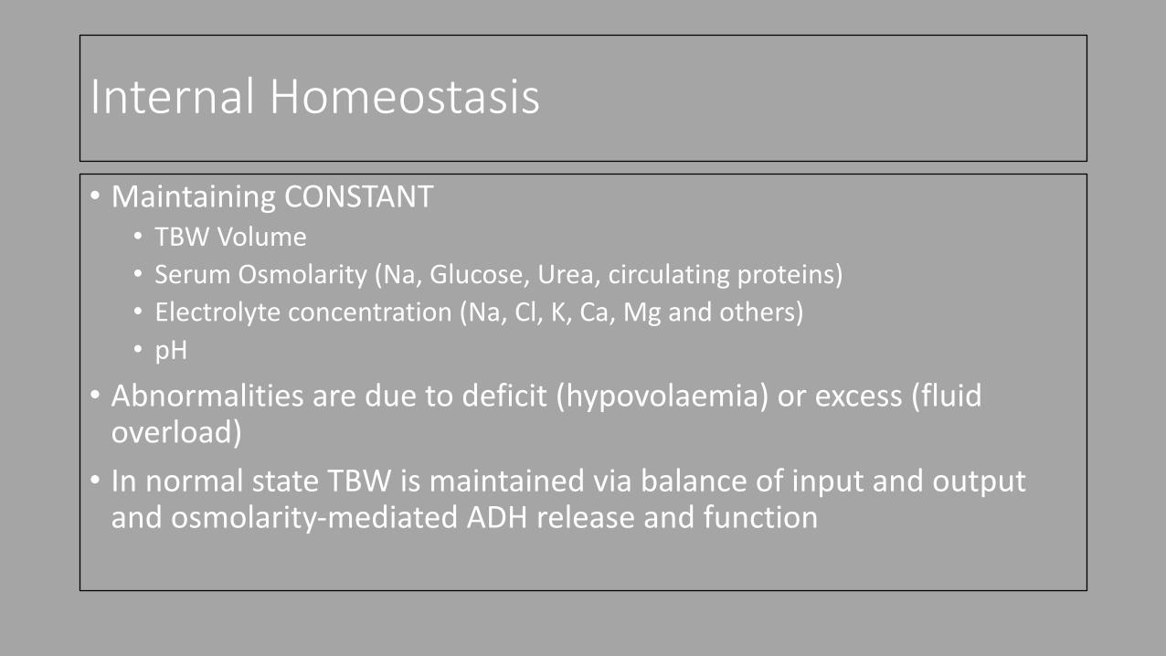
Internal Homeostasis
• Maintaining CONSTANT• TBW Volume• Serum Osmolarity (Na, Glucose, Urea, circulating proteins)• Electrolyte concentration (Na, Cl, K, Ca, Mg and others) • pH
• Abnormalities are due to deficit (hypovolaemia) or excess (fluid overload)
• In normal state TBW is maintained via balance of input and output and osmolarity-mediated ADH release and function

Input vs Output Adult
Input Output• Oral water (free water and water in food) 2000 -
2500 ml/day• Metabolic water (oxidation water) ± 250-300
ml/day
• Urine 1500 – 2000 ml/day (0.5-1ml/kg/h)*• Fecal loses ± 200 ml/day• Respiratory and Sweat loses : variable volume,
influenced by ambient temperature, level of physical activity and metabolic rate**
Daily Balance 2000 ± 500 ml
(*) Without use of diuretics(**) Oxidative water production is increased during catabolic state but is offset by respiratory and skin loses

TBW Regulation: ADH feed-back & Serum Osmolarity
• Normal Serum Osmolarity: 275-295 mOsm Increased Plasma osmolarity > 300 i.e. Net water loss, Hypovolaemia
R Decreased Plasma Osmolarity < 270i.e. Fluid Overload
Stimulates release of ADH from Neurohypophysis E Inhibits release of ADH
ADH Stimulates V2 receptors to opens up (express) renal tubular aquaporins S Renal tubular aquaporins closed
Aquaporins: Increases tubular water permeability U Decreases tubular water permeability
Urinary Water is retained (osmotic reabsorption)Urinary solutes are maintained(Concentrated Urine)
L
Urinary Water is increased with normal solute load(Urine diluted)
Plasma water increases T Plasma water decreases
Correction of osmolarity S Correction of osmolarity

IV Fluids commonly available in SA
Crystalloids (Semi) Synthetic Colloids Natural Colloids • Ringers Lactate• 0.9% NaCl (Normal Saline)• Plasmalyte B (Balsol)• Rehydration Solution (RHS)• Maintelyte (5%-10%)• 5% Dextrose in Water• 10% Dextrose in Water • 5% NaCl (Hypertonic Saline)• ½ DD (Half Darrow’s + Dextrose)• Neonatalyte
Starches (HES)• Voluven• VolulyteGelatins• Gelofusine
• Human Albumin 4 % (Albusol)• Blood Components: RC, FFP,
Platelets, Freeze Dried Plasma• Especial Fractions:
Cryoprecipitates, Factor Concentrates (Haemosolvate, Haemosolvex), Immunoglobulins

Composition of Crystalloid Fluids in SA
Na Cl K Buffer Ca Mg Glucose pH Osm Use Plasma 135-
14595-105 3.5-5.3 HCO3
24-322.2-2.6 0.8-1.2 3.5-5.5 7.35-
7.45275-295
Ringer Lactate (*)
130 109 4 Lact 28 1.4 0 0 6-7.5 273 Resusc.Replace
NaCL 0.9% 154 154 0 0 0 0 0 4.0 308 Resusc.Replace
Plasmalyte B
130 110 4 HCO3 27-28
0 1.5 0 7.4 273 Resusc.Replace
½ DD 61 52 17 Lact 27 0 0 50g 4.5 434 Resusc.Replace
Electrolytes in mmol/L (*) Safest Resuscitation fluid in non-bleeding patients

Composition of Crystalloid Fluids in SA
Na Cl K Buffer Ca Mg Glucose pH Osm Use Plasma 135-
14595-105 3.5-5.3 HCO3
24-322.2-2.6 0.8-1.2 3.5-5.5 7.35-
7.45275-295
5% Dextrose
0 0 0 0 0 0 50g 4.5 555 Replace
Maintelyte 5% (*)
35 65 25 0 0 2.5 50g 4.0 405 Maint.
Maintelyte 10%
35 65 25 0 0 2.5 100g 4.0 683 Maint.
Dextro-Saline (RHS)
154 154 0 0 0 0 50g 4.0 586 Maint.Replace
Electrolytes in mmol/L

Composition of Crystalloid Fluids in SA
Na Cl K Buffer Ca Mg Glucose pH Osm Use 5% NaCL 855 855 0 0 0 0 0 4.0 1710 ReplaceNeonatalyte 10% (*)
20 21 15 Lact 20 2.5 0.5 100g 4.0 670 Replace
Electrolyte 2 (**)
62 50 25 Lact 25 0 3 100 4.0 723 Replace
8.5% HCO3 1000 0 0 HCO31000
0 0 0 0 2000 Replace
(*) Contains HPO4 3,75 mmol/L(**) Contains HPO4 7 mmol/L

Composition of Colloids
Colloid Main Component Na – Cl – K – HCO3 Osmolarity/pH IndicationsVoluven(Isotonic)
6 % HES (Potato)130/0.4
154 – 154 – 0 - 0 308/4.5 – 5.5 Resuscitation
Volulyte(Balanced)
6 % HES (Maize)130/0.5
137 – 110 – 4 – 34 286.5/5.7 – 6.5 Resuscitation
Gelofusine 4 % Gelatin (Bovine) 154 – 120 – 0 – 0 274/ 7.5 Resuscitation
Albusol 4 4 % Human Albumin < 130 - < 2 – 0 – 0Citrate < 4
Hypertonic/7.0 Multiple
Albusol 20 20 % Human Albumin
< 100 - < 10 – 0 – 0Citrate < 20
Hypertonic/7.0 Multiple

IV Fluid Therapy
• Prescribed every day• Incorrect dosage and choice of fluid common• Lack of knowledge (composition and indications)• Complications• Understanding indications is essential

Key Concepts in IV Fluid Use
• Resuscitation: Plasma expansion to improve tissue perfusion, must contain Na, Isotonic (Ringers, Colloids) • Rehydration: purely to restore water deficits, electrolytes have to be
added depending on estimated-calculated deficits• Maintenance: provide basic daily requirements of water, electrolytes
and caloric support for patient no able to use oral-enteric route• Replacement-Redistribution: fluids and electrolytes added to or
subtracted from Maintenance fluids to restore daily balances in cases with superimposed deficits or excess (e.g. ECF, Ileus)

When using fluids….
Aim to restore internal homeostasis (balance) of:• Body Water• Electrolytes (Na, Cl, K, Ca, Mg, PO4)• Acid-base balance• Caloric needs (Carbohydrates-Lipids)• Protein Needs (essential amino acids)• Vitamins and Trace elements (Zinc)

Pyramid of Fluid Management
RESUSCITATION (PERFUSION)
ROUTINE MAINTENANCE (DAILY NEEDS: WATER, ELECTROLYTES,
CALORIES, PROTEINS)
REPLACEMENT OF LOSES
pH CORRECTION
PRIORITY
++++
+++
++
+

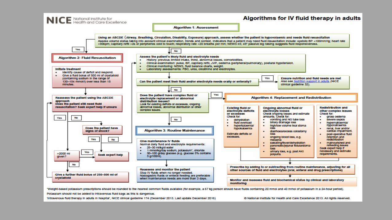
General Assessment Fluid Needs
(A) ABCDE TO EXCLUDE
HYPOVOLAEMIA
YES
INITIATE RESUSCITATION
ALGORITHM 2
NO
(B) ASSESS FLUID & ELECTROLYTE
NEEDS
(D) CAN PATIENT USE ORAL ENTERAL
ROUTE?
YES
FEED TO ACHIEVE WATER-ELECTROLYTES
& CALORIC NEEDS
NO
(C) Is there a Complex Fluid & Electrolyte
abnormality
(Excess-Deficit)?
YES
(F) REPLACE-REDISTRIBUTE
ALGORITHM 4
NO
(E) USE MAINTENANCE
ALGORITHM 3
(G) COMPLETED?
(H) ACHIEVED?
YES

Assessment of Hypovolaemia
• Volume status: clinical exam, trends and context• Common clinical tools are generally unreliable in critical illness but
may detect early changes and need for advanced monitoring• Simplest Tool
Urinary OutputNeonates and infant up to 1 y: 2ml/kg/hToddlers 12-36 mo: 1.5 ml/kg/hChildren > 3 y: 1 ml/kg/h Adult: 0.5-1 ml/kg/h
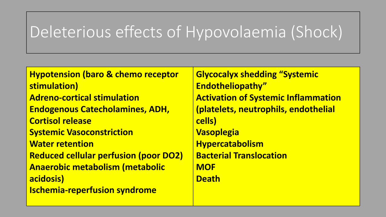
Deleterious effects of Hypovolaemia (Shock)
Hypotension (baro & chemo receptor stimulation)Adreno-cortical stimulationEndogenous Catecholamines, ADH, Cortisol releaseSystemic VasoconstrictionWater retentionReduced cellular perfusion (poor DO2)Anaerobic metabolism (metabolic acidosis)Ischemia-reperfusion syndrome
Glycocalyx shedding “Systemic Endotheliopathy”Activation of Systemic Inflammation (platelets, neutrophils, endothelial cells)Vasoplegia Hypercatabolism Bacterial TranslocationMOFDeath

Clinical “determinants” of Volume Status
Parameters Measure PitfallsVital Signs BP Could be normal despite volume changes, vasopressor therapy
Pulse Influenced by multiple factors: pain, fever, fluid loses, drugs
Orthostatic BP changes Affected by IAP, need objective quantification of SVV
Physical Exam GCS TBI, drugs
Capillary Refill Influenced by environment
Skin Changes Influenced by environment
Limb Temperature Influenced by environment
Urinary Output Stress response, vasopressors, IAP
Laboratory Na/Urea Influenced by multiple factors
Lactate Abnormally elevated: drugs, vasopressors, liver ischaemia
ScvO2 Surrogate measure, Influenced by high Hb concentration & SaO2

Monitoring Volume Status
• Non-invasive vs Invasive• Static vs Dynamic • Multiple tools available• Variability of results and poor correlation with actual volume status is
common• Equipment availability • Use of End-points of resuscitation: “safer” alternative in resources-
constrained environments?

Summary of Available ToolsMethod Type Assess Fluid Responsiveness Comments
History Findings Noninvasive, Static NO Poor correlation with invasive Press.
Physical exam NoninvasiveStatic & Dynamic
YES Serial exam may detect changes in organ perfusion
Chest X Ray Noninvasive NO No standard measurements of vascular pedicles and CT ratio, serial may help determine effects of Rx
CVP Invasive Static NO Poor correlation with fluid responsiveness
PCWP – PAC Invasive Static NO Poor correlation with fluid responsiveness
TT Echo Noninvasive Static NO Single measure of chamber volume difficult to interpret, serial best, labor intensive
SVV Invasive Dynamic YES Requires sedated, ventilated patient with N VT
Oesophageal Doppler Invasive Dynamic YES Not useful for continuous monitoring
VC diameter by US Noninvasive Dynamic YES Body habitus and operator dependent
Passive leg raising Noninvasive Dynamic(*) YES Affected by IAP
Bioimpedance Noninvasive Static NO Cannon assess intravascular volume
(*) Need FloTrac, PiCCo, LiDOO for SVVmeasurement

Resuscitation
(A) IS THE PATIENT
HYPOVOLAEMIC?
YES
Find-Control Cause
Administer Na containing
Crystalloid 500 ml bolus under 15
minutes. Reassess need
Additional 3-4 boluses can be
administered
(C) Not Bleeding?
(B) Bleeding?
(D) Initiate DCR
Urgent Surgical Control
Goals Achieved?
YES
Assess need for
Maintenance-
Replacement-
Redistribution
NO
CONTINUOUS ASSESSMENT OF RESUSCITATION GOALS
ASK FOR SENIOR HELP

Resuscitation (Paediatric)
• Ringer Lactate 20 ml/kg boluses (< 40 kg)• Avoid Hypertonic and Glucose containing solution as plasma
expanders• Hypoglycaemia common especially in neonates, infants and younger
children (Monitor Glucose and administer PRN)• Continuous re-assessment of resuscitation end-points (goals directed) • Monitor electrolytes, be wary of using IV K+ without proper
monitoring and acceptable urinary output

Routine Maintenance-Adult
• Prescribe and Administer Daily Fluids and Electrolyte Requirements(*)
• When a patient cannot meet daily requirements using oral-enteral route but does not have Replacements and Redistribution issues (no fistula, no vomiting, no sepsis, no organ failure, etc.)
(*) Dependent on level of physical activity and environmental factors (ambient Temperature)
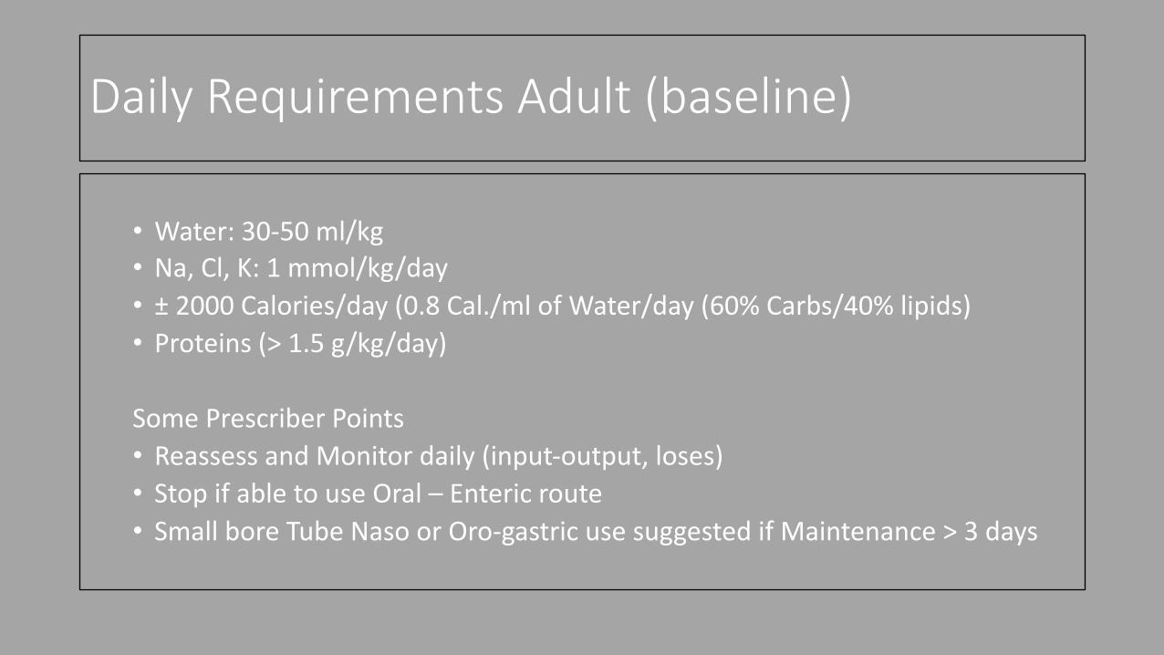
Daily Requirements Adult (baseline)
• Water: 30-50 ml/kg• Na, Cl, K: 1 mmol/kg/day • ± 2000 Calories/day (0.8 Cal./ml of Water/day (60% Carbs/40% lipids)• Proteins (> 1.5 g/kg/day)
Some Prescriber Points• Reassess and Monitor daily (input-output, loses)• Stop if able to use Oral – Enteric route• Small bore Tube Naso or Oro-gastric use suggested if Maintenance > 3 days

Adult Maintenance
Fluid Composition CommentMaintelyte 5% 5% dextrose 50g/L
Na 35 mmol/L Cl 65 mmol/LK 25 mmol/L Mg 2.5 mmol/LNo Buffer
Acid Solution pH 4.0High Osmolarity 405 mOsm(*)Should be fluid of choice after resuscitation is completed if patient cannot use oral-enteral route for feeding

Routine Maintenance Paediatric
• Isotonic Fluid maintenance to prevent hospital acquired Hyponatraemia secondary to SIADH (AAP CP Guidelines 2018, Feld LG et al Pediatrics 2018)
• Monitor and administer Glucose PRN
SIADH induced by:• Hypertonic fluids• Nausea/Vomiting• Pain• Fever• Sepsis• Hypotension• Mech. Ventilation

Routine Fluid Needs Paediatric (Baseline)
Fluid Requirement Age based Fluid Requirements (per kg/hour) Fluid Requirements (per kg/24 h)< 1 y = 120 ml/kg/24 h < 5 kg = 6 ml/kg/h 1-20 kg = 100 ml/kg/24h1-2 y = 100 ml/kg/24 h 5-10 kg = 5 ml/kg/h 11-20 kg = 1000 ml + 50 ml/kg/per
each kg over 10 kg in 24 h2-3 y = 85 ml/kg/24h 11-15 kg = 4 ml/kg/h > 20 kg = 1500 ml + 20 ml/kg/per
each kg over 20 kg in 24 h up to 40 kg
> 3 y = 70 ml/kg/24h 16-35 kg = 2-3 ml/kg/h > 40 kg 30-50 ml/kg/24 h> 36 kg = 1-2 ml/kg/h
Neonate 1,2 to 2 kg Neonate 2 to 2,5 kg Neonate > 2,5 kgDay 1 = 20 ml/kg/24h Day 1 = 25 ml/kg/24 h Day 1 = 30 ml/kg/24 hIncrease 20 ml/kg/day Max 160 ml Increase 25 ml/kg/day max 150 ml Increase 30 ml/kg/day Max 150 ml
Include term neonate

Routine Maintenance Paediatric
• Electrolyte requirements for 24 h (mEq/kg)• Meet Caloric Needs
Age Na Cl K Ca Mg PoNeonate 2-5 2-5 1-2 2-4 0.3-0.5 1-2Infant/child 2-5 2-5 2-4 0.5-4 0.3-0.5 0.5-3Adolescent 1-2 1-2 1-2 10-20 10-30 1-2
Solutions: Neonatalyte, ½ DD

Replacement and Redistribution Issues
Existing Fluid-electrolyte deficits or excess?
Ongoing abnormal Loses? Redistribution – complex issues?
Check & Estimate:1. Dehydration2. Fluid overload3. Degree of deficit-excess for:• Na & Cl• K• Ca, Mg, PO4• Acid-base status
Check & Estimate amount of:• Vomiting-NG output• Biliary drainage• SB Stomas• Diarrhoeas• Fistula• Polyuria• Fever-sweating
Check:• Gross oedema• Severe sepsis• Hyper-hyponatraemia• Renal-liver-cardiac failure• Post op fluid retention/mal
distribution• Malnourished – refeeding
syndrome• Ask for help• Estimate requirements

Composition of some Body Fluids Loses (70 kg patient)
Fluid Na K Cl HCO3 Volume/daySaliva 60 mmol/L 20 mmol/L 16 mmol/L 50 mmol/L ± 1500 mlGastric 60 mmol/L 10 mmol/L 90 mmol/L ± 1500 mlPancreas 140 mmol/L 5 mmol/L 75 mmol/L 70 mmol/L ± 2000 mlBile 145 mmol/L 5 mmol/L 100 mmol/L 50 mmol/L ± 750 mlSmall Bowel 120 mmol/L 5 mmol/L 105 mmol/L 20 mmol/L > 5 L produced

Results of Electrolyte Loses in Surgery
Loses Electrolytes Changes ResultsGastric (aspirate, fistula, vomiting) Low Na, Low Cl, Low K, high HCO3 Hypochloraemic metabolic alkalosis
Pancreatic Fistula Low Na, Low K, Low HCO3, Normal Cl
Hyponatraemic, hypokalaemic metabolic acidosis
SB ECF Low Na, Cl, K, HCO3 Hyponatraemic, hypokalaemic metabolic acidosis
Biliary Fistula Low Na, Cl, K, Normal HCO3 Alkalosis may occur
Diarrhoeas (water loss) High Na, Cl, HCO3 Low K Hyperchloraemic metabolic Acidosis (non anion GAP)

Replacement: Adult
• Deficit added or excess subtracted from baseline daily needs (Maintenance)• Usually done by monitoring 4-6 hourly loses of water and then using
Na containing crystalloid resuscitation fluid (Ringer, Plasmalyte B) to administer equal volume (the 1:1 formula)• i.e.: ECF loosing 450 ml every 6 hours, then 450 ml of Ringer Lactate should be
added to maintenance every 6 hours to keep water balance (i.e. 2500 ml Maintenance + 450 ml x 4 (1800 ml) = 4300 ml in 24 h)
• Monitor acid-base balance for early detection and correction of acidosis (hypoperfusion, HCO3 loses) or alkalosis (hypochloraemia)

Replacement: Adult
• Electrolytes are monitored daily (include CMP) and replaced PRN• Do not add KCL to IV maintenance fluids: dangerous practice!!• Replace K using infusions over 3-4 h and re-assess (Oral K is an option
is patient able to use oral-enteral route)• Remember Calories, Fat and Protein requirements (unless present in
maintenance solutions)• Add Vitamins and trace elements (unless present in maintenance
solutions)
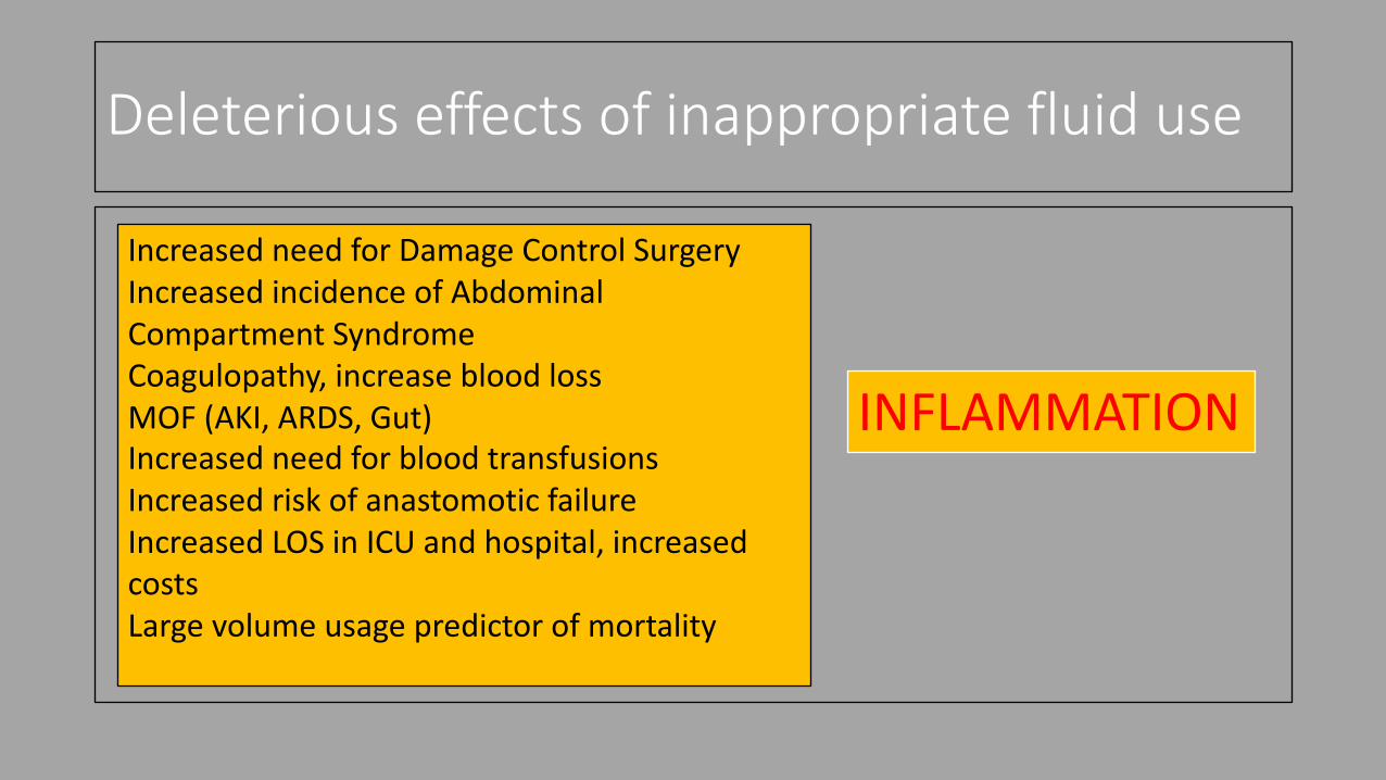
Deleterious effects of inappropriate fluid use
INFLAMMATION
Increased need for Damage Control SurgeryIncreased incidence of Abdominal Compartment SyndromeCoagulopathy, increase blood lossMOF (AKI, ARDS, Gut)Increased need for blood transfusionsIncreased risk of anastomotic failureIncreased LOS in ICU and hospital, increased costsLarge volume usage predictor of mortality

Synthetic Colloids
Starches – Gelatins - Dextran (no longer used)
• Known to increase rate of MOF, especially septic patients
• Coagulopathy and increase need for blood transfusions
• Data not validated in trauma cases (extrapolated results)
• “Unclear” relation between Colloids and Mortality in Critically Ill patients
• Limited data in bleeding trauma patients (increase mortality)
S6CHEST, CRYSTMASCRYSTAL, VISEP Trials
FIRST study - HES vs Saline in Trauma: no differences in mortality, improved Lactate clearance in penetrating trauma, criticized due to design flaws (James MF et al BMJ 2011)

Albumin Controversies
Reasons in Favor of Use as routine replacement in Critically Illness
Natural Protein responsible for Oncotic pressure (80%) Regulates volaemia Scavenger effect (free O2 radicals) (Cysteine residue - thiol)
Anti-Nitric Oxide (NOS): reduce vasodilatory effect of NOS (Cysteine residue - thiol)
Buffer effect in the extra-vascular space (Histidine Imidazole residues)
Transport protein (Domains I-II) Low levels may result in toxicity and poor drug efficacy
Low levels are associated with increased mortality, complications and LOS in ICU-Hospital
HALLMARKS OF CRITICAL ILLNESS PROCESSES: SYSTEMIC INFLAMMATION, HIGH OXIDATIVE STRESS (VASODILATION) AND METABOLIC ACIDOSIS (LACTATE)

Albumin Controversies
Reasons Against the use as routine replacement in Critically Illness
Natural protein: Allergic reactions Normal response in critical illness is to reduce production (preservation of proteins) Non pathological
Association with increased mortality is an expression of disease severity rather than Albumin DIRECT effect
Oncotic effect defeated by the Endotheliopathy of critical illness (shedding Glycocalyx, Leaky capillaries)
Albumin will leak to interstitial space and stay there due to impaired lymphatic clearance (↑oncotic pressure IS, worsening oedema, MOF)

Albumin for Routine Replacement
• Evidence: Seems to be safe
• No influence in survival compared with crystalloid (NaCl) (SAFE study ALBIOS trial)
• Not to be used in TBI with GCS < 8 (increased mortality first week –SAFE study post hoc analysis, linked to increased ICP-oedema)
SAFE study Finfer et al NEJM 2004; ALBIOS Trial Caironi et al NEJM 2014

Albumin for Resuscitation: Sepsis/Septic Shock
• Theoretically YES = anti-oxidant, anti NOS and Glycocalyx stabilizer• But marginal benefit only seen if HD stability is present (ALBIOS)• If endothelial damage is present (ongoing) it may worsen oedema and
organ dysfunction• Slight influence in survival (SAFE post hoc analysis)• HD effect is not clinically significant (ALBIOS)• Safe intervention but not effective to reduce mortality (Patel et al BJM
2014 Metanalysis)

Albumin in ARDS
• In combination with Furosemide seems to improve oxygenation in HD normal ARDS patients with hypoproteinemia• NO survival benefit is achieved

Albumin in Burns
• Safe to use• Anti-oxidant and oncotic effects • No differences in outcome irrespective of albumin levels (Melnyshyn
et al 2013)• No improvement of organ function when 4% albumin added to Ringer
Lactate (ALBUR - Cooper et al 2006)• Albumin use does not: Improve survival, LOS in ICU or healing rate
(Mohammadi et al 2014 RCT)

Other potential uses of Albumin
• Glycocalyx stabilizer (Albumin constitutes 70% of glycocalyx), may be used to stabilize endothelial surface (Counter argument: ongoing inflammation results in persistent endothelial damage)
• Fat scavenger in FES and TPN lipid overload (Free-fatty Acids bind to Albumin, especially Oleic acid, know to cause lung damage)

References
• Caironi P et al. The clinical use of albumin: the point of view of a specialist in intensive care Blood Transfusion 2009;7:259-267• Cantle Pm et al Balanced resuscitation in trauma management Surg Clin N
Am 2017;97:999-1014 • Kalantari K et al (Review) Assessment of intravascular status and fluid
responsiveness in critically ill patients Kidney International 2013;83:1017-28• National Institute for Health Care Excellence (NICE) Clinical Guidelines 2013
(update 2017)• SAFE study Finfer et al NEJM 2004; ALBIOS Trial Caironi et al NEJM 2014

Thank you!