Embed Size (px)
Citation preview

Journal ofMaterials Chemistry B
PAPER
Publ
ishe
d on
23
July
201
4. D
ownl
oade
d by
Ege
Uni
vers
itesi
on
25/0
2/20
15 1
9:06
:29.
View Article OnlineView Journal | View Issue
Folic acid modifi
aDepartment of Biochemistry, Faculty of Scien
Turkey. E-mail: [email protected] of Chemistry, Faculty of Scien
Urla, Izmir, TurkeycDepartment of Chemistry, Istanbul Techn
TurkeydDepartment of Polymer Engineering, Faculty
Yalova, TurkeyeDepartment of Materials Science and Eng
35430 Urla, Izmir, TurkeyfInstitute for Nanoscale Technology, UniversgEge University, Institute of Drug Abuse Toxi
Bornova, Izmir, TurkeyhCenter of Excellence for Advanced Mater
Chemistry, Faculty of Science, King Abdulazi
† Electronic supplementary informa10.1039/c4tb00850b
Cite this: J. Mater. Chem. B, 2014, 2,6412
Received 25th May 2014Accepted 23rd July 2014
DOI: 10.1039/c4tb00850b
www.rsc.org/MaterialsB
6412 | J. Mater. Chem. B, 2014, 2, 6412
ed clay/polymer nanocompositesfor selective cell adhesion†
F. B. Barlas,a D. Ag Seleci,a M. Ozkan,b B. Demir,a M. Seleci,a M. Aydin,c
M. A. Tasdelen,cd H. M. Zareie,ef S. Timur,*ag S. Ozcelikb and Y. Yagci*ch
A folic acid (FA) modified poly(epsilon-caprolactone)/clay nanocomposite (PCL/MMT–(CH2CH2OH)2–FA)
resulting in selective cell adhesion and proliferation was synthesized and characterized as a cell culture
and biosensing platform. For this purpose, first the FA modified clay (MMT–(CH2CH2OH)2–FA) was
prepared by treating the organo-modified clay, Cloisite 30B [MMT–(CH2CH2OH)2] with FA in chloroform
at 60 �C. Subsequent ring opening polymerization of 3-caprolactone in the presence of tin octoate
(Sn(Oct)2) using MMT–(CH2CH2OH)2–FA at 110 �C resulted in the formation of MMT–(CH2CH2OH)2–FA
with an exfoliated clay structure. The structures of intermediates and the final nanocomposite were
investigated in detail by FT-IR spectral analysis and DSC, TGA, XRD, SEM and AFM measurements. The
combination of FA, PCL and clay provides a simple and versatile route to surfaces that allows controlled
and selective cell adhesion and proliferation. FA receptor-positive HeLa and negative A549 cells were
used to prove the selectivity of the modified surfaces. Both microscopy and electrochemical sensing
techniques were applied to show the differences in cell adherence on the modified and pristine clay
platforms. This approach is expected to be adapted into various bio-applications such as ‘cell culture on
chip’, biosensors and design of tools for targeted diagnosis or therapy.
Introduction
Poly(epsilon-caprolactone)/clay (PCL/clay) nanocompositeshave attracted much attention due to the biocompatibility andbiodegradability of the aliphatic polyester matrix and highproperty enhancements that could result from the layered sili-cate dispersion.1,2 There are three common approaches for thepreparation of these nanocomposites: in situ polymerization,solution casting, and melt processing, which are known to leadto intercalated and/or exfoliated structures.3 Recently, severalcoupling reactions were also proposed as an alternative route to
ce, Ege University, 35100 Bornova, Izmir,
ce, Izmir Institute of Technology, 35430
ical University, Maslak, Istanbul 34469,
of Engineering, Yalova University, 77100
ineering, Izmir Institute of Technology,
ity of Technology, Sydney, Australia
cology & Pharmaceutical Sciences, 35100-
ials Research (CEAMR), Department of
z University, 21589, Jeddah, Saudi Arabia
tion (ESI) available. See DOI:
–6421
fabricate such clay based nanocomposites having variousstructurally different macromolecular chains.4–9 In the in situpolymerization technique, epsilon-caprolactone (CL) as amonomer, together with an alcohol functional initiator withstannous octanoate (Sn(Oct)2) as a catalyst, is intercalatedwithin the silicate layers and polymerization is initiated byheating the mixture at 110 �C.9 The chain growth in the claygalleries triggers the clay exfoliation and hence the nano-composite formation.10–18
In biomedical research, cultivation of cells on a substratum isone of the most crucial experimental procedures and extensivelyused in bio-investigations. In order to utilize polymer–claynanocomposites, they should have designed mechanical prop-erties as well as favourable interactions with biological inter-faces.19 The cultivation of various kinds of cells was carried out toinvestigate embryology, cytology, and tissue regeneration onscaffolds or to assess biocompatibility and in vitro toxicity ofnewly developed drug candidates, medical devices, as well asmaterials. For instance, in vitro cytocompatibility of laponitecross-linked poly(ethylene oxide) hydrogel lms using MC3T3-E1 mouse pre-osteoblast cells was reported.19 In the other study,thermo-sensitive poly(N-isopropyl acrylamide) nanocompositegels were used as a so and wet surface material with thecapability of thermally controlled cell adhesion and detachmentwithout the need for proteolytic enzyme treatment.20 Cell adhe-sion and spreading on porous poly(lactic acid)–montmorillonitenanocomposites and clay–gelatin–chitosan nanocomposite
This journal is © The Royal Society of Chemistry 2014

Paper Journal of Materials Chemistry B
Publ
ishe
d on
23
July
201
4. D
ownl
oade
d by
Ege
Uni
vers
itesi
on
25/0
2/20
15 1
9:06
:29.
View Article Online
lms were also investigated in the literature.21,22 On the otherhand, controlling and guiding cell adhesion on biomaterials areimportant for a variety of bio-applications. Biocompatibility canbe improved by limiting non-specic adsorption of proteins andpromoting specic cell–matrix interactions which are importantdue to their effects on regulation of cell function, tissuehomeostasis and cell shape.23–26
Recently, folic acid (FA) intercalated montmorillonite (MMT)was successfully used as a targeted surface for cell cultureapplications to discriminate the adhesion of folate receptorpositive and negative cell lines.27 The FA modied clay not onlyenhanced specicity for cell adhesion, but also exhibitedunique properties such as good mechanical and chemicalstabilities, high surface area and low toxicity. We describe here,FA modied PCL/clay nanocomposites as a non-toxic celladhesion material for various bio-investigations such as selec-tive cell biosensing.
ExperimentalMaterials
An organo-modied clay, Cloisite 30B [MMT–(CH2CH2OH)2]was purchased from Southern Clay Products (Gonzales, TX,USA). The organic content of organo-modied MMT, deter-mined by thermogravimetric analysis (TGA), was 21 wt%. Beforeuse, the clay was dried under vacuum at 110 �C for 1 h. Tin(II)2-ethyl-hexanoate (Sn(Oct)2, Aldrich, 95%) and folic acid (FA,Aldrich, 97%) were used as received. Epsilon-caprolactone (CL,Aldrich, 97%) was vacuum distilled over calcium hydride.Commercial grade solvents were puried by conventionaldrying and distillation procedures. Phosphate buffered saline(PBS, pH 7.4), 3-(4,5-dimethylthiazol-2-yl)-2,5-diphenyl tetrazo-lium bromide (MTT), 4,6-diamino-2-phenylindol (DAPI), tetra-hydrofuran (THF), sodium dodecyl sulphate (SDS),formaldehyde and bovine serum albumin (BSA) were purchasedfrom Sigma. Cell culture supplies including Dulbecco's Modi-ed Eagle Medium (DMEM), fetal calf serum (FCS Gold) andpenicillin/streptomycin (P/S, 100�) were purchased from Lonza(Basel, Switzerland).
Modication of MMT–(CH2CH2OH)2 with FA
MMT–(CH2CH2OH)2, 176 mg, 0.1 mmol, OH-content and FA(88 mg, 0.2 mmol) were added in 50 mL chloroform. The reac-tion mixture was heated up to 60 �C and stirred for 48 h. Aercooling to room temperature and removing the solvent byltration, modied MMT (MMT–(CH2CH2OH)2–FA) was washedwith chloroform and water three times and nally dried undervacuum.
Synthesis of poly(3-caprolactone)/MMT (PCL/MMT)nanocomposites containing FA
(MMT–(CH2CH2OH)2–FA) (20 mg, 0.17 mmol corresponding to1.0% of the monomer by weight) as the initiator was added inSchlenk tubes equipped with a magnetic stirrer and dried in anoil bath at 90 �C for 1 h with a vacuum pump. 1.94 mL (2.0 g,17 mmol) sample of monomer (CL), [Sn(Oct)2] (1/300 molar
This journal is © The Royal Society of Chemistry 2014
ratio with respect to monomer) and 2.0 mL of dry toluene wereadded under nitrogen. The CL polymerization reactions werecarried out at 110 �C. Aer 24 h the mixtures were diluted withTHF and poured into a 10-fold excess of cold methanol. Theproducts were collected aer ltration and dried at roomtemperature in a vacuum. Composites with higher clay contentswere prepared under similar experimental conditions.
Characterization
Fourier transform infrared (FT-IR) spectra were recorded on aPerkin-Elmer FT-IR Spectrum One B spectrometer. Molecularweights were determined by gel permeation chromatography(GPC) using an instrument consisting of a Viscotek GPCmaxAutosampler, a pump, three ViscoGEL GPC columns(G2000HHR, G3000HHR and G4000HHR), and a Viscotek differ-ential refractive index (RI) detector with a THF ow rate of1.0 mL min�1 at 30 �C. The RI detector was calibrated withpolystyrene standards having narrow molecular weight distri-bution. Data were analyzed using Viscotek OmniSEC Omni-01soware. Before the GPC measurement, the polymer wascleaved from the clay by LiBr reuxing in THF for about 24 h,followed by centrifugation and ltration through a lter.Differential scanning calorimetry (DSC) was performed on aPerkin-Elmer Diamond DSC with a heating rate of 20 �C min�1
under nitrogen ow (20mLmin�1). Thermogravimetric analysis(TGA) was performed on a Perkin-Elmer Diamond TA/TGA witha heating rate of 10 �C min�1 under nitrogen ow (200 mLmin�1). The surface morphology was monitored with a FEIQuanta250 FEG scanning electron microscope (SEM). Anaccelerating voltage of 5.0 kV was applied for each sample, and aspot size of 3 was used. X-ray diffraction (XRD) measurementswere carried out with a Panalytical X'Pert Pro MaterialsResearch Diffractometer with CuKa radiation (l ¼ 1.5406 A).The XRD data were collected in a step scanning mode in therange from 5.0� to 50�. The zeta potential of the synthesizednanocomposite samples was measured by using a MalvernZetasizer Nano ZS. 1.0 mL sample was loaded into a cell and thezeta potential was measured simultaneously three times and intriplicate. Atomic force microscopy (AFM) measurements wereconducted by using a Nanosurf exAFM (Liestel, Switzerland).
Cell culture on clay matrices
A549 and HeLa cells were grown in the DMEM containing 10%FCS and 1.0% P/S. All cells were cultivated in a medium at 37 �Cin a humidied environment with 5.0% CO2. The cell seedingdensity and passage number used for both cell lines were 30 000cells per mL and 10.
Cell adhesion on clay matrices was observed using a uo-rescence microscope and proliferation studies using the MTTassay.28 MMT–(CH2CH2OH)2, PCL/MMT–(CH2CH2OH)2, PCL/MMT–(CH2CH2OH)2–FA (5.0% FA) (NC-5) and PCL/MMT–(CH2CH2OH)2–FA (8.0% FA) (NC-8) were compared amongthemselves to observe the differences in their properties as cellculture materials. Therefore, 1.0 mg of each clay mineral wassuspended in 50 mL THF and 950 mL PBS, then 50 mL of thesuspension (per well) was added to 96 well tissue cultured
J. Mater. Chem. B, 2014, 2, 6412–6421 | 6413

Scheme 1 Modification of MMT–(CH2CH2OH)2 with FA and synthesisof PCL/MMT nanocomposites containing FA by in situ ring-openingpolymerization.
Journal of Materials Chemistry B Paper
Publ
ishe
d on
23
July
201
4. D
ownl
oade
d by
Ege
Uni
vers
itesi
on
25/0
2/20
15 1
9:06
:29.
View Article Online
polystyrene plates (TCPS) (Sarstedt). Plates were dried for 24 h atroom temperature. Aerward, clay covered plates were sterilizedunder UV radiation for 3 h and used for cell culture experi-ments. In the proliferation assay, cells which were cultivateddirectly on TCPS without clay matrices were considered asnegative control. Each trial was repeated 5 times. Cells wereincubated for 1 h, 17 h, 24 h, 48 h, 72 h and 96 h. At the end ofeach cultivation time, cells were treated with 110 mL, 10% MTTsolution (5.0 mg mL�1 PBS) per well plates, in the medium for4 h. Then, SDS solution (100 mL, 1.0 g SDS in 10 mL 0.01 M HCl)was added and aer 24 h incubation, UV-visible absorption wasmeasured at 570 nm with 630 nm as the reference wavelengthusing a microplate reader (Bio-Tek Instruments, Inc., Winooski,VT, USA). Furthermore, each cell was stained with DAPI tovisualize cell nuclei. For this aim, cell staining medium wasremoved and cells were washed once with PBS, then, cells weretreated with a DAPI solution (1.0 mg mL�1) for 15 min at 37 �C,then washed again with PBS several times. Fluorescence of thestained samples was monitored using an Olympus BX53Fuorescence microscope equipped with a CCD camera(Olympus DP72). For DAPI, a U-MWU excitation lter, BP330-385 (exciter lter), and BA420 (barrier lter) were used.
Aerwards, the adhered cell density on the surfaces wascalculated as cells per mm2 from the captured images whichwere taken from at least 4 or 5 different regions using HeLa andA549 cells aer nuclei staining via ImageJ Soware.
Bright-eld microscopy imaging
To investigate the morphological alteration of adhered cells ondifferent surfaces (NC-8, PCL/MMT–(CH2CH2OH)2) as well asTCPS as control, bright-eld microscopy images were taken byusing an Andor Revolution Confocal LaserMicroscope (OlympusIX-71 uorescence microscopy). Each sample (1.0 mgmL�1 in 50mL THF and 950 mL PBS) was coated onto TCPS m-Dishes (IbidiGmbH, Germany) except control. Aer drying, UV sterilizationwas performed for 3 h and HeLa cells were grown for 3 days onthe surfaces. Prior to bright-eld imaging, cells were washedwith PBS, then xed using 4.0% formaldehyde (dissolved in PBS)for 30 min and rinsed with PBS several times.
Electrochemical measurements
All electrochemical experiments were carried out on a Palmsenspotantiostat (Palm Instruments, Houten, Netherlands). A threeelectrode system consists of an Ag/AgCl reference electrode, aplatinum electrode as the auxiliary electrode and a 3.0 mmdiameter glassy carbon electrode (GCE) as the working electrodewas used. Prior to measurements, the GCE was polished withalumina slurry followed by sonication in the 1 : 1 distilledwater–ethanol mixture for 5 min. Aer that, mirror like GCEsurfaces were coated with 15 mL of 1.0 mg mL�1 clay samples(dissolved in 900 mL PBS, pH 7.4 and 100 mL THF) and 5.0 mL of1.0 mg mL�1 BSA (in PBS buffer). Electrodes were allowed to dryfor about 1 h. Finally, an appropriate amount of cells in PBS wasdropped onto the clay modied GCE surfaces and incubated for2 h under ambient conditions. HeLa (folate positive) and A549(folate negative) cell lines were applied during the experiments.
6414 | J. Mater. Chem. B, 2014, 2, 6412–6421
Cyclic voltammetry (CV) (between �0.4 V and 0.6 V) anddifferential pulse voltammetry (DPV) (between�0.2 V and 0.6 V)techniques were performed aer each modication using[Fe(CN)6]
3�/4� as a water soluble redox probe (10 mM). Cellbinding to the surface caused decreases in the response signalswhich were correlated with the cell loaded onto the surface.Differences between the current signals were calculated asfollows:
DI ¼ Io � Ic
where Io is the mean current at zero cell concentration and Ic isthe mean current at any concentration by aer cell binding ontothe clay covered surfaces.
Statistical analysis
All experiments were repeated 5 times. All data were expressedas average � SD (standard deviation) unless particularly out-lined. A one-way analysis of variance (ANOVA) was performedwith the Tukey test for multiple comparisons in statisticalevaluation. The difference between two groups was consideredto be signicant when the P value was less than 0.05 and highlysignicant when the P value was less than 0.01 or 0.001.
Results and discussionSynthesis and characterization of nanocomposites
Folic acid (folate, FA), which is composed of a pterin ring,p-amino benzoic acid and glutamic acid moieties, has beenused widely as a targeting ligand to deliver therapeutics tocancer cells because of its ability to react with the membrane-anchored protein called FA receptor. Use of FA in targetingstrategies is advantageous because it is nontoxic, non-immu-nogenic, inexpensive and stable. Conjugation of FA moleculeswith various nanoparticle types via polymer spacer units hasbeen extensively investigated.29 In order to impart its existingadvantages, initially, the FA salt was physically adsorbed at theedges and interlayers of commercially available Cloisite 30 B(a natural MMT clay fully modied with an organic surfactantcontaining two hydroxyl groups) in a manner similar to thatdescribed previously. These hydroxyl groups on the clay surfaceallowed the graing of initiators and the growth of PCL chainsfrom the clay surface, favoring the exfoliation of the platelets(Scheme 1).
This journal is © The Royal Society of Chemistry 2014

Paper Journal of Materials Chemistry B
Publ
ishe
d on
23
July
201
4. D
ownl
oade
d by
Ege
Uni
vers
itesi
on
25/0
2/20
15 1
9:06
:29.
View Article Online
FT-IR, XRD, and TGA techniques were used to characterizemodication of the MMT clay with FA. As shown in Fig. 1, thebands of pure FA between 3600 and 3400 cm�1 are due to thehydroxyl (OH) stretching bands of the glutamic acid moiety andthe NH group of the pterin ring. It also exhibited a very strongabsorption band at 1696, 1513 and 1485 cm�1 due to thestretching vibration of (C]O) and a characteristicabsorption band of the phenyl and pterin rings. The pristineMMT–(CH2CH2OH)2 showed typically broad O–H and Si–Ostretching bands at 3633 and 1010 cm�1. Besides these veri-cations for the MMT–(CH2CH2OH)2 clay, the spectra show allthe characteristic groups of FA.
The modication of MMT–(CH2CH2OH)2 with FA was alsoinvestigated by XRD and TGAmethods (Table 1, Fig. 2). The basalspacing (d001) of commercial and modied clay layers was foundto be 1.84 nm and 1.90 nm. The increment of interlayer spacingindicates an intercalated system with insertion of FA moleculesinto clay layers. The weight loss of MMT–(CH2CH2OH)2 andMMT–(CH2CH2OH)2–FA was found to be 20.3 and 41.3 wt% as aresult of degradation of the organic content of commercial clays.Both XRD and TGA results conrmed that the FAmolecules weresuccessfully incorporated into the MMT interlayer.
Fig. 1 FT-IR spectra of FA, MMT–(CH2CH2OH)2, MMT–(CH2CH2-OH)2–FA, and the nanocomposite (NC-8).
Fig. 2 X-ray diffractions of MMT–(CH2CH2OH)2–FA and the nano-composites (NC-1, NC-5 and NC-8).
This journal is © The Royal Society of Chemistry 2014
Ring opening polymerization of CL initiated by MMT–(CH2CH2OH)2–FA (Scheme 1) was carried under various condi-tions typical for such polymerization reactions. The molecularweight results and reaction conditions are summarized in Table1. With an increase in the nanoclay initiator amount, there wasan increase in the polymerization rate, which results in theslightly higher conversion and lower molecular weights of theresulting polymers. The molecular weight distributionremained narrow (1.24–1.35) and unimodal during the poly-merization. Aer the polymerization reactions, the XRD peak ofMMT–(CH2CH2OH)2–FA disappeared in the X-ray diffractionpattern for all nanocomposite samples, which indicates theformation of exfoliated structures of the clay. The DSC ther-mograms, performed on nanocomposites, showed a slightreduction of the melting temperature with the increase of theclay content, suggesting that the degree of crystallinity wasaffected by the restricted mobility of the chains (Fig. 4). Thethermal degradation temperatures of the nanocomposites withdifferent organic clay loadings were very close to each other, anda slight increase with 10% and 50% weight loss temperatureswas observed. Notably, the nal char yields of the nano-composites were increased from 1.2% to 8.5% by increasing clayloadings (Fig. 3).
The zeta potential of MMT–(CH2CH2OH)2, PCL/MMT–(CH2-CH2OH)2 and NC-8 is given in Table 2. The surface potential ofMMT–(CH2CH2OH)2 is found as highly negative. A less negativevalue was observed aer modication with PCL and nally, FAintercalation caused a remarkable decrease in the surface potentialof the nanocomposite. SEM micrographs of the nanocompositesare given in Fig. 5. The surface morphology of commerciallyavailable Cloisite 30B and PCL/MMT–(CH2CH2OH)2 nano-composites is shown in Fig. 5A and B, respectively. Aer treatmentof the polymer with the clay, small clay aggregates are observed onthe surface of nanocomposites (Fig. 5B). In addition, the SEMmicrograph of the FA intercalated PCL/MMT–(CH2CH2OH)2nanocomposite is given in Fig. 5C. It is clearly observed that thePCL/MMT–(CH2CH2OH)2 nanocomposite surface exhibits a roughsurface aer this modication step. Moreover, the surfacemorphologies with higher magnication (20 000�) are given inFig. S1 (ESI†).
AFM was also used to evaluate the morphology of thenanocomposite surfaces as well. Fig. 6 shows AFM images ofMMT–(CH2CH2OH)2, PCL/MMT–(CH2CH2OH)2 and NC-8. AFMimages were obtained by depositing the samples on siliconwafer. Tapping mode was used during the measurements.
Additionally, the roughness of (MMT–(CH2CH2OH)2, PCL/MMT–(CH2CH2OH)2 and NC-8) was assessed by measuring theroughness parameters (Table 3).
Table 3 presents the roughness values of MMT–(CH2CH2OH)2,PCL/MMT–(CH2CH2OH)2 and NC-8 nanocomposites. They weremeasured in terms of roughness average (Sa), mean value (Sm),root-mean-square roughness (Sq), valley depth (Sv), peak height(Sp), and peak-valley height (Sy). The roughness values werecalculated using integrated soware for image analysis. Here, thepeak-valley height is an estimate of z-values which is short or wideand tall or narrow. The average roughness (Sa) of MMT–(CH2-CH2OH)2, PCL/MMT–(CH2CH2OH)2 and NC-8 nanocomposites
J. Mater. Chem. B, 2014, 2, 6412–6421 | 6415

Table 1 Polymerization conditions and thermal properties of PCL/MMT–(CH2CH2OH)2–FA nanocomposites and their components
Entry Con.a Mnb (g mol�1) Mw/Mn
b Tmc (�C)
Weight losstemperatured (�C)
Char yieldd (%)10% 50%
MMT– — — — — 570 — 79.7MMT–FA — — — — 285 — 58.7NC-1e 91 13 400 1.24 56.8 302 327 1.2NC-5 93 8700 1.28 56.3 305 328 4.6NC-8 96 5500 1.35 55.7 310 330 8.5
a Determined gravimetrically. b Molecular weight and distribution were determined by gel permeation chromatography. c Determined by DSC andanalyses under a nitrogen ow at a heating rate of 10 �Cmin�1. d Determined by TGA analysis under a nitrogen ow at a heating rate of 10 �Cmin�1.e The number indicates MMT–FA loadings.
Fig. 4 DSC traces of the nanocomposites (NC-1, NC-5 and NC-8).
Fig. 3 TGA thermograms of MMT–(CH2CH2OH)2, MMT–(CH2CH2-OH)2–FA and the nanocomposites (NC-1, NC-5 and NC-8).
Table 2 Zeta potential values
Sample Zeta potential (mV)
MMT–(CH2CH2OH)2 �28.10PCL/MMT–(CH2CH2OH)2 �24.40NC-8 �9.94
Fig. 5 SEM images of MMT–(CH2CH2OH)2 (A), PCL/MMT–(CH2CH2OH)2(B) and NC-8 (C) with 10 000� magnification.
6416 | J. Mater. Chem. B, 2014, 2, 6412–6421
Journal of Materials Chemistry B Paper
Publ
ishe
d on
23
July
201
4. D
ownl
oade
d by
Ege
Uni
vers
itesi
on
25/0
2/20
15 1
9:06
:29.
View Article Online
was 37.0 nm, 23.0 nm, 29.0 nm, respectively. All values wereobtained with 1 � 1 mm scan area. The AFM results also supportthe SEM images. Here, the surface roughness of PCL/MMT–(CH2CH2OH)2 increases with the addition of FA.
Cell proliferation
The exibility, biocompatibility, biodegradability and mechan-ical stability of the nanocomposites are very advantageousallowing their use in various biomedical applications. Thesefeatures are important in the design of implants and drugdelivery systems, biosensors, in vitro diagnostics and cellculture matrices. Enhanced cell adhesion and proliferationwere observed on the intercalated structure of MMT–gelatin–chitosan for the stromal stem cells.22 Lewkowitz-Shpuntoff et al.used other nanocomposites made from ethylene vinyl acetateand Cloisite and reported clay dependent growth of humandermal broblasts on the polymer nanocomposite surfaces.30
Recently, MMT was modied with N-(2-hydroxy)propyl-3-tri-methylammonium chitosan chloride and human bone marrowmesenchymal stem cells were used to evaluate cell growth onthis matrix.31 Besides, poly(glycolic acid), PCL, poly(lactic acid)and these copolymers are very promising scaffolds for tissue
Mean potential (mV) Area (%) Width (mV)
�28.10 100 3.22�24.40 100 4.72�9.94 100 6.85
This journal is © The Royal Society of Chemistry 2014
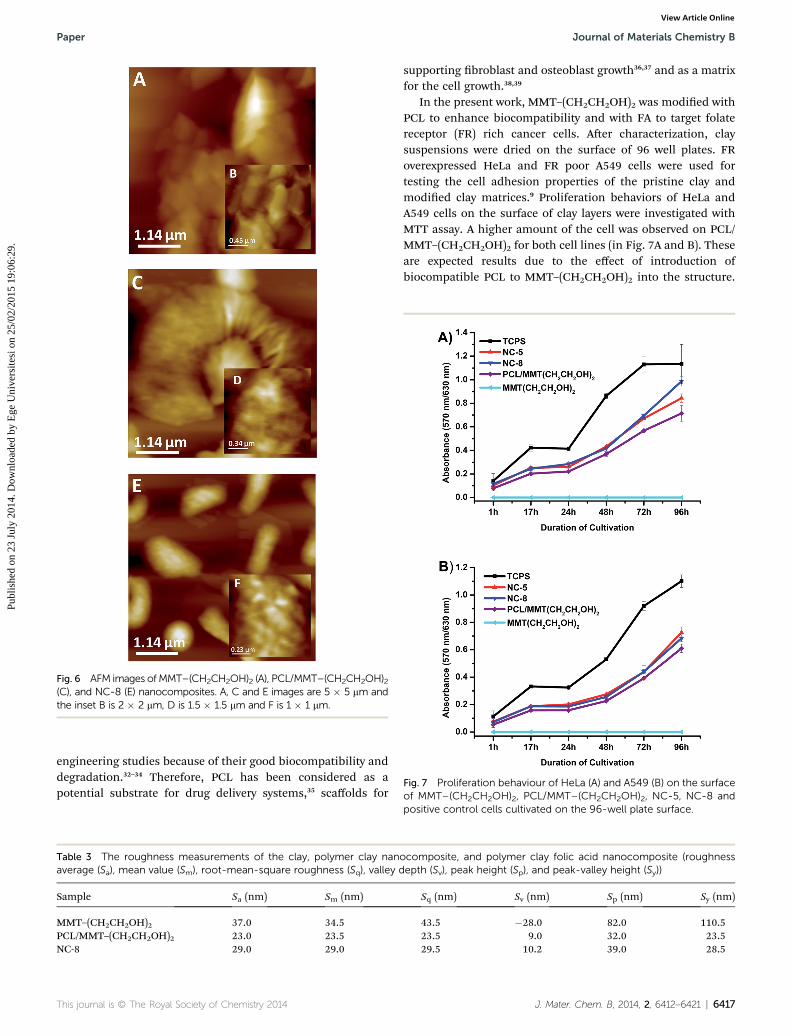
Fig. 6 AFM images ofMMT–(CH2CH2OH)2 (A), PCL/MMT–(CH2CH2OH)2(C), and NC-8 (E) nanocomposites. A, C and E images are 5 � 5 mm andthe inset B is 2 � 2 mm, D is 1.5 � 1.5 mm and F is 1 � 1 mm.
Fig. 7 Proliferation behaviour of HeLa (A) and A549 (B) on the surfaceof MMT–(CH2CH2OH)2, PCL/MMT–(CH2CH2OH)2, NC-5, NC-8 and
Paper Journal of Materials Chemistry B
Publ
ishe
d on
23
July
201
4. D
ownl
oade
d by
Ege
Uni
vers
itesi
on
25/0
2/20
15 1
9:06
:29.
View Article Online
engineering studies because of their good biocompatibility anddegradation.32–34 Therefore, PCL has been considered as apotential substrate for drug delivery systems,35 scaffolds for
Table 3 The roughness measurements of the clay, polymer clay nanaverage (Sa), mean value (Sm), root-mean-square roughness (Sq), valley d
Sample Sa (nm) Sm (nm)
MMT–(CH2CH2OH)2 37.0 34.5PCL/MMT–(CH2CH2OH)2 23.0 23.5NC-8 29.0 29.0
This journal is © The Royal Society of Chemistry 2014
supporting broblast and osteoblast growth36,37 and as a matrixfor the cell growth.38,39
In the present work, MMT–(CH2CH2OH)2 was modied withPCL to enhance biocompatibility and with FA to target folatereceptor (FR) rich cancer cells. Aer characterization, claysuspensions were dried on the surface of 96 well plates. FRoverexpressed HeLa and FR poor A549 cells were used fortesting the cell adhesion properties of the pristine clay andmodied clay matrices.9 Proliferation behaviors of HeLa andA549 cells on the surface of clay layers were investigated withMTT assay. A higher amount of the cell was observed on PCL/MMT–(CH2CH2OH)2 for both cell lines (in Fig. 7A and B). Theseare expected results due to the effect of introduction ofbiocompatible PCL to MMT–(CH2CH2OH)2 into the structure.
ocomposite, and polymer clay folic acid nanocomposite (roughnessepth (Sv), peak height (Sp), and peak-valley height (Sy))
Sq (nm) Sv (nm) Sp (nm) Sy (nm)
43.5 �28.0 82.0 110.523.5 9.0 32.0 23.529.5 10.2 39.0 28.5
positive control cells cultivated on the 96-well plate surface.
J. Mater. Chem. B, 2014, 2, 6412–6421 | 6417

Fig. 8 Fluorescence images of DAPI stained HeLa (A) and A549 cells (B)cultivated on TCPS, MMT–(CH2CH2OH)2, PCL/MMT–(CH2CH2OH)2,NC-1, NC-5 and NC-8 after 72 hours.
Journal of Materials Chemistry B Paper
Publ
ishe
d on
23
July
201
4. D
ownl
oade
d by
Ege
Uni
vers
itesi
on
25/0
2/20
15 1
9:06
:29.
View Article Online
Fig. 7A demonstrates that the cell amount of HeLa on NC-8increases compared to the control cells which are grown directlyon TCPS well plates. Also, better cell proliferation on NC-8 wasobserved in comparison with NC-5 (p < 0.05). We assume thatthe main reason for the better proliferation behaviour of HeLaon NC-8 with respect to NC-5 is due to the presence of more FAin the clay structure. On the other side no signicant differencewas observed on the proliferation behaviour of A549 cells onPCL/MMT–(CH2CH2OH)2, NC-5 and NC-8 (Fig. 7B). Aer 48 h,both HeLa and A549 cells were proliferated on nanocompositesurfaces between time intervals, signicantly (p < 0.05 for 48–72h and 72–96 h).
Furthermore, DAPI was used to visualize adhered cells on thematrices via cell nuclei staining. The inefficient adhesion ofboth cell lines on MMT–(CH2CH2OH)2 is observed aer 72 hcultivation time as shown in Fig. 8A and B. The cell amount forboth cell lines on PCL/MMT–(CH2CH2OH)2 was highercomparing to MMT–(CH2CH2OH)2. In the case of A549, there isno any signicant difference in proliferation features betweenPCL/MMT–(CH2CH2OH)2, NC-5 and NC-8. But on NC-5 andespecially on NC-8 FR, positive HeLa cells grew favorably. Alluorescence microscopic investigations are congruent with theproliferation results.
To obtain more informative data from the uorescent images,further analysis based on cell density (cells per mm2) was accom-plished using the ImageJ Soware. Fig. 9 illustrates the cell densityanalysis of both A549 and HeLa cells upon varying modiedsurfaces. It appears that both NC-5 and NC-8 surfaces have createda signicant difference between HeLa and A549 cells (p < 0.01for NC-5 and p < 0.001 for NC-8). Furthermore, the increase of FAamount in the nanocomposite structures caused the selectivecell adhesion compared to MMT–(CH2CH2OH)2 andPCL/MMT–(CH2CH2OH)2 surfaces, despite the fact that A549 cellsgrew better on the PCL/MMT–(CH2CH2OH)2 surface (p < 0.01).However, there is no considerable change among NC-1, NC-5 andNC-8 surfaces.
Bright-eld microscopy imaging
HeLa cells that were grown on PCL/MMT–(CH2CH2OH)2, NC-8and control groups were visualized by bright-eld microscopy tounderstand if there is any considerable change on themorphology of the cells (Fig. S2, in the ESI†). It can be statedthat the NC-8 coated surface did not affect the generalmorphology of the HeLa cell when compared to the controlgroups which are cultivated on TCPS.
Electrochemical studies
Electrochemical techniques are considered as good alternative todesign biosensors for label-free detection of cells.40 Detectionprinciples for cell analysis via electrochemical platforms aremainly based on creation of a self-assembled monolayer on theelectrode.41,42 These surfaces could be adapted properly to ‘Lab-on-a-Chip’ or microuidic based systems.43 On the other hand, deci-sion on the immobilization matrix affects the selectivity of thebiosensor for the analysis, signicantly. Herein, aer thesuccessful cell adhesion experiments, NC-8 was applied to create a
6418 | J. Mater. Chem. B, 2014, 2, 6412–6421
novel selective platform for the cultivation of folate positive HeLacells. CV and DPV experiments were performed using ferricyanideas a redox mediator. Fig. 10A shows the oxidation and reductionpeaks of bare and MMT–(CH2CH2OH)2, PCL/MMT–(CH2CH2OH)2andNC-8modied GCE surfaces, sequentially. The cationic natureof the MMT–(CH2CH2OH)2 clay exhibited sharper and higherredox peaks compared to the bare GCE due to the strongerattraction of negatively charged [Fe(CN)6]
3�/4� on the surface. DPVgraphs also showed the similar response characteristics. Adramatic decrease in the peak current was observed when the PCL
This journal is © The Royal Society of Chemistry 2014

Fig. 9 Cell density analysis using the DAPI stained fluorescent cellimages after 72 h cultivation via Image J software.
Fig. 10 Electrochemical behaviour of the bare GCE (a), MMT–(CH2CH2OH)2 (b), PCL/MMT–(CH2CH2OH)2 (c) and NC-8 (d) usingcyclic voltammetry (A) and differential pulse voltammetry (B) inphosphate buffer saline, pH 7.4 with the presence of 10 mM [Fe(CN)6]3�/4� at 50 mV s�1.
This journal is © The Royal Society of Chemistry 2014
Paper Journal of Materials Chemistry B
Publ
ishe
d on
23
July
201
4. D
ownl
oade
d by
Ege
Uni
vers
itesi
on
25/0
2/20
15 1
9:06
:29.
View Article Online
intercalated clay was covered on the electrode surface due to therather inefficient electron transfer properties. Similar electro-chemical characteristics were also obtained with NC-8 (Fig. 10). Ascan be seen from Fig. 10B, the peak current values were found tobe 54.35 mA (Ep¼ 0.2 V) for the bare GCE, 114.4 mA (Ep¼ 0.18 V) forMMT–(CH2CH2OH)2, 31.8 mA (Ep ¼ 0.2 V) for PCL/MMT–(CH2-CH2OH)2 and 22.6 mA (Ep ¼ 0.19 V) for NC-8, respectively.
Aerwards, cell sensing studies were carried out via CV andDPV techniques. Cyclic voltammograms of the bare GCE(Fig. 10A(a)), GCE/NC-8 (Fig. 10A(b)), A549 (1 � 104 cells)/NC-8/GCE (Fig. 11A(c)) and HeLa (1 � 104 cells)/NC-8/GCE(Fig. 11A(d)) were in an agreement with each other in thepresence of 10 mM [Fe(CN)6]3�/4� at 50 mV s�1. Redox peaks ofCVs are as follows; 64.47 mA (Epc ¼ 0.28 V) for the GCE, 48.08 mA(Epc ¼ 0.32 V) for GCE/NC-8, 33.7 mA (Epc ¼ 0.45 V) for GCE/NC-8/A549 and 18.72 mA (Epc ¼ 0.52 V). A drop in the peakcurrents and shiing in the peak potentials (Epc (cathodic) andEpa (anodic)) showed successful cell binding onto the NC-8surface. On the other hand, FR overexpressed HeLa cells displaybetter adherence than FR negative A549 cells as well. To prove
Fig. 11 (A) Cyclic voltammograms (B) differential pulse voltammo-grams of the bare GCE (a), GCE/NC-8 (b), GCE/NC-8/104 A549 cells(c) and GCE/NC-8/104 HeLa cells (d) in phosphate buffer saline, pH 7.4with the presence of 10 mM [Fe(CN)6]3�/4� at 50 mV s�1.
J. Mater. Chem. B, 2014, 2, 6412–6421 | 6419

Journal of Materials Chemistry B Paper
Publ
ishe
d on
23
July
201
4. D
ownl
oade
d by
Ege
Uni
vers
itesi
on
25/0
2/20
15 1
9:06
:29.
View Article Online
the cell capture of NC-8; both DPV and CV techniques were usedas previous studies in which the affinity type sensors werereported.44 Aer treatment of the NC-8 modied electrode withthe HeLa cells, a calibration curve was obtained by the decreasein the current signals (DI) depending on the cell amount withinDPV measurements. The linear relationship is dened by theequation of y ¼ 4.466x � 8.285 (R2 ¼ 0.999, y is DI (mA) and x isthe logarithm of the cell amount) between 1 � 102 and 1 � 104
cells per mL. As shown in Fig. 11A and B, treatment of cellscaused a decrease in the current response. To control theselectivity of the NC-8/GCE surface, the same amount of A549and HeLa cells (1 � 104) was compared. Fig. 11B displays theDPV responses of FR negative A549 (DI¼ 4.8 mA) and FR positiveHeLa cells (DI¼ 9.54 mA). Moreover, within the addition of HeLacells onto the NC-8 surface, a shi in the peak potentials wasobserved due to the strong adherence between HeLa cells andFA groups. Hence it can be claimed that NC-8 is an appropriateplatform for the selective and label free cell detection for furthercell sensing approaches.
Conclusions
In conclusion, a promising material that allows selective celladhesion and proliferation was synthesized and characterizedas a cell culture and biosensing platform. Besides, it provides anefficient way for the detection of FR positive cells via electro-chemical transduction as well as optical monitoring. Thisprinciple could be adapted for targeted detection of the othercells where different proteins are overexpressed in the case ofdifferent diseases. The advantage of this strategy resides in thefast and easy surface modication with the targeting ligands.
Acknowledgements
Ibidi GmbH is acknowledged for their supports to the cellculture studies.
Notes and references
1 B. Lepoittevin, M. Devalckenaere, N. Pantoustier,M. Alexandre, D. Kubies, C. Calberg, R. Jerome andP. Dubois, Polymer, 2002, 43, 4017–4023.
2 M. A. Tasdelen, Eur. Polym. J., 2011, 47, 937–941.3 M. A. Tasdelen, J. Kreutzer and Y. Yagci, Macromol. Chem.Phys., 2010, 211, 279–285.
4 J. Chen, H. Wang, W. Luo, J. Xiang, L. Zhang and B. Sun,Colloid Polym. Sci., 2010, 288, 173–179.
5 Y.-S. Ye, Y.-C. Yen, C.-C. Cheng, Y.-J. Syu, Y.-J. Huang andF.-C. Chang, Polymer, 2010, 51, 430–436.
6 H.-W. Cui and S.-W. Kuo, RSC Adv., 2012, 2, 12148–12152.7 M. A. Tasdelen, W. Van Camp, E. Goethals, P. Dubois, F. DuPrez and Y. Yagci, Macromolecules, 2008, 41, 6035–6040.
8 M. Aydin, M. A. Tasdelen, T. Uyar, S. Jockusch, N. J. Turroand Y. Yagci, J. Polym. Sci., Part A: Polym. Chem., 2013, 51,1024–1028.
6420 | J. Mater. Chem. B, 2014, 2, 6412–6421
9 B. Lepoittevin, N. Pantoustier, M. Devalckenaere,M. Alexandre, D. Kubies, C. Calberg, R. Jerome andP. Dubois, Macromolecules, 2002, 35, 8385–8390.
10 A. Oral, M. A. Tasdelen, A. L. Demirel and Y. Yagci, J. Polym.Sci., Part A: Polym. Chem., 2009, 47, 5328–5335.
11 A. Oral, M. A. Tasdelen, A. L. Demirel and Y. Yagci, Polymer,2009, 50, 3905–3910.
12 H. Akat, M. A. Tasdelen, F. Du Prez and Y. Yagci, Eur. Polym.J., 2008, 44, 1949–1954.
13 M. Aydin, M. A. Tasdelen, T. Uyar and Y. Yagci, J. Polym. Sci.,Part A: Polym. Chem., 2013, 51, 5257–5262.
14 C. Dizman, S. Ates, T. Uyar, M. A. Tasdelen, L. Torun andY. Yagci, Macromol. Mater. Eng., 2011, 296, 1101–1106.
15 K. D. Demir, M. A. Tasdelen, T. Uyar, A. W. Kawaguchi,A. Sudo, T. Endo and Y. Yagci, J. Polym. Sci., Part A: Polym.Chem., 2011, 49, 4213–4220.
16 C. Altinkok, T. Uyar, M. A. Tasdelen and Y. Yagci, J. Polym.Sci., Part A: Polym. Chem., 2011, 49, 3658–3663.
17 Z. Yenice, M. A. Tasdelen, A. Oral, C. Guler and Y. Yagci, J.Polym. Sci., Part A: Polym. Chem., 2009, 47, 2190–2197.
18 A. Nese, S. Sen, M. A. Tasdelen, N. Nugay and Y. Yagci,Macromol. Chem. Phys., 2006, 207, 820–826.
19 A. K. Gaharwar, P. J. Schexnailder, B. P. Kline andG. Schmidt, Acta Biomater., 2011, 7, 568–577.
20 K. Haraguchi, T. Takehisa and M. Ebato, Biomacromolecules,2006, 7, 3267–3275.
21 G. Ozkoc, S. Kemaloglu and M. Quaedieg, Polym. Compos.,2010, 31, 674–683.
22 H. Zhuang, J. P. Zheng, H. Gao and K. D. Yao, J. Mater. Sci.:Mater. Med., 2007, 18, 951–957.
23 F. Grinnell, Trends Cell Biol., 2003, 13, 264–269.24 A. Abbott, Nature, 2003, 424, 870–872.25 C. S. Chen, M. Mrksich, S. Huang, G. M. Whitesides and
D. E. Ingber, Science, 1997, 276, 1425–1428.26 R. McBeath, D. M. Pirone, C. M. Nelson, K. Bhadriraju and
C. S. Chen, Dev. Cell, 2004, 6, 483–495.27 R. Bongartz, D. Ag, M. Seleci, J.-G. Walter, E. E. Yalcinkaya,
D. O. Demirkol, F. Stahl, S. Timur and T. Scheper, J. Mater.Chem. B, 2013, 1, 522–528.
28 D. Ag, R. Bongartz, L. E. Dogan, M. Seleci, J.-G. Walter,D. O. Demirkol, F. Stahl, S. Ozcelik, S. Timur andT. Scheper, Colloids Surf., B, 2014, 114, 96–103.
29 K. Kaaki, K. Herve-Aubert, M. Chiper, A. Shkilnyy, M. Souce,R. Benoit, A. Paillard, P. Dubois, M.-L. Saboungi andI. Chourpa, Langmuir, 2011, 28, 1496–1505.
30 H. M. Lewkowitz-Shpuntoff, M. C. Wen, A. Singh,N. Brenner, R. Gambino, N. Pernodet, R. Isseroff,M. Rafailovich and J. Sokolov, Biomaterials, 2009, 30, 8–18.
31 M. Aliabadi, R. Dastjerdi and K. Kabiri, BioMed Res. Int.,2013, 749240.
32 S. Y. Lee, J. H. Oh, J. C. Kim, Y. H. Kim, S. H. Kim andJ. W. Choi, Biomaterials, 2003, 24, 5049–5059.
33 N. Isogai, S. Asamura, T. Higashi, Y. Ikada, S. Morita,J. Hillyer, R. Jacquet and W. J. Landis, Tissue Eng., 2004,10, 673–687.
34 K. W. Ng and D.W. Hutmacher, Biomaterials, 2006, 27, 4591–4598.
This journal is © The Royal Society of Chemistry 2014

Paper Journal of Materials Chemistry B
Publ
ishe
d on
23
July
201
4. D
ownl
oade
d by
Ege
Uni
vers
itesi
on
25/0
2/20
15 1
9:06
:29.
View Article Online
35 Z. K. Zhong and X. Z. S. Sun, Polymer, 2001, 42, 6961–6969.36 D. W. Hutmacher, T. Schantz, I. Zein, K. W. Ng, S. H. Teoh
and K. C. Tan, J. Biomed. Mater. Res., 2001, 55, 203–216.37 M. C. Serrano, R. Pagani, M. Vallet-Regi, J. Pena, A. Ramila,
I. Izquierdo and M. T. Portoles, Biomaterials, 2004, 25, 5603–5611.
38 M. Gumusderelioglu, S. Dalkiranoglu, R. S. T. Aydin andS. Cakmak, J. Biomed. Mater. Res., Part A, 2011, 98, 461–472.
39 S. Y. Kim, J. Appl. Polym. Sci., 2011, 121, 1921–1929.
This journal is © The Royal Society of Chemistry 2014
40 S. Andreescu and O. A. Sadik, Methods, 2005, 37, 84–93.41 R. Wang, J. Di, J. Ma and Z. Ma, Electrochim. Acta, 2012, 61,
179–184.42 J. Zhao, L. Zhu, C. Guo, T. Gao, X. Zhu and G. Li, Biosens.
Bioelectron., 2013, 49, 329–333.43 M. Moscovici, A. Bhimji and S. O. Kelley, Lab Chip, 2013, 13,
940–946.44 W. Cheng, L. Ding, J. Lei, S. Ding and H. Ju, Anal. Chem.,
2008, 80, 3867–3872.
J. Mater. Chem. B, 2014, 2, 6412–6421 | 6421


