Embed Size (px)
Citation preview
5PA R T
CHAPTER 1 Microbiology: Then and Now
CHAPTER 2 The Chemical Building Blocks of Life
CHAPTER 3 Concepts and Tools for Studying Microorganisms
CHAPTER 4 Structure of Bacterial and Archaeal Cells
CHAPTER 5 Microbial Growth and Nutrition
CHAPTER 6 Microbial Metabolism
CHAPTER 7 Control of Microorganisms: Physical and Chemical Methods
I n 1676, a century before the Declaration of Independence, a Dutch merchant named Antony van Leeuwenhoek sent a noteworthy letter to the Royal Society of London. Writing in the vernacular of his home in the United Netherlands, Leeuwenhoek described how he used a simple microscope to observe vast populations of minute, living creatures. His reports opened a chapter of science that would evolve into the study of microscopic organisms and the discipline of
microbiology. During the next three centuries scientists would discover how profoundly these organisms influence the quality of our lives and the environment around us.
We begin our study of the microorganisms by exploring the grassroot developments that led to the establishment of microbiology as a science. These developments are surveyed in Chapter 1, where we focus on some of the individuals who stood at the forefront of discovery. Today we are in the midst of a third Golden Age of microbiology and our understanding of microorganisms continues to grow even as you read this book.
Chapter 2 reviews basic chemistry, inasmuch as microbial growth, metabolism, and control are grounded in the molecules and macromolecules these organisms contain and in the biological processes they undergo. Chapter 3 sets down some basic microbiological concepts and describes one of the major tools for studying microorganisms. We will concentrate on the bacterial organisms in Chapter 4, where we survey their structural frameworks. In Chapter 5, we build on these frameworks by examining microbial growth patterns and nutritional requirements. Chapter 6 describes the metabolism of microbial cells, including those chemical reactions that produce and use energy. Part 1 concludes by considering the physical and chemical methods used to control microbial growth and metabolism (Chapter 7).
Much as the alphabet applies to word development, in each succeeding chapter we will formulate words into sentences and sentences into ideas as we construct an understanding of microorganisms and concentrate on their importance to public health and human welfare.
Foundations of Microbiology
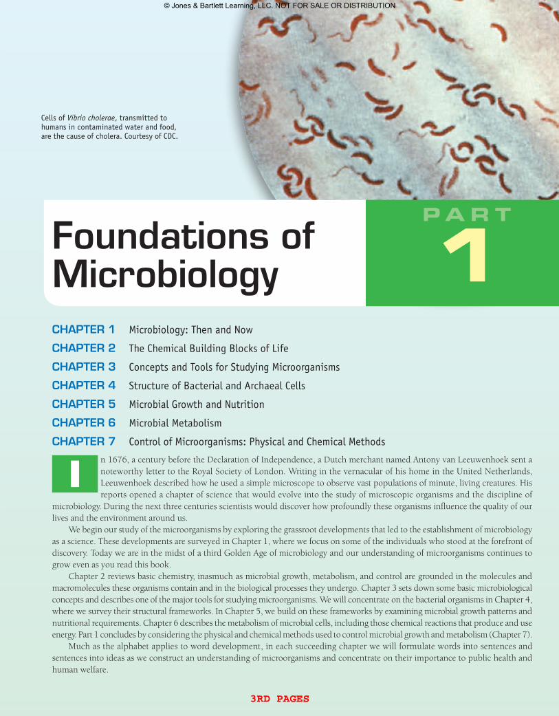
Cells of Vibrio cholerae, transmitted to humans in contaminated water and food, are the cause of cholera. Courtesy of CDC.
1PA R T
3rd pages
© Jones & Bartlett Learning, LLC. NOT FOR SALE OR DISTRIBUTION
microbiology pathways
Science may not seem like the most glamorous profession. So, as you read many of the chapters in this text, you might wonder why many scientists have the good fortune to make key discoveries. At times, it might seem like it is the luck of the draw, but actually many scientists have a set of characteristics that put them on the trail to success.
Robert S. Root-Bernstein, a physiology professor at Michigan State University, points out that many prominent scientists like to goof around, play games, and surround themselves with a type of chaos aimed at revealing the unexpected. Their labs may appear to be in disorder, but they know exactly where every tube or bottle belongs. Scientists also identify intimately with the organisms or creatures they study (it is said that Louis Pasteur actually dreamed about microorganisms), and this identification brings on an intuition—a “feeling for the organism.” In addition, there is the ability to recognize patterns that might bring a breakthrough. (Pasteur had studied art as a teenager and, therefore, he had an appreciation of patterns.)
The geneticist and Nobel laureate Barbara McClintock once remarked, “I was just so interested in what I was doing I could hardly wait to get up in the
morning and get at it. One of my friends, a geneticist, said I was a child, because only children can’t wait to get up in the morning to get at what they want to do.” Clearly, another characteristic of a scientist is having a child-like curiosity for the unknown.
Another Nobel laureate and immunologist, Peter Medawar, once said “Scientists are people of very dissimilar temperaments doing different things in very different ways. Among scientists are collectors, classifiers, and compulsive tidiers-up; many are detectives by temperament and many are explorers; some are artists and others artisans. There are poet-scientists and philosopher-scientists and even a few mystics.” In other words, scientists come from all walks of life.
For this author, I too have found science to be an extraordinary opportunity to discover and understand something never before known. Science is fun, yet challenging—and at times arduous, tedious, and frustrating. As with most of us, we will not make the headlines for a breakthrough discovery or find a cure for a disease. However, as scientists we all hope our hard work and achievements will contribute to a better understanding of a biological (or microbiological) phenomenon and will push back the frontiers of knowledge and have a positive impact on society.
Like any profession, being a scientist is not for everyone. Besides having a bachelor’s degree in biology or microbiology, you should be well read in the sciences and capable of working as part of an interdisciplinary team. Of course, you should have good quantitative and communication skills, have an inquisitive mind, and be goal oriented. If all this sounds interesting, then maybe you fit the mold of a scientist. Why not consider pursuing a career in microbiology? Some possibilities are described in other Microbiology Pathways included in this book, but you should also visit with your instructor. Simply stop by the student union, buy two cups of coffee, and you are on your way.
Being a Scientist
© Comstock/Thinkstock.
3rd pages
© Jones & Bartlett Learning, LLC. NOT FOR SALE OR DISTRIBUTION
3
CHAPTER PREVIEW
1.1 The Discovery of Microbes Leads to Questioning Their Origins
Investigating the Microbial World 1: Can Life Arise Spontaneously?
Microinquiry 1: Experimentation and Scientific Inquiry
1.2 Disease Transmission Can Be Interrupted
1.3 The Classical Golden Age of Microbiology Reveals the Germ
1.4 With the Discovery of Other Microbes, the Microbial World Expands
1.5 A Second Golden Age of Microbiology Involves the Birth of Molecular Biology and Chemotherapy
1.6 The Third Golden Age of Microbiology Is Now
Microbiology: Then and NowMicroorganisms account for most of the biomass on the planet and are an essential foundation on which the global ecosystem rests. They play an absolutely essential role in the survival of the human race.—Carl Woese (Professor of Microbiology, University of Illinois at Urbana-Champaign)
Space. The final frontier! Really? The final frontier? There are an esti-mated 350 billion large galaxies and more than 1023 stars in the visible universe. However, the invisible microbial universe consists of more than 1030 microorganisms (or microbes for short) scattered among an esti-mated 2 to 3 billion species. They may be microscopic in size but they are magnificent in their evolutionary diversity and astounding in their sheer numbers. Existing in such diversity and numbers in the oceans, the land masses, and the atmosphere means they must possess some amazing powers that contribute to the very survival of other organisms on planet Earth. So, could understanding these microscopic organisms on Earth be as important to us and all earthly creatures as studying stars and galaxies in space? Let’s uncover a few examples of what a “day in the life of a microorganism” is like.
A Day in the Life of a Microorganism
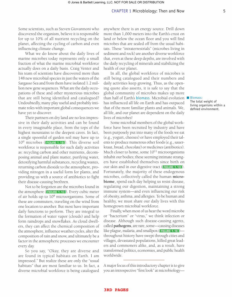
The oceans and seas cover 70% of planet Earth and are swarming with microbes—some 3 × 1029—and helping regulate life on Earth. Floating near the surface are photosynthetic groups that are part of the marine food chain on which all fish and ocean mammals depend. In addi-tion, they provide up to 50% of the oxygen gas we breathe and other organisms use to stay alive ( FiguRE 1.1A ). Other diverse marine and freshwater microbes are the engines that drive nutrient and mineral recycling needed to provide the building blocks to sustain all life. One particular microbe, called Pelagibacter ubique, accounts for 20% (2.4 × 1028 cells) of marine microbes—and 50% of the microbes in the surface waters of temperate oceans in the summer. What is its daily routine? Image courtesy of Dr. Fred Murphy/CDC.
1 CHAPTER PREVIEW
3rd pages
© Jones & Bartlett Learning, LLC. NOT FOR SALE OR DISTRIBUTION
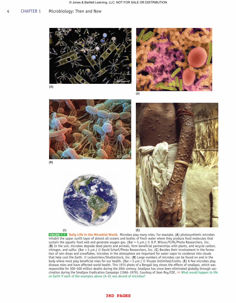
FiguRE 1.1 Daily Life in the Microbial World. Microbes play many roles. For example, (A) photosynthetic microbes inhabit the upper sunlit layer of almost all oceans and bodies of fresh water where they produce food molecules that sustain the aquatic food web and generate oxygen gas. (Bar = 5 µm.) © D.P. Wilson/FLPA/Photo Researchers, Inc. (B) In the soil, microbes degrade dead plants and animals, form beneficial partnerships with plants, and recycle carbon, nitrogen, and sulfur. (Bar = 5 µm.) © David Scharf/Photo Researchers, Inc. (C) Besides their involvement in the forma-tion of rain drops and snowflakes, microbes in the atmosphere are important for water vapor to condense into clouds that help cool the Earth. © Loskutnikov/Shutterstock, Inc. (D) Large numbers of microbes can be found on and in the body where most play beneficial roles for our health. (Bar = 5 µm.) © Visuals Unlimited/Corbis. (E) A few microbes play disease roles and have affected world health. This 1974 photo of a Bengali boy shows the effects of smallpox, which was responsible for 300–500 million deaths during the 20th century. Smallpox has since been eliminated globally through vac-cination during the Smallpox Eradication Campaign (1966–1979). Courtesy of Jean Roy/CDC. »» What would happen to life on Earth if each of the examples above (A–D) was devoid of microbes?
(B)
(A)
(C)
(D)
(E)
4 CHAPTER 1 Microbiology: Then and Now
3rd pages
© Jones & Bartlett Learning, LLC. NOT FOR SALE OR DISTRIBUTION
Some scientists, such as Steven Giovannoni who discovered the organism, believe it is responsible for up to 10% of all nutrient recycling on the planet, affecting the cycling of carbon and even influencing climate change.
What we do know about the daily lives of marine microbes today represents only a small fraction of what the marine microbial workforce actually does on a daily basis. Craig Venter and his team of scientists have discovered more than 148 new microbial species in just the waters of the Sargasso Sea and from them have isolated 1.2 mil-lion new gene sequences. What are the daily occu-pations of these and other mysterious microbes that are still being identified and catalogued? Undoubtedly, many play useful and probably inti-mate roles with important global consequences we have yet to discover.
Their partners on dry land are no less impres-sive in their daily activities and can be found in every imaginable place, from the tops of the highest mountains to the deepest caves. In fact, a single spoonful of garden soil may have up to 109 microbes ( FiguRE 1.1B ). This diverse soil workforce is responsible for such daily activities as: recycling carbon and other nutrients, decom-posing animal and plant matter, purifying water, detoxifying harmful substances, recycling wastes, returning carbon dioxide to the atmosphere, pro-viding nitrogen in a useful form for plants, and providing us with a source of antibiotics to fight their disease-causing brethren.
Not to be forgotten are the microbes found in the atmosphere ( FiguRE 1.1C ). Every cubic meter of air holds up to 108 microorganisms. Some of these are commuters, traveling on the wind from one location to another. But most have important daily functions to perform. They are integral to the formation of water vapor (clouds) and help form raindrops and snowflakes. As cloud dwell-ers, they can affect the chemical composition of the atmosphere, influence weather cycles, alter the composition of rain and snow, and ultimately be a factor in the atmospheric processes we encounter every day.
So you say, “Okay, they are diverse and are found in typical habitats on Earth. I am impressed.” But realize these are only the “usual habitats” that are most familiar to us. In fact, a diverse microbial workforce is being catalogued
anywhere there is an energy source. Drill down more than 1,000 meters into the Earth’s crust on land or below the ocean floor and you will find microbes that are sealed off from the usual habi-tats. These “intraterrestrials” (microbes living in sediment and rock) are another diverse workforce that, even at these deep depths, are involved with the daily recycling of minerals and stabilizing the health of our planet.
In all, the global workforce of microbes is still being catalogued and their numbers and daily activities keep growing. Thus, as the open-ing quote also asserts, it is safe to say that the global community of microbes makes up more than half of Earth’s biomass. Microbial evolution has influenced all life on Earth and has outpaced that of the more familiar plants and animals. We, all life, and our planet are dependent on the daily lives of microbes!
Some microbial members of the global work-force have been recruited by industry and have been purposely put into many of the foods we eat (e.g., yogurt, cheeses) or have employed their tal-ents to produce numerous other foods (e.g., sauer-kraut, bread, chocolate) or medicines (antibiotics). Much closer to home, some 1011 microorganisms inhabit our bodies; these seeming intimate strang-ers have established themselves since birth on our skin and in our digestive tract ( FiguRE 1.1D ). Fortunately, the majority of these endogenous microbes, collectively called the human micro-biome, spend each day helping us resist disease, regulating our digestion, maintaining a strong immune system—and even influencing our risk of obesity, asthma, and allergies. To be human and healthy, we must share our daily lives with this homegrown microbial workforce.
Finally, when most of us hear the word microbe or “bacterium” or “virus,” we think infection or disease. Although such disease-causing agents, called pathogens, are rare, some—causing diseases like plague, malaria, and smallpox ( FiguRE 1.1E )—throughout history have swept through cities and villages, devastated populations, killed great lead-ers and commoners alike, and, as a result, have transformed politics, economies, and public health worldwide.
A major focus of this introductory chapter is to give you an introspective “first look” at microbiology—
Biomass:The total weight of living organisms within a defined environment.
CHAPTER 1 Microbiology: Then and Now 5
3rd pages
© Jones & Bartlett Learning, LLC. NOT FOR SALE OR DISTRIBUTION
Chapter Challenge
How was this diverse global workforce of microorganisms and pathogens revealed, and what challenges do such organisms pose for microbiology today? Let’s investigate!
then and now. We will see how microbes were first discovered and how those that cause infectious disease preoccupied the minds and efforts of so many. Along the way, we will see how curiosity and scientific inquiry stimulated
the quest to understand the microbial world just as the science of microbiology does today. To begin our story, we reach back to the 1600s, where we encounter some very inquisitive individuals.
KEY CONCEPT 1.1 The Discovery of Microbes Leads to Questioning Their Origins
As the 17th century arrived, an observational revo-lution was about to begin: Dutch spectacle maker Zacharias Janssen was one of several individuals who discovered that if two convex lenses were put together, small objects could be magnified. Many individuals in Holland, England, and Italy further developed this combination of lenses that in 1625 would go by the term microscopio or “microscope.” This new invention would be the forerunner of the modern-day microscope.
Microscopy—Discovery of the Very Small
Robert Hooke, an English natural philosopher (the term “scientist” was not coined until 1833), was one of the most inventive and ingenious minds in the history of science. As the Curator of Experiments for the Royal Society of London, Hooke took advantage of the magnification abili-ties of the early compound microscope and made detailed studies of many living objects. One of the most important observations is contained in his Micrographia, published in 1665, where he describes and draws the structure of cork. Seeing a “great many little boxes,” he called these spaces cella (= rooms) and from that observation today we have the word “cell.”
Micrographia represents one of the most impor-tant books in science history because it awakened the learned and general population of Europe to
Convex:Referring to a surface that curves outward.
the world of the very small, revolutionized the art of scientific investigation, showed that the micro-scope was an important tool for unlocking the secrets of nature, and, notably, opened the door to a completely new world: the world of the cell.
At this same time, across the North Sea in Delft, Holland, Antony van Leeuwenhoek, a success-ful tradesman and dry goods dealer, was using hand lenses to inspect the quality of his cloth. As such, and without any scientific training, Leeuwenhoek became skilled at grinding single pieces of glass into fine magnifying lenses. Placing such a lens between two metal plates riveted together, Leeuwenhoek’s “simple microscope” could greatly out magnify Hooke’s microscope ( FiguRE 1.2A, B ).
Beginning in 1673 and lasting until his death in 1723, Leeuwenhoek communicated his micro-scope observations through letters to England’s Royal Society. In 1674, one letter described a sam-ple of cloudy surface water from a marshy lake. Placing the sample before his lens, he described hundreds of what he thought were tiny, living ani-mals, which he called animalcules. His curiosity aroused, Leeuwenhoek soon located even smaller animalcules in such materials as rainwater, scrap-ings from his teeth, and even his own feces. In fact, among the 165 letters sent to the Royal Society, he outlined structural details of yeast cells, described thread-like fungi and microscopic algae and pro-tozoa, and importantly was the first to describe
6 CHAPTER 1 Microbiology: Then and Now
3rd pages
© Jones & Bartlett Learning, LLC. NOT FOR SALE OR DISTRIBUTION
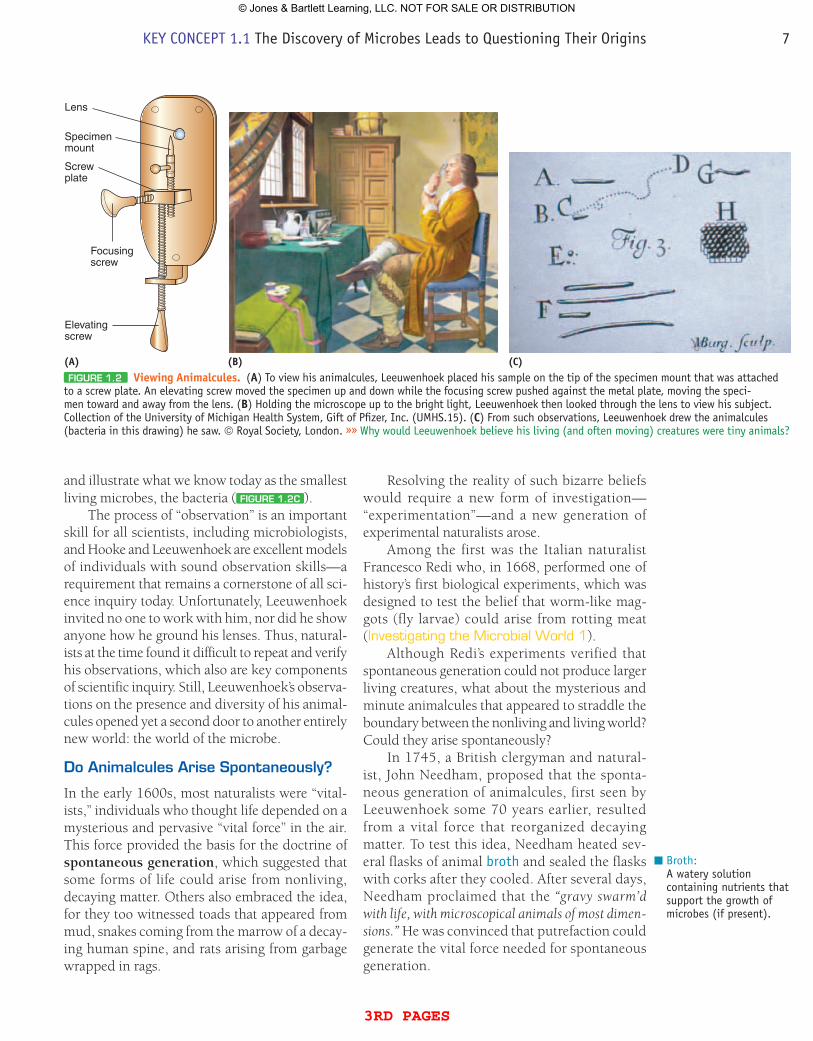
and illustrate what we know today as the smallest living microbes, the bacteria ( FiguRE 1.2C ).
The process of “observation” is an important skill for all scientists, including microbiologists, and Hooke and Leeuwenhoek are excellent models of individuals with sound observation skills—a requirement that remains a cornerstone of all sci-ence inquiry today. Unfortunately, Leeuwenhoek invited no one to work with him, nor did he show anyone how he ground his lenses. Thus, natural-ists at the time found it difficult to repeat and verify his observations, which also are key components of scientific inquiry. Still, Leeuwenhoek’s observa-tions on the presence and diversity of his animal-cules opened yet a second door to another entirely new world: the world of the microbe.
Do Animalcules Arise Spontaneously?
In the early 1600s, most naturalists were “vital-ists,” individuals who thought life depended on a mysterious and pervasive “vital force” in the air. This force provided the basis for the doctrine of spontaneous generation, which suggested that some forms of life could arise from nonliving, decaying matter. Others also embraced the idea, for they too witnessed toads that appeared from mud, snakes coming from the marrow of a decay-ing human spine, and rats arising from garbage wrapped in rags.
Resolving the reality of such bizarre beliefs would require a new form of investigation—“experimentation”—and a new generation of experimental naturalists arose.
Among the first was the Italian naturalist Francesco Redi who, in 1668, performed one of history’s first biological experiments, which was designed to test the belief that worm-like mag-gots (fly larvae) could arise from rotting meat (Investigating the Microbial World 1).
Although Redi’s experiments verified that spontaneous generation could not produce larger living creatures, what about the mysterious and minute animalcules that appeared to straddle the boundary between the nonliving and living world? Could they arise spontaneously?
In 1745, a British clergyman and natural-ist, John Needham, proposed that the sponta-neous generation of animalcules, first seen by Leeuwenhoek some 70 years earlier, resulted from a vital force that reorganized decaying matter. To test this idea, Needham heated sev-eral flasks of animal broth and sealed the flasks with corks after they cooled. After several days, Needham proclaimed that the “gravy swarm’d with life, with microscopical animals of most dimen-sions.” He was convinced that putrefaction could generate the vital force needed for spontaneous generation.
Broth:A watery solution containing nutrients that support the growth of microbes (if present).
Lens
Specimen mount
Screw plate
Focusingscrew
Elevating screw
(A) (B) (C)FiguRE 1.2 Viewing Animalcules. (A) To view his animalcules, Leeuwenhoek placed his sample on the tip of the specimen mount that was attached
to a screw plate. An elevating screw moved the specimen up and down while the focusing screw pushed against the metal plate, moving the speci-men toward and away from the lens. (B) Holding the microscope up to the bright light, Leeuwenhoek then looked through the lens to view his subject. Collection of the University of Michigan Health System, Gift of Pfizer, Inc. (UMHS.15). (C) From such observations, Leeuwenhoek drew the animalcules (bacteria in this drawing) he saw. © Royal Society, London. »» Why would Leeuwenhoek believe his living (and often moving) creatures were tiny animals?
KEY CONCEPT 1.1 The Discovery of Microbes Leads to Questioning Their Origins 7
3rd pages
© Jones & Bartlett Learning, LLC. NOT FOR SALE OR DISTRIBUTION
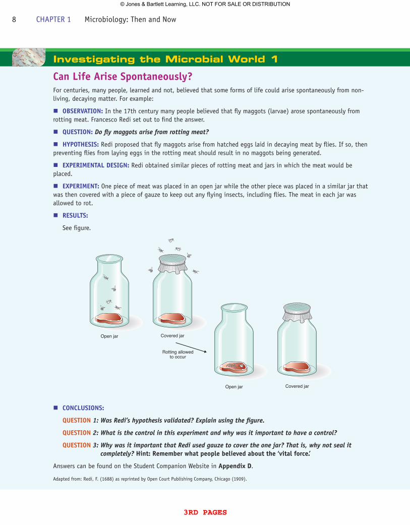
Investigating the Microbial World 1
Can Life Arise Spontaneously?For centuries, many people, learned and not, believed that some forms of life could arise spontaneously from non-living, decaying matter. For example:
nOBSERVATION: In the 17th century many people believed that fly maggots (larvae) arose spontaneously from rotting meat. Francesco Redi set out to find the answer.
nQuESTION: Do fly maggots arise from rotting meat?
nHypOTHESIS: Redi proposed that fly maggots arise from hatched eggs laid in decaying meat by flies. If so, then preventing flies from laying eggs in the rotting meat should result in no maggots being generated.
nExpERIMENTAL DESIGN: Redi obtained similar pieces of rotting meat and jars in which the meat would be placed.
nExpERIMENT: One piece of meat was placed in an open jar while the other piece was placed in a similar jar that was then covered with a piece of gauze to keep out any flying insects, including flies. The meat in each jar was allowed to rot.
nRESuLTS:
See figure.
nCONCLuSIONS:
QuESTION 1: Was Redi’s hypothesis validated? Explain using the figure.
QuESTION 2: What is the control in this experiment and why was it important to have a control?
QuESTION 3: Why was it important that Redi used gauze to cover the one jar? That is, why not seal it completely? Hint: Remember what people believed about the ‘vital force.’
Answers can be found on the Student Companion Website in Appendix D.
Adapted from: Redi, F. (1688) as reprinted by Open Court Publishing Company, Chicago (1909).
Open jar Covered jar
Open jar Covered jar
Rotting allowedto occur
8 CHAPTER 1 Microbiology: Then and Now
3rd pages
© Jones & Bartlett Learning, LLC. NOT FOR SALE OR DISTRIBUTION
Experiments often can be subject to varying interpretations. As such, the Italian cleric and nat-uralist Lazzaro Spallanzani challenged Needham’s conclusions and suggested that the animalcules came from the air and would therefore grow in the broth of the cooled flasks. So, in 1765, he repeated Needham’s experiments but with the few changes. He left some flasks with broth open to the air, others were stoppered loosely with corks, and the remaining flasks were sealed. All were then boiled. After 2 days, the open flasks were swarming with animalcules, but the loosely stop-pered ones had many fewer—and the sealed ones contained no animalcules. Spallanzani concluded that “the number of animalcula developed is propor-tional to the communication with the external air.”
Needham and others countered that Spallanzani’s experiments had destroyed the vital force because sealing the flasks prevented entry of this force necessary for the spontaneous genera-tion of animalcules.
The controversy over spontaneous generation of animalcules continued into the mid-1800s and only deepened when Rudolf Virchow, a German
pathologist, put forward, without direct evidence, the idea of biogenesis, which said that life only arises from life. To solve the debate, a new experi-mental strategy would be needed.
Louis Pasteur, a French chemist and scientist, took up the challenge in 1861 and, through an ele-gant series of experiments that were a variation of the methods of Needham and Spallanzani, discred-ited the idea. Microinquiry 1 outlines the process of scientific inquiry and Pasteur’s experiments.
Although Pasteur’s experiments generated considerable debate for several years, his exact-ing and carefully designed experiments marked the end of the belief in spontaneous generation and validated the idea of biogenesis.
However, today there is another form of “spontaneous generation”—this time occurring in the laboratory (MicroFocus 1.1).concept and reasoning checks 1a. If you were alive in Leeuwenhoek’s time, how would
you explain the origin for the animalcules he saw with his simple microscope?
b. Evaluate the role of experimentation as an important skill to the eventual rejection of spontaneous generation as an origin for animalcules.
KEY CONCEPT 1.2 Disease Transmission Can Be interrupted
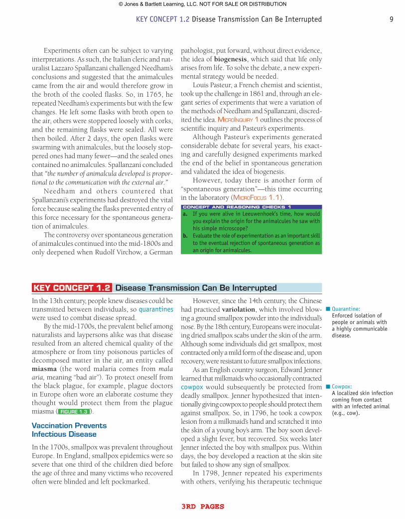
In the 13th century, people knew diseases could be transmitted between individuals, so quarantines were used to combat disease spread.
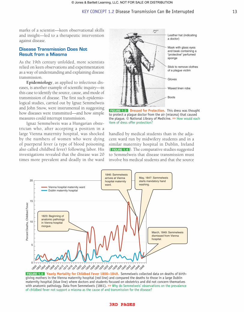
By the mid-1700s, the prevalent belief among naturalists and laypersons alike was that disease resulted from an altered chemical quality of the atmosphere or from tiny poisonous particles of decomposed matter in the air, an entity called miasma (the word malaria comes from mala aria, meaning “bad air”). To protect oneself from the black plague, for example, plague doctors in Europe often wore an elaborate costume they thought would protect them from the plague miasma ( FiguRE 1.3 ).
Vaccination Prevents infectious Disease
In the 1700s, smallpox was prevalent throughout Europe. In England, smallpox epidemics were so severe that one third of the children died before the age of three and many victims who recovered often were blinded and left pockmarked.
Quarantine:Enforced isolation of people or animals with a highly communicable disease.
However, since the 14th century, the Chinese had practiced variolation, which involved blow-ing a ground smallpox powder into the individual’s nose. By the 18th century, Europeans were inoculat-ing dried smallpox scabs under the skin of the arm. Although some individuals did get smallpox, most contracted only a mild form of the disease and, upon recovery, were resistant to future smallpox infections.
As an English country surgeon, Edward Jenner learned that milkmaids who occasionally contracted cowpox would subsequently be protected from deadly smallpox. Jenner hypothesized that inten-tionally giving cowpox to people should protect them against smallpox. So, in 1796, he took a cowpox lesion from a milkmaid’s hand and scratched it into the skin of a young boy’s arm. The boy soon devel-oped a slight fever, but recovered. Six weeks later Jenner infected the boy with smallpox pus. Within days, the boy developed a reaction at the skin site but failed to show any sign of smallpox.
In 1798, Jenner repeated his experiments with others, verifying his therapeutic technique
Cowpox:A localized skin infection coming from contact with an infected animal (e.g., cow).
KEY CONCEPT 1.2 Disease Transmission Can Be Interrupted 9
3rd pages
© Jones & Bartlett Learning, LLC. NOT FOR SALE OR DISTRIBUTION
INQUIRY
INQUIRY 1Experimentation and Scientific inquiry
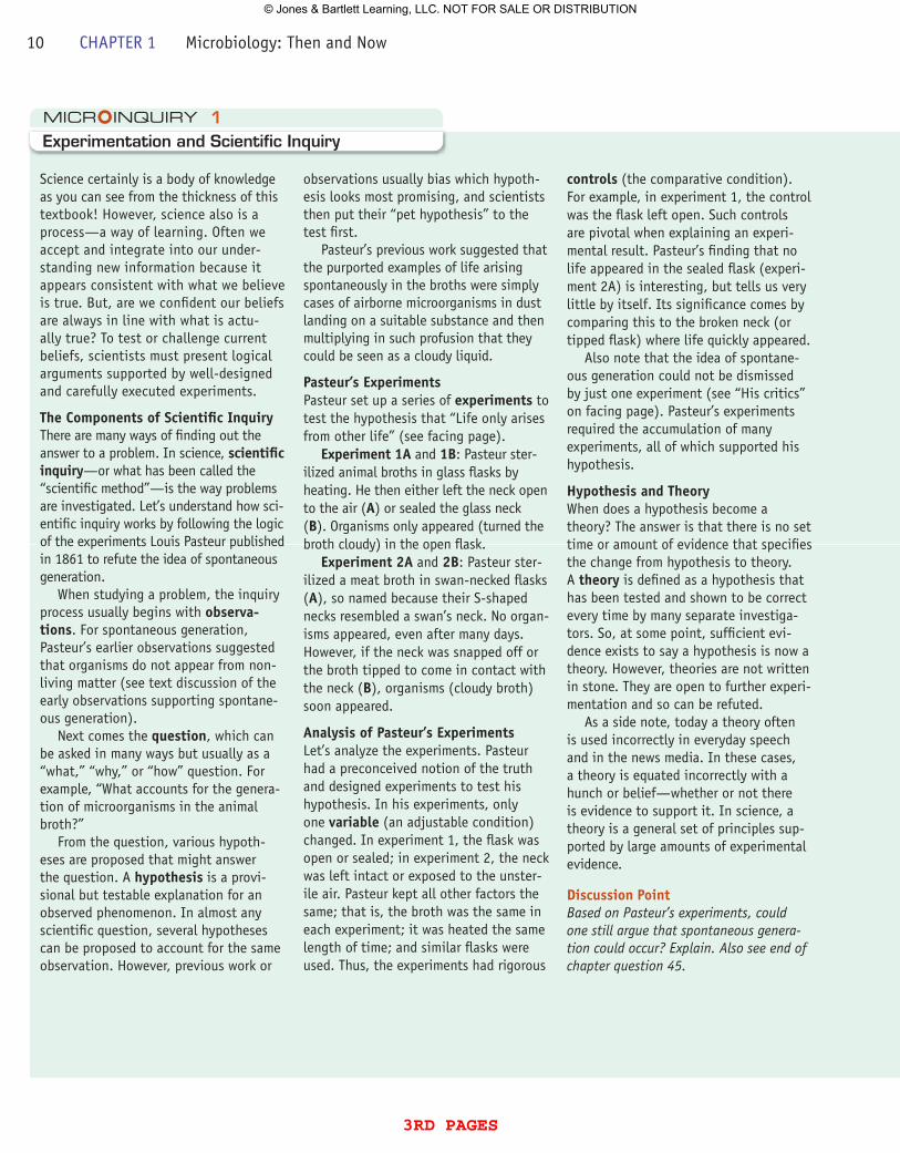
Science certainly is a body of knowledge as you can see from the thickness of this textbook! However, science also is a process—a way of learning. Often we accept and integrate into our under-standing new information because it appears consistent with what we believe is true. But, are we confident our beliefs are always in line with what is actu-ally true? To test or challenge current beliefs, scientists must present logical arguments supported by well-designed and carefully executed experiments.
The Components of Scientific InquiryThere are many ways of finding out the answer to a problem. In science, scientific inquiry—or what has been called the “scientific method”—is the way problems are investigated. Let’s understand how sci-entific inquiry works by following the logic of the experiments Louis Pasteur published in 1861 to refute the idea of spontaneous generation.
When studying a problem, the inquiry process usually begins with observa-tions. For spontaneous generation, Pasteur’s earlier observations suggested that organisms do not appear from non-living matter (see text discussion of the early observations supporting spontane-ous generation).
Next comes the question, which can be asked in many ways but usually as a “what,” “why,” or “how” question. For example, “What accounts for the genera-tion of microorganisms in the animal broth?”
From the question, various hypoth-eses are proposed that might answer the question. A hypothesis is a provi-sional but testable explanation for an observed phenomenon. In almost any scientific question, several hypotheses can be proposed to account for the same observation. However, previous work or
observations usually bias which hypoth-esis looks most promising, and scientists then put their “pet hypothesis” to the test first.
Pasteur’s previous work suggested that the purported examples of life arising spontaneously in the broths were simply cases of airborne microorganisms in dust landing on a suitable substance and then multiplying in such profusion that they could be seen as a cloudy liquid.
pasteur’s ExperimentsPasteur set up a series of experiments to test the hypothesis that “Life only arises from other life” (see facing page).
Experiment 1A and 1B: Pasteur ster-ilized animal broths in glass flasks by heating. He then either left the neck open to the air (A) or sealed the glass neck (B). Organisms only appeared (turned the broth cloudy) in the open flask.
Experiment 2A and 2B: Pasteur ster-ilized a meat broth in swan-necked flasks (A), so named because their S-shaped necks resembled a swan’s neck. No organ-isms appeared, even after many days. However, if the neck was snapped off or the broth tipped to come in contact with the neck (B), organisms (cloudy broth) soon appeared.
Analysis of pasteur’s ExperimentsLet’s analyze the experiments. Pasteur had a preconceived notion of the truth and designed experiments to test his hypothesis. In his experiments, only one variable (an adjustable condition) changed. In experiment 1, the flask was open or sealed; in experiment 2, the neck was left intact or exposed to the unster-ile air. Pasteur kept all other factors the same; that is, the broth was the same in each experiment; it was heated the same length of time; and similar flasks were used. Thus, the experiments had rigorous
controls (the comparative condition). For example, in experiment 1, the control was the flask left open. Such controls are pivotal when explaining an experi-mental result. Pasteur’s finding that no life appeared in the sealed flask (experi-ment 2A) is interesting, but tells us very little by itself. Its significance comes by comparing this to the broken neck (or tipped flask) where life quickly appeared.
Also note that the idea of spontane-ous generation could not be dismissed by just one experiment (see “His critics” on facing page). Pasteur’s experiments required the accumulation of many experiments, all of which supported his hypothesis.
Hypothesis and TheoryWhen does a hypothesis become a theory? The answer is that there is no set time or amount of evidence that specifies the change from hypothesis to theory. A theory is defined as a hypothesis that has been tested and shown to be correct every time by many separate investiga-tors. So, at some point, sufficient evi-dence exists to say a hypothesis is now a theory. However, theories are not written in stone. They are open to further experi-mentation and so can be refuted.
As a side note, today a theory often is used incorrectly in everyday speech and in the news media. In these cases, a theory is equated incorrectly with a hunch or belief—whether or not there is evidence to support it. In science, a theory is a general set of principles sup-ported by large amounts of experimental evidence.
Discussion pointBased on Pasteur’s experiments, could one still argue that spontaneous genera-tion could occur? Explain. Also see end of chapter question 45.
10 CHAPTER 1 Microbiology: Then and Now
3rd pages
© Jones & Bartlett Learning, LLC. NOT FOR SALE OR DISTRIBUTION
INQUIRY
INQUIRY
Each experiment begins with a boiled broth solution similar to that of Needham and Spallanzani.
Organisms appearafter neck snapped off
Sterile broth
Organisms appear
Flask open to air
Sterile broth
Sterile broth No organismsappear
No organisms appear
Flask sealed
Swan-necked flask
Air enters
Dust and microorganisms are trapped
(A)
(B)
(A)
(B)
Timepasses
Timepasses
Time
passes
Timepasses
Pasteur: The broth providesnutrients for the growth of unseenmicrobes in the air: life comes from other life.
His critics: The decomposedproducts in the broth give rise to lifethrough spontaneous generation.
Pasteur: The heat has killedthe microorganisms in the air.
His critics: Sealing the flaskprevents entry of the “life force”needed for spontaneous generation.
Exp
erim
ent 2
Exp
erim
ent 1
Flask tilted so broth enters neck
Timepasses
Organisms appearafter tilting
Pasteur: No life will appear in the flaskbecause microorganisms cannot reach the broth.
His critics: If the “life force” has free access tothe flask, life will appear, given enough time.
Many days later the intact flask is still free ofany life. Pasteur has refuted the doctrine ofspontaneous generation.
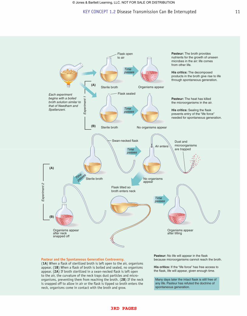
pasteur and the Spontaneous Generation Controversy. (1A) When a flask of sterilized broth is left open to the air, organisms appear. (1B) When a flask of broth is boiled and sealed, no organisms appear. (2A) If broth sterilized in a swan-necked flask is left open to the air, the curvature of the neck traps dust particles and micro-organisms, preventing them from reaching the broth. (2B) If the neck is snapped off to allow in air or the flask is tipped so broth enters the neck, organisms come in contact with the broth and grow.
KEY CONCEPT 1.2 Disease Transmission Can Be Interrupted 11
3rd pages
© Jones & Bartlett Learning, LLC. NOT FOR SALE OR DISTRIBUTION
of vaccination (vacca = “cow”). Prominent physi-cians soon confirmed his findings, and within a few years, Jenner’s method of vaccination spread through Europe and abroad. President Thomas Jefferson wrote to Jenner, “You have erased from the calendar of human afflictions one of its greatest. Yours is the comfortable reflection that mankind can never forget that you have lived.” However, Jefferson was
some 284 years premature in his pronouncement. It would not be until 1980 that the World Health Organization (WHO) would certify that smallpox had been eradicated globally through a massive vac-cination effort carried out between 1966 and 1979.
In retrospect, it is remarkable that without any knowledge of microbes or disease causation, Jenner accomplished what he did. Again, hall-
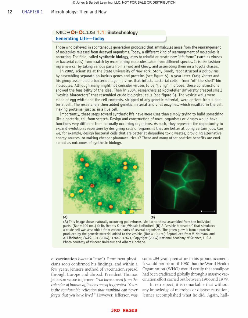
1.1: BiotechnologyGenerating Life—Today
Those who believed in spontaneous generation proposed that animalcules arose from the rearrangement of molecules released from decayed organisms. Today, a different kind of rearrangement of molecules is occurring. The field, called synthetic biology, aims to rebuild or create new “life forms” (such as viruses or bacterial cells) from scratch by recombining molecules taken from different species. It is like fashion-ing a new car by taking various parts from a Ford and Chevy, and assembling them on a Toyota chassis.
In 2002, scientists at the State University of New York, Stony Brook, reconstructed a poliovirus by assembling separate poliovirus genes and proteins (see figure A). A year later, Craig Venter and his group assembled a bacteriophage—a virus that infects bacterial cells—from “off-the-shelf” bio-molecules. Although many might not consider viruses to be “living” microbes, these constructions showed the feasibility of the idea. Then in 2004, researchers at Rockefeller University created small “vesicle bioreactors” that resembled crude biological cells (see figure B). The vesicle walls were made of egg white and the cell contents, stripped of any genetic material, were derived from a bac-terial cell. The researchers then added genetic material and viral enzymes, which resulted in the cell making proteins, just as in a live cell.
Importantly, these steps toward synthetic life have more uses than simply trying to build something like a bacterial cell from scratch. Design and construction of novel organisms or viruses would have functions very different from naturally occurring organisms. As such, they represent the opportunity to expand evolution’s repertoire by designing cells or organisms that are better at doing certain jobs. Can we, for example, design bacterial cells that are better at degrading toxic wastes, providing alternative energy sources, or making cheaper pharmaceuticals? These and many other positive benefits are envi-sioned as outcomes of synthetic biology.
(A) (B)(A) This image shows naturally occurring polioviruses, similar to those assembled from the individual parts. (Bar = 100 nm.) © Dr. Dennis Kunkel/Visuals Unlimited. (B) A “vesicle bioreactor” that simulates a crude cell was assembled from various parts of several organisms. The green glow is from a protein produced by the genetic material added to the vesicle. (Bar = 10 µm.) Reproduced from V. Noireaux and A. Libchaber, PNAS, 101 (2004), 17669–17674; Copyright (2004) National Academy of Science, U.S.A. Photo courtesy of Vincent Noireaux and Albert Libchabe.
12 CHAPTER 1 Microbiology: Then and Now
3rd pages
© Jones & Bartlett Learning, LLC. NOT FOR SALE OR DISTRIBUTION
marks of a scientist—keen observational skills and insight—led to a therapeutic intervention against disease.
Disease Transmission Does Not Result from a Miasma
As the 19th century unfolded, more scientists relied on keen observations and experimentation as a way of understanding and explaining disease transmission.
Epidemiology, as applied to infectious dis-eases, is another example of scientific inquiry—in this case to identify the source, cause, and mode of transmission of disease. The first such epidemio-logical studies, carried out by Ignaz Semmelweis and John Snow, were instrumental in suggesting how diseases were transmitted—and how simple measures could interrupt transmission.
Ignaz Semmelweis was a Hungarian obste-trician who, after accepting a position in a large Vienna maternity hospital, was shocked by the numbers of women who were dying of puerperal fever (a type of blood poisoning also called childbed fever) following labor. His investigations revealed that the disease was 20 times more prevalent and deadly in the ward
Leather hat (indicatinga doctor)
Mask with glass eyesand beak containing a“protective” perfumedsponge
Stick to remove clothesof a plague victim
Gloves
Waxed linen robe
Boots
FiguRE 1.3 Dressed for protection. This dress was thought to protect a plague doctor from the air (miasma) that caused the plague. © National Library of Medicine. »» How would each item of dress offer protection?
Vienna hospital maternity wardDublin maternity hospital
1823: Beginning ofanatomic pathologyin Vienna hospitalmorgue.
1846: Semmelweisarrives at Viennahospital maternityward.
May, 1847: Semmelweisstarts mandatory handwashing.
March, 1849: Semmelweisdismissed from Viennahospital.
20
15
10
5
0
Perc
ent D
eath
s, C
hild
bed
Feve
r
1800
1802
1804
1806
1808
1810
1812
1814
1816
1818
1820
1822
1824
1826
1828
1830
1832
1834
1836
1838
1840
1842
1844
1846
1848
FiguRE 1.4 yearly Mortality for Childbed Fever 1800–1849. Semmelweis collected data on deaths of birth-giving mothers in the Vienna maternity hospital (red line) and compared the deaths to those in a large Dublin maternity hospital (blue line) where doctors and students focused on obstetrics and did not concern themselves with anatomic pathology. Data from Semmelweis (1861). »» Why do Semmelweis’ observations on the prevalence of childbed fever not support a miasma as the cause of and transmission for the disease?
handled by medical students than in the adja-cent ward run by midwifery students and in a similar maternity hospital in Dublin, Ireland ( FiguRE 1.4 ). The comparative studies suggested to Semmelweis that disease transmission must involve his medical students and that the source
KEY CONCEPT 1.2 Disease Transmission Can Be Interrupted 13
3rd pages
© Jones & Bartlett Learning, LLC. NOT FOR SALE OR DISTRIBUTION
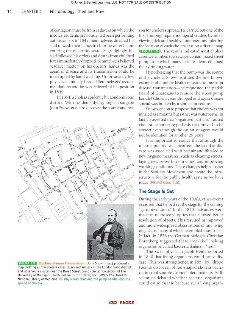
of contagion must be from cadavers on which the medical students previously had been performing autopsies. So, in 1847, Semmelweis directed his staff to wash their hands in chlorine water before entering the maternity ward. Begrudgingly, his staff followed his orders and deaths from childbed fever immediately dropped. Semmelweis believed “cadaver matter” on his doctors’ hands was the agent of disease and its transmission could be interrupted by hand washing. Unfortunately, few physicians initially heeded Semmelweis’ recom-mendations and he was relieved of his position in 1849.
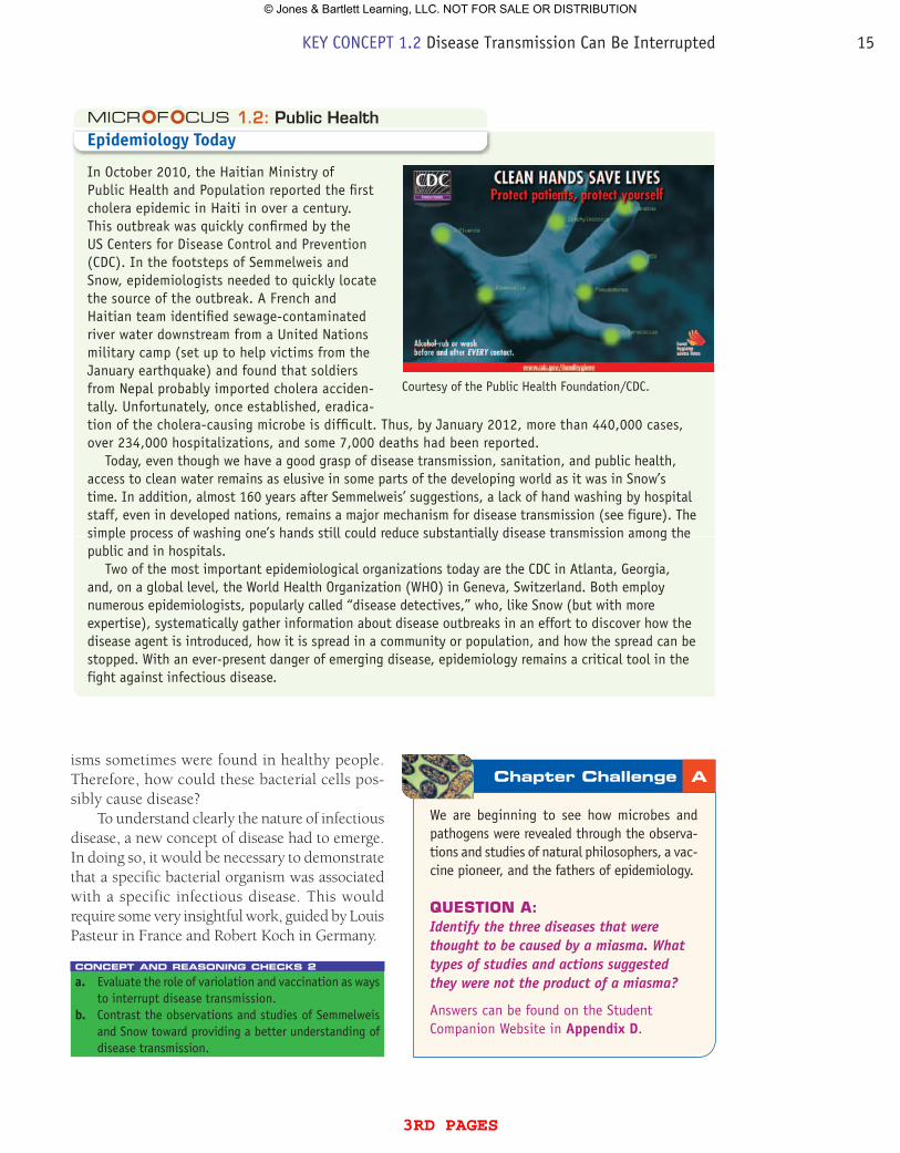
In 1854, a cholera epidemic hit London’s Soho district. With residents dying, English surgeon John Snow set out to discover the source and rea-
son for cholera’s spread. He carried out one of the first thorough epidemiological studies by inter-viewing sick and healthy Londoners and plotting the location of each cholera case on a district map ( FiguRE 1.5 ). The results indicated most cholera cases were linked to a sewage-contaminated street pump from which many local residents obtained their drinking water.
Hypothesizing that the pump was the source of the cholera, Snow instituted the first known example of a public health measure to interrupt disease transmission—he requested the parish Board of Guardians to remove the street pump handle! Cholera cases dropped and again disease spread was broken by a simple procedure.
Snow went on to propose that cholera was not inhaled as a miasma but rather was waterborne. In fact, he asserted that “organized particles” caused cholera—another hypothesis that proved to be correct even though the causative agent would not be identified for another 29 years.
It is important to realize that although the miasma premise was incorrect, the fact that dis-ease was associated with bad air and filth led to new hygiene measures, such as cleaning streets, laying new sewer lines in cities, and improving working conditions. These changes helped usher in the Sanitary Movement and create the infra-structure for the public health systems we have today (MicroFocus 1.2).
The Stage is Set
During the early years of the 1800s, other events occurred that helped set the stage for the coming “germ revolution.” In the 1830s, advances were made in microscope optics that allowed better resolution of objects. This resulted in improved and more widespread observations of tiny living organisms, many of which resembled short sticks. In fact, in 1838 the German biologist Christian Ehrenberg suggested these “rod-like” looking organisms be called bacteria (bakter = “rod”).
The Swiss physician Jacob Henle reported in 1840 that living organisms could cause dis-ease. This was strengthened in 1854 by Filippo Pacini’s discovery of rod-shaped cholera bacte-ria in stool samples from cholera patients. Still, scientists debated whether bacterial organisms could cause disease because such living organ-
FiguRE 1.5 Blocking Disease Transmission. John Snow (inset) produced a map plotting all the cholera cases (black rectangles) in the London Soho district and observed a cluster near the Broad Street pump (circle). Collection of the University of Michigan Health System, Gift of Pfizer, Inc. (UMHS.26). Inset © National Library of Medicine. »» Why would removing the pump handle stop the spread of cholera?
14 CHAPTER 1 Microbiology: Then and Now
3rd pages
© Jones & Bartlett Learning, LLC. NOT FOR SALE OR DISTRIBUTION
1.2: Public HealthEpidemiology Today
In October 2010, the Haitian Ministry of Public Health and Population reported the first cholera epidemic in Haiti in over a century. This outbreak was quickly confirmed by the US Centers for Disease Control and Prevention (CDC). In the footsteps of Semmelweis and Snow, epidemiologists needed to quickly locate the source of the outbreak. A French and Haitian team identified sewage-contaminated river water downstream from a United Nations military camp (set up to help victims from the January earthquake) and found that soldiers from Nepal probably imported cholera acciden-tally. Unfortunately, once established, eradica-tion of the cholera-causing microbe is difficult. Thus, by January 2012, more than 440,000 cases, over 234,000 hospitalizations, and some 7,000 deaths had been reported.
Today, even though we have a good grasp of disease transmission, sanitation, and public health, access to clean water remains as elusive in some parts of the developing world as it was in Snow’s time. In addition, almost 160 years after Semmelweis’ suggestions, a lack of hand washing by hospital staff, even in developed nations, remains a major mechanism for disease transmission (see figure). The simple process of washing one’s hands still could reduce substantially disease transmission among the public and in hospitals.
Two of the most important epidemiological organizations today are the CDC in Atlanta, Georgia, and, on a global level, the World Health Organization (WHO) in Geneva, Switzerland. Both employ numerous epidemiologists, popularly called “disease detectives,” who, like Snow (but with more expertise), systematically gather information about disease outbreaks in an effort to discover how the disease agent is introduced, how it is spread in a community or population, and how the spread can be stopped. With an ever-present danger of emerging disease, epidemiology remains a critical tool in the fight against infectious disease.
isms sometimes were found in healthy people. Therefore, how could these bacterial cells pos-sibly cause disease?
To understand clearly the nature of infectious disease, a new concept of disease had to emerge. In doing so, it would be necessary to demonstrate that a specific bacterial organism was associated with a specific infectious disease. This would require some very insightful work, guided by Louis Pasteur in France and Robert Koch in Germany.
concept and reasoning checks 2a. Evaluate the role of variolation and vaccination as ways
to interrupt disease transmission.b. Contrast the observations and studies of Semmelweis
and Snow toward providing a better understanding of disease transmission.
We are beginning to see how mic robes and pathogens were revealed through the observa-tions and studies of natural philosophers, a vac-cine pioneer, and the fathers of epidemiology.
QuEsTiON A:Identify the three diseases that were thought to be caused by a miasma. What types of studies and actions suggested they were not the product of a miasma?
Answers can be found on the Student Companion Website in Appendix D.
Chapter Challenge A
Courtesy of the Public Health Foundation/CDC.
KEY CONCEPT 1.2 Disease Transmission Can Be Interrupted 15
3rd pages
© Jones & Bartlett Learning, LLC. NOT FOR SALE OR DISTRIBUTION
Beginning around 1854, the association of microbes in the disease process blossomed and continued until the advent of World War I. Over these 60 years, the foundations were laid for the maturing process that has led to the modern science of microbiol-ogy. We refer to this period as the first, or classical, Golden Age of microbiology.
Louis Pasteur Proposes That germs Cause infectious Disease
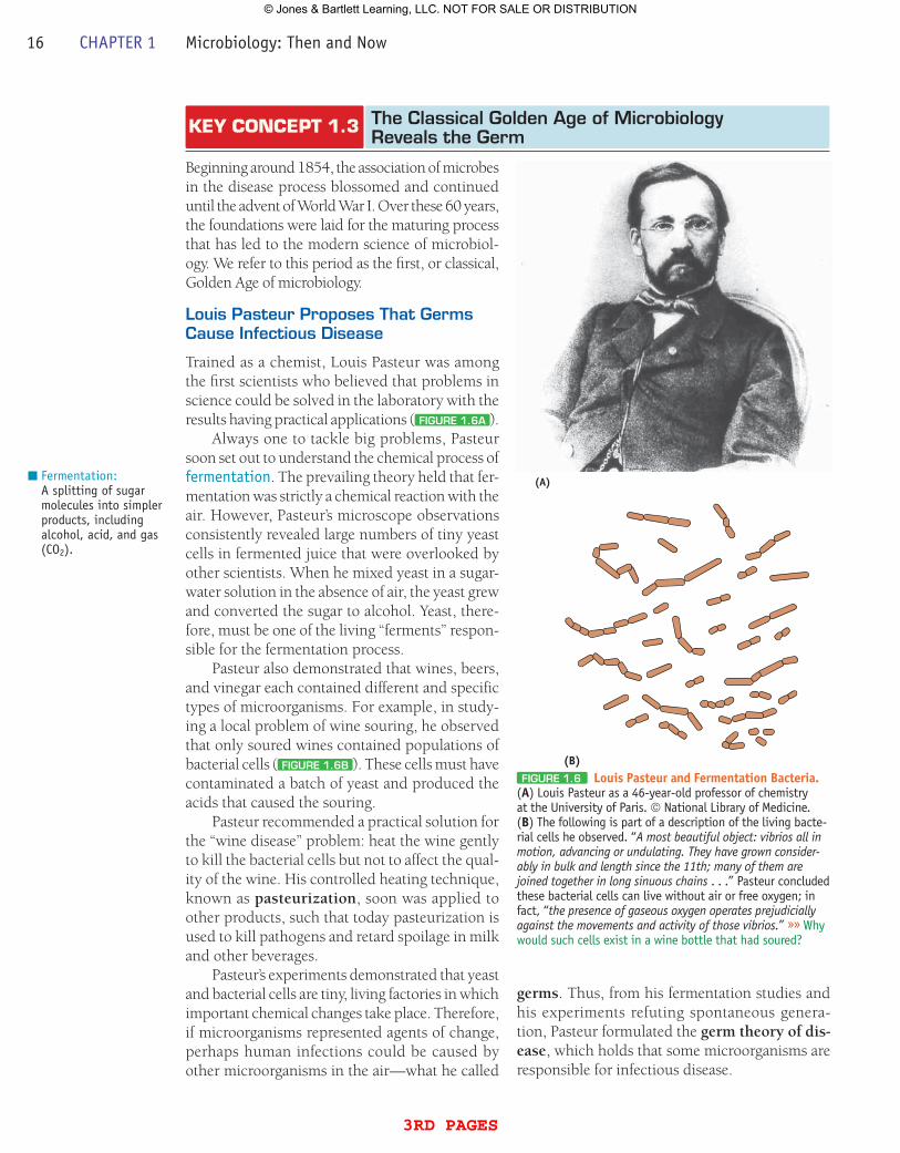
Trained as a chemist, Louis Pasteur was among the first scientists who believed that problems in science could be solved in the laboratory with the results having practical applications ( FiguRE 1.6A ).
Always one to tackle big problems, Pasteur soon set out to understand the chemical process of fermentation. The prevailing theory held that fer-mentation was strictly a chemical reaction with the air. However, Pasteur’s microscope observations consistently revealed large numbers of tiny yeast cells in fermented juice that were overlooked by other scientists. When he mixed yeast in a sugar-water solution in the absence of air, the yeast grew and converted the sugar to alcohol. Yeast, there-fore, must be one of the living “ferments” respon-sible for the fermentation process.
Pasteur also demonstrated that wines, beers, and vinegar each contained different and specific types of microorganisms. For example, in study-ing a local problem of wine souring, he observed that only soured wines contained populations of bacterial cells ( FiguRE 1.6B ). These cells must have contaminated a batch of yeast and produced the acids that caused the souring.
Pasteur recommended a practical solution for the “wine disease” problem: heat the wine gently to kill the bacterial cells but not to affect the qual-ity of the wine. His controlled heating technique, known as pasteurization, soon was applied to other products, such that today pasteurization is used to kill pathogens and retard spoilage in milk and other beverages.
Pasteur’s experiments demonstrated that yeast and bacterial cells are tiny, living factories in which important chemical changes take place. Therefore, if microorganisms represented agents of change, perhaps human infections could be caused by other microorganisms in the air—what he called
Fermentation:A splitting of sugar molecules into simpler products, including alcohol, acid, and gas (CO2).
KEY CONCEPT 1.3 The Classical golden Age of Microbiology Reveals the germ
(A)
(B)FiguRE 1.6 Louis pasteur and Fermentation Bacteria.
(A) Louis Pasteur as a 46-year-old professor of chemistry at the University of Paris. © National Library of Medicine. (B) The following is part of a description of the living bacte-rial cells he observed. “A most beautiful object: vibrios all in motion, advancing or undulating. They have grown consider-ably in bulk and length since the 11th; many of them are joined together in long sinuous chains . . .” Pasteur concluded these bacterial cells can live without air or free oxygen; in fact, “the presence of gaseous oxygen operates prejudicially against the movements and activity of those vibrios.” »» Why would such cells exist in a wine bottle that had soured?
germs. Thus, from his fermentation studies and his experiments refuting spontaneous genera-tion, Pasteur formulated the germ theory of dis-ease, which holds that some microorganisms are responsible for infectious disease.
16 CHAPTER 1 Microbiology: Then and Now
3rd pages
© Jones & Bartlett Learning, LLC. NOT FOR SALE OR DISTRIBUTION

Pasteur’s Work Stimulates Disease Control and Reinforces Disease Causation
Pasteur had reasoned that if germs were acquired from the environment, their spread could be controlled and the chain of disease transmission broken.
Joseph Lister was Professor of Surgery at Glasgow Royal Infirmary in Scotland, where more than half his amputation patients died—not from the surgery—but rather from postopera-tive infections. Hearing of Pasteur’s germ theory, Lister hypothesized that these surgical infections resulted from germs in the air. Knowing that car-bolic acid had been effective on sewage control, in 1865 he used a carbolic acid spray in surgery and on surgical wounds ( FiguRE 1.7 ). The result was spectacular—the wounds healed without infec-tion. His technique would soon not only revolu-tionize medicine and the practice of surgery, but also lead to the practice of antisepsis, the use of chemical methods for disinfection of external liv-ing surfaces, such as the skin. So, germs can come from the environment and they can be controlled.
In an effort to familiarize himself with bio-logical problems, Pasteur had the opportunity to study pébrine, a disease of silkworms. After several setbacks, he finally identified a new type of germ, unlike the bacterial cells and yeast he had observed with his microscope. These tiny globules, called “corpuscular parasites” were the infectious agent in silkworms and on the mulberry leaves fed to the worms. By separating the healthy silkworms from the diseased silkworms and their food, he managed to quell the spread of disease. The identification of the pathogen was crucial to supporting the germ theory and Pasteur would never again doubt the ability of germs to cause infectious disease. Now infectious disease would be his only interest.
In 1865, cholera engulfed Paris, killing 200 people a day. Pasteur tried to capture the responsible pathogen by filtering the hospital air and trapping the bacterial cells in cotton. Unfortunately, Pasteur could not grow or sepa-rate one bacterial species apart from the others because his broth cultures allowed the organisms to mix freely. Although Pasteur demonstrated that bacterial inoculations made animals ill, he could not pinpoint an exact cause.
To completely validate the germ theory, what was missing was the ability to isolate a specific
germ from a diseased individual and demonstrate the isolated germ caused the same disease.
Robert Koch Formalizes Standards to Equate germs with infectious Disease
Robert Koch ( FiguRE 1.8A ) was a German country doctor who was well aware of anthrax, a deadly disease that periodically ravaged cattle and sheep, and could cause disease in humans.
In 1875, Koch injected mice with the blood from such diseased sheep and cattle. He then per-formed meticulous autopsies and noted the same symptoms in the mice that had appeared in the sheep and cattle. Next, he isolated from the blood a few rod-shaped bacterial cells and, with his micro-scope, watched for hours as the bacterial cells mul-tiplied, formed tangled threads, and finally reverted to spores. He then took several spores on a sliver of wood and injected them into healthy mice. The symptoms of anthrax soon appeared and when Koch autopsied the animal, he found their blood swarming with the same bacterial cells. He reiso-lated the cells in fresh aqueous humor. The cycle was now complete. Here was the first evidence that a specific germ was the causative agent of anthrax.
Growing bacterial cells was not very conve-nient. Then, in 1880, Koch observed a slice of potato on which small masses of bacterial cells, which he termed colonies, were growing and multiplying. So, Koch tried adding gelatin to his broth to prepare a similar solid culture surface. He then inoculated bacterial cells on the surface and set the dish aside to incubate. Within 24 hours,
FiguRE 1.7 Lister and Antisepsis. By 1870, Joseph Lister (inset) and his students were using a carbolic acid spray in surgery and on surgical wounds to prevent postopera-tive infections. © Mary Evans Picture Library/Alamy Images. Inset © National Library of Medicine. »» Hypothesize how carbolic acid prevented surgical infections.
KEY CONCEPT 1.3 The Classical Golden Age of Microbiology Reveals the Germ 17
3rd pages
© Jones & Bartlett Learning, LLC. NOT FOR SALE OR DISTRIBUTION
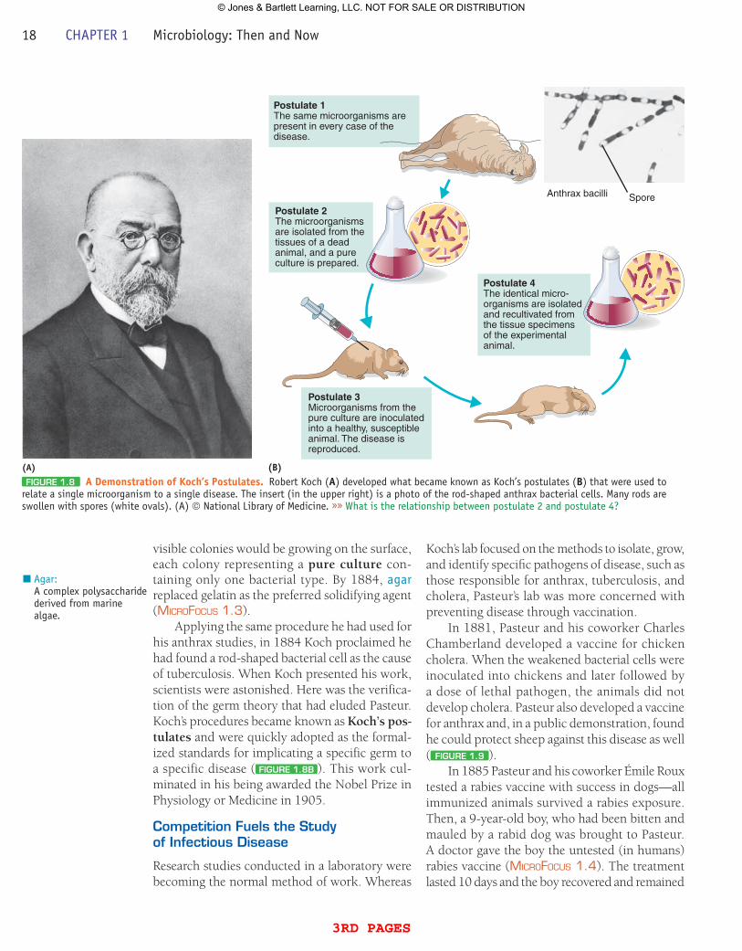
visible colonies would be growing on the surface, each colony representing a pure culture con-taining only one bacterial type. By 1884, agar replaced gelatin as the preferred solidifying agent (MicroFocus 1.3).
Applying the same procedure he had used for his anthrax studies, in 1884 Koch proclaimed he had found a rod-shaped bacterial cell as the cause of tuberculosis. When Koch presented his work, scientists were astonished. Here was the verifica-tion of the germ theory that had eluded Pasteur. Koch’s procedures became known as Koch’s pos-tulates and were quickly adopted as the formal-ized standards for implicating a specific germ to a specific disease ( FiguRE 1.8B ). This work cul-minated in his being awarded the Nobel Prize in Physiology or Medicine in 1905.
Competition Fuels the Study of infectious Disease
Research studies conducted in a laboratory were becoming the normal method of work. Whereas
Agar:A complex polysaccharide derived from marine algae.
Koch’s lab focused on the methods to isolate, grow, and identify specific pathogens of disease, such as those responsible for anthrax, tuberculosis, and cholera, Pasteur’s lab was more concerned with preventing disease through vaccination.
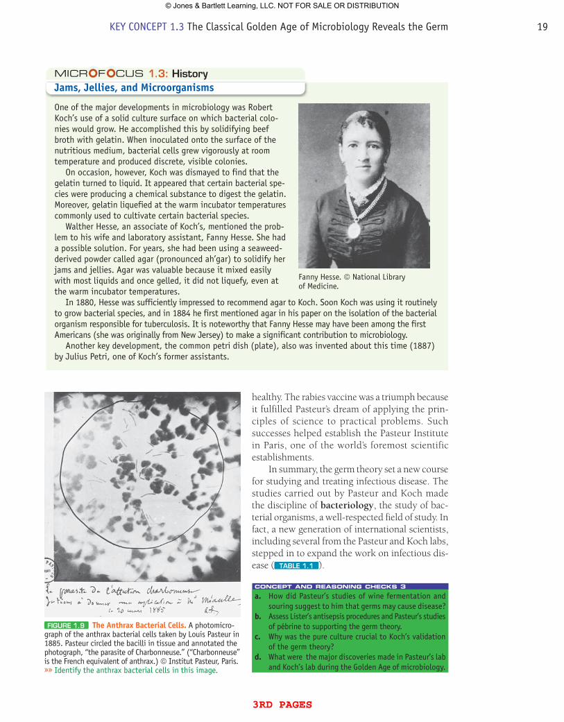
In 1881, Pasteur and his coworker Charles Chamberland developed a vaccine for chicken cholera. When the weakened bacterial cells were inoculated into chickens and later followed by a dose of lethal pathogen, the animals did not develop cholera. Pasteur also developed a vaccine for anthrax and, in a public demonstration, found he could protect sheep against this disease as well ( FiguRE 1.9 ).
In 1885 Pasteur and his coworker Émile Roux tested a rabies vaccine with success in dogs—all immunized animals survived a rabies exposure. Then, a 9-year-old boy, who had been bitten and mauled by a rabid dog was brought to Pasteur. A doctor gave the boy the untested (in humans) rabies vaccine (MicroFocus 1.4). The treatment lasted 10 days and the boy recovered and remained
(A)
Postulate 4The identical micro-organisms are isolatedand recultivated from the tissue specimens of the experimentalanimal.
Postulate 1The same microorganisms arepresent in every case of the disease.
Postulate 2The microorganisms are isolated from the tissues of a deadanimal, and a pure culture is prepared.
Postulate 3Microorganisms from the pure culture are inoculated into a healthy, susceptible animal. The disease is reproduced.
Anthrax bacilli Spore
(B)FiguRE 1.8 A Demonstration of Koch’s postulates. Robert Koch (A) developed what became known as Koch’s postulates (B) that were used to
relate a single microorganism to a single disease. The insert (in the upper right) is a photo of the rod-shaped anthrax bacterial cells. Many rods are swollen with spores (white ovals). (A) © National Library of Medicine. »» What is the relationship between postulate 2 and postulate 4?
18 CHAPTER 1 Microbiology: Then and Now
3rd pages
© Jones & Bartlett Learning, LLC. NOT FOR SALE OR DISTRIBUTION
healthy. The rabies vaccine was a triumph because it fulfilled Pasteur’s dream of applying the prin-ciples of science to practical problems. Such successes helped establish the Pasteur Institute in Paris, one of the world’s foremost scientific establishments.
In summary, the germ theory set a new course for studying and treating infectious disease. The studies carried out by Pasteur and Koch made the discipline of bacteriology, the study of bac-terial organisms, a well-respected field of study. In fact, a new generation of international scientists, including several from the Pasteur and Koch labs, stepped in to expand the work on infectious dis-ease ( TABLE 1.1 ).
concept and reasoning checks 3a. How did Pasteur’s studies of wine fermentation and
souring suggest to him that germs may cause disease?b. Assess Lister’s antisepsis procedures and Pasteur’s studies
of pébrine to supporting the germ theory.c. Why was the pure culture crucial to Koch’s validation
of the germ theory?d. What were the major discoveries made in Pasteur’s lab
and Koch’s lab during the Golden Age of microbiology.
1.3: HistoryJams, Jellies, and Microorganisms
One of the major developments in microbiology was Robert Koch’s use of a solid culture surface on which bacterial colo-nies would grow. He accomplished this by solidifying beef broth with gelatin. When inoculated onto the surface of the nutritious medium, bacterial cells grew vigorously at room temperature and produced discrete, visible colonies.
On occasion, however, Koch was dismayed to find that the gelatin turned to liquid. It appeared that certain bacterial spe-cies were producing a chemical substance to digest the gelatin. Moreover, gelatin liquefied at the warm incubator temperatures commonly used to cultivate certain bacterial species.
Walther Hesse, an associate of Koch’s, mentioned the prob-lem to his wife and laboratory assistant, Fanny Hesse. She had a possible solution. For years, she had been using a seaweed-derived powder called agar (pronounced ah’gar) to solidify her jams and jellies. Agar was valuable because it mixed easily with most liquids and once gelled, it did not liquefy, even at the warm incubator temperatures.
In 1880, Hesse was sufficiently impressed to recommend agar to Koch. Soon Koch was using it routinely to grow bacterial species, and in 1884 he first mentioned agar in his paper on the isolation of the bacterial organism responsible for tuberculosis. It is noteworthy that Fanny Hesse may have been among the first Americans (she was originally from New Jersey) to make a significant contribution to microbiology.
Another key development, the common petri dish (plate), also was invented about this time (1887) by Julius Petri, one of Koch’s former assistants.
Fanny Hesse. © National Library of Medicine.
FiguRE 1.9 The Anthrax Bacterial Cells. A photomicro-graph of the anthrax bacterial cells taken by Louis Pasteur in 1885. Pasteur circled the bacilli in tissue and annotated the photograph, “the parasite of Charbonneuse.” (“Charbonneuse” is the French equivalent of anthrax.) © Institut Pasteur, Paris. »» Identify the anthrax bacterial cells in this image.
KEY CONCEPT 1.3 The Classical Golden Age of Microbiology Reveals the Germ 19
3rd pages
© Jones & Bartlett Learning, LLC. NOT FOR SALE OR DISTRIBUTION
Although the list of identified microbes was grow-ing, the agents responsible for diseases such as measles, mumps, smallpox, and yellow fever con-tinued to elude identification.
Other global Pioneers Contribute to New Disciplines in Microbiology
In 1892, a Russian scientist, Dimitri Ivanowsky, used a ceramic filter developed by Pasteur’s group to trap what he thought were bacterial cells respon-sible for tobacco mosaic disease, which produces mottled and stunted tobacco leaves. Surprisingly, Ivanowsky discovered that when he applied the liquid that passed through the filter to healthy tobacco plants, the leaves became mottled and stunted. Ivanowsky, not understanding the sig-nificance of this, simply assumed bacterial cells somehow had slipped through the filter.
Unaware of Ivanowsky’s work, Martinus Beijerinck, a Dutch investigator, did similar experi-ments in 1898 but suggested tobacco mosaic disease was a “contagious, living liquid” that acted like a poison or virus (virus = “poison”). Also in 1898, the causative agent for an animal disease—hoof-and-mouth disease—was found to be another filterable liquid, and in 1901 American Walter Reed concluded that the agent responsible for yellow fever in humans also was a virus. With these discoveries, the discipline of virology, the study of viruses, was launched.
While scientists like Pasteur and Koch were investigating the bacterial contribution to the infectious disease process, others were identify-ing other types of disease-causing microbes. That fungi could cause plant diseases was known since 1767 and such diseases were studied extensively by Anton de Bary in the 1860s. As already men-tioned in this chapter, Pasteur identified the role of fungal yeasts (first seen by Leeuwenhoek) with fermentation. Importantly, the recognition that some fungi were linked to human skin diseases was proposed as early as 1841 when a Hungarian physician, David Gruby, discovered a fungus asso-ciated with human scalp infections.
The realization that infectious disease could be caused by yet another group of microbes, the protozoa (again first seen by Leeuwenhoek), was another major milestone in understanding infectious disease. In fact, Pasteur’s “corpuscu-lar parasites” of pébrine were protozoa. Other advances in the study of these types of microbes were dependent on studies in tropical medicine. Major advances in understanding these microbes included Charles Laveran’s discovery (1880) that the protozoan parasite causing malaria could be found in human blood and David Bruce’s studies (1903) that another protozoan parasite was the agent of human sleeping sickness. These and many other investigations with the fungi and protozoa
1.4: HistoryThe private pasteur
The notebooks of Louis Pasteur had been an enduring mystery of science ever since the scientist him-self requested his family not to show them to anyone. But in 1964, Pasteur’s last surviving grandson donated the notebooks to the National Library in Paris, and after soul-searching for a decade, the directors made them available to a select group of scholars. Among the group was Gerald Geison of Princeton University. What Geison found stripped away part of the veneration conferred on Pasteur and showed another side to his work.
In 1881, Pasteur conducted a trial of his new anthrax vaccine by inoculating half a flock of animals with the vaccine, then exposing the entire flock to the disease. When the vaccinated half survived, Pasteur was showered with accolades. However, Pasteur’s notebooks, according to Geison, reveal that he had prepared the vaccine not by his own method, but by a competitor’s.
Pasteur also apparently sidestepped established protocols when he inoculated two boys with a rabies vaccine before it was tested on animals. Fortunately, the two boys survived, possibly because they were not actually infected or because the vaccine was, indeed, safe and effective. Nevertheless, the untested treatment should not have been used, says Geison. His book, The Private Science of Louis Pasteur (Princeton University Press, 1995) places the scientist in a more realistic light and shows that today’s pressures to succeed in research are little different than they were more than a century ago.
KEY CONCEPT 1.4 With the Discovery of Other Microbes, the Microbial World Expands
20 CHAPTER 1 Microbiology: Then and Now
3rd pages
© Jones & Bartlett Learning, LLC. NOT FOR SALE OR DISTRIBUTION
TABLE
TABLE
TABLE
TABLE TABLE
TABLE TABLE
TABLE
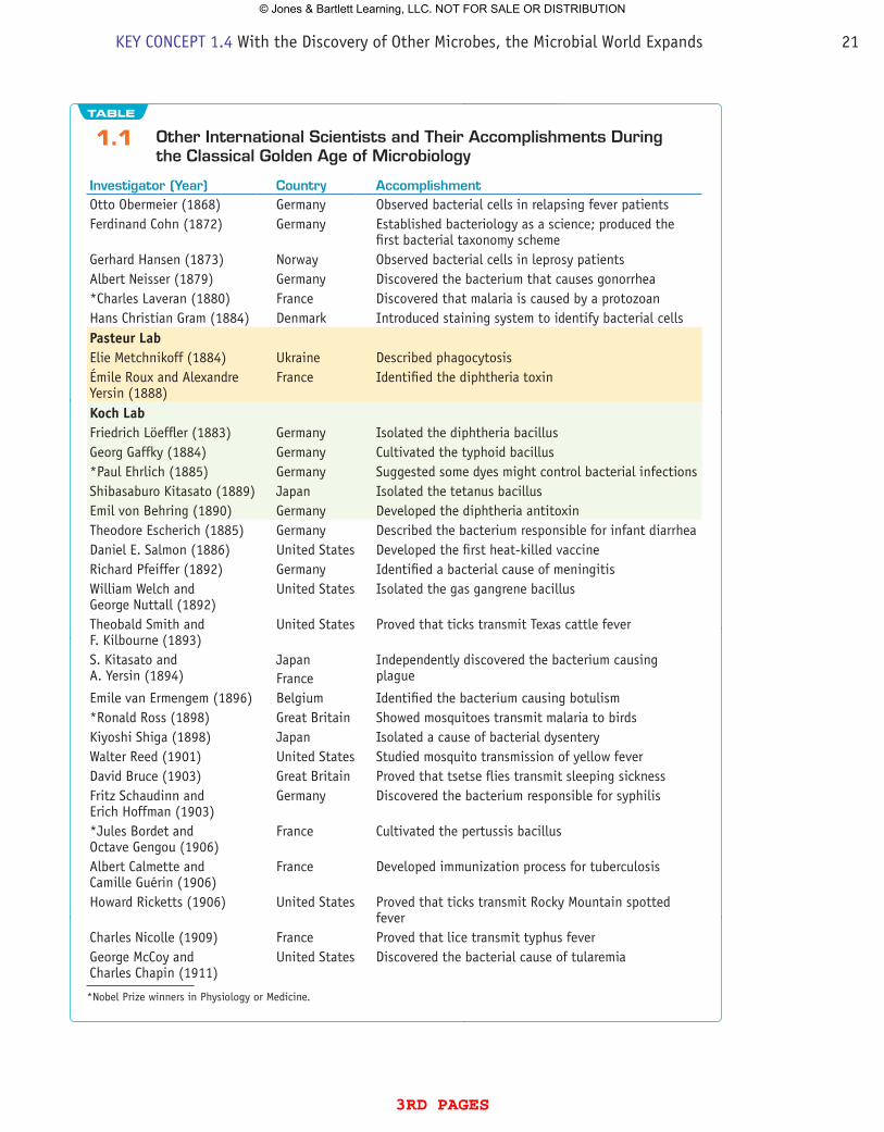
1.1 Other international Scientists and Their Accomplishments During the Classical golden Age of Microbiology
investigator (Year) Country AccomplishmentOtto Obermeier (1868) Germany Observed bacterial cells in relapsing fever patientsFerdinand Cohn (1872) Germany Established bacteriology as a science; produced the
first bacterial taxonomy schemeGerhard Hansen (1873) Norway Observed bacterial cells in leprosy patientsAlbert Neisser (1879) Germany Discovered the bacterium that causes gonorrhea*Charles Laveran (1880) France Discovered that malaria is caused by a protozoanHans Christian Gram (1884) Denmark Introduced staining system to identify bacterial cellspasteur LabElie Metchnikoff (1884) Ukraine Described phagocytosisÉmile Roux and Alexandre Yersin (1888)
France Identified the diphtheria toxin
Koch LabFriedrich Löeffler (1883) Germany Isolated the diphtheria bacillusGeorg Gaffky (1884) Germany Cultivated the typhoid bacillus*Paul Ehrlich (1885) Germany Suggested some dyes might control bacterial infectionsShibasaburo Kitasato (1889) Japan Isolated the tetanus bacillusEmil von Behring (1890) Germany Developed the diphtheria antitoxinTheodore Escherich (1885) Germany Described the bacterium responsible for infant diarrheaDaniel E. Salmon (1886) United States Developed the first heat-killed vaccineRichard Pfeiffer (1892) Germany Identified a bacterial cause of meningitisWilliam Welch and George Nuttall (1892)
United States Isolated the gas gangrene bacillus
Theobald Smith and F. Kilbourne (1893)
United States Proved that ticks transmit Texas cattle fever
S. Kitasato and A. Yersin (1894)
JapanFrance
Independently discovered the bacterium causing plague
Emile van Ermengem (1896) Belgium Identified the bacterium causing botulism*Ronald Ross (1898) Great Britain Showed mosquitoes transmit malaria to birdsKiyoshi Shiga (1898) Japan Isolated a cause of bacterial dysenteryWalter Reed (1901) United States Studied mosquito transmission of yellow feverDavid Bruce (1903) Great Britain Proved that tsetse flies transmit sleeping sicknessFritz Schaudinn and Erich Hoffman (1903)
Germany Discovered the bacterium responsible for syphilis
*Jules Bordet and Octave Gengou (1906)
France Cultivated the pertussis bacillus
Albert Calmette and Camille Guérin (1906)
France Developed immunization process for tuberculosis
Howard Ricketts (1906) United States Proved that ticks transmit Rocky Mountain spotted fever
Charles Nicolle (1909) France Proved that lice transmit typhus feverGeorge McCoy and Charles Chapin (1911)
United States Discovered the bacterial cause of tularemia
*Nobel Prize winners in Physiology or Medicine.
KEY CONCEPT 1.4 With the Discovery of Other Microbes, the Microbial World Expands 21
3rd pages
© Jones & Bartlett Learning, LLC. NOT FOR SALE OR DISTRIBUTION
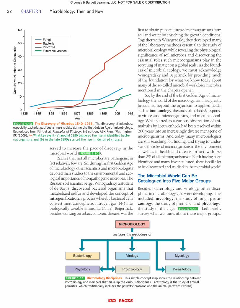
served to increase the pace of discovery in the microbial world ( FiguRE 1.10 ).
Realize that not all microbes are pathogens; in fact relatively few are. So, during the first Golden Age of microbiology, other scientists and microbiologists devoted their studies to the environmental and eco-logical importance of nonpathogenic microbes. The Russian soil scientist Sergei Winogradsky, a student of de Bary’s, discovered bacterial organisms that metabolized sulfur and developed the concept of nitrogen fixation, a process whereby bacterial cells convert inert atmospheric nitrogen gas (N2) into biologically useable ammonia (NH3). Beijerinck, besides working on tobacco mosaic disease, was the
first to obtain pure cultures of microorganisms from soil and water by enriching the growth conditions. Together with Winogradsky, they developed many of the laboratory methods essential to the study of microbial ecology, while revealing the physiological significance of soil microbes and discovering the essential roles such microorganisms play in the recycling of matter on a global scale. As the found-ers of microbial ecology, we must acknowledge Winogradsky and Beijerinck for providing much of the foundation for what we know today about many of the so-called microbial workforce microbes mentioned in the chapter opener.
So, by the end of the first Golden Age of micro-biology, the world of the microorganism had greatly broadened beyond the organism to applied fields, such as immunology, the study of the body’s response to viruses and microorganisms, and microbial ecol-ogy. What started as a curious observation of ani-malcules by Leeuwenhoek had been resolved within 200 years into an increasingly diverse menagerie of microorganisms. And today, many microbiologists are still searching for, finding, and trying to under-stand the roles of microorganisms in the environment as well as in health and disease. In fact, with less than 2% of all microorganisms on Earth having been identified and many fewer cultured, there is still a lot to be discovered and studied in the microbial world!
The Microbial World Can Be Catalogued into Five Major groups
Besides bacteriology and virology, other disci-plines in microbiology also were developing. This included: mycology, the study of fungi; proto-zoology, the study of protozoa; and phycology, the study of the algae ( FiguRE 1.11 ). Let’s briefly survey what we know about these major groups.
FiguRE 1.10 The Discovery of Microbes 1840–1915. The discovery of microbes, especially bacterial pathogens, rose rapidly during the first Golden Age of microbiology. Reproduced from Flint et al, Principles of Virology, 3rd edition, ASM Press, Washington DC (2009). »» What key event (a) around 1880 triggered the rise in identified bacte-rial organisms and (b) in the late 1890s started the rise in identified viruses?
FungiBacteriaProtozoaFilterable viruses
60
50
40
30
20
10
01835 1845 1855 1865 1875
Year
Cum
ulat
ive
Num
ber o
f Dis
cove
ries
1885 1895 1905 1915
FiguRE 1.11 Microbiology Disciplines. This simple concept map shows the relationship between microbiology and members that make up the various disciplines. Parasitology is the study of animal parasites, which traditionally includes the parasitic protozoa and the animal parasites (worms).
MICROBIOLOGY
includes the disciplines of
Bacteriology Virology Mycology
Phycology Protozoology Parasitology
22 CHAPTER 1 Microbiology: Then and Now
3rd pages
© Jones & Bartlett Learning, LLC. NOT FOR SALE OR DISTRIBUTION
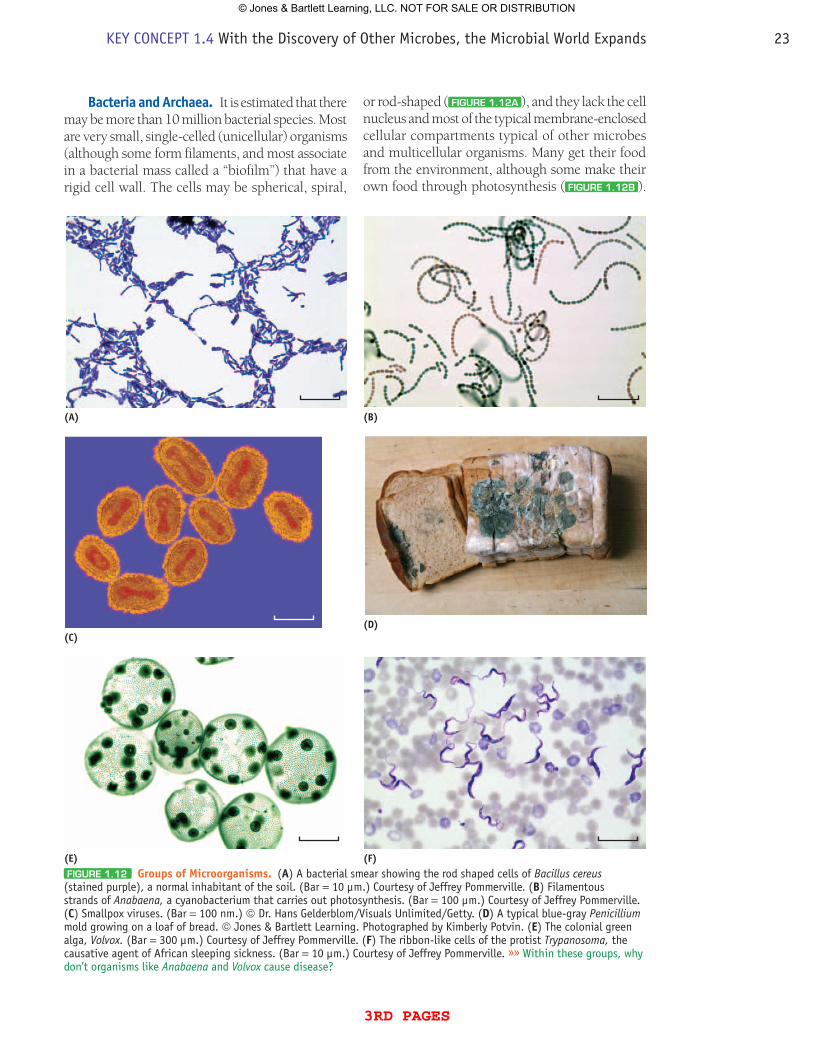
Bacteria and Archaea. It is estimated that there may be more than 10 million bacterial species. Most are very small, single-celled (unicellular) organisms (although some form filaments, and most associate in a bacterial mass called a “biofilm”) that have a rigid cell wall. The cells may be spherical, spiral,
or rod-shaped ( FiguRE 1.12A ), and they lack the cell nucleus and most of the typical membrane-enclosed cellular compartments typical of other microbes and multicellular organisms. Many get their food from the environment, although some make their own food through photosynthesis ( FiguRE 1.12B ).
(A) (B)
(C)(D)
(E) (F)FiguRE 1.12 Groups of Microorganisms. (A) A bacterial smear showing the rod shaped cells of Bacillus cereus (stained purple), a normal inhabitant of the soil. (Bar = 10 µm.) Courtesy of Jeffrey Pommerville. (B) Filamentous strands of Anabaena, a cyanobacterium that carries out photosynthesis. (Bar = 100 µm.) Courtesy of Jeffrey Pommerville. (C) Smallpox viruses. (Bar = 100 nm.) © Dr. Hans Gelderblom/Visuals Unlimited/Getty. (D) A typical blue-gray Penicillium mold growing on a loaf of bread. © Jones & Bartlett Learning. Photographed by Kimberly Potvin. (E) The colonial green alga, Volvox. (Bar = 300 µm.) Courtesy of Jeffrey Pommerville. (F) The ribbon-like cells of the protist Trypanosoma, the causative agent of African sleeping sickness. (Bar = 10 µm.) Courtesy of Jeffrey Pommerville. »» Within these groups, why don’t organisms like Anabaena and Volvox cause disease?
KEY CONCEPT 1.4 With the Discovery of Other Microbes, the Microbial World Expands 23
3rd pages
© Jones & Bartlett Learning, LLC. NOT FOR SALE OR DISTRIBUTION
Bacterial cells are found in most all environments, making up a large percentage of the Earth’s microbial workforce.
Besides the disease-causing members, some are responsible for food spoilage while others are use-ful in the food industry. Many bacterial members, along with several fungi, are decomposers, organ-isms that recycle nutrients from dead organisms.
Based on recent biochemical and molecular studies, many bacterial organisms have been reas-signed into another evolutionary group, called the Archaea. Although they look like bacterial cells, many grow in environments that are extremely hot (such as the Yellowstone hot springs), extremely salty (such as the Dead Sea), or of extremely low pH (such as acid mine drainage). Surviving in these environments has brought about many evolution-ary adaptations and changes to their cell structure and chemical composition. As such, no archaeal members are known to be pathogens. In fact, many normally grow in soils and water, and are an inte-gral part of animal digestive tracts.
Viruses. Although not correctly labeled as microorganisms, currently there are more than 3,600 known types of viruses. Viruses are not cel-lular and cannot be grown in pure culture. They have a core of nucleic acid (DNA or RNA) sur-rounded by a protein coat. Among the features used to identify viruses are morphology (size, shape), genetic material (RNA, DNA), and bio-logical properties (organism or tissue infected).
Viruses infect organisms for one reason only—to replicate. Viruses in the air or water, for example, cannot replicate because they need the metabolic machinery and chemical building blocks found inside living cells. Of the known viruses, only a small percentage causes disease in humans. Polio, the flu, measles, AIDS, and smallpox are examples ( FiguRE 1.12C ).
The other groups of microbes have a cell nucleus and a variety of internal, membrane-bound cellular compartments.
Fungi. The fungi include the unicellular yeasts and the multicellular mushrooms and molds ( FiguRE 1.12D ). About 100,000 species of fungi have been described; however, there may be as many as 1.5 million species in nature.
Other than the yeasts, molds tend to grow as filaments with unique rigid cell walls. Most fungi grow best in warm, moist places and secrete diges-tive enzymes that break down nutrients into smaller
bits that can be absorbed easily across a rigid cell wall. Fungi thus live in their own food supply. If that “food supply” is a human, diseases such as ringworm or vaginal yeast infections may result.
For the pharmaceutical industry, some fungi are sources for useful products, such as antibiotics. Others are used in the food industry to impart dis-tinctive flavors in foods such as Roquefort cheeses. Together with many bacterial species, numerous molds play a major role as decomposers.
protists. The protists consist mostly of single-celled algae and protozoa. Some are free- living while others live in association with plants or animals. Movement, if present, is achieved by flagella or cilia, or by a crawling movement.
Protists obtain nutrients in different ways. Some absorb nutrients from the surrounding environment or ingest smaller microorganisms. The unicellular, colonial, and filamentous algae have a rigid cell wall and can carry out photo-synthesis ( FiguRE 1.12E ). The aquatic protists also provide energy and organic compounds for the lower trophic levels of the food web. Some protists (the protozoa) are capable of causing diseases in animals, including humans; these include malaria, several types of diarrhea, and sleeping sickness ( FiguRE 1.12F ).concept and reasoning checks 4a. Describe how viruses were discovered as disease-causing
agents.b. What significant discoveries added the fungi and pro-
tozoa to the growing list of microbes?c. Judge the significance of the pioneering studies carried
out by Winogradsky and Beijerinck.d. Identify which of the microbial groups were originally
seen by Leeuwenhoek.
With the end of the first Golden Age of micro-biology, all the major groups of microbes had been identified.
QuEsTiON B:Briefly recount how each group of microbes in the diverse global workforce was revealed and identify the characteristics that separate them into different groups.
Answers can be found on the Student Companion Website in Appendix D.
Chapter Challenge B
24 CHAPTER 1 Microbiology: Then and Now
3rd pages
© Jones & Bartlett Learning, LLC. NOT FOR SALE OR DISTRIBUTION
The 1940s brought the birth of molecular genet-ics to biology. Many biologists focused on under-standing the genetics of organisms, including the nature of the genetic material and its regulation.
Molecular Biology Relies on Microorganisms as Model Systems
In 1943, the Italian-born microbiologist Salvador Luria and the German physicist Max Delbrück carried out a series of experiments with bacterial cells and viruses that marked the second Golden Age of microbiology. They used a common gut-inhabiting bacterial organism, Escherichia coli, to address a basic question regarding evolution-ary biology: Do mutations occur spontaneously or does the environment induce them? Luria and Dulbrück showed that bacterial cells could develop spontaneous mutations that generate resistance to viral infection. Such uses of microbial model systems showed to other researchers that microorganisms could be used to study general principles of biology.
So, biologists were quick to jump on the “microbial bandwagon.” For example:
• Americans George Beadle and Edward Tatum (1941) use the fungus Neurospora to demonstrate that “one gene codes for one enzyme”;
• Oswald Avery, Colin MacLeod, and Maclyn McCarty (1944) use the bacterial organism Streptococcus pneumoniae to sug-gest that DNA is the genetic material in cells;
• Alfred Hershey and Martha Chase (1952) use a virus that infects bacterial cells to assert that DNA is the substance of the genetic material;
• Francis Crick (1958) uses E. coli and a virus to show how the DNA genetic code works to make individual proteins.
Again, microbes were at the forefront in answer-ing fundamental questions that applied to all of biology. In addition, during this second Golden Age, other studies illuminated the basic structure of microbes and led to one of the greatest breakthroughs (antibiotics) that would revolutionize medicine’s ability to treat and eliminate infectious disease.
Mutations:Permanent alterations in DNA base sequences.
Two Types of Cellular Organization Are Realized
The small size of bacterial cells hindered sci-entists’ abilities to confirm whether these cells were similar to other cellular organisms in orga-nization. In the 1940s and 1950s, a new type of microscope—the electron microscope—was being developed that could magnify objects and cells thousands of times better than typical light microscopes. With the electron microscope, for the first time bacterial cells were seen as being cellular like all other microbes, plants, and ani-mals. However, studies showed that they were organized in a structurally different way from other organisms.
It was known that animal and plant cells contained a cell nucleus that houses the genetic instructions in the form of chromosomes and was separated physically from other cell structures by a membrane envelope ( FiguRE 1.13A ). This type of cellular organization is called eukaryotic (eu = “true”; karyon = “nucleus”). Microscope observations of the protists and fungi had revealed that these organisms also have a eukaryotic orga-nization. Thus, not only are all plants and animals eukaryotes, so are the microorganisms that com-prise the fungi and protists.
Studies with the electron microscope revealed that bacterial (and archaeal) cells had few of the membranous compartments typical of eukaryotic cells. They lacked a cell nucleus, indicating the bacterial chromosome (DNA) was not sur-rounded by a membrane envelope ( FiguRE 1.13B ). Therefore, members of the Bacteria and Archaea have a prokaryotic (pro = “before”) type of cellular organization and represent prokaryotes. Importantly, there are also many differences between bacterial and archaeal cells, account-ing for their split into separate microbial groups. By the way, because viruses lack a cellular organization, they are neither prokaryotes nor eukaryotes.
Antibiotics Are used to Cure infectious Disease
In 1910, another coworker of Koch’s, Paul Ehrlich, synthesized the first “magic bullet”—a chemical
KEY CONCEPT 1.5 A Second golden Age of Microbiology involves the Birth of Molecular Biology and Chemotherapy
KEY CONCEPT 1.5 A Second Golden Age of Microbiology 25DQ1
3rd pages
© Jones & Bartlett Learning, LLC. NOT FOR SALE OR DISTRIBUTION
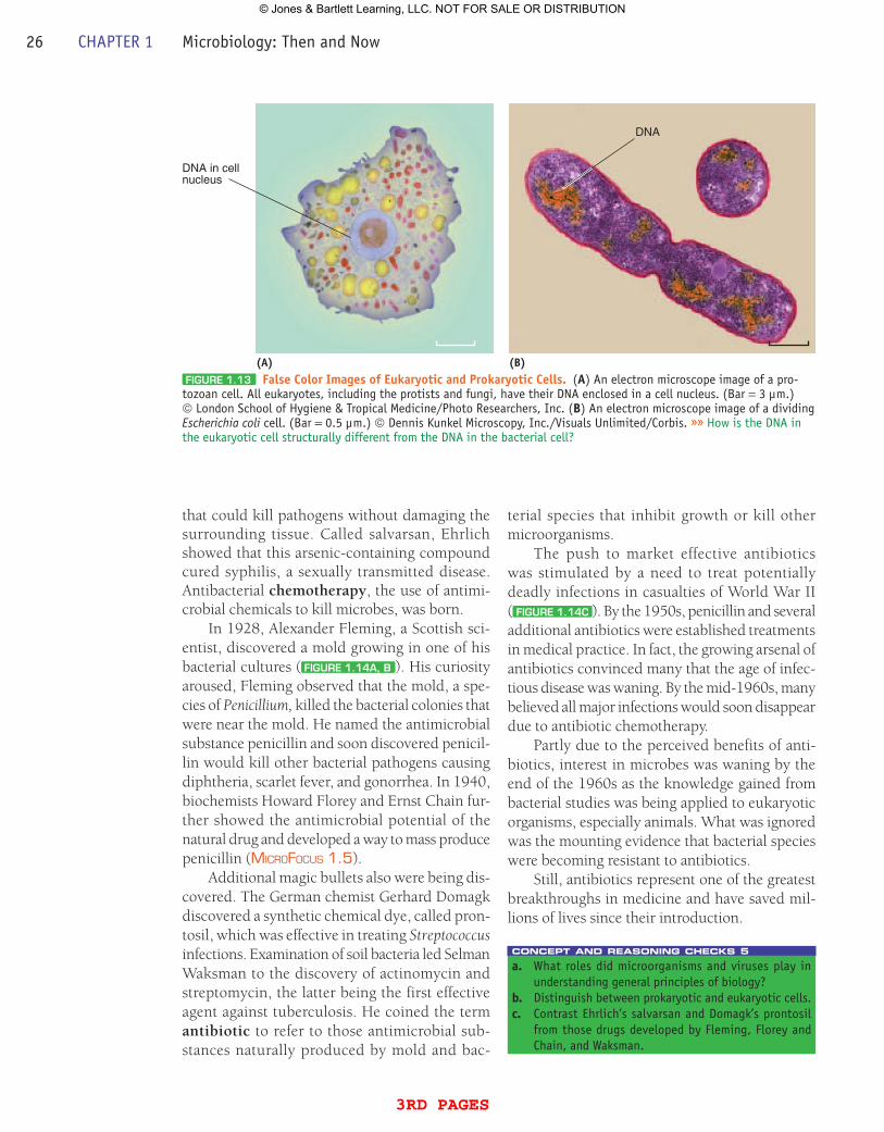
that could kill pathogens without damaging the surrounding tissue. Called salvarsan, Ehrlich showed that this arsenic-containing compound cured syphilis, a sexually transmitted disease. Antibacterial chemotherapy, the use of antimi-crobial chemicals to kill microbes, was born.
In 1928, Alexander Fleming, a Scottish sci-entist, discovered a mold growing in one of his bacterial cultures ( FiguRE 1.14A, B ). His curiosity aroused, Fleming observed that the mold, a spe-cies of Penicillium, killed the bacterial colonies that were near the mold. He named the anti microbial substance penicillin and soon discovered penicil-lin would kill other bacterial pathogens causing diphtheria, scarlet fever, and gonorrhea. In 1940, biochemists Howard Florey and Ernst Chain fur-ther showed the antimicrobial potential of the natural drug and developed a way to mass produce penicillin (MicroFocus 1.5).
Additional magic bullets also were being dis-covered. The German chemist Gerhard Domagk discovered a synthetic chemical dye, called pron-tosil, which was effective in treating Streptococcus infections. Examination of soil bacteria led Selman Waksman to the discovery of actino mycin and streptomycin, the latter being the first effective agent against tuberculosis. He coined the term antibiotic to refer to those antimicrobial sub-stances naturally produced by mold and bac-
terial species that inhibit growth or kill other microorganisms.
The push to market effective antibiotics was stimulated by a need to treat potentially deadly infections in casualties of World War II ( FiguRE 1.14C ). By the 1950s, penicillin and several additional antibiotics were established treatments in medical practice. In fact, the growing arsenal of antibiotics convinced many that the age of infec-tious disease was waning. By the mid-1960s, many believed all major infections would soon disappear due to antibiotic chemotherapy.
Partly due to the perceived benefits of anti-biotics, interest in microbes was waning by the end of the 1960s as the knowledge gained from bacterial studies was being applied to eukaryotic organisms, especially animals. What was ignored was the mounting evidence that bacterial species were becoming resistant to antibiotics.
Still, antibiotics represent one of the greatest breakthroughs in medicine and have saved mil-lions of lives since their introduction.
concept and reasoning checks 5a. What roles did microorganisms and viruses play in
understanding general principles of biology?b. Distinguish between prokaryotic and eukaryotic cells.c. Contrast Ehrlich’s salvarsan and Domagk’s prontosil
from those drugs developed by Fleming, Florey and Chain, and Waksman.
DNA
DNA in cellnucleus
(A) (B)FiguRE 1.13 False Color Images of Eukaryotic and prokaryotic Cells. (A) An electron microscope image of a pro-tozoan cell. All eukaryotes, including the protists and fungi, have their DNA enclosed in a cell nucleus. (Bar = 3 µm.) © London School of Hygiene & Tropical Medicine/Photo Researchers, Inc. (B) An electron microscope image of a dividing Escherichia coli cell. (Bar = 0.5 µm.) © Dennis Kunkel Microscopy, Inc./Visuals Unlimited/Corbis. »» How is the DNA in the eukaryotic cell structurally different from the DNA in the bacterial cell?
26 CHAPTER 1 Microbiology: Then and Now
3rd pages
© Jones & Bartlett Learning, LLC. NOT FOR SALE OR DISTRIBUTION
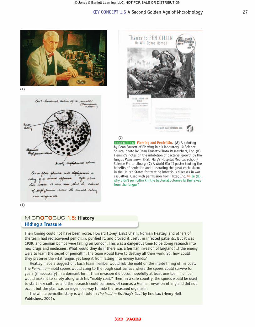
FiguRE 1.14 Fleming and penicillin. (A) A painting by Dean Fausett of Fleming in his laboratory. © Science Source, photo by Dean Fausett/Photo Researchers, Inc. (B) Fleming’s notes on the inhibition of bacterial growth by the fungus Penicillium. © St. Mary’s Hospital Medical School/Science Photo Library. (C) A World War II poster touting the benefits of penicillin and illustrating the great enthusiasm in the United States for treating infectious diseases in war casualties. Used with permission from Pfizer, Inc. »» In (B), why didn’t penicillin kill the bacterial colonies farther away from the fungus?
(A)
(B)
(C)
1.5: HistoryHiding a Treasure
Their timing could not have been worse. Howard Florey, Ernst Chain, Norman Heatley, and others of the team had rediscovered penicillin, purified it, and proved it useful in infected patients. But it was 1939, and German bombs were falling on London. This was a dangerous time to be doing research into new drugs and medicines. What would they do if there was a German invasion of England? If the enemy were to learn the secret of penicillin, the team would have to destroy all their work. So, how could they preserve the vital fungus yet keep it from falling into enemy hands?
Heatley made a suggestion. Each team member would rub the mold on the inside lining of his coat. The Penicillium mold spores would cling to the rough coat surface where the spores could survive for years (if necessary) in a dormant form. If an invasion did occur, hopefully at least one team member would make it to safety along with his “moldy coat.” Then, in a safe country, the spores would be used to start new cultures and the research could continue. Of course, a German invasion of England did not occur, but the plan was an ingenious way to hide the treasured organism.
The whole penicillin story is well told in The Mold in Dr. Flory’s Coat by Eric Lax (Henry Holt Publishers, 2004).
KEY CONCEPT 1.5 A Second Golden Age of Microbiology 27
3rd pages
© Jones & Bartlett Learning, LLC. NOT FOR SALE OR DISTRIBUTION
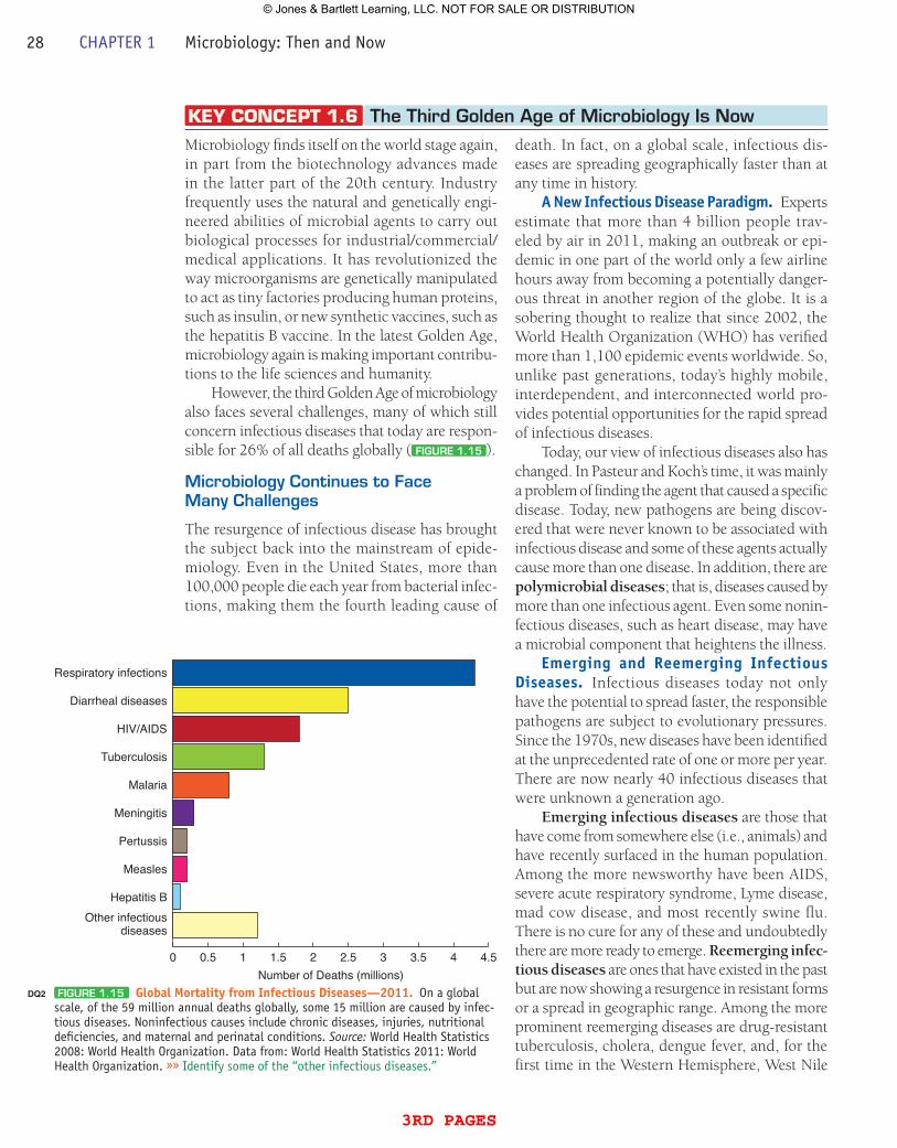
Microbiology finds itself on the world stage again, in part from the biotechnology advances made in the latter part of the 20th century. Industry frequently uses the natural and genetically engi-neered abilities of microbial agents to carry out biological processes for industrial/commercial/medical applications. It has revolutionized the way microorganisms are genetically manipulated to act as tiny factories producing human proteins, such as insulin, or new synthetic vaccines, such as the hepatitis B vaccine. In the latest Golden Age, microbiology again is making important contribu-tions to the life sciences and humanity.
However, the third Golden Age of microbiology also faces several challenges, many of which still concern infectious diseases that today are respon-sible for 26% of all deaths globally ( FiguRE 1.15 ).
Microbiology Continues to Face Many Challenges
The resurgence of infectious disease has brought the subject back into the mainstream of epide-miology. Even in the United States, more than 100,000 people die each year from bacterial infec-tions, making them the fourth leading cause of
KEY CONCEPT 1.6 The Third golden Age of Microbiology is Now
death. In fact, on a global scale, infectious dis-eases are spreading geographically faster than at any time in history.
A New Infectious Disease paradigm. Experts estimate that more than 4 billion people trav-eled by air in 2011, making an outbreak or epi-demic in one part of the world only a few airline hours away from becoming a potentially danger-ous threat in another region of the globe. It is a sobering thought to realize that since 2002, the World Health Organization (WHO) has verified more than 1,100 epidemic events worldwide. So, unlike past generations, today’s highly mobile, interdependent, and interconnected world pro-vides potential opportunities for the rapid spread of infectious diseases.
Today, our view of infectious diseases also has changed. In Pasteur and Koch’s time, it was mainly a problem of finding the agent that caused a specific disease. Today, new pathogens are being discov-ered that were never known to be associated with infectious disease and some of these agents actually cause more than one disease. In addition, there are polymicrobial diseases; that is, diseases caused by more than one infectious agent. Even some nonin-fectious diseases, such as heart disease, may have a microbial component that heightens the illness.
Emerging and Reemerging Infectious Diseases. Infectious diseases today not only have the potential to spread faster, the responsible pathogens are subject to evolutionary pressures. Since the 1970s, new diseases have been identified at the unprecedented rate of one or more per year. There are now nearly 40 infectious diseases that were unknown a generation ago.
Emerging infectious diseases are those that have come from somewhere else (i.e., animals) and have recently surfaced in the human population. Among the more newsworthy have been AIDS, severe acute respiratory syndrome, Lyme disease, mad cow disease, and most recently swine flu. There is no cure for any of these and undoubtedly there are more ready to emerge. Reemerging infec-tious diseases are ones that have existed in the past but are now showing a resurgence in resistant forms or a spread in geographic range. Among the more prominent reemerging diseases are drug-resistant tuberculosis, cholera, dengue fever, and, for the first time in the Western Hemisphere, West Nile
0 0.5 1 1.5 2 2.5Number of Deaths (millions)
3 3.5 4 4.5
Respiratory infections
Diarrheal diseases
HIV/AIDS
Tuberculosis
Malaria
Meningitis
Pertussis
Measles
Hepatitis BOther infectious
diseases
FiguRE 1.15 Global Mortality from Infectious Diseases—2011. On a global scale, of the 59 million annual deaths globally, some 15 million are caused by infec-tious diseases. Noninfectious causes include chronic diseases, injuries, nutritional deficiencies, and maternal and perinatal conditions. Source: World Health Statistics 2008: World Health Organization. Data from: World Health Statistics 2011: World Health Organization. »» Identify some of the “other infectious diseases.”
DQ2
28 CHAPTER 1 Microbiology: Then and Now
3rd pages
© Jones & Bartlett Learning, LLC. NOT FOR SALE OR DISTRIBUTION
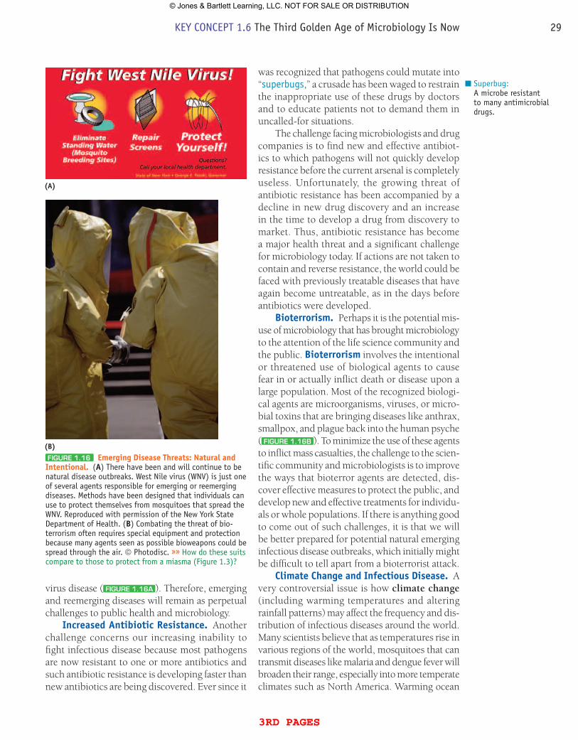
virus disease ( FiguRE 1.16A ). Therefore, emerging and reemerging diseases will remain as perpetual challenges to public health and microbiology.
Increased Antibiotic Resistance. Another challenge concerns our increasing inability to fight infectious disease because most pathogens are now resistant to one or more antibiotics and such antibiotic resistance is developing faster than new antibiotics are being discovered. Ever since it
was recognized that pathogens could mutate into “superbugs,” a crusade has been waged to restrain the inappropriate use of these drugs by doctors and to educate patients not to demand them in uncalled-for situations.
The challenge facing microbiologists and drug companies is to find new and effective antibiot-ics to which pathogens will not quickly develop resistance before the current arsenal is completely useless. Unfortunately, the growing threat of antibiotic resistance has been accompanied by a decline in new drug discovery and an increase in the time to develop a drug from discovery to market. Thus, antibiotic resistance has become a major health threat and a significant challenge for microbiology today. If actions are not taken to contain and reverse resistance, the world could be faced with previously treatable diseases that have again become untreatable, as in the days before antibiotics were developed.
Bioterrorism. Perhaps it is the potential mis-use of microbiology that has brought microbiology to the attention of the life science community and the public. Bioterrorism involves the intentional or threatened use of biological agents to cause fear in or actually inflict death or disease upon a large population. Most of the recognized biologi-cal agents are microorganisms, viruses, or micro-bial toxins that are bringing diseases like anthrax, smallpox, and plague back into the human psyche ( FiguRE 1.16B ). To minimize the use of these agents to inflict mass casualties, the challenge to the scien-tific community and microbiologists is to improve the ways that bioterror agents are detected, dis-cover effective measures to protect the public, and develop new and effective treatments for individu-als or whole populations. If there is anything good to come out of such challenges, it is that we will be better prepared for potential natural emerging infectious disease outbreaks, which initially might be difficult to tell apart from a bioterrorist attack.
Climate Change and Infectious Disease. A very controversial issue is how climate change (including warming temperatures and altering rainfall patterns) may affect the frequency and dis-tribution of infectious diseases around the world. Many scientists believe that as temperatures rise in various regions of the world, mosquitoes that can transmit diseases like malaria and dengue fever will broaden their range, especially into more temperate climates such as North America. Warming ocean
Superbug:A microbe resistant to many antimicrobial drugs.
FiguRE 1.16 Emerging Disease Threats: Natural and Intentional. (A) There have been and will continue to be natural disease outbreaks. West Nile virus (WNV) is just one of several agents responsible for emerging or reemerging diseases. Methods have been designed that individuals can use to protect themselves from mosquitoes that spread the WNV. Reproduced with permission of the New York State Department of Health. (B) Combating the threat of bio-terrorism often requires special equipment and protection because many agents seen as possible bioweapons could be spread through the air. © Photodisc. »» How do these suits compare to those to protect from a miasma (Figure 1.3)?
(A)
(B)
KEY CONCEPT 1.6 The Third Golden Age of Microbiology Is Now 29
3rd pages
© Jones & Bartlett Learning, LLC. NOT FOR SALE OR DISTRIBUTION
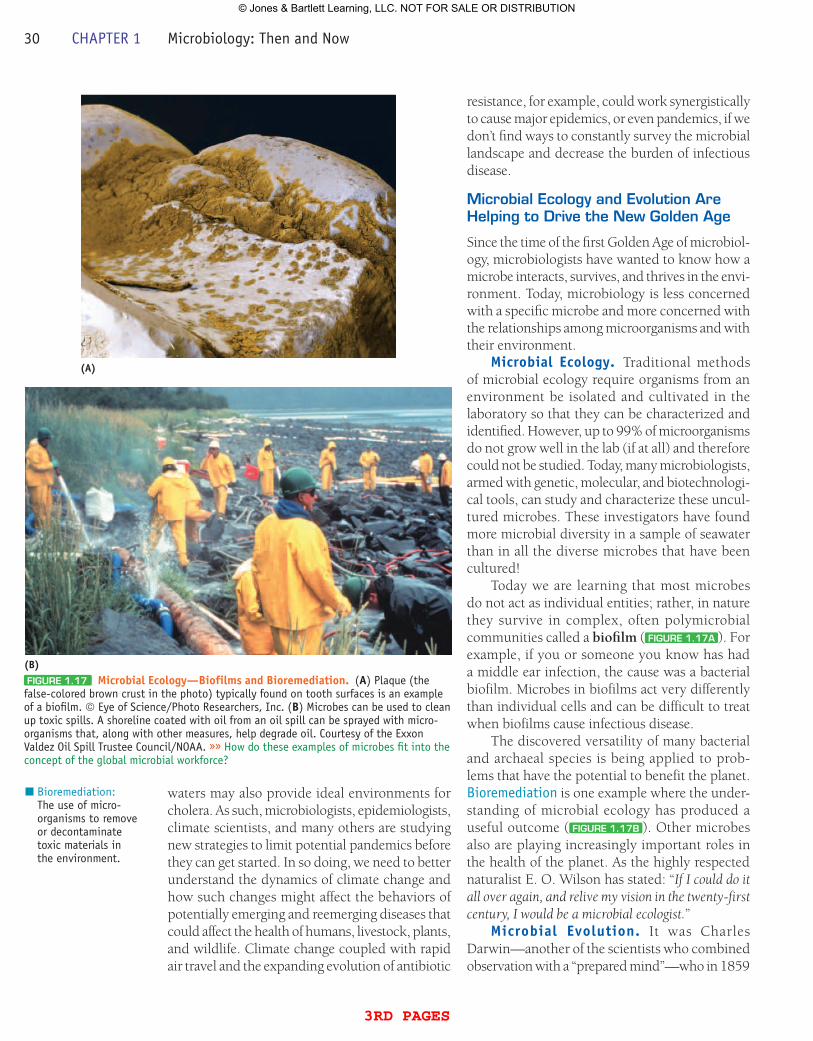
waters may also provide ideal environments for cholera. As such, microbiologists, epidemiologists, climate scientists, and many others are studying new strategies to limit potential pandemics before they can get started. In so doing, we need to better understand the dynamics of climate change and how such changes might affect the behaviors of potentially emerging and reemerging diseases that could affect the health of humans, livestock, plants, and wildlife. Climate change coupled with rapid air travel and the expanding evolution of antibiotic
resistance, for example, could work synergistically to cause major epidemics, or even pandemics, if we don’t find ways to constantly survey the microbial landscape and decrease the burden of infectious disease.
Microbial Ecology and Evolution Are Helping to Drive the New golden Age
Since the time of the first Golden Age of microbiol-ogy, microbiologists have wanted to know how a microbe interacts, survives, and thrives in the envi-ronment. Today, microbiology is less concerned with a specific microbe and more concerned with the relationships among microorganisms and with their environment.
Microbial Ecology. Traditional methods of microbial ecology require organisms from an environment be isolated and cultivated in the laboratory so that they can be characterized and identified. However, up to 99% of microorganisms do not grow well in the lab (if at all) and therefore could not be studied. Today, many microbiologists, armed with genetic, molecular, and biotechnologi-cal tools, can study and characterize these uncul-tured microbes. These investigators have found more microbial diversity in a sample of seawater than in all the diverse microbes that have been cultured!
Today we are learning that most microbes do not act as individual entities; rather, in nature they survive in complex, often polymicrobial communities called a biofilm ( FiguRE 1.17A ). For example, if you or someone you know has had a middle ear infection, the cause was a bacterial biofilm. Microbes in biofilms act very differently than individual cells and can be difficult to treat when biofilms cause infectious disease.
The discovered versatility of many bacterial and archaeal species is being applied to prob-lems that have the potential to benefit the planet. Bioremediation is one example where the under-standing of microbial ecology has produced a useful outcome ( FiguRE 1.17B ). Other microbes also are playing increasingly important roles in the health of the planet. As the highly respected naturalist E. O. Wilson has stated: “If I could do it all over again, and relive my vision in the twenty-first century, I would be a microbial ecologist.”
Microbial Evolution. It was Charles Darwin—another of the scientists who combined observation with a “prepared mind”—who in 1859
Bioremediation:The use of micro-organisms to remove or decontaminate toxic materials in the environment.
(A)
(B)FiguRE 1.17 Microbial Ecology—Biofilms and Bioremediation. (A) Plaque (the false-colored brown crust in the photo) typically found on tooth surfaces is an example of a biofilm. © Eye of Science/Photo Researchers, Inc. (B) Microbes can be used to clean up toxic spills. A shoreline coated with oil from an oil spill can be sprayed with micro-organisms that, along with other measures, help degrade oil. Courtesy of the Exxon Valdez Oil Spill Trustee Council/NOAA. »» How do these examples of microbes fit into the concept of the global microbial workforce?
30 CHAPTER 1 Microbiology: Then and Now
3rd pages
© Jones & Bartlett Learning, LLC. NOT FOR SALE OR DISTRIBUTION
first described the principles of evolution, which today is the unifying force in modern biology, tying together such distinct fields as genetics, ecology, medicine—and yes, microbiology.
Like all life, microorganisms evolve. In fact, they were evolving for some 2 billion years before the first truly eukaryotic cells appeared on the planet. Because most microbes have relatively short generation times, they represent experi-mental (model) systems in which evolutionary processes can be observed and tested and, in so doing, help us better understand the origin of all microorganisms—and larger organisms as well. Although challenging to study, it is possible today to “replay history” by following the accumulation of unpredictable, chance events that lead to evo-lutionary novelty. In addition, when considering the challenges facing microbiology today, current research on microbial evolution is putting together new approaches for the treatment of infectious disease, to improve agricultural productivity, to monitor and assess climate change, and even to produce clean fuels and energy.
Today, thanks to the availability of sequenced genomes for groups of related and unrelated microbes, and new analytical approaches, micro-biologists and microbial ecologists are studying the processes that drive evolutionary diversification and constructing a family tree that more clearly illustrates evolutionary relationships among all organisms. Such developments are giving us a bet-ter appreciation for the roles microbes have played and are continuing to play in Earth’s evolution. I
wonder what Darwin would make of the microbial world if he were alive today?
concept and reasoning checks 6a. Give some examples of how microbial ecology and
evolution are helping drive the new Golden Age of microbiology.
Many of the microbes that first occupied the minds and work of the early microbial pioneers like Pasteur and Koch still challenge microbiologists today.
QuEsTiON C:Describe the natural and intentional disease threats challenging microbiology today, explaining why they are still so prominent even with all the advances in medical science and microbiology.
Answers can be found on the Student Companion Website in Appendix D.
Chapter Challenge C
n In conclusion, microbiology (from then until now) has gone from observing the first microbes (Leeuwenhoek) to identifying and studying indi-vidual microorganisms (Pasteur and Koch) to sequencing all species in a sample of seawater. Yet, over these 300+ years, microbiologists have only discovered perhaps 1% of all microbial spe-cies. Microbiology from then until now has come a long way, but has a much longer way yet to go.
1.1 The Discovery of Microbes Leads to Questioning Their Origins1. The observations with the microscope made by Hooke
and especially Leeuwenhoek, who reported the existence of animalcules (microorganisms), sparked interest in an unknown world of microscopic life. (Fig. 1.2)
2. The controversy over spontaneous generation initiated the need for accurate scientific experimentation, which then provided the means to refute the concept.
1.2 Disease Transmission Can Be Interrupted3. Edward Jenner determined that disease (smallpox) could be
prevented through vaccination with a similar but milder disease-causing agent.
4. Semmelweis and Snow believed that infectious disease could be caused by particles transmitted from the environment and that the transmission could be interrupted. (Figs. 1.4, 1.5)
1.3 The Classical Golden Age of Microbiology Reveals the Germ5. Pasteur’s fermentation experiments indicated that
microorganisms could induce chemical changes. He proposed the germ theory of disease, which stated that human disease could be due to chemical changes brought about by microorganisms in the body.
6. Lister’s use of antisepsis techniques and Pasteur’s studies of pébrine supported the germ theory and showed how diseases can be controlled.
SuMMARY OF KEY CONCEPTS
Summary of Key Concepts 31
3rd pages
© Jones & Bartlett Learning, LLC. NOT FOR SALE OR DISTRIBUTION
32 CHAPTER 27 Microbiology: Then and Now
For STEpS A–D, answers to even-numbered questions and problems can be found in Appendix C on the Student Companion Website at http://microbiology.jbpub.com/10e. In addition, the site features eLearning, an online review area that provides quizzes and other tools to help you study for your class. You can also follow useful links for in-depth information, read more MicroFocus stories, or just find out the latest microbiology news.
CHAPTER SELF-TEST
Multiple Choice
Read each question carefully, then select the one answer that best fits the question or statement. 1. Who was the first person to see bacterial cells with the microscope? A. Pasteur B. Koch C. Leeuwenhoek D. Hooke 2. What process was studied by Redi and Spallanzani? A. Spontaneous generation B. Fermentation C. Variolation D. Antisepsis 3. The process of _____ involved the inoculation of dried smallpox
scabs under the skin. A. vaccination B. antisepsis C. variolation D. immunization
4. What is the name for the field of study established by Semmelweis and Snow in the mid 1800s?
A. Immunology B. Bacteriology C. Virology D. Epidemiology 5. The process of controlled heating, called _____, was used to keep
wine from spoiling. A. curdling B. fermentation C. pasteurization D. variolation 6. What surgical practice was established by Lister? A. Antisepsis B. Chemotherapy C. Variolation D. Sterilization
STEP A: REViEW OF FACTS AND TERMS
7. Koch’s work with anthrax allowed him to formalize the methods (Koch’s postulates) for relating a specific microorganism to a specific disease. These postulates were only valid after he discovered how to make pure cultures of bacterial species. (Fig. 1.8)
8. Laboratory science arose as Pasteur and Koch hunted down the microorganisms of infectious disease. Pasteur’s lab studied the mechanisms for infection and developed vaccines for chicken cholera, animal anthrax, and human rabies. Koch’s lab focused on isolation, cultivation, and identification of pathogens such as those responsible for cholera and tuberculosis.
1.4 With the Discovery of Other Microbes, the Microbial World Expands9. Ivanowsky and Beijerinck provided the first evidence for
viruses as infectious agents.10. Winogradsky and Beijerinck were the first to recognize the
beneficial roles played by microorganisms found in the environment.
11. Microbes include the “bacteria” (Bacteria and Archaea), viruses, fungi, and protists. (Fig. 1.11)
1.5 A Second Golden Age of Microbiology Involves the Birth of Molecular Biology and Chemotherapy12. Many of the advances toward understanding molecular
biology and general principles in biology were based on experiments using microbial model systems.
13. With the advent of the electron microscope, microbiologists realized that there were two basic types of cellular organization: eukaryotic and prokaryotic. (Fig. 1.13)
14. Following from the initial work by Ehrlich, antibiotics were developed as “magic bullets” to cure many infectious diseases.
1.6 The Third Golden Age of Microbiology Is Now15. In the 21st century, fighting infectious disease, identifying
emerging and reemerging infectious diseases, combating increasing antibiotic resistance, countering the bioterrorism threat, and addressing the potential spread of infectious diseases due to climate change are challenges facing microbiology, healthcare systems, and society. (Fig. 1.15)
16. Microbial ecology is providing new clues to the roles of microorganisms in the environment. The understanding of microbial evolution through the use of genomic technologies has expanded our understanding of microorganism relationships.
32 CHAPTER 1 Microbiology: Then and Now
3rd pages
© Jones & Bartlett Learning, LLC. NOT FOR SALE OR DISTRIBUTION
7. Which one of the following statements is NOT part of Koch’s postulates?
A. The microorganism must be isolated from a dead animal and pure cultured.
B. The microorganism and disease can be identified from a mixed culture.
C. The pure cultured organism is inoculated into a healthy, susceptible animal.
D. The same microorganism must be present in every case of the disease.
8. Match the lab with the correct set of identified diseases. A. Pasteur: tetanus and tuberculosis B. Koch: anthrax and rabies C. Koch: cholera and tuberculosis D. Pasteur: diphtheria and typhoid fever 9. What group of microbial agents would eventually be identified
from the work of Ivanowsky and Beijerinck? A. Viruses B. Fungi C. Protists D. Bacteria10. What microbiological field was established by Winogradsky and
Beijerinck? A. Virology B. Microbial ecology C. Bacteriology D. Mycology11. What group of microorganisms has a variety of internal cell
compartments and where some members act as decomposers? A. Bacteria B. Viruses C. Archaea D. Fungi12. Which one of the following organisms was NOT a model organism
related to the birth of molecular genetics? A. Streptococcus B. Penicillium C. Escherichia D. Neurospora13. Which group of microbial agents is eukaryotic? A. Bacteria B. Viruses C. Archaea D. Algae
14. The term antibiotic was coined by _____ to refer to antimicrobial substances naturally derived from _____.
A. Waksman; bacteria and fungi B. Domagk; other living organisms C. Fleming; fungi and bacteria D. Ehrlich; bacteria15. Which one of the following is NOT considered an emerging
infectious disease? A. Polio B. Severe acquired respiratory syndrome C. Lyme disease D. AIDS16. A _____ is a mixture of _____ that form as a complex community. A. genome; genes B. biofilm; microbes C. biofilm; chemicals D. miasma; microbes
True-False
Each of the following statements is true (T) or false (F). If the state-ment is false, substitute a word or phrase for the underlined word or phrase to make the statement true.17. _____ Leeuwenhoek believed that animalcules arose spontaneously
from decaying matter.18. _____ Pasteur proposed that “wine disease” was a souring of
wine caused by yeast cells.19. _____ Separate bacterial colonies can be observed in a broth
culture.20. _____ Semmelweis proposed that cholera was a waterborne
disease.21. _____ Some bacterial cells can convert nitrogen gas (N2) into
ammonia (NH3).22. _____ Fungi are eukaryotic microorganisms, some of which are
decomposers.23. _____ Koch proposed the germ theory.24. _____ Variolation involved inoculating individuals with smallpox
scabs.25. _____ Mycology is the scientific study of viruses.
STEP B: CONCEPT REViEW
26. Describe the concept of spontaneous generation and distinguish between the experiments that supported and refuted the belief. (Key Concept 1)
27. Assess the importance of the work carried out by Semmelweis and by Snow that went against the miasma view of disease and established the field of epidemiology. (Key Concept 2)
28. Compare Jenner’s work on smallpox and Pasteur’s studies on anthrax and rabies to the concept of preventing disease through vaccination. (Key Concepts 2 and 3)
29. Discuss Pasteur’s early studies and Lister’s surgical work suggesting that germs cause disease. (Key Concept 3)
30. Judge the importance of (a) the germ theory of disease and (b) Koch’s postulates to the identification of microbes as agents of infectious disease. (Key Concept 3)
31. Provide evidence to support the statement: “Not all microbes cause disease; many play important roles in the environment.” (Key Concept 4)
32. Construct a concept map for Microbial Agents, using the following terms. (Key Concept 4)
Algae Fungi ProtistsArchaea Microorganisms ProtozoaBacteria Nucleated cells VirusesDecomposers Pathogens (germs)
33. Distinguish between the “new generation” of scientists in the second Golden Age of microbiology that set the stage for the antibiotic revolution. (Key Concept 5)
34. Assess the importance of microbial ecology and microbial evolution to the current Golden Age of microbiology. (Key Concept 6)
Step B: Concept Review 33
3rd pages
© Jones & Bartlett Learning, LLC. NOT FOR SALE OR DISTRIBUTION
34 CHAPTER 27 Microbiology: Then and Now
STEP C: APPLiCATiONS AND PROBLEM SOLViNg
35. As a microbiologist in the 1940s, you are interested in discovering new antibiotics that will kill bacterial pathogens. You have been given a liquid sample of a chemical substance to test in order to determine if it kills bacterial cells. Drawing on the culture techniques of Robert Koch, design an experiment that would allow you to determine the killing properties of the sample substance.
36. As a microbial ecologist, you discover a new species of microbe. How could you determine if it has a prokaryotic or eukaryotic cell structure? Suppose it has a eukaryotic structure. What information would be needed to determine if it is a member of the protista or fungi?
37. One of the foundations of scientific inquiry is proper experimental design involving the use of controls. What is the role of a control in an experiment? For each of the experiments described in the section on spontaneous generation, identify the control(s) and explain how the interpretation of the experimental results would change without such controls.
38. You isolate and pure culture a bacterial organism from ill humans that you believe causes the disease. However, you cannot find a susceptible animal for testing that contracts the disease. What would you conclude from these observations?
STEP D: QuESTiONS FOR THOugHT AND DiSCuSSiON
39. Many people are fond of pinpointing events that alter the course of history. In your mind, which single event described in this chapter had the greatest influence on (a) the development of microbiology and (b) medicine?
40. Louis Pasteur once stated: “In the field of observation, chance favors only the prepared mind.” How does this quote apply to the work done by (a) Semmelweis, (b) Snow, and (c) Fleming?
41. One reason for the rapid advance in knowledge concerning molecular biology during the second Golden Age of microbiology was because many researchers used microorganisms as model systems. Why would bacterial cells be more advantageous to use for research than, say, rats or guinea pigs?
42. When you tell a friend that you are taking microbiology this semester, she asks, “Exactly what is microbiology?” How do you answer her?
43. As microbiologists continue to explore the microbial universe, it is becoming more apparent that microbes are “invisible emperors” that rule the world. Now that you have completed Chapter 1, provide examples to support the statement: Microbes Rule!
44. Who would you select as the “first microbiologist?” (a) Leeuwenhoek, (b) Hooke, or (c) Pasteur and Koch. Support your decision.
45. Felix Pouchet was a French biologist and science writer who believed in spontaneous generation. As such, he was often in debate with Pasteur because he was not convinced that Pasteur’s experiments refuted the idea of spontaneous generation. As proof, Pouchet set up a series of swan-necked flasks identical to those used by Pasteur to refute spontaneous generation. He then filled the flasks with a broth made from hay, boiled the flasks for one hour, and allowed them to cool. Everything was identical to Pasteur’s experiments except Pasteur used a sugar and yeast extract broth and only boiled the flasks for a few minutes.
In all cases, Pouchet saw growth of microorganisms in all his flasks, even with boiling for one hour. Propose a solution for the contradictory results of Pasteur and Pouchet, knowing that (a) what both scientists saw was valid and correct, and (b) spontaneous generation does not occur.
If you remain stumped, check out the short paper enti-tled: John Tyndall and the Spontaneous Generation Debate (Microbiology Today, November 2005-http:www.sgm.ac.uk/pubs/micro_today/pdf/110501.pdf).
34 CHAPTER 1 Microbiology: Then and Now
3rd pages
© Jones & Bartlett Learning, LLC. NOT FOR SALE OR DISTRIBUTION

Design Query1. Running Head for Key Concept 1.5 doesn’t fit in a single line
with the given text (A-Head). Please verify.2. Please check correction for Figure 1.15. No particular colors
mentioned to change the color of the bars.
© Jones & Bartlett Learning, LLC. NOT FOR SALE OR DISTRIBUTION


