Embed Size (px)
Citation preview

CB28CH20-Tryggvason ARI 11 September 2012 12:41
Functional Diversityof LamininsAnna Domogatskaya, Sergey Rodin,and Karl TryggvasonDivision of Matrix Biology, Department of Medical Biochemistry and Biophysics,Karolinska Institute, 171 77 Stockholm, Sweden; email: [email protected],[email protected], [email protected]
Annu. Rev. Cell Dev. Biol. 2012. 28:523–53
The Annual Review of Cell and DevelopmentalBiology is online at cellbio.annualreviews.org
This article’s doi:10.1146/annurev-cellbio-101011-155750
Copyright c© 2012 by Annual Reviews.All rights reserved
1081-0706/12/1110-0523$20.00
Keywords
cell-matrix interactions, basement membranes, evolution, integrinsignaling, embryonic stem cells, laminin disease
Abstract
Laminins are a large family of conserved, multidomain trimeric base-ment membrane proteins that contribute to the structure of extracellularmatrix and influence the behavior of associated cells, such as adhesion,differentiation, migration, phenotype stability, and resistance to anoikis.In lower organisms such as Hydra there is only one isoform of laminin,but higher organisms have at least 16 trimeric isoforms with varyingdegrees of cell/tissue specificity. In vitro protein and cell culture stud-ies, gene manipulation in animals, and laminin gene mutations in hu-man diseases have provided insight into the specific functions of somelaminins, but the biological roles of many isoforms are still largely unex-plored, mainly owing to difficulties in isolating them in pure form fromtissues or cells. In this review, we elucidate the evolution of laminins, de-scribe their molecular complexity, and explore the current knowledge oftheir diversity and functional aspects, including laminin-mediated sig-naling via membrane receptors, in vitro cell biology, and involvementin various tissues gained from animal model and human disease studies.The potential use of laminins in cell biology research and biotechnologyis discussed.
523
Ann
u. R
ev. C
ell D
ev. B
iol.
2012
.28:
523-
553.
Dow
nloa
ded
from
ww
w.a
nnua
lrev
iew
s.or
gby
KA
RO
LIN
SKA
IN
STIT
UT
ET
UN
IVE
RSI
TY
LIB
RA
RY
on
10/1
3/12
. For
per
sona
l use
onl
y.

CB28CH20-Tryggvason ARI 11 September 2012 12:41
Contents
INTRODUCTION . . . . . . . . . . . . . . . . . . 524EVOLUTION OF LAMININS . . . . . . 525STRUCTURAL COMPLEXITY
OF LAMININS . . . . . . . . . . . . . . . . . . . 528Molecular Structure and Isoforms . . 528Laminin Interactions and Role in
Basement Membrane Assembly . . 528LAMININ SIGNALING AND
CELLULAR EFFECTS . . . . . . . . . . . 530MAMMALIAN LAMININS IN
BIOLOGY AND DISEASE. . . . . . . . 532Laminins in Embryogenesis and
Pluripotent Stem Cells . . . . . . . . . . 532Peripheral Nervous System . . . . . . . . . 533Central Nervous System . . . . . . . . . . . 535Epithelial Laminins and Hair
Follicles . . . . . . . . . . . . . . . . . . . . . . . . 535Laminins in Muscle . . . . . . . . . . . . . . . . 539Vascular System . . . . . . . . . . . . . . . . . . . 540Pancreas . . . . . . . . . . . . . . . . . . . . . . . . . . . 540Kidney . . . . . . . . . . . . . . . . . . . . . . . . . . . . 541Immune System . . . . . . . . . . . . . . . . . . . 541Laminin-332 in Epithelial Cancer . . 543
INTRODUCTION
Basement membranes (BMs) are sheet-likeextracellular matrix (ECM) structures thatare located in the immediate vicinity of mostcells. They contain specific highly conservedproteins, mainly laminins, type IV collagens,perlecan, agrin, and nidogens (Miner &Yurchenco 2004, Yurchenco 2011). Theseproteins are the first ECM proteins to appearin the early embryo. Type IV collagens andlaminins form insoluble networks that bothhave a structural role, and they also influencethe behavior and properties of BM-associatedcells. Nidogens link BM proteins together, andthe glycosaminoglycan-containing perlecanand agrin contribute to the matrix volume viatheir hygroscopic properties as well as bindgrowth factors and introduce them to the cellsurface. BMs are associated with practically
all organized cells in the body; they underlieall epithelia and endothelia as ultrathin ≈100–350-nm-thick sheets, and they surround singlecells such as muscle fibers, adipocytes, andperipheral nerves.
The laminins are a family of large het-erotrimeric multidomain proteins that consistof three chains, α, β, and γ (Aumailley et al.2005, Miner & Yurchenco 2004), which exist infive, four, and three genetically distinct forms,respectively. Laminins have multiple, oftencell type–specific functions in processes suchas adhesion, differentiation, migration, andphenotype maintenance, and they also provideresistance to apoptosis. Laminins influence cellfunction by inducing various signaling path-ways via cell membrane receptors. In simpleorganisms, such as Hydra, there is only onelaminin type in a BM containing ECM betweentwo endoderm and ectoderm layers (Sarraset al. 1991, 1993, 1994; Zhang et al. 2002). Incomplex organisms such as vertebrates, whichhave multiple tissue types and organs, at least 16different isoforms are present (Aumailley et al.2005, Yurchenco 2011). The laminin isoformsare named based on their chain composition(Aumailley et al. 2005). For example, the com-position of LM-111 is α1β1γ1, and that of LM-511 is α5β1γ1. Laminins are important for dif-ferentiation and morphogenesis, and mutationsin laminin genes often lead to embryonic lethal-ity or severe diseases affecting many organs.
Knowledge about the properties and biolog-ical roles of laminins has come mainly fromin vitro protein and cell culture experiments,from studies on human laminin diseases, orgene inactivation in animals. Most protein levelstudies on a whole αβγ heterotrimer or aspecific chain domain have been carried outon the first laminin isoform to be described(Timpl et al. 1979), which can be obtained inlarge quantities from the BM-like matrix ofthe murine Engelbreth-Holm-Swarm (EHS)sarcoma (Orkin et al. 1977). Recently, severalother isoforms, most produced as recombinanthuman proteins (Doi et al. 2002, Kortesmaaet al. 2000, Miyazaki et al. 2008, Yurchencoet al. 1997), have become available.
524 Domogatskaya · Rodin · Tryggvason
Ann
u. R
ev. C
ell D
ev. B
iol.
2012
.28:
523-
553.
Dow
nloa
ded
from
ww
w.a
nnua
lrev
iew
s.or
gby
KA
RO
LIN
SKA
IN
STIT
UT
ET
UN
IVE
RSI
TY
LIB
RA
RY
on
10/1
3/12
. For
per
sona
l use
onl
y.

CB28CH20-Tryggvason ARI 11 September 2012 12:41
One problem with the analysis of the cellu-lar and tissue expression patterns of laminins isthat they are trimers. It is difficult to determinethe specific location of a specific trimer in situwith chain-specific antibodies, because an anti-body to a specific chain such as β1 reacts withseven trimer combinations, and a polyclonal an-tibody against one trimeric molecule will reactwith over ten distinct trimer isoforms. Knowl-edge about the biological roles of laminins hascome mainly from studies on human laminindiseases or gene inactivation in animals. Thishas had its drawbacks, as inactivation of genesencoding specific laminin monomer chains usu-ally affects several different isoforms, and partialcompensation by others chains may mask thefunction of the knocked-out isoform (Simon-Assmann et al. 2011, Yurchenco 2011). In thisreview, we describe the current understandingof the evolution, structure, properties, and dis-eases of the laminin protein family. Special em-phasis is placed on the biological roles of andsignaling processes mediated by these versatileand important matrix molecules.
EVOLUTION OF LAMININS
In recent years, the development of DNAsequencing techniques has enabled robustsequencing of whole genomes from a varietyof animal species, and this has facilitatedcomparative genomics and studies on theevolution of proteins and protein families. Wehave used available expression data and genomesequences currently available in the publicdomain to analyze the evolution of laminins bycomparing patterns of laminin chain expressionin vivo in several species that are separated byenormous evolutionary gaps. Furthermore, wehave analyzed well-sequenced species for whichwe established highly probable laminin chaincandidates as illustrated in SupplementalTable 1 (follow the Supplemental Materiallink from the Annual Reviews home page athttp://www.annualreviews.org). We also an-alyzed protein and RNA sequence similarities aswell as gene microsyntheny, in which candidatelaminin genes were compared with orthologs
in evolutionarily close species and with otherlaminin genes within the same species (seeSupplemental Methods Section 1). Theresulting evolutionary tree of the lamininprotein family is illustrated in Figure 1a.
As shown in Figure 1a, the Animalia branchdivides into Radiata and Bilateria in the earlyphylogenetic tree. Hydra vulgaris, which isclassified within Radiata, contains the earliestevolutionary form of trimeric laminin charac-terized to date (Sarras et al. 1994, Zhang et al.2002). Its chain composition is αβγ or αββ, ofwhich α and β have been cloned and sequenced;it is not yet known if this laminin has a γ chain.Laminin is expressed by the endoderm onlyand is required for ectoderm to express typeI collagen (Shimizu et al. 2002, Zhang et al.2002). Lack of any laminin chain leads to lossof regenerative ability (Figure 1b) (Sarras et al.1993, Shimizu et al. 2002, Zhang et al. 2002).
Bilateria, which contains creatures withbilateral symmetry, divides into Protostomiaand Deuterostomia. Protostomia, the lowestof the Bilateria branches, includes nematodessuch as Caenorhabditis elegans and insects suchas Drosophila melanogaster. In these species, twoα, one β, and one γ chain have been identified.Expression of the laminin chains starts at thegastrulation stage in C. elegans, in which it con-siderably precedes type IV collagen expression(Huang et al. 2003). Lack of the β (lam-1) or γ
(lam-2) chain causes embryonic/larval lethalityin C. elegans owing to organogenesis failure(Kao et al. 2006). Absence of the laminin β
(LanB1) chain in Drosophila causes completeabsence of BMs, impaired cell migration, andlate embryonic lethality (Urbano et al. 2009).The two ancestral branches of α chains appearin Bilateria. One of them, α1,2 (lam-3 in C.elegans and wb in Drosophila), is the ancestorof mammalian α1 and α2, and lack of thatchain is 100% embryonic/larval lethal. Theother, α3,5, is ancestral to mammalian α3 andα5 chains, the lack of which is 100% lethalin Drosophila (LanA), but only 72% embry-onic/larval lethal in C. elegans (epi-1) (Huanget al. 2003, Martin et al. 1999, Yarnitzky & Volk1995).
www.annualreviews.org • Functional Diversity of Laminins 525
Supplemental Material
Supplemental Material
Ann
u. R
ev. C
ell D
ev. B
iol.
2012
.28:
523-
553.
Dow
nloa
ded
from
ww
w.a
nnua
lrev
iew
s.or
gby
KA
RO
LIN
SKA
IN
STIT
UT
ET
UN
IVE
RSI
TY
LIB
RA
RY
on
10/1
3/12
. For
per
sona
l use
onl
y.

CB28CH20-Tryggvason ARI 11 September 2012 12:41
α1, α?, α??, α5β1, β?γ1, γ?
α, β, (γ?)
CnidariaHydra
α1,2α3,5
βγ
MammalsChickenAnole lizardFrogZebrafish α3
α5
α2
α1
α4
a
b
Animalia
Radiata
Bil
ate
ria
Proteostomia
DeuterostomiaEchinodermata
Sea urchin
Chordata
CephalochordataLancelet
Vertebrata
NematodaCaenorhabditis elegans
InsectaFruit flyHoneybee
TunicataCiona intestinalis
γ3
γ1
γ2 β2
β1
β3β2l
β1b
β4
Hydra
C. elegans
Fruit fly
Zebrafish
Mouse
Human
β4β2 β3γ2
γ
γ3
β
α5
Cardio
myopath
y,
glo
meru
lar s
clero
sis
JEB, L
OC
JEB
Piers
on syndro
me
JEB
MDC1A
Brain
anom
aly
α4 α3 α2α1
?
lam-3epi-1 lam-2 lam-1
wb *LanA LanB2 LanB1
fin bal caf sly softy gup
γ1 β1
α3,5 α1,2
526 Domogatskaya · Rodin · Tryggvason
Ann
u. R
ev. C
ell D
ev. B
iol.
2012
.28:
523-
553.
Dow
nloa
ded
from
ww
w.a
nnua
lrev
iew
s.or
gby
KA
RO
LIN
SKA
IN
STIT
UT
ET
UN
IVE
RSI
TY
LIB
RA
RY
on
10/1
3/12
. For
per
sona
l use
onl
y.

CB28CH20-Tryggvason ARI 11 September 2012 12:41
Deuterostomia, includes several phyla suchas Echinodermata (e.g., sea urchin) andChordata. The latter branches into threesubphyla: lower Tunicata (ascidians), higherCephalochordata (contains lancelet), and Ver-tebrata (includes fishes, reptiles, amphibians,birds, and mammals). It appears that the fourancestral laminin chain branches have under-gone multiple bifurcations in some of thosespecies.
Sea urchin has laminin α1, α5, β1, and γ1-like chains, but it has also developed two addi-tional α chains, one β and one γ-like chain thatcannot be classified as clearly correspondingto particular analogous chains in vertebrates.The ascidians are soft, nonvertebrate primitivechordates, of which Ciona intestinalis has onlyfour ancestral laminin chains, as in Proteosto-mia (Sasakura et al. 2003), even though it is evo-lutionarily closer to vertebrates than sea urchin.It is not clear if the extra ancestral genes thatemerged in early Deuterostomia were lost inascidians or if echinoderms developed an alter-native laminin system.
All vertebrates, including jawed fishes, am-phibians, reptiles, birds, and mammals, with theexception of the anole lizard and frog, havefive α, three γ, and three to six β laminingenes. Sequence similarity analysis of the threelaminin gene types allowed detection of similar-ity clusters referred to here to as constellations(Figure 1a). The relative similarity of eachpair of laminin chains within a chain constel-lation is conserved in all vertebrate species ana-lyzed. For some laminin genes (e.g., lama2) thefunctional role was conserved across species [itcauses muscular dystrophy in zebrafish (Hallet al. 2007), mouse, and human]; however,lama1 and lama5 deficiency affects zebrafish dif-ferently than mammals. Zebrafish lama5 mu-tants probably die because they fail to in-flate swim bladder (Webb et al. 2007), whileLama5-deficient mouse embryos die probablyowing to placental dysfunction (Miner et al.1998). Interestingly, vertebrates have not de-veloped any new laminin genes since jawedfishes; rather, they have lost some β chains(Figure 1b; Supplemental Table 1). We could
←−−−−−−−−−−−−−−−−−−−−−−−−−−−−−−−−−−−−−−−−−−−−−−−−−−−−−−−−−−−−−−−−−−−−−−−−Figure 1Laminins in evolution. (a) Evolutionary tree of the laminin chain family. Radiata, lower developmentalbranch, is represented by Hydra, which has only one laminin trimer. Bilateria, the higher branch, splits intotwo branches according to organism gastrulation mechanism: lower Protostomia and higher Deuterostomia.Protostomia is represented by the nematode Caenorhabditis elegans and insects (fruit fly and honeybee); theseorganisms have two α, one β, and one γ laminin chains that can form two isoforms. Deuterostomia splitsinto several phyla, including Echinodermata (represented by the sea urchin, which has developed extra α, β,and γ chains) and Chordata. The latter has three branches: lower Tunicata (ascidians), higherCephalochordata (lancelet, laminin composition unknown) and Vertebrata (represented by zebrafish, frog,anole lizard, chicken, and mammals). Notably, all Vertebrata have multiple laminin α, β, and γ chains; nonew chains have developed since bony fishes, but some chains have been lost (see Supplemental Table 1).Chain constellations, i.e., evolutionarily conserved protein similarity clusters of laminin chains, are indicatedat the top by the solid, dotted, and dashed lines. (b) Severity of knockout phenotypes for single lamininchains. Absence or mutation of most laminin chains in higher and lower species results in early lethality orsevere pathology. Here, colored spheres are used to illustrate phenotype severity: red, 100% earlydevelopmental lethality (e.g., embryonic for mammals, embryonic/larval for insects and worms); orange,early postnatal lethality (e.g., junctional epidermolysis bullosa); yellow, early diagnosed severe disease,sometimes lethal [e.g., congenital muscular dystrophy type 1A (MDC1A)]; green, disease diagnosed at adultstage; blue, no definite pathology discovered. Black spheres represent genes for which embryonic lethality inhuman cannot be verified. Light gray spheres (e.g., in zebrafish or LAMB4 in humans) indicate unknownphenotypes. ∗ A particular mutation in the wb (wing blister) gene in Drosophila melanogaster causes a rathermild phenotype well known for wing blistering. However, total lack of the gene results in 100% earlylethality (Martin et al. 1999). Abbreviations: JEB, junctional epidermolysis bullosa; LOC, laryngo-onycho-cutaneous; MDC1A: merosin-deficient congenital muscular dystrophy, type 1A.
www.annualreviews.org • Functional Diversity of Laminins 527
Supplemental Material
Ann
u. R
ev. C
ell D
ev. B
iol.
2012
.28:
523-
553.
Dow
nloa
ded
from
ww
w.a
nnua
lrev
iew
s.or
gby
KA
RO
LIN
SKA
IN
STIT
UT
ET
UN
IVE
RSI
TY
LIB
RA
RY
on
10/1
3/12
. For
per
sona
l use
onl
y.

CB28CH20-Tryggvason ARI 11 September 2012 12:41
not pinpoint multiple bifurcation points in thelaminin evolutionary tree precisely, but theyare likely to be located within nonvertebrateDeuterostomia. Future sequencing of interme-diate species may elucidate why evolution hasturned 3 laminin genes in some lower speciesinto as many as 14 in zebrafish.
STRUCTURAL COMPLEXITYOF LAMININS
Molecular Structure and Isoforms
The highly glycosylated α, β, and γ lamininchains assemble through a coiled coil at theC-terminal end (long arm) of the chains(Aumailley et al. 2005, Miner & Yurchenco2004, Yurchenco 2011). The α chains range insize between 200 and 440 kDa, and the β and γ
chains vary between 120 and 200 kDa. Thus, atrimer can vary between approximately 400 and800 kDa. All laminin chains share a homologyof tandem repeats of structural motifs, but thereare also significant differences, particularly be-tween the α chains on the one hand and the β
and γ chains on the other (Figure 2a). The α
chains have a large C-terminal globular G do-main containing five LG subdomains (LG1–5)next to a long coiled-coil domain that is homol-ogous to domains in the β and γ chains. TheN termini of the α, β, and γ chains, often re-ferred to as the short arms, vary in length, but allhave varying numbers of laminin-type epider-mal growth factor-like (LE) repeats located inrod-like domains between globular L domains.With exception of the α3A and α4 chains, theshort arm has one to three globular domainsbetween the LE domains or at the N terminus(Figure 2a) (Aumailley et al. 2005).
Theoretically, the twelve mammalian α, β,and γ chains can form 60 different trimericcombinations, but thus far, only 16 combina-tions have been identified in tissues (Figure 2b;Supplemental Table 2). Isoforms containingthe α3 chain variants α3A and α3B and β1and γ1 chains are correspondingly referred toas LM-3A11 and LM-3B11. Previously, α3Aand α3B chains were not always distinguishedby authors, and therefore, we use LM-332
in such cases. Intracellular trimer assemblyis controlled by highly specific interactionsin the C terminus of the coiled-coil domainthat limit the number of allowed heterotrimerformations (Macdonald et al. 2010). Somelaminin isoforms are modified extracellularlyby proteolytic processing at the N- or C-terminal ends prior to their binding to cellularreceptors or other matrix molecules.
Laminin Interactions and Role inBasement Membrane Assembly
Knowledge about the molecular interactionsand signaling effects of laminins is essentialfor understanding their functional complex-ity. Several specific binding domains have beendetermined through in vitro experiments. Mostof those studies have been carried out on themurine EHS-tumor-derived LM-111 (Timplet al. 1979) and its domains generated by pro-teolytic treatments, or by using recombinantlaminin fragments (Sasaki & Timpl 2001), butseveral studies have also been done on lamininssecreted by cultured cells.
The cell adhesion properties of laminins aremediated primarily via binding of the G do-main of the α chains to integrins, dystroglycan,Lutheran glycoprotein, or sulfated glycolipids.The N-terminal globular domains of the α1(Colognato-Pyke et al. 1995) and α2 chains(Colognato et al. 1997) as well as the globulardomains VI (LN) (Nielsen & Yamada 2001)and IVa (L4a) (Sasaki & Timpl 2001) of theα5 chains can bind to several integrin isoforms(α1β1, α2β1, α3β1, and αVβ3), which enablescell binding on both ends of laminins con-taining these three α chains (Figure 2a).Thelaminin γ2 chain has been reported to bindα2β1 integrin (Decline & Rousselle 2001).The N-terminal globular domains of someα-chains can also bind sulfatides. This may alsolink the laminin molecules to the cell surface.
Integrins are heterodimeric transmembranebidirectional signaling receptors that binddifferent laminin isoforms. These interactionsgenerate dynamic links between the ECMand intracellular spaces involving induction of
528 Domogatskaya · Rodin · Tryggvason
Supplemental Material
Ann
u. R
ev. C
ell D
ev. B
iol.
2012
.28:
523-
553.
Dow
nloa
ded
from
ww
w.a
nnua
lrev
iew
s.or
gby
KA
RO
LIN
SKA
IN
STIT
UT
ET
UN
IVE
RSI
TY
LIB
RA
RY
on
10/1
3/12
. For
per
sona
l use
onl
y.

CB28CH20-Tryggvason ARI 11 September 2012 12:41
signaling pathways and organization of theintracellular cytoskeleton (Berrier & Yamada2007). The major binding site for integrinsα6β1, α6β4, α3β1, α7X1β1, and α7X2β1is located in LG motifs 1–3 on the α chains(Figure 2a) (Hirosaki et al. 2000, Smirnovet al. 2002, Talts et al. 2000, Yu & Talts 2003).A mixture of LM-511 and LM-521 has beenshown to have the highest affinity for α6β1 andα3β1, LM-3A32 has the highest affinity forα6β4, a mixture of LM-211 and LM-221 forα7X1β1, and LN-111 for α7X2β1. LM-411is the poorest ligand; it has modest affinitiesfor α6β1 and α7X1β1 (Nishiuchi et al. 2006).Interestingly, laminin β and γ chains canmodulate laminin-integrin binding. Thus,β2-containing laminins have higher affinity
−−−−−−−−−−−−−−−−−−−−−−−−−−−−−−−−−−→Figure 2Laminin chain structure, isoforms, and their cellularreceptors. (a) Laminin chains contain tandem arraysof globular and rod-like domains. The α chains havefive globular domains (LG1–5) at the C terminus. Inα3, α4, and α5, LG4–5 are believed to be normallycleaved off extracellularly (red dashed line), whereasall LG domains are probably intact in functional α1and α2 chains (LG domains of α2 stay attached tothe molecule after the cleavage). The LG domainscontain the main cellular binding region. LG1–3bind mostly, but not exclusively, to integrins (α1β1,α2β1, α6β1, α6β4, α3β1, and α7β1), whereasLG4 and LG5 contain binding sites for dystroglycan(αDG) and sulfated glycolipids (SGLs). A smallglobular motif (Lβ) in the coiled-coil domain of theβ chains binds to another extracellular matrixmolecule, agrin. The N-terminal end of all of thechains contains a variable number of epidermalgrowth factor–like repeats in short rod-like domains(LEa–c), as well as 1–3 globular domains (LN, L4,L4a, L4b, LFx), some of which can interact withintegrins and SGLs. The N terminus of laminin γ2interacts with α2β1 integrin. LM-332 can interactwith collagen XVII, also known as bullouspemphigoid protein 180 (BP-180), but the exactlocation of the binding site is unclear and is oftenattributed to γ2 laminin chain (Van den Bergh et al.2011). (b) The α, β, and γ chains assemble to form acoiled coil in at least 16 combinations. The exactnumber of laminin heterotrimers in mammaliantissues is not clear. Currently, no heterotrimerscontaining the β4 chain have been described. Figuremodified from Aumailley et al. (2005).
1 23
4 5
LM-111, LM-121LM-211, LM-221
LM-213
α1, α2
β1, β2 γ1, γ3
LM-3A11, LM-3A21
α3Aβ1, β2 γ1
α3B
β3 γ2
LM-411, LM-421LM-423
α4β1, β2 γ1, γ3
1 23
4 5
1 23
4 5
1 23
4 5
1 23
4 5
LM-511, LM-521LM-523
α5
β1, β2 γ1, γ3
LM-3A32
α3Aβ3 γ2
α3B
β1 γ1
LM-3B11
LM-3B32
b
a
α2
LNLEa LEb LEc
Coiled coil
L4a L4b
α7β1, αDG αDG, SGL
SGL,weak αDG
α3B
α6β4, α3β1, α6β1
LN LFx L4 Cleavage
α3A
α6β4, α3β1, α6β1
α4
SGL, weak α6β1,weak α7β1, weak αDG
α5
α6β1, α6β4, α3β1,α7β1, Lutheran, SGL
α2β1
LN LFx L4
α3β1,SGL
αDG, SGLαVβ3
β1, β2
Lβ
β3
Lβ
LN LFLEbLEa
LN
γ1, γ3
γ2
LN L4 LEbLEa
L4
Cleavage
α1
LNLEa LEb LEc
Coiled coil
L4a L4b
1 2 3 4 5
LG
α6β1, α7β1 αDG, SGLα1β1, α2β1,SGL
α1β1, α2β1,SGL
SGL
1 23
4 5
1 23
4 5
www.annualreviews.org • Functional Diversity of Laminins 529
Ann
u. R
ev. C
ell D
ev. B
iol.
2012
.28:
523-
553.
Dow
nloa
ded
from
ww
w.a
nnua
lrev
iew
s.or
gby
KA
RO
LIN
SKA
IN
STIT
UT
ET
UN
IVE
RSI
TY
LIB
RA
RY
on
10/1
3/12
. For
per
sona
l use
onl
y.

CB28CH20-Tryggvason ARI 11 September 2012 12:41
for α3β1 and α7X2β1 integrins than do β1laminins in binding assays in vitro (Taniguchiet al. 2009). The difference has been attributedto the 20 C-terminal amino acid residues inthe coiled-coil domain of the laminin longarm. The glutamic acid residue in the thirdposition from the C termini of the γ1 and γ2laminin chains is necessary for laminin bindingto integrins (Ido et al. 2007). Importantly, thelaminin γ3 chain lacks that residue (Ido et al.2008). It has been suggested on the basis of theuse of recombinant chain fragments that γ3laminins are unable to bind integrins (Ido et al.2008). LG motifs 4–5 bind to α-dystroglycanand glycosylated sulfatides (Talts et al. 2000,Wizemann et al. 2003, Yu & Talts 2003).Dystroglycan is a heavily glycosylated proteinthat is cleaved into two noncovalently asso-ciated α and β subunits. The first containsa laminin α binding site, and the latter is atransmembrane subunit that associates withdystrophin/utrophin to form the dystrophinglycoprotein complex (DGC). DGC binds toF-actin, which provides a link between theECM and the cellular cytoskeleton. Interest-ingly, LG motifs 1–3 in the C terminus of thelaminin α2 chain contain an additional bindingsite for α-dystroglycan (Smirnov et al. 2002,Talts et al. 1999). In addition to their structuralrole, integrins and dystroglycan can participatein direct outside-in signaling (see below). TheN-terminal globular domains and LG domains1–3 in the C terminus of several α chains alsocontain binding sites for sulfatides (Garbe et al.2002, Talts et al. 2000, Yu & Talts 2003).
In addition to common laminin receptors,some laminin isoforms (laminin α5 chain) caninteract specifically with surface molecules suchas the Lutheran blood group glycoprotein,which is also known as basal cell adhesionmolecule (Udani et al. 1998). LM-332 also caninteract with collagen XVII also known as bul-lous pemphigoid protein 180 (BP-180) (Vanden Bergh et al. 2011), which is an importantpart of hemidesmosomes (see below). Otherlaminin isoforms might have some as yet un-known receptors. For instance, LM-411 has lowaffinity for integrins (Nishiuchi et al. 2006) and
α-dystroglycan (Talts et al. 2000), but its pro-cessed form, which also lacks LG4-5 domains(Talts et al. 2000), induces Dll4 expression inendothelial cells during vasculogenesis (Stenzelet al. 2011), which suggests an unknown signal-ing interaction.
Some laminin isoforms interact with eachother at the cell surface in a self-assembly pro-cess in which they form a laminin network thatcontributes to the general type IV collagen–containing BM structure. LM-111, LM-211,and LM-221 self-assemble through calcium-dependent thermal gelation in a process involv-ing interactions between the N-terminal shortarm domains (Yurchenco & Cheng 1993). Incontrast, LM-3A32 and LM-3A11, which lackthe typical short arm of laminin chains, neitherpolymerize nor copolymerize with LM-111.
Laminin molecules interact with other com-ponents of the BM matrix and thus contributeto the superstructure, but they also can inter-act with components in the underlying inter-stitial stroma. For example, an LE motif in theLEb domain of the γ1 and γ3 chains interactswith nidogen-1 (Gersdorff et al. 2005, Poschlet al. 1994, Takagi et al. 2003), which in turninteracts with type IV collagen and perlecan(Yurchenco 2011). The coiled-coil domain ofthe β chain and the LG4 motif of α1 can inter-act with agrin. LM-332 interacts with the NCdomain of type VII collagen (Rousselle et al.1997); this link is important for proper anchor-age of epithelial cells (see below).
LAMININ SIGNALING ANDCELLULAR EFFECTS
The cellular effects of laminins are mediatedlargely via ligand binding to cell membranereceptors, and this signaling can alter tran-scription levels of genes and even influencechromatin remodeling of the gene promoters.Furthermore, polarization of cells generatedby interaction with laminins can affect theirresponse to signaling from the extracellularspace, e.g., growth factor treatments (Xu et al.2009). Broad studies on laminin-mediatedsignaling hitherto have been hampered by lack
530 Domogatskaya · Rodin · Tryggvason
Ann
u. R
ev. C
ell D
ev. B
iol.
2012
.28:
523-
553.
Dow
nloa
ded
from
ww
w.a
nnua
lrev
iew
s.or
gby
KA
RO
LIN
SKA
IN
STIT
UT
ET
UN
IVE
RSI
TY
LIB
RA
RY
on
10/1
3/12
. For
per
sona
l use
onl
y.

CB28CH20-Tryggvason ARI 11 September 2012 12:41
of access to most laminin isoforms; as manyof them are now available, knowledge aboutlaminin-mediated signaling is likely to increaseextensively in the near future.
In one study, signals from LM-511/521 con-veyed via α3β1 integrin could rescue apop-tosis of serum-deprived adenocarcinoma cellsthrough activation of the phosphatidylinositol3-kinase/Akt (PI3K/Akt) pathway (Gu et al.2002). In another study, ligation of LM-332 toα6β4 integrin caused RAC-dependent activa-tion of the NF-κB pathway, mediating survivalof mammary tumor cells (Zahir et al. 2003).Furthermore, laminins, via integrin β1 signal-ing, can induce insulin expression in pancre-atic β-cells (Nikolova et al. 2006), but the ex-act mechanism of the laminin action is unclear.Binding of LM-521 to α6β1 integrin expressedby pluripotent human embryonic stem (hES)cells induces the PI3K/Akt pathway, which,in turn, stabilizes the pluripotent phenotype(S. Rodin, manuscript submitted). Binding ofthe LG domain to integrins influences develop-ment of the epidermal-dermal junction and at-tachment; muscle, eye, and brain development;and kidney and lung development (DiPersioet al. 1997; Georges-Labouesse et al. 1996,1998; Kreidberg et al. 1996; Mayer et al. 1997).
Laminins can also exert signaling effects vianonintegrin signaling receptors such as dys-troglycan. LM-111 binding to dystroglycan onSchwann cells triggers phosphorylation of c-Srcand Fyn kinases, which promotes cell survival(Li et al. 2005). In muscle cells, laminin inter-action with dystroglycan activates PI3K/Aktand downstream signaling, which mediates cellsurvival (Langenbach & Rando 2002). Signal-ing pathways induced in muscle microsomes byLM-111 have been analyzed (Zhou et al. 2006),but the biological relevance of such results is notobvious because muscle cells are not in contactwith this laminin isoform in vivo, and micro-somes may not be optimal for such studies.
Another level of complexity arises from thecurious fact that laminins are capable of cosig-naling with various growth factors. Evidence ofthat comes from observations that LM-411 in-duces Dll4 expression in endothelial cells in co-
operation with vascular endothelial growth fac-tor A (VEGF-A) (Stenzel et al. 2011). Both LM-411 signaling through integrin β1 and VEGF-A signaling through its receptor 2 (VEGFR2)induce Dll4 expression, but cooperative actionis necessary for Dll4 to be fully functional. Be-cause none of the integrin α subunits are equallyimportant for Dll4 expression, it is unclear ifthe signaling events are triggered by several β1-containing integrin heterodimers or by a core-ceptor interacting with integrin β1. Xu et al.(2009) showed that LM-111 polarizes mam-mary epithelial cells and in cooperation withprolactin constitutively activates signal trans-ducer and activator of transcription protein 5(STAT5). In mammary epithelial cell cultures,neither LM-111 nor prolactin alone can sustainthe STAT5 activation necessary for mammary-specific function, e.g., expression of caseins.Moreover, LM-111/prolactin-dependent acti-vation of STAT5 induces chromatin remodel-ing of the casein gene promoters. Furthermore,LM-111 directly activates the PI3K/Rac1 path-way in mammary epithelial cells, which in turnmaintains STAT5 signaling (Xu et al. 2010).Similarly, Leonoudakis et al. (2010) observedthat LM-111 modulates prolactin- and growthhormone–induced activation of STAT5 andincreases β-casein and insulin growth factor-1(IGF-1) expression in mouse mammary epithe-lial cells. The effect is dependent on the bindingof LM-111 to dystroglycan, but independentof the dystroglycan cytoplasmic domain. Theauthors speculate that a coreceptor of dystro-glycan, possibly β1 integrin, initiates signalsfrom laminins. Recently, Galvin et al. (2010)showed that the LM-211-dystroglycan interac-tion is capable of enhancing IGF-1-dependentdifferentiation of rat precursor cells into oligo-dendrocytes. These examples imply that insome cases growth factors alone are insufficientfor the differentiation and full physiologicalactivity of cells and that signals from ECMmolecules, particularly laminins, are necessaryto complement them. Furthermore, Gao et al.(2008) reported that LM-511, but not LM-111, can induce Noggin expression in dermalpapilla cells through cooperative signaling
www.annualreviews.org • Functional Diversity of Laminins 531
Ann
u. R
ev. C
ell D
ev. B
iol.
2012
.28:
523-
553.
Dow
nloa
ded
from
ww
w.a
nnua
lrev
iew
s.or
gby
KA
RO
LIN
SKA
IN
STIT
UT
ET
UN
IVE
RSI
TY
LIB
RA
RY
on
10/1
3/12
. For
per
sona
l use
onl
y.

CB28CH20-Tryggvason ARI 11 September 2012 12:41
with platelet-derived growth factor, but a laterstudy contradicts this finding (DeRouen et al.2010).
Understanding of laminin signaling is stillnot complete, which leaves many obvious dis-crepancies without an adequate explanation.Thus, the mediating effects of LM-521 on Aktphosphorylation and migration of hES cells invitro via binding to α6β1 integrin is muchstronger than that of LM-511, which binds thesame integrin. The C terminus of the β2 chaincan potentiate integrin-mediated signaling inhES cells (S. Rodin, manuscript submitted).Dorsal root ganglion cells that express α3β1and α7β1 integrins bind both LM-111 andLM-211 and develop neurite outgrowth, butthe effect of LM-111 is much stronger than thatof LM-211 (Plantman et al. 2008). Similarly,LM-511, which preferentially binds α6β1, gen-erates much longer neurites in dorsal root gan-glion cells than does LM-411, which also bindsthis integrin. The differences in the intensityof signaling effects between laminins not onlymay be the result of specific laminin-integrininteractions, but also can depend, e.g., on theaffinity of laminin for the integrins (Nishiuchiet al. 2006) or interactions with yet unidentifiedintegrin coreceptors.
MAMMALIAN LAMININS INBIOLOGY AND DISEASE
Laminins in Embryogenesisand Pluripotent Stem Cells
Laminins containing α1, α5, β1, and γ1 arethe first ECM proteins identified in the earlymammalian preimplantation embryo (Cooper& MacQueen 1983, Klaffky et al. 2001, Leivoet al. 1980, Miner et al. 2004a) (Figure 3a).Expression of single laminin β- and γ-chainsstarts as early as the 2–4-cell embryo stage, andthe whole trimer is detected at the 8–16-cellstage (Cooper & MacQueen 1983, Dziadek &Timpl 1985). The β1, γ1, α1, and α5 lamininchains are essential for early embryogenesisand initiation of morphogenesis. In the earlyembryo LM-511 is the dominating laminin
isoform in the embryonic BM and inner cellmass, whereas LM-111 is the most abundantone in the Reichert’s membrane that supportsthe outer extraembryonic layer of trophoblasts(Klaffky et al. 2001, Miner et al. 2004a). Invivo, cells of the inner cell mass give rise to allembryonic tissues and, if cultured in vitro, toembryonic stem cells that maintain a require-ment for α5 laminins and the ability to expressthem. Deletion of the mouse β1 (Lamb1) andγ1 (Lamc1) chain genes causes embryonic deathwithin one day of implantation [embryonic day(E) 5.5] owing to total lack of BMs and tissuedisorganization (Miner et al. 2004a, Smythet al. 1999). Inactivation of Lama1 (α1) causeslack of Reichert’s membrane as well as failureof epiblast differentiation and trophoblastfunction, which causes embryo lethality byE6.5–E7, although it allows formation of theembryonic BM, cavitation of the embryonicectoderm, and differentiation of the parietalendoderm, probably owing to compensationfrom α5 laminin chain (Miner et al. 2004a).LM-111 (that functionally requires the lamininα1 LG4–5 domains) is essential for epiblastdifferentiation, a developmental step that iscrucial for formation of the three embryonicgerm layers (Figure 3b) (Li et al. 2002, Li et al.2004, Scheele et al. 2005). Absence of the α5chains does not interfere with Reichert’s mem-brane formation and early morphogenesis,but it leads in mice to multiple developmentaldefects and lethality at E13–17 (Miner et al.1998).
In agreement with laminin expression in theearly embryo, pluripotent mouse embryonicstem (mES) and hES cells established in vitroand derived from the blastocyst inner cell massalso express laminins. Thus, cultured pluripo-tent hES cells express α1, α5, β1, β2, and γ1laminin chains (Rodin et al. 2010). Only LM-511 and LM-521 maintain pluripotency of theES cells by α6β1 integrin signaling (Figure 3b)(Domogatskaya et al. 2008, Rodin et al. 2010),which in hES cells induces the PI3K/Akt signal-ing pathway (S. Rodin, manuscript submitted).LM-511 and LM-521 prevent apoptosis of hEScells in vitro (S. Rodin, manuscript submitted).
532 Domogatskaya · Rodin · Tryggvason
Ann
u. R
ev. C
ell D
ev. B
iol.
2012
.28:
523-
553.
Dow
nloa
ded
from
ww
w.a
nnua
lrev
iew
s.or
gby
KA
RO
LIN
SKA
IN
STIT
UT
ET
UN
IVE
RSI
TY
LIB
RA
RY
on
10/1
3/12
. For
per
sona
l use
onl
y.
CB28CH20-Tryggvason ARI 11 September 2012 12:41
Usually, culture of mES cells requires the useof leukemia inhibitory factor (LIF) or fibroblastfeeder cells for maintenance of pluripotency invitro, but when cultured solely on human re-combinant LM-511, mES cells do not requireany LIF or feeder cells (Domogatskaya et al.2008). Such cells cultured over 150 doublingsgenerate chimeras and new mice via germlinetransmission (A. Domogatskaya, unpublishedmanuscript). Derivation and expansion of hEScells have been possible only for approximatelya decade (Thomson et al. 1998). For deriva-tion, researchers mainly have used feeder cells,Matrigel, undefined mixtures of ECM pro-teins, or feeder cell–derived ECM. Under thoseconditions the cells grow in clumps with ap-proximately 10–15% of the cells being par-tially differentiated. The use of human recom-binant LM-511 and LM-521 has solved thoseproblems, as it enables culture of homogenouspluripotent hES and induced pluripotent stem(iPS) cells in a chemically defined and xeno-freeenvironment. Additionally, the cells grow in amonolayer and can be expanded clonally froma single-cell solution (S. Rodin, manuscriptsubmitted).
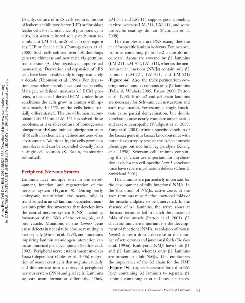
Peripheral Nervous System
Laminins have multiple roles in the devel-opment, function, and regeneration of thenervous system (Figure 4). During earlyembryonic neurulation, the neural tube istransformed in an α5 laminin–dependent man-ner into primitive structures that develop intothe central nervous system (CNS), includingformation of the BMs of the retina, pia, andotic vesicle. Mutations in the Lama5 genecause defects in neural tube closure resulting inexencephaly (Miner et al. 1998), and mutationsimpairing laminin γ1-nidogen interaction cancause abnormal pial development (Halfter et al.2002). Peripheral nerve establishment involvesLama5-dependent (Coles et al. 2006) migra-tion of neural crest cells that migrate caudallyand differentiate into a variety of peripheralnervous system (PNS) and glial cells. Lamininssupport axon formation differently. Thus,
LM-511 and LM-111 support good spreadingin vitro, whereas LM-211, LM-411, and someunspecific coatings do not (Plantman et al.2008).
The complex mature PNS exemplifies theneed for specific laminin isoforms. For instance,isoforms containing β1 and β2 chains do notcolocate. Axons are covered by β1 laminins(LM-211, LM-411, LM-511), whereas the neu-romuscular junctions (NMJs) contain only β2laminins (LM-221, LM-421, and LM-521)(Figure 4a). Also, the thick perineurium cov-ering nerve bundles contains only β2 laminins(Feltri & Wrabetz 2005; Patton 2000; Pattonet al. 1998). Both α2 and α4 chain lamininsare necessary for Schwann cell maturation andaxon myelination. For example, single knock-outs cause partial demyelination, but doubleknockouts cause nearly complete amyelinationand severe neuropathy (Wallquist et al. 2005,Yang et al. 2005). Muscle-specific knock-in ofthe Lama2 gene into Lama2 knockout mice withmuscular dystrophy rescues the skeletal musclephenotype but not hind leg paralysis (Kuanget al. 1998). Schwann cell laminins contain-ing the γ1 chain are important for myelina-tion, as Schwann cell–specific Lamc1 knockoutmice have severe myelination defects (Chen &Strickland 2003).
The laminins are particularly important forthe development of fully functional NMJs. Inthe formation of NMJs, active zones at theaxon terminus must fit the junctional folds ofthe muscle endplate to be innervated. In theabsence of α4 laminins, the active zones atthe axon terminus fail to match the junctionalfolds of the muscle (Patton et al. 2001). β2chain laminins are important for the develop-ment of functional NMJs, as ablation of mouseLamb2 causes a drastic decrease in the num-ber of active zones and junctional folds (Noakeset al. 1995a). Embryonic NMJs have both β1and β2 laminins, whereas only β2 lamininsare present in adult NMJs. This emphasizesthe importance of the β2 chain for the NMJ(Figure 4b). It appears essential for a thin BMlayer containing β2 laminins to separate β1laminin–containing axon and muscle surfaces.
www.annualreviews.org • Functional Diversity of Laminins 533
Ann
u. R
ev. C
ell D
ev. B
iol.
2012
.28:
523-
553.
Dow
nloa
ded
from
ww
w.a
nnua
lrev
iew
s.or
gby
KA
RO
LIN
SKA
IN
STIT
UT
ET
UN
IVE
RSI
TY
LIB
RA
RY
on
10/1
3/12
. For
per
sona
l use
onl
y.

CB28CH20-Tryggvason ARI 11 September 2012 12:41
If the β2 chain is absent, the NMJ does not or-ganize itself properly and neuropathy results asSchwann cells migrate into the cleft, which pre-vents neurotransmission to the muscle (Noakeset al. 1995a, Patton et al. 1998). Furthermore,
the β2 chain, unlike β1, is involved in orga-nizing the calcium channels needed for neuro-transmission (Nishimune et al. 2004), and α5and α4 are needed for postsynaptic membranematuration (Nishimune et al. 2008).
E0 E5.5 E6.5 E17.5
Lamb1–/–
Lamc1–/–Lama1–/–
Lama1ΔLG45–/– Lama5–/–
E1 E2 E4.5E3
a
b
Fallopian tube
Uterus
Implantation Placenta
Cleavage Morula
BlastocystFertilization
Laminin β/γ
Zona pellucida
Laminin αβγ
ICM/epiblast: α5
Placenta: α1, α2, α4, α5,LM-511/521, 121, 213...
RM: α1, (α5) EBM: α5, (α1)
Morula
Trophoblast
ICM
Primitiveendoderm
Epiblast PolarizationDifferentiation
LM-111
Ectoderm
Mesoderm
Endoderm
Cleavage
Fertilization
Oct4
Nanog
Mouse ES cell(naive)
Human ES cell(primed)
LM-511
LM-521LM-511
In vitro
Lama1
Sox7
Sox17
Parietal endoderm
534 Domogatskaya · Rodin · Tryggvason
Ann
u. R
ev. C
ell D
ev. B
iol.
2012
.28:
523-
553.
Dow
nloa
ded
from
ww
w.a
nnua
lrev
iew
s.or
gby
KA
RO
LIN
SKA
IN
STIT
UT
ET
UN
IVE
RSI
TY
LIB
RA
RY
on
10/1
3/12
. For
per
sona
l use
onl
y.
CB28CH20-Tryggvason ARI 11 September 2012 12:41
Central Nervous System
Laminins are not detected at many locationsin the CNS, but they seem to have impor-tant roles where they are present. The CNSis protected from the blood circulation and im-mune system by the blood-brain barrier, part ofwhich is a double BM around capillary endothe-lial cells (Figure 6b, Supplemental Figure 1).The subendothelial BM of the capillary con-tains typical endothelial LM-411 and LM-511,whereas the peripheral BM produced by astro-cyte endfeet partially covering the capillariescontains α1 and α2 laminins (Sixt et al. 2001).Lama4 knockout mice that target lymphocyteinteractions with endothelial laminins providea means of inhibiting CNS disease withoutcompromising innate immune responses (Wuet al. 2009). T lymphocyte extravasation wasshown to correlate with sites expressing lamininα4 but not α5. In laminin α4 knockout mice,laminin α5 is ubiquitously expressed along thevascular tree, which results in marked and se-lective reduction of T lymphocyte infiltrationinto the brain and reduced disease suscepti-bility and severity. Vessel phenotype and im-mune response were not affected in these mice.
It appears that laminin α5 directly inhibits in-tegrin α6β1–mediated migration of T lympho-cytes adherent to laminin α4. For other roles oflaminins in the CNS, development and main-tenance (CNS synapsis function, oligodendro-cyte maturation, cortical gyration development,and protection of hippocampal neurons), seeSupplemental Figure 1.
Epithelial Laminins and Hair Follicles
Epithelial tissues line the surfaces and cavitiesof the body, such as the skin, oral cavity, andlung as well as gastrointestinal and urinarytracts, and they also form hair follicles andmany glands. The epithelia contain polarizedcells with their basolateral surface resting ona BM sheet. LM-332 is highly enriched inall epithelial BMs, in which it forms a stronglink between the epithelium and the stromaunderneath via a protein complex termed ahemidesmosome (Figure 5b). In normal skin,stationary epithelial cells bind LM-332 withhigh affinity for the α6β4 laminin–specificintegrin as well as the plasma membraneprotein BP-180 and intracellular plectin, bothof which are components of hemidesmosomes
←−−−−−−−−−−−−−−−−−−−−−−−−−−−−−−−−−−−−−−−−−−−−−−−−−−−−−−−−−−−−−−−−−−−−−−−−Figure 3Laminins in the mammalian embryo. (a) Embryonic development. The first laminin chains are expressed atthe cleavage and morula stages. In the blastocyst, laminins reside in three locations: the Reichert’s membrane(RM) that underlies the trophectoderm that is responsible for implantation (blue external line), the embryonicbasement membrane (EBM) that separates epiblast cells that give rise to all three embryonic layers fromextraembryonic primitive endoderm (blue internal line), and between cells of the inner cell mass (ICM) and inepiblast cells (yellow inner space) (Klaffky et al. 2001, Miner et al. 2004a). Various laminins are expressed inplacenta (Champliaud et al. 2000, Kikkawa & Miner 2006, Koch et al. 1999, Wondimu et al. 2006). Lack ofα5 laminin is fatal for placental function, as trophoblasts fail to attach to the vascular BMs, and lacoons thatinhibit exchange of nutrients and gases between maternal and embryonic bloodflows are formed (Miner et al.1998). (b) LM-111 is indispensable for the first embryonic differentiation. Of all laminin chains, only Lama1,Lamb1, and Lamc1-null phenotypes result in early embryonic lethality at E6.5, E5.5, and E5.5, respectively(Miner et al. 2004a, Smyth et al. 1999), which implies that LM-111 plays a key role in early development ofthe mammalian embryo. Indeed, LM-111, which is provided by the primary endoderm, an extraembryonictissue, is needed for epiblast polarization and differentiation into the three primary germ layers: ectoderm,mesoderm, and endoderm. Notably, the laminin α1 LG4–5 domain is needed for this, as proven by a specificknockout mouse model Lama1�LG45 (Scheele et al. 2005). Lack of LM-111 may be compensated for byapplication of exogenous LM-111 (Li et al. 2002). In parietal endoderm (a derivative of primitive endoderm),Sox7 and Sox17 induce expression of the Lama1 gene (Niimi et al. 2004). Both mouse embryonic stem (ES)cells derived from the blastocyst ICM (naive state) and human ES cells derived from epiblast cells [primedstate, according to Nichols & Smith (2009)] can self-renew on α5 laminins in vitro for long time periodswithout pluripotency loss (Domogatskaya et al. 2008, Rodin et al. 2010).
www.annualreviews.org • Functional Diversity of Laminins 535
Supplemental Material
Ann
u. R
ev. C
ell D
ev. B
iol.
2012
.28:
523-
553.
Dow
nloa
ded
from
ww
w.a
nnua
lrev
iew
s.or
gby
KA
RO
LIN
SKA
IN
STIT
UT
ET
UN
IVE
RSI
TY
LIB
RA
RY
on
10/1
3/12
. For
per
sona
l use
onl
y.

CB28CH20-Tryggvason ARI 11 September 2012 12:41
(Ghohestani et al. 2001, Litjens et al. 2006,Nishiuchi et al. 2006). In wound healing follow-ing epithelial damage, migratory epithelial cellsprimarily make use of α3β1 integrin via activa-tion of Rac1 (Choma et al. 2004, Frank & Carter2004). Within the normal BM zone, LM-332binds tightly to the noncollagenous domain of
type VII collagen of anchoring fibrils that ex-tends into the anchoring plaques located in theunderlying stroma (Pulkkinen & Uitto 1999,Rousselle et al. 1997). Disruption of the epithe-lial α6β4 integrin/LM-332/type VII collagenadhesion complex results in a skin blisteringdisease known as epidermolysis bullosa (EB).
Development: establishment of NMJ Mature NMJ
Lama2–/–
Lama4–/–
Lamc1–/–
Lama2mut
Lama4–/–
Lamb2–/–
Lama5–/–
Lama4–/–
a
b
Schwann cellsBM: α2, α4, β1, γ1
ProliferationMaturation
Myelin production
Skeletal muscleLM-211
Endoneuriumβ1 laminin
Perineuriumβ2 laminin
NMJBM: α2, α4,α5, β2, γ1
Nodes of RanvierBM: α2, α5, β1, γ1
Na+ channel clustering
Synapticvesicles
Schwanncell
Active zones
Ca2+ channelclustering
X
l t iclusteringclustering
XXXX
Muscleendplate
Nerveterminal
Junctionalfolds
Postsynapticmaturation
β2 laminin
536 Domogatskaya · Rodin · Tryggvason
Ann
u. R
ev. C
ell D
ev. B
iol.
2012
.28:
523-
553.
Dow
nloa
ded
from
ww
w.a
nnua
lrev
iew
s.or
gby
KA
RO
LIN
SKA
IN
STIT
UT
ET
UN
IVE
RSI
TY
LIB
RA
RY
on
10/1
3/12
. For
per
sona
l use
onl
y.

CB28CH20-Tryggvason ARI 11 September 2012 12:41
Mutations in the integrin α6β4 genes ITGA6and ITGB4 lead to junctional EB ( JEB) asso-ciated with pyloric atresia (Pulkkinen & Uitto1999, Pulkkinen et al. 1997, Vidal et al. 1995).Mutations in or absence of any of the three LM-332 chain genes in humans leads to autosomalrecessive JEB (Aberdam et al. 1994, Kivirikkoet al. 1996, Pulkkinen & Uitto 1999, Pulkkinenet al. 1994). A mutation specific for the shorterα3 chain, α3A, causes rare autosomal recessivelaryngo-onycho-cutaneous syndrome, whichis characterized by cutaneous erosions, naildystrophy, and exuberant vascular granula-tion tissue in certain epithelia, especially theconjunctiva and larynx. As only the basal ker-atinocytes of stratified epithelia secrete the α3Asplice variant, the mutation is likely to causedysfunction of keratinocyte-mesenchymalcommunication (McLean et al. 2003).
Epithelial BMs also contain LM-311 (andto a lesser extent LM-321), which shares theα3 chain of LM-332 but is assembled withlaminin γ1 and β1 chains. This laminin variantforms a covalent complex with LM-332 in tis-sue (Champliaud et al. 1996). LM-311 also hasbeen shown to participate in mechanical signaltransduction by complexing with perlecan, dys-troglycan, and integrins to regulate epithelialcells after mechanical stimulation ( Jones et al.2005)
In addition, α5 laminins are essential forhair follicle embryonic development and forthe adult hair cycle. In that process, epithelium-derived LM-511 is a critical early signal thatdirects dermal papilla development. Skin graftsfrom Lama5-null mouse embryos grafted ontohealthy mice failed to grow hair (Li et al.2003). Apparently, LM-511 is needed for hair
←−−−−−−−−−−−−−−−−−−−−−−−−−−−−−−−−−−−−−−−−−−−−−−−−−−−−−−−−−−−−−−−−−−−−−−−−Figure 4Laminins in the peripheral nervous system (PNS). (a) Mature neuromuscular system. In PNS development,motor neurons spread axons that establish contacts with muscle fibers via neuromuscular junctions (NMJs).The axons are arranged in bundles (fascicles) coated by a thick basement membrane (BM) termedperineurium; individual axons are enveloped by a BM termed endoneurium. Schwann cells are glial cells thatmyelinate the axons and thus enable a higher signal transduction rate. During development they migratealong axons, wrap them, and myelinate them. Schwann cells express γ1 laminins, particularly those includingα2 and α4, which are essential for their development (proliferation, maturation, myelin production, andsurvival). Lack of either chain results in dismyelination and neuropathy (Chen & Strickland 2003, Wallquistet al. 2005, Yang et al. 2005). The α2 laminins are required for proper clustering of sodium channels withinthe nodes of Ranvier (gaps between Schwann cells), which is needed to generate action potentials thatdepend on laminin α2 (Occhi et al. 2005). Laminin chains α2, α4, α5, β1, β2, and γ1 are restricted tocertain PNS locations and have unique functions (Feltri & Wrabetz 2005). β1 laminins are restricted toaxon, Schwann cell, and non-NMJ skeletal muscle surfaces, whereas β2 laminins are specific for NMJs andperineurium. Laminin α2 envelops the whole axon, Schwann cell BM, NMJ, and skeletal muscle surface,possibly to maintain integrity between neural and muscular BMs. Laminin α4 is expressed in the Schwanncell BM (probably to support Schwann cell function), whereas laminin α5 is restricted to the nodes ofRanvier. (b) For efficient signal transduction across the NMJ, it is essential that presynaptic active zones onthe nerve terminal are directly opposed to the postsynaptic junctional folds of the muscle endplate. Lamininα4 is required for this; in Lama4-null mice, both active zones and junctional folds are formed in normalamounts, but they fail to oppose each other (Patton et al. 2001). For NMJ function, laminin β1–coated axonand skeletal muscle surfaces must be separated by the laminin β2 layer located at the nerve terminal, muscleendplate, and junctional folds (Noakes et al. 1995a). First, β2 laminin is required for development of activezones and junctional folds in normal amounts. Second, β2 laminin, unlike β1, binds and clusters calciumchannels at the nerve terminal, which enables controlled release of neurotransmitters upon arrival of theaction potential (Nishimune et al. 2004). Third, β2 laminin, unlike β1, prevents Schwann cells fromspreading into the synaptic cleft. In Lamb2-null mice, lack of the β2 laminin chain is compensated for by β1,which enables Schwann cell migration into synaptic cleft and thus impairs released neurotransmittermigration. Laminin α5, together with α4, is important for postsynaptic maturation of acetylcholine receptorclusters; in Lama5-null mice such maturation is delayed, and in double Lama5-null Lama4-null knockouts, itis arrested completely (Nishimune et al. 2008).
www.annualreviews.org • Functional Diversity of Laminins 537
Ann
u. R
ev. C
ell D
ev. B
iol.
2012
.28:
523-
553.
Dow
nloa
ded
from
ww
w.a
nnua
lrev
iew
s.or
gby
KA
RO
LIN
SKA
IN
STIT
UT
ET
UN
IVE
RSI
TY
LIB
RA
RY
on
10/1
3/12
. For
per
sona
l use
onl
y.

CB28CH20-Tryggvason ARI 11 September 2012 12:41
follicle epithelial invagination to form hairpapillae, but papilla cells can form in itsabsence (DeRouen et al. 2010). Adult hairundergoes a cycle that involves three stages:anagen (growth), catagen (apoptosis), andtelogen (pause before anagen) (Figure 5a).Alopecia (hair loss) occurs when the balancebetween those stages changes. Knowledge offactors that regulate the hair cycle may help
in devising methods to enhance hair growth(for instance, for patients recovering afterchemotherapy). LM-511 is upregulated inanagen versus catagen; in contrast, LM-332 isupregulated in catagen (Sugawara et al. 2007).Exogenous α5 laminins made adult humanhair follicles grow faster; however, LM-332did not inhibit hair growth (Sugawara et al.2007).
Granulationtissue formation
α3 lamininsN terminus
LM-332
AnagenProliferation
CatagenApoptosis
TelogenPause
a
LM-511
LM-332
FACIR α3β1
LM-332
Cell membrane
BMγ2
α3
β3γ2
α3β3
Co
lla
ge
nV
II
Stroma
HDIR α6β4
b
Epidermis
HD
FACBM
Dermis
c E12.5 d Skin blistering e Wound healing
Lama5–/–
mesenchymeoutburst
LOC syndrome:uncontrolledgranulationand scarring
JEB:LAMA3–/–
LAMB3–/–
LAMC2–/–
Pemphigoids: autoimmuneantibodies to LM-332/311γ1-laminin
Lama5–/–
HD HD
538 Domogatskaya · Rodin · Tryggvason
Ann
u. R
ev. C
ell D
ev. B
iol.
2012
.28:
523-
553.
Dow
nloa
ded
from
ww
w.a
nnua
lrev
iew
s.or
gby
KA
RO
LIN
SKA
IN
STIT
UT
ET
UN
IVE
RSI
TY
LIB
RA
RY
on
10/1
3/12
. For
per
sona
l use
onl
y.

CB28CH20-Tryggvason ARI 11 September 2012 12:41
Laminins in Muscle
The LM-211 isoform is crucial for muscle de-velopment and function. Skeletal muscle fibersare covered by a thin BM that is tightly linkedwith the sarcolemma. In the embryo, the mus-cle BM contains α2 chain laminin (Sanes et al.1990) but also α4 and α5 laminins in lesseramounts (Patton et al. 1999). However, inadults, LM-211 is practically the only lamininisoform in muscle, in addition to small amountsof LM-221, LM-521, and LM-421 that are ex-pressed in NMJs (Patton et al. 1999).
The α2 chain laminin is important forsmooth muscle myogenesis. For example,when mesenchymal smooth muscle precursorschange shape from round to spread/elongatedcells, Lama2 expression is induced; this isswitched off if the cell shape changes backto round (Relan et al. 1999). Also, antibod-ies to the α2 chain inhibit further differenti-
ation into smooth muscle cells. One can alsodifferentiate dy/dy (Lama2 mutants) lung cellsinto smooth muscle cells in vitro by provid-ing exogenous LM-211. Bolcato-Bellemin et al.(2003) also showed that α5 chain laminins playa major role in smooth muscle organization anddifferentiation.
Laminin plays an important structural rolefor muscle fibers by forming a bridge connect-ing a complex composed of transmembraneproteins, α-dystroglycan, and β1 integrins,on the one hand, with type VI collagen andpossibly other ECM collagens on the other.In the intracellular space, the dystroglycan,integrin, and the sarcoglycan complex con-nects with dystrophin, which binds the actincytoskeleton. Lack or dysfunction of any of thecomponents that connect dystrophin with thecollagen-containing ECM can lead to a varietyof muscular disorders that vary in severity.
←−−−−−−−−−−−−−−−−−−−−−−−−−−−−−−−−−−−−−−−−−−−−−−−−−−−−−−−−−−−−−−−−−−−−−−−−Figure 5Surface ectoderm: skin and hair. (a) α5 laminins are essential for hair follicle embryonic development andthe adult hair cycle (Li et al. 2003, Sugawara et al. 2007). The adult hair cycle includes three stages: anagen(proliferation), catagen (apoptosis), and telogen (pause before anagen). LM-511 expression is downregulatedin catagen, whereas LM-332 is upregulated. Exogenous LM-511 stimulates hair growth; however, LM-332does not inhibit it (Sugawara et al. 2007). (b) Two types of adhesion contacts are essential for epithelial cellfunction: focal adhesion contacts (FACs), which enable temporary adhesion contacts on the migrating cellleading edge, and hemidesmosomes (HDs), protein complexes that enable stable attachment of epithelialcells, via LM-332, to stromal type VII collagen. In a FAC, the G domain of an unprocessed laminin α3 chain(in LM-332) binds integrin receptor (IR) α3β1, whereas in an HD a processed G domain of α3 binds tointegrin α6β4 (Marinkovich 2007). (c) In embryonic skin development, α5 laminins are essential forepidermal basement membrane (BM) integrity and durability. For example, Lama5-null mice exhibitsyndactyly in which the digit septation fails (Miner et al. 1998). Owing to epithelial BM weakness, themesenchyme penetrates the surface ectoderm layer, and a second BM is formed to separate those layers.(d ) Failure in the dermal-epidermal junction can cause severe, even lethal skin blistering. A mutation in anyof the genes for LM-332, i.e., LAMA3, LAMB3, or LAMC2, can result in junctional epidermolysis bullosa( JEB), a severe neonatal skin-blistering disorder. The Herlitz form of JEB is the most severe of all EB formsand results in death within days of birth owing to malnutrition, fluid loss, infections, and/or organ failure.JEB occurs when any of the LM-332 chains is not produced or is nonfunctional. This leads to detachment ofthe epithelium in the skin, oral cavity, intestine, lung, and other organs. A non-Herlitz form of JEB is lesssevere: the laminin chains are mutated, but LM-332 trimers can form, although in reduced amounts (Hamill& McLean 2005). Autoantibodies against epithelial laminins can also cause skin blistering. For example,anti-LM-332/311 autoantibodies can in some cases cause cicatricial pemphigoid (Chan et al. 1999), andanti-γ1-laminin autoantibodies can cause anti-p200 pemphigoid (Dainichi et al. 2009).(e) LM-332 plays a double role in wound healing. First, it promotes fast spreading of epithelial cells over thehealing wound surface. Second, it controls vascularized granulation tissue formation. Early healing eventsinitiate granulation tissue formation, which stops upon a signal from the N-terminal part of laminin α3A innewly synthesized LM-3A32. In laryngo-onycho-cutaneous (LOC) syndrome, the N terminus of the α3Achain is impaired owing to a mutation in the LAMA3 gene leading to uncontrolled vascularized granulationtissue formation in patients (McLean et al. 2003).
www.annualreviews.org • Functional Diversity of Laminins 539
Ann
u. R
ev. C
ell D
ev. B
iol.
2012
.28:
523-
553.
Dow
nloa
ded
from
ww
w.a
nnua
lrev
iew
s.or
gby
KA
RO
LIN
SKA
IN
STIT
UT
ET
UN
IVE
RSI
TY
LIB
RA
RY
on
10/1
3/12
. For
per
sona
l use
onl
y.

CB28CH20-Tryggvason ARI 11 September 2012 12:41
LM-211 is important for normal muscle func-tion because it connects the cell membrane tothe type VI collagen of the interstitial ECMlocated between muscle fibers. DysfunctionalLM-211 can more or less uncouple the musclecell from the ECM, which leads to ineffec-tive and uncoordinated muscle contractionsthat cause cell damage and replacement byfibrous tissue, as occurs in muscular dystrophy.LAMA2 mutations are the most common causeof congenital muscular dystrophy type 1A(MDC1A; Helbling-Leclerc et al. 1995), whichfrequently leads to death in early childhood.Inactivation of the Lama2 gene in mice alsoleads to a muscular dystrophy phenotype (Xuet al. 1994b), and dy/dy mice that have a Lama2gene mutation and only weak expression ofthe laminin α2 chain also develop a musculardystrophy phenotype (Xu et al. 1994a). Lackof the laminin α2 chain is, however, not alwayslethal, as some individuals lacking the proteindo not exhibit muscle symptoms ( Jones et al.2001). However, it is not known if thoseindividuals have some compensatory changes,e.g., overexpression of another laminin chainthat can rescue the muscle phenotype. Gawliket al. (2004) reported that α1 chain expressionin muscle fibers can rescue the muscularphenotype in Lama2 knockout mice.
Vascular System
The vascular bed is covered on the luminalside by a single layer of endothelial cells thatrest on a subendothelial BM. In certain tis-sues, such as renal glomeruli, lung, and pancre-atic islets, podocytes, alveolar epithelial cells,or islet β-cells, respectively, also rest on thesubendothelial BM. The subendothelial BMsof most vessels contain two main laminin types,LM-511 and LM-411 (Hallmann et al. 2005,Simon-Assmann et al. 2011), but Mori et al.(2010) recently identified a third less commonisoform, LM-3B11, expressed by vascular en-dothelial cells. Strict developmental regulationof laminin expression appears to occur. Duringembryonic mouse angiogenesis, when the BMis under extensive remodeling, LM-411 is the
main laminin. LM-511 normally first appearsafter birth when the BM is being stabilized. Therole of laminins in vasculogenesis is discussed inthe Laminin Signaling and Cellular Effects sec-tion above and represented by Figure 6a.
Postcapillary venules and their BMs arethe main sites for extravasation of immuno-cytes, and the combination of laminins presenthas been shown to be of importance in thatregard. Permissive and restrictive locationsfor T cell extravasation through blood ves-sel walls are determined by the ratio be-tween extravasation-permissive laminin α4 andextravasation-restrictive laminin α5, as demon-strated in a mouse model of autoimmune Tlymphocyte extravasation into the CNS (Sixtet al. 2001, Wu et al. 2009). Locations of Tcell extravasation correlate with sites express-ing laminin α4 and small amounts of laminin α5(Figure 6b) (Sixt et al. 2001). Using a Lama4knockout mouse model (Thyboll et al. 2002),such pathological extravasation can be inhib-ited by removing permissive laminin α4 fromvascular BMs (Wu et al. 2009).
Pancreas
The endocrine glands of the pancreas are lo-cated in clusters of cells, termed the islets ofLangerhans (Figure 6), that have an imper-ative role in regulation of blood glucose andmetabolism. The islets contain an array ofcell types: glucagon-producing α cells, insulin-producing β cells, somatostatin-producing δ
cells, and pancreatic polyptide–producing PPcells. The islets are highly vascularized withcapillaries, the lumen of which is covered byan endothelial layer. Between the endotheliumand islet cells, such as β cells, there is a thinBM that in the mouse only contains α4 andα5 (Nikolova et al. 2006) (Figure 6). Immuno-staining suggests that the fetal human pancreascontains LM-411 (Petajaniemi et al. 2002).However, adult human islets have what appearsto be a double-layer BM with a subendothe-lial layer containing both LM-511 and LM-411and a β cell–proximal layer containing LM-511
540 Domogatskaya · Rodin · Tryggvason
Ann
u. R
ev. C
ell D
ev. B
iol.
2012
.28:
523-
553.
Dow
nloa
ded
from
ww
w.a
nnua
lrev
iew
s.or
gby
KA
RO
LIN
SKA
IN
STIT
UT
ET
UN
IVE
RSI
TY
LIB
RA
RY
on
10/1
3/12
. For
per
sona
l use
onl
y.

CB28CH20-Tryggvason ARI 11 September 2012 12:41
only; neither contains any LM-111 (Otonkoskiet al. 2008, Virtanen et al. 2008).
The β cells constitute 60–85% of the isletcells responsible for the synthesis, storage, andsecretion of insulin, and dysfunction or de-struction of those cells leads to type 1 dia-betes. The β cells require endothelial signalsfor their differentiation and for normal func-tion (Lammert et al. 2003, Yoshitomi & Zaret2004). During late embryonic development, β
cells express high levels of VEGF-A (Inoueet al. 2002) and attract VEGFR2-expressing en-dothelial cells that, in turn, form the vasculatureof the islets (Lammert et al. 2003). Only thecapillary endothelial cells produce a BM ma-trix. In a study exploring which endothelial sig-nals promote β cell proliferation and function,Nikolova et al. (2006) showed that laminins(LM-111, LM-411, and LM-511) stimulate β1integrin signaling–dependent insulin produc-tion in vitro. Because LM-111 is not a compo-nent of the islet BM in vivo, it appears that LM-411 and especially LM-511 are an importantpart of the natural niche for pancreatic β cells.
Kidney
The development of the mammalian kidneystarts with a metanephric mesenchyme in-duction of a ureteric bud protrusion from theWolffian duct (in mice at E11). The processis dependent on a laminin γ1-nidogen inter-action, as mice with a dysfunctional nidogenbinding site in the γ1 chain do not developa Wolffian duct, which hinders ureteric budformation with 80% renal agenesis (Willemet al. 2002). The glomeruli that constitute theprincipal renal filtration system are located atthe proximal end of the approximately 1 millionnephrons present in each human kidney. Theglomerulus is a tuft of capillaries located in aBowman’s capsule (Tryggvason et al. 2006).The actual filter is composed of fenestratedcapillary endothelial cells, the glomerular BM(GBM), and epithelial podocytes containinginterdigitating foot processes separated bya narrow slit containing an ultrathin slitdiaphragm. The glomerular filter is size and
charge selective. As the glomerulus develops,there is a switch in laminin isoforms (Mineret al. 1997, Sorokin et al. 1997), most of whichare indispensable for kidney development andfunction. Thus, early α1 and α4 chains are re-placed by α5 and β1 chains after birth, similarto the developmental regulation of GBM typeIV collagens (Hudson et al. 2003). Thus, theGBM undergoes a transition from LM-111to LM-511 and finally to LM-521, whichis the only isoform in healthy adult GBM.Furthermore, α5 and α3 laminins are essentialfor glomerulogenesis, and Lama5-null miceexhibit GBM breakdown leading to glomerularcell disorganization and failed glomerularvascularization (Miner & Li 2000). Mesangialcells organize glomerular capillaries by bindingthe laminin α5 G domain via α3β1 integrinand Lutheran receptor (Kikkawa et al. 2003).The endothelial cells of Lama3-null mice failedto attach to the developing podocyte-producedBM, so they could not merge (Abrass et al.2006). Laminin β2 is essential for GBM post-natal function. Mutations in human LAMB2lead to Pierson syndrome, a severe and oftenearly lethal disorder that also affects the ner-vous system and eyes. Lamb2-null mice appearto have normal glomeruli and GBMs, butneonatally severe proteinuria develops, whichcauses early lethality (Noakes et al. 1995b).Notably, proteinuria in Lamb2-null mice mani-fests a week before podocyte and slit diaphragmdamage is observed ( Jarad et al. 2006). Appar-ently, both the GBM and slit diaphragm are anindispensable part of the filtration barrier. Lackof laminin α4 impairs vascular maturation(via the mechanisms discussed in previoussections). Therefore, Lama4-null mice developchronic glomerular and tubulointerstitialfibrosis (Abrass et al. 2010).
Immune System
All blood cells originate from hematopoieticstem cells that reside in the bone marrow.These include immune cell types such as B cells,T cells, natural killer (NK) cells, monocytes,macrophages (phagocytic immunocytes), mast
www.annualreviews.org • Functional Diversity of Laminins 541
Ann
u. R
ev. C
ell D
ev. B
iol.
2012
.28:
523-
553.
Dow
nloa
ded
from
ww
w.a
nnua
lrev
iew
s.or
gby
KA
RO
LIN
SKA
IN
STIT
UT
ET
UN
IVE
RSI
TY
LIB
RA
RY
on
10/1
3/12
. For
per
sona
l use
onl
y.

CB28CH20-Tryggvason ARI 11 September 2012 12:41
cells, granulocytes (neutrophils, eosinophils,basophils), and dendritic cells. Expression anal-yses have revealed that laminin α2, α4, and α5chains, but not α1, are expressed in mouse andhuman bone marrow (Gu et al. 1999, Siler et al.2000). Normal stem cells adhere well to LM-511/521 and fibronectin, which are a naturalpart of the bone marrow stem cell niche, butpoorly to LM-111, which is foreign to them.Lymphoid progenitor cells giving rise to Tcells, B cells, and NK cells are released into
the bloodstream. T cell progenitors temporar-ily settle in the thymus, where they undergomaturation into double-negative (DN), double-positive (DP), and finally single-positive (SP)cells. During maturation, they change loca-tion within the thymus, and each location hasa different laminin composition (Kutlesa et al.2002). DN cells migrate from the vessels at themedulla-cortex border to the subcapsular ep-ithelium, where LN-332 appears to contributeto their survival (Kim et al. 2000). In vitro, DN
Tip cell
Stalk cells
Dll4
LM-411
VEGF
Notch1
a
External(astroglial) BMα1, α2 laminins
Low–α5laminins
region
Astrocyteendfeet
T cellextravasation
b
c
d
Lama4–/–
Lama4–/–
Lama4–/–
Internal(vascular) BMα4, α5 laminins
Vascularendothelium
Islet ofLangerhans
Exocrineacini
Acinar cellsα2, α4 laminins
β cell
Bloodcapillary
Vascularendothelial cell
α4, α5 laminins but not α1, α2, α3
VEGF-A
Ins1Ins2
Insulin-producingpancreatic β cells
β cell
542 Domogatskaya · Rodin · Tryggvason
Ann
u. R
ev. C
ell D
ev. B
iol.
2012
.28:
523-
553.
Dow
nloa
ded
from
ww
w.a
nnua
lrev
iew
s.or
gby
KA
RO
LIN
SKA
IN
STIT
UT
ET
UN
IVE
RSI
TY
LIB
RA
RY
on
10/1
3/12
. For
per
sona
l use
onl
y.

CB28CH20-Tryggvason ARI 11 September 2012 12:41
cells adhere strongly to α5 laminins, unlike DPcells, which reside within the cortex (Kutlesaet al. 2002). Lack of α2 laminin impairs thy-mus development by reducing the amount of Tcell progenitors, especially DP cells (Iwao et al.2000, Magner et al. 2000). In vitro, LM-211supports the survival of thymocytes (Iwao et al.2000).
Laminins appear to have significant rolesin the migration and inflammation of immunecells. For example, α4 laminin is required forrecruitment of immune cells to sites of inflam-mation. In Lama4-null mice, recruitment of allmajor leukocyte types (neutrophils, monocytes,and leukocytes) to inflammation loci is impairedowing to abnormal extravasation through thepostcapillary venule wall, of which LM-411 is amain BM component (Kenne et al. 2010). Mastcells, which are cytokine-releasing inflamma-tory cells, are positioned on the BMs of bloodvessels, epithelia, and nerves. In vitro, LM-332and -511, but not LM-111, -211, or -411, pro-mote mast cell adhesion and migration (Simeet al. 2009). LM-332, but not LM-511, stimu-lates release of interleukin 8 in immunoglobulinE–stimulated mast cells. Neutrophils expressLM-411, which is needed for their migration
and survival (Wondimu et al. 2004). They in-teract with it via integrin α6β1 and αMβ2 (thelatter drives neutrophil migration on LM-411).Traversal of T cells through the capillary vesselwalls has been shown to occur in regions wherethe amount of α5 laminin is low, but α4 chainlaminin is permissive for such penetration (Sixtet al. 2001, Wu et al. 2009).
Laminin-332 in Epithelial Cancer
Approximately 85% of all cancers (carcinomas)originate from epithelial cells. As describedabove, epithelial cells are dependent on prosur-vival signaling from the epithelial BM lamininsthat constitute their natural niche. Dependenceon such signaling is a common mechanismfor preventing cells from leaving their niches,and loss of contact with the natural niche canresult in anoikis, i.e., apoptosis from lack ofprosurvival signaling from the ECM (Chiarugi& Giannoni 2008). Malignantly transformedcells, however, can survive either by changingtheir intrinsic integrin signaling pattern toadjust to a foreign ECM or by overexpressingthe ECM proteins on which they depend(Chiarugi & Giannoni 2008). Carcinoma cells
←−−−−−−−−−−−−−−−−−−−−−−−−−−−−−−−−−−−−−−−−−−−−−−−−−−−−−−−−−−−−−−−−−−−−−−−−Figure 6Vascular system and pancreas. (a) α4 laminins in concert with vascular endothelial growth factor (VEGF)signaling are needed for proper function of Notch-Dll4 control of angiogenesis. In Lama4-null mice,Dll4/Notch signaling is impaired, which leads to excessive formation of tip cells and uncontrolledangiogenesis. LM-411 signaling, which is mediated by β1 integrins, specifically induces Dll4, thus activatingNotch signaling (Stenzel et al. 2011). LAMA4 mutations in mice and humans result in cardiomyopathy(Knoll et al. 2007, Wang et al. 2006) and glomerular sclerosis (Abrass et al. 2010) owing to irregularvascularization of the heart and kidney glomerulus, respectively. (b) Blood-brain barrier. The central nervoussystem is protected from the immune system by the blood-brain barrier, part of which is a double basementmembrane (BM) surrounding small blood vessels. An internal BM contains LM-411 and LM-511 producedby vascular endothelial cells. An external BM contains α1 and α2 laminins, which are provided by astrocyteendfeet. In the case of autoimmune T cell invasion, T cells easily penetrate vascular BM via low–laminin α5regions (Sixt et al. 2001, Wu et al. 2009). (c) The pancreas is composed of exocrine acini and the islets ofLangerhans, which include several endocrine cell types (glucagon-producing α cells, insulin-producing β
cells, somatostatin-producing δ cells, and pancreatic peptide-producing PP cells) as well as capillaries. Theacinar cells lie upon BMs containing several laminin isoforms, but laminins α2 and α4 are the essential ones:double Lama2-null Lama4-null mice lack pancreatic acinar BMs, and the acinar cells lose polarization (Mineret al. 2004b). The capillary endothelial cells within the pancreatic islets are separated from the endocrinecells by a single BM in mouse and a double BM in humans. (d ) The pancreatic endocrine cell niche isestablished and maintained through an interplay between β cells and vascular endothelial cells. The β cellsexpress angiogenic VEGF-A to attract vessel growth, whereas the endothelial cells produce α5 and α4laminins that are needed for β cell maintenance and insulin production (Nikolova et al. 2006).
www.annualreviews.org • Functional Diversity of Laminins 543
Ann
u. R
ev. C
ell D
ev. B
iol.
2012
.28:
523-
553.
Dow
nloa
ded
from
ww
w.a
nnua
lrev
iew
s.or
gby
KA
RO
LIN
SKA
IN
STIT
UT
ET
UN
IVE
RSI
TY
LIB
RA
RY
on
10/1
3/12
. For
per
sona
l use
onl
y.

CB28CH20-Tryggvason ARI 11 September 2012 12:41
often overexpress LM-332, which providesthem with survival signals and enables theirmigration away from the normal epithelialenvironment. Marinkovich (2007) reviews themolecular mechanisms of LM-332 in squamouscell carcinoma in detail. Numerous reportshave described increased expression of theLM-332 γ2 chain as a prognostic marker formultiple types of invasive carcinomas by usingimmunohistology and serum enzyme-linkedimmunosorbent assay analysis (Katayamaet al. 2005; Marinkovich 2007; Pyke et al.1994, 1995). Furthermore, antibodies against
LM-332 target primary mouse carcinoma andits lung metastasis in vivo (Salo et al. 2009), andthey significantly inhibit tumor proliferationand metastatic dissemination, possibly byincreasing apoptosis. A monoclonal antibodytargeting the LG4–5 motifs of the laminin α3chain induced squamous cell carcinoma cellapoptosis in vivo and thus inhibited tumorproliferation, but it did not affect normalepithelial tissue adhesion because the LG4–5domain is normally cleaved before LM-332 isintegrated into hemidesmosomes (Tran et al.2008).
SUMMARY POINTS
1. Laminins are highly biologically active multifunctional cell/tissue type–specific BM com-ponents. Laminin heterotrimers contain α, β, and γ chains that exist in several geneticallydistinct forms.
2. During evolution, a single laminin heterotrimer in low multicellular organisms has de-veloped into more than 16 different trimeric isoforms.
3. The laminins are not passive adhesion proteins. Rather, they modulate cell behavior;influence differentiation, migration, and phenotype stability; and inhibit apoptosis bysignaling via cell membrane receptors such as integrins and dystroglycan. However, thedetails of laminin signaling are still largely unexplored.
4. Laminins constitute the first ECM in the developing embryo, and embryonic lamininshave found an important use as culture matrices for stem cells. Other laminins are crucialfor, e.g., nerve, epithelium, blood vessel, and kidney function.
5. The unavailability of most laminin isoforms has hampered in vitro studies. However,many isoforms have become available recently as recombinant proteins, which may enabledeeper insight into functional properties. Laminins may find numerous new applicationsin cell biology and cell therapy research.
6. The vast complexity of laminin effects cannot be explained solely by simple integrinbinding and signaling. The existence of coreceptors is likely.
FUTURE ISSUES
1. Knowledge about the exact locations of laminin trimers in different tissues is still limited.State-of-the-art, precise biochemical methodologies may provide a more sophisticatedmeans than immunocytochemistry to explore and elucidate the nature of laminin nichesin native tissues.
544 Domogatskaya · Rodin · Tryggvason
Ann
u. R
ev. C
ell D
ev. B
iol.
2012
.28:
523-
553.
Dow
nloa
ded
from
ww
w.a
nnua
lrev
iew
s.or
gby
KA
RO
LIN
SKA
IN
STIT
UT
ET
UN
IVE
RSI
TY
LIB
RA
RY
on
10/1
3/12
. For
per
sona
l use
onl
y.
CB28CH20-Tryggvason ARI 11 September 2012 12:41
2. There is a need to determine the biological properties of laminin isoforms and theirvarious functional domains. For such studies, availability of the different isoforms isessential. As most laminins are difficult or impossible to obtain in pure forms from tissues,emphasis should be placed on obtaining various laminins with recombinant techniques.Structural analyses of functionally important domains of different laminins as well as themolecular nature of binding interactions are important future tasks.
3. One future challenge is to explore the potential for laminins in cell differentiation pro-tocols and to study the use of biologically relevant laminins or their combination in 2Dand 3D systems for primary culture and expansion as well as maintenance of cellularphenotypes.
4. Sophisticated laminin knockout and knock-in models, such as conditional tissue- anddevelopmental stage–specific variants as well as chimeric and combined knockouts, couldreveal novel tissue-specific roles of certain laminins in vivo.
DISCLOSURE STATEMENT
S.R. and K.T. are shareholders in BioLamina AB.
ACKNOWLEDGMENTS
Many scientists have made significant contributions to the laminin field. Owing to space limita-tions, it unfortunately has not been possible to give everybody proper credit via citations, but theirinput is greatly respected and appreciated. Laminin research in our laboratory has been sponsoredin part by the Swedish Medical Research Council, Swedish Foundation for Strategic Research,Knut and Alice Wallenberg Foundation, and AFA Insurance.
LITERATURE CITED
Aberdam D, Galliano MF, Vailly J, Pulkkinen L, Bonifas J, et al. 1994. Herlitz’s junctional epidermolysisbullosa is linked to mutations in the gene (LAMC2) for the γ2 subunit of nicein/kalinin (LAMININ-5).Nat. Genet. 6(3):299–304
Abrass CK, Berfield AK, Ryan MC, Carter WG, Hansen KM. 2006. Abnormal development of glomerularendothelial and mesangial cells in mice with targeted disruption of the lama3 gene. Kidney Int. 70(6):1062–71
Abrass CK, Hansen KM, Patton BL. 2010. Laminin α4-null mutant mice develop chronic kidney disease withpersistent overexpression of platelet-derived growth factor. Am. J. Pathol. 176(2):839–49
Aumailley M, Bruckner-Tuderman L, Carter WG, Deutzmann R, Edgar D, et al. 2005. A simplified lamininnomenclature. Matrix Biol. 24(5):326–32
Berrier AL, Yamada KM. 2007. Cell-matrix adhesion. J. Cell. Physiol. 213(3):565–73Bolcato-Bellemin AL, Lefebvre O, Arnold C, Sorokin L, Miner JH, et al. 2003. Laminin α5 chain is required
for intestinal smooth muscle development. Dev. Biol. 260(2):376–90Champliaud MF, Lunstrum GP, Rousselle P, Nishiyama T, Keene DR, Burgeson RE. 1996. Human amnion
contains a novel laminin variant, laminin 7, which like laminin 6, covalently associates with laminin 5 topromote stable epithelial-stromal attachment. J. Cell Biol. 132(6):1189–98
Champliaud MF, Virtanen I, Tiger CF, Korhonen M, Burgeson R, Gullberg D. 2000. Posttranslationalmodifications and β/γ chain associations of human laminin α1 and laminin α5 chains: purifications oflaminin-3 from placenta. Exp. Cell Res. 259:326–35
www.annualreviews.org • Functional Diversity of Laminins 545
Ann
u. R
ev. C
ell D
ev. B
iol.
2012
.28:
523-
553.
Dow
nloa
ded
from
ww
w.a
nnua
lrev
iew
s.or
gby
KA
RO
LIN
SKA
IN
STIT
UT
ET
UN
IVE
RSI
TY
LIB
RA
RY
on
10/1
3/12
. For
per
sona
l use
onl
y.

CB28CH20-Tryggvason ARI 11 September 2012 12:41
Chan LS, Lapiere JC, Chen M, Traczyk T, Mancini AJ, et al. 1999. Bullous systemic lupus erythematosus withautoantibodies recognizing multiple skin basement membrane components, bullous pemphigoid antigen1, laminin-5, laminin-6, and type VII collagen. Arch. Dermatol. 135(5):569–73
Chen ZL, Strickland S. 2003. Laminin γ1 is critical for Schwann cell differentiation, axon myelination, andregeneration in the peripheral nerve. J. Cell Biol. 163(4):889–99
Chiarugi P, Giannoni E. 2008. Anoikis: a necessary death program for anchorage-dependent cells. Biochem.Pharmacol. 76(11):1352–64
Choma DP, Pumiglia K, DiPersio CM. 2004. Integrin α3β1 directs the stabilization of a polarized lamel-lipodium in epithelial cells through activation of Rac1. J. Cell Sci. 117(Pt. 17):3947–59
Coles EG, Gammill LS, Miner JH, Bronner-Fraser M. 2006. Abnormalities in neural crest cell migration inlaminin α5 mutant mice. Dev. Biol. 289(1):218–28
Colognato H, MacCarrick M, O’Rear JJ, Yurchenco PD. 1997. The laminin α2-chain short arm mediates celladhesion through both the α1β1 and α2β1 integrins. J. Biol. Chem. 272(46):29330–36
Colognato-Pyke H, O’Rear JJ, Yamada Y, Carbonetto S, Cheng YS, Yurchenco PD. 1995. Mapping ofnetwork-forming, heparin-binding, and α1β1 integrin-recognition sites within the α-chain short arm oflaminin-1. J. Biol. Chem. 270(16):9398–406
Cooper AR, MacQueen HA. 1983. Subunits of laminin are differentially synthesized in mouse eggs and earlyembryos. Dev. Biol. 96(2):467–71
Dainichi T, Kurono S, Ohyama B, Ishii N, Sanzen N, et al. 2009. Anti-laminin gamma-1 pemphigoid. Proc.Natl. Acad. Sci. USA 106(8):2800–5
Decline F, Rousselle P. 2001. Keratinocyte migration requires α2β1 integrin-mediated interaction with thelaminin 5 γ2 chain. J. Cell Sci. 114:811–23
DeRouen MC, Zhen H, Tan SH, Williams S, Marinkovich MP, Oro AE. 2010. Laminin-511 and integrinbeta-1 in hair follicle development and basal cell carcinoma formation. BMC Dev. Biol. 10:112
DiPersio CM, Hodivala-Dilke KM, Jaenisch R, Kreidberg JA, Hynes RO. 1997. α3β1 Integrin is requiredfor normal development of the epidermal basement membrane. J. Cell Biol. 137(3):729–42
Doi M, Thyboll J, Kortesmaa J, Jansson K, Iivanainen A, et al. 2002. Recombinant human laminin-10(α5β1γ1). Production, purification, and migration-promoting activity on vascular endothelial cells.J. Biol. Chem. 277(15):12741–48
Domogatskaya A, Rodin S, Boutaud A, Tryggvason K. 2008. Laminin-511 but not -332, -111, or -411 enablesmouse embryonic stem cell self-renewal in vitro. Stem. Cells 26(11):2800–9
Dziadek M, Timpl R. 1985. Expression of nidogen and laminin in basement membranes during mouse em-bryogenesis and in teratocarcinoma cells. Dev. Biol. 111(2):372–82
Ezratty EJ, Stokes N, Chai S, Shah AS, Williams SE, Fuchs E. 2011. A role for the primary cilium in Notchsignaling and epidermal differentiation during skin development. Cell 145(7):1129–41
Feltri ML, Wrabetz L. 2005. Laminins and their receptors in Schwann cells and hereditary neuropathies.J. Peripher. Nerv. Syst. 10(2):128–43
Frank DE, Carter WG. 2004. Laminin 5 deposition regulates keratinocyte polarization and persistent migra-tion. J. Cell Sci. 117(Pt. 8):1351–63
Galvin J, Eyermann C, Colognato H. 2010. Dystroglycan modulates the ability of insulin-like growth factor-1to promote oligodendrocyte differentiation. J. Neurosci. Res. 88(15):3295–307
Gao J, DeRouen MC, Chen CH, Nguyen M, Nguyen NT, et al. 2008. Laminin-511 is an epithelial mes-sage promoting dermal papilla development and function during early hair morphogenesis. Genes Dev.22(15):2111–24
Garbe JH, Gohring W, Mann K, Timpl R, Sasaki T. 2002. Complete sequence, recombinant analysis andbinding to laminins and sulphated ligands of the N-terminal domains of laminin α3B and α5 chains.Biochem. J. 362(Pt. 1):213–21
Gawlik K, Miyagoe-Suzuki Y, Ekblom P, Takeda S, Durbeej M. 2004. Laminin α1 chain reduces musculardystrophy in laminin α2 chain deficient mice. Hum. Mol. Genet. 13(16):1775–84
Georges-Labouesse E, Mark M, Messaddeq N, Gansmuller A. 1998. Essential role of α6 integrins in corticaland retinal lamination. Curr. Biol. 8(17):983–86
Georges-Labouesse E, Messaddeq N, Yehia G, Cadalbert L, Dierich A, Le Meur M. 1996. Absence of integrinα6 leads to epidermolysis bullosa and neonatal death in mice. Nat. Genet. 13(3):370–73
546 Domogatskaya · Rodin · Tryggvason
Ann
u. R
ev. C
ell D
ev. B
iol.
2012
.28:
523-
553.
Dow
nloa
ded
from
ww
w.a
nnua
lrev
iew
s.or
gby
KA
RO
LIN
SKA
IN
STIT
UT
ET
UN
IVE
RSI
TY
LIB
RA
RY
on
10/1
3/12
. For
per
sona
l use
onl
y.

CB28CH20-Tryggvason ARI 11 September 2012 12:41
Gersdorff N, Kohfeldt E, Sasaki T, Timpl R, Miosge N. 2005. Laminin γ3 chain binds to nidogen and islocated in murine basement membranes. J. Biol. Chem. 280(23):22146–53
Ghohestani RF, Li K, Rousselle P, Uitto J. 2001. Molecular organization of the cutaneous basement membranezone. Clin. Dermatol. 19(5):551–62
Gu J, Fujibayashi A, Yamada KM, Sekiguchi K. 2002. Laminin-10/11 and fibronectin differentially preventapoptosis induced by serum removal via phosphatidylinositol 3-kinase/Akt- and MEK1/ERK-dependentpathways. J. Biol. Chem. 277(22):19922–28
Gu Y, Sorokin L, Durbeej M, Hjalt T, Jonsson JI, Ekblom M. 1999. Characterization of bone marrow lamininsand identification of α5-containing laminins as adhesive proteins for multipotent hematopoietic FDCP-Mix cells. Blood 93(8):2533–42
Halfter W, Dong S, Yip YP, Willem M, Mayer U. 2002. A critical function of the pial basement membranein cortical histogenesis. J. Neurosci. 22(14):6029–40
Hall TE, Bryson-Richardson RJ, Berger S, Jacoby AS, Cole NJ, et al. 2007. The zebrafish candyfloss mutantimplicates extracellular matrix adhesion failure in laminin α2-deficient congenital muscular dystrophy.Proc. Natl. Acad. Sci. USA 104:7092–97
Hallmann R, Horn N, Selg M, Wendler O, Pausch F, Sorokin LM. 2005. Expression and function of lamininsin the embryonic and mature vasculature. Physiol. Rev. 85(3):979–1000
Hamill KJ, McLean WH. 2005. The α3 polypeptide chain of laminin 5: insight into wound healing responsesfrom the study of genodermatoses. Clin. Exp. Dermatol. 30(4):398–404
Helbling-Leclerc A, Zhang X, Topaloglu H, Cruaud C, Tesson F, et al. 1995. Mutations in the laminin α2-chain gene (LAMA2) cause merosin-deficient congenital muscular dystrophy. Nat. Genet. 11(2):216–18
Hirosaki T, Mizushima H, Tsubota Y, Moriyama K, Miyazaki K. 2000. Structural requirement of carboxyl-terminal globular domains of laminin α3 chain for promotion of rapid cell adhesion and migration bylaminin-5. J. Biol. Chem. 275(29):22495–502
Huang CC, Hall DH, Hedgecock EM, Kao G, Karantza V, et al. 2003. Laminin α subunits and their role inC. elegans development. Development 130(14):3343–58
Hudson BG, Tryggvason K, Sundaramoorthy M, Neilson EG. 2003. Alport’s syndrome, Goodpasture’s syn-drome, and type IV collagen. N. Engl. J. Med. 348(25):2543–56
Ido H, Ito S, Taniguchi Y, Hayashi M, Sato-Nishiuchi R, et al. 2008. Laminin isoforms containing the γ3chain are unable to bind to integrins due to the absence of the glutamic acid residue conserved in theC-terminal regions of the γ1 and γ2 chains. J. Biol. Chem. 283(42):28149–57
Ido H, Nakamura A, Kobayashi R, Ito S, Li S, et al. 2007. The requirement of the glutamic acid residue atthe third position from the carboxyl termini of the laminin γ chains in integrin binding by laminins.J. Biol. Chem. 282(15):11144–54
Inoue M, Hager JH, Ferrara N, Gerber HP, Hanahan D. 2002. VEGF-A has a critical, nonredundant role inangiogenic switching and pancreatic β cell carcinogenesis. Cancer Cell 1(2):193–202
Iwao M, Fukada S, Harada T, Tsujikawa K, Yagita H, et al. 2000. Interaction of merosin (laminin 2) with verylate activation antigen-6 is necessary for the survival of CD4+ CD8+ immature thymocytes. Immunology99(4):481–88
Jarad G, Cunningham J, Shaw AS, Miner JH. 2006. Proteinuria precedes podocyte abnormalities inLamb2−−/−− mice, implicating the glomerular basement membrane as an albumin barrier. J. Clin. Investig.116(8):2272–79
Jones JC, Lane K, Hopkinson SB, Lecuona E, Geiger RC, et al. 2005. Laminin-6 assembles into multimolecularfibrillar complexes with perlecan and participates in mechanical-signal transduction via a dystroglycan-dependent, integrin-independent mechanism. J. Cell Sci. 118(Pt. 12):2557–66
Jones KJ, Morgan G, Johnston H, Tobias V, Ouvrier RA, et al. 2001. The expanding phenotype of lamininα2 chain (merosin) abnormalities: case series and review. J. Med. Genet. 38(10):649–57
Kao G, Huang CC, Hedgecock EM, Hall DH, Wadsworth WG. 2006. The role of the laminin β subunitin laminin heterotrimer assembly and basement membrane function and development in C. elegans. Dev.Biol. 290(1):211–19
Katayama M, Funakoshi A, Sumii T, Sanzen N, Sekiguchi K. 2005. Laminin γ2-chain fragment circulatinglevel increases in patients with metastatic pancreatic ductal cell adenocarcinomas. Cancer Lett. 225(1):167–76
www.annualreviews.org • Functional Diversity of Laminins 547
Ann
u. R
ev. C
ell D
ev. B
iol.
2012
.28:
523-
553.
Dow
nloa
ded
from
ww
w.a
nnua
lrev
iew
s.or
gby
KA
RO
LIN
SKA
IN
STIT
UT
ET
UN
IVE
RSI
TY
LIB
RA
RY
on
10/1
3/12
. For
per
sona
l use
onl
y.

CB28CH20-Tryggvason ARI 11 September 2012 12:41
Kenne E, Soehnlein O, Genove G, Rotzius P, Eriksson EE, Lindbom L. 2010. Immune cell recruitment toinflammatory loci is impaired in mice deficient in basement membrane protein laminin α4. J. Leukoc.Biol. 88(3):523–28
Kikkawa Y, Miner JH. 2006. Molecular dissection of laminin α5 in vivo reveals separable domain-specificroles in embryonic development and kidney function. Dev. Biol. 296(1):265–77
Kikkawa Y, Virtanen I, Miner JH. 2003. Mesangial cells organize the glomerular capillaries by adhering tothe G domain of laminin α5 in the glomerular basement membrane. J. Cell Biol. 161(1):187–96
Kim MG, Lee G, Lee SK, Lolkema M, Yim J, et al. 2000. Epithelial cell-specific laminin 5 is required forsurvival of early thymocytes. J. Immunol. 165(1):192–201
Kivirikko S, McGrath JA, Pulkkinen L, Uitto J, Christiano AM. 1996. Mutational hotspots in the LAMB3gene in the lethal (Herlitz) type of junctional epidermolysis bullosa. Hum. Mol. Genet. 5(2):231–37
Klaffky E, Williams R, Yao CC, Ziober B, Kramer R, Sutherland A. 2001. Trophoblast-specific expressionand function of the integrin α7 subunit in the peri-implantation mouse embryo. Dev. Biol. 239(1):161–75
Kleinman HK, Martin GR. 2005. Matrigel: basement membrane matrix with biological activity. Semin. CancerBiol. 15(5):378–86
Knoll R, Postel R, Wang J, Kratzner R, Hennecke G, et al. 2007. Laminin-α4 and integrin-linked kinasemutations cause human cardiomyopathy via simultaneous defects in cardiomyocytes and endothelialcells. Circulation 116(5):515–25
Koch M, Olson PF, Albus A, Jin W, Hunter DD, et al. 1999. Characterization and expression of the lamininγ3 chain: a novel, non-basement membrane-associated, laminin chain. J. Cell Biol. 145(3):605–18
Kortesmaa J, Yurchenco P, Tryggvason K. 2000. Recombinant laminin-8 (α4β1γ1). Production, purification,and interactions with integrins. J. Biol. Chem. 275(20):14853–59
Kreidberg JA, Donovan MJ, Goldstein SL, Rennke H, Shepherd K, et al. 1996. Alpha 3 beta 1 integrin has acrucial role in kidney and lung organogenesis. Development 122(11):3537–47
Kuang W, Xu H, Vachon PH, Liu L, Loechel F, et al. 1998. Merosin-deficient congenital muscular dystrophy.Partial genetic correction in two mouse models. J. Clin. Investig. 102(4):844–52
Kutlesa S, Siler U, Speiser A, Wessels JT, Virtanen I, et al. 2002. Developmentally regulated interactions ofhuman thymocytes with different laminin isoforms. Immunology 105(4):407–18
Lammert E, Gu G, McLaughlin M, Brown D, Brekken R, et al. 2003. Role of VEGF-A in vascularization ofpancreatic islets. Curr. Biol. 13(12):1070–74
Langenbach KJ, Rando TA. 2002. Inhibition of dystroglycan binding to laminin disrupts the PI3K/AKTpathway and survival signaling in muscle cells. Muscle Nerve 26(5):644–53
Leivo I, Vaheri A, Timpl R, Wartiovaara J. 1980. Appearance and distribution of collagens and laminin in theearly mouse embryo. Dev. Biol. 76(1):100–14
Leonoudakis D, Singh M, Mohajer R, Mohajer P, Fata JE, et al. 2010. Dystroglycan controls signaling ofmultiple hormones through modulation of STAT5 activity. J. Cell Sci. 123(Pt. 21):3683–92
Li J, Tzu J, Chen Y, Zhang YP, Nguyen NT, et al. 2003. Laminin-10 is crucial for hair morphogenesis.EMBO J. 22(10):2400–10
Li L, Arman E, Ekblom P, Edgar D, Murray P, Lonai P. 2004. Distinct GATA6- and laminin-dependentmechanisms regulate endodermal and ectodermal embryonic stem cell fates. Development 131(21):5277–86
Li S, Harrison D, Carbonetto S, Fassler R, Smyth N, et al. 2002. Matrix assembly, regulation, and survivalfunctions of laminin and its receptors in embryonic stem cell differentiation. J. Cell Biol. 157(7):1279–90
Li S, Liquari P, McKee KK, Harrison D, Patel R, et al. 2005. Laminin-sulfatide binding initiates base-ment membrane assembly and enables receptor signaling in Schwann cells and fibroblasts. J. Cell Biol.169(1):179–89
Litjens SH, de Pereda JM, Sonnenberg A. 2006. Current insights into the formation and breakdown ofhemidesmosomes. Trends Cell Biol. 16(7):376–83
Macdonald PR, Lustig A, Steinmetz MO, Kammerer RA. 2010. Laminin chain assembly is regulated by specificcoiled-coil interactions. J. Struct. Biol. 170(2):398–405
Magner WJ, Chang AC, Owens J, Hong MJ, Brooks A, Coligan JE. 2000. Aberrant development of thymocytesin mice lacking laminin-2. Dev. Immunol. 7(2–4):179–93
548 Domogatskaya · Rodin · Tryggvason
Ann
u. R
ev. C
ell D
ev. B
iol.
2012
.28:
523-
553.
Dow
nloa
ded
from
ww
w.a
nnua
lrev
iew
s.or
gby
KA
RO
LIN
SKA
IN
STIT
UT
ET
UN
IVE
RSI
TY
LIB
RA
RY
on
10/1
3/12
. For
per
sona
l use
onl
y.

CB28CH20-Tryggvason ARI 11 September 2012 12:41
Marinkovich MP. 2007. Tumour microenvironment: laminin 332 in squamous-cell carcinoma. Nat. Rev.Cancer 7(5):370–80
Martin D, Zusman S, Li X, Williams EL, Khare N, et al. 1999. wing blister, a new Drosophila laminin α
chain required for cell adhesion and migration during embryonic and imaginal development. J. Cell Biol.145(1):191–201
Mayer U, Saher G, Fassler R, Bornemann A, Echtermeyer F, et al. 1997. Absence of integrin α7 causes anovel form of muscular dystrophy. Nat. Genet. 17(3):318–23
McLean WH, Irvine AD, Hamill KJ, Whittock NV, Coleman-Campbell CM, et al. 2003. An unusual N-terminal deletion of the laminin α3a isoform leads to the chronic granulation tissue disorder laryngo-onycho-cutaneous syndrome. Hum. Mol. Genet. 12(18):2395–409
Miner JH, Cunningham J, Sanes JR. 1998. Roles for laminin in embryogenesis: exencephaly, syndactyly, andplacentopathy in mice lacking the laminin α5 chain. J. Cell Biol. 143(6):1713–23
Miner JH, Li C. 2000. Defective glomerulogenesis in the absence of laminin α5 demonstrates a developmentalrole for the kidney glomerular basement membrane. Dev. Biol. 217(2):278–89
Miner JH, Li C, Mudd JL, Go G, Sutherland AE. 2004a. Compositional and structural requirements forlaminin and basement membranes during mouse embryo implantation and gastrulation. Development131(10):2247–56
Miner JH, Li C, Patton BL. 2004b. Laminins α2 and α4 in pancreatic acinar basement membranes are requiredfor basal receptor localization. J. Histochem. Cytochem. 52(2):153–56
Miner JH, Patton BL, Lentz SI, Gilbert DJ, Snider WD, et al. 1997. The laminin α chains: expression,developmental transitions, and chromosomal locations of α1–5, identification of heterotrimeric laminins8–11, and cloning of a novel α3 isoform. J. Cell Biol. 137(3):685–701
Miner JH, Yurchenco PD. 2004. Laminin functions in tissue morphogenesis. Annu. Rev. Cell Dev. Biol. 20:255–84
Miyazaki T, Futaki S, Hasegawa K, Kawasaki M, Sanzen N, et al. 2008. Recombinant human laminin isoformscan support the undifferentiated growth of human embryonic stem cells. Biochem. Biophys. Res. Commun.375(1):27–32
Mori T, Ono K, Kariya Y, Ogawa T, Higashi S, Miyazaki K. 2010. Laminin-3B11, a novel vascular-typelaminin capable of inducing prominent lamellipodial protrusions in microvascular endothelial cells.J. Biol. Chem. 285(45):35068–78
Nichols J, Smith A. 2009. Naive and primed pluripotent states. Cell Stem Cell 4(6):487–92Nielsen PK, Yamada Y. 2001. Identification of cell-binding sites on the Laminin α5 N-terminal domain by
site-directed mutagenesis. J. Biol. Chem. 276(14):10906–12Niimi T, Hayashi Y, Futaki S, Sekiguchi K. 2004. SOX7 and SOX17 regulate the parietal endoderm-specific
enhancer activity of mouse laminin α1 gene. J. Biol. Chem. 279(36):38055–61Nikolova G, Jabs N, Konstantinova I, Domogatskaya A, Tryggvason K, et al. 2006. The vascular basement
membrane: a niche for insulin gene expression and β cell proliferation. Dev. Cell 10(3):397–405Nishimune H, Sanes JR, Carlson SS. 2004. A synaptic laminin-calcium channel interaction organizes active
zones in motor nerve terminals. Nature 432(7017):580–87Nishimune H, Valdez G, Jarad G, Moulson CL, Muller U, et al. 2008. Laminins promote postsynaptic
maturation by an autocrine mechanism at the neuromuscular junction. J. Cell Biol. 182(6):1201–15Nishiuchi R, Takagi J, Hayashi M, Ido H, Yagi Y, et al. 2006. Ligand-binding specificities of laminin-binding
integrins: a comprehensive survey of laminin-integrin interactions using recombinant α3β1, α6β1, α7β1and α6β4 integrins. Matrix Biol. 25(3):189–97
Noakes PG, Gautam M, Mudd J, Sanes JR, Merlie JP. 1995a. Aberrant differentiation of neuromuscularjunctions in mice lacking s-laminin/laminin β2. Nature 374(6519):258–62
Noakes PG, Miner JH, Gautam M, Cunningham JM, Sanes JR, Merlie JP. 1995b. The renal glomerulus ofmice lacking s-laminin/laminin β2: nephrosis despite molecular compensation by laminin β1. Nat. Genet.10(4):400–6
Occhi S, Zambroni D, Del Carro U, Amadio S, Sirkowski EE, et al. 2005. Both laminin and Schwann celldystroglycan are necessary for proper clustering of sodium channels at nodes of Ranvier. J. Neurosci.25(41):9418–27
www.annualreviews.org • Functional Diversity of Laminins 549
Ann
u. R
ev. C
ell D
ev. B
iol.
2012
.28:
523-
553.
Dow
nloa
ded
from
ww
w.a
nnua
lrev
iew
s.or
gby
KA
RO
LIN
SKA
IN
STIT
UT
ET
UN
IVE
RSI
TY
LIB
RA
RY
on
10/1
3/12
. For
per
sona
l use
onl
y.

CB28CH20-Tryggvason ARI 11 September 2012 12:41
Orkin RW, Gehron P, McGoodwin EB, Martin GR, Valentine T, Swarm R. 1977. A murine tumor producinga matrix of basement membrane. J. Exp. Med. 145(1):204–20
Otonkoski T, Banerjee M, Korsgren O, Thornell LE, Virtanen I. 2008. Unique basement membrane struc-ture of human pancreatic islets: implications for β-cell growth and differentiation. Diabetes Obes. Metab.10(Suppl. 4):119–27
Patarroyo M, Tryggvason K, Virtanen I. 2002. Laminin isoforms in tumor invasion, angiogenesis and metas-tasis. Semin. Cancer Biol. 12(3):197–207
Patton BL. 2000. Laminins of the neuromuscular system. Microsc. Res. Tech. 51(3):247–61Patton BL, Chiu AY, Sanes JR. 1998. Synaptic laminin prevents glial entry into the synaptic cleft. Nature
393(6686):698–701Patton BL, Connoll AM, Martin PT, Cunningham JM, Mehta S, et al. 1999. Distribution of ten laminin
chains in dystrophic and regenerating muscles. Neuromuscul. Disord. 9(6–7):423–33Patton BL, Cunningham JM, Thyboll J, Kortesmaa J, Westerblad H, et al. 2001. Properly formed but im-
properly localized synaptic specializations in the absence of laminin α4. Nat. Neurosci. 4(6):597–604Petajaniemi N, Korhonen M, Kortesmaa J, Tryggvason K, Sekiguchi K, et al. 2002. Localization of laminin
α4-chain in developing and adult human tissues. J. Histochem. Cytochem. 50(8):1113–30Plantman S, Patarroyo M, Fried K, Domogatskaya A, Tryggvason K, et al. 2008. Integrin-laminin interactions
controlling neurite outgrowth from adult DRG neurons in vitro. Mol. Cell. Neurosci. 39(1):50–62Poschl E, Fox JW, Block D, Mayer U, Timpl R. 1994. Two non-contiguous regions contribute to nidogen
binding to a single EGF-like motif of the laminin γ1 chain. EMBO J. 13(16):3741–47Pulkkinen L, Christiano AM, Airenne T, Haakana H, Tryggvason K, Uitto J. 1994. Mutations in the γ2
chain gene (LAMC2) of kalinin/laminin 5 in the junctional forms of epidermolysis bullosa. Nat. Genet.6(3):293–97
Pulkkinen L, Kimonis VE, Xu Y, Spanou EN, McLean WH, Uitto J. 1997. Homozygous α6 integrin mutationin junctional epidermolysis bullosa with congenital duodenal atresia. Hum. Mol. Genet. 6(5):669–74
Pulkkinen L, Uitto J. 1999. Mutation analysis and molecular genetics of epidermolysis bullosa. Matrix Biol.18(1):29–42
Pyke C, Romer J, Kallunki P, Lund LR, Ralfkiaer E, et al. 1994. The γ2 chain of kalinin/laminin 5 ispreferentially expressed in invading malignant cells in human cancers. Am. J. Pathol. 145(4):782–91
Pyke C, Salo S, Ralfkiaer E, Romer J, Dano K, Tryggvason K. 1995. Laminin-5 is a marker of invading cancercells in some human carcinomas and is coexpressed with the receptor for urokinase plasminogen activatorin budding cancer cells in colon adenocarcinomas. Cancer Res. 55(18):4132–39
Relan NK, Yang Y, Beqaj S, Miner JH, Schuger L. 1999. Cell elongation induces laminin α2 chain expressionin mouse embryonic mesenchymal cells: role in visceral myogenesis. J. Cell Biol. 147(6):1341–50
Rodin S, Domogatskaya A, Strom S, Hansson EM, Chien KR, et al. 2010. Long-term self-renewal of humanpluripotent stem cells on human recombinant laminin-511. Nat. Biotechnol. 28(6):611–15
Rousselle P, Keene DR, Ruggiero F, Champliaud MF, Rest M, Burgeson RE. 1997. Laminin 5 binds theNC-1 domain of type VII collagen. J. Cell Biol. 138(3):719–28
Salo S, Boutaud A, Hansen AJ, He L, Sun Y, et al. 2009. Antibodies blocking adhesion and matrix bindingdomains of laminin-332 inhibit tumor growth and metastasis in vivo. Int. J. Cancer 125(8):1814–25
Sanes JR, Engvall E, Butkowski R, Hunter DD. 1990. Molecular heterogeneity of basal laminae: isoforms oflaminin and collagen IV at the neuromuscular junction and elsewhere. J. Cell Biol. 111:1685–99
Sarras MP Jr, Madden ME, Zhang XM, Gunwar S, Huff JK, Hudson BG. 1991. Extracellular matrix (mesoglea)of Hydra vulgaris: I. Isolation and characterization. Dev. Biol. 148(2):481–94
Sarras MP Jr, Yan L, Grens A, Zhang X, Agbas A, et al. 1994. Cloning and biological function of laminin inHydra vulgaris. Dev. Biol. 164(1):312–24
Sarras MP Jr, Zhang X, Huff JK, Accavitti MA, St John PL, Abrahamson DR. 1993. Extracellular matrix(mesoglea) of Hydra vulgaris: III. Formation and function during morphogenesis of hydra cell aggregates.Dev. Biol. 157(2):383–98
Sasakura Y, Shoguchi E, Takatori N, Wada S, Meinertzhagen IA, et al. 2003. A genomewide survey ofdevelopmentally relevant genes in Ciona intestinalis. X. Genes for cell junctions and extracellular matrix.Dev. Genes Evol. 213(5–6):303–13
550 Domogatskaya · Rodin · Tryggvason
Ann
u. R
ev. C
ell D
ev. B
iol.
2012
.28:
523-
553.
Dow
nloa
ded
from
ww
w.a
nnua
lrev
iew
s.or
gby
KA
RO
LIN
SKA
IN
STIT
UT
ET
UN
IVE
RSI
TY
LIB
RA
RY
on
10/1
3/12
. For
per
sona
l use
onl
y.

CB28CH20-Tryggvason ARI 11 September 2012 12:41
Sasaki T, Timpl R. 2001. Domain IVa of laminin α5 chain is cell-adhesive and binds β1 and αVβ3 integrinsthrough Arg-Gly-Asp. FEBS Lett. 509(2):181–85
Scheele S, Falk M, Franzen A, Ellin F, Ferletta M, et al. 2005. Laminin α1 globular domains 4–5 induce fetaldevelopment but are not vital for embryonic basement membrane assembly. Proc. Natl. Acad. Sci. USA102(5):1502–6
Shimizu H, Zhang X, Zhang J, Leontovich A, Fei K, et al. 2002. Epithelial morphogenesis in hydra re-quires de novo expression of extracellular matrix components and matrix metalloproteinases. Development129(6):1521–32
Siler U, Seiffert M, Puch S, Richards A, Torok-Storb B, et al. 2000. Characterization and functional analysisof laminin isoforms in human bone marrow. Blood 96(13):4194–203
Sime W, Lunderius-Andersson C, Enoksson M, Rousselle P, Tryggvason K, et al. 2009. Human mast cellsadhere to and migrate on epithelial and vascular basement membrane laminins LM-332 and LM-511 viaα3β1 integrin. J. Immunol. 183(7):4657–65
Simon-Assmann P, Orend G, Mammadova-Bach E, Spenle C, Lefebvre O. 2011. Role of laminins in physio-logical and pathological angiogenesis. Int. J. Dev. Biol. 55(4–5):455–65
Sixt M, Engelhardt B, Pausch F, Hallmann R, Wendler O, Sorokin LM. 2001. Endothelial cell lamininisoforms, laminins 8 and 10, play decisive roles in T cell recruitment across the blood-brain barrier inexperimental autoimmune encephalomyelitis. J. Cell Biol. 153(5):933–46
Smirnov SP, McDearmon EL, Li S, Ervasti JM, Tryggvason K, Yurchenco PD. 2002. Contributions of theLG modules and furin processing to laminin-2 functions. J. Biol. Chem. 277(21):18928–37
Smyth N, Vatansever HS, Murray P, Meyer M, Frie C, et al. 1999. Absence of basement membranes aftertargeting the LAMC1 gene results in embryonic lethality due to failure of endoderm differentiation.J. Cell Biol. 144(1):151–60
Sorokin LM, Pausch F, Durbeej M, Ekblom P. 1997. Differential expression of five laminin α (1–5) chains indeveloping and adult mouse kidney. Dev. Dyn. 210(4):446–62
Stenzel D, Franco CA, Estrach S, Mettouchi A, Sauvaget D, et al. 2011. Endothelial basement membranelimits tip cell formation by inducing Dll4/Notch signalling in vivo. EMBO Rep. 12(11):1135–43
Sugawara K, Tsuruta D, Kobayashi H, Ikeda K, Hopkinson SB, et al. 2007. Spatial and temporal control oflaminin-332 (5) and -511 (10) expression during induction of anagen hair growth. J. Histochem. Cytochem.55(1):43–55
Takagi J, Yang Y, Liu JH, Wang JH, Springer TA. 2003. Complex between nidogen and laminin fragmentsreveals a paradigmatic β-propeller interface. Nature 424(6951):969–74
Talts JF, Andac Z, Gohring W, Brancaccio A, Timpl R. 1999. Binding of the G domains of laminin α1 andα2 chains and perlecan to heparin, sulfatides, α-dystroglycan and several extracellular matrix proteins.EMBO J. 18(4):863–70
Talts JF, Sasaki T, Miosge N, Gohring W, Mann K, et al. 2000. Structural and functional analysis of therecombinant G domain of the laminin α4 chain and its proteolytic processing in tissues. J. Biol. Chem.275(45):35192–99
Taniguchi Y, Ido H, Sanzen N, Hayashi M, Sato-Nishiuchi R, et al. 2009. The C-terminal region of lamininβ chains modulates the integrin binding affinities of laminins. J. Biol. Chem. 284(12):7820–31
Thomson JA, Itskovitz-Eldor J, Shapiro SS, Waknitz MA, Swiergiel JJ, et al. 1998. Embryonic stem cell linesderived from human blastocysts. Science 282(5391):1145–47
Thyboll J, Kortesmaa J, Cao R, Soininen R, Wang L, et al. 2002. Deletion of the laminin α4 chain leads toimpaired microvessel maturation. Mol. Cell. Biol. 22(4):1194–202
Timpl R, Rohde H, Robey PG, Rennard SI, Foidart JM, Martin GR. 1979. Laminin—a glycoprotein frombasement membranes. J. Biol. Chem. 254(19):9933–37
Tran M, Rousselle P, Nokelainen P, Tallapragada S, Nguyen NT, et al. 2008. Targeting a tumor-specificlaminin domain critical for human carcinogenesis. Cancer Res. 68(8):2885–94
Tryggvason K, Patrakka J, Wartiovaara J. 2006. Hereditary proteinuria syndromes and mechanisms of pro-teinuria. N. Engl. J. Med. 354(13):1387–401
Udani M, Zen Q, Cottman M, Leonard N, Jefferson S, et al. 1998. Basal cell adhesion molecule/lutheranprotein. The receptor critical for sickle cell adhesion to laminin. J. Clin. Investig. 101(11):2550–58
www.annualreviews.org • Functional Diversity of Laminins 551
Ann
u. R
ev. C
ell D
ev. B
iol.
2012
.28:
523-
553.
Dow
nloa
ded
from
ww
w.a
nnua
lrev
iew
s.or
gby
KA
RO
LIN
SKA
IN
STIT
UT
ET
UN
IVE
RSI
TY
LIB
RA
RY
on
10/1
3/12
. For
per
sona
l use
onl
y.

CB28CH20-Tryggvason ARI 11 September 2012 12:41
Urbano JM, Torgler CN, Molnar C, Tepass U, Lopez-Varea A, et al. 2009. Drosophila laminins act as keyregulators of basement membrane assembly and morphogenesis. Development 136(24):4165–76
Van den Bergh F, Eliason SL, Giudice GJ. 2011. Type XVII collagen (BP180) can function as a cell-matrixadhesion molecule via binding to laminin 332. Matrix Biol. 30(2)100–8
Vidal F, Aberdam D, Miquel C, Christiano AM, Pulkkinen L, et al. 1995. Integrin β4 mutations associatedwith junctional epidermolysis bullosa with pyloric atresia. Nat. Genet. 10(2):229–34
Virtanen I, Banerjee M, Palgi J, Korsgren O, Lukinius A, et al. 2008. Blood vessels of human islets of Langer-hans are surrounded by a double basement membrane. Diabetologia 51(7):1181–91
Wallquist W, Plantman S, Thams S, Thyboll J, Kortesmaa J, et al. 2005. Impeded interaction between Schwanncells and axons in the absence of laminin α4. J. Neurosci. 25(14):3692–700
Wang J, Hoshijima M, Lam J, Zhou Z, Jokiel A, et al. 2006. Cardiomyopathy associated with microcirculationdysfunction in laminin α4 chain-deficient mice. J. Biol. Chem. 281(1):213–20
Webb AE, Sanderford J, Frank D, Talbot WS, Driever W, Kimelman D. 2007. Laminin α5 is essential forthe formation of the zebrafish fins. Dev. Biol. 311(2):369–82
Willem M, Miosge N, Halfter W, Smyth N, Jannetti I, et al. 2002. Specific ablation of the nidogen-bindingsite in the laminin γ1 chain interferes with kidney and lung development. Development 129(11):2711–22
Wizemann H, Garbe JH, Friedrich MV, Timpl R, Sasaki T, Hohenester E. 2003. Distinct requirements forheparin and α-dystroglycan binding revealed by structure-based mutagenesis of the laminin α2 LG4-LG5domain pair. J. Mol. Biol. 332(3):635–42
Wondimu Z, Geberhiwot T, Ingerpuu S, Juronen E, Xie X, et al. 2004. An endothelial laminin isoform,laminin 8 (α4β1γ1), is secreted by blood neutrophils, promotes neutrophil migration and extravasation,and protects neutrophils from apoptosis. Blood 104(6):1859–66
Wondimu Z, Gorfu G, Kawataki T, Smirnov S, Yurchenco P, et al. 2006. Characterization of commer-cial laminin preparations from human placenta in comparison to recombinant laminins 2 (α2β1γ1), 8(α4β1γ1), 10 (α5β1γ1). Matrix Biol. 25(2):89–93
Wu C, Ivars F, Anderson P, Hallmann R, Vestweber D, et al. 2009. Endothelial basement membrane lamininα5 selectively inhibits T lymphocyte extravasation into the brain. Nat. Med. 15(5):519–27
Xu H, Christmas P, Wu XR, Wewer UM, Engvall E. 1994a. Defective muscle basement membrane and lackof M-laminin in the dystrophic dy/dy mouse. Proc. Natl. Acad. Sci. USA 91(12):5572–76
Xu H, Wu XR, Wewer UM, Engvall E. 1994b. Murine muscular dystrophy caused by a mutation in thelaminin α2 (Lama2) gene. Nat. Genet. 8(3):297–302
Xu R, Nelson CM, Muschler JL, Veiseh M, Vonderhaar BK, Bissell MJ. 2009. Sustained activation of STAT5 isessential for chromatin remodeling and maintenance of mammary-specific function. J. Cell Biol. 184(1):57–66
Xu R, Spencer VA, Groesser DL, Bissell MJ. 2010. Laminin regulates PI3K basal localization and activationto sustain STAT5 activation. Cell Cycle 9(21):4315–22
Yang D, Bierman J, Tarumi YS, Zhong YP, Rangwala R, et al. 2005. Coordinate control of axon defasciculationand myelination by laminin-2 and -8. J. Cell Biol. 168(4):655–66
Yarnitzky T, Volk T. 1995. Laminin is required for heart, somatic muscles, and gut development in theDrosophila embryo. Dev. Biol. 169(2):609–18
Yoshitomi H, Zaret KS. 2004. Endothelial cell interactions initiate dorsal pancreas development by selectivelyinducing the transcription factor Ptf1a. Development 131(4):807–17
Yu H, Talts JF. 2003. β1 integrin and α-dystroglycan binding sites are localized to different laminin-G-domain-like (LG) modules within the laminin α5 chain G domain. Biochem. J. 371(Pt. 2):289–99
Yurchenco PD. 2011. Basement membranes: cell scaffoldings and signaling platforms. Cold Spring Harb.Perspect. Biol. 3(2):a004911
Yurchenco PD, Cheng YS. 1993. Self-assembly and calcium-binding sites in laminin. A three-arm interactionmodel. J. Biol. Chem. 268(23):17286–99
Yurchenco PD, Quan Y, Colognato H, Mathus T, Harrison D, et al. 1997. The α chain of laminin-1 isindependently secreted and drives secretion of its β- and γ-chain partners. Proc. Natl. Acad. Sci. USA94(19):10189–94
552 Domogatskaya · Rodin · Tryggvason
Ann
u. R
ev. C
ell D
ev. B
iol.
2012
.28:
523-
553.
Dow
nloa
ded
from
ww
w.a
nnua
lrev
iew
s.or
gby
KA
RO
LIN
SKA
IN
STIT
UT
ET
UN
IVE
RSI
TY
LIB
RA
RY
on
10/1
3/12
. For
per
sona
l use
onl
y.

CB28CH20-Tryggvason ARI 11 September 2012 12:41
Zahir N, Lakins JN, Russell A, Ming W, Chatterjee C, et al. 2003. Autocrine laminin-5 ligates α6β4 integrinand activates RAC and NFκB to mediate anchorage-independent survival of mammary tumors. J. CellBiol. 163(6):1397–407
Zhang X, Fei K, Agbas A, Yan L, Zhang J, et al. 2002. Structure and function of an early divergent form oflaminin in hydra: a structurally conserved ECM component that is essential for epithelial morphogenesis.Dev. Genes Evol. 212(4):159–72
Zhou YW, Thomason DB, Gullberg D, Jarrett HW. 2006. Binding of laminin α1-chain LG4-5 domainto α-dystroglycan causes tyrosine phosphorylation of syntrophin to initiate Rac1 signaling. Biochemistry45(7):2042–52
www.annualreviews.org • Functional Diversity of Laminins 553
Ann
u. R
ev. C
ell D
ev. B
iol.
2012
.28:
523-
553.
Dow
nloa
ded
from
ww
w.a
nnua
lrev
iew
s.or
gby
KA
RO
LIN
SKA
IN
STIT
UT
ET
UN
IVE
RSI
TY
LIB
RA
RY
on
10/1
3/12
. For
per
sona
l use
onl
y.


