Embed Size (px)
Citation preview

Ž .Matrix Biology 18 1999 19!28
Laminins of the dermo!epidermal junction
Monique Aumailley a,! , Patricia Rousselleb
aInstitut II fur Biochemie, Medical Faculty, Joseph-Stelzmann-Str. 52, 50931 Cologne, Germany¨
bInstitut de Biologie et Chimie des Proteines, CNRS, 69367 Lyon, France´
Accepted 18 November 1998
Abstract
Laminins are the most abundant structural non-collagenous glycoproteins ubiquitously present in basement membranes.They are multidomain molecules constituting a family of possibly more than 50 members. Some members such as laminins 5, 6and 10 are specific of the basal lamina present under stratified epithelia. Although only few intact laminin isoforms have beenpurified from cultivated cells or tissues, genetic engineering has opened the way for a rapid development of laminin structuralbiology. Moreover, the phenotypes resulting from gene targeting in mouse or from laminin defects in acquired or inheritedhuman diseases highlight the pivotal role of laminins in morphogenesis, development, and physiology. Indeed, the lamininsdisplay a remarkable repertoire of functions, most importantly as structural elements forming a network throughout thebasement membrane to which other collagenous or non-collagenous glycoproteins and proteoglycans attach. Furthermore,they are signaling molecules providing adjacent cells with diverse information by interacting with cell surface components.! 1999 Elsevier Science B.V.!International Society of Matrix Biology. All rights reserved.
Keywords: Laminins; Non-collagenous glycoproteins; Basement membranes
1. Introduction
The basal lamina of epithelia with secretory orprotective functions, such as in skin or mucosa, ischaracterized at the ultrastructural level by the pres-ence of numerous and regularly distributed struc-tures, the anchoring complexes. These structures se-cure the epidermis to the basement membrane and
Ž .the underlying sub-lamina Eady, 1986 . They consistof electron-dense thickenings at the basal plasmamembrane of epithelial cells, the hemidesmosomes, ofthe anchoring filaments, which span the lamina lucidaand insert into the lamina densa, and of the an-choring fibrils which originate within the lamina densaand project into the upper regions of the papillary
Ž .dermis Ellison and Garrod, 1984; Eady, 1986 . This
!
Corresponding author. Tel.: "49 221 4786991; fax: "49 2214786977; e-mail: [email protected]
highly complex architecture contributes to the resis-tance of external epithelia to applied forces and it isbuilt up by specific collagenous and non-collagenousproteins, in particular certain laminin isoforms.
Laminins are a family of glycoproteins comprisingprobably more than 50 members. They are consti-tuted by the assembly of three different classes ofpolypeptides, the " , # and $ chains, each class con-
Ž .taining several variants Timpl, 1996 . Laminin 5Ž ." 3# 3$ 2 was the first isoform shown to be specificof the dermo!epidermal basement membrane where
Žit localizes to anchoring filaments Rousselle et al.,.1991 . Although less abundant, other isoforms are
present at this location, including laminins 6Ž . Ž ." 3# 1$1 and 10 "5# 1$1 in humans, and lamininsŽ . Ž .7 " 3# 2$1 and 11 "5# 2$1 in some other species.
The characteristic structural and biological featuresof these laminins will be discussed here in the contextof skin physiology. A large portion of our knowledgeof laminin structure and function is derived from
0945-053X!99!$ - see front matter ! 1999 Elsevier Science B.V.!International Society of Matrix Biology. All rights reserved.Ž .PII: S 0 9 4 5 - 0 5 3 X 9 8 0 0 0 0 4 - 3

( )M. Aumailley, P. Rousselle ! Matrix Biology 18 1999 19!2820
Ž .studies of laminin 1 "1# 1$1 purified from theŽ . Ž .Engelbreth!Holm!Swarm EHS tumor Timpl, 1996 .
Therefore, we will often refer to this isoform althoughit may not represent a typical component of the
Ždermo!epidermal junction Falk and Ekblom, perso-.nal communication .
2. General characteristics of laminin structure
For all laminin chain variants the deduced se-quences show the presence in the carboxy terminalregion of a "600 residue-stretch of repeated heptadpeptides, the domains I and II, that fold into coil-
Ž .coiled "#$ trimers Timpl, 1996 . The # chains havea discontinuity in the heptad repeat inserted as a loopbetween domains I and II. An additional carboxy-terminal domain, specific and conserved in the "
chains, is formed by five distinct 180!200-residue se-quences, separately folded into small globes, the
Ž .G1!G5 or LG subdomains Fig. 1 . The amino-termi-nal regions of laminin chains are much more variablein their overall structure than the carboxy-terminalparts, in spite of being formed by only three types ofhomologous modules, the LE, L4 and LN modulesŽ .Timpl and Brown, 1996 . Rows of LE motifs formrod domains III and V. The L4 motifs are eitherinserted in or between LE modules and the LN motifsare N-terminal. L4 and LN modules are folded intothe globular domains IV and VI, respectively. Thevarious laminin chains differ, however, by the loca-tion, and the numbers of these different modules.
Ž .Full-length " chains "1, " 2, " 3B, and "5 containŽ17!21 LE motifs, two LE-inserted L4 motifs domains
.IVa and IVb , and one amino-terminal LN module,domain VI. By contrast the truncated " 3A and "4chains have only two to three LE motifs and anN-terminal rudimentary domain IV. The # 1 and # 2chains contain 13 LE modules, one intercalated L4
Fig. 1. Schematic representation of the domain organization of different laminin chains. LE modules are formed by "60 residues and havehomology to the epidermal growth factor, except for the presence of six cysteine residues in EGF vs. eight residues in laminin. The L4 and LN
Ž .modules are folded into globular structures located between or within LE modules domain IV or at the amino-terminus of the chainsŽ . Ždomain VI , respectively. The coil-coiled regions are shown by lines. #4 and $ 3 denote newly identified laminin chains Drs M.F. Champliaud
.and P. Olson, personal communication .

( )M. Aumailley, P. Rousselle ! Matrix Biology 18 1999 19!28 21
motif and one amino-terminal LN domain, while the# 3 chain has six LE motifs, no L4 module, and aterminal domain VI. The $ chains have 8!11 LE
Ž .motifs, one LE-inserted L4 motif, and either one $1Ž .or no $ 2 amino-terminal LN module. The # 3 and
$ 2 chains are therefore the most divergent fromŽ .other laminin chains Fig. 1 .
Possibly due to the strength of the ionic interac-tions between laminin chains, assembly of "!#!$
polypeptides is restricted to only some combinations.The $1 and either the # 1 or # 2 chains have beenshown, or are predicted, to associate with all " chain
Ž .variants Beck et al., 1993 , while the # 3 and $ 2chains appear to associate exclusively with the " 3
Ž .chain. These associations result Fig. 2 or shouldresult in three different types of shape for the amino-terminal regions, either molecules with three arms
Žlike laminins 1, 2, 4, 10 or 11 Paulsson and Saladin,1989; Beck et al., 1993; Brown et al., 1994; Lindblom
. Žet al., 1994 , two arms like laminins 6, 7, 8 or 9 Edgaret al., 1988; Marinkovich et al., 1992a; Sorokin et al.,
.1994 , or as a rod flanked by globular ends for lamininŽ . Ž .5 Rousselle et al., 1991 Fig. 2 . By contrast, all
isoforms have a similar carboxy-terminal rod termi-nated by the globular G domains. The stability of thecoiled-coil fold contributing the rod varies, however,
Žbetween laminin isoforms, that of laminin 5 T #m
. Ž .72%C being the highest Rousselle et al., 1995 . Thismay reflect protein adaptation to specific tissue con-straints, such as the high mechanical and environmen-tal stress in the skin.
3. Synthesis, tissue-specific expression and processing
of laminin chains
Most laminin chains are distinct gene products.Exceptions are the alternatively spliced " 3A and
Ž" 3B variants Ryan et al., 1994; Galliano et al., 1995;.Miner et al., 1997 . For the "5 chain, however, it is
not clear whether the different transcripts result fromŽalternative splicing or extracellular processing Miner
.et al., 1997; Sorokin et al., 1997a . Intracellular as-sembly of "!#!$ trimers involves two major steps,first is the formation of stable, disulfide-linked #!$
dimers, followed by incorporation and disulfide cross-linking to the dimers of the " chain which drives
Žsecretion of the heterotrimers Marinkovich et al.,.1992b; Utani et al., 1994; Yurchenco et al., 1997 .
Except for cleavage of the signal peptide, process-ing has so far not been observed for the "1, # 1, # 2,# 3, or $1 chains. In contrast, the " 2, " 3A, possiblythe "5, and the $ 2 chains are extracellularlyprocessed. In particular, human laminin 5, synthe-sized as a "490-kDa precursor, is proteolyticallycleaved in the tissue to 400- and 440-kDa forms byamino- and!or carboxyterminus truncation of the
Fig. 2. Electron microscopy images of rotary-shadowed lamininmolecules. A, laminin 1; B, laminin 6; C, laminin 5; D, dimerbetween laminin 5 and 6. Laminin 1 was extracted from the EHS
Ž .tumor commercial source . Laminin 5 and 6 monomers and dimerswere purified from cell culture medium as previously describedŽ .Rousselle et al., 1991; Champliaud et al., 1996 . Pictures by cour-tesy of Dr D. Keene, Shriners Hospital for Children, Portland, OR,USA.
200-kDa " 3A chain into 165 and possibly 145-kDapolypeptides, and by shortening the 155-kDa $ 2 chainat the amino-terminus into a 105-kDa polypeptideŽRousselle et al., 1991; Marinkovich et al., 1992b;
.Goldfinger et al., 1998 . For the latter, the reportedŽ .clivage site is within a LE motif Vailly et al., 1994 , in
a presumably disulfide-linked loop, and it is not clearif the amino-terminal portion of the chain is sepa-rated from the rest of the molecule under physiologi-cal conditions. A larger truncation has been reportedfor the $ 2 chain synthesized by a rat tumor cell lineŽ .Giannelli et al., 1997 . Interestingly, the extent ofprocessing may vary in vivo since two major forms of
Ž .laminin 5 400 and 440 kDa , which could have physi-ologically different roles, have been purified from cell
Žculture media or human placenta Rousselle et al.,.1991; Goldfinger et al., 1998 . For the laminin "5
chain several polypeptides of 380, 350, and 210 kDaŽ .were detected in mouse Miner et al., 1997 as well as
Ž .in bovine tissue extracts Dogic et al., submittedtogether with small amounts of a 450-kDa polypeptide
Žwhich may represent the "5 precursor chain Miner.et al., 1997 .
Laminin chain expression is tissue-specific and de-velopmental stage-specific, certain chains being syn-thesized by both mesenchymal and parenchymal cells,
Žand others by parenchymal cells only Simo et al.,1992; Thomas and Dziadek, 1993; Schuler and
.Sorokin, 1995; Tiger et al., 1997 . Expression of

( )M. Aumailley, P. Rousselle ! Matrix Biology 18 1999 19!2822
laminin chains starts very early and is absolutely re-quired for development in mouse embryos. Laminin 1is the earliest isoform produced during mouse em-
Ž .bryogenesis Wu et al., 1983 . At the two to fourŽcell-stage, the # 1 and $1 chains are detected Dzia-
.dek and Timpl, 1985 and at the 16 cell-stage, when abasement membrane appears, laminin containing the
Ž"1, # 1 and $1 chains is extracellular Cooper and.MacQueen, 1983 . Laminin 1 plays an important role
in morphogenic epithelial!mesenchymal interactionsŽ .Ekblom, 1996; Schuger et al., 1997 , and deletion ofthe LAMC1 gene precludes the formation of laminintrimers and of a basal lamina, and the development of
Ž .embryos Smyth et al., 1999 . At later stages of devel-opment, the laminin "1 chain is transiently expressed
Žduring mesenchymal!epithelial transition for review.see Ekblom, 1996 . By contrast to the rather broad
expression of the " 2 chain, which is mainly a mes-enchymal product, the laminin "1 chain is synthe-sized by both epithelial and mesenchymal cells, andits synthesis by epithelial cells requires contact
Žbetween epithelial and mesenchymal cells Schuger et.al., 1997 . Expression of laminin " 3, # 3, and $ 2
chains is detected at the 7th week of gestation inŽ .human Heagerty et al., 1987 and at mid-gestation inŽ .mouse Aberdam et al., 1994 and it is restricted to
certain epithelial cells, while that of the "4 chain isŽmainly of endothelial origin Kallunki et al., 1992;
Ryan et al., 1994; Iivanainen et al., 1995; Sorokin et.al., 1997a . By contrast, the # 1, # 2, $1 and "5 chains
Žare produced by a broader repertoire of cells Miner.et al., 1997; Tiger et al., 1997 . Interestingly, expres-
sion of the "5 chain appears to succeed that of theŽ"1 chain in diverse mature epithelias Durbeej et al.,
.1996; Sorokin et al., 1997b . Based on the presence ofsignals specific for mRNA and polypeptides corre-sponding to the " 3, "5, # 1, # 3, $1, and $ 2 chainsŽ .Fig. 3 and references in preceding paragraph , it canbe deduced that the basal lamina of the dermo!epi-dermal junction contains, at least, laminin 5, 6 and 10.
Ž .By contrast, laminin 7 " 3# 2$1 , characterised in anextract of human amnion, is absent in the basementmembrane of human neonatal skin, but present infetal and adult bovine skin as reflected by # 2 chain-
Ž .specific signals Champliaud et al., 1996 . Thus, thedistinct expression profile of laminin chains togetherwith the resulting deposition of characteristic lamininisoforms in the basal lamina at the dermo!epidermaljunction suggest strongly a functional diversity oflaminins.
4. Laminin assemblies and network formation
In vitro, laminin 1 self-associates and forms hexago-nal networks similar to those observed in situ, inde-pendently of the presence of collagen IV networks
Fig. 3. Indirect immunofluorescence staining of human skin withantibodies against diverse laminin chains. Human foreskin wasindirectly stained with monoclonal antibodies against the " 3Ž . Ž . Ž . Ž . Ž . Ž .BM165 , "5 4C7 , # 1 S45 , # 2 C4 , # 3 6F12 , $1 D18 and
Ž .$ 2 GB3 laminin chains.
Ž .Yurchenco et al., 1985, 1992 . The polymerizationprocess is reversible and concentration- and divalentcation-dependent, allowing laminin extraction from
Žtissues by neutral buffers containing EDTA for re-.view see Timpl and Brown, 1996 . This assembly model
involves the aminoterminal LN modules of the " , # ,and $ chains. It was first shown for laminin 1Ž .Yurchenco and Cheng, 1993 and applies to laminins2, 4, and probably other isoforms with homologousdomain organisation, i.e. with LN modules on each of
Ž .the short arms Cheng et al., 1997 . Such a network islikely present in the basement membrane of thedermo!epidermal junction since it contains, at least,the full-length "5, # 1 and $1 chains, and possiblythe " 3B chain.
Besides self-assembly, laminin 1 or other $1 chain-containing variants form a stable and equimolecular
Žcomplex with nidogen Paulsson et al., 1987; Brown et.al., 1994; Lindblom et al., 1994 . The complex results
from an high affinity interaction, K # 0.5 nM,d
between one LE motif in domain III of the laminin
( )M. Aumailley, P. Rousselle ! Matrix Biology 18 1999 19!28 23
$1 chain, $1III4, and the carboxy-terminal G3 do-Ž .main of nidogen Fox et al., 1991; Mayer et al., 1993 .
By its amino-terminal G2 domain nidogen binds toŽcollagen IV or to perlecan Fox et al., 1991; Aumail-
.ley et al., 1993 . So, despite the fact that collagen IVand laminin do not directly interact, the formation ofternary complexes allows for connecting the two ma-
Ž .jor networks Fig. 4 . Relevance of these interactionsin vivo has been highlighted by experiments showingthat antibody-inhibition of the interactions dramati-cally perturbs branching epithelial morphogenesisŽ .Kadoya et al., 1997 .
The three arm-based laminin assembly modelŽ .Yurchenco and Cheng, 1993 apparently does notapply to isoforms with less than three LN modules, inparticular to those containing the " 3, "4, or $ 2chains, or a mutated " 2 chain, which all lack domain
Ž .VI Cheng et al., 1997 . Similarly, nidogen cannotbridge $ 2 chain-containing laminins to collagen IV,because nidogen affinity for the $ 2 chain is not highenough due to the replacement of two critical residues
Žexposed at the surface of the $1III4 motif Mayer et.al., 1995; Baumgartner et al., 1996 .
An alternative assembly pattern involves dimeriza-tion of laminin 5 with laminin 6 in the cutaneousbasement membrane and!or laminin 7 in amniotic
Ž .membranes Champliaud et al., 1996 and direct in-Žteractions between laminin 5 and collagen VII Chen
.et al., 1997; Rousselle et al., 1997 . Rotary shadowingŽ .images of laminin 5!6 or 5!7 dimers Fig. 2 show
that the short arm of laminin 5 interacts with anamino-terminal site close to the intersection of thelaminin 6!7 short arms. These complexes are most
likely stabilized by a disulfide bridge between a con-served unpaired cysteine in the LN module of the # 3
Ž .chain of laminin 5 and domain III LE motifs of theŽprocessed " 3 chain in laminin 6 or 7 Marinkovich et
.al., 1992a; Champliaud et al., 1996 . Laminin 5!6 or5!7 dimers therefore contain three LN domains, onecontributed by the # 3 chain of laminin 5, and two bylaminin 6 or 7, i.e. one by the # 1 or # 2 chain,
Ž .respectively, and another by the $1 chain Fig. 4 . Thedimers could, theoretically, self-associate, but it re-mains to be demonstrated. In addition, the $1 chainof laminin 6 or 7 provides the binding site for nido-gen, and, although it has not been experimentallyproven, the laminin 5!6 or 5!7 dimers could so beanchored to the collagen IV network.
The second mechanism for integrating laminin 5within the architectural scaffold of the basal laminarelies on interactions between laminin 5 localized inthe anchoring filaments and collagen VII in the an-choring fibrils. Collagen VII binds to laminin 5 or tothe laminin 5!6 dimer, but not to laminin 6, whichimplies an involvement of either the # 3 or $ 2 chain,
Ž .but not of the " 3 chain Rousselle et al., 1997 . Oncollagen VII the binding site is located on the large
Žnon-collagenous domain, NC1 Chen et al., 1997;.Rousselle et al., 1997 , contributed by the amino-Ž .terminus of the three "1 VII chains separately folded
Ž .into 36-nm arms Bruckner-Tuderman, this issue .This extended anatomy of the NC1 domain and thepresence of several motifs potentially involved in pro-tein!protein binding appear well suited for the inter-actions. Moreover, NC1 is a large homotrimer andcould therefore anchor three molecules of laminin 5
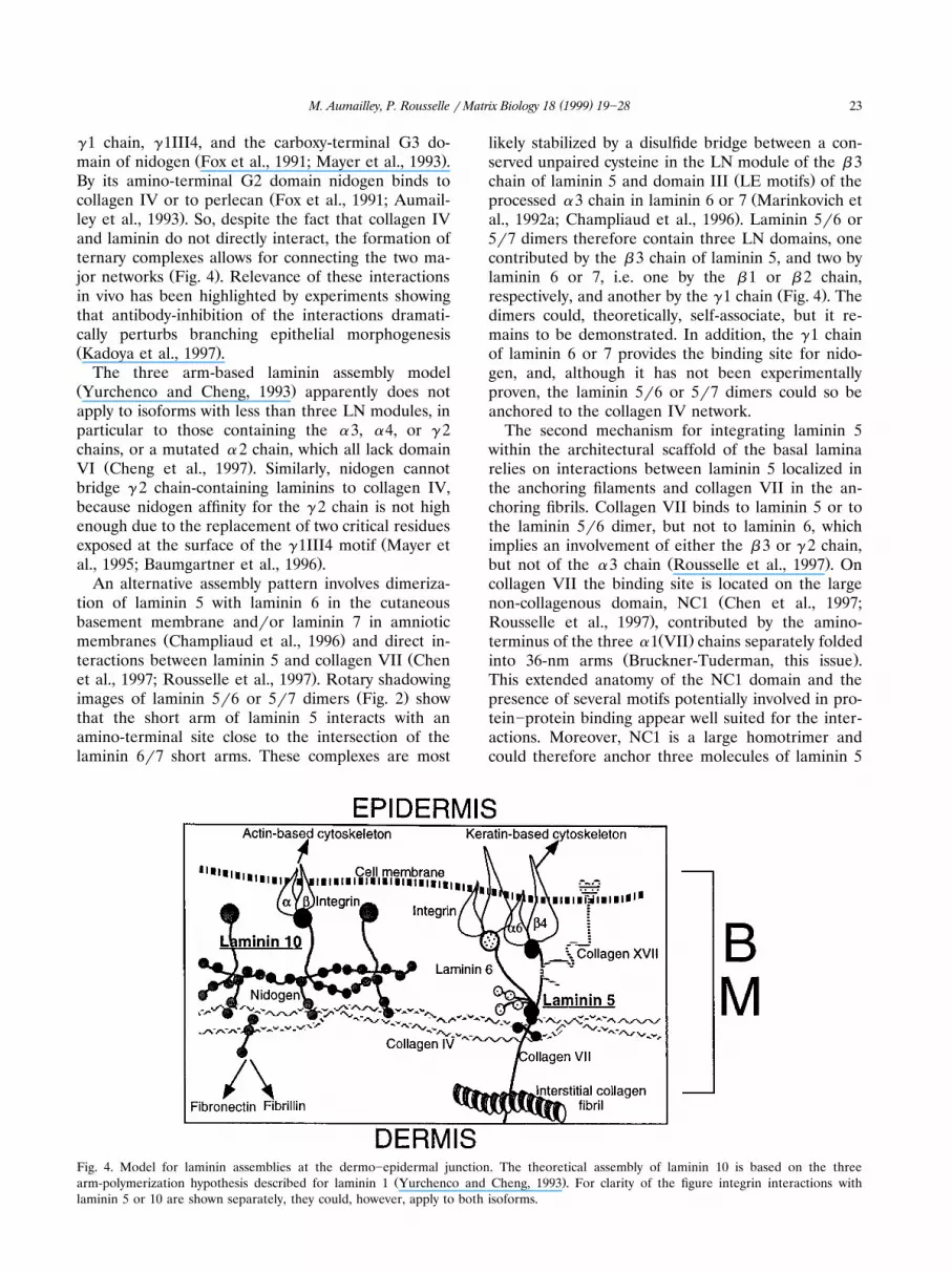
Fig. 4. Model for laminin assemblies at the dermo!epidermal junction. The theoretical assembly of laminin 10 is based on the threeŽ .arm-polymerization hypothesis described for laminin 1 Yurchenco and Cheng, 1993 . For clarity of the figure integrin interactions with
laminin 5 or 10 are shown separately, they could, however, apply to both isoforms.

( )M. Aumailley, P. Rousselle ! Matrix Biology 18 1999 19!2824
Ž .Chen et al., 1997; Rousselle et al., 1997 , and interactŽ .with collagen IV Burgeson, 1993 and probably other
components located in the lamina densa such as theGDA-J!F3 antigen, a small 45!50-kDa proteinŽ .Gayraud et al., 1997 .
Several observations suggest that interactions mayalso exist between laminin 5 and collagen XVII. Thelatter is a transmembrane protein with a type IIorientation and a carboxy-ectodomain folded into a60!70-nm rod adjacent to the cell membrane and a
Ždistal 100!130-nm flexible tail Li et al., 1993; Hirako.et al., 1996; Balding et al., 1997 . Both proteins are
Žco-localized in the anchoring filaments Rousselle et.al., 1991; Masunaga et al., 1997 . In cicatricial pem-
phigoid, the autoantibodies directed against either theŽC terminus of collagen XVII or laminin 5 Domloge-
Hultsch et al., 1992; Shimizu et al., 1995; Bedane et.al., 1997 could impair sticking of laminin 5 to colla-
gen XVII, a condition that will result in thedermo!epidermal dehiscence observed in this humanbullous disorder. Furthermore, in human skin lackinglaminin 5, the extracellular domain of collagen XVII
Ž .is abnormally localized Masunaga et al., 1998 whichsuggests that laminin 5 helps to position the C-terminus of collagen XVII. This agrees with prelimi-nary in vitro interaction studies showing direct bind-ing of the collagen XVII ectodomain to laminin 5Ž .Reddy et al., 1998 .
Additional interactions of laminins with extracellu-lar components were mapped to the LG domainswhich have heparin-binding activity. Other proteinslike the fibulins may connect laminins to the underly-
Žing stroma Timpl and Brown, 1996; Aumailley and.Gayraud, 1998 . In particular, fibulin 2 has affinity for
a recombinant L4 module of the laminin $ 2 chainŽ .Utani et al., 1997 which is, however, probably cleaved
Žoff during extracellular processing of the chain Vailly.et al., 1994; Giannelli et al., 1997 .
An attractive hypothesis is that an extremely highstructural and biological diversity will be achieved bymixing varying proportions of several laminin iso-forms and by superimposing laminin 1- and laminin5-based assemblies. The multiple interactions oflaminins presumably regulate their biological activityby affecting the conformation and the spatial orienta-tion of the different isoforms and of their subdo-mains. Conversely, multiple interactions could restrictmolecular flexibility and maintain the different com-ponents of the anchoring complexes in appropriatepositions in spite of strong environmental tensions.
5. Laminins and cellular interactions
By interacting with integrins and other cell surfacecomponents, laminins are endowed with the propertyto control cellular activities such as migration, differ-
entiation, polarity, proliferation, apoptosis, or geneexpression. Several integrin binding sites were mapped
Žfirst on laminin 1 and later on other isoforms for.references see Aumailley et al., 1996 . Specificly for
laminin 1, there is a cryptic binding site for RGD-de-pendent integrins in the domain IIIa of laminin "1chain. For some laminin isoforms, the aminoterminalregions are recognized by the classical collagen-bind-ing integrins, "1# 1 or " 2# 1. For all isoforms, themajor cell binding sites are located in the carboxyterminal region, and the triple-stranded coil-coiledconformation of " , # and $ chains is required forintegrin binding to laminin 1, 2 and 4, 5, and pre-
Ž .sumably other isoforms Aumailley et al., 1996 . Thecurrent interpretation is, however, that integrins bindto proximal LG domains, whose correct folding de-pends on the presence of the adjacent helical rod.Indeed, the activity of recombinant LG domains alonedoes not always reproduce that of native lamininsŽ .Sung et al., 1993; Mizushima et al., 1997 and differ-ent activities may be expressed in the presence or
Žabsence of the coil-coiled fold Sung et al., 1993;.Brandenberger et al., 1996: Patton et al., 1997 . This
highlights the fact that protein folding is crucial forthe biological activity of laminin adhesion motifs, andthat ascribing a cell binding site at the amino acidlevel probably requires site-directed mutagenesis inthe context of heterotrimers. The " 3# 1, " 6# 1, " 6#4and " 7# 1 integrins are involved in these conforma-tion-dependent interactions with all laminins investi-gated to date, with the exception of laminin 5 for the" 7# 1 integrin and laminin 1 for the " 3# 1 integrinŽ .Aumailley et al., 1996; Yao et al., 1996 . In addition,the two last carboxyterminal LG domains, proteolyti-cally processed in some " chains, interact with "-dys-troglycan, a component of the dystrophin!glycopro-
Ž .tein complex Henry and Campbell, 1996 . The "-dys-troglycan-mediated cellular interactions may be
Žlaminin isoform-specific Pall et al., 1996; Angoli et.al., 1997 and regulate epithelial morphogenesis
Ž .Durbeej and Ekblom, 1997 .Integrins mediate the bidirectional transfer of in-
formation between the extracellular matrix and thecell interior and an obvious and important issue is todetermine the precise and possibly specific roles ofeach laminin-binding integrin. Activation of " 3# 1 or" 6#4 integrins triggers different signaling pathwaysŽJewell et al., 1995; Wary et al., 1996; Xia et al., 1996;
.Mainiero et al., 1997 and the " 3# 1 integrin trans-Ždominantly regulates the " 6 integrin subunit Dogic
.et al., 1998 . Furthermore, laminin-binding integrinsŽdiffer in their affinity for laminins Delwel et al., 1993,
.1994 and in their sub-cellular localization. In particu-lar, in resting keratinocytes " 6#4 integrins are con-fined to hemidesmosomes while " 3# 1 integrins arelocated to cell!cell contacts, i.e. respectively, to the

( )M. Aumailley, P. Rousselle ! Matrix Biology 18 1999 19!28 25
Žbasal and lateral plasma membranes Nievers et al.,.this issue . Moreover, the " 3# 1 and " 6#4 integrins
Ž .participate in different anchorage systems Fig. 4 andconnect laminins to, respectively, the actin- or ker-
Ž .atin-based cytoskeleton Nievers et al., this issue .The typical sub-cellular distribution and intracellularconnections typical of the " 3# 1 and " 6#4 integrinsare linked to distinct functions. The " 3# 1 integrin
Žinitiates binding of cells to laminins Carter et al.,.1991; Rousselle and Aumailley, 1994 , whereas the
" 6#4 integrin is involved in the stable anchorage ofthe basal layer of resting keratinocytes to lamininsŽ .Carter et al., 1990 . Consequently, alterations of theselinkages, either due to changes in the laminin subs-trates or in the expression of integrins, are associatedwith modifications of cell movement under physiologi-cal or pathological conditions. For instance ker-atinocytes have an increased motility when they con-
Žstitutively lack the #4 integrin chain Niessen et al.,.1996 , or when they loose contact with laminin fol-
Ž .lowing injury Carter et al., 1991 . Interestingly, theprecursor and mature forms of laminin 5 may have
Žopposite effects on cell migration Giannelli et al.,.1997; Goldfinger et al., 1998 . The " 3# 1 integrin may
also be involved in matrix assembly as inferred by thephenotype of " 3 integrin chain-deficient mice, pre-senting with a disorganization of the kidney and skin
Žbasal lamina Kreidberg et al., 1996; DiPersio et al.,.1997 . Although the molecular mechanisms underly-
ing this particular role are elusive, integrins mayinduce, in vivo, clustering of laminins at the cellsurface, a nucleation step possibly needed prior to
Ž .laminin polymerization Fleischmajer et al., 1998 .The " 6# 1 integrin, "-dystroglycan and laminin 1 areinvolved in epithelial cell polarisation at the moment
Žof epithelial!mesenchymal transition Ekblom, 1996;.Durbeej and Ekblom, 1997 .
6. Hierarchy of laminin interactions
A hierarchy in the physiological relevance of theversatile interaction repertoire of laminins can bededuced from the phenotypes of patients affectedwith inborn or acquired diseases and of mice bearingsite-directed mutations. Targeted extinction of the
Žgenes coding for the laminin $1 chain Smyth et al.,. Ž1999 , the # 1 integrin chain Fassler et al., 1995;¨
. ŽStephens et al., 1995 or dystroglycan Williamson et.al., 1997 lead to early embryonic lethality which
indicates that laminin 1 and possibly other isoformsand their receptors are absolutely required for embry-onic development. By contrast, certain laminins orindividual receptors, in particular laminins 5 and 6and the " 3# 1, " 6# 1, and " 6#4 integrins, are ap-parently not involved in cell movement during em-
bryogenesis and morphogenesis or compensatorymechanisms may occur in these events.
However, it is clear that there is no compensationfor the structural and mechanical roles of laminin 5and for its binding to " 6#4 integrins at the cellsurface and to collagens in the sub-lamina and laminadensa. Indeed, absence of laminin 5 or " 6#4 integrin,or alterations of their interactions are associated inman as well as in mouse with a dramatic loss ofcohesion between epidermis and dermis and withcomplications leading most frequently to deathŽAumailley and Krieg, 1996; Bruckner-Tuderman, this
.issue . The phenotypes resulting from the absence oflaminin 5 are, at least in man, more severe than thoseresulting from the absence of the " 6#4 integrin. Itindicates that under circumstances of high tensionother laminin isoforms cannot replace the structuralfunction of laminin 5, while that of the " 6#4 integrinis apparently compensated partially by other linkageswith cell surface components, such as may be otherintegrins, collagen XVII or "-dystroglycan. The lattertwo may also function as signal transducers since theintracellular domain of collagen XVII contains poten-
Ž .tial phosphorylation sites Li et al., 1993 and "-dys-troglycan is linked to #-dystroglycan, a transmem-brane polypeptide containing phosphotyrosine con-sensus sequence and several proline-rich regionsŽ .Ibraghimov-Beskrovnaya et al., 1993 .
In conclusion, laminin 1 and its receptors are abso-lutely required for morphogenesis and embryogenesiswhile other isoforms, such as laminin 5, are not indis-pensable for these early events. Nevertheless, lamininisoforms have tissue-specific functions at later devel-opment stages. In particular, laminin 5 and its inter-actions with other extracellular or transmembranemolecules of the anchoring complexes are most im-portant to maintain the architecture and the stabilityof the basement membrane at the epidermal junctionand there is no redundancy for this specific mechani-cal function. In contrast, redundancy may exist forsignal transduction regulating cellular behavior.
Acknowledgements
The authors gratefully acknowledge Dr Neil Smythfor critically reading the manuscript, Dr D. Keene formaking the rotary-shadowed imaging of laminins, andthe Centre National de la Recherche Scientifique, theAssociation pour la Recherche sur le Cancer, the
Ž .PROCOPE programme MA and PR , the DeutscheForschungsgemeinschaft and the University of
Ž .Cologne MA for financial support.
References
Aberdam, D., Aguzzi, A., Baudoin, C., Galliano, M.F., Ortonne,J.R., Meneguzzi, G., 1994. Developmental expression of nicein

( )M. Aumailley, P. Rousselle ! Matrix Biology 18 1999 19!2826
Ž .adhesion protein laminin 5 subunits suggests multiple mor-phogenetic roles. Cell Adhes. Commun. 2, 115!129.
Angoli, D., Corona, R., Baresi, R., Mora, M., Wanke, E., 1997.Laminin-" 2 but not -" 1-mediated adhesion of humanŽ . Ž .Duchenne and murine mdx dystrophic myotubes is seriouslydefective. FEBS Lett. 408, 341!344.
Aumailley, M., Gayraud, B., 1998. Structure and biological activityof the extracellular matrix. J. Mol. Med. 76, 253!265.
Aumailley, M., Krieg, T., 1996. Laminins: a family of diversemultifunctional molecules of basement membranes. J. Invest.Dermatol. 106, 209!214.
Aumailley, M., Battaglia, C., Mayer, U., et al., 1993. Nidogenmediates the formation of ternary complexes of basement mem-brane components. Kidney Int. 43, 7!12.
Aumailley, M., Gimond, C., Rousselle, R., 1996. Integrin-mediatedcellular interactions with laminins. In: Ekblom, P., Timpl, R.Ž .Eds. , The Laminins Harwood. Academic Publishers, pp.127!158.
Balding, S.D., Diaz, L.A., Giudice, G.A., 1997. A recombinant formof the human BP180 ectodomain forms a collagen-like ho-motrimeric complex. Biochemistry 36, 5821!8830.
Baumgartner, R., Czisch, M., Mayer, U., et al., 1996. Structure ofthe nidogen binding LE module of the laminin $1 chain insolution. J. Mol. Biol. 257, 658!668.
Beck, K., Dixon, T.W., Engel, J., Parry, D.A.D., 1993. Ionic interac-tions in the coiled-coil domain of laminin determine the speci-ficity of chain assembly. J. Mol. Biol. 231, 311!323.
Bedane, C., McMillan, J.R., Balding, S.D., et al., 1997. Bullouspemphigoid and cicatricial pemphigoid autoantibodies react withultrastructurally separable epitopes on the BP180 ectodomain:evidence that BP180 spans the lamina lucida. J. Invest. Derma-tol. 108, 901!907.
Brandenberger, R., Kammerer, R.A., Engel, J., Chiquet, M., 1996.Ž .Native chick laminin-4 containing the # 2 chain s-laminin pro-
motes motor axon growth. J. Cell Biol. 135, 1583!1592.
Brown, J.C., Wiedemann, H., Timpl, R., 1994. Protein binding andŽcell adhesion properties of two laminin isoforms AmB1eB2e,
.AmB1sB2e from human placenta. J. Cell Sci. 107, 329!338.
Burgeson, R.E., 1993. Type VII collagen, anchoring fibrils, andepidermolysis bullosa. J. Invest. Dermatol. 101, 252!255.
Carter, W.G., Kaur, R., Gil, S.G., Gahr, P.J., Wayner, E.A., 1990.Distinct functions for integrins " 3# 1 in focal adhesions and" 6#4!bullous pemphigoid antigen in a new stable anchoring
Ž .contact SAC of keratinocytes: relation to hemidesmosomes. J.Cell Biol. 111, 2141!3154.
Carter, W.G., Ryan, M.C., Gahr, P.J., 1991. Epiligrin, a new celladhesion ligand for integrin " 3# 1 in epithelial basement mem-branes. Cell 65, 599!610.
Champliaud, M.F., Lunstrum, G.R., Rousselle, R., Nishiyama, T.,Keene, D.R., Burgeson, R.E., 1996. Human amnion contains anovel laminin variant, laminin 7, which like laminin 6, covalentlyassociates with laminin 5 to promote stable epithelial-stromalattachment. J. Cell Biol. 132, 1189!1198.
Chen, W., Marinkovich, M.P., Veis, A., et al., 1997. Interactions ofŽ .the amino-terminal noncollagenous NC1 domain of type VII
collagen with extracellular matrix components. A potential rolein epidermal!dermal adherence in human skin. J. Biol. Chem.272, 14516!14522.
Cheng, V.S., Champliaud, M.F., Burgeson, R.E., Marinkovich, M.R.,Yurchenco, P.D., 1997. Self-assembly of laminin isoforms. J.Biol. Chem. 272, 21525!31532.
Cooper, A.R., MacQueen, H.A., 1983. Subunits of laminin aredifferentially synthesized in mouse eggs and early embryos. Dev.Biol. 96, 467!471.
Delwel, G.O., Hogervorst, R., Kuikman, L., Paulsson, M., Timpl,R., Sonnenberg, K., 1993. Expression and function of cyto-plasmic variants of the integrin " 6 subunit in transfected K562
cells: activation-dependent adhesion and interaction with iso-forms of laminin. J. Biol. Chem. 268, 25865!25875.
Delwel, G.O., de Melker, A.A., Hogervorst, F., et al., 1994. Distinctand overlapping ligand specificities of the " 3Ab1 and " 6Ab1integrins: recognition of laminin isoforms. Mol. Biol. Cell 5,203!215.
DiPersio, C.M., Hodivala-Dike, K.M., Jaenisch, R., Kreidberg, J.A.,Hynes, R.O., 1997. " 3# 1 integrin is required for normal devel-opment of the epidermal basement membrane. J. Cell Biol. 137,729!742.
Dogic, D., Rousselle, R., Aumailley, W., 1998. Cell adhesion tolaminin 1 or 5 induces isoform-specific clustering of integrinsand other focal adhesion components. J. Cell Sci. 111, 793!802.
Dogic, D., Holsmann, H., Sherman, W., Fox, J.W., Broermann, R.,Paulsson, M., Aumailley, M., submitted. Cell adhesion to apopulation of laminin isoforms isolated from normal renal tis-sue.
Domloge-Hultsch, N., Gammon, W.R., Briggaman, R.A., Gil, S.G.,Carter, W.G., Yancey, K.B., 1992. Epiligrin, the major humankeratinocyte integrin ligand, is a target in both an acquiredautoimmune and an inherited subepidermal blistering skin dis-ease. J. Clin. Invest. 90, 1628!1633.
Durbeej, M., Ekblom, R., 1997. Dystroglycan and laminins: glyco-conjugates involved in branching epithelial morphogenesis. Exp.Lung Res. 23, 109!118.
Durbeej, M., Fecker, L., Hjalt, T., et al., 1996. Expression oflaminins "1, "5 and # 2 chains during embryogenesis of thekidney and vasculature. Matrix Biol. 15, 397!413.
Dziadek, M., Timpl, R., 1985. Expression of nidogen and laminin inbasement membranes during mouse embryogenesis and in tera-tocarcinoma cells. Dev. Biol. 111, 372!382.
Eady, R.A.J., 1986. Babes, blisters and basement membranes: fromsticky molecules to epidermolysis bullosa. Clin. Exp. Dermatol.12, 161!170.
Edgar, D., Timpl, R., Thoenen, H., 1988. Structural requirementsfor the stimulation of neurite outgrowth by two variants oflaminin and their inhibition by antibodies. J. Cell Biol. 106,1299!1306.
Ekblom, R., 1996. Receptors for laminins during epithelial mor-phogenesis. Curr. Opin. Cell Biol. 8, 700!706.
Ellison, J., Garrod, D.R., 1984. Anchoring filaments of the amphib-ian epidermal!dermal junction transverse the basal lamina en-tirely from plasma membranes of hemidesmosomes to the dermis.J. Cell Sci. 72, 163!172.
Fassler, R., Pfaff, M., Murphy, J., et al., 1995. The lack of # 1¨integrin gene in embryonic stem cells affects cell morphology,migration and adhesion but not integration into the inner cellmass of blastocysts. J. Cell Biol. 128, 979!988.
Fleischmajer, R., Utani, A., MacDonald, E.D., et al., 1998. Initia-tion of skin basement membrane formation at theepidermo!dermal interface involves assembly of laminin throughbinding to cell membrane receptors. J. Cell Sci. 111, 1929!1940.
Fox, J., Mayer, U., Nischt, R., et al., 1991. Recombinant nidogenconsists of three globular domains and mediates binding oflaminin to collagen IV. EMBO J. 10, 3137!3146.
Galliano, M.F., Aberdam, D., Aguzzi, A., Ortonne, J.R., Meneguzzi,W., 1995. Cloning and complete primary structure of the mouselaminin " 3 chain. J. Biol. Chem. 270, 21820!21826.
Gayraud, B., Hopfner, B., Jassim, A., Aumailley, M., Bruckner-Tuderman, L., 1997. Characterization of a novel component ofepithelial basement membranes using GDA-J!F3 monoclonalantibody J. Biol. Chem. 272, 9531!9538.
Giannelli, G., Falk-Marzillier, J., Schiraldi, O., Stetler-Stevenson,W.G., Quaranta, V., 1997. Induction of cell migration by matrixmetalloprotease-2 cleavage of laminin-5. Science 277, 225!228.
Goldfinger, L.E., Stack, M.S., Jones, J.C.R., 1998. Processing of

( )M. Aumailley, P. Rousselle ! Matrix Biology 18 1999 19!28 27
laminin-5 and its functional consequences: role of plasmin andtissue-type plasminogen activator. J. Cell Biol. 141, 255!265.
Heagerty, A.H.M., Eady, R.A.J., Kennedy, A.A., Nicolaides, K.H.,Rodek, C.H., Hsi, B.L., Ortonne, J.P., 1987. Rapid prenataldiagnosis of epidermolysis bullosa lethalis using GB3 monoclo-nal antibody. Br. J. Dermatol. 117, 271!276.
Henry, M.D., Campbell, K.P., 1996. Dystroglycan & an extracellu-lar matrix receptor linked to the cytoskeleton. Curr. Opin. CellBiol. 8, 625!631.
Hirako, Y., Usukura, J., Nishizawa, V., Owaribe, K., 1996. Demon-stration of the molecular shape of BP180, a 180-kDa bullouspemphigoid antigen and its potential trimer formation. J. Biol.Chem. 271, 13739!13745.
Ibraghirnov-Beskrovnaya, O., Milatovich, A., Ozcelik, T., et al.,1993. Human dystroglycan: skeletal muscle cDNA, genomicstructure, origin of tissue specific isoforms and chromosomallocalization. Hum. Mol. Genet. 2, 1651!1657.
livanainen, A., Sainio, K., Sariola, H., Tryggvason, K., 1995. Primarystructure and expression of a novel human laminin "4 chain.FEBS Lett. 365, 183!188.
Jewell, K., Kapron-Bras, C., Jeevaratnam, R., Dedhar, S., 1995.Stimulation of tyrosine phosphorylation of distinct proteins inresponse to antibody-mediated ligation and clustering of " 3 and" 6 integrins. J. Cell Sci. 108, 1165!1174.
Kadoya, V., Salmivirta, K., Talts, J.F., et al., 1997. Importance ofnidogen binding to laminin $1 for branching epithelial mor-phogenesis of the submandibular gland. Development 124,683!691.
Kallunki, R., Sainio, K., Eddy, R., et al., 1992. A truncated lamininchain homologous to the B2 chain: structure, spatial expression,and chromosomal assignment. J. Cell Biol. 119, 679!693.
Kreidberg, J.A., Donovan, M.J., Goldstein, S.L., et al., 1996. " 3# 1integrin has a crucial role in kidney and lung organogenesis.Development 122, 3537!3547.
Li, K., Tamai, K., Tan, E.M.L., Uitto, J., 1993. Cloning of type XVIIcollagen complementary and genomic DNA sequences of mouse
Ž .180 kilodalton bullous pemphigoid antigen BPAG2 predict aninterrupted collagenous domain, a transmembrane segment, andunusual features in the 5'-end of the gene and the 3'-untrans-lated region of the mRNA. J. Biol. Chem. 268, 8825!8834.
Lindblom, A., Marsh, T., Fauser, C., Engel, J., Paulsson, M., 1994.Characterization of native laminin from bovine kidney and com-parison with other laminin variants. Eur. J. Biochem. 219,383!392.
Mainiero, F., Murgia, C., Wary, K.K., et al., 1997. The coupling of" 6#4 integrin to Ras-MAP kinase pathways mediated by Shrcontrols keratinocyte proliferation. EMBO J. 16, 2365!2375.
Marinkovich, M.R., Lunstrum, G.R., Keene, D.R., Burgeson, R.E.,1992a. The dermal!epidermal junction of human skin contains anovel laminin variant. J. Cell Biol. 119, 695!703.
Marinkovich, M.P., Lunstrum, G.R., Burgeson, R.E., 1992b. Theanchoring filament protein kalinin is synthesized and secreted asa high molecular weight precursor. J. Biol. Chem. 267,17900!17906.
Masunaga, T., Shimizu, H., Yee, C., et al., 1997. The extracellulardomain of BPAG2 localizes to anchoring filaments and itscarboxyl terminus extends to the lamina densa of normal humanepidermal basement membrane. J. Invest. Dermatol. 109,200!206.
Masunaga, T., Shimizu, H., Yancey, K.B., Nishikawa, T., 1998.Altered ultrastructural localization of the C-terminus of Bullous
Ž .Pemphigoid antigen BPAG2 in human skin lacking laminin 5.J. Invest. Dermatol. 110, 508.
Mayer, U., Nischt, R., Poschl, E., et al., 1993. A single EGF-like¨motif of laminin is responsible for high affinity nidogen binding.EMBO J. 12, 1879!1885.
Mayer, U., Poschl, E., Gerecke, D.A., Wagman, D.W., Burgeson,¨
R.E., Timpl, R., 1995. Low nidogen affinity of laminin-5 can beattributed to two serine residues in EGF-like motif $ 2III4.FEBS Lett. 365, 129!132.
Miner, J.H., Patton, B.L., Lentz, S.I., et al., 1997. The laminin "
chains: expression, developmental transitions, and chromosomallocations of "1!5, identification of heterotrimeric laminins 8!11,and cloning of a novel " 3 isoform. J. Cell Biol. 137, 685!701.
Mizushima, H., Takamura, H., Miyagi, V., et al., 1997. Identifica-tion of integrin-dependent and independent cell adhesion do-mains in COOH-terminal globular region of laminin-5 " 3 chain.Cell Growth Diff. 8, 979!987.
Niessen, C.M., van der Raaij-Helmer, M.H., Hulsman, E.H., vander Neut, R., Jonkman, M.F., Sonnenberg, A., 1996. Deficiencyof the integrin #4 subunit in junctional epidermolysis bullosawith pyloric atresia: consequences for hemidesmosome forma-tion and adhesion properties. J. Cell Sci. 109, 1695!1706.
Pall, E.A., Bolton, K.M., Ervasti, J.M., 1996. Differential heparininhibition of skeletal muscle "-dystroglycan binding to laminins.J. Biol. Chem. 271, 2817!3821.
Patton, B.L., Miner, J.H., Chiu, A.X., Sanes, J.R., 1997. Distribu-tion and function of laminins in the neuromuscular system ofdeveloping, adult, and mutant mice. J. Cell Biol. 139, 1507!1521.
Paulsson, M., Saladin, K., 1989. Mouse heart laminin. Purificationof the native protein and structural comparison with Engel-breth!Holm!Swarm tumor laminin. J. Biol. Chem. 264,18726!18732.
Paulsson, M., Aumailley, M., Deutzmann, R., Timpl, R., Beck, K.,Engel, J., 1987. Laminin!nidogen complex. Extraction withchelating agents and structural characterization. Eur. J. Biochem.166, 11!19.
Reddy, D., Muller, P., Tran, H., et al., 1998. The extracellulardomain of BP180 binds laminin-5. J. Invest. Dermatol. 110, 593.
Rousselle, R., Aumailley, M., 1994. Kalinin is more efficient thanlaminin in promoting adhesion of primary keratinocytes andsome other epithelial cells and has a different requirement forintegrin receptors. J. Cell Biol. 125, 1205!1214.
Rousselle, P., Lunstrum, G.R., Keene, D.A., Burgeson, R.E., 1991.Kalinin: an epithelium-specific basement membrane adhesionmolecule that is a component of anchoring filaments. J. CellBiol. 114, 567!576.
Rousselle, P., Golbik, R., van der Rest, M., Aumailley, M., 1995.Ž .Structural requirement for cell adhesion to kalinin laminin-5 .
J. Biol. Chem. 270, 13766!13770.
Rousselle, P., Keene, D.R., Ruggiero, F., Champliaud, M.F., vander Rest, M., Burgeson, R.E., 1997. Laminin 5 binds the NC-1domain of type VII collagen. J. Cell Biol. 138, 719!728.
Ryan, M.C., Tizard, R., VanDevanter, D.R., Carter, W.A., 1994.Cloning of the LamA3 gene encoding the " 3 chain of theadhesive ligand epiligrin. Expression in wound repair. J. Biol.Chem. 269, 22779!22787.
Schuger, L., Skubitz, A.P., Zhang, J., Sorokin, L., He, L., 1997.Laminin "1 chain synthesis in the mouse developing lung:requirement for epithelial-mesenchymal contact and possiblerole in bronchial smooth muscle development. J. Cell Biol. 139,553!562.
Schuler, F., Sorokin, L.M., 1995. Expression of laminin isoforms inmouse myogenic cells in vitro and in vivo. J. Cell Sci. 108,3795!3805.
Shimizu, W., Masunaga, T., lshiko, A., et al., 1995. Autoantibodiesfrom patients with cicatricial pemphigoid target different sites inepidermal basement membrane. J. Invest. Dermatol. 104,370!373.
Simo, P., Bouziges, F., Lissitzky, J.C., Sorokin, L., Kedinger, M.,Simon-Assmann, P., 1992. Dual and asynchronous deposition oflaminin chains at the epithelial!mesenchymal interface in thegut. Gastroenterology 102, 1835!1845.
Smyth, N., Vantansever, H.S., Meyer, M., Frie, C., Paulsson, M.,

( )M. Aumailley, P. Rousselle ! Matrix Biology 18 1999 19!2828
Edgar, D., 1999. Absence of basement membranes after target-ing the LAMC1 gene results in embryonic lethality due tofailure of endoderm differentiation. J. Cell. Biol. 144, 151!160.
Sorokin, L., Girg, W., Gopfert, T., Hallmann, R., Deutzmann, R.,1994. Expression of novel 400-kDa laminin chains by mouse andbovine endothelial cells. Eur. J. Biochem. 223, 603!610.
Sorokin, L.M., Pausch, F., Frieser, M., Kroger, S., Ohage, E.,¨Deutzmann, R., 1997a. Developmental regulation of the laminin"5 chain suggests a role in epithelial and endothelial cell matu-ration. Dev. Biol. 189, 285!300.
Sorokin, L.M., Pausch, F., Burbeel, M., Ekblom, P., 1997b. Differ-Ž .ential expression of five laminin " $5 chains in developing and
adult mouse kidney. Dev. Dyn. 210, 446!462.Stephens, L.E., Sutherland, A.E., Klimanskaya, I.M., et al., 1995.
Deletion of # 1 integrins in mice results in inner cell massfailure and peri-implantation lethality. Genes Dev. 9, 1883!1895.
Sung, U., O’Rear, J.J., Yurchenco, P.D., 1993. Cell and heparinbinding in the distal long arm of laminin: identification of activeand cryptic sites with recombinant and hybrid glycoprotein. J.Cell Biol. 123, 1255!1268.
Thomas, T., Dziadek, M., 1993. Genes coding for basement mem-brane glycoproteins laminin, nidogen, and collagen IV are dif-ferentially expressed in the nervous system and by epithelial,endothelial, and mesenchymal cells of the mouse embryo. Exp.Cell Res. 208, 54!67.
Tiger, C.R., Champliaud, M.F., Pedrosa-Domellof, F., Thornell,L.E., Ekblom, P., Gullberg, D., 1997. Presence of laminin "5chain and lack of laminin "1 chain during human muscledevelopment and in muscular dystrophies. J. Biol. Chem. 272,28590!28595.
Timpl, R., 1996. Macromolecular organization of basement mem-branes. Curr. Opin. Cell Biol. 8, 618!624.
Timpl, R., Brown, J.C., 1996. Supramolecular assembly of basementmembranes. Bioessays 18, 123!132.
Utani, A., Nomizu, M., Timpl, R., Roller, P.P., Yamada, Y., 1994.Laminin chain assembly. Specific sequences at the C terminus ofthe long arm are required for the formation of specific double-and triple-stranded coiled-coil structures. J. Biol. Chem. 269,19167!19175.
Utani, A., Nomizu, W., Yamada, Y., 1997. Fibulin-2 binds to the
short arms of laminin-5 and laminin-1 via conserved amino acidsequences. J. Biol. Chem. 272, 2814!2820.
Vailly, J., Verrando, R., Champliaud, M.F., et al., 1994. The 100-kDa
chain of nicein!kalinin is a laminin B2 chain variant. Eur. J.Biochem. 219, 209!218.
Wary, K.K., Mainiero, F., Isakoff, S.A., Marcantonio, E.E., Gian-
cotti, F.G., 1996. The adaptor protein Shc couples a class ofintegrins to the control of cell cycle progression. Cell 87, 733!743.
Williamson, R.A., Henry, M.D., Daniels, K.J., et al., 1997. Dystrog-
lycan is essential for early embryonic development: disruption of
Reichert’s membrane in Dag1-null mice. Hum. Mol. Genet. 6,831!841.
Wu, T.C., Wan, Y.J., Chung, E.A., Damjanov, L., 1983. Immunohis-
tochemical localization of entactin and laminin in mouse em-bryos and fetuses. Dev. Biol. 100, 496!505.
Xia, Y., Gil, S.G., Carter, W.G., 1996. Anchorage mediated byŽ .integrin " 6#4 to laminin 5 epiligrin regulates tyrosine phos-
phorylation of a membrane-associated 80-kD protein. J. CellBiol. 132, 727!740.
Yao, C.C., Ziober, B.L., Squillace, R.M., Kramer, R.H., 1996.
Alpha7 integrin mediates cell adhesion and migration on specificlaminin isoforms. J Biol. Chem. 271, 25598!25603.
Yurchenco, P.D., Tsilibary, E.C., Charonis, A.S., Furthmayr, H.,
1985. Laminin polymerization in vitro. Evidence for a two-stepassembly with domain specificity. J. Biol. Chem. 260, 7636!7644.
Yurchenco, P.D., Cheng, Y.S., Colognato, H., 1992. Laminin forms
an independent network in basement membranes. J. Cell Biol.117, 1119!1133.
Yurchenco, P.D., Cheng, Y.S., 1993. Self-assembly and calcium-bi-
nding sites in laminin. A three-arm interaction model. J. Biol.Chem. 268, 17286!17299.
Yurchenco, R.D., Quan, Y., Colognato, H., et al., 1997. The "
chain of laminin-1 is independently secreted and drives secretion
of its #- and $-chain partners. Proc. Natl. Acad. Sci. USA 94,10189!10194.