Embed Size (px)
Citation preview
Molecular & Biochemical Parasitology 131 (2003) 65–75
Functional expression and characterizationof Schistosoma mansonicathepsin B and itstrans-activation
by an endogenous asparaginyl endopeptidase�
Mohammed Sajida,∗, James H. McKerrowa, Elizabeth Hansella,Mary A. Mathieua, Kimberley D. Lucasa, Ivy Hsieha, Doron Greenbauma,
Matthew Bogyob, Jason P. Saltera, Kee C. Lima, Christopher Franklina,Jea-Hyoun Kima, Conor R. Caffreya
a Department of Pathology, Tropical Disease Research Unit and Sandler Centre for Basic Research in Parasitic Diseases,University of California San Francisco, Box 0511, San Francisco, CA 94143, USA
b Department of Biochemistry and Biophysics, University of California San Francisco,San Francisco, CA 94143, USA
Received 10 February 2003; received in revised form 2 July 2003; accepted 3 July 2003
Abstract
Peptidases are essential for the establishment and survival of the medically important parasite,Schistosoma mansoni. This helminthexpresses a number of gut-associated peptidases that degrade host blood proteins, including hemoglobin, as a means of nutrition. Usingirreversible affinity probes, we demonstrate thatS. mansonicathepsin B-like endopeptidase 1 (SmCB1) is the most abundant papainfamily cysteine peptidase in both the parasite gut and somatic extracts. SmCB1 zymogen (SmCB1pm) was functionally expressed inPichia pastoris(4–11 mg l−1). Monospecific and immunoselected antibodies raised against SmCB1pm localized the enzyme exclusivelyto the gut lumen and surrounding gastrodermis of adult worms. Recombinant SmCB1pm was unable to catalyze its activation, even atlow pH. However, recombinantS. mansoniasparaginyl endopeptidase (SmAE), another gut-associated cysteine peptidase, processed andactivated SmCB1pmin trans. Consistent with the known specificity of AEs, processing occurred on the carboxyl side of an asparagineresidue, two residues upstream of the start of the mature SmCB1 sequence. The remaining pro-region dipeptide was removed by ratcathepsin C (dipeptidyl-peptidase I)—an action conceivably performed by an endogenous cathepsin C in vivo. The activated recom-binant SmCB1 is biochemically identical to the native enzyme with respect to dipeptidyl substrate kinetics and pH profiles. Also, theserum proteins, hemoglobin, serum albumin, IgG, and�-2 macroglobulin were efficiently degraded. Further, a novel application of anassay to measure the peptidyl carboxypeptidase activity of SmCB1 and other cathepsins B was developed using the synthetic substratebenzoyl-glycinyl-histidinyl-leucine (Bz-Gly-His-Leu). This study characterizes the major digestive cysteine peptidase in schistosomesand defines noveltrans-processing events required to activate the SmCB1 zymogen in vitro which may facilitate the digestive processin vivo.© 2003 Elsevier B.V. All rights reserved.
Keywords:Cathepsin B; Asparaginyl endopeptidase;trans-Processing; Peptidyl-dipeptidase;Schistosoma
Abbreviations:ACC, 7-amino-4-carbamoylmethyl coumarin; Bz-Gly-His-Leu, benzoyl-glycinyl-histidinyl-leucine; DPP I, dipeptidyl peptidase I (cathep-sin C); DTT, dithiothreitol; E-64,l-trans epoxysuccinyl-leucylamido-(4-guanidino)-butane; GIC, gastrointestinal contents; GSH, glutathione (reduced);K11777,N-methylpiperazine-urea-phenylalanyl-homophenylalanyl-vinylsulfone-benzene; LC-ESI-ToF, liquid chromatography-electrospray ionization-timeof flight; NaOAc, sodium acetate; PAGE, polyacrylamide gel electrophoresis; PCR, polymerase chain reaction; PMSF, phenylmethylsulfonyl flouride;Nle, norleucine; SmCB1,S. mansonicathepsin B1 (a.k.a. Sm31); SmAE,S. mansoniasparaginyl endopeptidase (a.k.a.S. mansonilegumain;Sm32); Z-Ala-Ala-Asn, benzoyloxycarbanoyl alanyl-alanyl-asparaginyl 4-methyl-7-amido-coumarin; Z-Arg-Arg-AMC, benzoyloxycarbanoyl arginyl-arginyl4-methyl-7-amido-coumarin; Z-Phe-Arg-AMC, benzoyloxycarbonyl phenylalanyl-arginyl 4-methyl-7-amido-coumarin
� Note: Both SmCB1.1 (accession no. AJ506157) and SmCB1.2 (accession no. AJ506158) sequences were deposited in the EMBL nucleotide database.∗ Corresponding author.E-mail address:[email protected] (M. Sajid).
0166-6851/$ – see front matter © 2003 Elsevier B.V. All rights reserved.doi:10.1016/S0166-6851(03)00194-4
66 M. Sajid et al. / Molecular & Biochemical Parasitology 131 (2003) 65–75
1. Introduction
Human schistosomiasis causes almost one million deathsper year, affects an estimated 200–250 million people,and is second only to malaria as a global parasitic healthproblem. Caused by a trematode bloodfluke, adult wormsingest whole erythrocytes using the globin component ofhemoglobin as a nutrient source[1]. Though chemother-apy of the disease is available, concerns over possibledrug resistance encourage the search for new drug targets.One group of potential targets are those cysteine pepti-dases that are associated with the parasite gut and digesthost proteins to absorbable nutrients[2]. Interruption oftheir function would limit the parasite’s ability to feed andreproduce. Indeed, inhibitors of cysteine peptidases havebeen shown to decrease worm burden and egg productionin mice infected withS. mansoni[3]. Characterizing suchalimentary peptidases and understanding their biochemicaldifferences compared to orthologous host enzymes is afundamental requirement to rational and biospecific drugdesign.
A number of gut-associated cysteine peptidases have beenidentified in adultS. mansoni. These include cathepsin B1(SmCB1; a.k.a. Sm31)[4,5], cathepsin L1[6], cathepsinL2 [7], asparaginyl endopeptidase (SmAE; a.k.a Sm32), orschistosome legumain[8] and cathepsin C[9]. In addition,a schistosome cathepsin D-like aspartic endopeptidase hasbeen described[10].
Cathepsin B activity, presumably due to CB1, is the mostabundant papain-like, cysteine protease detected in the lu-men of the schistosome gut[11]. The gene for SmCB1 en-codes a putative signal sequence, pro-region and catalyticdomain. The native SmCB1 zymogen is processed to a ma-ture 31 kDa glycosylated protein. SmCB1 is secreted intothe gut lumen of adult schistosomes and has also been foundin the gastrodermal cells[12,13]. The enzyme has beenused as a serodiagnostic marker of schistosomiasis[12].Though recombinant SmCB1 has been heterologously ex-pressed in insect cells[14], Saccharomyces cerevisiae[15]and a cell-free system[16], biochemical analysis has beenhampered by poor protein yield or lack of ‘correct’ pro-cessing. As a result, questions as to how pro-SmCB1 is ac-tivated, and whether it can efficiently degrade hemoglobinhave been raised[10].
For this report, we have overexpressed SmCB1 inPichia pastoris in reagent quantities and characterizedthe enzyme with respect to zymogen activation, peptidyl-and protein-substrate specificity, tissue localization, andabundance relative to otherS. mansonipapain-familycysteine peptidases. We also demonstrate for the firsttime a specifictrans-processing event necessary for fullactivation of the SmCB1 zymogen involving a secondand biologically relevant clan CD cysteine peptidase,SmAE.
2. Material and methods
2.1. Chemicals
Unless stated all chemicals were obtained from SigmaChemical Co, St. Loius, MO, USA. Restriction endonucle-ases were purchased from Roche Molecular Biochemicals,Indianapolis, IN. The plasmid vector pPICZ�A, the an-tibiotic Zeocin and X33 strain ofP. pastoriswere fromInvitrogen, Carlsbad, CA.N-methylpiperazine-urea-phenyl-alanine-homophenylalanine-vinylsulfone-phenyl (K11777)was synthesized by Dr. Jim Palmer, Celera Genomics,South San Francisco, CA. Benzoyloxycarbonyl phenylala-nyl-arginyl 4-methyl-7-amido-coumarin (Z-Phe-Arg-AMC)and benzoyloxycarbanoyl arginyl-arginyl 4-methyl-7-amido-coumarin (Z-Arg-Arg-AMC) were obtained fromBachem, King of Prussia, PA. Recombinant rat cathepsin C(dipeptidyl-peptidase I or DPP I) was a kind gift from Dr.John Pedersen of Unizyme, Denmark. The parasite endopep-tidases, cruzain, rhodesain, and SmCB2 are available in thislaboratory and falcipain 2 was a gift from Dr. Phil Rosenthal(UCSF).
2.2. Parasite material
Adult S. mansoniwere obtained as described previously[17]. Prior to the collection of gastrointestinal contents(GIC), parasites were washed in RPMI and then three timeswith distilled water over a wire mesh to remove any RPMI.GIC were then collected as described[13,18].
2.3. Inhibitor iodination
The E-64 analogue DCG-04 was iodinated as previouslydescribed[19].
2.4. Radiolabeling cysteine peptidases in S. mansoniwhole worm extract and GIC
Parasite material was incubated with radio-iodinatedDCG-04 and processed as described[20]. Prior to radio-labeling, samples were either preheated or labeled in theabsence or presence of 10�M of the cathepsin B-selectiveinhibitor, MB-074 [21] for 60 min at room temperature.Labeled proteins were visualized by autoradiography usingstandard methodology.
2.5. pH measurements of GIC
The pH of GIC contents was measured in triplicate atroom temperature using either a Micro comb pH probe(Lazar Research Laboratories, Inc., Los Angeles, CA) orColorpHast indicator strips (pH 6–7; 0.2 unit accuracy; EMScience, Gibbstown, NJ).
M. Sajid et al. / Molecular & Biochemical Parasitology 131 (2003) 65–75 67
2.6. Construction of smcb1 plasmid
Total RNA from mixed-sex adultS. mansoniwas ob-tained using the Quickprep Total RNA kit obtained (Am-bion, Austin, TX). First strand cDNA was generated usinga poly-T oligonucleotide primer and reverse transcriptase(Invitrogen) under standard conditions. This was followedby polymerase chain reaction (PCR)-amplification of thesmcb1open reading frame encoding the pro- and maturedomains (smcb1pm).
The start of the pro-region was predicted using the soft-ware SigPep atwww.exspasy.ch. The forward primer, SmF1,5′AGCTACTCGAGAAAAGACATAT T T CAGTTAAGAA-CGAAAAG-3′ contained sequence complementary for thepredicted pro-region of SmCB1pm and also included akexin endopeptidase cleavage site (Lys-Arg; in italic) andan XhoI restriction site (underlined) upstream of the genespecific sequence. The reverse primer, SmR1, 5′-GTAA-TAGCCGGTCGAATAAACTAAGCGGCCGCAC T GC-3′-end incorporated the complementary sequence for the cod-ing region of the 3′ of SmCB1 and included the nativetranslation termination codon (italics) and aNotI restric-tion site (underlined). Thesmcb1pmnucleotide sequencewas confirmed by bidirectional sequencing at the Biomed-ical Resource Centre, UCSF using both vector specificand gene specific primers. The pPICZ�A-smcb1pmplas-mid was propagated as recommended by manufacturer’sguidelines.
SmCB1pm was also subcloned into the prokaryoteexpression vector pTrcHis2A by PCR (Invitrogen). The for-ward primer, pTF1, 5′-TCGGCGGATCCACATCATCAT-CATCATCATATTTCAGTTAAG-3′ contained sequencecomplementary to the predicted pro-region ofsmcb1pm,a BamHI restriction site (underlined) and a sequence en-coding a polyhistidine fusion tag immediately upstreamof the gene specific sequence. The reverse primer, pTR1,5′-GCCGGTCGAATAAACTAACTGCAGAATAG-3′ inco-rporated the complementary sequence for the codingregion of the 3′ of smcb1pmand included the nativestop site (italics) and aPstI restriction site (underlined).The construct was used to transform chemically com-petent Escherichia colistrain DH5�. The nucleotide se-quence of the pTrcHis2A-smcb1pmplasmid was verifiedby bidirectional sequencing at the Biomedical ResourceCentre.
2.7. Overexpression of smcb1pm in P. pastoris andE. coli
Transfection of X33 strain ofP. pastoriswith SmCB1was carried out using the methodology previously de-scribed[8]. Expression of SmCB1 inE. coli was carriedout as described by the manufacturer’s recommendation(Invitrogen).
2.8. Expression of recombinant SmAE
The expression of SmAE inP. pastoriswas performed asdescribed previously[8].
2.9. Processing of recombinant Pichia-derived SmCB1pm
The ability of SmCB1pm to undergo inter- or intramolec-ular processing and activation was studied by incubating therecombinant protein in 100 mM sodium phosphate, 50 mMcitrate buffer, 2 mM DTT at 0.5 incremental pH values be-tween pH 3.0 and 8.0. Endopeptidolytic activity was mea-sured using Z-Phe-Arg-AMC and the samples also subjectedto SDS–polyacrylamide gel electrophoresis (PAGE) fol-lowed by either western blotting or Coomassie Blue staining.The ability of SmAE totrans-process the SmCB1pm wasalso studied. SmAE was activated as described previously[8] and immobilized usingN-hydroxysuccinamide-activatedsepharose (Amersham Biosciences, Piscataway, NJ) usingthe manufacturer’s guidelines. Immobilized SmAE waswashed extensively in 50 mM sodium acetate (NaOAc; pH4.5), until no detectable unbound activity against the SmAEsubstrate Z-Ala-Ala-Asn-AMC was obtained. The immo-bilized SmAE was incubated at 37◦C with SmCB1pm(≈15–20:1 mg ml−1 protein, SmAE:SmCB1, respectively)in 100 mM sodium phosphate, 50 mM citrate buffer, 2 mMDTT, at 0.5 incremental pH values between pH 3.0 and 8.0for 3 h at 37◦C on a orbital shaker. Processing and activa-tion of SmCB1pm was monitored using Z-Phe-Arg-AMCand SDS–PAGE followed by Coomassie Blue staining.Processing sites were identified by N-terminal protein se-quencing. The activated mature SmCB1 (SmCB1m∗) wascollected by passing the SmAE/sepharose through an emptychromatography column.
2.10. Processing of SmCB1m∗ by cathepsin C
Rat cathepsin C (DPP I; 0.5 U) was incubated with10�g of activated SmCB1m∗ in 50 mM NaOAc, (pH 5.5),2 mM DTT at 37◦C for 40 min. To determine the spe-cific activity and processing of SmCB1m∗, activity againstZ-Phe-Arg-AMC was measured and theN-terminal se-quence identified, respectively.
2.11. Peptidase assays
Peptidase activities were carried out using the fluoro-metric substrates Z-Phe-Arg-AMC, Z-Arg-Arg-AMC (both20�M final concentration) for SmCB1m∗ and Z-Ala-Ala-Asn-AMC (20�M final concentration) for SmAE. Assayswere performed in the presence of 2 mM dithiothreitol(DTT) and at 25◦C using an automated microtiter platespectrofluorimeter (Labsystems Fluoroscan II) in a finalvolume of 250�l. The appearance of 7-amino-4-methyl
68 M. Sajid et al. / Molecular & Biochemical Parasitology 131 (2003) 65–75
coumarin (AMC) was measured with excitation and emis-sion wavelengths of 355 and 460 nm, respectively. One unitof activity was defined as that releasing 1�mol of AMCmin−1. All assays were repeated three times.
A novel exopeptidase activity assay of SmCB1m∗was developed using a modified method that employedpeptidyl-dipeptidase activity[22] to generate a fluorescentadduct. The substrate benzoyl-glycinyl-histidinyl-leucine(Bz-Gly-His-Leu; final 2 mM) is hydrolyzed by a peptidyl-dipeptidase activity which results in the appearance ofHis-Leu. The free amine-of His-Leu spontaneously reactswith fluorescamine (0.05 mg ml−1 from a 0.1 mg ml−1 stockin acetone) and the fluorescent adduct formed is excited at390 nm and measured at 475 nm. SmCB1m∗ was incubatedwith substrate in 100 mM NaOAc, 2 mM DTT (pH 5.5),and the reaction allowed to proceed at 37◦C for 90 min.The reaction was terminated by the addition of 1.0 Mglycine/NaOH buffer (pH 9.0) to a final concentration of200 mM.
A 2D-gel analysis approach was used to monitor serumprotein degradation by SmCB1m∗ (data not shown). Proteinspots that either appeared or disappeared in the presence ofSmCB1 were excised and digested with trypsin using stan-dard methodologies. Peptide fragments analysis was carriedout at the Protein and Nucleic Acid Facility, Stanford Uni-versity.
2.12. Identification of the initial hydrolysis sites ofhemoglobin by SmCB1m∗
A 4% (w/v) filtered bovine hemoglobin solution in100 mM ammonium acetate buffer (pH 5.5), 2 mM GSH,was incubated with SmCB1m∗ (1–2 RFU min−1 withZ-Phe-Arg-AMC). The reaction was carried out for 0, 15,30, and 60 s, after which activity was stopped by additionof 10�M E-64 and immediate placement in liquid nitrogen.Samples were stored at−70◦C until required and analyzedby liquid chromatography-electrospray ionization-time offlight (LC-ESI-ToF) using the Mariner software (AppliedBiosystems, Foster City, CA) by Dr. David Maltby at theMass Spectrometry Unit, UCSF.
2.13. Production of monospecific anti-SmCB1pmantibodies and immunoelectron microscopy
Pure recombinant SmCB1pm (3.0 mg) expressed inP. pastoris was subjected to SDS–PAGE stained withCoomassie Blue and the corresponding protein band excisedand supplied to Covance, CA, for generation of polyclonalsera in New Zealand White Rabbits. Serum from the fourthbleed was used in all subsequent experiments and wasimmunoglobulin purified using a 1 ml HiTrap protein-Gcolumn (Amersham Biosciences) using the manufacturer’sguidelines.
Immunoselection of the monovalent antibody prepara-tion was carried out using standard methodologies. Briefly,
800�g of SmCB1pm expressed inE. coli (see above) wassubjected to SDS–PAGE. Following electrophoresis, pro-teins were electrotransferred to polyvinylidene difluoride(PVDF) membranes as described below. The transferredproteins were visualized using 0.1% (v/v) Ponceau-Red andSmCB1pm excised and washed briefly with 0.1 M sodiumhydroxide to remove the stain. The excised PVDF wasblocked with 5% (w/v) BSA in PBS containing 0.05% (w/v)Tween 20 (PBS/T), washed extensively with PBS/T andincubated with the Protein-G purified anti-SmCB1 antibodyfor 15 h at room temperature. After further washing, thebound antibodies were eluted with 100 mM glycine–HCl(pH 2.7), and the preparation immediately adjusted to pH7.2 using 1.0 M phosphate buffer. The eluted antibodieswere used in western blot analysis to confirm their speci-ficity (data not presented).
Immunoelectron microscopy was carried out as describedby Yezzi et al.[23].
2.14. SDS–PAGE and western blotting
Proteins were separated by SDS–PAGE using precast In-vitrogen 4–12% NuPage gradient gels. The gels were eitherstained with Coomassie Blue or electroblotted onto nitro-cellulose (Schleicher and Schuell, Keene, NH) or PVDFmembranes (Millipore, Bedford, MA). After transfer, themembranes were blocked with 5% (w/v) bovine serumalbumin in PBS/T and incubated for 1 h with polyclonalanti-SmCB1pm rabbit serum diluted 1:3000. After wash-ing with PBS/T, membranes were incubated for a further1 h with goat anti-rabbit antibodies coupled to alkalinephosphatase (1:4000 in PBS/T; Invitrogen). The mem-branes were washed extensively with PBS/T and stainedwith 5-bromo-4-chloro-3-indolyl phosphate/nitro blue tetra-zolium (Promega, St Luis Obispo, CA).
2.15. Amino terminal sequencing
Following separation by SDS–PAGE and electroblot-ting onto PVDF, membranes were stained briefly withCoomassie blue and destained (45% (v/v) methanol, 10%(v/v) glacial acetic acid) and washed extensively in dis-tilled water. Amino-terminal sequencing was carried outby Dr. Ralph Reid at the Protein Sequencing Unit of theBiomolecular Research Centre, UCSF.
3. Results
3.1. SmCB1 is the most abundant papain family cysteinepeptidase in the parasite gut
Schistosome extracts and GIC incubated with the radio-labeled site-directed cysteine peptidase probe,125I-DCG-04[20], identified a number of cysteine peptidases by autora-diography (Fig. 1). As might be expected, there were more
M. Sajid et al. / Molecular & Biochemical Parasitology 131 (2003) 65–75 69
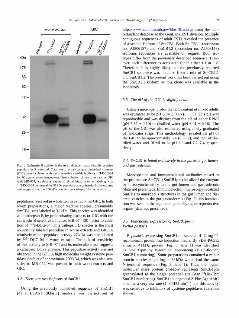
Fig. 1. Cathepsin B activity is the most abundant papain-family cysteinepeptidase inS. mansoni. Total worm extract or gastrointestinal contents(GIC) were incubated with the irreversible epoxide inhibitor125I-DCG-04for 60 min at room temperature. Preincubation of worm extract or GICwith MB-074, a selective cathepsin B inhibitor, prior to labeling with125I-DCG-04 confirmed the 31 kDa peptidase as a cathepsin B-like enzymeand suggests that the 200 kDa doublet has cathepsin B-like activity.
peptidases resolved in whole worm extract than GIC. In bothworm preparations, a major reactive species, presumablySmCB1, was labeled at 31 kDa. This species was identifiedas a cathepsin B by preincubating extracts or GIC with thecathepsin B-selective inhibitor, MB-074[20], prior to addi-tion of 125I-DCG-04. This cathepsin B species is the mostabundantly labeled peptidase in worm extracts and GIC. Arelatively minor peptidase activity 27 kDa was also labeledby 125I-DCG-04 in worm extracts. The lack of sensitivityof this activity to MB-074 and its molecular mass suggestsa cathepsin L-like enzyme. This peptidase activity was notobserved in the GIC. A high molecular weight cysteine pep-tidase doublet of approximate 200 kDa, which was also sen-sitive to MB-074, was present in both worm extracts andGIC.
3.2. There are two isoforms of SmCB1
Using the previously published sequence of SmCB1[4] a BLAST (tblastn) analysis was carried out at
http://www.ncbi.nlm.nih.gov/blast/Blast.cgiusing the non-redundant database at the GenBank EST division. Multiplecontiguous sequences of adult ESTs revealed the presenceof a second isoform of SmCB1. Both SmCB1.1 (accessionno. AJ506157) and SmCB1.2 (accession no. AJ506158)isoforms sequences are available on request. Both iso-types differ from the previously described sequence. How-ever, each difference is accounted for in either 1.1 or 1.2.Therefore, it is highly likely that the previously reportedSmCB1 sequence was obtained from a mix of SmCB1.1and SmCB1.2. The present work has been carried out usingthe SmCB1.1 isoform as this clone was available in thelaboratory.
3.3. The pH of the GIC is slightly acidic
Using a micro-pH probe, the GIC content of mixed adultswas estimated to be pH 6.84± 0.16 (n = 5). This pH wasreproducible and was distinct from the pH of either RPMI(pH 7.57± 0.10) or distilled water (pH 6.01± 0.16). ThepH of the GIC was also estimated using finely graduatedpH indicator strips. This methodology revealed the pH ofthe GIC to be approximately 6.4 (n = 2), and that of dis-tilled water and RPMI to be pH 6.0 and 7.2–7.4, respec-tively.
3.4. SmCB1 is found exclusively in the parasite gut lumenand gastrodermis
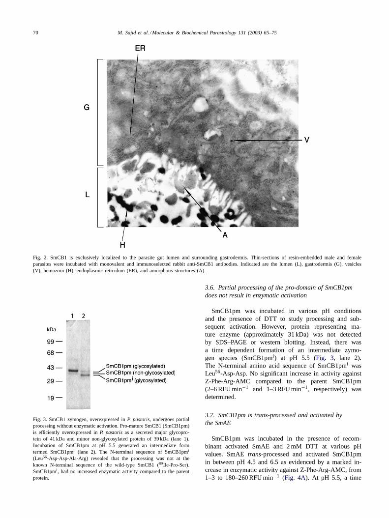
Monospecific and immunoselected antibodies raised tothe pro-mature SmCB1 (SmCB1pm) localized the enzymeby histocytochemistry to the gut lumen and gastrodermis(data not presented). Immunoelectron microscopy localizedSmCB1 to amorphous structures in the gut lumen and dis-crete vesicles in the gut gastrodermis (Fig. 2). No localiza-tion was seen in the tegument, parenchyma, or reproductiveorgans (data not presented).
3.5. Functional expression of SmCB1pm inPichia pastoris
P. pastorisexpressing SmCB1pm secreted 4–11 mg l−1
recombinant protein into induction media. By SDS–PAGE,a major 41 kDa protein (Fig. 3, lane 1) was identifiedas SmCB1pm by N-terminal sequencing (His18-Ile-Ser;SmCB1 numbering). Some preparations contained a minorprotein species migrating at 38 kDa which had the sameN-terminal sequence (Fig. 3, lane 1). Thus, the highermolecular mass protein probably represents SmCB1pmglycosylated at the single potential site (Asn183His-Thr;SmCB1 numbering). SmCB1pm degraded Z-Phe-Arg-AMCalbeit at a very low rate (1–3 RFU min−1) and this activitywas sensitive to inhibitors of cysteine peptidases (data notshown).
70 M. Sajid et al. / Molecular & Biochemical Parasitology 131 (2003) 65–75
Fig. 2. SmCB1 is exclusively localized to the parasite gut lumen and surrounding gastrodermis. Thin-sections of resin-embedded male and femaleparasites were incubated with monovalent and immunoselected rabbit anti-SmCB1 antibodies. Indicated are the lumen (L), gastrodermis (G), vesicles(V), hemozoin (H), endoplasmic reticulum (ER), and amorphous structures (A).
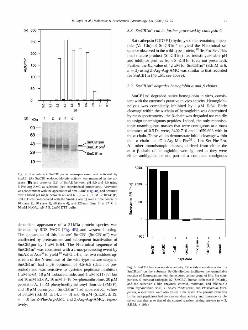
Fig. 3. SmCB1 zymogen, overexpressed inP. pastoris, undergoes partialprocessing without enzymatic activation. Pro-mature SmCB1 (SmCB1pm)is efficiently overexpressed inP. pastorisas a secreted major glycopro-tein of 41 kDa and minor non-glycosylated protein of 39 kDa (lane 1).Incubation of SmCB1pm at pH 5.5 generated an intermediate formtermed SmCB1pmi (lane 2). The N-terminal sequence of SmCB1pmi
(Leu56-Asp-Asp-Ala-Arg) revealed that the processing was not at theknown N-terminal sequence of the wild-type SmCB1 (89Ile-Pro-Ser).SmCB1pmi, had no increased enzymatic activity compared to the parentprotein.
3.6. Partial processing of the pro-domain of SmCB1pmdoes not result in enzymatic activation
SmCB1pm was incubated in various pH conditionsand the presence of DTT to study processing and sub-sequent activation. However, protein representing ma-ture enzyme (approximately 31 kDa) was not detectedby SDS–PAGE or western blotting. Instead, there wasa time dependent formation of an intermediate zymo-gen species (SmCB1pmi) at pH 5.5 (Fig. 3, lane 2).The N-terminal amino acid sequence of SmCB1pmi wasLeu56-Asp-Asp. No significant increase in activity againstZ-Phe-Arg-AMC compared to the parent SmCB1pm(2–6 RFU min−1 and 1–3 RFU min−1, respectively) wasdetermined.
3.7. SmCB1pm is trans-processed and activated bythe SmAE
SmCB1pm was incubated in the presence of recom-binant activated SmAE and 2 mM DTT at various pHvalues. SmAEtrans-processed and activated SmCB1pmin between pH 4.5 and 6.5 as evidenced by a marked in-crease in enzymatic activity against Z-Phe-Arg-AMC, from1–3 to 180–260 RFU min−1 (Fig. 4A). At pH 5.5, a time
M. Sajid et al. / Molecular & Biochemical Parasitology 131 (2003) 65–75 71
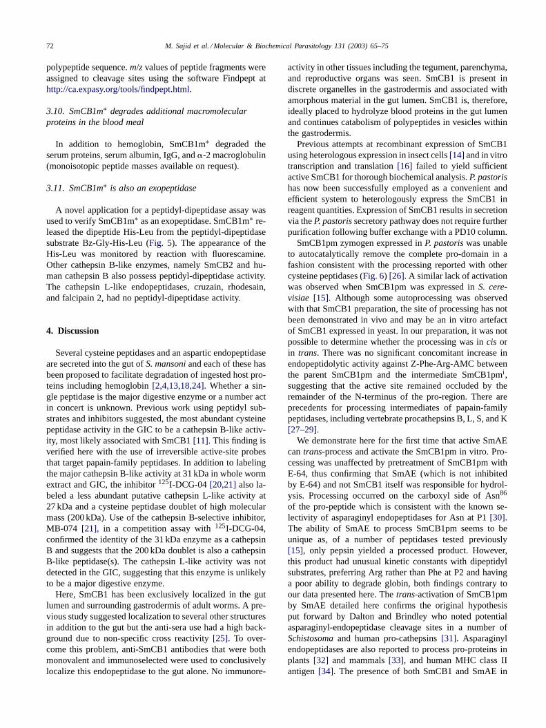
Fig. 4. Recombinant SmCB1pm istrans-processed and activated bySmAE; (A) SmCB1 endopeptidolytic activity was measured in the ab-sence (�) and presence (�) of SmAE between pH 3.0 and 8.0 usingZ-Phe-Arg-AMC as substrate (see experimental procedures). Activationwas concomitant with the appearance of SmCB1m∗ (Fig. 4B) and occurredover a broad pH range between 4.5 and 6.5 (n = 3, S.E.M. < 10%); (B)SmCB1 was co-incubated with the SmAE (lane 1) over a time course of10 (lane 2), 30 (lane 3), 60 (lane 4), and 120 min (lane 5) at 37◦C in50 mM NaOAc, pH 5.5, 2 mM DTT buffer.
dependent appearance of a 31 kDa protein species wasdetected by SDS–PAGE (Fig. 4B) and western blotting.The appearance of this ‘mature’ SmCB1 (SmCB1m∗) wasunaffected by pretreatment and subsequent inactivation ofSmCB1pm by 1�M E-64. The N-terminal sequence ofSmCB1m∗ was consistent with atrans-processing event bySmAE at Asn86 to yield 87Val-Glu-Ile, i.e. two residues up-stream of the N-terminus of the wild-type mature enzyme.SmCB1m∗ had a pH optimum of 4.5–6.5 (data not pre-sented) and was sensitive to cysteine peptidase inhibitors1�M E-64, 10�M iodoacetamide, and 1�M K11777, butnot 10 mM EDTA, 10 mM 1–10bis-phenanthroline, 20�Mpepstatin A, 1 mM phenylmethylsulfonyl flouride (PMSF),and 10�M puromycin. SmCB1m∗ had apparentKm valuesof 38�M (S.E.M. ± 14, n = 3) and 46�M (S.E.M. ± 19,n = 3) for Z-Phe-Arg-AMC and Z-Arg-Arg-AMC, respec-tively.
3.8. SmCB1m∗ can be further processed by cathepsin C
Rat cathepsin C (DPP I) hydrolyzed the remaining dipep-tide (Val-Glu) of SmCB1m∗ to yield the N-terminal se-quence observed in the wild-type protein,89Ile-Pro-Ser. Thisfinal mature product (SmCB1m) had indistinguishable pHand inhibitor profiles from SmCB1m (data not presented).Further, theKm value of 42�M for SmCB1m∗ (S.E.M.±6,n = 3) using Z-Arg-Arg-AMC was similar to that recordedfor SmCB1m (46�M; see above).
3.9. SmCB1m∗ degrades hemoglobinα andβ chains
SmCB1m∗ degraded native hemoglobin in vitro, consis-tent with the enzyme’s putative in vivo activity. Hemoglobi-nolysis was completely inhibited by 1�M E-64. Earlycleavage within the�-chain of hemoglobin was determinedby mass spectrometry; the�-chain was degraded too rapidlyto assign unambiguous peptides. Indeed, the only monoiso-topic unambiguous masses that were contiguous at a masstolerance of 0.5 Da were, 3402.710 and 11659.603 with inthe�-chain. These values demonstrate initial cleavage withinthe �-chain at Glu-Arg-Met-Phe33-↓-Leu-Ser-Phe-Pro.All other monoisotopic masses, derived from either the� or � chain of hemoglobin, were ignored as they wereeither ambiguous or not part of a complete contiguous
Fig. 5. SmCB1 has exopeptidase activity. Dipeptidyl-peptidase action bySmCB1m∗ on the substrate Bz-Gly-His-Leu facilitates the quantifiablereaction of fluorescamine with the exposed amino group of His. For com-parison,S. mansonicathepsin B2 (SmCB2), human cathepsin B (hCatB),and the cathepsin L-like enzymes, cruzain, rhodesain, and falcipain-2from Trypanosoma cruzi, T. brucei rhodesiense, and Plasmodium falci-parum, respectively, were also tested in the assay. The parasite cathepsinL-like endopeptidases had no exopeptidase activity and fluorescence ob-tained was similar to that of the control reaction lacking enzyme (n = 3;S.E.M. < 10%).
72 M. Sajid et al. / Molecular & Biochemical Parasitology 131 (2003) 65–75
polypeptide sequence.m/zvalues of peptide fragments wereassigned to cleavage sites using the software Findpept athttp://ca.expasy.org/tools/findpept.html.
3.10. SmCB1m∗ degrades additional macromolecularproteins in the blood meal
In addition to hemoglobin, SmCB1m∗ degraded theserum proteins, serum albumin, IgG, and�-2 macroglobulin(monoisotopic peptide masses available on request).
3.11. SmCB1m∗ is also an exopeptidase
A novel application for a peptidyl-dipeptidase assay wasused to verify SmCB1m∗ as an exopeptidase. SmCB1m∗ re-leased the dipeptide His-Leu from the peptidyl-dipeptidasesubstrate Bz-Gly-His-Leu (Fig. 5). The appearance of theHis-Leu was monitored by reaction with fluorescamine.Other cathepsin B-like enzymes, namely SmCB2 and hu-man cathepsin B also possess peptidyl-dipeptidase activity.The cathepsin L-like endopeptidases, cruzain, rhodesain,and falcipain 2, had no peptidyl-dipeptidase activity.
4. Discussion
Several cysteine peptidases and an aspartic endopeptidaseare secreted into the gut ofS. mansoniand each of these hasbeen proposed to facilitate degradation of ingested host pro-teins including hemoglobin[2,4,13,18,24]. Whether a sin-gle peptidase is the major digestive enzyme or a number actin concert is unknown. Previous work using peptidyl sub-strates and inhibitors suggested, the most abundant cysteinepeptidase activity in the GIC to be a cathepsin B-like activ-ity, most likely associated with SmCB1[11]. This finding isverified here with the use of irreversible active-site probesthat target papain-family peptidases. In addition to labelingthe major cathepsin B-like activity at 31 kDa in whole wormextract and GIC, the inhibitor125I-DCG-04[20,21] also la-beled a less abundant putative cathepsin L-like activity at27 kDa and a cysteine peptidase doublet of high molecularmass (200 kDa). Use of the cathepsin B-selective inhibitor,MB-074 [21], in a competition assay with125I-DCG-04,confirmed the identity of the 31 kDa enzyme as a cathepsinB and suggests that the 200 kDa doublet is also a cathepsinB-like peptidase(s). The cathepsin L-like activity was notdetected in the GIC, suggesting that this enzyme is unlikelyto be a major digestive enzyme.
Here, SmCB1 has been exclusively localized in the gutlumen and surrounding gastrodermis of adult worms. A pre-vious study suggested localization to several other structuresin addition to the gut but the anti-sera use had a high back-ground due to non-specific cross reactivity[25]. To over-come this problem, anti-SmCB1 antibodies that were bothmonovalent and immunoselected were used to conclusivelylocalize this endopeptidase to the gut alone. No immunore-
activity in other tissues including the tegument, parenchyma,and reproductive organs was seen. SmCB1 is present indiscrete organelles in the gastrodermis and associated withamorphous material in the gut lumen. SmCB1 is, therefore,ideally placed to hydrolyze blood proteins in the gut lumenand continues catabolism of polypeptides in vesicles withinthe gastrodermis.
Previous attempts at recombinant expression of SmCB1using heterologous expression in insect cells[14] and in vitrotranscription and translation[16] failed to yield sufficientactive SmCB1 for thorough biochemical analysis.P. pastorishas now been successfully employed as a convenient andefficient system to heterologously express the SmCB1 inreagent quantities. Expression of SmCB1 results in secretionvia theP. pastorissecretory pathway does not require furtherpurification following buffer exchange with a PD10 column.
SmCB1pm zymogen expressed inP. pastoriswas unableto autocatalytically remove the complete pro-domain in afashion consistent with the processing reported with othercysteine peptidases (Fig. 6) [26]. A similar lack of activationwas observed when SmCB1pm was expressed inS. cere-visiae [15]. Although some autoprocessing was observedwith that SmCB1 preparation, the site of processing has notbeen demonstrated in vivo and may be an in vitro artefactof SmCB1 expressed in yeast. In our preparation, it was notpossible to determine whether the processing was incis orin trans. There was no significant concomitant increase inendopeptidolytic activity against Z-Phe-Arg-AMC betweenthe parent SmCB1pm and the intermediate SmCB1pmi,suggesting that the active site remained occluded by theremainder of the N-terminus of the pro-region. There areprecedents for processing intermediates of papain-familypeptidases, including vertebrate procathepsins B, L, S, and K[27–29].
We demonstrate here for the first time that active SmAEcantrans-process and activate the SmCB1pm in vitro. Pro-cessing was unaffected by pretreatment of SmCB1pm withE-64, thus confirming that SmAE (which is not inhibitedby E-64) and not SmCB1 itself was responsible for hydrol-ysis. Processing occurred on the carboxyl side of Asn86
of the pro-peptide which is consistent with the known se-lectivity of asparaginyl endopeptidases for Asn at P1[30].The ability of SmAE to process SmCB1pm seems to beunique as, of a number of peptidases tested previously[15], only pepsin yielded a processed product. However,this product had unusual kinetic constants with dipeptidylsubstrates, preferring Arg rather than Phe at P2 and havinga poor ability to degrade globin, both findings contrary toour data presented here. Thetrans-activation of SmCB1pmby SmAE detailed here confirms the original hypothesisput forward by Dalton and Brindley who noted potentialasparaginyl-endopeptidase cleavage sites in a number ofSchistosomaand human pro-cathepsins[31]. Asparaginylendopeptidases are also reported to process pro-proteins inplants [32] and mammals[33], and human MHC class IIantigen[34]. The presence of both SmCB1 and SmAE in
M. Sajid et al. / Molecular & Biochemical Parasitology 131 (2003) 65–75 73
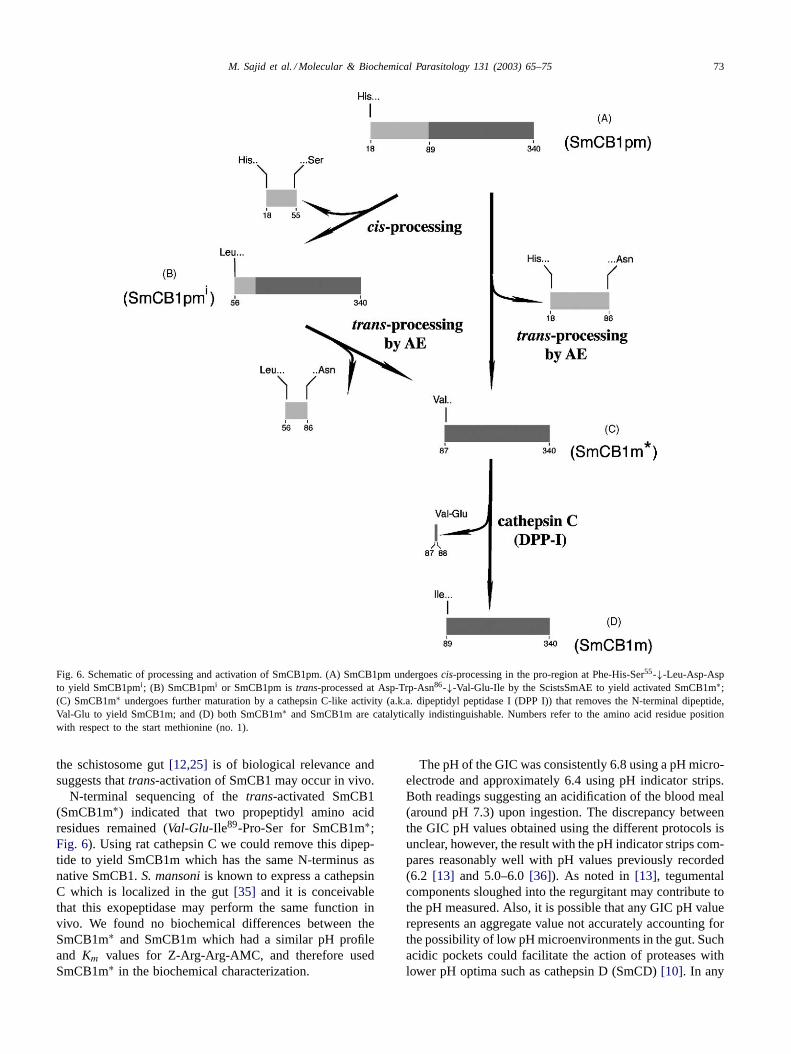
Fig. 6. Schematic of processing and activation of SmCB1pm. (A) SmCB1pm undergoescis-processing in the pro-region at Phe-His-Ser55-↓-Leu-Asp-Aspto yield SmCB1pmi; (B) SmCB1pmi or SmCB1pm istrans-processed at Asp-Trp-Asn86-↓-Val-Glu-Ile by the ScistsSmAE to yield activated SmCB1m∗;(C) SmCB1m∗ undergoes further maturation by a cathepsin C-like activity (a.k.a. dipeptidyl peptidase I (DPP I)) that removes the N-terminal dipeptide,Val-Glu to yield SmCB1m; and (D) both SmCB1m∗ and SmCB1m are catalytically indistinguishable. Numbers refer to the amino acid residue positionwith respect to the start methionine (no. 1).
the schistosome gut[12,25] is of biological relevance andsuggests thattrans-activation of SmCB1 may occur in vivo.
N-terminal sequencing of thetrans-activated SmCB1(SmCB1m∗) indicated that two propeptidyl amino acidresidues remained (Val-Glu-Ile89-Pro-Ser for SmCB1m∗;Fig. 6). Using rat cathepsin C we could remove this dipep-tide to yield SmCB1m which has the same N-terminus asnative SmCB1.S. mansoniis known to express a cathepsinC which is localized in the gut[35] and it is conceivablethat this exopeptidase may perform the same function invivo. We found no biochemical differences between theSmCB1m∗ and SmCB1m which had a similar pH profileand Km values for Z-Arg-Arg-AMC, and therefore usedSmCB1m∗ in the biochemical characterization.
The pH of the GIC was consistently 6.8 using a pH micro-electrode and approximately 6.4 using pH indicator strips.Both readings suggesting an acidification of the blood meal(around pH 7.3) upon ingestion. The discrepancy betweenthe GIC pH values obtained using the different protocols isunclear, however, the result with the pH indicator strips com-pares reasonably well with pH values previously recorded(6.2 [13] and 5.0–6.0[36]). As noted in[13], tegumentalcomponents sloughed into the regurgitant may contribute tothe pH measured. Also, it is possible that any GIC pH valuerepresents an aggregate value not accurately accounting forthe possibility of low pH microenvironments in the gut. Suchacidic pockets could facilitate the action of proteases withlower pH optima such as cathepsin D (SmCD)[10]. In any
74 M. Sajid et al. / Molecular & Biochemical Parasitology 131 (2003) 65–75
case, both thetrans-activation of SmCB1pm by SmAE andthe activity of SmCB1 against ingested proteins would pro-ceed efficiently given the pH data presented herein or byothers.
Sequence comparison with human cathepsin B revealedthat SmCB1 has the predicted occluding loop, which hasbeen shown to confer exopeptidase (specifically peptidyldipeptidase) activity in vertebrate cathepsin Bs[37]. A novelassay for such activity was developed and confirmed thatSmCB1 acts as an exopeptidase in addition to its endopep-tidolytic activity described herein and elsewhere. The ex-opeptidase activity may be important in the downstreamcatabolism of ingested blood proteins. The assay also con-firmed SmCB2 and human cathepsin B as exopeptidases. Incontrast, none of the cathepsin L-like endopeptidases stud-ied namely, cruzain, rhodesain and falcipain 2, could act aspeptidyl dipeptidases.
Activated recombinant SmCB1 has very similar bio-chemical properties to the native parasite enzyme[5]. Theseinclude a similar pH optimum with Z-Phe-Arg-AMC as asubstrate (pH 5.5–6.0) and similarKm values for Z-Phe-Arg-AMC (wild-type, 21�M; recombinant, 38�M) andZ-Arg-Arg-AMC (wild-type 41�M; recombinant, 46�M).
A major protein in the schistosome blood meal ishemoglobin. Accordingly, it was pertinent to attempt tomap the initial cleavage sites produced by recombinantSmCB1m∗ in the � and� chains of hemoglobin. Such ex-periments were carried out using a dilute enzyme prepara-tion with reaction samples taken at 0, 15, 30, and 60 s. Massspectrometry revealed the appearance of peptide fragmentsin a time dependent manner. However, due to the extremelyrapid degradation of substrate by SmCB1, it was difficult toassign unambiguous monoisotopic masses to unique linearregions within the� or � chains of hemoglobin. The onlycomplete contiguous sequence occurred with two polypep-tides (3402.7 and 11659.6 Da) in the�-chain, consistentwith initial hydrolysis at Arg-Met-Phe33-↓-Leu-Ser-Phe-.Interestingly, the same cleavage site is also targeted as theinitial point of hemoglobin hydrolysis by other peptidasesimplicated in parasite nutrition. These include,S. mansonicathepsin D[10] the hookworm cathepsin Ds, Ac-APR-1and Na-APR-1[38], and a preparation ofPlasmodium fal-ciparum digestive vacuoles[39]. This may represent anevolutionary convergence across parasites and peptidaseclasses for the most appropriate site of initial hydrolysis ofhemoglobin. This area within hemoglobin is referred to asthe ‘hinge region’ and is involved in maintaining stabilityof the quaternary tetrameric structure. Hydrolysis at thispoint would relax the molecule and facilitate subsequentcatabolism of hemoglobin. In addition to hemoglobin,SmCB1m∗ also degraded a number of other macromolec-ular proteins in the blood meal. These included, serumalbumin, IgG, and�-2 macroglobulin. Hydrolysis of im-munoglobulins may facilitate immune evasion by the para-site. In addition, effective degradation of macromolecularinhibitors such as�-2 macroglobulin, would be conceiv-
ably important in maintaining a full repertoire of activepeptidases, thus aiding parasite survival.
In conclusion, we have expressed SmCB1 in quantitiessufficient for detailed enzymic characterization and haveshown that recombinant SmCB1 zymogen requires SmAEfor trans-processing and activation. SmCB1 efficiently de-grades hemoglobin and, as the major cysteine peptidase inthe parasite gut, is a potential target for novel chemothera-pies of schistosomiasis.
Acknowledgements
Many thanks to Dr. John Pedersen of Unizyme, Denmarkand Dr. Phil Rosenthal at UCSF for the kind gifts of re-combinant rat cathepsin C and falcipain 2, respectively. Thiswork was supported by NIAID Grant AI053247, a WellcomeInternational Fellowship in Tropical Medicine to M.S., andthe Sandler Family Supporting Foundation.
References
[1] Lawrence JD. The ingestion of red blood cells bySchistosomamansoni. J Parasitol 1973;59(1):60–3.
[2] Brindley PJ, Kalinna BH, Dalton JP, Day SR, Wong JY, Smythe ML,et al. Proteolytic degradation of host hemoglobin by schistosomes.Mol Biochem Parasitol 1997;89(1):1–9.
[3] Wasilewski MM, Lim KC, Phillips J, McKerrow JH. Cysteine pro-tease inhibitors block schistosome hemoglobin degradation in vitroand decrease worm burden and egg production in vivo. Mol BiochemParasitol 1996;81(2):179–89.
[4] Klinkert MQ, Felleisen R, Link G, Ruppel A, Beck E. Primarystructures of Sm31/32 diagnostic proteins ofSchistosoma man-soni and their identification as proteases. Mol Biochem Parasitol1989;33(2):113–22.
[5] Ghoneim H, Klinkert MQ. Biochemical properties of purified cathep-sin B fromSchistosoma mansoni. Int J Parasitol 1995;25(12):1515–9.
[6] Day SR, Dalton JP, Clough KA, Leonardo L, Tiu WU, Brindley PJ.Characterization and cloning of the cathepsin L proteinases ofSchis-tosoma japonicum. Biochem Biophys Res Commun 1995;217(1):1–9.
[7] Bogitsh BJ, Dalton JP, Brady CP, Brindley PJ. Gut-associated im-munolocalization of theSchistosoma mansonicysteine proteases,SmCL1 and SmCL2. J Parasitol 2001;87(2):237–41.
[8] Caffrey CR, Mathieu MA, Gaffney AM, Salter JP, Sajid M, LucasKD, et al. Identification of a cDNA encoding an active asparaginylendopeptidase ofSchistosoma mansoniand its expression inPichiapastoris. FEBS Lett 2000;466(2/3):244–8.
[9] Hola-Jamriska L, Tort JF, Dalton JP, Day SR, Fan J, Aaskov J,et al. Cathepsin C fromSchistosoma japonicum—cDNA encodingthe preproenzyme and its phylogenetic relationships. Eur J Biochem1998;255(3):527–34.
[10] Brindley PJ, Kalinna BH, Wong JY, Bogitsh BJ, King LT, Smyth DJ,et al. Proteolysis of human hemoglobin by schistosome cathepsin D.Mol Biochem Parasitol 2001;112(1):103–12.
[11] Caffrey CR, Ruppel A. Cathepsin B-like activity predominates overcathepsin L-like activity in adultSchistosoma mansoniandS. japon-icum. Parasitol Res 1997;83(6):632–5.
[12] Ruppel A, Shi YE, Wei DX, Diesfeld HJ. Sera ofSchistosomajaponicum-infected patients cross-react with diagnostic 31/32 kD pro-teins of S. mansoni. Clin Exp Immunol 1987;69(2):291–8.
M. Sajid et al. / Molecular & Biochemical Parasitology 131 (2003) 65–75 75
[13] Chappell CL, Dresden MH.Schistosoma mansoni: proteinase activityof “hemoglobinase” from the digestive tract of adult worms. ExpParasitol 1986;61(2):160–7.
[14] Gotz B, Felleisen R, Shaw E, Klinkert MQ. Expression of an activecathepsin B-like protein Sm31 fromSchistosoma mansoniin insectcells. Trop Med Parasitol 1992;43(4):282–4.
[15] Lipps G, Fullkrug R, Beck E. Cathepsin B ofSchistosoma mansoni.Purification and activation of the recombinant proenzyme secretedby Saccharomyces cerevisiae. J Biol Chem 1996;271(3):1717–25.
[16] Felleisen R, Klinkert MQ. In vitro translation and processing ofcathepsin B ofSchistosoma mansoni. EMBO J 1990;9(2):371–7.
[17] Ruppel A, Shi YE, Moloney NA.Schistosoma mansoniandS. japon-icum: comparison of levels of ultraviolet irradiation for vaccinationof mice with cercariae. Parasitology 1990;101:23–6.
[18] Caffrey CR, Rheinberg CE, Mone H, Jourdane J, Li YL, Ruppel A.Schistosoma japonicum, S. mansoni, S. haematobium, S. intercalatum,andS. rodhaini: cysteine-class cathepsin activities in the vomitus ofadult worms. Parasitol Res 1997;83:37–41.
[19] Xing R, Addington AK, Mason RW. Quantification of cathepsins Band L in cells. Biochem J 1998;332(Pt 2):499–505.
[20] Greenbaum D, Medzihradszky KF, Burlingame A, Bogyo M. Epoxideelectrophiles as activity-dependent cysteine protease profiling anddiscovery tools. Chem Biol 2000;7(8):569–81.
[21] Bogyo M, Verhelst S, Bellingard-Dubouchaud V, Toba S, GreenbaumD. Selective targeting of lysosomal cysteine proteases with radiola-beled electrophilic substrate analogs. Chem Biol 2000;7(1):27–38.
[22] Lamango NS, Sajid M, Isaac RE. The endopeptidase activity and theactivation by Cl- of angiotensin-converting enzyme is evolutionarilyconserved: purification and properties of an angiotensin-convertingenzyme from the housefly,Musca domestica. Biochem J 1996;314(Pt2):639–46.
[23] Yezzi MJ, Hsieh IE, Caughey GH. Mast cell and neutrophil ex-pression of dog mast cell protease-3. A novel tryptase-related serineprotease. J Immunol 1994;152(6):3064–72.
[24] Brinkworth RI, Prociv P, Loukas A, Brindley PJ. Hemoglobin-degrading, aspartic proteases of blood-feeding parasites: sub-strate specificity revealed by homology models. J Biol Chem2001;276(42):38844–51.
[25] Skelly PJ, Shoemaker CB.Schistosoma mansoniproteases Sm31(cathepsin B) and Sm32 (legumain) are expressed in the cecum andprotonephridia of cercariae. J Parasitol 2001;87(5):1218–21.
[26] Turk V, Turk B, Turk D. Lysosomal cysteine proteases: facts andopportunities. EMBO J 2001;20(17):4629–33.
[27] McQueney MS, Amegadzie BY, D’Alessio K, Hanning CR,McLaughlin MM, McNulty D, et al. Autocatalytic activation of hu-man cathepsin K. J Biol Chem 1997;272(21):13955–60.
[28] Menard R, Carmona E, Takebe S, Dufour E, Plouffe C, Mason P,et al. Autocatalytic processing of recombinant human procathepsinL. Contribution of both intermolecular and unimolecular events inthe processing of procathepsin L in vitro. J Biol Chem 1998;273(8):4478–84.
[29] Quraishi O, Storer AC. Identification of internal autoproteolytic cleav-age sites within the prosegments of recombinant procathepsin B andprocathepsin S. Contribution of a plausible unimolecular autoprote-olytic event for the processing of zymogens belonging to the papainfamily. J Biol Chem 2001;276(11):8118–24.
[30] Mathieu MA, Bogyo M, Caffrey CR, Choe Y, Lee J, ChapmanH, et al. Substrate specificity of schistosome versus human legu-main determined by P1-P3 peptide libraries. Mol Biochem Parasitol2002;121(1):99–105.
[31] Dalton JP, Brindley PJ. Schistosome asparaginyl endopeptidase Sm32in hemoglobin digestion. Parasitol Today 1996;12(Pt 3):125.
[32] Okamoto T, Minamikawa T. Molecular cloning and characterizationof Vigna mungo processing enzyme 1 (VmPE-1), an asparaginylendopeptidase possibly involved in post-translational processingof a vacuolar cysteine endopeptidase (SH-EP). Plant Mol Biol1999;39(1):63–73.
[33] Dando PM, Fortunato M, Smith L, Knight CG, McKendrick JE,Barrett AJ. Pig kidney legumain: an asparaginyl endopeptidase withrestricted specificity. Biochem J 1999;339(Pt 3):743–9.
[34] Manoury B, Hewitt EW, Morrice N, Dando PM, Barrett AJ, WattsC. An asparaginyl endopeptidase processes a microbial antigen forclass II MHC presentation. Nature 1998;396(6712):695–9.
[35] Bogitsh BJ, Dresden MH. Fluorescent histochemistry of acid pro-teases in adultSchistosoma mansoniand Schistosoma japonicum. JParasitol 1983;69(1):106–10.
[36] Senft AW. Observations on the physiology of the gut ofSchistosomamansoni. In: H. Van den Bossche, editor. Biochemistry of parasitesand host parasite relationships. Amsterdam: Elsevier/North-HollandBiomedical Press; 1976. p. 335–42.
[37] Krupa JC, Hasnain S, Nagler DK, Menard R, Mort JS. S2′ substratespecificity and the role of His110 and His111 in the exopepti-dase activity of human cathepsin B. Biochem J 2002;361(Pt 3):613–9.
[38] Williamson AL, Brindley PJ, Abbenante G, Prociv P, Berry C,Girdwood K, et al. Cleavage of hemoglobin by hookworm cathepsinD aspartic proteases and its potential contribution to host specificity.FASEB J 2002;16(11):1458–60.
[39] Kolakovich KA, Gluzman IY, Duffin KL, Goldberg DE. Gener-ation of hemoglobin peptides in the acidic digestive vacuole ofPlasmodium falciparumimplicates peptide transport in amino acidproduction. Mol Biochem Parasitol 1997;87(2):123–35.


