Embed Size (px)
Citation preview
Heliyon 8 (2022) e09630
Contents lists available at ScienceDirect
Heliyon
journal homepage: www.cell.com/heliyon
Review article
Functional foods: promising therapeutics for Nigerian Children with sicklecell diseases
Oladeji John Alabi a,b,*, Fikayo Noah Adegboyega a,c, Dolapo Samuel Olawoyin a,d,Oluwakemi Arinola Babatunde e
a Department of Biochemistry, Faculty of Basic Medical Sciences, Ladoke Akintola University of Technology, Ogbomoso, Nigeriab Department of Biochemistry, Institute for Agriculture & Natural Sciences, College of Arts & Sciences, University of Nebraska-Lincoln, USAc Department of Biotechnology, Egypt-Japan University of Science and Technology, Alexandria, Egyptd School of Food Science and Nutrition, University of Leeds, Leeds, UKe Department of Nursing, Achievers University, Owo, Nigeria
A R T I C L E I N F O
Keywords:Functional foodsSickle cell disease (SCD)Pediatric/children
* Corresponding author.E-mail addresses: [email protected], john
https://doi.org/10.1016/j.heliyon.2022.e09630Received 25 September 2021; Received in revised2405-8440/© 2022 The Author(s). Published by Els
A B S T R A C T
Sickle cell disease (SCD), also known as sickle cell anemia (SCA) is one of the structural hemoglobinopathies thatoccurs due to a single nucleotide mutation from GAG to GTG, which changes the amino acid of a β-globin chain ofhemoglobin (Hb) from glutamate to valine. This singular mutation results to disorderliness in red blood cells(RBCs) with advent of changes in RBC morphology and other pathological conditions. In the 1980s, intermittentred blood cell transfusions, opioids, and penicillin prophylaxis were the only available therapy for SCA and werecommonly reserved for acute, life threatening complications. So far, the US Food and Drug Administration (FDA)has granted a total of four drugs approval for the prophylaxis and treatment of the clinical complications of SCD.Due to limitations (adherence, safety, adverse effects) of existing therapies in the prophylaxis and treatment ofSCD complications in Nigerian children and their inaccessibility to approved drugs, the present study discussesthe therapeutic effects of readily available functional food as one of the therapies or an adjunct therapy to tacklethe sickle cell crisis in Nigerian Children.
1. Introduction
Sickle cell disease (SCD) is a group of genetic ailments affecting thered blood cell [1]. It is sometimes referred to as drepanocytosis consistingof sickle thalassemia and sickle cell anemia (HbSS). Heterozygous ge-notypes (HbAS) expressing parents are sickle cell carriers and theiroffspring have 25 % chances of giving rise to a homozygous sickle ge-notype (HbSS) progenies or a 75 % homozygous normal genotype(HbAA) [2]. Sickle cell derangement was first identified as a hemato-logical disease by Herrick in 1910 [2] and its biochemical pathology wasproven by Linus Pauling in 1949 [2]. Epidemiological information onSCD has majorly been reported among Jamaica, India and US population,with less statistics in Africa where the trait of SCD is said to be largest [3].It is mostly common among the black Africa, and some other racesoccupying the Mediterranean, India, and Middle East [4]. Further studieson children born with SCD have recorded the major occurrence in thedeveloping world, with an estimated 200,000 annual sickle genotypebirths in Sub-Saharan Africa [5].
[email protected] (O.J. Alabi)
form 30 November 2021; Acceptevier Ltd. This is an open access
Sickle cell disease is one of the causes of childhood mortality in Af-rica. 80% of people with SCD globally live in Sub-Saharan Africa. Thisproportion is projected to reach 88% by 2050 [6,7]. Nigeria has thelargest burden of sickle cell disease in Africa and the world, withapproximately 150,000 births annually [8] and 50–90% of them diebefore age five [9]. Poor quality healthcare, poor education, unaware-ness, poverty, and poor nutritional choices contribute to excess mortality.In West African for example, historical and scientific records havedescribed different names used to qualify SCD children among the threemain Nigerian tribes. The Yorubas, constituting the dominant populationin the west called them “abiku” translated “sufferers'' or “children thatbring sadness” [10, 11], the Ibos called them “Ogbanje” [12], and theHausas called them “Sankara-miji” [13]. It is the major innate disorder inNigeria, affecting about 4 million population at prevalence of 2% at birthwhile more than 40 million persons have sickle cell traits.
Nigeria shows a record for about 75% of children with SCD in Africa[14]. Though having normal weight at birth, babies affected by SCDshow weight loss during the first year and gradually lingers until
.
ed 26 May 2022article under the CC BY license (http://creativecommons.org/licenses/by/4.0/).
O.J. Alabi et al. Heliyon 8 (2022) e09630
adulthood incidentally followed by prolonged skeletal maturation inboth boys and girls with a protracted menarche in girls [15]. At molec-ular and genetic levels, the hereditary sickle cell derangement is visual-ized when a single nucleotide base substitution occurs in the gene codinghuman β-globin subunit. Such substitution causes the replacement ofhydrophilic amino acid (glutamic acid) at position 6 in normal hemo-globin (HbA) by a hydrophobic amino acid (valine) in abnormal hemo-globin, culminating in the disease state of sickle red cells [16]. Theoccurred hydrophobic replacement results in insolubility of sickle cellhemoglobin (HbS) when deoxygenated. HbS molecules that areconstantly formed polymerize to long crystalline intracellular mass offibers that deform the original biconcave shape of the red blood cell(erythrocyte) into a sickled shape cell. The magnitudes of this reshapingare noticed with hemolytic anemia and tissue disruption emanated fromthe blockage of blood vessels by the sickled cells.
Clinical appearances of this condition are vascular necrosis, hypo-sthenuria, proliferative retinopathy, priapism, aplastic crises, pulmonarydisease and nephropathy. In most cases, the complications are severedand include periodic attacks of pain and progressive organ dysfunctionleading to a much reduced biologic life span [17]. Likewise, reactiveoxygen species [18] are considered to play a crucial role in the SCDpathogenesis. The chronically increased oxidative stress in SCD mightplay a significant role in the emergence of SCD related organ complica-tions [19].
Pharmacologically, many drugs with various targeted pathways haveemerged. However, majority of them have failed to display benefit inmedical trials, few have produced encouraging results but with lessavailability making them out of reach for low – income countries likeNigeria [20]. According to WHO (2002), up to 80% of individuals livingin Africa depend on traditional plant-based treatment, with slight sideeffects, for their primary health care needs [18]. Phytomedicine has beenwidely utilized as effective remedies for the prevention and treatment ofmultiple health conditions for centuries by almost every known culture inNigeria [18].
Table 1. Clinical trials of the FDA approved pharmaceutical drugs for sickle cell dise
Clinical Trials Mechanisms and Results Reco
HydroxyureaMulticenter Study ofHydroxyurea (MSH)NCT00000586 [54]Multicentre, randomised,controlled trial (BABY HUG)NCT00006400 [55]
Targeting HbS polymerization: InhibitsRibonucleotide reductase; thereby increasing HBFlevels, which in turn retard sickling.Drug was well tolerated. Reduction in the need forblood transfusions and frequency of painfulepisodes, lower hospitalization rates, and reducednumber of episodes of acute chest syndrome inchildren and adults.
Startincreweek(MTthe a
L-glutaminePhase III randomized trialNCT01179217 [69]
Targeting hemolysis-mediated endothelialdysfunction: Increase NADH and NAD redoxpotential; thereby maintaining vascular tone &impairing adhesion.Drug was well tolerated; reduction in the number ofpain crises, lower hospitalization rates, and reducednumber of episodes of acute chest syndrome inchildren and adults with SCD with or withouthydroxyurea compared to those that receivedplacebo. No improvement in Hb or reticulocytecount.
0.3 gtwicmonwhic[65]
CrizanlizumabPhase II randomized trialNCT01895361 [70]
Targeting vasocclusion: Humanized monoclonalantibody against P-selectin; thereby reducing paincrises.All SCD genotypes experienced tremendousreduction in the percentage of crisis episodes by45% in high-dose arm; no improvement in markersof hemolysis.
5 mgperiomg/whil[ICE
Voxelotor (Oxbryta/GBT440)1/2, RDBPC, NCT02285088/NCT03041909 [74]Phase III randomized trialNCT03036813 [75]
Targeting HbS polymerization: Binds to HbS a-globin chain and stabilizes it in the R-state; therebyincreasing its affinity for oxygen.Voxelotor was well tolerated. Significantimprovement in hemoglobin levels and markers ofhemolysis.
1500mg otable
2
Polyphenols, alkaloids, flavonoids, and other Phenolic compoundshave been reported [21] to have antisickling and antioxidant effectswhich are capable of restoring normalcy to the titled redox balance thatoriginally contributes to sickle cell crisis in children. Such foods con-taining polyphenols are often referred to as functional foods i.e. foodscontaining bioactive compounds that can promote the health of a personbeyond rudimentary diets, or serve as a measure for prevention ormanagement of chronic diseases [22].
Nevertheless, the application of renowned drugs and potential cura-tive treatments will most likely be limited to high-income countries whoare able to possess them at high cost, leaving out the low – incomecountries with severe complications due to unavailability of these ther-apeutics (Table 1). In this review, we discuss the readily available uniquefunctional foods that can be used in ameliorating the sickle cell crisis inNigerian Children.
2. Pathophysiology of Sickle Cell Disease
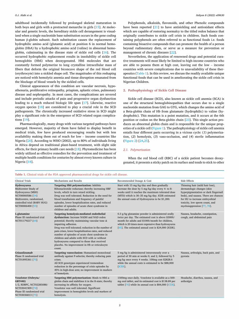
Sickle cell disease (SCD), also known as sickle cell anemia (SCA) isone of the structural hemoglobinopathies that occurs due to a singlenucleotide mutation from GAG to GTG, which changes the amino acid ofa Beta-globin chain of Hb from glutamate (hydrophilic) to valine (hy-drophobic). This mutation is a point mutation, and it occurs at the 6thposition or codon on the Beta-globin chain [23]. This single action pro-duces an abnormal globin chain and is responsible for the unique prop-erties of a sickle cell (Figure 1). The pathophysiology of sickle cell anemiaentails four different parts occurring in a vicious cycle: (1) polymeriza-tion, (2) hemolysis, (3) vaso-occlusion, and (4) sterile inflammation(Figure 2) [24,25].
2.1. Polymerization
When the red blood cell (RBC) of a sickle patient becomes deoxy-genated, it presents a sticky patch on its surface and tends to stick to other
ase.
mmended Dosage & Cost Side Effects
with 15 mg/kg/day and then graduallyase the dose by 5 mg/kg/day every 4- to 8-s until it reaches the maximum tolerated doseD) which is 30–35 mg/kg/day. ICER estimatednnual costs of Hydroxyurea to be $1,200.
Thinning hair (mild hair loss),dermatologic changes (skinhyperpigmentation or dark fingernailbeds), and nausea. There are chancesfor HU to increase embryofetaltoxicity, low sperm count, andmyelosuppression [77, 78].
/kg glutamine powder is administered orallye per day. The estimated cost is above $3000/th for adults and $1000/month for children,h is 20 times more expensive than hydroxyurea. The estimated annual cost is $24,000 [ICER].
Nausea, headache, constipation,cough, and abdominal pain
/kg is administered intravenously over ad of 30 min at weeks 0, and 2, followed by 5kg once every 4 weeks. 100mg cost $2828.6e the annual costs is estimated to be $88,000R].
Nausea, arthralgia, back pain, andpyrexia
mg once daily. Voxelotor is available as a 500-ral tablet, and its estimated cost is $138.89 pert [76] while its annual cost is $84,000 [ICER].
Headache, diarrhea, nausea, andarthralgia
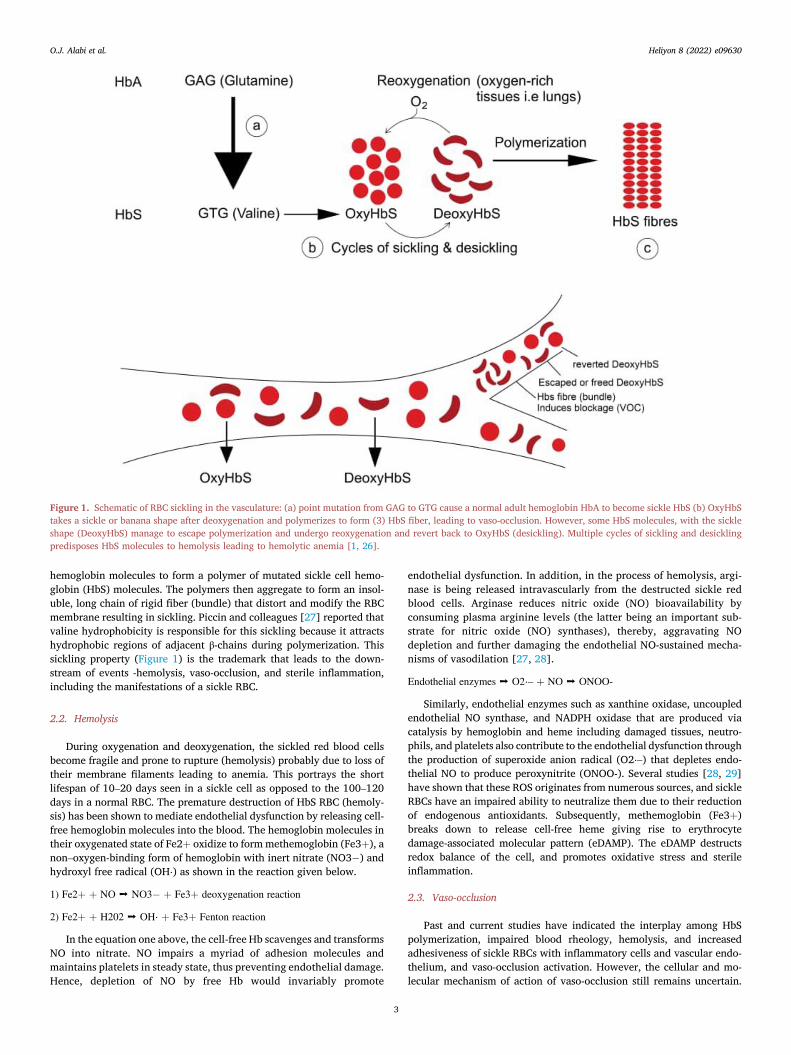
Figure 1. Schematic of RBC sickling in the vasculature: (a) point mutation from GAG to GTG cause a normal adult hemoglobin HbA to become sickle HbS (b) OxyHbStakes a sickle or banana shape after deoxygenation and polymerizes to form (3) HbS fiber, leading to vaso-occlusion. However, some HbS molecules, with the sickleshape (DeoxyHbS) manage to escape polymerization and undergo reoxygenation and revert back to OxyHbS (desickling). Multiple cycles of sickling and desicklingpredisposes HbS molecules to hemolysis leading to hemolytic anemia [1, 26].
O.J. Alabi et al. Heliyon 8 (2022) e09630
hemoglobin molecules to form a polymer of mutated sickle cell hemo-globin (HbS) molecules. The polymers then aggregate to form an insol-uble, long chain of rigid fiber (bundle) that distort and modify the RBCmembrane resulting in sickling. Piccin and colleagues [27] reported thatvaline hydrophobicity is responsible for this sickling because it attractshydrophobic regions of adjacent β-chains during polymerization. Thissickling property (Figure 1) is the trademark that leads to the down-stream of events -hemolysis, vaso-occlusion, and sterile inflammation,including the manifestations of a sickle RBC.
2.2. Hemolysis
During oxygenation and deoxygenation, the sickled red blood cellsbecome fragile and prone to rupture (hemolysis) probably due to loss oftheir membrane filaments leading to anemia. This portrays the shortlifespan of 10–20 days seen in a sickle cell as opposed to the 100–120days in a normal RBC. The premature destruction of HbS RBC (hemoly-sis) has been shown to mediate endothelial dysfunction by releasing cell-free hemoglobin molecules into the blood. The hemoglobin molecules intheir oxygenated state of Fe2þ oxidize to form methemoglobin (Fe3þ), anon–oxygen-binding form of hemoglobin with inert nitrate (NO3�) andhydroxyl free radical (OH⋅) as shown in the reaction given below.
1) Fe2þ þ NO ➝ NO3� þ Fe3þ deoxygenation reaction
2) Fe2þ þ H202 ➝ OH⋅ þ Fe3þ Fenton reaction
In the equation one above, the cell-free Hb scavenges and transformsNO into nitrate. NO impairs a myriad of adhesion molecules andmaintains platelets in steady state, thus preventing endothelial damage.Hence, depletion of NO by free Hb would invariably promote
3
endothelial dysfunction. In addition, in the process of hemolysis, argi-nase is being released intravascularly from the destructed sickle redblood cells. Arginase reduces nitric oxide (NO) bioavailability byconsuming plasma arginine levels (the latter being an important sub-strate for nitric oxide (NO) synthases), thereby, aggravating NOdepletion and further damaging the endothelial NO-sustained mecha-nisms of vasodilation [27, 28].
Endothelial enzymes ➝ O2⋅– þ NO ➝ ONOO-
Similarly, endothelial enzymes such as xanthine oxidase, uncoupledendothelial NO synthase, and NADPH oxidase that are produced viacatalysis by hemoglobin and heme including damaged tissues, neutro-phils, and platelets also contribute to the endothelial dysfunction throughthe production of superoxide anion radical (O2⋅–) that depletes endo-thelial NO to produce peroxynitrite (ONOO-). Several studies [28, 29]have shown that these ROS originates from numerous sources, and sickleRBCs have an impaired ability to neutralize them due to their reductionof endogenous antioxidants. Subsequently, methemoglobin (Fe3þ)breaks down to release cell-free heme giving rise to erythrocytedamage-associated molecular pattern (eDAMP). The eDAMP destructsredox balance of the cell, and promotes oxidative stress and sterileinflammation.
2.3. Vaso-occlusion
Past and current studies have indicated the interplay among HbSpolymerization, impaired blood rheology, hemolysis, and increasedadhesiveness of sickle RBCs with inflammatory cells and vascular endo-thelium, and vaso-occlusion activation. However, the cellular and mo-lecular mechanism of action of vaso-occlusion still remains uncertain.
Figure 2. Pathophysiology of sickle cell disease. Adapted from [24].
O.J. Alabi et al. Heliyon 8 (2022) e09630
Also, evidence has shown that blood vessels occlusion can be initiated bydiverse mechanisms involving different inflammatory and/or environ-mental stimuli such as TNFα, hypoxia, heme, dehydration, Hb, infection,hypoxia, acidosis, lipopolysaccharide, and several others [30, 31, 32, 33,34]. It has been reported that these stimuli or triggers may cause painevents due to a link or connection between the vaso-occlusion andautonomic nervous system [35]. Further, it has been explained that thecellular and molecular paradigm of vaso-occlusion is not similar in allvascular beds or organs [24].
After deoxygenation, the HbS bundle takes a sickle or needle-likeshape that protrudes through the lipid bilayer to cause a leakage [36],which causes the influx of calcium ions inside the HbS. This action ac-tivates the HbS RBC ion channel (Ca2-dependent K channel, K–Clcotransport) resulting in a corresponding efflux of potassium ion andwater out of RBC [27, 37]. This further distorts the RBC and makes theRBC more viscous and dehydrated. The HbS polymer bundles alsostimulate membrane lipids peroxidation to cause the release of peroxi-dation products which can damage the membrane structure, alter waterpermeability, and increase cell deformability. Disruption of the RBCmembrane phospholipids causes the exposure of phosphatidylserine (PS)to the outer cell surface [38]. This loss in RBC integrity makes itdetectable and markable by macrophages and spleen for destruction,thereby elevating the level of hemolysis, and thus contributing to chronicanemia. Most destruction of sickle hemoglobin takes place extracellu-larly, while only a fraction occurs intravascularly and make up 30% oftotal hemolysis [36]. During hemolysis, heme is produced. Hemolysis,heme, and dehydration have been indicated to propagate or contribute to
4
vaso-occlusion. In an attempt to compensate for blood shortage during orfollowing hemolysis, the bone marrow produces more reticulocytes thannormal, therefore, it becomes stressed and produces immature re-ticulocytes, known as “stress reticulocytes” with adhesion molecules-α4β1 integrin (VLA-4) and CD36 expressed on their surface [1, 31, 39].
2.4. Sterile inflammation
The sterile inflammation pathway has been indicated to be activatedby a concerted action of both hemolysis-mediated endothelial dysfunc-tion and increased adhesion-mediated vaso-occlusion [24, 40, 41](Figure 2). Either of the cell-free heme or ischemia-reperfusion (I-R)injury promotes sterile inflammation in SCA by activating the inflam-masome pathways in vascular and inflammatory cells to release Inter-leukin-1β (IL-1β). The released or activated IL-1β aggravates theprogression of vaso-occlussion by promoting the adhesiveness of plate-lets, neutrophils, and endothelial cells, upregulation of P-selectin,E-selectin, intercellular adhesion molecule-1 (ICAM-1), vascular celladhesion molecule-1 (VCAM-1), and chemokines [28, 42]. In the processof inflammasome activation, heme and I-R injury have been shown to beinvolved in the generation of reactive oxygen species (ROS), neutrophilextracellular trap (NET), generations of DAMPs, and DNA, and activationof the Toll-like receptor 4 (TLR4) [40,43,44,45,46,47]. In other words,ROS, NET, TLR4, DAMPs, and DNA play a critical role in Inflammasomesactivation following their stimulation and/or generation by heme or I-Rinjury. Therefore, deletion/inhibition of any of these molecules mightprevent or attenuate sterile inflammation in SCD.
O.J. Alabi et al. Heliyon 8 (2022) e09630
3. FDA approved pharmaceutical drugs for sickle cell disease inclinical practice
So far, the US Food and Drug Administration (FDA) has granted a totalof four drugs approval —Hydroxyurea, L-glutamine, voxelotor, and cri-zanlizumab—for the prophylaxis and treatment of the clinical compli-cations of SCD. In the 1980s, intermittent red blood cell transfusionswere the only available therapy for SCA and were commonly reserved foracute, life threatening complications [48]. Blood transfusions wereclaimed to be associated with a number of risks, including acute trans-fusion reactions, transmission of infection, the development of allo- andauto-antibodies to erythrocyte and human leukocyte antigens, andtransfusional hemosiderosis (iron overload) leading to further damage toorgans such as the heart, pancreas, and liver [49, 50]. In addition, thereare other supportive therapies such as hydration and opioids analgesicsthat are commonly used along with blood transfusion to manage thepainful events of sickle cell disease.
3.1. Hydroxyurea (hydroxycarbamide)
In the 1990s, hydroxyurea (HU) emerged as a promising pharmaco-logic therapy for SCA [48] and was approved for use in adults with SCDby the US Food and Drug Administration in 1998 [29]. Further, hy-droxyurea received US FDA approval for the treatment of children (pe-diatric patients) from 2 years of age and older with severe SCA in 2017.The clinical studies of hydroxyurea pluripotency to induce fetal hemo-globin (HbF) in sickle cell anemia started in the mid- 1980s [51,52],followed by the phase I/II [53], phase III/MSH [54], and BABY HUG [55]trials which led to its approval in adults first- 1998 and thenchildren-2017 (Table 1). These approvals were granted to reduce thefrequency of painful crises and the need for blood transfusions in bothchildren and adults of SCA with recurrent moderate to severe painfulcrises.
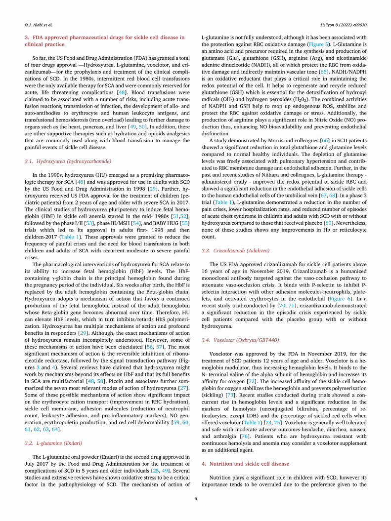
The pharmacological interventions of hydroxyurea for SCA relate toits ability to increase fetal hemoglobin (HbF) levels. The HbF-containing γ-globin chain is the principal hemoglobin found duringthe pregnancy period of the individual. Six weeks after birth, the HbF isreplaced by the adult hemoglobin containing the Beta-globin chain.Hydroxyurea adopts a mechanism of action that favors a continuedproduction of the fetal hemoglobin instead of the adult hemoglobinwhose Beta-globin gene becomes abnormal over time. Therefore, HUcan elevate HbF levels, which in turn inhibits/retards HbS polymeri-zation. Hydroxyurea has multiple mechanisms of action and profoundbenefits in responders [29]. Although, the exact mechanisms of actionof hydroxyurea remain incompletely understood. However, some ofthese mechanisms of action have been elucidated [56, 57]. The mostsignificant mechanism of action is the reversible inhibition of ribonu-cleotide reductase, followed by the signal transduction pathway (Fig-ures 3 and 4). Several reviews have claimed that hydroxyurea mightwork by mechanisms beyond its effects on HbF and that its full benefitsin SCA are multifactorial [48, 58]. Piccin and associates further sum-marized the seven most relevant modes of action of hydroxyurea [27].Some of these possible mechanisms of action show significant impacton the erythrocyte cation transport (improvement in RBC hydration),sickle cell membrane, adhesion molecules (reduction of neutrophilcount, leukocyte adhesion, and pro-inflammatory markers), NO gen-eration, erythropoietin production, and red cell deformability [59, 60,61, 62, 63, 64].
3.2. L-glutamine (Endari)
The L-glutamine oral powder (Endari) is the second drug approved inJuly 2017 by the Food and Drug Administration for the treatment ofcomplications of SCD in 5 years and older individuals [25, 49]. Severalstudies and extensive reviews have shown oxidative stress to be a criticalfactor in the pathophysiology of SCD. The mechanism of action of
5
L-glutamine is not fully understood, although it has been associated withthe protection against RBC oxidative damage (Figure 5). L-Glutamine isan amino acid and precursor required in the synthesis and production ofglutamate (Glu), glutathione (GSH), arginine (Arg), and nicotinamideadenine dinucleotide (NADH), all of which protect the RBC from oxida-tive damage and indirectly maintain vascular tone [65]. NADH/NADPHis an oxidative reductant that plays a critical role in maintaining theredox potential of the cell. It helps to regenerate and recycle reducedglutathione (GSH) which is essential for the detoxification of hydroxylradicals (OH⋅) and hydrogen peroxides (H202). The combined activitiesof NADPH and GSH help to mop up endogenous ROS, stabilize andprotect the RBC against oxidative damage or stress. Additionally, theproduction of arginine plays a significant role in Nitric Oxide (NO) pro-duction thus, enhancing NO bioavailability and preventing endothelialdysfunction.
A study demonstrated by Morris and colleagues [66] in SCD patientsshowed a significant reduction in total glutathione and glutamine levelscompared to normal healthy individuals. The depletion of glutaminelevels was freely associated with pulmonary hypertension and contrib-uted to RBC membrane damage and endothelial adhesion. Further, in thepast and recent studies of Niihara and colleagues, L-glutamine therapy -administered orally - improved the redox potential of sickle RBC andshowed a significant reduction in the endothelial adhesion of sickle cellsto the human endothelial cells of the umbilical vein [67, 68]. In a phase 3trial (Table 1), L-glutamine demonstrated a reduction in the number ofpain crises, lower hospitalization rates, and reduced number of episodesof acute chest syndrome in children and adults with SCD with or withouthydroxyurea compared to those that received placebo [69]. Nevertheless,none of these studies shows any improvements in Hb or reticulocytecount.
3.3. Crizanlizumab (Adakveo)
The US FDA approved crizanlizumab for sickle cell patients above16 years of age in November 2019. Crizanlizumab is a humanizedmonoclonal antibody targeted against the vaso-occlusion pathway toattenuate vaso-occlusion crisis. It binds with P-selectin to inhibit P-selectin interaction with other adhesion molecules-neutrophils, plate-lets, and activated erythrocytes in the endothelial (Figure 6). In arecent study trial conducted by [70, 71], crizanlizumab demonstrateda significant reduction in the episodic crisis experienced by sicklecell patients compared with the placebo group with or withouthydroxyurea.
3.4. Voxelotor (Oxbryta/GBT440)
Voxelotor was approved by the FDA in November 2019, for thetreatment of SCD patients 12 years of age and older. Voxelotor is a he-moglobin modulator, thus increasing hemoglobin levels. It binds to theN- terminal valine of the alpha subunit of hemoglobin and increases itsaffinity for oxygen [72]. The increased affinity of the sickle cell hemo-globin for oxygen stabilizes the hemoglobin and prevents polymerization(sickling) [73]. Recent studies conducted during trials showed a con-current rise in hemoglobin levels and a significant reduction in themarkers of hemolysis (unconjugated bilirubin, percentage of re-ticulocytes, except LDH) and the percentage of sickled red cells whenoffered voxelotor (Table 1) [74, 75]. Voxelotor is generally well toleratedand safe with moderate adverse outcomes-headache, diarrhea, nausea,and arthralgia [76]. Patients who are hydroxyurea resistant withcontinuous hemolysis and anemia may consider a voxelotor supplementas an additional agent.
4. Nutrition and sickle cell disease
Nutrition plays a significant role in children with SCD; however itsimportance tends to be overruled due to the preference given to the
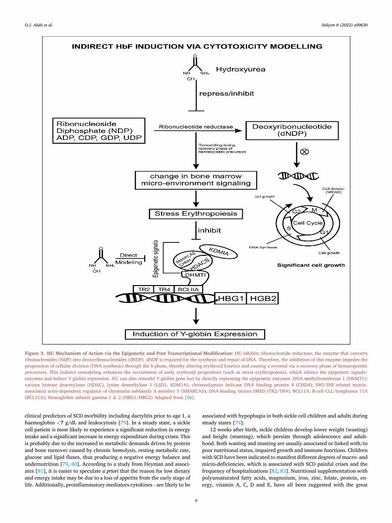
Figure 3. HU Mechanism of Action via the Epigenetic and Post Transcriptional Modification: HU inhibits ribonucleotide reductase, the enzyme that convertsribonucleosides (NDP) into deoxyribonucleosides (dNDP). dNDP is required for the synthesis and repair of DNA. Therefore, the inhibition of this enzyme impedes theprogression of cellular division (DNA synthesis) through the S-phase, thereby altering erythroid kinetics and causing a reversal via a recovery phase of hematopoieticprecursors. This indirect remodeling enhances the recruitment of early erythroid progenitors (such as stress erythropoiesis), which silence the epigenetic signals/enzymes and induce Y-globin expression. HU can also remodel Y-globin gene loci by directly repressing the epigenetic enzymes. DNA methyltransferase 1 (DNMT1);various histone deacetylases (HDAC), lysine demethylase 1 (LSD1, KDM1A); chromodomain helicase DNA binding protein 4 (CHD4); SWI/SNF-related matrix-associated actin-dependent regulator of chromatin subfamily A member 5 (SMARCA5); DNA-binding factors DRED (TR2/TR4); BCL11A: B-cell CLL/lymphoma 11A(BCL11A); Hemoglobin subunit gamma-1 & 2 (HBG1/HBG2) Adapted from [56].
O.J. Alabi et al. Heliyon 8 (2022) e09630
clinical predictors of SCD morbidity including dactylitis prior to age 1, ahaemoglobin <7 g/dL and leukocytosis [79]. In a steady state, a sicklecell patient is most likely to experience a significant reduction in energyintake and a significant increase in energy expenditure during crises. Thisis probably due to the increased in metabolic demands driven by proteinand bone turnover caused by chronic hemolysis, resting metabolic rate,glucose and lipid fluxes, thus producing a negative energy balance andundernutrition [79, 80]. According to a study from Heyman and associ-ates [81], it is easier to speculate a priori that the reason for low dietaryand energy intake may be due to a loss of appetite from the early stage oflife. Additionally, proinflammatory mediators-cytokines - are likely to be
6
associated with hypophagia in both sickle cell children and adults duringsteady states [79].
12 weeks after birth, sickle children develop lower weight (wasting)and height (stunting), which persists through adolescence and adult-hood. Both wasting and stunting are usually associated or linked with/topoor nutritional status, impaired growth and immune functions. Childrenwith SCD have been indicated to manifest different degrees of macro- andmicro-deficiencies, which is associated with SCD painful crises and thefrequency of hospitalizations [82, 83]. Nutritional supplementation withpolyunsaturated fatty acids, magnesium, iron, zinc, folate, protein, en-ergy, vitamin A, C, D and E, have all been suggested with the great
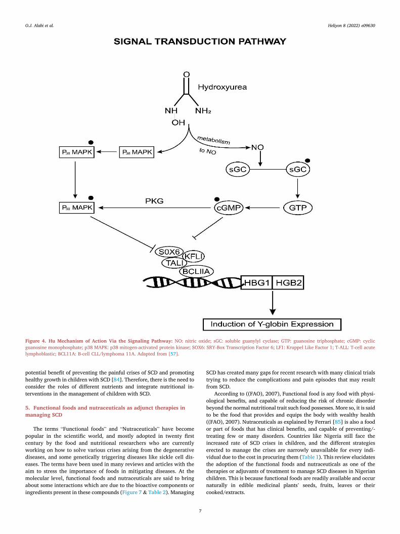
Figure 4. Hu Mechanism of Action Via the Signaling Pathway: NO: nitric oxide; sGC: soluble guanylyl cyclase; GTP: guanosine triphosphate; cGMP: cyclicguanosine monophosphate; p38 MAPK: p38 mitogen-activated protein kinase; SOX6: SRY-Box Transcription Factor 6; LF1: Kruppel Like Factor 1; T-ALL: T-cell acutelymphoblastic; BCL11A: B-cell CLL/lymphoma 11A. Adapted from [57].
O.J. Alabi et al. Heliyon 8 (2022) e09630
potential benefit of preventing the painful crises of SCD and promotinghealthy growth in children with SCD [84]. Therefore, there is the need toconsider the roles of different nutrients and integrate nutritional in-terventions in the management of children with SCD.
5. Functional foods and nutraceuticals as adjunct therapies inmanaging SCD
The terms “Functional foods” and “Nutraceuticals” have becomepopular in the scientific world, and mostly adopted in twenty firstcentury by the food and nutritional researchers who are currentlyworking on how to solve various crises arising from the degenerativediseases, and some genetically triggering diseases like sickle cell dis-eases. The terms have been used in many reviews and articles with theaim to stress the importance of foods in mitigating diseases. At themolecular level, functional foods and nutraceuticals are said to bringabout some interactions which are due to the bioactive components oringredients present in these compounds (Figure 7 & Table 2). Managing
7
SCD has created many gaps for recent research with many clinical trialstrying to reduce the complications and pain episodes that may resultfrom SCD.
According to ((FAO), 2007), Functional food is any food with physi-ological benefits, and capable of reducing the risk of chronic disorderbeyond the normal nutritional trait such food possesses. More so, it is saidto be the food that provides and equips the body with wealthy health((FAO), 2007). Nutraceuticals as explained by Ferrari [85] is also a foodor part of foods that has clinical benefits, and capable of preventing/-treating few or many disorders. Countries like Nigeria still face theincreased rate of SCD crises in children, and the different strategieserected to manage the crises are narrowly unavailable for every indi-vidual due to the cost in procuring them (Table 1). This review elucidatesthe adoption of the functional foods and nutraceuticals as one of thetherapies or adjuvants of treatment to manage SCD diseases in Nigerianchildren. This is because functional foods are readily available and occurnaturally in edible medicinal plants’ seeds, fruits, leaves or theircooked/extracts.
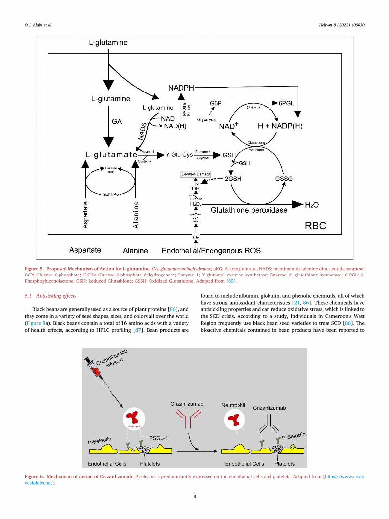
Figure 5. Proposed Mechanism of Action for L-glutamine: GA: glutamine aminohydrolase; aKG: A-ketoglutarate; NADS: nicotinamide adenine dinucleotide synthase;G6P: Glucose 6-phosphate; G6PD: Glucose 6-phosphate dehydrogenase; Enzyme 1; Y-glutamyl cysteine synthetase; Enzyme 2: glutathione synthetase; 6-PGL: 6-Phosphogluconolactone; GSH: Reduced Glutathione; GSSH: Oxidized Glutathione. Adapted from [65].
O.J. Alabi et al. Heliyon 8 (2022) e09630
5.1. Antisickling effects
Black beans are generally used as a source of plant proteins [86], andthey come in a variety of seed shapes, sizes, and colors all over the world(Figure 8a). Black beans contain a total of 16 amino acids with a varietyof health effects, according to HPLC profiling [87]. Bean products are
Figure 6. Mechanism of action of Crizanlizumab. P-selectin is predominantly expvebiolabs.net].
8
found to include albumin, globulin, and phenolic chemicals, all of whichhave strong antioxidant characteristics [21, 86]. These chemicals haveantisickling properties and can reduce oxidative stress, which is linked tothe SCD crisis. According to a study, individuals in Cameroon's WestRegion frequently use black bean seed varieties to treat SCD [88]. Thebioactive chemicals contained in bean products have been reported to
ressed on the endothelial cells and platelets. Adapted from [https://www.creati
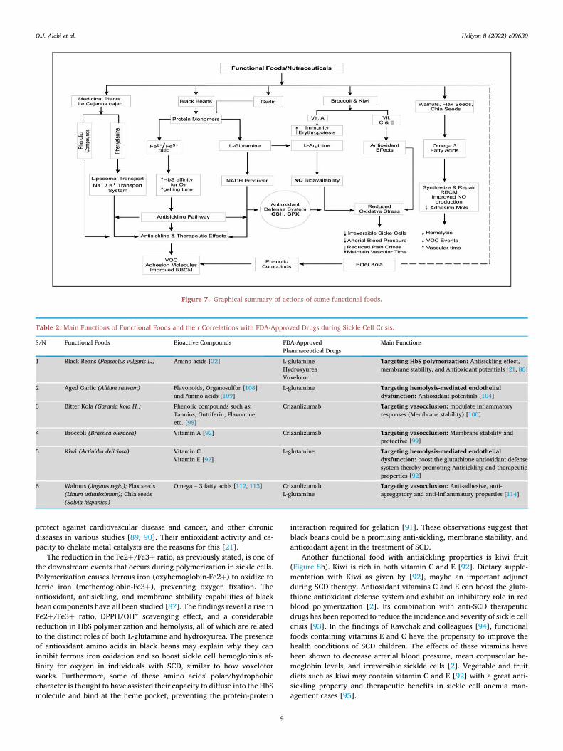
Figure 7. Graphical summary of actions of some functional foods.
Table 2. Main Functions of Functional Foods and their Correlations with FDA-Approved Drugs during Sickle Cell Crisis.
S/N Functional Foods Bioactive Compounds FDA-ApprovedPharmaceutical Drugs
Main Functions
1 Black Beans (Phaseolus vulgaris L.) Amino acids [22] L-glutamineHydroxyureaVoxelotor
Targeting HbS polymerization: Antisickling effect,membrane stability, and Antioxidant potentials [21, 86]
2 Aged Garlic (Allium sativum) Flavonoids, Organosulfur [108]and Amino acids [109]
L-glutamine Targeting hemolysis-mediated endothelialdysfunction: Antioxidant potentials [104]
3 Bitter Kola (Garania kola H.) Phenolic compounds such as:Tannins, Guttiferin, Flavonone,etc. [98]
Crizanlizumab Targeting vasocclusion: modulate inflammatoryresponses (Membrane stability) [100]
4 Broccoli (Brassica oleracea) Vitamin A [92] Crizanlizumab Targeting vasocclusion: Membrane stability andprotective [99]
5 Kiwi (Actinidia deliciosa) Vitamin CVitamin E [92]
L-glutamine Targeting hemolysis-mediated endothelialdysfunction: boost the glutathione antioxidant defensesystem thereby promoting Antisickling and therapeuticproperties [92]
6 Walnuts (Juglans regia); Flax seeds(Linum usitatissimum); Chia seeds(Salvia hispanica)
Omega – 3 fatty acids [112, 113] CrizanlizumabL-glutamine
Targeting vasocclusion: Anti-adhesive, anti-agreggatory and anti-inflammatory properties [114]
O.J. Alabi et al. Heliyon 8 (2022) e09630
protect against cardiovascular disease and cancer, and other chronicdiseases in various studies [89, 90]. Their antioxidant activity and ca-pacity to chelate metal catalysts are the reasons for this [21].
The reduction in the Fe2þ/Fe3þ ratio, as previously stated, is one ofthe downstream events that occurs during polymerization in sickle cells.Polymerization causes ferrous iron (oxyhemoglobin-Fe2þ) to oxidize toferric iron (methemoglobin-Fe3þ), preventing oxygen fixation. Theantioxidant, antisickling, and membrane stability capabilities of blackbean components have all been studied [87]. The findings reveal a rise inFe2þ/Fe3þ ratio, DPPH/OH* scavenging effect, and a considerablereduction in HbS polymerization and hemolysis, all of which are relatedto the distinct roles of both L-glutamine and hydroxyurea. The presenceof antioxidant amino acids in black beans may explain why they caninhibit ferrous iron oxidation and so boost sickle cell hemoglobin's af-finity for oxygen in individuals with SCD, similar to how voxelotorworks. Furthermore, some of these amino acids' polar/hydrophobiccharacter is thought to have assisted their capacity to diffuse into the HbSmolecule and bind at the heme pocket, preventing the protein-protein
9
interaction required for gelation [91]. These observations suggest thatblack beans could be a promising anti-sickling, membrane stability, andantioxidant agent in the treatment of SCD.
Another functional food with antisickling properties is kiwi fruit(Figure 8b). Kiwi is rich in both vitamin C and E [92]. Dietary supple-mentation with Kiwi as given by [92], maybe an important adjunctduring SCD therapy. Antioxidant vitamins C and E can boost the gluta-thione antioxidant defense system and exhibit an inhibitory role in redblood polymerization [2]. Its combination with anti-SCD therapeuticdrugs has been reported to reduce the incidence and severity of sickle cellcrisis [93]. In the findings of Kawchak and colleagues [94], functionalfoods containing vitamins E and C have the propensity to improve thehealth conditions of SCD children. The effects of these vitamins havebeen shown to decrease arterial blood pressure, mean corpuscular he-moglobin levels, and irreversible sicklde cells [2]. Vegetable and fruitdiets such as kiwi may contain vitamin C and E [92] with a great anti-sickling property and therapeutic benefits in sickle cell anemia man-agement cases [95].
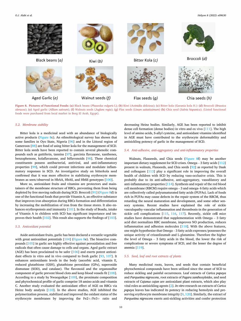
Figure 8. Pictures of Functional Foods: (a) Black beans (Phaseolus vulgaris L); (b) Kiwi (Actinidia deliciosa); (c) Bitter kola (Garania kola H.): (d) Broccoli (Brassicaoleracea); (e) Aged garlic (Allium sativum); (f) Walnuts seeds (Juglans regia); (g) Flax seeds (Linum usitatissimum) (h) Chia seed (Salvia hispanica). (Listed functionalfoods were purchased from local market in Borg El Arab, Egypt).
O.J. Alabi et al. Heliyon 8 (2022) e09630
5.2. Membrane stability
Bitter kola is a medicinal seed with an abundance of biologicallyactive products (Figure 8c). An ethnobiological survey has shown thatsome families in Oyo State, Nigeria [96] and in the Littoral region ofCameroon [88] are fond of using bitter kola for the management of SCD.Bitter kola seeds have been reported to contain several phenolic com-pounds such as guttiferin, tannins [97], garcinia flavanone, xanthenes,benzophenone, kolaflavanone, and biflavonoids [98]. These chemicalconstituents possess antibacterial, antiviral, and anti-inflammatoryproperties [99], which could prevent infections and modulate inflam-matory responses in SCD. An investigative study on bitterkola seedconfirmed that it was more effective in stabilizing erythrocyte mem-branes as seen/observed in HbAA, HbAS, and HbSS genotypes [100].
More so, antioxidant fruits and vitamins are protectors and main-tainers of the membrane structure of RBCs, preventing them from beingdepleted by free moving radicals during SCD. Broccoli [91] (Figure 8d) isone of the functional foods that provide Vitamin A, an effective substancethat improves iron absorption during RBCs formation and differentiationby increasing the mobilization of iron from the tissue stores. It also en-hances erythropoiesis and immunity [101]. In the study of Boadu, intakeof Vitamin A in children with SCD has significant importance and im-proves their health [102]. This result also supports the findings of [103].
5.3. Antioxidant potential
Aside antioxidant fruits, garlic has been declared a versatile vegetablewith great antioxidant potentials [104] (Figure 8e). The bioactive com-pounds [105] in garlic are highly effective against peroxidation and freeradicals that often cause damage to cells and organs. Aged garlic extract(AGE) has been proclaimed to be safer [106] and exerts potent antioxi-dant effects in vitro and in vivo compared to fresh garlic [93, 107]. Itenhances antioxidants levels in the body (ascorbic acid, vitamin E,reduced glutathione (GSH), glutathione peroxidase (GPx), superoxidedismutase (SOD), and catalase). The flavonoid and the organosulfurcomponent of garlic prevent blood clots and keep blood vessels fit [108].According to a study by Nwaoguikpe [109], the proximate compositionand phytochemical profile of garlic comprise 18 amino acids and vitaminC. Another study evaluated the antioxidant effect of AGE on RBCs viaHeinz body analysis [110]. In the above studies, AGE inhibited thepolymerization process, stabilized and improved the oxidant status of theerythrocyte membranes by improving the Fe2þ/Fe3þ ratio and
10
decreasing Heinz bodies. Similarly, AGE has been reported to inhibitdense cell formation (dense bodies) in vitro and ex vivo [111]. The highlevel of amino acids, S-allyl cysteine, and antioxidant vitamins identifiedin AGE must have contributed to the erythrocyte deformability andantisickling potency of garlic in the management of SCD.
5.4. Anti-adhesive, anti-aggregatory and anti-inflammatory properties
Walnuts, Flaxseeds, and Chia seeds (Figure 8f) may be anotherimportant dietary supplement for SCD crises. Omega – 3 fatty acids [112]present in walnuts, Flaxseeds, and Chia seeds [92] as reported by Daakand colleagues [113] play a significant role in improving the overallhealth of children with SCD by reducing vaso-occlusive crisis. This isprobably due to its anti-adhesive, anti-aggregatory, vasodilatory andanti-inflammatory properties [114]. Synthesis and repair of the red bloodcell membrane (RBCM) require omega – 3 and omega- 6 fatty acids whichare collectively called polyunsaturated fatty acids (PUFAs). Lack of foodsrich in PUFAs may cause defects in the repair system of the RBCM, thusretarding the neural maturation and development, and some other sen-sory systems. Recent studies have explained the role of sicklevasculopathy-vascular inflammation and thrombosis-in the generation ofsickle cell complications [115, 116, 117]. Recently, sickle cell micestudies have demonstrated that supplementation with Omega – 3 fattyacid diet normalizes RBC membrane, improves NO production, reducesinflammation and adhesion molecules [118]. With the above features,onemight hypothesize that Omega – 3 fatty acids expresses/possesses theunique activity of crizanlizumab and L-glutamine. Therefore the higherthe level of Omega – 3 fatty acids in the blood, the lower the risk ofcomplications or severe symptoms of SCD, and the lesser the degree ofanemia [119].
5.5. Seed, leaf and root extracts of plants
Many medicinal roots, leaves, and seeds that contain beneficialphytochemical compounds have been utilized since the onset of SCD toreduce sickling and painful occurrences. Leaf extracts of Carica papayaand Parquetina nigrescens, root extracts of Fagara zanthoxyloides, and seedextracts of Cajanus cajan are antioxidant plant extracts, which also playvital roles as antisickling agents [2]. In vitro research on extracts of Caricapapaya leaves has indicated its potency in reducing hemolysis and pre-serving erythrocyte membrane integrity [9, 120]. Similarly, the extract ofParquetina nigrescens exerts anti-sickling activities and confer protection
O.J. Alabi et al. Heliyon 8 (2022) e09630
on the integrity of the RBC membrane [121]. Another study conductedon a cocktail of Carica papaya, Parquetina nigrescens, and Fagara zan-thoxyloides extracts shows promising results [122]. These pharmacolog-ical properties are due to the presence of several phenolic compoundsand free amino acids found in these plant extracts [123, 124, 125].Eugenia caryophyllata and Piper guineense are other examples of medicinalplants thatcontain vanilloids like Shikimic acid and cannaboids that areof benefit in reducing the painful symptoms of SCD [126]. Additionalphytomedicines that have been reported to possess antisickling poten-tials are Pterocarpa osun, Justicia secunda, and Sorghum bicolor extracts[127,128].
Taken together, these functional foods can be used in the form of foodor decoction. The consumption of a cocktail of functional foods is highlyrecommended for sickle cell disease patients. Consuming functionalfoods and nutraceuticals alongside with/without FDA-approved drugscan be one of the first–line and promising therapies in managing thecrises of SCD in Nigerian children especially during their infanthoodstages.
6. Conclusion
For SCD challenging children in Nigeria, the functional food researchand development must be motivated on how to decrease the number ofcrises during SCD and thus increase the overall survival of the SCDchildren. Various bioactive ingredients in the foods and edible plants maybe extracted and screened to drug prologue during management of SCDcrisis. Research to experimentally identify these bioactive ingredients isat hand. Therefore, capability of functional foods in managing SCDwithout painful episodes in children involves myriad of research intofoods and nutraceuticals which is a current trend in nutritionalbiochemistry and genetic disorders like sickle cell disease, with aresponse to find readily available, cheaper and alternative novel mole-cule that the Nigerian children susceptible to SCD can get within theirreach. This review paper indicates the therapeutic, antisickling, antiox-idant and inhibitory effects of functional foods as combatants for sicklecell disease crises in Nigerian children (Table 2).
Declarations
Author contribution statement
All authors listed have significantly contributed to the developmentand the writing of this article.
Funding statement
This research did not receive any specific grant from funding agenciesin the public, commercial, or not-for-profit sectors.
Data availability statement
No data was used for the research described in the article.
Declaration of interests statement
The authors declare no conflict of interest.
Additional information
No additional information is available for this paper.
Acknowledgements
We would like to thank Festus Adebayo Atiba, (1Department ofBiochemistry, Ladoke Akintola University of Technology, Nigeria &
11
2Department of Biochemistry, University of S~ao Paulo Brazil), forassisting with the illustration.
References
[1] H.F. Bunn, Pathogenesis and treatment of sickle cell disease, N. Engl. J. Med. 337(11) (1997) 762–769.
[2] N.A. Imaga, Phytomedicines and nutraceuticals: alternative therapeutics for sicklecell anemia, Sci. World J. (2013).
[3] I. Boadu, A. Ohemeng, L.A. Renner, Dietary intakes and nutritional status ofchildren with sickle cell disease at the Princess Marie Louise Hospital, Accra – asurvey, BMC Nutr. 4 (2018) 33.
[4] M.H. Steinberg, Sickle cell disease, Hematology 1 (2004) 53.[5] D. Ansong, A. Osei-Akoto, D. Ocloo, K.-F. Ohene-Frempong, Sickle cell disease:
management options and challenges in developing countries, Mediterr. J.Hematol. Infect. Dis. 5 (1) (2013).
[6] M.R. Islam, M. Moinuddin, A. Ahmed, S.M. Rahman, Association of sickle celldisease with anthropometric indices among under-five children: evidence from2018 Nigeria Demographic and Health Survey, BMC Med. 19 (1) (2021) 5.
[7] I. Odame, Perspective: we need a global solution, Nature 515 (7526) (2014) S10.[8] K.A. Anie, F.E. Egunjobi, O.O. Akinyanju, Psychosocial impact of sickle cell
disorder: perspectives from a Nigerian setting, Glob. Health 6 (2010) 2.[9] S.D. Grosse, I. Odame, H.K. Atrash, D.D. Amendah, F.B. Piel, T.N. Williams, Sickle
cell disease in Africa: a neglected cause of early childhood mortality, Am. J. Prev.Med. 41 (6 Suppl 4) (2011) S398–S405.
[10] G. Jones, Dictionary of Mythology Folklore and Symbols, The Scarecrow Press,New York, NY, USA, 1961.
[11] L.A. Alli, M.P. Okoh, Phyto-Medicine in gene(s) targeting future direction forsickle cell disease management, Hered. Genet. 5 (2016) 2.
[12] T. Oduola, F. Adeniyi, E. Ogunyemi, I. Bello, T. Idowu, Antisickling agent in anextract of unripe pawpaw (Carica papaya): is it real? Afr. J. Biotechnol. 5 (2006)1947.
[13] N. Imaga, O. Adepoju, Analyses of antisickling potency of Carica papaya dried leafextract and fractions, J. Pharmacogn. Phytotherapy 2 (2010) 97–102.
[14] N. Galadanci, B. Wudil, T. Balogun, G. Ogunrinde, A. Akinsulie, et al., Currentsickle cell disease management practices in Nigeria, Int. Health 6 (2015) 23–28.
[15] A. Adewoyin, Management of sickle cell disease: a review for physician educationin Nigeria (sub-saharan Africa), Anemia 2015 (2015) 7914–7998.
[16] L. Kenmoe, T. Kotue, K. Chandra, F. Djouhou, A. Pieme, G. Kansci, E. Fokou,N. Arumugam, Albumin and globulin fractions from black bean seeds (Phaseolusvulgaris L.) used in the management of sickle cell disease (SCD) in the west regionof Cameroon have antisickling and antioxidant properties, Biotechnol. Biomed. 3(2) (2020) 78–92.
[17] E. Svarch, P. Hern�andez, J. Ballester, Sickle Cell Disease in Cuba, Institute deHematologiae Inmunologia (IHI) La Habana, Cuba, 2001, pp. 13–45.
[18] C.A. Kingsley, A. Merit, O.I. Rose, N.O. Paulinus, O.E. Olusola, F. Abiodun,N.E. Lauretta, C.T. Mercy, J.O. John, K.O. Sarah, R.E. Oghenebrozie, A.O. Joyce,I.K. Amina, O.A. Faith, Biochemical investigation of the upstream anti-sicklingmechanisms of soursop (Annona muricata): 15-acetyl guanacone as an inhibitor ofdeoxyhaemoglobin polymerisation, J. Biomol. Struct. Dyn. (2020).
[19] W. Xu, L. Chi, B. Row, et al., Increased oxidative stress is associated with chronicintermittent hypoxia-mediated brain cortical neuronal cell apoptosis in a mousemodel of sleep apnea, Neuroscience 126 (2004) 313–323.
[20] N.-M. Amina, B. Valentine, R. David, Emerging therapies in sickle cell disease, Br.J. Haematol. (2020).
[21] D.C. Nkenmeni, T.C. Kotue, P. Kumar, F.M. Djouhou, S.F. Ngo, A.C. Pieme,G. Kansci, E. Fokou, N. Arumugam, HPLC profiling, in vitro antisickling andantioxidant activities of phenolic compound extracts from black bean seeds(Phaseolus vulgarus L.) used in the management of sickle cell disease in the WestRegion of Cameroon, Int. J. Food Nutr. Res. 3 (2019) 30.
[22] J. Teibo, S. Bello, A. Olagunju, F. Olorunfemi, O. Ajao, O. Fabunmi, Functionalfoods and bioactive compounds: roles in the prevention, treatment andmanagement of neurodegenerative diseases, GSC Biol. Pharmaceut. Sci. 11 (2)(2020) 297–313.
[23] J. Goldstein, W. Konigsberg, R.J. Hill, The structure of human hemoglobin. VI. Thesequence of amino acids in the tryptic peptides of the beta chain, J. Biol. Chem.238 (1963) 2016–2027.
[24] P. Sundd, M.T. Gladwin, E.M. Novelli, Pathophysiology of sickle cell disease, Ann.Rev. Pathol, 14 (2019) 263–292.
[25] G.S. Cisneros, S.L. Thein, Recent advances in the treatment of sickle cell disease,Front. Physiol. 11 (2020) 435.
[26] R.P. Hebbel, Reconstructing sickle cell disease: a data-based analysis of the"hyperhemolysis paradigm" for pulmonary hypertension from the perspective ofevidence-based medicine, Am. J. Hematol. 86 (2) (2011) 123–154.
[27] A. Piccin, C. Murphy, E. Eakins, M.B. Rondinelli, M. Daves, C. Vecchiato, D. Wolf,C. Mc Mahon, O.P. Smith, Insight into the complex pathophysiology of sickle cellanaemia and possible treatment, Eur. J. Haematol. 102 (4) (2019) 319–330.
[28] G.J. Kato, M.H. Steinberg, M.T. Gladwin, Intravascular hemolysis and thepathophysiology of sickle cell disease, J. Clin. Invest. 127 (3) (2017) 750–760.
[29] S. Moerdler, D. Manwani, New insights into the pathophysiology and developmentof novel therapies for sickle cell disease, Hematology. Americ an Society ofHematology. Edu. Program 2018 (1) (2018) 493–506.
[30] A. Hidalgo, J. Chang, J.E. Jang, A.J. Peired, E.Y. Chiang, P.S. Frenette, Heterotypicinteractions enabled by polarized neutrophil microdomains mediatethromboinflammatory injury, Nat. Med. 15 (4) (2009) 384–391.
O.J. Alabi et al. Heliyon 8 (2022) e09630
[31] D.C. Rees, T.N. Williams, M.T. Gladwin, Sickle-cell disease, Lancet (London,England) 376 (9757) (2010) 2018–2031.
[32] J.D. Belcher, C. Chen, J. Nguyen, L. Milbauer, F. Abdulla, A.I. Alayash, A. Smith,K.A. Nath, R.P. Hebbel, G.M. Vercellotti, Heme triggers TLR4 signaling leading toendothelial cell activation and vaso- occlusion in murine sickle cell disease, Blood123 (3) (2014) 377–390.
[33] D.R. Gutsaeva, P. Montero-Huerta, J.B. Parkerson, S.D. Yerigenahally, T. Ikuta,C.A. Head, Molecular mechanisms underlying synergistic adhesion of sickle redblood cells by hypoxia and low nitric oxide bioavailability, Blood 123 (12) (2014)1917–1926.
[34] D.S. Darbari, V.A. Sheehan, S.K. Ballas, The vaso-occlusive pain crisis in sickle celldisease: definition, pathophysiology, and management, Eur. J. Haematol. 105 (3)(2020) 237–246.
[35] T.D. Coates, P. Chalacheva, L. Zeltzer, M. Khoo, Autonomic nervous systeminvolvement in sickle cell disease, Clin. Hemorheol. Microcirc. 68 (2-3) (2018)251–262.
[36] M.H. Steinberg, Pathophysiology of sickle cell disease, Bailliere. Clin. Haematol.11 (1) (1998) 163–184.
[37] M.J. Stuart, R.L. Nagel, Sickle-cell disease, Lancet (London, England) 364 (9442)(2004) 1343–1360.
[38] E. Nur, B.J. Biemond, H.M. Otten, D.P. Brandjes, J.J. Schnog, CURAMA StudyGroup, Oxidative stress in sickle cell disease; pathophysiology and potentialimplications for disease management, Am. J. Hematol. 86 (6) (2011) 484–489.
[39] D.K. Kaul, E. Finnegan, G.A. Barabino, Sickle red cell-endothelium interactions,Microcirculation (N. Y.) 16 (1) (2009) 97–111.
[40] G.Y. Chen, G. Nu~nez, Sterile inflammation: sensing and reacting to damage, Nat.Rev. Immunol. 10 (12) (2010) 826–837.
[41] F.F. Dutra, L.S. Alves, D. Rodrigues, P.L. Fernandez, R.B. de Oliveira,D.T. Golenbock, D.S. Zamboni, M.T. Bozza, Hemolysis- induced lethality involvesinflammasome activation by heme, Proc. Nat. Acad. Sci. USA 111 (39) (2014)E4110–E4118.
[42] D. Zhang, C. Xu, D. Manwani, P.S. Frenette, Neutrophils, platelets, andinflammatory pathways at the nexus of sickle cell disease pathophysiology, Blood127 (7) (2016) 801–809.
[43] S.M. Camus, J.A. De Moraes, P. Bonnin, P. Abbyad, S. Le Jeune, F. Lionnet,L. Loufrani, L. Grimaud, J.C. Lambry, D. Charue, L. Kiger, J.M. Renard,C. Larroque, H. Le Cl�esiau, A. Tedgui, P. Bruneval, C. Barja-Fidalgo,A. Alexandrou, P.L. Tharaux, C.M. Boulanger, O.P. Blanc-Brude, Circulating cellmembrane microparticles transfer heme to endothelial cells and triggervasoocclusions in sickle cell disease, Blood 125 (24) (2015) 3805–3814.
[44] G. Chen, D. Zhang, T.A. Fuchs, D. Manwani, D.D. Wagner, P.S. Frenette, Heme-induced neutrophil extracellular traps contribute to the pathogenesis of sickle celldisease, Blood 123 (24) (2014) 3818–3827.
[45] D.V. Krysko, P. Agostinis, O. Krysko, A.D. Garg, C. Bachert, B.N. Lambrecht,P. Vandenabeele, Emerging role of damage-associated molecular patterns derivedfrom mitochondria in inflammation, Trends Immunol. 32 (4) (2011) 157–164.
[46] R.T. Figueiredo, P.L. Fernandez, D.S. Mourao-Sa, B.N. Porto, F.F. Dutra, L.S. Alves,M.F. Oliveira, P.L. Oliveira, A.V. Graça-Souza, M.T. Bozza, Characterization ofheme as activator of Toll-like receptor 4, J. Biol. Chem. 282 (28) (2007)20221–20229.
[47] A.P. Monteiro, C.S. Pinheiro, T. Luna-Gomes, L.R. Alves, C.M. Maya-Monteiro,B.N. Porto, C. Barja-Fidalgo, C.F. Benjamim, M. Peters-Golden, C. Bandeira-Melo,M.T. Bozza, C. Canetti, Leukotriene B4 mediates neutrophil migration induced byheme, J. Immunol. 186 (11) (2011) 6562–6567.
[48] P.T. McGann, R.E. Ware, Hydroxyurea therapy for sickle cell anemia, Expet Opin.Drug Saf. 14 (11) (2015) 1749–1758.
[49] M.A. Ali, A. Ahmad, H. Chaudry, W. Aiman, S. Aamir, M.Y. Anwar, A. Khan,Efficacy and safety of recently approved drugs for sickle cell disease: a review ofclinical trials, Exp. Hematol. 92 (2020) 11–18.e1.
[50] D. Sharma, A.A. Ogbenna, A. Kassim, J. Andrews, Transfusion support in patientswith sickle cell disease, Semin. Hematol. 57 (2) (2020) 3950.
[51] N.L. Letvin, D.C. Linch, G.P. Beardsley, K.W. McIntyre, D.G. Nathan,Augmentation of fetal-hemoglobin production in anemic monkeys byhydroxyurea, N. Engl. J. Med. 310 (14) (1984) 869–873.
[52] O.S. Platt, S.H. Orkin, G. Dover, G.P. Beardsley, B. Miller, D.G. Nathan,Hydroxyurea enhances fetal hemoglobin production in sickle cell anemia, J. Clin.Invest. 74 (2) (1984) 652–656.
[53] S. Charache, G.J. Dover, R.D. Moore, S. Eckert, S.K. Ballas, M. Koshy, P.F. Milner,E.P. Orringer, G. Phillips Jr., O.S. Platt, Hydroxyurea: effects on hemoglobin Fproduction in patients with sickle cell anemia, Blood 79 (10) (1992) 2555–2565.
[54] S. Charache, M.L. Terrin, R.D. Moore, G.J. Dover, F.B. Barton, S.V. Eckert,R.P. McMahon, D.R. Bonds, Effect of hydroxyurea on the frequency of painfulcrises in sickle cell anemia. Investigators of the Multicenter Study of Hydroxyureain Sickle Cell Anemia, N. Engl. J. Med. 332 (20) (1995) 1317–1322.
[55] W.C. Wang, R.E. Ware, S.T. Miller, R.V. Iyer, J.F. Casella, C.P. Minniti, S. Rana,C.D. Thornburg, Z.R. Rogers, R.V. Kalpatthi, J.C. Barredo, R.C. Brown,S.A. Sarnaik, T.H. Howard, L.W. Wynn, A. Kutlar, F.D. Armstrong, B.A. Files,J.C. Goldsmith, M.A. Waclawiw, BABY HUG investigators, Hydroxycarbamide invery young children with sickle-cell anaemia: a multicentre, randomised,controlled trial (BABY HUG), Lancet (London, England) 377 (9778) (2011)1663–1672.
[56] Y. Saunthararajah, Targeting sickle cell disease root-cause pathophysiology withsmall molecules, Haematologica 104 (9) (2019) 1720–1730.
[57] A. Pandey, J.H. Estepp, D. Ramkrishna, Hydroxyurea treatment of sickle celldisease: towards a personalized model-based approach, J. Transl. Genet. Genom. 5(2021) 22–36.
12
[58] M.H. Steinberg, Sickle cell anemia, the first molecular disease: overview ofmolecular etiology, pathophysiology, and therapeutic approaches,TheScientificWorldJOURNAL 8 (2008) 1295–1324.
[59] D.T. Covas, I. de Lucena Angulo, P. Vianna Bonini Palma, M.A. Zago, Effects ofhydroxyurea on the membrane of erythrocytes and platelets in sickle cell anemia,Haematologica 89 (3) (2004) 273–280.
[60] C.A. Hillery, M.C. Du, W.C. Wang, J.P. Scott, Hydroxyurea therapy decreases thein vitro adhesion of sickle erythrocytes to thrombospondin and laminin, Br. J.Haematol. 109 (2) (2000) 322–327.
[61] Z. Huang, J.G. Louderback, S.B. King, S.K. Ballas, D.B. Kim-Shapiro, In vitroexposure to hydroxyurea reduces sickle red blood cell deformability, Am. J.Hematol. 67 (3) (2001) 151–156.
[62] N. Lemonne, B. M€ockesch, K. Charlot, Y. Garnier, X. Waltz, Y. Lamarre, S. Antoine-Jonville, M. Etienne-Julan, M.D. Hardy-Dessources, M. Romana, P. Connes, Effectsof hydroxyurea on blood rheology in sickle cell anemia: a two-years follow-upstudy, Clin. Hemorheol. Microcirc. 67 (2) (2017) 141–148.
[63] I. Papassotiriou, E. Voskaridou, A. Stamoulakatou, D. Loukopoulos, Increasederythropoietin level induced by hydroxyurea treatment of sickle cell patients,Hematol. J. : Off. J. Eur. Haematol. Assoc. 1 (5) (2000) 295–300.
[64] V.P. Cokic, R.D. Smith, B.B. Beleslin-Cokic, J.M. Njoroge, J.L. Miller,M.T. Gladwin, A.N. Schechter, Hydroxyurea induces fetal hemoglobin by the nitricoxide- dependent activation of soluble guanylyl cyclase, J. Clin. Invest. 111 (2)(2003) 231–239.
[65] C.T. Quinn, l-Glutamine for sickle cell anemia: more questions than answers,Blood 132 (7) (2018) 689–693.
[66] C.R. Morris, J.H. Suh, W. Hagar, S. Larkin, D.A. Bland, M.H. Steinberg,E.P. Vichinsky, M. Shigenaga, B. Ames, F.A. Kuypers, E.S. Klings, Erythrocyteglutamine depletion, altered redox environment, and pulmonary hypertension insickle cell disease, Blood 111 (1) (2008) 402–410.
[67] Y. Niihara, C.R. Zerez, D.S. Akiyama, K.R. Tanaka, Oral L-glutamine therapy forsickle cell anemia: I. Subjective clinical improvement and favorable change in redcell NAD redox potential, Am. J. Hematol. 58 (2) (1998) 117–121.
[68] Y. Niihara, N.M. Matsui, Y.M. Shen, D.A. Akiyama, C.S. Johnson, M.A. Sunga,J. Magpayo, S.H. Embury, V.K. Kalra, S.H. Cho, K.R. Tanaka, L- glutamine therapyreduces endothelial adhesion of sickle red blood cells to human umbilical veinendothelial cells, BMC Blood Disord. 5 (2005) 4.
[69] Y. Niihara, S.T. Miller, J. Kanter, S. Lanzkron, W.R. Smith, L.L. Hsu, V.R. Gordeuk,K. Viswanathan, S. Sarnaik, I. Osunkwo, E. Guillaume, S. Sadanandan, L. Sieger,J.L. Lasky, E.H. Panosyan, O.A. Blake, T.N. New, R. Bellevue, L.T. Tran,R.L. Razon, Investigators of the Phase 3 Trial of l- Glutamine in Sickle Cell Disease,A phase 3 trial of l-glutamine in sickle cell disease, N. Engl. J. Med. 379 (3) (2018)226–235.
[70] K.I. Ataga, A. Kutlar, J. Kanter, D. Liles, R. Cancado, J. Friedrisch, T.H. Guthrie,J. Knight-Madden, O.A. Alvarez, V.R. Gordeuk, S. Gualandro, M.P. Colella,W.R. Smith, S.A. Rollins, J.W. Stocker, R.P. Rother, Crizanlizumab for theprevention of pain crises in sickle cell disease, N. Engl. J. Med. 376 (5) (2017)429–439.
[71] A. Kutlar, J. Kanter, D.K. Liles, O.A. Alvarez, R.D. Cançado, J.R. Friedrisch,J.M. Knight-Madden, A. Bruederle, M. Shi, Z. Zhu, K.I. Ataga, Effect ofcrizanlizumab on pain crises in subgroups of patients with sickle cell disease: aSUSTAIN study analysis, Am. J. Hematol. 94 (1) (2019) 55–61.
[72] M.B. Strader, H. Liang, F. Meng, J. Harper, D.A. Ostrowski, E.R. Henry, A.S. Shet,W.A. Eaton, S.L. Thein, A.I. Alayash, Interactions of an anti-sickling drug withhemoglobin in red blood cells from a patient with sickle cell anemia, BioconjugateChem. 30 (3) (2019) 568–571.
[73] D. Oksenberg, K. Dufu, M.P. Patel, C. Chuang, Z. Li, Q. Xu, A. Silva-Garcia,C. Zhou, A. Hutchaleelaha, L. Patskovska, Y. Patskovsky, S.C. Almo, U. Sinha,B.W. Metcalf, D.R. Archer, GBT440 increases haemoglobin oxygen affinity,reduces sickling and prolongs RBC half-life in a murine model of sickle celldisease, Br. J. Haematol. 175 (1) (2016) 141–153.
[74] J. Howard, C.J. Hemmaway, P. Telfer, D.M. Layton, J. Porter, M. Awogbade,T. Mant, D.D. Gretler, K. Dufu, A. Hutchaleelaha, M. Patel, V. Siu, S. Dixon,N. Landsman, M. Tonda, J. Lehrer-Graiwer, A phase 1/2 ascending dose study andopen-label extension study of voxelotor in patients with sickle cell disease, Blood133 (17) (2019) 1865–1875.
[75] Vichinsky,E.,Hoppe,C.C.,Ataga,K. I.,Ware,R.E.,Nduba,V., El-Beshlawy,A.,Hassab,H., Achebe, M. M., Alkindi, S., Brown, R. C., Diuguid, D. L., Telfer, P., Tsitsikas, D. A.,Elghandour, A., Gordeuk, V. R., Kanter, J., Abboud, M. R., Lehrer-Graiwer, J., Tonda,M., Intondi, A., HOPE trial Investigators (2019). A phase 3 randomized trial ofvoxelotor in sickle cell disease. N. Engl. J. Med. , 381(6), 509– 519.
[76] L.B. Herity, D.M. Vaughan, L.R. Rodriguez, D.K. Lowe, Voxelotor: a noveltreatment for sickle cell disease, Ann. Pharmacother. 55 (2) (2021) 240–245.
[77] M.R. DeBaun, Hydroxyurea therapy contributes to infertility in adult men withsickle cell disease: a review, Expet Rev. Hematol. 7 (6) (2014) 767–773.
[78] I. Berthaut, G. Guignedoux, F. Kirsch-Noir, V. de Larouziere, C. Ravel, D. Bachir, etal., Influence of sickle cell disease and treatment with hydroxyurea on spermparameters and fertility of human males, Haematologica 93 (2008) 988–993.
[79] M. Reid, Nutrition and sickle cell disease, Comptes Rendus Biol. 336 (3) (2013)159–163.
[80] H.I. Hyacinth, O.A. Adekeye, C.S. Yilgwan, Malnutrition in sickle cell anemia:implications for infection, growth, and maturation, J. Social Behav. Health Sci. 7(1) (2013).
[81] M.B. Heyman, E. Vichinsky, R. Katz, B. Gaffield, D. Hurst, R. Castillo, D. Chiu,K. Kleman, A.J. Ammann, M.M. Thaler, Growth retardation in sickle-cell diseasetreated by nutritional support, Lancet (London, England) 1 (8434) (1985)903–906.
O.J. Alabi et al. Heliyon 8 (2022) e09630
[82] N.T. Gray, J.M. Bartlett, K.M. Kolasa, S.P. Marcuard, C.T. Holbrook, R.D. Horner,Nutritional status and dietary intake of children with sickle cell anemia, Am. J.Pediatr. Hematol. Oncol. 14 (1) (1992) 57–61.
[83] E.M. Barden, D.A. Kawchak, K. Ohene-Frempong, V.A. Stallings, B.S. Zemel, Bodycomposition in children with sickle cell disease, Am. J. Clin. Nutr. 76 (1) (2002)218–225.
[84] A. Ohemeng, I. Boadu, The role of nutrition in the pathophysiology andmanagement of sickle cell disease among children: a review of literature, Crit. Rev.Food Sci. Nutr. 58 (14) (2018) 2299–2305.
[85] C. Ferrari, Functional foods, herbs and nutraceuticals: towards biochemicalmechanisms of healthy aging, Biogerontology 5 (2004) 275–289.
[86] C.W. Beninger, G.L. Hosfield, Antioxidant activity of extracts, condensed tanninfractions, and pure flavonoids from Phaseolus vulgaris L. seed coat colorgenotypes, J. Agric. Food Chem. 51 (27) (2003) 7879–7883.
[87] T.C. Kotue, L.N. Wirba, P. Jayamurthy, A.C. Pieme, G. Kansci, E. Fokou, HPLCprofiling, in vitro antisickling and antioxidant activities of amino acids from blackbean seeds (Phaseolus vulgarus L.) used in the management of Sickle Cell Disease(SCD) in the West Region of Cameroon, Int. J. Curr. Res. 11 (8) (2019) 5872–5880.
[88] T.C. Kotue, A.C. Pieme, E. Fokou, Ethnobotanicals usages in the management ofsickle cell disease (SDC) in some localities of Cameroon, Pharmacophore 7 (4)(2016) 192–200.
[89] Z. Zhu, W. Jiang, H.J. Thompson, Edible dry bean consumption (Phaseolusvulgaris L.) modulates cardiovascular risk factors and diet-induced obesity in ratsand mice, Br. J. Nutr. 108 (Suppl 1) (2012) S66–S73.
[90] F. Finetti, M. Biagi, J. Ercoli, G. Macrì, E. Miraldi, L. Trabalzini, Phaseolus vulgaris L.Var. Venanzio grown in Tuscany: chemical composition and in vitro investigation ofpotential effects on colorectal cancer, Antioxidants 9 (12) (2020) 1181.
[91] M. Gorecki, J.R. Votano, A. Rich, Peptide inhibitors of sickle hemoglobinaggregation: effect of hydrophobicity, Biochemistry 19 (8) (1980) 1564–1568.
[92] K. Umeakunne, J.M. Hibbert, Nutrition in sickle cell disease: recent insights, Nutr.Diet. Suppl. 11 (2019) 9–17.
[93] S. Ohnishi, T. Ohnishi, G. Ogunmola, Sickle cell anemia: a potential nutritionalapproach for a molecular disease, J. Nutr. 16 (5) (2000) 330–338.
[94] D. Kawchak, J. Schall, B. Zemel, K. Ohene-Frempong, V. Stallings, Adequacy ofdietary intake declines with age in children with sickle cell disease, J. Am. DietAssoc. 107 (2007) 843–848.
[95] R. Tang, R. Tian, J. Cai, et al., Acute and sub-chronic toxicity of Cajanus cajan leafextracts, Pharm. Biol. 55 (91) (2017) 1740–1746.
[96] T.I. Borokini, M. Clement, N.J. Dickson, D.E. Edagbo, Ethnobiological survey oftraditional medicine practice for Circulatory and nervous system related diseasesin Oyo State, Nigeria”Topclass J. Herbal Med. 2 (6) (2013) 111–120.
[97] N.L. Etkin, A Hausa herbal pharmacopoeia: biomedical evaluation of commonlyused plant medicines, J. Ethnopharmacol. 4 (1981) 75–98.
[98] M.M. Iwu, O. Igboko, Flavonoids of Garcinia kola seeds, J. Nat. Prod. 45 (1982)650–651.
[99] J.F. Akoachere, R.N. Ndip, E.B. Chenwi, L.M. Ndip, T.E. Njock, D.N. Anong,Antibacterial effect of Zingiber officinale and Garcinia kola on respiratory tractpathogens, East Afr. Med. J. 79 (11) (2002) 588–592.
[100] I. Elekwa, M.O. Monanu, E.O. Anosike, Studies on the effect of aqueous extracts ofGarcinia kola seed on the human erythrocytes adenosine triphosphatase of HbAA,HbAS, and HbSS genotypes, Global J. Med. Sci. 2 (2) (2003) 107–114.
[101] E. Villamor, R. Mbise, D. Spiegelman, G. Ndossi, W. Fawzi, Vitamin asupplementation and other predictors of anemia among children from Dar EsSalaam. Tanzania, Am. J. Trop. Med. Hyg. 62 (5) (2000) 590–597.
[102] I. Boadu, A. Ohemeng, L.A. Renner, Dietary intakes and nutritional status ofchildren with sickle cell disease at the Princess Marie Louise Hospital, Accra – asurvey, BMC Nutr. 4 (2018) 33.
[103] J. Schall, B. Zemel, D. Kawchak, K. Ohene-Frempong, V. Stallings, Vitamin astatus, hospitalizations, and other outcomes in young children with sickle celldisease, J. Pediatr. 145 (1) (2004) 99–106.
[104] A.B. Phyllistin, F.B. James, Tips for Preventing Food Poisoning “Herbs” Americas No.1Guide To Natural Health, third ed., Publ. Averg, New York, 2000, pp. 383–398.
[105] Y. Zhang, X. Liu, J. Ruan, X. Zhuang, X. Zhang, Z. Li, Phytochemicals of garlic:promising candidates for cancer therapy, Biomed. Pharmacother. Biomed.Pharmacother. 123 (2020), 109730.
13
[106] M. Miraghajani, N. Rafie, H. Hajianfar, B. Larijani, L. Azadbakht, Aged garlic andcancer: a systematic review, Int. J. Prev. Med. 9 (2018) 84.
[107] S. Balasenthil, S. Arivazhagan, S. Nagini, Garlic enhances circulatory antioxidantsduring 7, 12-dimethylbenz[a]anthracene-induced hamster buccal pouchcarcinogenesis, J. Ethnopharmacol. 72 (3) (2000) 429–433.
[108] L. Schwingshackl, B. Missbach, G. Hoffmann, An umbrella review of garlic intakeand risk of cardiovascular disease, Phytomedicine : Int. J. phytother.phytopharmacol. 23 (11) (2016) 1127–1133.
[109] R.N. Nwaoguikpe, The antisickling effects of some edible vegetables, Int. J.BiolChemSci. 3 (5) (2009) 1005–1012.
[110] J. Takasu, R. Uykimpang, M.A. Sunga, H. Amagase, Y. Niihara, Aged garlic extractis a potential therapy for sickle-cell anemia, J. Nutr. 136 (3 Suppl) (2006)803S–805S.
[111] S.T. Ohnishi, T. Ohnishi, In vitro effects of aged garlic extract and other nutritionalsupplements on sickle erythrocytes, J. Nutr. 131 (3s) (2001), 1085S–92S.
[112] S.A. Khan, G. Damanhouri, A. Ali, S.A. Khan, A. Khan, A. Bakillah, S. Marouf,G.A. Harbi, S.H. Halawani, A. Makki, Precipitating factors and targeted therapiesin combating the perils of sickle cell disease— a special nutritional consideration,Nutr. Metabol. 13 (2016) 50.
[113] A.A. Daak, K. Ghebremeskel, Z. Hassan, B. Attallah, H.H. Azan, M.I. Elbashir,M. Crawford, Effect of omega-3 (n-3) fatty acid supplementation in patients withsickle cell anemia: randomized, double-blind, placebo-controlled trial, Am. J. Clin.Nutr. 97 (1) (2013) 37–44.
[114] T. Mori, L. Beilin, Omega-3 fatty acids and implantation Current, AtheroscleroRep. 6 (2004) 461–467.
[115] R.P. Hebbel, G. Vercellotti, K.A. Nath, A systems biology consideration of thevasculopathy of sickle cell anemia: the need for multi-modality chemo-prophylaxsis, Cardiovasc. Haematol. Disord. - Drug Targets 9 (4) (2009) 271–292.
[116] L. De Franceschi, M.D. Cappellini, O. Olivieri, Thrombosis and sickle cell disease,Semin. Thromb. Hemost. 37 (3) (2011) 226–236.
[117] E. Sparkenbaugh, R. Pawlinski, Interplay between coagulation and vascularinflammation in sickle cell disease, Br. J. Haematol. 162 (1) (2013) 3–14.
[118] B.T. Kalish, A. Matte, I. Andolfo, et al., Dietary v-3 fatty acids protect againstvasculopathy in a transgenic mouse model of sickle cell disease, Haematologica100 (7) (2015) 870–880.
[119] H. Ren, I. Okpala, K. Ghebremeskel, C. Ugochukwu, O. Ibegbulam, M. Crawford,Blood mononuclear cells and platelets have abnormal fatty acid composition inhomozygous sickle cell disease, Ann. Hematol. 84 (2005) 578–583.
[120] N.O.A. Imaga, G.O. Gbenle, V.I. Okochi, et al., Antisickling property of Caricapapaya leaf extract, Afr. J. Biochem. Res. 3 (4) (2009) 102–106.
[121] N.A. Imaga, G.O. Gbenle, V.I. Okochi, et al., Antisickling and toxicological profilesof leaf and stem of Parquetina nigrescens L, J. Med. Plants Res. 4 (8) (2010)639–643.
[122] N.A. Imaga, S.O. Adenekan, G.A. Yussuph, T.I. Nwoyimi, O.O. Balogun,T.A. Eguntola, Assessment of antioxidation potential of selected plants withantisickling property, J. Med. Plants Res. 4 (21) (2010) 2217–2221.
[123] J. Ogoda Onah, P.I. Akubue, G.B. Okide, The kinetics of reversal of pre-sicklederythrocytes by the aqueous extract of Cajanus cajan seeds, Phytother Res. : PTR16 (8) (2002) 748–750.
[124] A. Canini, D. Alesiani, G.D. Arcangelo, P. Tagliatesta, Gas chromatography-massspectrometry analysis of phenolic compounds from Carica papaya L. leaf, J. FoodCompos. Anal. 20 (7) (2007) 584–590.
[125] F. Alam, K.M. Din, R. Rasheed, A. Sadiq, M.S. Jan, A.M. Minhas, A. Khan,Phytochemical investigation, anti-inflammatory, antipyretic and antinociceptiveactivities of Zanthoxylum armatum DC extracts-in vivo and in vitro experiments,Heliyon 6 (11) (2020) e05571.
[126] S. Ameh, F. Tarfa, B. Ebeshi, Traditional Herbal Management of Sickle CellAnemia: Lessons from Nigeria. Anemia, 2012, p. 607436.
[127] P. Mpiana, V. Mudogo, D. Tshibangu, E. Kitwa, A. Kanangila, J. Lumbu,K. Ngbolua, E. Atibu, M. Kakule, Antisickling activity of anthocyanins fromBombax pentadrum, Ficus capensis and Ziziphus mucronata: photodegradationeffect, J. Ethnopharmacol. 120 (2008) 413–418.
[128] C. Wambebe, H. Khamofu, J.A. Momoh, Double-blind, placebo-controlled,randomised cross-over clinical trial of NIPRISAN in patients with sickle celldisorder, Phytomedicine 8 (4) (2001) 252–261.


