Embed Size (px)
Citation preview

REVIEW ARTICLE OPEN
Immunomodulatory functional foods and their molecularmechanismsJae Hwan Kim1,2, Da Hyun Kim1,2, Seongin Jo1, Min Je Cho1, Ye Ryeong Cho1, Yong Joon Lee1 and Sanguine Byun 1✉
© The Author(s) 2022
The immune system comprises a complex group of processes that provide defense against diverse pathogens. These defenses canbe divided into innate and adaptive immunity, in which specific immune components converge to limit infections. In addition togenetic factors, aging, lifestyle, and environmental factors can influence immune function, potentially affecting the susceptibility ofthe host to disease-causing agents. Chemical compounds in certain foods have been shown to regulate signal transduction and cellphenotypes, ultimately impacting pathophysiology. Research has shown that the consumption of specific functional foods canstimulate the activity of immune cells, providing protection against cancer, viruses, and bacteria. Here, we review a number offunctional foods reported to strengthen immunity, including ginseng, mushrooms, chlorella, and probiotics (Lactobacillusplantarum). We also discuss the molecular mechanisms involved in regulating the activity of various types of immune cells.Identifying immune-enhancing functional foods and understanding their mechanisms of action will support new approaches tomaintain proper health and combat immunological diseases.
Experimental & Molecular Medicine (2022) 54:1–11; https://doi.org/10.1038/s12276-022-00724-0
INTRODUCTIONThe immune system is a network composed of various structuresand biological processes that defend the host against pathogens.Impairment of the immune system affects the susceptibility of thehost to foreign pathogens and may lead to diseases such ascancer and viral infections1. A study reported that the response tovaccines is significantly reduced in elderly individuals, as immunefunction declines with age2. On the other hand, immuneenhancement positively correlates with lower cancer incidence.According to a cohort study, individuals with higher lymphocytecytotoxic activity had a reduced risk of cancer3. With the increasein life expectancy, concerns about the age-mediated weakening ofimmune functions are considered an important social healthissue4. In addition, lifestyles, dietary patterns, and environmentalhazards can also affect immunity, further emphasizing theimportance of maintaining a healthy immune system5–7. Certainfoods have been shown to have immunostimulatory effects,providing protection against microbial pathogens and cancerprogression8. In this review, we discuss functional foods reportedto improve immunity as well as their molecular mechanisms ofaction.
THE IMMUNE SYSTEMInnate immunityThe immune system can be grouped into two categories: innateimmunity and adaptive immunity. The innate defense system is animmediate nonspecific response mediated by various types ofimmune cells, including macrophages, natural killer (NK) cells, anddendritic cells (DCs)9. Macrophages are essential cells of the innate
immune system that can remove pathogens through phagocy-tosis and subsequently recruit other immune cells to fight againstinvaders10. Additionally, activated macrophages secrete cytokinessuch as tumor necrosis factor (TNF)-α, which acts as a meditatorfor activating/recruiting NK cells, neutrophils, and eosinophils11,12.In addition to cytokine secretion, nitric oxide (NO) production byinducible NO synthase (iNOS) is a method that macrophages useto destroy foreign microbial agents13. Toll-like receptors (TLRs) arepattern recognition receptors that play an important role in theregulation of the immune system by macrophages. Activation ofTLR2, which in turn induces mitogen-activated protein kinase(MAPK) signaling pathways, and nuclear factor κ-light-chain-enhancer of activated B cells (NF-κB) has been known to be amajor mechanism controlling the immune response in macro-phages14–16. NK cells play a pivotal role in surveillance anddefense against virus infection and malignant cells. NK cellssecrete interferon (IFN)-γ as a signal to activate macrophages forphagocytosis, further augmenting the immune response17. DCsoriginate from hematopoietic bone marrow progenitor cells. DCsare professional antigen-presenting cells that link the innate andadaptive immune systems by processing antigens and presentingthem to T lymphocytes18. Overall, these dedicated immune cellsare involved in the first line of defense against external microbesas a part of the innate immune system, with a role in identifyingnonself elements and generating cytotoxic effects.
Adaptive immunityAdaptive immunity is an antigen-specific defense system char-acterized by the activity of B and T cells. The adaptive immuneresponse takes much longer than the innate immune response
Received: 4 April 2021 Revised: 4 August 2021 Accepted: 29 September 2021Published online: 25 January 2022
1Department of Biotechnology, Yonsei University, Seoul 03722, Korea. 2These authors contributed equally: Jae Hwan Kim, Da Hyun Kim ✉email: [email protected]
www.nature.com/emm
1234567890();,:

but is more specific to the pathogen and uses immunologicalmemory to enhance the response when re-exposed in the future.B cells produce unique antibodies in response to antigens ofinvading pathogens. The binding of antibodies to specificantigens can neutralize the pathogen directly and/or activatemacrophages to phagocytose foreign entities. These antibodiesalso promote the formation of the complement system on themicrobe’s membrane to initiate destruction19.Cytotoxic T cells directly kill host cells that harbor foreign
molecules, while helper T (TH) cells enhance the immune responseby controlling the activity of other immune cells, such as B cellsand cytotoxic T cells. Proper differentiation of naive T cells intospecific types of T cells after exposure to viral or bacterial antigensis crucial for fine-tuning the immune response against antigenicchallenge20. Cytotoxic T cells induce cell death of victim cells bycell-mediated destruction, which requires direct physical contact.Cytotoxic T cells release granzymes and perforins, which disruptmembrane integrity and trigger apoptosis of the target cell.Additionally, the Fas ligand (FasL) expressed on the surface ofcytotoxic T cells binds to the Fas receptor of the target cell,causing apoptosis of the target cell via the caspase cascade21. THcells communicate with both B cells and T cells. TH cells aredivided into several subsets, of which TH 1 cells play an importantrole in regulating cell-mediated responses related to cytotoxicT cells and macrophages22. TH 1 cells produce TNF-α, IL-12, IL-2, orIFN-γ to induce cellular immunity and are related to defenseagainst intracellular microbes. TH 2 cells participate in protectionagainst parasites and produce IL-4, IL-5, and IL-10 to orchestrateimmune responses, such as the control of B cells23. TH 17 cellsdefend against pathogens by secreting IL-6, IL-17, or IL-22 and areinvolved in host defense against bacteria and fungi24,25.
PANAX GINSENG C.A. MEYER (GINSENG)Ginseng is one of the most well-known medicinal foods and hasbeen studied for its immunostimulatory effects26. Ginseng hasbeen widely consumed in two major forms: white ginseng and redginseng. White ginseng is produced by dehydration of ginseng,while red ginseng is produced by steaming and drying rawginseng multiple times27,28. Ginseng roots as a whole, as well astheir constituents, have been studied as immunostimulants (Table 1).
White ginsengWhite ginseng is reported to contain various immunomodulatingcomponents, including ginsenosides and polysaccharides29,30. Theinnate immunostimulatory effects of white ginseng extracts andtheir constituents have been extensively studied, focusing onmacrophages, DCs, and NK cells. Treatment with white ginsengextract increased the phagocytic activity of RAW 264.7 murinemacrophages and dose-dependently upregulated iNOS expres-sion and NO production31–33. White ginseng extracts alsoenhanced the expression of proinflammatory cytokines, includingIL-6, IL-1β, and TNF-α, in RAW264.7 cells33,34. In addition tocytokine secretion, Lim et al.31 demonstrated that white ginsengextract stimulated the recruitment of immune cells to the site ofinfection and increased the expression of the proinflammatorycytokines TNF-α, IL-1α, and IL-23 in RAW 264.7 macrophage cellsthrough the activation of the MAPK kinase (MKK)4-c-JunN-terminal kinase (JNK)-c-Jun signaling pathway. The phosphory-lated levels of JNK1, JNK2, and ERK2 were found to increase uponadministration of white ginseng extracts31. Pretreatment with JNKinhibitors in white ginseng extract-activated RAW264.7 cellssignificantly reduced the production of immunomodulators suchas NO, IL-6, and TNF-α31. This result confirms that the immuno-modulatory effects of white ginseng extracts are dependent onthe activation of the MAPK/JNK pathway. Previous studiesreported that MAPK, NF-κB, and PI3K/AKT signaling are dependenton TLR2/4-mediated immune responses35,36. Um et al.33 revealed
that treatment with white ginseng extract in RAW 264.7 cellsenhanced macrophage phagocytosis through TLR2/4-dependentactivation of the MAPK, NF-κB, and PI3K/AKT signaling pathways.Um et al.33 further demonstrated that white ginseng extract-induced activation of NF-κB and PI3K/AKT signaling was primarilydependent on TLR4. In addition, administration of white ginsengoligopeptides to BALB/c mice enhanced the innate immuneresponse, demonstrated by enhancement of the phagocyticcapacity of macrophages and NK cell activity37.White ginseng extracts promoted the maturation of DCs and
upregulated the production of the proinflammatory cytokinesTNF-α and IL-12 in human-derived peripheral blood mononuclearcells (PBMCs)38. Ginsan is an acidic polysaccharide isolated fromwhite ginseng that has been studied for its immunomodulatoryeffects39–41. Cytokine-mediated major histocompatibility complex(MHC) class II expression and the costimulatory molecule CD86 areessential for upregulating T cell activation and maturation ofDCs42. Ginsan was shown to induce the expression of CD86 andMHC class II markers in bone marrow-derived DCs (BMDCs) frommice and DCs derived from human monocytes in vitro39,42. Kimet al.39 demonstrated that ginsan induced the expression of theproinflammatory cytokines IL-12 and TNF-α and stimulated theproliferation of BMDCs harvested from C57BL/6 mice. Further-more, ginsan-treated DCs induced the proliferation of allogeneicCD4+ T cells that markedly increased the production of both IFN-γand IL-439. These results suggest that ginsan may activate thecostimulatory signal in DC-T lymphocyte interactions.White ginseng extracts have also been reported to regulate NK
cell activity. In vivo mouse studies revealed that treatment withwhite ginseng extracts induced the proliferation and cytotoxicactivity of NK cells. With the enhanced activity of NK cells, theproduction of NK cell-secreted cytokines and IFN-γ expressionwere also increased31,43. The oral administration of white ginsengextracts enhanced the cytotoxic activity of NK cells isolated fromwild-type B6 mice and BALB/c mice but not from IFN-γ knockoutB6 mice, suggesting the involvement of IFN-γ in white ginseng’simmunostimulatory effects44. A randomized, double-blind clinicalstudy on twenty healthy volunteers conducted by Scaglioneet al.45 revealed that 8 weeks of ginseng extract consumptionsignificantly increased NK cell activity compared to that ofplacebo-treated individuals. This study implies that the immunos-timulatory effects of ginseng extracts may also be reproduced inhumans. Treatment with the ginsenoside Rg1 stimulated thecytolytic activity of NK cells isolated from mice and augmented IL-1 production by macrophages46. Another mouse study alsoconfirmed that Rg1 treatment enhanced the cytolytic activity ofNK cells and restored the impairment of the immune response bycyclophosphamide treatment, suggesting that certain ginseno-sides are at least partially responsible for the NK cell-activatingeffect of white ginseng46,47.The immunomodulatory activity of white ginseng appears to
also impact the adaptive immune system. In a mouse study, theoral administration of white ginseng extracts significantlyincreased the level of IgA in the spleen and serum48. Similarly,the administration of white ginseng extracts to mice for threeconsecutive days upregulated IgM and IgG production comparedto that of the control group49. In addition, studies have shown thestimulatory effects of ginseng components on the production ofantibodies against bacterial antigens. The effect of the ginseno-side Rg1 on antibody production was evaluated using Toxoplasmagondii (T. gondii) recombinant surface antigen 1 (rSAG1)50.Subcutaneous injection of Rg1 upregulated splenocyte prolifera-tion and significantly enhanced the secretion of T. gondii-specificIgG antibodies50. When ginsan was administered to BALB/c miceimmunized with Salmonella, the ginsan-treated mice secretedsignificantly greater amounts of serum IgG1, IgG2, and IgA againstSalmonella than the control mice51. Studies have also demon-strated the immunostimulatory effects of white ginseng against
J.H. Kim et al.
2
Experimental & Molecular Medicine (2022) 54:1 – 11

viral infections. Su et al.52 studied the effects of the ginsenoside Reon the immune response against rabies virus (RV)-immunizedBALB/c mice52. Treatment with Re significantly induced serumrabies-specific antibody production and enhanced CD4+ and CD8+ T cell expression. Compared to control BALB/c mice, read-ministered mice also displayed increased levels of the proin-flammatory cytokines IL-4, IL-10, IL-12, and IFN-γ52.Research has shown the involvement of white ginseng in
controlling T cell activity. White ginseng extracts, polysaccharides,and ginsenosides were found to regulate TH 1 and TH 2 immuneresponses. White ginseng extracts induced a TH 1-specific immuneresponse, as demonstrated by enhanced proinflammatory IFN-γand IL-12 cytokine production in human PBMCs38. Similarly, Lim
et al.31 reported that white ginseng extract induced the expressionof TH 1 cytokines, TNF-α, IL-1α, and IL-23 and increased thephosphorylation of JNK1 and JNK2 in murine macrophage cells.The TH 1 axis-stimulating effect of white ginseng was further
supported through a mouse model study. The administration ofginsan to BALB/c mice enhanced the proliferation and activity of Tlymphocytes53. Treatment with ginsan also induced the produc-tion of IL-1, IL-6, IL-12, and IFN-γ53. Ginsenoside Rg1-treated BALB/c mice had enhanced TH 2 immune activity, as demonstrated byincreased CD4+ T lymphocyte counts and differentiation into TH 2cells54. In this study, Rg1 induced the mRNA expression of the TH2-specific IL-4 cytokine in CD4+ T cells while reducing thesecretion of the TH 1 cytokine IFN-γ54. Lee et al.54 reported that
Table 1. The immunomodulatory functions and mechanism of ginseng.
Source Cell type Function Model Reference
White ginseng PBMCs IL-12, IFN-γ↑ Animal 38
Splenocytes IgA, IgG, IgM, IL-2, IL-4, IL-10, IFN-γ, TNF- α↑ Animal 43,48,49
IgG1, IgG2a, IL-2, IL-4, IL-5, IL-6, IL-10, TNF- α, IFN-γ, Animal 58,60
Macrophages Phagocytosis, NO, iNOS↑IL-1α, IL-1β, IL-6, IL-23↑COX-2, TNF- α, JNK↑
In vitro 31–33,35,36
NK cells Cytotoxicity↑ Animal,human 43–45
T cells Cell proliferation↑ Human 45
Ginsan Splenocytes IgG1, IgG2, IgA, COX-1 ↑T cell proliferation↑IL-1, IL-6, IL-12, IFN-γ↑
Animal 51,53
Macrophages NO, iNOS, IFN-γ, TLR-2↑IL-1β, IL-6, IL-12, IL-18, TNF- α↑
Animal 40,41
CD4+ T cells IL-4, IFN-γ ↑ Animal 39
DCs IL-12, IFN-γ, TNF- α↑MHC class II, CD86↑
Animal 39
Red ginseng PBMCs CD25, CD69 expression↑ Ex vivo 63
Macrophages NO, iNOS, IL-1, IL-6↑ In vitro 64,135
NK cells Cytotoxicity, IFN-γ, NKp46↑ Animal 63
CD25, CD69 expression↑Cell proliferation↑
Ex vivo 63
Blood IgA, all types of IgG, IFN-γ↑ Animal 63,65,68
T cells, B cells, WBCs↑ Human 74
ARI frequency rate↓ Human 75
Ginsenoside Rg1 Splenocytes Cell proliferation↑IL-4, IL-10, IL-12, IFN-γ↑
Animal 50,71
Macrophages NF-κB, IL-1, IL-2↑ Animal 46,71
NK cells Cytotoxicity↑ Animal 46
CD4+ T cells IL-2, IL-4, IL-10, IL-12, IFN-γ↑ Animal 54,56
Blood IgG1, IgG2a↑ Animal 50
Ginsenoside Rd Splenocytes Cell proliferation↑IL-2, IFN-γ, IL-4, IL-10↑
Animal 69
NK cells Cytotoxicity↑ In vitro 55
T cells TH 2 cell proliferation↑ In vitro 55
Blood IgG, IgG1, IgG2b↑ Animal 69
Ginsenoside Re Splenocytes Cell proliferation↑IL-4, IL-5, IL-10, IL-12, IFN-γ↑
Animal 52,70,71
Macrophages NF-κB↑ Animal 71
Blood IgG, IgG1, IgG2a, IgG2b↑ Animal 70,71
Ginsenoside Rb1 Splenocytes IL-2, IL-4, IL-10, IFN-γ, TNF- α↑IgG1, IgG2a, IgG2b↑
Animal 57
Ginsenoside Rc NK cells Cytotoxicity↑ In vitro 55
T cells TH 2 cell proliferation↑ In vitro 55
J.H. Kim et al.
3
Experimental & Molecular Medicine (2022) 54:1 – 11

Rg1 stimulated the activity of CD4+ T cells and promoteddifferentiation into TH 2 cells more than TH 1 cells. In contrast tothe promotion of TH 1-specific immune responses in PBMCs bywhite ginseng extract38, the treatment of PBMCs with ginsenosideRc and Rd compounds increased the differentiation and prolifera-tion of TH 2 cells more than those of TH 1 cells55. The discrepanciesbetween the effect of white ginseng extracts and single ginseno-sides in mediating either a TH 1 or TH 2 immune responsedemonstrate that there is a complex interplay of constituents inwhite ginseng extracts and suggest that a more thorough studyon individual components may be needed.The contribution of white ginseng to controlling helper T cell
responses has been translated into a protective effect againstbacterial and viral infections. Pseudomonas aeruginosa-infectedmice and live Candida albicans (C. albicans)-infected mice weretreated with white ginseng extracts and ginsenoside Rg1,respectively43,56. Treatment with white ginseng extracts and Rg1induced TH 1 cell proliferation and the production of TH 1-specificproinflammatory cytokines, including IFN-γ, IL-2, and TNF-α43,56.Pretreatment with Rg1 enhanced the protection of mice against C.albicans, as determined by the reduced number of colony-formingunits (CFU) and prolonged survival of mice compared to those ofthe control mice56. Lee et al.56 also examined the relationshipbetween increased TH 1-specific IFN-γ production and enhancedprotection against C. albicans. Anti-mouse IFN-γ antibody admi-nistered to Rg1-treated mice abrogated the protective effect ofRg1 against C. albicans, which reveals that the immunostimulatoryeffects of Rg1 are dependent on IFN-γ56. Additionally, thepotential adjuvant role of white ginseng against porcineparvovirus (PPV), a virus causing reproductive failure in swine,was also studied57. The coadministration of the ginsenoside Rb1with the PPV vaccine significantly stimulated IL-4 and IL-10proinflammatory cytokine secretion in vaccinated mice57. Whiteginseng stem-leaf saponin extract, in combination with selenium(Se), was reported to significantly improve immune responsesupon vaccination against pseudorabies virus (aPrV), a contagiousherpesvirus in swine58. In this study, Wang et al.58 revealed thatthe adjuvant effect of this extract and immune responseenhancement were dependent on the JAK-STAT pathway.Cotreatment with the extract and Se upregulated the productionof both TH 1-specific IgG2a and the cytokines IL-2, TNF-α, and IFN-γ as well as the TH 2 response cytokines IL-4, IL-5, IL-6, and IL-10and IgG158–60. This study showed that the administration of whiteginseng saponin extract may enhance both TH 1 and TH 2 immuneresponses59,61. The immunostimulatory and adjuvant effects ofwhite ginseng demonstrated in these studies support thepotential applications of white ginseng as a functional food.
Red ginsengThe immunomodulatory effect of red ginseng has been primarilystudied with respect to the innate immune system. In mice fed redginseng, the size of the spleen and thymus and the number ofwhite blood cells, including macrophages and NK cells, wereincreased62. In addition, red ginseng extract treatment of H1N1virus-infected mice increased the expression of NKp46 on NK cellsand upregulated IFN-γ production63. As a result of boosted NK cellactivity, red ginseng was able to increase the survival rate of virus-infected mice. Research has shown that red ginseng can alsoactivate macrophages. One study described that acidic polysac-charides from red ginseng increased NO production and themRNA levels of iNOS in RAW264.7 macrophages through theregulation of extracellular signal-regulated kinase (ERK) and theJNK, AP-1, and NF-κB pathways64. Red ginseng acidic polysacchar-ides were also reported to induce the production of cytokinessuch as IL-1 and IL-6 in macrophages64. However, treatment with acombination of red ginseng acidic polysaccharide and IFN-γsignificantly increased the production of IL-1 and IL-6 as well asTNF-α through the activation of NF-κB64. Therefore, the
consumption of red ginseng may enhance innate immunity bycontrolling the activity of NK cells and macrophages.Red ginseng extracts have also been reported to inhibit
bacterial and viral infections by stimulating the adaptive immuneresponse. The effects of red ginseng on the infection of influenzaviruses, including influenza virus A/PR8, H1N1 virus, and H9N2virus, were investigated63,65–67. The administration of red ginsengextract to mice infected with influenza A/PR8 significantlyincreased the production of serum IgA and all IgG subtypes65.Treatment of human PBMCs with red ginseng extracts upregu-lated the expression of CD25 and CD69, which are responsible forthe proliferation of CD3+ T cells63. In the same study, theadministration of red ginseng extracts to H1N1 virus-infected miceameliorated H1N1 virus-induced lytic gene expression and viralplaque accumulation and increased the survival rate of mice63.Yoo et al.68 reported that oral administration of red ginsengextracts had antiviral effects against H1N1 and H3N2 influenzavirus in mice68. Oral administration of red ginseng extracts wasfound to enhance cross-protection against antigenically distinctH1N1 and H3N2 influenza viruses. Treatment of H1N1 virus-infected mice with red ginseng extract resulted in a significantreduction in lung viral titers and increased expression of theantiviral cytokine IFN-γ compared to those of the untreatedmice68.The potential adjuvant properties of red ginseng ginsenosides
on cellular and humoral immune responses in ovalbumin (OVA)-immunized mouse models have been studied69–72. The redginseng ginsenosides Rg1, Rd, and Re induced TH 1 and TH 2lymphocyte proliferation and enhanced OVA-specific IgG antibodyproduction against OVA in ICR and BALB/c mice69–72. Rg1, Rd, andRe significantly induced the expression of IL-4, IL-10, IFN-γ, IL-5,and IL-2, as well as the production of IgG1 and IgG2a69–72. Suet al.71 studied the molecular mechanisms involved in theadjuvant effects of Rg1 and Re using OVA-immunized C3H/HeBmice and TLR-4 defective C3H/HeJ mice. Both Rg1 and Restimulated the activation of NF-κB and the expression ofproinflammatory cytokines73 in C3H/HeB mice but not in TLR-4knockout C3H/HeJ mice71. This result suggests that TH 1/TH 2immune enhancement and adjuvant effects of red ginsengginsenosides are dependent on the TLR-4 signaling pathway71.The immunostimulatory and adjuvant properties of red ginsengagainst viral and bacterial infections demonstrate the potentialapplications of red ginseng as a functional food to improve thehost immune response. In a randomized, double-blind, placebo-controlled clinical trial on 100 healthy subjects, the red ginsengintake group had significantly increased T cell and B cell countscompared to those of the placebo group at week 874. Secondaryefficacy evaluation measured by vital signs and hematologicaltests confirmed that the administration of red ginseng extracts didnot cause any adverse responses74. A randomized, double-blind,placebo-controlled clinical trial was performed on 100 healthysubjects for 12 weeks to investigate the effects of red ginsengextract on acute respiratory illness (ARI), a self-limiting viral diseasecaused by viruses including rhinovirus and coronavirus75. Thisstudy reported that the red ginseng-treated group had a lower ARIfrequency rate, lower symptom score, and shorter symptomduration than the placebo group over the 12-week study75. Theseclinical studies support the hypothesis that the consumption ofred ginseng may improve immunity in healthy human subjectsand promotes the potential applications of red ginseng extractsfor use as a complementary treatment of influenza A virus74,75.
MUSHROOMSThe mushroom is a fruiting body of a fungus, which is producedby more than 1000 different species. Mushrooms have beenwidely consumed worldwide as a food ingredient, and variousbioactivities of mushrooms have been reported76 (Table 2).
J.H. Kim et al.
4
Experimental & Molecular Medicine (2022) 54:1 – 11
Agaricus blazei (A. blazei)A. blazei is known for its immunostimulatory properties, whichinclude the enhancement of cytokine production and induction ofimmune cell proliferation. A study showed that the stimulation ofimmune responses may have generated antitumor effects in mice.Spleen cells from A. blazei-treated tumor-bearing mice showedincreased IFN-γ production and IFN-γ-inducible protein (IP-10)mRNA expression compared to those of spleen cells from thecontrol mice. In addition, mice treated with A. blazei extractsshowed decreased tumor size and weight and increased expres-sion of CD69, which is an activation marker of infiltrating T cells intumors77. These results could imply that A. blazei extracts mayactivate immune cells to promote tumor rejection. In addition,administration of A. blazei extracts increased NK cell activity in aMeth A-bearing mouse model78 in a dose-dependent manner andpromoted the phagocytic activity of macrophages in a murineleukemia BALB/c mouse model79. These changes in immune cellactivity were accompanied by a decrease in tumor size andweight78. In addition to its effect on tumors, the potentialtherapeutic implication of A. blazei against cerebral malaria (CM)was examined80. CM is caused by Plasmodium berghei, whichinduces lipid peroxidation through the release of ROS. Theadministration of A. blazei for three days significantly reducedROS activity, inhibited lipid peroxidation, and ameliorated parasiteinfection severity in mice80. A. blazei-treated mice also hadreduced cytokine production compared to that of untreated mice.Mice treated with A. blazei exhibited increased parasitemia levels,elevated survival rates and reduced weight loss, which led topreventive effects on CM development80.
Ganoderma lucidum (G. lucidum, reishi)G. lucidum has shown immunostimulatory effects that might betranslated into cancer therapeutic activity. A study examining theeffect of G. lucidum on macrophages in vitro confirmed that TNF-α
and IL-6 secretion was significantly stimulated in a dose-dependent manner compared with that in untreated controlmacrophages81. In this study, the researchers identified that theeffect of G. lucidum on macrophages occurred through theupregulation of MAPK signaling pathways, including ERK1/2, p38,and JNK, in murine resident peritoneal macrophages. Whentumor-bearing mice were treated with G. lucidum, there was anincrease in the concentration of IFN-γ and IL-2 in the blood as wellas in the cytotoxicity of NK cells82. This study showed upregulationof NF-κB expression in the spleen of mice treated with G. lucidum,suggesting that the immunomodulatory function of G. lucidumoccurs be through the activation of the NF-κB signaling path-way82. In addition, proliferation of spleen lymphocytes waspromoted, and NK cell activity and macrophage phagocyticactivity were augmented83. In a clinical study, patients withadvanced lung, colon, or breast cancers treated with G. lucidum for12 weeks showed increased levels of IL‐2, IL‐6, and IFN‐γ in theirplasma and an increased number of CD56+ cells. Additionally, G.lucidum treatment induced a significant increase in the mean NKcell activity compared with the baseline (34.5% ± 11.8% vs. 26.6%± 8.3%) in advanced‐stage cancer patients84. In summary, G.lucidum treatment induces changes in cytokine production andimmune cell activity, which could contribute to suppressing tumorgrowth in vivo.
Grifola frondosa (maitake D)A polysaccharide designated the maitake D fraction, extractedfrom Grifola frondosa has been reported to exert antitumor effectsby activating macrophages and T cells and increasing theexpression of TH 1 cytokines while simultaneously suppressingthe production of the TH 2 cytokine IL-485. As TH 2 activationdecreases TH 1 levels, by suppressing TH 2 responses, G. frondosaestablishes a TH 1-dominant phenotype86. When G. frondosa wasadministered to mice, the expression of IFN-γ and IL-12 in antigen-
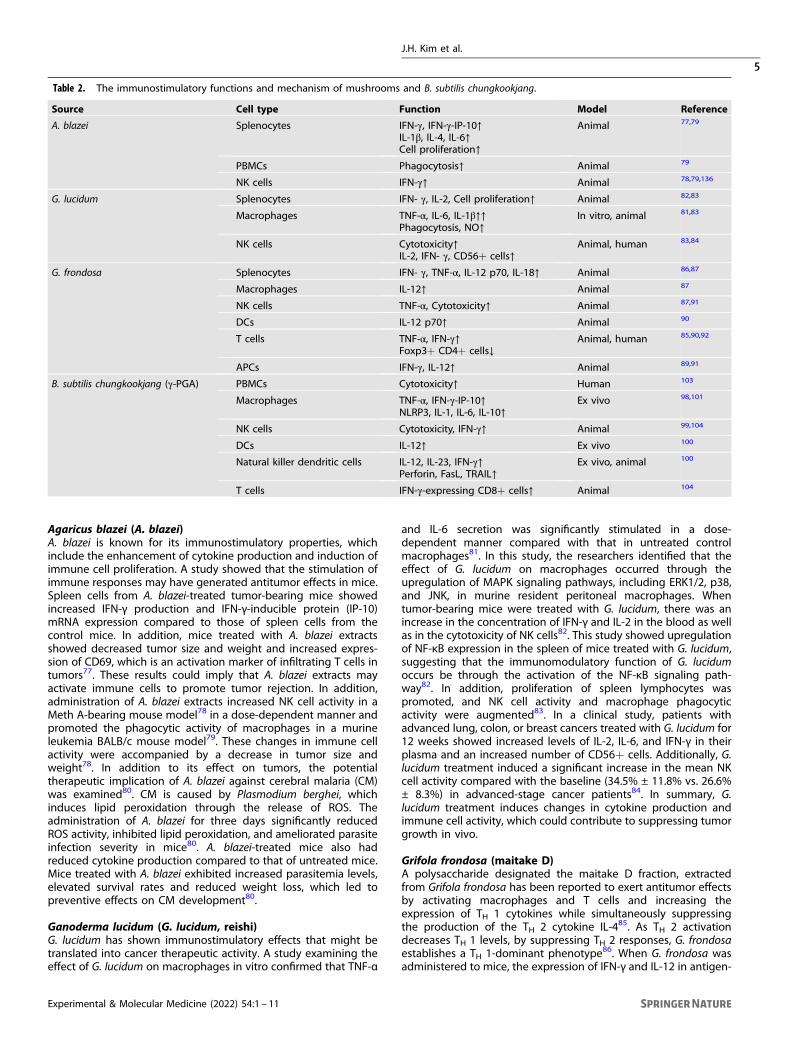
Table 2. The immunostimulatory functions and mechanism of mushrooms and B. subtilis chungkookjang.
Source Cell type Function Model Reference
A. blazei Splenocytes IFN-γ, IFN-γ-IP-10↑IL-1β, IL-4, IL-6↑Cell proliferation↑
Animal 77,79
PBMCs Phagocytosis↑ Animal 79
NK cells IFN-γ↑ Animal 78,79,136
G. lucidum Splenocytes IFN- γ, IL-2, Cell proliferation↑ Animal 82,83
Macrophages TNF-α, IL-6, IL-1β↑↑Phagocytosis, NO↑
In vitro, animal 81,83
NK cells Cytotoxicity↑IL-2, IFN- γ, CD56+ cells↑
Animal, human 83,84
G. frondosa Splenocytes IFN- γ, TNF-α, IL-12 p70, IL-18↑ Animal 86,87
Macrophages IL-12↑ Animal 87
NK cells TNF-α, Cytotoxicity↑ Animal 87,91
DCs IL-12 p70↑ Animal 90
T cells TNF-α, IFN-γ↑Foxp3+ CD4+ cells↓
Animal, human 85,90,92
APCs IFN-γ, IL-12↑ Animal 89,91
B. subtilis chungkookjang (γ-PGA) PBMCs Cytotoxicity↑ Human 103
Macrophages TNF-α, IFN-γ-IP-10↑NLRP3, IL-1, IL-6, IL-10↑
Ex vivo 98,101
NK cells Cytotoxicity, IFN-γ↑ Animal 99,104
DCs IL-12↑ Ex vivo 100
Natural killer dendritic cells IL-12, IL-23, IFN-γ↑Perforin, FasL, TRAIL↑
Ex vivo, animal 100
T cells IFN-γ-expressing CD8+ cells↑ Animal 104
J.H. Kim et al.
5
Experimental & Molecular Medicine (2022) 54:1 – 11

presenting cells (APCs) was increased, and the activities of NK cellsand macrophages were augmented87–89. Orally administered G.frondosa fraction inhibited tumor growth in various models whilestimulating immune responses. It has been revealed that the G.frondosa fraction increases the number of IFN-γ-expressing CD4+and CD8+ T cells88 and IL-12 p70 production through theregulation of dectin-1 in DCs in colon cancer models90.Intraperitoneal administration of the maitake D fraction 2 daysbefore tumor implantation significantly inhibited lung metastasis.Moreover, the G. frondosa fraction increased the production of IL-12 in APCs and the cytotoxicity of NK cells91. The maitake Dfraction directly stimulated DC maturation through a C-type lectinreceptor dectin-1 pathway. Oral administration of the maitake Dfraction was reported to increase systemic tumor antigen-specificT cell responses and T cell infiltration into tumor sites. Oral intakeof the maitake D fraction also reduced the number of regulatoryT cells and myeloid-derived suppressor cells90. In a clinical study (aphase II study examining the effects of G. frondosa onmyelodysplastic syndromes, MDSs), G. frondosa β-glucan con-sumption improved neutrophil and monocyte function in lower-risk MDS patients. Moreover, treatment with G. frondosa extractincreased the ROS response to Escherichia coli (E. coli) ex vivo,indicating that G. frondosa might enhance the immune responseto bacterial infections in MDS patients. Similarly, in a G. frondosaphase I/II trial of breast cancer patients, the intermediate dose(5–7mg/kg/day) was associated with increased TNF-α and IFN-γproduction by T cells92.Mushrooms are known to exert immunomodulatory and
antitumor effects through the activation of immune cells. Studieshave been conducted to examine cytokine production, cellactivation, and effects on tumors upon treatment with A. blazei,G. lucidum, and G. frondosa. In most studies of these mushrooms,the production of IFN-γ, along with that of IL-1, IL-6, IL-8, IL-12, andTNF-α, was increased after treatment. The polysaccharide fraction,mainly β-glucans, is the component that is known to beresponsible for the immunomodulatory effects exerted by mush-rooms, and its effect appears to depend on the structuralcharacteristics of β-glucans93. The receptors of these β-glucansinclude dectin-1, TLR, and CR3 (also known as CD11b/CD18)94–96.Dectin-1 and TLR, which are expressed on macrophages, bind toβ-glucans, which are pathogen-associated molecular patterns,thereby inducing macrophage activation and promoting theproduction of inflammatory cytokines. In addition, β-glucansinduce the functional maturation of DCs and the production of IL-12 and IFN-γ, indirectly promoting the activation of T cells.β-glucans also bind to CR3, which is highly expressed onneutrophils, monocytes, and NK cells, and prime these cells tobind to inactivated complement 3b (iC3b), which directs β-glucan-activated cells to induce the lysis of iC3b-coated cells94,96. Moststudies investigating the immunomodulatory effects of mush-rooms have been conducted using mushroom extracts. Therefore,studies using highly purified β-glucans could benefit fromunderstanding a more precise mechanism by which β-glucanand mushrooms affect immunity.
BACILLUS SUBTILIS CHUNGKOOKJANG (POLY-γ-GLUTAMATE,γ-PGA)Bacillus subtilis subsp. chungkookjang is found in chungkookjang, atraditional Korean fermented soybean food. Bacillus subtilis subsp.chungkookjang naturally produces an edible biomaterial calledpoly-γ-glutamate (γ-PGA), which is a polymer with a γ-amide bondbetween D- or L-glutamate with a molecular weight of 1000 kDaor more97. The immunomodulatory functions of γ-PGA and itspotential use as a drug or dietary supplement have been studiedin cancer and virus infection models (Table 2). γ-PGA induced theexpression of proinflammatory cytokines, including IL-1β, TNF-α,IL-6, and NLRP3, through its interaction with TLR4 in BMDMs98.
The treatment of γ-PGA stimulated the production of IFN-γ in NKcells and enhanced NK cell cytotoxicity in a melanoma mousemodel99. Treatment with high-molecular-mass (2000 kDa) γ-PGAled to an increase in the number of NK cells, with a concomitantreduction in tumor size, in a lung cancer model99. Additionally,TNF-α, IFN-γ, and IL-12 secretion from activated NK DCs wereinduced by γ-PGA100. Moreover, a study demonstrated the crucialinvolvement of TLR4 in γ-PGA-mediated antitumor immunityusing MyD88 knockout and TLR4-defective mice101. In this study,Lee et al. reported that the stimulatory potency toward macro-phages and immature DCs as well as the antitumor effect of γ-PGAwas lost when TLR4 signaling was genetically blocked, revealingan important functional target of γ-PGA. In a multicenter,randomized, double-blind clinical trial of 195 patients with cervicalintraepithelial neoplasia (CIN) 1, 42.4% of the patients whoreceived oral administration of γ-PGA showed histologicalremission of CIN 1, compared to 27.1% of the control subjects102.High-risk human papillomavirus (HPV) clearance was observed in43.5% of the patients receiving γ-PGA, whereas 26.7% clearancewas found in the control subjects102. However, while there was amild increase in NK cell activity induced by γ-PGA administrationat week 8, the activity was not higher at week 12102. On the otherhand, in a randomized double-blind placebo-controlled clinicalstudy with healthy volunteers, oral administration of γ-PGA for8 weeks caused higher NK cell cytotoxicity compared to that ofthe placebo group when examined at the end of the study103. Thediscrepancy in NK cell activity between the two human studiesimplies that the effect of γ-PGA may not be sustained for a longtime after cessation of consumption because the γ-PGA admin-istration period was 4 weeks shorter in the CIN 1 patient studythan in the study conducted with healthy volunteers. In anotherstudy focusing on the anti-infection effect, intranasal γ-PGAadministration protected against H1N1 influenza A virus in vivoby inhibiting viral infection, which in turn led to an increase in thesurvival rate of infected mice104. Moreover, influenza-specificcytotoxic T cell activity increased upon γ-PGA treatment101,104.
CHLORELLAChlorella is a unicellular green algae widely used as a functionalfood and nutraceutical due to its rich content in proteins, dietaryfibers, vitamins, minerals, and other bioactive compounds105.Chlorella vulgaris and Chlorella pyrenoidosa are the most studiedspecies among the chlorella for use as dietary supplements106.
Chlorella vulgaris (C. vulgaris)C. vulgaris has been reported to enhance immunity in animalmodels as well as human studies and may improve defenseagainst microbial infections (Table 3). In a cyclophosphamide-mediated immunosuppression mouse experiment, dried C.vulgaris rescued the cyclophosphamide-induced downregulationof IL-2, TNF-α, IFN-γ, and IL-12 expression. More importantly, C.vulgaris treatment was able to increase the cytotoxic activity of NKcells in cyclophosphamide-treated mice. In addition, the prolifera-tion of lymphocytes and phagocytic activity of macrophages wasalso increased by C. vulgaris107. In vitro examination revealed anincrease in IFN-γ and IL-2 levels in MOLT-4 cells108. In an 8-weekrandomized, double-blinded, placebo-controlled study, treatmentof healthy participants with dried C. vulgaris extract tabletsresulted in higher NK cell activity than that in the placebo-treatedsubjects. Additionally, serum cytokine concentrations, especiallythose of IFN-γ and IL-1β, were significantly increased in theparticipants who received chlorella supplementation, suggesting apotential immunostimulatory effect in healthy individuals109.When chlorella was fed to chickens as a supplement, theconcentrations of plasma IgG and IgM increased compared tothose in the group without supplementation110. Interestingly, thenumber of lymphocytes and white blood cells increased in broiler
J.H. Kim et al.
6
Experimental & Molecular Medicine (2022) 54:1 – 11

chickens treated with 1% fresh liquid chlorella but not in thosetreated with 1% dried chlorella powder111. This finding impliesthat while chlorella might provide immunomodulatory effectsin vivo, the type of supplement may be important in determiningbioactivity.C. vulgaris has been reported to enhance antibacterial immunity
against several types of bacterial infections. C. vulgaris waterextract lowered the number of bacteria in the peritoneal cavity orspleen of mice after Listeria monocytogenes (L. monocytogenes)infection, which appeared to occur through augmentation of the Tcell-mediated immune response112. Further studies demonstratedthat the resistance to L. monocytogenes conveyed by C. vulgarisinvolved the activation of the TH 1 response driven by IFN-γ andIL-12113,114. C. vulgaris also augmented resistance against intraper-itoneal E. coli infection in rats. Oral administration of C. vulgarisdecreased the viable number of bacteria in the blood, spleen, andliver while increasing the activity of polymorphonuclear leuko-cytes112. Treatment with C. vulgaris extracts also improvedantitumor activity in mice by inducing the production of IL-12p40. To investigate which biological pathway mediates theantitumor activity of C. vulgaris, spleen-adherent cells derivedfrom TLR4-lacking C3H/HeJ mice and TLR2 knockout mice wereseparately treated with C. vulgaris extract. IL-12 p40 cytokineproduction was significantly impaired in C. vulgaris-stimulatedspleen-adherent cells isolated from TLR2 knockout mice comparedto that in corresponding cells from WT mice. This study suggeststhat the immunomodulatory effects of C. vulgaris are dependenton TLR2 signaling115. These recent findings reveal the potential ofC. vulgaris as a potent immunostimulatory agent. Further researchon the biological pathways involved in the immunomodulatoryeffects of C. vulgaris would provide a better understanding andencourage the potential uses of C. vulgaris to improve the immunesystem.
Chlorella pyrenoidosa (C. pyrenoidosa)Immunomodulatory activities of C. pyrenoidosa have been foundmainly in macrophages and humoral immune responses (Table 3).The effect on macrophage activation was measured based onphagocytic activity and intracellular NO generation in murinemacrophage cell lines. The phagocytic activity of macrophageswas elevated by hot water extracts of C. pyrenoidosa. Theseextracts also enhanced macrophage proliferation and NO
generation116. Treatment with hot-water-soluble polysaccharidesof C. pyrenoidosa induced the expression of human leukocyteantigen (HLA)-DA and HLA-DC in human blood monocyte-derivedmacrophages117. The expression of the proinflammatory cytokinesTNF-α and IL-1β and the production of the costimulatorymolecules CD80 and CD86 were found to be upregulated by C.pyrenoidosa117. In this study, Hsu et al.117 suggested that theimmunostimulatory effects of C. pyrenoidosa were mediated bythe TLR4-mediated signaling pathway. In a similar study, thepolysaccharide fraction of C. pyrenoidosa was also found toincrease the expression of TNF-α and IL-1βmRNA and cause NF-κBactivation in THP-1 cells118. These findings encourage furtherresearch on the potential use of C. pyrenoidosa as an agent toimprove immune responses.The immune-enhancing function of C. pyrenoidosa supplements
was examined in a clinical experiment on healthy subjects above50 years of age who have treated with an influenza A vaccine. Nomeaningful results were obtained, but subjects aged 50–55 yearsexhibited increased anti-influenza A antibody production119.When C. pyrenoidosa tablets were administered to pregnantwomen, concentrations of IgA significantly increased in breastmilk compared to those in the control group. This finding impliesthat C. pyrenoidosa supplementation during pregnancy couldreduce the probability of infection in nursing infants by increasingIgA levels in breast milk120. In addition, oral administration of C.pyrenoidosa powder to rats enhanced the production of IgM inspleen and mesenteric lymph node lymphocytes and increasedserum concentrations of IgM and IgG121.
LACTOBACILLUS PLANTARUM (L. PLANTARUM)Probiotics are widely used as health-promoting food ingredientsdue to their various bioactivities122. Recent studies have shownthat not only live probiotics but also heat-killed probiotics orfractionated cellular components (exopolysaccharides, EPSs) of thebacteria can have beneficial effects on the host immunesystem123. Among various probiotics, L. plantarum was chosenbecause it is one of the most-researched species due to itsimmunomodulatory functionality (Table 4).It has been reported that heat-killed and micronized L.
plantarum and EPS from L. plantarum enhance phagocytic activityand cytokine production (e.g., TNF-α and IL-6) in macrophages
Table 3. The immunostimulatory functions and mechanism of chlorella.
Source Cell type Function Model Reference
C. vulgaris White blood cells Cell number↑ Animal 111
Peritoneal adherent cells IL-1α, IL-12, GM-CSF, MIP, TNF- α↑ Animal 113
Splenocytes Cell proliferation↑IL-2, IL-12, TNF-α, IFN-γ↑
Animal 107,113
IL-12 p40↑ Animal 115
Macrophages Phagocytosis↑Cell proliferation↑
Animal 107
HLA-DA, HLA-DC↑TNF-α, IL-1β, CD80, CD86↑
Animal, human 117
NK cells Cytotoxicity↑ Animal, human 107,109
T cells Cell proliferation↑IFN-γ, IL-2, IL-4↑
Animal 108
Blood IFN-γ, IL-1β, IL-2↑IgA, IgG, IgM↑
Animal, human 108–111
C. pyrenoidosa Blood Antibody production↑ Human 119
Macrophages IL-1β, TNF- α↑ In vitro 118
Macrophages Phagocytosis, NO↑ In vitro 116
Breast milk IgA ↑ Human 120
J.H. Kim et al.
7
Experimental & Molecular Medicine (2022) 54:1 – 11

through the activation of the NF-κB and MAPK signalingpathways124–127. A study reported that micronized and heat-killed L. plantarum LM1004 resulted in a marked increase in TLR2mRNA expression levels in macrophages. Moreover, various typesof L. plantarum, such as heat-killed L. plantarum Ln1, live L.plantarum nF1 and dead nanosized L. plantarum nF1, have beenreported to enhance NO production and iNOS expression levels inmacrophages24,124,128. In addition, mouse DCs treated with L.plantarum showed upregulated levels of cytokines, including IL-10, IL-12 p40, IL-20 p70, and TNF-α, through the upregulation ofthe NF-κB and MAPK signaling pathways129.L. plantarum is known to modulate the adaptive immune
system through the regulation of T cells as well as IgA production.Dead L. plantarum has been reported to promote TH 1 (TNF-α andIL-12 p70) and TH 17 (IL-6 and IL-17A) responses rather than TH 2(IL-4 and IL-5) responses in splenocytes isolated from normal maleC57BL/6J mice24. In an OVA/alum-immunized mouse model, L.plantarum YU-treated mice produced higher levels of IFN-γ, whilethe levels of the TH 2-related cytokines IL-4 and IL-10 were notsignificantly different than those in the control mice130. In additionto T cell regulation, L. plantarum can enhance IgA production. IgAproduction was significantly stimulated by oral administration of L.plantarum YU130. Additionally, in a cyclophosphamide-inducedimmunosuppression mouse model, the number of cells secretingIgA and the level of secretory IgA (sIgA) in the small intestine wasincreased by the administration of L. plantarum131. Treatment withL. plantarum in mice challenged with intranasal inoculation ofinfluenza A virus (IFV) enhanced IFV-specific sIgA levels inbronchoalveolar lavage fluid (BALF). As a result, viral proliferationand the viral titer in the lung were strongly suppressed by theadministration of L. plantarum130,132.The immunomodulatory effects of L. plantarum were also
demonstrated in a clinical study. The study showed that L.plantarum restored the number of regulatory T cells in the serumthat were suppressed by nonsteroidal anti-inflammatory drugs(NSAIDs). L. plantarum also enhanced memory responses of T cellsagainst tetanus toxoid (TT)-antigen and upregulated expression ofgenes associated with T and B cell function in the small intestinalmucosa133. L. plantarum was also found to improve immuneresponses in older subjects. In this study, researchers demon-strated that a high dose of L. plantarum increased the compositionof activated NK cells, whereas a low dose of L. plantarum increasedactivated T cells, B cells, and APCs134. This clinical study revealedthat the concentration of L. plantarum may elicit different immuneresponses, suggesting that the concentration may be animportant factor when consuming L. plantarum to control immunefunction. These various previous studies have shown that varioustypes of L. plantarum can activate both the innate immuneresponse and the adaptive immune response.
CONCLUSIONIn this review, we summarized food materials with immunomo-dulatory effects that have been studied in vitro, in vivo, and inclinical models (Tables 1–4). The immune system is essential forproviding protection against pathogens. Hosts with compromisedor weakened immune function are more vulnerable to cancer or
infectious diseases. The consumption of functional foods such asginseng, mushroom, poly-γ-glutamate, chlorella, and L. plantarummay improve the immunological defense system, which in turncould help protect the body from diseases. Further identificationof novel functional foods with immunomodulatory activity andstudy of their molecular mechanisms could aid in improvingpublic health.
REFERENCES1. Frisch, M., Biggar, R. J., Engels, E. A., Goedert, J. J. & Group, A. I.-C. M. R. S.
Association of cancer with AIDS-related immunosuppression in adults. J. Am.Med. Assoc. 285, 1736–1745 (2001).
2. Rier, S. E. Environmental immune disruption: a comorbidity factor for repro-duction? Fertil. Steril. 89, e103–e108 (2008).
3. Imai, K., Matsuyama, S., Miyake, S., Suga, K. & Nakachi, K. Natural cytotoxicactivity of peripheral-blood lymphocytes and cancer incidence: an 11-year fol-low-up study of a general population. Lancet 356, 1795–1799 (2000).
4. Montecino-Rodriguez, E., Berent-Maoz, B. & Dorshkind, K. Causes, consequences,and reversal of immune system aging. J. Clin. Invest. 123, 958–965 (2013).
5. Nakano, Y. et al. Immune function and lifestyle of taxi drivers in Japan. Ind.Health 36, 32–39 (1998).
6. Thorburn, A. N., Macia, L. & Mackay, C. R. Diet, metabolites, and “western-life-style” inflammatory diseases. Immunity 40, 833–842 (2014).
7. Cava, E., Neri, B., Carbonelli, M. G., Riso, S. & Carbone, S. Obesity pandemicduring COVID-19 outbreak: Narrative review and future considerations. Clin.Nutr. 40, 1637–1643 (2021).
8. Hachimura, S., Totsuka, M. & Hosono, A. Immunomodulation by food: impact ongut immunity and immune cell function. Biosci. Biotechnol. Biochem. 82,584–599 (2018).
9. Iwasaki, A. & Pillai, P. S. Innate immunity to influenza virus infection. Nat. Rev.Immunol. 14, 315–328 (2014).
10. Linehan, E. & Fitzgerald, D. C. Ageing and the immune system: focus on mac-rophages. Eur. J. Microbiol. Immunol. 5, 14–24 (2015).
11. Lukacs, N. W., Strieter, R. M., Chensue, S. W., Widmer, M. & Kunkel, S. L. TNF-alphamediates recruitment of neutrophils and eosinophils during airway inflamma-tion. J. Immunol. 154, 5411–5417 (1995).
12. Almishri, W. et al. TNFalpha augments cytokine-induced NK Cell IFNgammaproduction through TNFR2. J. Innate Immun. 8, 617–629 (2016).
13. Sharma, J. N., Al-Omran, A. & Parvathy, S. S. Role of nitric oxide in inflammatorydiseases. Inflammopharmacology 15, 252–259 (2007).
14. Lee, J., Choi, J. W., Sohng, J. K., Pandey, R. P. & Park, Y. I. The immunostimulatingactivity of quercetin 3-O-xyloside in murine macrophages via activation of theASK1/MAPK/NF-κB signaling pathway. Int. Immunopharmacol. 31, 88–97 (2016).
15. Hirschfeld, M., Ma, Y., Weis, J., Vogel, S. & Weis, J. Cutting edge: repurification oflipopolysaccharide eliminates signaling through both human and murine toll-like receptor 2. J. Immunol. 165, 618–622 (2000).
16. Takeuchi, O. & Akira, S. Toll-like receptors; their physiological role and signaltransduction system. Int. Immunopharmacol. 1, 625–635 (2001).
17. Vivier, E., Nunes, J. A. & Vely, F. Natural killer cell signaling pathways. Science 306,1517–1519 (2004).
18. Guermonprez, P., Valladeau, J., Zitvogel, L., Thery, C. & Amigorena, S. Antigenpresentation and T cell stimulation by dendritic cells. Annu. Rev. Immunol. 20,621–667 (2002).
19. Iwasaki, A. & Medzhitov, R. Control of adaptive immunity by the innate immunesystem. Nat. Immunol. 16, 343–353 (2015).
20. Hahn, H. & Kaufmann, S. H. The role of cell-mediated immunity in bacterialinfections. Rev. Infect. Dis. 3, 1221–1250 (1981).
21. Andersen, M. H., Schrama, D., Thor Straten, P. & Becker, J. C. Cytotoxic T cells. J.Invest. Dermatol. 126, 32–41 (2006).
22. Ekkens, M. J. et al. Th1 and Th2 cells help CD8 T-cell responses. Infect. Immun.75, 2291–2296 (2007).
Table 4. The immunostimulatory functions and mechanism of L. plantarum.
Source* Cell type Function Model Reference
L. plantarum AYA DCs IL-6, TGF-β, IgA ↑ Animal 132
L. plantarum nF1 Blood IgA, IgG↑ Animal 24
Splenocytes IL-4, IL-5, IL-17A, IL-12, TNF- α ↑
Macrophages NO, IL-6, TNF- α ↑
L. plantarum YU Blood IgE ↑ Animal 130
J.H. Kim et al.
8
Experimental & Molecular Medicine (2022) 54:1 – 11

23. Walker, J. A. & McKenzie, A. N. J. TH2 cell development and function. Nat. Rev.Immunol. 18, 121–133 (2018).
24. Lee, H. A., Kim, H., Lee, K. W. & Park, K. Y. Dead Lactobacillus plantarum stimulatesand skews immune responses toward T helper 1 and 17 Polarizations in RAW264.7 cells and mouse splenocytes. J. Microbiol. Biotechnol. 26, 469–476 (2016).
25. Yu, S.-L., Kuan, W.-P., Wong, C., Li, E. & Tam, L.-S. Immunopathological roles ofcytokines, chemokines, signaling molecules, and pattern-recognition receptorsin systemic lupus erythematosus. Clin. Dev. Immunol. 2012, 715190 (2012).
26. Tan, B. K. & Vanitha, J. Immunomodulatory and antimicrobial effects of some tra-ditional chinese medicinal herbs: a review. Curr. Med. Chem. 11, 1423–1430 (2004).
27. Jin, Y. et al. Effect of white, red and black ginseng on physicochemical prop-erties and ginsenosides. Plant Foods Hum. Nutr. 70, 141–145 (2015).
28. Zhang, H. M. et al. Holistic quality evaluation of commercial white and redginseng using a UPLC-QTOF-MS/MS-based metabolomics approach. J. Pharm.Biomed. 62, 258–273 (2012).
29. Lee, S. Y. et al. Chemical constituents and biological activities of the berry ofPanax ginseng. J. Med. Plants Res. 4, 349–353 (2010).
30. Shibata, S. Chemistry and cancer preventing activities of ginseng saponins andsome related triterpenoid compounds. J. Korean Med. Sci. 16, S28–S37 (2001).
31. Lim, T. G. et al. White ginseng extract induces immunomodulatory effects viathe MKK4-JNK pathway. Food Sci. Biotechnol. 25, 1737–1744 (2016).
32. Jang, H. I. & Shin, H. M. Wild Panax ginseng (Panax ginseng C.A. Meyer) protectsagainst methotrexate-induced cell regression by enhancing the immuneresponse in RAW 264.7 macrophages. Am. J. Chin. Med. 38, 949–960 (2010).
33. Um, Y. et al. Wild simulated ginseng activates mouse macrophage, RAW264.7cells through TRL2/4-dependent activation of MAPK, NF-kappaB and PI3K/AKTpathways. J. Ethnopharmacol. 263, 113218 (2020).
34. Kang, S. & Min, H. Ginseng, the ‘Immunity Boost’: the effects of Panax ginsengon immune system. J. Ginseng Res. 36, 354–368 (2012).
35. Shen, T. et al. Polysaccharide from wheat bran induces cytokine expression viathe toll-like receptor 4-mediated p38 MAPK signaling pathway and preventscyclophosphamide-induced immunosuppression in mice. Food Nutr. Res. 61,1344523 (2017).
36. Yang, Y. et al. Immunostimulatory effects of sulfated chitosans on RAW 264.7mouse macrophages via the activation of PI3K/Akt signaling pathway. Int. J. Biol.Macromol. 108, 1310–1321 (2018).
37. He, L. X. et al. Ginseng (Panax ginseng Meyer) oligopeptides regulate innate andadaptive immune responses in mice via increased macrophage phagocytosiscapacity, NK cell activity and Th cells secretion. Food Funct. 8, 3523–3532 (2017).
38. Larsen, M. W., Moser, C., Hoiby, N., Song, Z. & Kharazmi, A. Ginseng modulatesthe immune response by induction of interleukin-12 production. APMIS 112,369–373 (2004).
39. Kim, M. H. et al. Immunomodulatory activity of ginsan, a polysaccharide of Panaxginseng, on dendritic cells. Korean J. Physiol. Pharmacol. 13, 169–173 (2009).
40. Ahn, J. Y. et al. The immunomodulator ginsan induces resistance to experi-mental sepsis by inhibiting Toll-like receptor-mediated inflammatory signals.Eur. J. Immunol. 36, 37–45 (2006).
41. Song, J. Y. et al. Induction of secretory and tumoricidal activities in peritonealmacrophages by ginsan. Int. Immunopharmacol. 2, 857–865 (2002).
42. Takei, M., Tachikawa, E., Hasegawa, H. & Lee, J. J. Dendritic cells maturationpromoted by M1 and M4, end products of steroidal ginseng saponins meta-bolized in digestive tracts, drive a potent Th1 polarization. Biochem. Pharmacol.68, 441–452 (2004).
43. Song, Z. et al. Cytokine modulating effect of ginseng treatment in a mouse modelof Pseudomonas aeruginosa lung infection. J. Cyst. Fibros. 2, 112–119 (2003).
44. Takeda, K. & Okumura, K. Interferon-gamma-mediated natural killer cell activa-tion by an aqueous Panax ginseng extract. Evid. Based Complement. Altern. Med.2015, 603198 (2015).
45. Scaglione, F. et al. Immunomodulatory effects of two extracts of Panax ginsengC.A. Meyer. Drugs Exp. Clin. Res. 16, 537–542 (1990).
46. Kenarova, B., Neychev, H., Hadjiivanova, C. & Petkov, V. D. Immunomodulatingactivity of ginsenoside Rg1 from Panax ginseng. Jpn. J. Pharmacol. 54, 447–454(1990).
47. Kim, J. Y., Germolec, D. R. & Luster, M. I. Panax ginseng as a potential immu-nomodulator: studies in mice. Immunopharm. Immunot. 12, 257–276 (1990).
48. Liou, C. J., Huang, W. C. & Tseng, J. Short-term oral administration of ginsengextract induces type-1 cytokine production. Immunopharm. Immunot. 28,227–240 (2006).
49. Liou, C. J., Li, M. L. & Tseng, J. Intraperitoneal injection of ginseng extractenhances both immunoglobulin and cytokine production in mice. Am. J. Chin.Med. 32, 75–88 (2004).
50. Qu, D. F. et al. Ginsenoside Rg1 enhances immune response induced byrecombinant Toxoplasma gondii SAG1 antigen. Vet. Parasitol. 179, 28–34 (2011).
51. Na, H. S., Lim, Y. J., Yun, Y. S., Kweon, M. N. & Lee, H. C. Ginsan enhances humoralantibody response to orally delivered antigen. Immune Netw. 10, 5–14 (2010).
52. Su, X., Pei, Z. & Hu, S. Ginsenoside Re as an adjuvant to enhance the immuneresponse to the inactivated rabies virus vaccine in mice. Int. Immunopharmacol.20, 283–289 (2014).
53. Han, S. K., Song, J. Y., Yun, Y. S. & Yi, S. Y. Ginsan improved Th1 immune responseinhibited by gamma radiation. Arch. Pharm. Res. 28, 343–350 (2005).
54. Lee, E. J. et al. Ginsenoside Rg1 enhances CD4(+) T-cell activities and modulatesTh1/Th2 differentiation. Int. Immunopharmacol. 4, 235–244 (2004).
55. Berek, L. et al. Effects of naturally occurring glucosides, solasodine glucosides,ginsenosides and parishin derivatives on multidrug resistance of lymphomacells and leukocyte functions. Vivo 15, 151–156 (2001).
56. Lee, J. H. & Han, Y. Ginsenoside Rg1 helps mice resist to disseminated candi-diasis by Th1 type differentiation of CD4+ T cell. Int. Immunopharmacol. 6,1424–1430 (2006).
57. Rivera, E., Ekholm Pettersson, F., Inganas, M., Paulie, S. & Gronvik, K. O. The Rb1fraction of ginseng elicits a balanced Th1 and Th2 immune response. Vaccine23, 5411–5419 (2005).
58. Wang, Y. et al. A solution with ginseng saponins and selenium as vaccine diluent toincrease Th1/Th2 immune responses in mice. J. Immunol. Res. 2020, 2714257 (2020).
59. Peck, A. & Mellins, E. D. Precarious balance: Th17 cells in host defense. Infect.Immun. 78, 32–38 (2010).
60. Maqbool, B. et al. Ginseng stem-leaf saponins in combination with seleniumenhance immune responses to an attenuated pseudorabies virus vaccine.Microbiol. Immunol. 63, 269–279 (2019).
61. Cox, J. C. & Coulter, A. R. Adjuvants—a classification and review of their modesof action. Vaccine 15, 248–256 (1997).
62. Shin, K. K. et al. Korean red ginseng plays an anti-aging role by modulatingexpression of aging-related genes and immune cell subsets. Molecules 25, 1492(2020).
63. Kim, H. et al. Red ginseng and vitamin C increase immune cell activity anddecrease lung inflammation induced by influenza A virus/H1N1 infection. J.Pharm. Pharmacol. 68, 406–420 (2016).
64. Byeon, S. E. et al. Molecular mechanism of macrophage activation by red gin-seng acidic polysaccharide from Korean red ginseng. Mediat. Inflamm. 2012,732860 (2012).
65. Quan, F. S., Compans, R. W., Cho, Y. K. & Kang, S. M. Ginseng and Salviae herbsplay a role as immune activators and modulate immune responses duringinfluenza virus infection. Vaccine 25, 272–282 (2007).
66. Kwok, H. H. et al. Anti-inflammatory effects of indirubin derivatives on influenzaA virus-infected human pulmonary microvascular endothelial cells. Sci. Rep. 6,18941 (2016).
67. Chan, L. Y. et al. Dual functions of ginsenosides in protecting human endothelialcells against influenza H9N2-induced inflammation and apoptosis. J. Ethno-pharmacol. 137, 1542–1546 (2011).
68. Yoo, D. G. et al. Protective effect of Korean red ginseng extract on the infectionsby H1N1 and H3N2 influenza viruses in mice. J. Med. Food 15, 855–862 (2012).
69. Yang, Z., Chen, A., Sun, H., Ye, Y. & Fang, W. Ginsenoside Rd elicits Th1 and Th2immune responses to ovalbumin in mice. Vaccine 25, 161–169 (2007).
70. Song, X., Chen, J., Sakwiwatkul, K., Li, R. & Hu, S. Enhancement of immuneresponses to influenza vaccine (H3N2) by ginsenoside Re. Int. Immuno-pharmacol. 10, 351–356 (2010).
71. Su, F., Yuan, L., Zhang, L. & Hu, S. Ginsenosides Rg1 and Re act as adjuvant viaTLR4 signaling pathway. Vaccine 30, 4106–4112 (2012).
72. Sun, J., Song, X. & Hu, S. Ginsenoside Rg1 and aluminum hydroxide synergis-tically promote immune responses to ovalbumin in BALB/c mice. Clin. Vaccin.Immunol. 15, 303–307 (2008).
73. Bremner, P. & Heinrich, M. Natural products as targeted modulators of thenuclear factor-kappaB pathway. J. Pharm. Pharmacol. 54, 453–472 (2002).
74. Hyun, S. H. et al. Immuno-enhancement effects of Korean Red Ginseng inhealthy adults: a randomized, double-blind, placebo-controlled trial. J. GinsengRes. 45, 191–198 (2021).
75. Lee, C. S. et al. Preventive effect of Korean red ginseng for acute respiratoryillness: a randomized and double-blind clinical trial. J. Korean Med. Sci. 27,1472–1478 (2012).
76. Mizuno, T. Bioactive biomolecules of mushrooms—food, function and medicinaleffect of mushroom fungi. Food Rev. Int. 11, 7–21 (1995).
77. Takimoto, H., Kato, H., Kaneko, M. & Kumazawa, Y. Amelioration of skewed Th1/Th2 balance in tumor-bearing and asthma-induced mice by oral administrationof Agaricus blazei extracts. Immunopharm. Immunotoxicol. 30, 747–760 (2008).
78. Takimoto, H., Wakita, D., Kawaguchi, K. & Kumazawa, Y. Potentiation of cytotoxicactivity in naive and tumor-bearing mice by oral administration of hot-waterextracts from Agaricus brazei fruiting bodies. Biol. Pharm. Bull. 27, 404–406(2004).
79. Lin, J. G. et al. An extract of Agaricus blazei Murill administered orally promotesimmune responses in murine leukemia BALB/c mice in vivo. Integr. Cancer Ther.11, 29–36 (2012).
J.H. Kim et al.
9
Experimental & Molecular Medicine (2022) 54:1 – 11

80. Val, C. H. et al. Effect of mushroom Agaricus blazei on immune response anddevelopment of experimental cerebral malaria. Malar. J. 14, 311 (2015).
81. Guo, L. et al. Characterization and immunostimulatory activity of a poly-saccharide from the spores of Ganoderma lucidum. Int. Immunopharmacol. 9,1175–1182 (2009).
82. Wang, G. et al. Enhancement of IL-2 and IFN-gamma expression and NK cellsactivity involved in the anti-tumor effect of ganoderic acid Me in vivo. Int.Immunopharmacol. 7, 864–870 (2007).
83. Wang, P. Y., Zhu, X. L. & Lin, Z. B. Antitumor and immunomodulatory effects ofpolysaccharides from broken-spore of Ganoderma lucidum. Front. Pharmacol. 3,135 (2012).
84. Gao, Y., Zhou, S., Jiang, W., Huang, M. & Dai, X. Effects of ganopoly (a Ganodermalucidum polysaccharide extract) on the immune functions in advanced-stagecancer patients. Immunol. Invest. 32, 201–215 (2003).
85. Kodama, N., Harada, N. & Nanba, H. A polysaccharide, extract from Grifolafrondosa, induces Th-1 dominant responses in carcinoma-bearing BALB/c mice.Jpn. J. Pharmacol. 90, 357–360 (2002).
86. Inoue, A., Kodama, N. & Nanba, H. Effect of maitake (Grifola frondosa) D-fractionon the control of the T lymph node Th-1/Th-2 proportion. Biol. Pharm. Bull. 25,536–540 (2002).
87. Kodama, N., Komuta, K., Sakai, N. & Nanba, H. Effects of D-Fraction, a poly-saccharide from Grifola frondosa on tumor growth involve activation of NK cells.Biol. Pharm. Bull. 25, 1647–1650 (2002).
88. Harada, N., Kodama, N. & Nanba, H. Relationship between dendritic cells and theD-fraction-induced Th-1 dominant response in BALB/c tumor-bearing mice.Cancer Lett. 192, 181–187 (2003).
89. Kodama, N., Mizuno, S., Nanba, H. & Saito, N. Potential antitumor activity of alow-molecular-weight protein fraction from Grifola frondosa through enhance-ment of cytokine production. J. Med. Food 13, 20–30 (2010).
90. Masuda, Y. et al. Oral administration of soluble beta-glucans extracted fromGrifola frondosa induces systemic antitumor immune response and decreasesimmunosuppression in tumor-bearing mice. Int. J. Cancer 133, 108–119 (2013).
91. Masuda, Y., Murata, Y., Hayashi, M. & Nanba, H. Inhibitory effect of MD-Fractionon tumor metastasis: involvement of NK cell activation and suppression ofintercellular adhesion molecule (ICAM)-1 expression in lung vascular endothelialcells. Biol. Pharm. Bull. 31, 1104–1108 (2008).
92. Deng, G. et al. A phase I/II trial of a polysaccharide extract from Grifola frondosa(Maitake mushroom) in breast cancer patients: immunological effects. J. CancerRes. Clin. Oncol. 135, 1215–1221 (2009).
93. Volman, J. J. et al. Effects of mushroom-derived beta-glucan-rich polysaccharideextracts on nitric oxide production by bone marrow-derived macrophages andnuclear factor-kappaB transactivation in Caco-2 reporter cells: can effects beexplained by structure? Mol. Nutr. Food Res. 54, 268–276 (2010).
94. Akramiene, D., Kondrotas, A., Didziapetriene, J. & Kevelaitis, E. Effects of beta-glucans on the immune system. Medicines 43, 597–606 (2007).
95. Chan, G. C., Chan, W. K. & Sze, D. M. The effects of beta-glucan on humanimmune and cancer cells. J. Hematol. Oncol. 2, 25 (2009).
96. Kim, H. S., Hong, J. T., Kim, Y. & Han, S. B. Stimulatory effect of beta-glucans onimmune cells. Immune Netw. 11, 191–195 (2011).
97. Ashiuchi, M. et al. Isolation of Bacillus subtilis (chungkookjang), a poly-gamma-glutamate producer with high genetic competence. Appl. Microbiol. Biot. 57,764–769 (2001).
98. Ahn, H. et al. Poly-gamma-glutamic acid from Bacillus subtilis upregulates pro-inflammatory cytokines while inhibiting NLRP3, NLRC4 and AIM2 inflammasomeactivation. Cell. Mol. Immunol. 15, 111–119 (2018).
99. Kim, T. W. et al. Oral administration of high molecular mass poly-gamma-glutamate induces NK cell-mediated antitumor immunity. J. Immunol. 179,775–780 (2007).
100. Lee, S. W., Park, H. J., Park, S. H., Kim, N. & Hong, S. Immunomodulatory effect ofpoly-gamma-glutamic acid derived from Bacillus subtilis on natural killer den-dritic cells. Biochem. Biophys. Res. Commun. 443, 413–421 (2014).
101. Lee, T. Y. et al. Oral administration of poly-gamma-glutamate induces TLR4- anddendritic cell-dependent antitumor effect. Cancer Immunol. Immunother. 58,1781–1794 (2009).
102. Cho, H. W. et al. Short-term clinical and immunologic effects of poly-gamma-glutamic acid (gamma-PGA) in women with cervical intraepithelial neoplasia 1(CIN 1): a multicenter, randomized, double blind, phase II trial. PLoS ONE 14,e0217745 (2019).
103. Kim, K. S. et al. A single-center, randomized double-blind placebo-controlledstudy evaluating the effects of poly-gamma-glutamate on human NK cellactivity after an 8-week oral administration in healthy volunteers. Evid. BasedComplement. Altern. Med. 2013, 635960 (2013).
104. Kim, E. H., Choi, Y. K., Kim, C. J., Sung, M. H. & Poo, H. Intranasal administration ofpoly-gamma glutamate induced antiviral activity and protective immuneresponses against H1N1 influenza A virus infection. Virol. J. 12, 160 (2015).
105. Rani, K., Sandal, N. & Sahoo, P. A comprehensive review on chlorella-its com-position, health benefits, market and regulatory scenario. Pharma Innov. J. 7,584–589 (2018).
106. Chakka, S., Concha, J. S. S., Bax, C. E., Zeidi, M. & Werth, V. P. The effects ofimmunostimulatory herbal supplements on autoimmune skin diseases. J. Am.Acad. Dermatol. 84, 1051–1058 (2020).
107. Cheng, D. et al. Dietary Chlorella vulgaris ameliorates altered immunomodula-tory functions in cyclophosphamide-induced immunosuppressive mice. Nutri-ents 9, 708 (2017).
108. An, H. J. et al. Effect of Chlorella vulgaris on immune-enhancement and cytokineproduction in vivo and in vitro. Food Sci. Biotechnol. 17, 953–958 (2008).
109. Kwak, J. H. et al. Beneficial immunostimulatory effect of short-term Chlorellasupplementation: enhancement of natural killer cell activity and early inflam-matory response (randomized, double-blinded, placebo-controlled trial). Nutr. J.11, 53 (2012).
110. An, B. K., Kim, K. E., Jeon, J. Y. & Lee, K. W. Effect of dried Chlorella vulgaris andChlorella growth factor on growth performance, meat qualities and humoralimmune responses in broiler chickens. Springerplus 5, 718 (2016).
111. Kang, H. K. et al. Effect of various forms of dietary Chlorella supplementation ongrowth performance, immune characteristics, and intestinal microflora popu-lation of broiler chickens. J. Appl. Poult. Res. 22, 100–108 (2013).
112. Hasegawa, T. et al. Augmentation of the resistance against Escherichia coli byoral-administration of a hot water extract of Chlorella vulgaris in rats. Int. J.Immunopharmacol. 11, 971–976 (1989).
113. Hasegawa, T. et al. Effect of hot water extract of Chlorella vulgaris on cytokineexpression patterns in mice with murine acquired immunodeficiency syndromeafter infection with Listeria monocytogenes. Immunopharmacology 35, 273–282(1997).
114. Hasegawa, T. et al. Hot water extracts of Chlorella vulgaris reduce opportunisticinfection with Listeria monocytogenes in C57BL/6 mice infected with LP-BM5murine leukemia viruses. Int. J. Immunopharmacol. 17, 505–512 (1995).
115. Hasegawa, T. et al. Toll-like receptor 2 is at least partly involved in the antitumoractivity of glycoprotein from Chlorella vulgaris. Int. Immunopharmacol. 2,579–589 (2002).
116. Zhuang, X. et al. A comparison on the preparation of hot water extracts fromChlorella pyrenoidosa (CPEs) and radical scavenging and macrophage activationeffects of CPEs. Food Funct. 5, 3252–3260 (2014).
117. Hsu, H. Y. et al. Immunostimulatory bioactivity of algal polysaccharides fromChlorella pyrenoidosa activates macrophages via Toll-like receptor 4. J. Agric.Food Chem. 58, 927–936 (2010).
118. Pugh, N., Ross, S. A., ElSohly, H. N., ElSohly, M. A. & Pasco, D. S. Isolation of threehigh molecular weight polysaccharide preparations with potent immunosti-mulatory activity from Spirulina platensis, aphanizomenon flos-aquae andChlorella pyrenoidosa. Planta Med. 67, 737–742 (2001).
119. Halperin, S. A., Smith, B., Nolan, C., Shay, J. & Kralovec, J. Safety and immu-noenhancing effect of a Chlorella-derived dietary supplement in healthy adultsundergoing influenza vaccination: randomized, double-blind, placebo-controlled trial. Can. Med. Assoc. J. 169, 111–117 (2003).
120. Nakano, S., Takekoshi, H. & Nakano, M. Chlorella (Chlorella pyrenoidosa) sup-plementation decreases dioxin and increases immunoglobulin A concentrationsin breast milk. J. Med. Food 10, 134–142 (2007).
121. Kanouchi, H. et al. Dietary effect of Chlorella pyrenoidosa powder on immu-noglobulin productivity of Sprague-Dawley rats. J. Jpn. Soc. Food Sci. 48,634–636 (2001).
122. Heczko, P., Strus, M. & Kochan, P. Critical evaluation of probiotic activity of lacticacid bacteria and their effects. J. Physiol. Pharmacol. 57, 5–12 (2006).
123. Adams, C. The probiotic paradox: live and dead cells are biological responsemodifiers. Nutr. Res. Rev. 23, 37–46 (2010).
124. Jang, H. J., Yu, H. S., Lee, N. K. & Paik, H. D. Immune-stimulating effect ofLactobacillus plantarum Ln1 Isolated from the traditional Korean fermentedfood, Kimchi. J. Microbiol. Biotechnol. 30, 926–929 (2020).
125. Jeong, M. et al. Heat-killed Lactobacillus plantarum KCTC 13314BP enhancesphagocytic activity and immunomodulatory effects via activation of MAPK andSTAT3 pathways. J. Microbiol. Biotechnol. 29, 1248–1254 (2019).
126. Lee, J. et al. Micronized and heat-treated Lactobacillus plantarumLM1004 stimulates host immune responses via the TLR-2/MAPK/NF-kappaBsignalling pathway in vitro and in vivo. J. Microbiol. Biotechnol. 29, 704–712(2019).
127. Wang, J., Wu, T., Fang, X., Min, W. & Yang, Z. Characterization and immuno-modulatory activity of an exopolysaccharide produced by Lactobacillus plan-tarum JLK0142 isolated from fermented dairy tofu. Int. J. Biol. Macromol. 115,985–993 (2018).
128. Ren, D. Y., Wang, D., Liu, H. Y., Shen, M. H. & Yu, H. S. Two strains of probioticLactobacillus enhance immune response and promote naive T cell polarizationto Th1. Food Agr. Immunol. 30, 281–295 (2019).
J.H. Kim et al.
10
Experimental & Molecular Medicine (2022) 54:1 – 11
129. Rigaux, P. et al. Immunomodulatory properties of Lactobacillus plantarum and itsuse as a recombinant vaccine against mite allergy. Allergy 64, 406–414 (2009).
130. Kawashima, T. et al. Lactobacillus plantarum strain YU from fermented foodsactivates Th1 and protective immune responses. Int. Immunopharmacol. 11,2017–2024 (2011).
131. Junhua, X. et al. Effects of Lactobacillus plantarum NCU116 on intestine mucosalimmunity in immunosuppressed mice. J. Agr. Food Chem. 63, 10914–10920(2015).
132. Kikuchi, Y. et al. Oral administration of Lactobacillus plantarum strain AYAenhances IgA secretion and provides survival protection against influenza virusinfection in mice. PLoS ONE 9, e86416 (2014).
133. de Vos, P. et al. Lactobacillus plantarum strains can enhance human mucosal andsystemic immunity and prevent non-steroidal anti-inflammatory drug inducedreduction in T regulatory cells. Front. Immunol. 8, 1000 (2017).
134. Mane, J. et al. A mixture of Lactobacillus plantarum CECT 7315 and CECT 7316enhances systemic immunity in elderly subjects. A dose-response, double-blind,placebo-controlled, randomized pilot trial. Nutr. Hosp. 26, 228–235 (2011).
135. Choi, H. S. et al. Red ginseng acidic polysaccharide (RGAP) in combination withIFN-gamma results in enhanced macrophage function through activation of theNF-kappaB pathway. Biosci. Biotechnol. Biochem. 72, 1817–1825 (2008).
136. Yuminamochi, E., Koike, T., Takeda, K., Horiuchi, I. & Okumura, K. Interleukin-12-and interferon-gamma-mediated natural killer cell activation by Agaricus blazeiMurill. Immunology 121, 197–206 (2007).
ACKNOWLEDGEMENTSThis work was supported by a National Research Foundation of Korea (NRF) grantfunded by the Korean government (MSIP) (NRF-2020R1A2C1010703). This researchwas also supported in part by the Brain Korea 21 (BK21) program.
COMPETING INTERESTSThe authors declare no competing interests.
ADDITIONAL INFORMATIONCorrespondence and requests for materials should be addressed to Sanguine Byun.
Reprints and permission information is available at http://www.nature.com/reprints
Publisher’s note Springer Nature remains neutral with regard to jurisdictional claimsin published maps and institutional affiliations.
Open Access This article is licensed under a Creative CommonsAttribution 4.0 International License, which permits use, sharing,
adaptation, distribution and reproduction in anymedium or format, as long as you giveappropriate credit to the original author(s) and the source, provide a link to the CreativeCommons license, and indicate if changes were made. The images or other third partymaterial in this article are included in the article’s Creative Commons license, unlessindicated otherwise in a credit line to the material. If material is not included in thearticle’s Creative Commons license and your intended use is not permitted by statutoryregulation or exceeds the permitted use, you will need to obtain permission directlyfrom the copyright holder. To view a copy of this license, visit http://creativecommons.org/licenses/by/4.0/.
© The Author(s) 2022
J.H. Kim et al.
11
Experimental & Molecular Medicine (2022) 54:1 – 11


