Embed Size (px)
Citation preview

MOLECULAR AND SYNAPTIC MECHANISMS
GABAA receptors can initiate the formation of functionalinhibitory GABAergic synapses
Celine Fuchs,1,* Karine Abitbol,1,* Jemima J. Burden,2 Audrey Mercer,1 Laura Brown,1 Jonathan Iball,1
F. Anne Stephenson,1 Alex M. Thomson1,# and Jasmina N. Jovanovic1,#1UCL School of Pharmacy, University College London, 29–39 Brunswick Square, London, WC1N 1AX, UK2MRC Laboratory of Molecular Cell Biology, University College London, London, UK
Keywords: inhibition, neuroligin-2, postsynaptic, presynaptic, synaptic adhesion, synaptogenesis
Abstract
The mechanisms that underlie the selection of an inhibitory GABAergic axon’s postsynaptic targets and the formation of the firstcontacts are currently unknown. To determine whether expression of GABAA receptors (GABAARs) themselves – the essentialfunctional postsynaptic components of GABAergic synapses – can be sufficient to initiate formation of synaptic contacts, a novelco-culture system was devised. In this system, the presynaptic GABAergic axons originated from embryonic rat basal gangliamedium spiny neurones, whereas their most prevalent postsynaptic targets, i.e. a1/b2/c2-GABAARs, were expressed constitu-tively in a stably transfected human embryonic kidney 293 (HEK293) cell line. The first synapse-like contacts in these co-cultureswere detected by colocalization of presynaptic and postsynaptic markers within 2 h. The number of contacts reached a plateau at24 h. These contacts were stable, as assessed by live cell imaging; they were active, as determined by uptake of a fluorescentlylabelled synaptotagmin vesicle-luminal domain-specific antibody; and they supported spontaneous and action potential-drivenpostsynaptic GABAergic currents. Ultrastructural analysis confirmed the presence of characteristics typical of active synapses.Synapse formation was not observed with control or N-methyl-D-aspartate receptor-expressing HEK293 cells. A prominentincrease in synapse formation and strength was observed when neuroligin-2 was co-expressed with GABAARs, suggesting acooperative relationship between these proteins. Thus, in addition to fulfilling an essential functional role, postsynaptic GABAARscan promote the adhesion of inhibitory axons and the development of functional synapses.
Introduction
GABAA receptors (GABAARs) are the essential functional postsyn-aptic components of GABAergic synapses (Schofield et al., 1987;Sieghart, 2006; Farrant & Kaila, 2007). Gene deletion studies ofsome of the most abundant subunits of GABAARs in mice havedemonstrated specific structural changes in inhibitory synapses(Fritschy et al., 2012), suggesting that GABAARs may play a directrole in regulating synapse formation. A number of neuronal adhe-sion proteins have also been suggested to play a direct structuralrole in inhibitory GABAergic synapse formation (Sudhof, 2008;Shen & Scheiffele, 2010; Siddiqui & Craig, 2011). Among these,the most prominent role has been attributed to the neuroligin familyof postsynaptic adhesion proteins (Scheiffele et al., 2000) and theirpresynaptic binding partners neurexins (Dean et al., 2003; Grafet al., 2004; Kang et al., 2008; Futai et al., 2013). The synaptogenicactivity of neuroligin-2 (NL2) was first demonstrated in heterolo-gous neuron–HEK293 co-culture systems, in which this protein
alone was found to be necessary and sufficient to promote presynap-tic differentiation, via neurexin-mediated recruitment of the essentialvesicular release machinery (Dean et al., 2003; Missler et al., 2003;Zhang et al., 2005). Surprisingly, gene deletion studies of neuroli-gins in mice revealed that the density of synaptic contacts was unal-tered, suggesting that their presence is not required for the initialformation of synapses (Varoqueaux et al., 2006; Poulopoulos et al.,2009). However, the prominent functional impairments in GABAergicsynaptic transmission in these mice suggest that neuroligins maybe required later for consolidation and functional maturation ofGABAergic synapses.The question addressed here was whether GABAARs alone, under
appropriate conditions, could constitute the primary target recog-nized by inhibitory axons and initiate the formation of synapticcontacts. This would impart selectivity in axonal adhesion to thedomains of the plasma membrane enriched in GABAARs, therebyallowing coordinated consolidation of the structure and the functionof synaptic contacts.To investigate the ability of GABAARs to promote the formation
of contacts with GABAergic neurones, a co-culture model systemwas developed in which the ‘match’ between the GABAergic neuro-nal cell type and the postsynaptic GABAAR subtype would closelyresemble the situation in vivo. This ‘matching’ of presynaptic andpostsynaptic elements was an attempt to provide the optimal
Correspondence: Jasmina N. Jovanovic and Alex M. Thomson, as above.E-mails: [email protected] and [email protected]
*C.F. and K.A. contributed equally to this work.
#J.N.J. and A.M.T. jointly directed this work.
Received 30 April 2013, revised 3 July 2013, accepted 8 July 2013
© 2013 The Authors. European Journal of Neuroscience published by Federation of European Neuroscience Societies and John Wiley & Sons Ltd.This is an open access article under the terms of the Creative Commons Attribution License, which permits use, distribution and reproductionin any medium, provided the original work is properly cited.
European Journal of Neuroscience, Vol. 38, pp. 3146–3158, 2013 doi:10.1111/ejn.12331

conditions for synapse formation. Accordingly, the neuronal culturesused contained a predominantly GABAergic medium spiny neuronepopulation, which was derived from dissociated embryonic basalganglia tissue (Ventimiglia & Lindsay, 1998; Goffin et al., 2010).The postsynaptic counterparts of medium spiny neurones in theseco-cultures were stably transfected human embryonic kidney 293(HEK293) cells that constitutively expressed a1/b2/c2-GABAARs.This GABAAR subtype was selected because it was shown to bepresent in the majority of synapses formed by medium spiny neuronsin vivo (Gross et al., 2011). Synapse formation in this co-culturesystem was then studied, to assess whether the presence of appropri-ate GABAARs is sufficient to promote axon adhesion and formationof functional synapses.
Materials and methods
Primary neuronal cultures
Embryonic basal ganglia and hippocampal primary neuronal cultureswere prepared as described previously (Ventimiglia & Lindsay, 1998),with minor modifications (Goffin et al., 2010). Sprague-Dawley rats(Harlan, UK; the number of pregnant females used was ~30) werehoused and killed according to UK Home Office [and European Com-munities Council Directive of 24 November 1986 (86/609/EEC)]guidelines. The project was formally approved by the UCL School ofPharmacy Ethics Committee. Basal ganglia regions or hippocampiwere dissected from embryonic day 16–17 rat embryos, and dissoci-ated by trituration in Ca2+/Mg2+-free Hepes-buffered saline solution(Invitrogen). Cells were plated at a density of 30 000 cells/cm2 inneurobasal medium containing B27 supplement, glutamine (2 mM),penicillin (100 units), streptomycin (100 lg) and glucose (6 mM) (allfrom Invitrogen) on glass coverslips or glass-bottomed dishes(MatTek, Ashland, MA, USA) coated with poly-L-lysine (0.1 mg/mL)and laminin (0.01 mg/mL) (both from Sigma-Aldrich). Cultures wereincubated in a humidified 37 °C/5% CO2 incubator for 12–14 daysprior to experimentation.
Co-cultures
Prior to the formation of co-cultures, control HEK293 cells, or HEK293cells stably expressing a1b2c2-GABAARs (Sanofi-Synth�elabo, Paris,France), were transiently transfected with either pCherry or NL2–pCherry, by the use of Effectene reagent (Qiagen). The cells expressingpCherry were referred to as either HEK293 cells or HEK293-GABAARcells. The cells expressing NL2–pCherry were referred to as HEK293-NL2 cells or HEK293-GABAAR-NL2 cells. In separate experiments,HEK293 cells were transiently transfected with yellow fluorescentprotein (YFP) or with N-methyl- D-aspartate (NMDA) receptor NR1–YFP and NR2C subunits, by the use of lipofectamine LTX (Invitrogen)(Chazot et al., 1994). These cells were referred to as HEK293-YFPcells or HEK293-NMDAR–YFP cells, respectively. The appropriateHEK293 cells were trypsinized 48 h post-transfection, and added tocultures of medium spiny neurons or hippocampal neurones. Theformation of synapses was analysed 2–48 h after plating byelectrophysiological recordings and immunolabelling.
Immunofluorescence
Cells in co-culture were fixed with 4% paraformaldehyde (PFA)/4%sucrose/phosphate-buffered saline (PBS) for 15 min, washedextensively, and incubated with 1% bovine serum albumin (Sigma-Aldrich)/PBS for 30 min to reduce non-specific binding. Cultures
were incubated with rabbit anti-GABAAR a1 subunit antibodies(1 : 200, directed against the extracellular a1 N-terminal domain)(Fujiyama et al., 2000), mouse anti-b2/3 subunit antibodies (10 lg/mL,directed against the extracellular b2/3 N-terminal domain; MAB341;Merck Millipore, Billerica, MA, USA) or guinea pig anti-c2 subunitantibodies (1 : 3000, directed against the extracellular c2 N-terminaldomain) (Fritschy & Mohler, 1995) for 14–16 h without permeabili-zation. Following washing and permeabilization with 0.1% TritonX-100 (Sigma-Aldrich) for 30 min, cultures were incubated withmouse anti-glutamic acid decarboxylase (GAD)65 antibodies(1 : 4000, MAB351; Merck Millipore) for 120 min. Primary anti-bodies were visualized after incubation with the appropriate goatanti-rabbit, anti-mouse or anti-guinea pig secondary antibodies con-jugated to Alexa405, Alexa488, Alexa555, or Cy5 (3 lg/mL; MerckMillipore). The samples were analysed with laser scanning confocalmicroscopy (Zeiss LSM 510 or 710 Meta) with a 9 63 oil-immer-sion objective. Light levels and detector gain and offset wereadjusted to avoid any saturation. Images from at least eight cellsfrom two independent co-cultures, in each of the experimental con-ditions, were analysed quantitatively. Contacts were identified asregions of colocalization of presynaptic (GAD65), postsynaptic(GABAARs) or HEK293 cell markers (i.e. pCherry, NL2–pCherry,YFP, or NMDA–YFP) (Figs 1E and 2E; Fig. S1C). The number ofcontacts between GABAergic axons and HEK293, HEK293-GABAAR or HEK293-GABAAR-NL2 cells was counted in z-seriesof optical sections (8–10) through a depth of 4–5 lm (Figs 1E andS1C) with LSM 510 software, and the number of contacts withHEK293-YFP or HEK293-NMDAR–YFP cells was counted inz-series of 20 sections through a depth of 2–3 lm with LSM 710software (Fig. 2E). The number of contacts formed between hippo-campal glutamatergic axons and HEK293 or HEK293-GABAARcells was counted in z-series of 20 sections through a depth of2–3 lm with LSM 710 software (Fig. 2E).
Activity-dependent uptake of synaptotagmin antibody
To label presynaptic boutons that released synaptic vesicle contentsduring the incubation period, Cy3-labelled or Cy5-labelled anti-synaptotagmin vesicle-luminal domain-specific antibodies (1 : 50,105311C3/C5; Synaptic Systems, Goettingen, Germany) were addedto co-cultures for 30 min at 37 °C. These antibodies only haveaccess to the luminal domain of synaptotagmin when the vesiclefuses with the plasma membrane and there is continuity between thevesicle lumen and the extracellular space. This occurs specifically inactive presynaptic nerve terminals during neurotransmitter release,so these antibodies are used as specific markers of active terminals(Fernandez-Alfonso et al., 2006). Co-cultures were washed thor-oughly, fixed with 4% PFA/sucrose/PBS, and processed for immu-nolabelling with anti-GAD65 antibodies, or for electron microscopy.Immunoreactivity was visualized with a Zeiss LSM 710 Meta confo-cal microscope with a 9 63 oil-immersion objective. For eachexperimental condition, three-colour images of pCherry-positivecells (HEK293-GABAAR, n = 16 cells, and HEK293-GABAAR-NL2, n = 14 cells, from two independent co-cultures; and HEK293,n = 20 cells, and HEK293-NL2, n = 22 cells, from four indepen-dent co-cultures) were analysed quantitatively with IMAGEJ software(NIH, USA). Briefly, for each cell stack (8–10 sequential z-sectionsof 4–5 lm), the co-localization of GAD65 and synaptotagminimmunolabelling was estimated first, and then compared withpCherry immunolabelling. The labelled area fraction of positivepixels for all three channels was measured, and this value was
© 2013 The Authors. European Journal of Neuroscience published by Federation of European Neuroscience Societies and John Wiley & Sons Ltd.European Journal of Neuroscience, 38, 3146–3158
GABAA receptors initiate synaptogenesis 3147

expressed as the colocalization GAD65/synaptotagmin/pCherry ratioin arbitrary units (Figs 3C and 5C).
Correlated light and electron microscopy
Co-cultures grown on photo-etched glass-bottomed dishes (MatTek)were fixed immediately, or incubated with horseradish peroxidase(HRP) (10 lg/mL; Sigma-Aldrich) and Cy3-labelled or Cy5-labelledanti-synaptotagmin luminal domain antibodies (1 : 50; 105311C3/C5; Synaptic Systems) prior to fixation with 4% PFA/0.1 M sodiumcacodylate buffer. Cells were then imaged (Zeiss LSM 710 Meta) todocument the locations of synaptotagmin antibody-positive puncta
on HEK293 cells, for later correlation with ultrastructure. Cells wereadditionally fixed with 2% PFA/1.5% glutaraldehyde/0.1 M sodiumcacodylate buffer, and post-fixed with 1% osmium tetroxide/1.5%potassium ferricyanide at 4 °C, and then with 1% tannic acid/0.05 M
sodium cacodylate, before being dehydrated and embedded in Epon(TAAB, UK). In HEK293-GABAAR cells, HRP taken up duringvesicular release was visualized with the HRP/diaminobenzidinereaction (TAAB) prior to osmication. Previously identified HEK293cells were located on the block face, and 70-nm serial sections werecollected on Formvar-coated slot grids, post-stained with lead citrate,and imaged with an FEI Tecnai 20 microscope (FEI, Hillsboro, OR,USA), equipped with an Olympus-SIS Morada CCD camera (Olym-
A B
C D
E F
Fig. 1. HEK293-GABAAR cells expressing functional a1b2c2-GABAARs are innervated by embryonic GABAergic medium spiny neurones in co-culture. (A)Cell surface-labelling with antibodies recognizing the extracellular domains of GABAAR a1, b2/3 and c2 subunits in the stably transfected HEK293-GABAARcell line. Scale bar: 20 lm. (B) HEK293 cells responded to GABA (1 lM), puff-applied (10 s) in close proximity, with a large inward current. This responsewas enhanced by zolpidem (0.4 lM) co-applied in the bathing medium (n = 3). (C) Immunolabelling of synapse-like contacts formed between GAD65-positiveterminals of GABAergic neurones and postsynaptic a1 and c2 GABAAR subunits expressed in HEK293-GABAAR cells. Scale bar: 10 lm. (D) Recordings ofsIPSCs in a HEK293-GABAAR cell (upper trace) in control medium, and, following TTX application (1 lM), of mIPSCs, in the absence (middle trace) or pres-ence (lower trace) of bicuculline (10 lM). (E) Time course of contact formation. The number of putative contacts per HEK293-GABAAR cell at 2, 4, 8, 24 and48 h, and per control HEK293 cell at 4 and 24 h (mean � SEM, n = 8 in each condition, from two independent experiments). (F) Proportion of HEK293-GABAAR cells showing IPSCs increases with time in co-culture to 24 h (mean � SEM at 2, 4, 8, 24 and 48 h, from two independent experiments).
© 2013 The Authors. European Journal of Neuroscience published by Federation of European Neuroscience Societies and John Wiley & Sons Ltd.European Journal of Neuroscience, 38, 3146–3158
3148 C. Fuchs et al.

pus, Tokyo, Japan) and ITEM software, or with a CM 120 Bio-Twinmicroscope (Philips). Contacts were reconstructed in three dimen-sions by combining ultrastructural data from 10–19 70-nm serialsections by the use of RECONSTRUCT software (Fiala, 2005).
Time-lapse imaging
Cultured medium spiny neurones (7 days in vitro) were transfectedwith pEGFP (Clontech), by the use of magnetofection (OZ Bio-sciences, Marseille, France), as described previously (Buerli et al.,2007). At 12–13 days in vitro, HEK293-GABAAR cells transfectedwith pCherry were added to the neurones. Time-lapse recording wasperformed 24 h later (Zeiss LSM 710 Meta), with serial imagesbeing taken (9 20 objective) every minute for 120 min.
Immunoblotting
Cultures of HEK293, HEK293-GABAAR or HEK293-GABAAR-NL2 cells were washed with PBS and lysed with pre-warmed 2%sodium dodecyl sulphate. The protein concentration was estimatedwith the bicinchoninic acid assay (Thermo Scientific), and the sameamount of total protein (200 lg/lane) was subjected to sodiumdodecyl sulphate–polyacrylamide gel electrophoresis with 8% gels.Immunoblotting was carried out with primary rabbit anti-NL2-spe-cific antibodies (1 : 1000 dilution, 129 202; Synaptic Systems) andsecondary alkaline phosphatase-conjugated anti-rabbit antibodies(1 : 2000; Jackson ImmunoResearch, West Grove, PA, USA), withdetection with nitroblue tetrazolium/5-bromo-4-chloro-3′-indolyphos-phate substrate (Promega, Madison, WI, USA).
Electrophysiology
HEK293-GABAAR or HEK293-GABAAR-NL2 cells were identifiedby their fluorescence (X-Cite series 120Q light source; EXFO) andrecorded in visually guided whole-cell mode with infrared differen-tial interference contrast optics (Olympus BX51). The extracellularmedium contained 130 mM NaCl, 4 mM KCl, 10 mM Hepes, 20 mM
NaHCO3, 10 mM glucose, 1 mM MgCl2, and 2 mM CaCl2, and wasequilibrated with 5% CO2/95% O2 (pH 7.4; 330 mosmol/L; flowrate, 1.8 mL/min). All recordings of spontaneous inhibitory postsyn-aptic currents (IPSCs) [sIPSCs, which include both miniature IPSCs(mIPSCs) and action potential (AP)-driven IPSCs (AP-IPSCs)],mIPSCs and AP-IPSCs in HEK293-GABAAR-NL2 cells, all record-ings of sIPSCs, mIPSCs, stimulus-elicited AP-IPSCs and half of theAP-IPSCs recorded in dual recordings in HEK293-GABAAR cellswere made at 32 °C, whereas 67 of the 125 dual whole-cell record-ings with HEK293-GABAAR cells (Figs 4E–G) were made at roomtemperature. Patch pipettes had a final resistance of 3–8 MΩ whenfilled with an intracellular solution containing 130 mM KCl, 3 mM
NaCl, 4.5 mM phosphocreatine, 10 mM Hepes, 1 mM EGTA,3.5 mM Na-ATP, 0.45 mM Na-GTP, and 2 mM MgCl2 (adjusted topH 7.2 with KOH, 290–300 mosmol/L). To test the responsivenessof the HEK293 cells to GABA and to investigate the pharmacologi-cal properties of the expressed receptors, GABA (1 lM; TocrisBioscience, Bristol, UK) was puffer-applied, and zolpidem [N,N-dimethyl-2-(6-methyl-2-p-tolylimidazo[1,2-a]pyridin-3-yl)acetamide,0.4 lM; Sigma/RBI] was added to the bathing medium. Zolpidem isa GABAAR benzodiazepine site agonist with a higher affinity for a1subunit-containing GABAARs than for a2-containing and a3-con-taining receptors, and very low affinity for a5-containing receptors(a4-containing and a6-containing receptors are insensitive tobenzodiazepines). Given that the responsiveness to zolpidem is
A
B
C
D
E
Fig. 2. In ‘mismatch’ co-culture experiments, expression of GABAAR orNMDA receptor in HEK293 cells failed to promote the formation of contactswith glutamatergic or GABAergic neurones, respectively. (A and B) Immu-nolabelling of GAD65-positive terminals of GABAergic neurones in co-culture with (A) HEK293-NMDAR–YFP-expressing cells, or (B) HEK293cells expressing only YFP. Scale bar: 10 lm. (C and D) Immunolabelling ofvesicular glutamate transporter 1 (VGLUT1)-positive terminals of hippocam-pal glutamatergic neurones in co-culture with (C) HEK293-GABAAR cellsexpressing pCherry and GABAARs (c2 subunit), or (D) HEK293 cellsexpressing only pCherry. Scale bar: 10 lm. (E) Quantification of putativecontacts formed after 24 h in co-culture between hippocampal glutamatergicneurones and HEK293-GABAAR cells or HEK293 cells (mean � SEM,n = 16, from two independent experiments), or between GABArgic neuronesand HEK293-NMDAR–YFP cells or HEK293-YFP cells (mean � SEM,n = 16, from two independent experiments), in comparison with the numberof contacts formed between GABAergic neurones and HEK293-GABAARcells or HEK293 cells both expressing pCherry (mean � SEM, n = 8, fromtwo independent experiments in each condition).
© 2013 The Authors. European Journal of Neuroscience published by Federation of European Neuroscience Societies and John Wiley & Sons Ltd.European Journal of Neuroscience, 38, 3146–3158
GABAA receptors initiate synaptogenesis 3149

dependent on the presence of a c2 subunit and surface expression isdetermined by the inclusion of a b subunit, we can conclude thatenhancement of the GABA response by zolpidem demonstrates thatthe GABAARs expressed at the surface of HEK293-GABAAR cellswere predominantly a1b2c2-GABAARs.sIPSCs, mIPSCs and AP-IPSCs were recorded at a membrane
potential of �60 mV (MultiClamp 700B, with series resistancecompensation; Molecular Devices), and, in preliminary experiments,in the presence of 20 lM 6,7-dinitroquinoxaline-2,3-dione (DNQX,first dissolved in dimethyl sulphoxide; Tocris Bioscience) and50 lM D-2-amino-5-phosphonovalerate (D-AP5) (Abcam Biochemi-cals, Cambridge, UK). As neither D-AP5 nor DNQX influencedHEK293 cell properties or spontaneous postsynaptic currents inthese cells, subsequent experiments were performed without theseblockers. mIPSCs were recorded in the presence of 1 lM tetrodo-
toxin citrate (TTX) (Abcam Biochemicals) (Fig. 1D; Fig. S1B). Insome of these recordings, the GABAAR antagonist bicucullinemethochloride (10 lM; Abcam Biochemicals) was also added(Fig. 1D; Fig. S1B), to determine whether these spontaneous minia-ture events were mediated by GABAARs. IPSC frequencies wereobtained from periods of continuous recording of not < 5 min.These frequencies are expressed as the number of events per secondthat exceeded a current threshold (2 9 noise) and resembled aver-aged IPSCs in shape.
Dual whole-cell recordings
Presynaptic medium spiny neurones were recorded in current-clampmode, and presynaptic APs were elicited by injecting depolarizingcurrent. Responses in neighbouring HEK293 cells were recorded in
A
Da b a
b
c
d
e
f
g
h
c
a
d g
e
f
b c
E
B C
Fig. 3. GABAARs promote the formation of active synaptic contacts. (A and B) Immunolabelling of contacts formed by presynaptic nerve terminals (anti-GAD65), incorporating a vesicle-luminal domain-specific anti-synaptotagmin antibody (Cy5), and the surface of (A) HEK293-GABAAR cells, or (B) HEK293cells expressing only pCherry (scale bar: 10 lm). (C) Quantification of active terminals in contact with the surface of HEK293 cells expressed as colocalizationbetween presynaptic markers and pCherry (arbitrary units; mean � SEM; HEK293-GABAAR, n = 16, from two independent experiments; HEK293, n = 20,from four independent experiments; **P < 0.05, Student's t-test). (D) Ultrastructure of contacts between neurones and HEK293-GABAAR cells labelled andidentified (see insert) with anti-synaptotagmin antibodies (Cy3) (scale bar: 10 lm) (D1a and D2a from two independent experiments). Sections of selected con-tacts (i and ii) are shown in D1b and c and D2b–f. D1b and D1c and D2b and D2c show, in each case, the same section photographed without and with tilt(tilted) (scale bar: 1 lm). The tilt in D2c reveals a second labelled contact on a fine HEK293-GABAAR cell process. Serial sections of a single contact areshown in D2d–f, and D2g represents a three-dimensional reconstruction of this contact. Presynaptic vesicles labelled with HRP–diaminobenzidine are indicatedby white arrowheads in D2b–f. (E) Ultrastructure of a potential contact with a control HEK293 cell labelled with anti-synaptotagmin antibodies (a, Cy3) (scalebar: 10 lm). Eb–g show serial sections of the selected contact marked with * in Ea (scale bar: 1 lm). Eh represents a three-dimensional reconstruction of thiscontact. Contacts with control HEK293 cells did not show any structural characteristics typical of synaptic contacts.
© 2013 The Authors. European Journal of Neuroscience published by Federation of European Neuroscience Societies and John Wiley & Sons Ltd.European Journal of Neuroscience, 38, 3146–3158
3150 C. Fuchs et al.
voltage-clamp mode. If a test produced no response in the simulta-neously recorded HEK293 cell, both electrodes were withdrawn andtwo new cells were ‘patched’. Extracellular stimulation of neuroneswas performed with a patch electrode containing extracellular med-ium. This electrode was positioned close to a neurone, the neuronewas stimulated to elicit APs, and, if the test was negative, the elec-trode was moved to appose another neurone. The axons of theseneurones often formed bundles in culture. Extracellular stimulationmay therefore have activated several axons, accounting for the largeIPSCs elicited by stimulation in some cultures and their longer timecourse than those of sIPSCs and the AP-IPSCs recorded during dualrecordings, when the output of only one neurone was activated andrecorded.
Data acquisition and analysis
Continuous electrophysiological recordings were filtered at 5 kHz,digitized at 10 kHz (CED 1401; Cambridge Electronic Design) andcollected with SPIKE2 (Cambridge Electronic Design). Putative post-synaptic events were detected according to a current threshold, andselected (manually, by shape) before offline analysis (MSpike, D.C.West, UCL School of Pharmacy). Access resistance was monitored,and recordings were discarded if it exceeded 15 MΩ. For computedaverages of mIPSCs, sIPSCs, and AP-IPSCs, the fast rising phase ofthe IPSC, the fast rising phase of the presynaptic AP (dual record-ings) or the extracellular stimulus was used, respectively, as a trigger.The shape of the averaged IPSC and the timing of its peak informed
A DB
C
E F G
H I
J
K
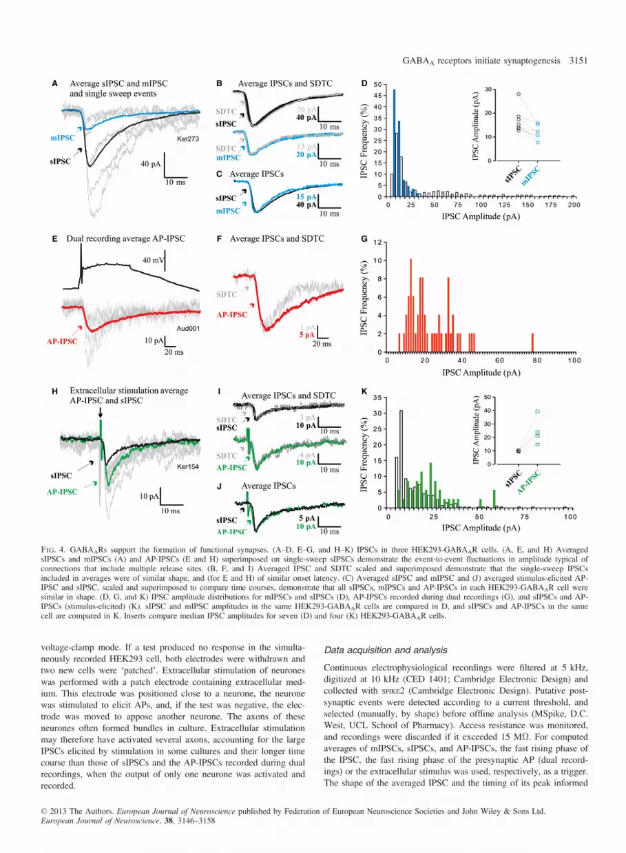
Fig. 4. GABAARs support the formation of functional synapses. (A–D, E–G, and H–K) IPSCs in three HEK293-GABAAR cells. (A, E, and H) AveragedsIPSCs and mIPSCs (A) and AP-IPSCs (E and H) superimposed on single-sweep sIPSCs demonstrate the event-to-event fluctuations in amplitude typical ofconnections that include multiple release sites. (B, F, and I) Averaged IPSC and SDTC scaled and superimposed demonstrate that the single-sweep IPSCsincluded in averages were of similar shape, and (for E and H) of similar onset latency. (C) Averaged sIPSC and mIPSC and (J) averaged stimulus-elicited AP-IPSC and sIPSC, scaled and superimposed to compare time courses, demonstrate that all sIPSCs, mIPSCs and AP-IPSCs in each HEK293-GABAAR cell weresimilar in shape. (D, G, and K) IPSC amplitude distributions for mIPSCs and sIPSCs (D), AP-IPSCs recorded during dual recordings (G), and sIPSCs and AP-IPSCs (stimulus-elicited) (K). sIPSC and mIPSC amplitudes in the same HEK293-GABAAR cells are compared in D, and sIPSCs and AP-IPSCs in the samecell are compared in K. Inserts compare median IPSC amplitudes for seven (D) and four (K) HEK293-GABAAR cells.
© 2013 The Authors. European Journal of Neuroscience published by Federation of European Neuroscience Societies and John Wiley & Sons Ltd.European Journal of Neuroscience, 38, 3146–3158
GABAA receptors initiate synaptogenesis 3151

individual manual IPSC amplitude measurements. The standard devi-ation time course (SDTC), which plots the standard deviation aboutthe mean of the averaged IPSC, was computed in parallel with eachaverage, prior to further analysis, to ensure that events included inaverages were of similar shape. When events with different shapes,or different latencies, are included in an average, the peak of theSDTC does not coincide with the peak of the average, indicating var-iation in the onset latency or the rising and/or falling phase of theevents. The IPSC 10–90% rise time (RT, the time taken for the IPSCto rise from 10% to 90% of its peak amplitude) and width at halfamplitude (HW) were measured from averages, and amplitude distri-butions were constructed from single event measurements. As it ispossible that very high sIPSC frequencies could have obscured thefalling phase of the averaged IPSC, averages of a subset of records,selected as being devoid of such spontaneous events, were used tomeasure the IPSC HW. However, the median IPSC amplitude wascalculated from measures of the entire population of detected synapticevents for each cell, and amplitude distributions contained all events.Data are given as mean � standard error of the mean (SEM), andStudent’s t-test (e.g. for sIPCS or mIPSC frequencies and IPSC timecourse parameters) was used to test for significant differencesbetween populations. For skewed distributions, however, median val-ues plus the 25th and 75th percentiles are given. sIPSC, mIPSC andAP-IPSC amplitude distributions were first tested for normality(Shapiro–Wilk test), and, because the majority were found not to benormally distributed, a Mann–Whitney U-test was used for unpairedcomparisons (e.g. sIPSC or mIPSC amplitudes in HEK293-GABAARcells vs. sIPSC or mIPSC amplitudes in HEK293-GABAAR-NL2cells), and a Wilcoxon test was used for paired comparisons (e.g.sIPSC amplitudes vs. mIPSC amplitudes, and sIPSC amplitudes vs.AP-IPSC amplitudes). In the figures, some electrophysiologicaltraces were filtered to reduce high-frequency noise (three-point run-ning average), and stimulus artefacts were reduced graphically, forclarity. PSI-PLOT (Poly Software International), GRAPHPAD PRISM
(GraphPad Software) and Excel (Microsoft) were used for analysis,plotting, and statistical tests.
Results
Postsynaptic GABAARs mediate adhesion of GABAergicaxons
To test whether GABAARs can play a direct role in synaptic targetrecognition, a novel co-culture model system was established. Thisconsisted of a homogeneous population of GABAergic basal gangliamedium spiny neurones co-cultured with HEK293 cells stablyexpressing a1/b2/c2-GABAARs (HEK293-GABAAR; Fig. 1A), themost prevalent postsynaptic receptor subtype present in synapses ofthese neurons (Gross et al., 2011).HEK293-GABAAR cells responded to GABA (1 lM), puff-applied
(10 s) in close proximity, with a large inward current (�518 �24.2 pA, n = 9). This response was enhanced by the a1 subunit-preferring GABAAR benzodiazepine site agonist zolpidem (0.4 lM)co-applied in the bathing medium (58 � 6.4% enhancement, n = 3),confirming the expression of functional a1/b2/c2-GABAARs in thiscell line with the expected pharmacological response (Fig. 1B).The presence of functional a1/b2/c2-GABAARs at the plasma
membrane was sufficient to initiate adhesion of GAD65-positivepresynaptic terminals and the formation of contacts between neuro-nes and HEK293 cells, as early as 2 h after plating (Fig. 1E). Thenumber of putative synaptic contacts per HEK293 cell increasedrapidly, reaching 57.1 � 6.2 in HEK293-GABAAR cells at 24 h
(mean � SEM, n = 8; Fig. 1E). Electrophysiological recordingsshowed that the proportion of HEK293-GABAAR cells in co-culture that showed sIPSCs (Fig. 1D) also reached a maximum at24 h (56%; Fig. 1F). This correlated well with the time scale ofputative contact formation (Fig. 1E). The smaller, TTX-resistantmIPSCs, recorded when APs were blocked, were abolished bythe GABA antagonist bicuculline (10 lM, n = 6; Fig. 1D),indicating their mediation by GABA release from the mediumspiny neurones.This process of rapid contact formation between neurones and
HEK293-GABAAR cells was not observed in experiments with con-trol HEK293 cells (Figs 1E, 2B, and 2E). This was also the casewith ‘mismatch’ experiments, when HEK293 cells expressingNMDA receptors were co-cultured with GABAergic medium spinyneurones (Figs 2A and E), or when HEK293-GABAAR cells(Fig. 2C) or control HEK293 cells (Fig. 2D) were co-cultured withglutamatergic hippocampal neurones (Fig. 2E). Thus, the expressionof ‘mismatched’ receptors in HEK293 cells was not sufficient topromote contact formation in co-cultures with GABAergic mediumspiny neurones or glutamatergic hippocampal neurones.
GABAARs initiate the formation of functional synaptic contacts
Many of the contacts between medium spiny neurones and HEK293-GABAAR cells showed activity-dependent uptake of fluorescentlylabelled anti-synaptotagmin synaptic vesicle-luminal domain-specificantibodies (Fig. 3A). Co-localization between GAD65-positive andluminal synaptotagmin-positive terminals that formed contacts withthe surface of HEK293-GABAAR cells (Fig. 3A) or control HEK293cells (Fig. 3B) was quantified with IMAGEJ. This analysis demon-strated that the number of active contacts formed with HEK293-GABAAR cells was significantly greater than the number of contactsformed with control HEK293 cells (means � SEM: 6.5 � 1.7 arbi-trary units, n = 11, vs. 1.01 � 0.28 arbitrary units, n = 18; Student’st-test, P = 0.015; Fig. 3C). Ultrastructural analysis of synaptotag-min-positive contacts between neurones and HEK293-GABAAR cellsrevealed characteristics typical of active synapses (Figs 3D1 andD2). These included a region of close membrane apposition betweenpresynaptic and postsynaptic elements, and multiple membrane-bound vesicles and mitochondria in the same, or adjacent, sections.Dark, diaminobenzidine-positive synaptic vesicles observed withinthe contacts formed with HEK293-GABAAR cells confirmed theactivity-dependent incorporation of HRP (Fig. 3D2). This demon-strates that, during the incubation, prior to fixation, the lumens ofsome synaptic vesicles were in continuity with the extracellularspace; that is, these vesicles had undergone exocytosis and neuro-transmitter release. These characteristics were not observed in rarecontacts formed between neurones and control HEK293 cells(Fig. 3E). Time-lapse confocal imaging demonstrated that contactsformed between neurones and HEK293-GABAAR cells were stableover a time period of 120 min (Movie S1).
Recordings of inhibitory postsynaptic potentials in HEK293-GABAAR cells
Electrophysiological recordings were made from HEK293-GABAARcells to determine whether the putative contacts identified with immu-nolabelling and electron microscopy could support synaptic activity.After 22–26 h in co-culture, HEK293-GABAAR cells were identifiedby fluorescence and recorded in whole-cell mode. In these recordings,sIPSCs were detected at a frequency of 2.5 � 0.75/s (mean � SEM,n = 20; Figs 1D and 4A–C). In contrast, in unmodified HEK293
© 2013 The Authors. European Journal of Neuroscience published by Federation of European Neuroscience Societies and John Wiley & Sons Ltd.European Journal of Neuroscience, 38, 3146–3158
3152 C. Fuchs et al.
cells (n = 10) and control HEK293 cells (expressing only pCherry,n = 10), no synaptic events were recorded (data not shown). sIPSCamplitude distributions were skewed, with a large population ofsmall-amplitude events, followed by a ‘tail’, or one or more discretepeaks of larger events (Fig. 4D) (median sIPSC amplitude, 15.3 pA;25–75%, 13.7–18.7 pA; n = 7). These larger events were blocked byTTX (1 lM; Figs 1D and 4A–D), suggesting that these larger sponta-neous events represented AP-driven release of GABA, and amplitudedistributions became more discrete (compare blue with white bars inthe histogram shown in Fig. 4D; median mIPSC amplitude, 12.3 pA;25–75%, 10.7–15.3 pA). This demonstrates that spontaneous synap-tic events recorded under control conditions included a population oflarger events that were dependent on APs.In some HEK293-GABAAR cells, sIPSC frequency estimates
were compromised by the ability of large spontaneous events toobscure the smaller events when they overlapped in time. The fre-quencies of sIPSCs and mIPSCs cannot, therefore, be directly com-pared for all seven HEK293-GABAAR cells treated with TTX, butin three such cells in which events could be distinguished satisfacto-rily, the sIPSC frequency was 8.973 � 1.66/s (mean � SEM), andthe mIPSC frequency was 4.69 � 2.07/s. For all seven cells subse-quently treated with TTX, the mean sIPSC frequency was5.5 � 1.58/s and the mean mIPSC frequency was 4.23 � 1.25/s.These sIPSC frequencies are higher than the average for the largerpopulation given above, which includes cells not treated with TTX,because those selected for mIPSC analysis were those with thehigher spontaneous frequencies.
Paired whole-cell recordings
To confirm that these synapse-like contacts could indeed supportAP-driven GABA release (AP-IPSCs), paired whole-cell recordingswere performed for presynaptic basal ganglia medium spiny neuro-nes and neighbouring HEK293-GABAAR cells (Figs 4E–G). Theproportion of such paired recordings that revealed a connectionwas 1 : 125. An event of the appropriate shape that follows eachpresynaptic AP at fixed latency can be assumed to be the result ofthe synchronous release of transmitter in response to that AP, i.e.AP-driven release. Despite their similarity in shape and onsetlatency shape, however, AP-IPSCs fluctuated in amplitude fromevent to event, because several synaptic contacts typically contrib-ute to each synaptic connection between two cells, and the releaseof transmitter is stochastic. This fluctuation can be seen in Figs 4Eand H, in which an averaged AP-IPSC is superimposed on severalof the single-sweep AP-IPSCs that contributed to the average.That the shapes and onset latencies of all events included inthe averaged AP-IPSCs were similar is indicated by the shape ofthe SDTC, which matches the shape of the average (Figs 4B, F,and I). To increase test numbers, extracellular stimulation wasalso employed (Fig. 4H–K). These AP-IPSCs, whether from dualrecordings or extracellular stimulation, were generally of similarduration to sIPSCs in the same HEK293-GABAAR cells. This canbe seen in Fig. 4J, in which the averaged IPSCs shown in Fig. 4H(sIPSC and AP-IPSC) are scaled to match amplitudes and superim-posed. This is also demonstrated in Figs 7A and B, which comparethe RTs and HWs of these IPSCs. Like sIPSCs (Fig. 4D), AP-IPSCs were larger than mIPSCs (23.32 pA for the dual recordingAP-IPSC and 24.83 � 5.22 pA, n = 4, for the stimulus-elicitedAP-IPSCs) (Fig. 4; compare mIPSC mean amplitudes, insert in D,with AP-IPSC amplitude distributions in G and K), suggesting thatseveral synapse-like contacts from one axon contributed to eachAP-IPSC.
GABAARs expressed with NL2 promote furthersynaptogenesis
The experiments described above demonstrate that the presence ofonly one type of neuronal GABAAR expressed stably in HEK293cells can promote the adhesion, formation and stabilization of func-tional presynaptic GABAergic axon terminals (Figs 1–4). This pro-cess occurs independently of the neuronal adhesion protein NL2, asdemonstrated by the lack of expression of this protein in HEK293-GABAAR cells by immunoblotting (data not shown). In the light ofpreviously published evidence for an equivalent role for NL2 in syn-apse assembly, NL2–pCherry was transiently expressed in HEK293-GABAAR cells (HEK293-GABAAR-NL2; Fig. S1A) to determinewhether the co-expression of NL2 with a1/b2/c2-GABAARs mayfurther promote synapse formation and maturation in our co-cul-tures. Contacts between the basal ganglia medium spiny neuronesand HEK293-GABAAR-NL2 cells formed rapidly, reaching76.2 � 12.1 per cell at 24 h (mean � SEM, n = 8; Fig. S1C). Thetiming of contact formation paralleled the detection of synapticGABAergic currents (Fig. S1B and D).The majority of contacts formed with HEK293-GABAAR-NL2 or
HEK293-NL2 cells incorporated the fluorescently labelled anti-synaptotagmin vesicle-luminal domain-specific antibodies (Figs 5Aand B). Quantification revealed that the innervation of HEK293-GABAAR-NL2 cells (mean � SEM, 39.2 � 8.7 arbitrary units,n = 15) was significantly greater than that of HEK293-NL2 cells(mean � SEM, 22.4 � 2.7 arbitrary units, n = 18; Student’s t-test,P = 0.03; Fig. 5C), or of HEK293-GABAAR cells (mean � SEM,6.5 � 1.7 arbitrary units, n = 11; Student’s t-test, P = 0.001;Fig. 3C). Ultrastructural analysis of contacts formed between med-ium spiny neurones and HEK293-GABAAR-NL2 or HEK293-NL2cells also demonstrated close membrane appositions between presyn-aptic and postsynaptic elements, multiple membrane-bound vesicles,and mitochondria, i.e. structural characteristics of mature synapses(Figs S2A and B).
Co-expression of GABAARs and NL2 enhances synapticefficacy
Electrophysiological recordings were made from HEK293-GABAAR-NL2 cells to determine the functional properties of puta-tive synaptic contacts formed between these cells and medium spinyneurones in co-culture. In these recordings, a high sIPSC frequencywas observed (mean � SEM, 12.8 � 2.96/s, n = 16; Fig. S1B;Figs 6A–C). In contrast, no synaptic events were observed whenrecordings were made from HEK293-NL2 cells (n = 6). As inHEK293-GABAAR cells, sIPSCs in HEK293-GABAAR-NL2 cellsfluctuated in amplitude, and were larger than mIPSCs in the samecells (Figs 6A and D), but were of similar shape (Figs 6C and 7).In 10 paired recordings, APs in five medium spiny neurones elicitedAP-IPSCs in a neighbouring, simultaneously recorded HEK293-GABAAR-NL2 cell (Figs 6E–G) (mean amplitude, 106.14 �17.33 pA). This 1 : 2 ‘hit rate’ is considerably higher than that seenin HEK293-GABAAR cells (1 : 125). Extracellular stimulation wasalso employed (Figs 6I–K) (hit rates of 5 : 8 in HEK293-GABAAR-NL2 cells vs. 4 : 46 in HEK293-GABAAR cells; meanamplitude, 1187.2 � 253.68 pA). The dual recording AP-IPSCswere generally of similar duration to sIPSCs in the same HEK293cells (Figs 6G and K) and larger than mIPSCs in similar cells(Fig. 6; compare D with H and L), suggesting, again, that severalsynapse-like contacts from one axon contributed to each AP-IPSC.The much larger amplitudes of the stimulus-elicited AP-IPSCs in
© 2013 The Authors. European Journal of Neuroscience published by Federation of European Neuroscience Societies and John Wiley & Sons Ltd.European Journal of Neuroscience, 38, 3146–3158
GABAA receptors initiate synaptogenesis 3153

HEK293-GABAAR-NL2 cells suggest the activation of severalconnected axons.Overall, the shape and time course of IPSCs recorded in
HEK293-GABAAR and HEK293-GABAAR-NL2 cells were similar(Fig. 7). In HEK293-GABAAR cells, the RTs of sIPSCs and ofmIPSCs (2.2 � 0.25 ms and 1.5 � 0.25 ms, respectively; Fig. 7A)and the HWs (10.4 � 1.23 ms and 8.5 � 1.09 ms, respectively,n = 7; Fig. 7B) were similar to those in HEK293-GABAAR-NL2cells (sIPSC RT, 1.9 � 0.35 ms; mIPSC RT, 1.5 � 0.35 ms; sIPSCHW, 11.4 � 2.25 ms; mIPSC HW, 8.7 � 1.69 ms; n = 6). Thebriefer time course of mIPSCs than of sIPSCs was apparent in bothconditions, but reached significance only in HEK293-GABAAR cells(RT, P = 0.0047; HW, P = 0.03; Student’s paired t-test; Figs 7Aand B).Despite these similarities, spontaneous IPSCs were significantly
larger in HEK293-GABAAR-NL2 cells (median, �56.6 pA; 25/75%,�44/�159 pA; n = 6; Fig. 6D, insert) than in HEK293-GABAARcells (median, �15.3 pA; 25/75%, �13.7/�18.7 pA; n = 7; Fig. 4D,insert; P = 0.0012, Mann–Whitney U-test). Miniature IPSC ampli-tudes were also significantly larger in HEK293-GABAAR-NL2 cellsthan in HEK293-GABAAR cells (median, �27.2 pA; 25/75%,�25.1/�29 pA; Fig. 6D, insert; and median, �12.3 pA, 25/75%,�10.7/�15.3 pA; Fig. 4D, insert; P = 0.0012, Mann–WhitneyU-test). However, the difference in mIPSC amplitude (2.2-fold) doesnot entirely explain the large increase in sIPSC amplitude seen whenNL2 was co-expressed (3.7-fold). This may indicate that both thenumber of active synapses provided by each axon and the size of theresponse produced by each contact are larger in HEK293-GABAAR-NL2 cells. Thus, whereas GABAARs and NL2 can initiate contactformation independently of each other, when co-expressed they actcooperatively to promote further formation and strengthening ofsynaptic connections. Expressed together, they appear to increaseboth the quantal amplitudes and the number of active release sitesprovided by each axon.
Discussion
This study demonstrates, for the first time, that the expression ofGABAARs by a postsynaptic cell is sufficient to initiate the adhe-sion of GABAergic axons and the formation of functional synapses.When GABAARs were co-expressed with NL2 in this system, theeffect on synapse formation exceeded the individual effects of thesetwo proteins. In addition, connections with HEK293-GABAAR cellswere strengthened when NL2 was added, suggesting a cooperativeinteraction.
The structural role of GABAARs in synapse formation foundhere, with an in vitro co-culture model system, is supported byresults emerging from the in vivo analysis of mutant mice lackingspecific GABAAR a subunits. For example, in a1 subunit knockoutmice, the function and synaptic localization of gephyrin, a majorpostsynaptic scaffold protein, at inhibitory synapses, is disrupted(Fritschy et al., 2012). Similarly, in CA1 pyramidal neurones of a2subunit knockout mice, clustering of both gephyrin and of NL2 isdecreased in many subcellular compartments, but most prominentlyin the region of the axon initial segment, where a2 subunit-contain-ing GABAergic synapses are abundant (Nusser et al., 1996; Nyiriet al., 2001; Panzanelli et al., 2011). The result presented here mayalso help to explain why deleting all neuroligin isoforms does notprevent synapse formation in vivo, but impairs their functional matu-ration (Varoqueaux et al., 2006; Poulopoulos et al., 2009).The presence of gephyrin in control HEK293 cells has been
reported previously, particularly in dividing cells (Wu et al., 2012).A role for gephyrin in the consolidation of synapse-like contacts inthese co-cultures cannot therefore be excluded. That gephyrin, ifpresent, does not initiate contact formation is indicated by the lack ofcontacts with control HEK293 cells, or with HEK293 cells transfectedwith glutamate receptors. Another postsynaptic scaffold protein,collybistin, has been shown to play an important role in the postsyn-aptic accumulation of GABAARs in neurones, but not to be synapto-genic (Chiou et al., 2011). The very high-density plasma membraneexpression of GABAARs in the present HEK293-GABAAR cellsmay have removed the need for collybistin to concentrate receptorsin these co-cultures, as previous studies have indicated that collybi-stin is not expressed in HEK293 cells (Kins et al., 2000).Direct in vivo evidence for a role for GABAARs in synapse
assembly has yet to emerge. The multiplicity of GABAAR subtypesexpressed in neurones (Schofield et al., 1987; Sieghart, 2006), thevast array of presynaptic and postsynaptic proteins, in addition tothe receptors, found to populate the synaptic cleft, and the possibil-ity that removal or modification of any one building block mayresult either in its replacement by another or in a string of knock-onconsequences, present enormous difficulties in the interpretation ofany study designed to identify the unique role(s) of a specific pro-tein. Although they are far from the situation in vivo and are subjectto all of the caveats that should surround any study in a reducedsystem, these co-cultures have allowed the potential for GABAARsto participate directly in synapse formation to be demonstrated. Inagreement with studies of synapse formation in NL2 knockout micein vivo (Varoqueaux et al., 2006; Blundell et al., 2009; Gibsonet al., 2009), although in contrast to the conclusions reached in
A B C
Fig. 5. GABAARs expressed with NL2 promote further synaptogenesis. (A and B) Immunolabelling of contacts between presynaptic nerve terminals (anti-GAD65), incorporating a luminal domain-specific anti-synaptotagmin antibody (Cy5) and the surface of HEK293-GABAAR-NL2 cells (A) or of HEK293-NL2cells (B) (NL2–pCherry) (scale bar: 10 lm). (C) Quantification of active terminals at the surface of HEK293-GABAAR-NL2 or of HEK293-NL2 cells,expressed as colocalization between presynaptic markers and postsynaptic NL2–pCherry (arbitrary units; mean � SEM; HEK293-GABAAR-NL2, n = 14 fromtwo independent experiments; HEK293-NL2, n = 22 from four independent experiments; **P < 0.05, Student's t-test).
© 2013 The Authors. European Journal of Neuroscience published by Federation of European Neuroscience Societies and John Wiley & Sons Ltd.European Journal of Neuroscience, 38, 3146–3158
3154 C. Fuchs et al.

relation to some of the previous co-culture studies (Scheiffele et al.,2000; Dean et al., 2003; Graf et al., 2004; Chih et al., 2005; Donget al., 2007), we show that NL2 is not an absolute requirement forthe formation of functional GABAergic synapses, because synapse-like contacts, capable of supporting both spontaneous ‘miniature’synaptic events and AP-driven GABA release, were induced byGABAARs alone. The discrepancies between our study and previousin vitro studies could perhaps be explained, at least in part, by thedifferent combinations of neuronal cell types and postsynapticGABAAR subtypes tested. This, in addition, to the high level andconsistency of cell surface expression of GABAAR subunits in the
stably transfected HEK293 cell line used in our study, and in con-trast to the transiently expressed GABAARs in previous studies,may have been crucial for the reliable detection of synapse forma-tion and activity across the population of cells in co-culture.The number of functional contacts was enhanced significantly by
concomitant overexpression of NL2, as seen in neurones (Fu &Vicini, 2009). Stable connections, involving several synapse-likecontacts per axon, do occur in the absence of NL2. However, com-parison of sIPSC, AP-IPSC and mIPSC amplitudes indicates thatsingle axon connections may involve more presynaptic terminals,and that each terminal elicits a stronger postsynaptic response when
A B
C
D
E F
G
H
I J
K
L
Fig. 6. Co-expression of GABAARs and NL2 enhances synaptic efficacy. IPSCs in three HEK293-GABAAR-NL2 cells (A–D, E–H, and I–L). (A, E, and I).Averaged sIPSCs and mIPSCs (A), and averaged AP-IPSCs (dual recordings vs. extracellular stimulation) and sIPSCs (E and I), superimposed on single-sweepsIPSCs. (B, F, and J) Averaged IPSC and SDTC scaled and superimposed demonstrates the similarity in shape of events included in the averages. (C) Aver-aged sIPSC and mIPSC, (G) averaged sIPSC and AP-IPSC (dual recording), and (K) averaged AP-IPSC (stimulus-elicited) and sIPSC, scaled and superimposedto compare time courses. (D, H, and L). IPSC amplitude distributions to compare the amplitudes of sIPSCs and mIPSCs (D) and of AP-IPSCs and sIPSCs(H and L) in the same cells. Inserts compare median IPSC amplitudes for seven (D), five (H) and five (L) cells.
© 2013 The Authors. European Journal of Neuroscience published by Federation of European Neuroscience Societies and John Wiley & Sons Ltd.European Journal of Neuroscience, 38, 3146–3158
GABAA receptors initiate synaptogenesis 3155

NL2 is co-expressed together with GABAARs. NL2 may also beimportant for the rigid membrane appositions typical of synapsesin situ, and contribute to the stabilization of contacts (compareFig. 3D and Figs S2A and B), and may increase the number ofsynapses formed by each axon, thereby playing an essential role innormal synaptic activity. However, NL2 does not appear to be essen-tial for GABAergic synapse formation, either in vivo (Varoqueauxet al., 2006; Blundell et al., 2009; Gibson et al., 2009) or in vitro.That these a1/b2/c2-GABAARs were sufficient alone to support
and stabilize functional synapse-like contacts is interesting in thelight of a study by Gibson et al. (2009). In this study, the synapsesinnervated by fast-spiking, parvalbumin-containing interneurones inthe hippocampus, which are mediated by a1-GABAARs (Thomsonet al., 2000; Nyiri et al., 2001), were found to be the most power-fully affected in NL2 knockouts. Both quantal amplitude and quan-tal content (i.e. the number of quanta, or synapses, contributing to
each event) were lower than at wild-type connections. These find-ings in NL2 knockout mice have a striking parallel in the presentstudy, where the absence of NL2 coincided with decreases in boththe number of functional synapses and the quantal amplitude, in amuch more reduced system employing a different class of presynapticneurone.A larger mIPSC, or quantal amplitude, is typically explained
either by a larger number of postsynaptic receptors, or by anincrease in their single channel conductance. HEK293-GABAAR-NL2 cells received a large number of synapse-like contacts, whichwere often very close neighbours (Fig. 5A; Fig. S2A), whereasHEK293-GABAAR cells received more sparse innervation (Figs 3Aand D). If such a finding were obtained in a neuronal system, itmight suggest that the larger quantal amplitudes seen in HEK293-GABAAR-NL2 cells are attributable to spill-over from one terminalto receptors lying under one or more neighbouring terminals. How-ever, although these cultures did not contain glial cells, whose activere-uptake of GABA might otherwise have curtailed its diffusion, theextracellular space in the co-cultures is very large, and the releasedGABA can be expected to have diffused rapidly away from theHEK293 cell. There was, moreover, little evidence for clustering ofreceptors in these HEK293-GABAAR cells (Fig. 1C; Fig. S1A), andthere was no evidence that the surface expression of GABAARs dif-fered between HEK293-GABAAR and HEK293-GABAAR-NL2cells. Spread of GABA to a larger, more widely distributed popula-tion of receptors is therefore not an adequate explanation for the lar-ger quantal amplitudes seen here with NL2 overexpression.How, then, might overexpression of NL2 enhance the activity of
GABAARs in this system? An economical, currently not easilyrefutable hypothesis, for which there is at present only indicativeevidence, is suggested by a recent finding (Zhang et al., 2010).Neurexin-2b, either overexpressed in the postsynaptic cell or appliedto the culture as free protein, decreased GABA currents via directinteraction with a1-GABAARs, in a neuroligin-independent manner.It is possible that, in the absence of NL2, neurexin-2b interacts withGABAARs in a way that promotes adhesion but suppresses receptoractivity, and that this suppression is relieved or reduced when NL2is also present. During synapse formation, an interaction betweenthe appropriate presynaptic neurexin and postsynaptic GABAARmight be sufficient to establish a synaptic connection. This connec-tion would then become stronger as NL2 became colocalized withthe receptors, and the interaction between receptor and neurexin wasthereby modified. In vivo, NL2 would then recruit further postsynap-tic density proteins (Poulopoulos et al., 2009), and perhaps this,together with the increased synaptic activity, would furtherstrengthen and stabilize the connection (Hartman et al., 2006;Varoqueaux et al., 2006; Chubykin et al., 2007). As a very reducedsystem was used for this study, it is not possible to propose thatcomplex developments at the postsynaptic density could have initi-ated an increase in the stability or density of the innervation here.The most likely explanation for this is therefore that the enhancedsynaptic activity, resulting from the larger quantal amplitudes inHEK293-GABAAR-NL2 cells, contributes to the increase in thedensity of innervation. Although the proposed modulatory role ofneurexins and NL2 in synapse formation initiated by GABAARsawaits further investigation, the co-culture system described heremay be particularly advantageous in these experiments, as it pro-vides tight control of expression and precise molecular manipulationof component players, both individually and in combination.In conclusion, using a multidisciplinary approach, we have dem-
onstrated that functional synapses can form in the absence of neuro-nal trans-synaptic adhesion molecules, if GABAARs are present. By
A
B
Fig. 7. Time course of IPSCs: properties of mIPSCs, sIPSCs, dual recordingAP-IPSCs and stimulus-elicited AP-IPSCs (means � SEM) in HEK293-GABAAR cells (left) and HEK293-GABAAR-NL2 cells (right). (A) Compari-son of the RTs. (B) Comparison of the HWs. Asterisks indicate where twopopulations differed significantly from each other (*P < 0.05, ***P < 0.001,one-way ANOVA). The numbers in the bars indicate the number of cellsincluded in each population. Time course parameters for AP-IPSCs (dualrecordings) without NL2 are not shown, as one of the two examples of theseAP-IPSCs was recorded at room temperature and is not therefore comparable.
© 2013 The Authors. European Journal of Neuroscience published by Federation of European Neuroscience Societies and John Wiley & Sons Ltd.European Journal of Neuroscience, 38, 3146–3158
3156 C. Fuchs et al.

promoting the adhesion of inhibitory axon terminals and their stabil-ization, GABAARs may play an important role in mechanismsunderlying the development of inhibitory synapses.
Supporting Information
Additional supporting information can be found in the online ver-sion of this article:Fig. S1. Innervation of HEK293-GABAAR-NL2 cells by embryonicbasal ganglia GABAergic medium spiny neurones in co-culture.Fig. S2. Innervation of HEK293-GABAAR-NL2 and HEK293 cellsby medium spiny neurones at the EM level.Movie S1. GABAergic medium spiny neurones and HEK293-GABAAR cells in co-culture form stable contacts.
Acknowledgements
We thank Professor J.-M. Fritschy, University of Zurich, for providingGABAAR subunit-specific antibodies, Professor R. J. Harvey (UCL Schoolof Pharmacy) and Professor N. Brose (Max Planck Institute of ExperimentalMedicine) for providing us with the NL2–pCherry construct, and Drs C.Porcher (INMED–INSERM U901), A. Bremaud, K. Brickley and S. Cousins(UCL School of Pharmacy) for technical advice and scientific discussion.We gratefully acknowledge financial support from the MRC UK (G0800498)and The Wellcome Trust. J. J. Burden is supported by the MRC Laboratoryfor Molecular Cell Biology. The authors declare no conflict of interest.
Abbreviations
AP, action potential; AP-IPSC, action potential-driven inhibitory postsynapticcurrent; D-AP5, D-2-amino-5-phosphonovalerate; DNQX, 6,7-dinitroquinoxa-line-2,3-dione; GABAAR, GABA type A receptor; GAD, glutamic aciddecarboxylase; HEK293, human embryonic kidney 293; HRP, horseradishperoxidase; HW, width at half amplitude; IPSC, inhibitory postsynaptic cur-rent; mIPSC, miniature inhibitory postsynaptic current; NL2, neuroligin-2;NMDA, N-methyl-D-aspartate; PBS, phosphate-buffered saline; PFA, parafor-maldehyde; RT, 10–90% rise time; SDTC, standard deviation time course;SEM, standard error of the mean; sIPSC, spontaneous inhibitory postsynapticcurrent; TTX, tetrodotoxin citrate; YFP, yellow fluorescent protein.
References
Blundell, J., Tabuchi, K., Bolliger, M.F., Blaiss, C.A., Brose, N., Liu, X.,Sudhof, T.C. & Powell, C.M. (2009) Increased anxiety-like behavior inmice lacking the inhibitory synapse cell adhesion molecule neuroligin 2.Genes Brain Behav., 8, 114–126.
Buerli, T., Pellegrino, C., Baer, K., Lardi-Studler, B., Chudotvorova, I., Frits-chy, J.M., Medina, I. & Fuhrer, C. (2007) Efficient transfection of DNAor shRNA vectors into neurons using magnetofection. Nat. Protoc., 2,3090–3101.
Chazot, P.L., Coleman, S.K., Cik, M. & Stephenson, F.A. (1994) Molecularcharacterization of N-methyl-D-aspartate receptors expressed in mamma-lian cells yields evidence for the coexistence of three subunit types withina discrete receptor molecule. J. Biol. Chem., 269, 24403–24409.
Chih, B., Engelman, H. & Scheiffele, P. (2005) Control of excitatory andinhibitory synapse formation by neuroligins. Science, 307, 1324–1328.
Chiou, T.T., Bonhomme, B., Jin, H., Miralles, C.P., Xiao, H., Fu, Z.,Harvey, R.J., Harvey, K., Vicini, S. & De Blas, A.L. (2011) Differentialregulation of the postsynaptic clustering of gamma-aminobutyric acid typeA (GABAA) receptors by collybistin isoforms. J. Biol. Chem., 286,22456–22468.
Chubykin, A.A., Atasoy, D., Etherton, M.R., Brose, N., Kavalali, E.T.,Gibson, J.R. & Sudhof, T.C. (2007) Activity-dependent validation ofexcitatory versus inhibitory synapses by neuroligin-1 versus neuroligin-2.Neuron, 54, 919–931.
Dean, C., Scholl, F.G., Choih, J., DeMaria, S., Berger, J., Isacoff, E. & Sche-iffele, P. (2003) Neurexin mediates the assembly of presynaptic terminals.Nat. Neurosci., 6, 708–716.
Dong, N., Qi, J. & Chen, G. (2007) Molecular reconstitution of functionalGABAergic synapses with expression of neuroligin-2 and GABAA recep-tors. Mol. Cell. Neurosci., 35, 14–23.
Farrant, M. & Kaila, K. (2007) The cellular, molecular and ionic basis ofGABA(A) receptor signalling. Prog. Brain Res., 160, 59–87.
Fernandez-Alfonso, T., Kwan, R. & Ryan, T.A. (2006) Synaptic vesiclesinterchange their membrane proteins with a large surface reservoir duringrecycling. Neuron, 51, 179–186.
Fiala, J.C. (2005) Reconstruct: a free editor for serial section microscopy.J. Microsc., 218, 52–61.
Fritschy, J.M. & Mohler, H. (1995) GABAA-receptor heterogeneity in theadult rat brain: differential regional and cellular distribution of seven majorsubunits. J. Comp. Neurol., 359, 154–194.
Fritschy, J.M., Panzanelli, P. & Tyagarajan, S.K. (2012) Molecular and func-tional heterogeneity of GABAergic synapses. Cell. Mol. Life Sci., 69,2485–2499.
Fu, Z. & Vicini, S. (2009) Neuroligin-2 accelerates GABAergic synapsematuration in cerebellar granule cells. Mol. Cell. Neurosci., 42, 45–55.
Fujiyama, F., Fritschy, J.M., Stephenson, F.A. & Bolam, J.P. (2000)Synaptic localization of GABA(A) receptor subunits in the striatum of therat. J. Comp. Neurol., 416, 158–172.
Futai, K., Doty, C.D., Baek, B., Ryu, J. & Sheng, M. (2013) Specific trans-synaptic interaction with inhibitory interneuronal neurexin underlies differ-ential ability of neuroligins to induce functional inhibitory synapses.J. Neurosci., 33, 3612–3623.
Gibson, J.R., Huber, K.M. & Sudhof, T.C. (2009) Neuroligin-2 deletionselectively decreases inhibitory synaptic transmission originating from fast-spiking but not from somatostatin-positive interneurons. J. Neurosci., 29,13883–13897.
Goffin, D., Ali, A.B., Rampersaud, N., Harkavyi, A., Fuchs, C., Whitton,P.S., Nairn, A.C. & Jovanovic, J.N. (2010) Dopamine-dependent tuning ofstriatal inhibitory synaptogenesis. J. Neurosci., 30, 2935–2950.
Graf, E.R., Zhang, X., Jin, S.X., Linhoff, M.W. & Craig, A.M. (2004) Neu-rexins induce differentiation of GABA and glutamate postsynaptic special-izations via neuroligins. Cell, 119, 1013–1026.
Gross, A., Sims, R.E., Swinny, J.D., Sieghart, W., Bolam, J.P. & Stanford,I.M. (2011) Differential localization of GABA(A) receptor subunits in rela-tion to rat striatopallidal and pallidopallidal synapses. Eur. J. Neurosci.,33, 868–878.
Hartman, K.N., Pal, S.K., Burrone, J. & Murthy, V.N. (2006) Activity-dependent regulation of inhibitory synaptic transmission in hippocampalneurons. Nat. Neurosci., 9, 642–649.
Kang, Y., Zhang, X., Dobie, F., Wu, H. & Craig, A.M. (2008) Induction ofGABAergic postsynaptic differentiation by alpha-neurexins. J. Biol.Chem., 283, 2323–2334.
Kins, S., Betz, H. & Kirsch, J. (2000) Collybistin, a newly identified brain-specific GEF, induces submembrane clustering of gephyrin. Nat. Neuro-sci., 3, 22–29.
Missler, M., Zhang, W., Rohlmann, A., Kattenstroth, G., Hammer, R.E.,Gottmann, K. & Sudhof, T.C. (2003) Alpha-neurexins couple Ca2+ chan-nels to synaptic vesicle exocytosis. Nature, 423, 939–948.
Nusser, Z., Sieghart, W., Benke, D., Fritschy, J.M. & Somogyi, P. (1996)Differential synaptic localization of two major gamma-aminobutyric acidtype A receptor alpha subunits on hippocampal pyramidal cells. Proc.Natl. Acad. Sci. USA, 93, 11939–11944.
Nyiri, G., Freund, T.F. & Somogyi, P. (2001) Input-dependent synaptic tar-geting of alpha(2)-subunit-containing GABA(A) receptors in synapses ofhippocampal pyramidal cells of the rat. Eur. J. Neurosci., 13, 428–442.
Panzanelli, P., Gunn, B.G., Schlatter, M.C., Benke, D., Tyagarajan, S.K.,Scheiffele, P., Belelli, D., Lambert, J.J., Rudolph, U. & Fritschy, J.M.(2011) Distinct mechanisms regulate GABAA receptor and gephyrin clus-tering at perisomatic and axo-axonic synapses on CA1 pyramidal cells.J. Physiol., 589, 4959–4980.
Poulopoulos, A., Aramuni, G., Meyer, G., Soykan, T., Hoon, M., Papadopoulos,T., Zhang, M., Paarmann, I., Fuchs, C., Harvey, K., Jedlicka, P., Schwarzach-er, S.W., Betz, H., Harvey, R.J., Brose, N., Zhang, W. & Varoqueaux, F.(2009) Neuroligin 2 drives postsynaptic assembly at perisomatic inhibitorysynapses through gephyrin and collybistin.Neuron, 63, 628–642.
Scheiffele, P., Fan, J., Choih, J., Fetter, R. & Serafini, T. (2000) Neuroliginexpressed in nonneuronal cells triggers presynaptic development in con-tacting axons. Cell, 101, 657–669.
Schofield, P.R., Darlison, M.G., Fujita, N., Burt, D.R., Stephenson, F.A.,Rodriguez, H., Rhee, L.M., Ramachandran, J., Reale, V. & Glencorse,T.A. (1987) Sequence and functional expression of the GABA A receptorshows a ligand-gated receptor super-family. Nature, 328, 221–227.
© 2013 The Authors. European Journal of Neuroscience published by Federation of European Neuroscience Societies and John Wiley & Sons Ltd.European Journal of Neuroscience, 38, 3146–3158
GABAA receptors initiate synaptogenesis 3157

Shen, K. & Scheiffele, P. (2010) Genetics and cell biology of buildingspecific synaptic connectivity. Annu. Rev. Neurosci., 33, 473–507.
Siddiqui, T.J. & Craig, A.M. (2011) Synaptic organizing complexes. Curr.Opin. Neurobiol., 21, 132–143.
Sieghart, W. (2006) Structure, pharmacology, and function of GABAAreceptor subtypes. Adv. Pharmacol., 54, 231–263.
Sudhof, T.C. (2008) Neuroligins and neurexins link synaptic function tocognitive disease. Nature, 455, 903–911.
Thomson, A.M., Bannister, A.P., Hughes, D.I. & Pawelzik, H. (2000) Differ-ential sensitivity to Zolpidem of IPSPs activated by morphologically iden-tified CA1 interneurons in slices of rat hippocampus. Eur. J. Neurosci.,12, 425–436.
Varoqueaux, F., Aramuni, G., Rawson, R.L., Mohrmann, R., Missler, M.,Gottmann, K., Zhang, W., Sudhof, T.C. & Brose, N. (2006) Neuroliginsdetermine synapse maturation and function. Neuron, 51, 741–754.
Ventimiglia, R. & Lindsay, R.M. (1998). Rat striatal neurons in low-density,serum-free culture. In Goslin, K. & Banker G. (Eds), Culturing NerveCells. MIT Press, Cambridge, MA, pp. 371–393.
Wu, X., Wu, Z., Ning, G., Guo, Y., Ali, R., Macdonald, R.L., De Blas,A.L., Luscher, B. & Chen, G. (2012) gamma-Aminobutyric acid type A(GABAA) receptor alpha subunits play a direct role in synaptic versus ex-trasynaptic targeting. J. Biol. Chem., 287, 27417–27430.
Zhang, C., Atasoy, D., Arac, D., Yang, X., Fucillo, M.V., Robison, A.J., Ko,J., Brunger, A.T. & Sudhof, T.C. (2010) Neurexins physically and func-tionally interact with GABA(A) receptors. Neuron, 66, 403–416.
Zhang, W., Rohlmann, A., Sargsyan, V., Aramuni, G., Hammer, R.E.,Sudhof, T.C. & Missler, M. (2005) Extracellular domains of alpha-neurexins participate in regulating synaptic transmission by selec-tively affecting N- and P/Q-type Ca2+ channels. J. Neurosci., 25, 4330–4342.
© 2013 The Authors. European Journal of Neuroscience published by Federation of European Neuroscience Societies and John Wiley & Sons Ltd.European Journal of Neuroscience, 38, 3146–3158
3158 C. Fuchs et al.


