Embed Size (px)
Citation preview

ORIGINAL CLINICAL ARTICLE
Gait in children with arthrogryposis multiplex congenita
Marie Eriksson • Elena M. Gutierrez-Farewik •
Eva Brostrom • Asa Bartonek
Received: 14 October 2009 / Accepted: 21 December 2009 / Published online: 16 January 2010
� EPOS 2010
Abstract
Purpose Lower limb contractures and muscle weakness
are common in children with arthrogryposis multiplex
congenita (AMC). To enhance or facilitate ambulation,
orthoses may be used. The aim of this study was to describe
gait pattern among individuals wearing their habitual
orthotic devices.
Methods Fifteen children with AMC, mean age 12.4 (4.3)
years, with some lower limb involvement underwent 3-D
gait analysis. Three groups were defined based on orthosis
use; Group 1 used knee–ankle–foot orthoses with locked
knee joints, Group 2 used ankle–foot orthoses or knee–
ankle–foot orthoses with open knee joints and Group 3
used no orthoses.
Results The greatest trunk and pelvis movements in all
planes and the greatest hip abduction were observed in
Group 1, compared to Groups 2 and 3, as well as to the gait
laboratory control group. Maximum hip extension was
similar in Groups 1 and 2, but in Group 3, there was less
hip extension and large deviations from the control data.
Lower cadence and walking speed were observed in Group
1 than in Groups 2 and 3. The step length was similar in all
groups and also with respect to the gait laboratory refer-
ence values.
Conclusions Children with AMC were subdivided
according to orthoses use. Kinematic data as recorded with
3-D gait analysis showed differences among the groups in
trunk, pelvis and knee kinematics, and in cadence and
walking speed. The step length was similar in all groups
and to the gait laboratory reference values, which may
be attributable to good hip extension strength in all
participants.
Keywords Motion analysis � AMC � Amyoplasia �Ambulation
Introduction
Arthrogryposis multiplex congenita (AMC) can be
described as a complex condition characterised by
deformed joints with an intact sensory system [1, 2]. To
be defined as AMC, there should be contractures in
multiple joints in multiple body areas present at birth
[1]. Arthrogryposis refers to a large heterogeneous group
of conditions [3], with an incidence reported from
1/3,000 to 1/5,100 live births [1, 4]. The underlying
cause of AMC is decreased foetal movements due to
various factors, such as neuropathic and myopathic
processes, abnormality of connective tissue, maternal
diseases or impaired foetal or intrauterine vascularity.
A correlation has been found between the early onset of
insufficient movement and the severity of contractures
[1].
AMC has been classified into three groups; disorders
with mainly limb involvement (four limb, lower limb or
upper limb), disorders with limb involvement associated
M. Eriksson � E. M. Gutierrez-Farewik � E. Brostrom �A. Bartonek (&)
Department of Women’s and Children’s Health,
Karolinska Institutet, MotorikLab Q2:07,
171 76 Stockholm, Sweden
e-mail: [email protected]
E. M. Gutierrez-Farewik
KTH Mechanics, Royal Institute of Technology,
Stockholm, Sweden
123
J Child Orthop (2010) 4:21–31
DOI 10.1007/s11832-009-0234-1

with other organs, and disorders with limb involvement and
central nervous dysfunction [1]. Various subtypes have
also been distinguished, such as amyoplasia, distal arthro-
gryposis and Larsen syndrome. The most common subtype
is amyoplasia, which represents one-third of all cases and is
also usually referred to as classic arthrogryposis. In
approximately half of the patients, a specific subtype
diagnosis cannot be given [1].
In children with amyoplasia, hip deformities are
common and range from soft tissue contractures to hip
dislocation [3, 5]. Knee joint involvement has been
reported in 70%, with flexion contractures being the
most common, followed by extension contractures [5].
Foot deformities are frequently observed, with equino-
varus adductus foot being the most common [5, 6].
Children with AMC frequently undergo orthopaedic
surgery, often with multiple procedures [3, 7]. The goals
for combining operative procedures are to avoid multiple
immobilisation periods [8] and reduction of muscle
strength [9].
The ability to walk depends on the extent of joint range
of motion, in particular in the hips and knees, as well as in
the foot, with possibility of plantigrade foot position.
Muscle weakness in the lower extremities, primarily in the
hip and knee extensor muscles, has also been reported to
influence walking ability [10], and in children with amy-
oplasia, muscle weakness was considered to be more
influential on walking ability than the severity of contrac-
tures [9]. The involvement of upper extremities with
inability of hand and arm support has also been found to
influence walking function [10, 11]. Most children with
AMC can achieve functional ambulation and 85% have
been reported as ambulators by the age of 5 years, but for
efficient community ambulation, a wheelchair may be
required [3, 12, 13].
To enhance or facilitate ambulation in children with
AMC, orthoses may be used to compensate for muscle
weakness and to support the lower extremities in an aligned
position [8, 10, 13]. Orthoses are made of different mate-
rials and with or without knee-locking mechanisms [14].
The use of ankle carbon fibre spring orthoses has report-
edly led to increased plantarflexor moment and stride
length in children with AMC [15]. Shoe wedges have also
been recommended to compensate for plantarflexion con-
tractures [13]. In our clinical practice, effort is made to
analyse the needs of orthoses in children with AMC and to
evaluate gait outcome. With the exception of one study
comparing two different types of orthoses in a group which
included four children with AMC [15], no published
description of gait in children with AMC has been found.
The aim of this study was to describe gait pattern among
individuals with AMC wearing their habitual orthotic
devices.
Participants and methods
The study was approved by the local ethics committee and
informed consent was obtained from the participants and
their parents.
Participants
Of 27 children with AMC born between the years 1989 and
2003 and who were treated at the orthopaedic department
at Karolinska University Hospital, 17 children fulfilled the
criteria of some lower limb motor involvement and inde-
pendent ambulation, and were invited to participate in a
gait analysis study. Inclusion criteria were independent
ambulation with or without orthoses and age between 4
and 18 years. Two children were excluded, one child
due to an unclear diagnosis and one child with unilateral
leg involvement who used a prosthesis, thus, 15 children
(8 males, 7 females; mean age 12.4 [4.3] years [range 4.7–
17.7]) participated in the study (Table 1). Six children had
four-limb and nine had lower-limb involvement (Table 1).
Orthosis subgroups
Twelve of the 15 participants used orthoses (Table 1). The
children were designated into subgroups with respect to
orthosis use. Prescriptions of orthoses were based on the
presence of muscle weakness, joint contractures or need of
joint stabilisation according to the current orthotic pro-
gramme. Group 1, represented by four participants, used
knee–ankle–foot orthoses with locked knee joints (KAFO-L)
(Fig. 1). Three participants had grade B3 in knee extensor
muscle strength and one participant had grade 4 and bilateral
knee flexion contractures of 25–30�. Two children had the
knee locking mechanisms anteriorly and two posteriorly,
depending on the child’s hand function. Two participants had
a carbon fibre ankle joint in combination with their KAFOs
with locked knee joints (KAFO-L-C) (Fig. 2).
Group 2, with eight participants, used knee–ankle–foot
orthoses with open knee joints (KAFO-O) or ankle–foot
orthoses (AFO) of different types. Two participants had open
knee joints with an extension stop and carbon fibre spring
ankle joint (KAFO-O-C) (Fig. 3). In one child, this was
required bilaterally to control knee hyperextension and knee
valgus due to the lack of both anterior and posterior cruciate
ligaments. In the other child, the control of knee valgus and
knee hyperextension was indicated unilaterally, and an ankle–
foot orthoses with a carbon fibre spring ankle joint (AFO-C)
(Fig. 4) was used on the contralateral limb. Five participants
used AFOs bilaterally and one unilaterally to stabilise the
ankle joint. Four participants used an AFO-C with the aim to
utilise the properties of the material to restore energy [15], one
used a hinged (AFO-H) and one a solid (AFO-S).
22 J Child Orthop (2010) 4:21–31
123

All orthoses were made by the same orthotic company.
Group 3, with three participants, used no orthoses. Among
the subjects in Group 3, one child preferred to walk barefoot
and two children used shoes, of which one child had a uni-
lateral lift due to leg length discrepancy and the other had
heel wedges to compensate for plantarflexion contractures.
Table 1 Distribution of patient characteristics, limb involvement, functional ambulation orthoses type and group according to orthosis use
Subject Gender Age (years) Height (cm) Weight (kg) Limb
involvement
Functional
ambulation
Orthoses type (L/R) Group
1 M 12.5 149 31.4 FL III KAFO-L-C 1
2 M 12.7 141 38.1 FL III KAFO-L-C
3 M 13.0 165 80.0 LL III KAFO-L
4 M 16.7 165 48.2 LL III KAFO-L
5 M 4.7 112 19.7 LL II Shoe/AFO-C 2
6a F 6.5 131 27.6 LL II KAFO-O-C
7a F 7.4 119 21.9 LL II AFO-C
8a M 9.2 136 28.8 LL II AFO-C
9a F 12.3 155 37.1 FL II AFO-H
10 M 15.3 166 44.6 LL II AFO-C
11 F 16.2 159 46.3 FL III KAFO-O-C/AFO-C
12a M 17.7 170 55.8 FL II AFO-S
13 F 12.4 151 37.9 LL I Barefoot 3
14 F 14.0 145 37.1 LL I Shoes with heel wedges
15 F 13.9 159 38.6 FL II Shoes with heel height, unilateral
R right; L left; FL four limb; LL lower limb; KAFO knee–ankle–foot orthoses; L locked knee joint; C carbon fibre spring ankle joint; AFO ankle–
foot orthoses; O open knee joint with extension stop; H hinged; S solida Gait analysis performed both with orthosis and barefoot
Fig. 1 Knee–ankle–foot orthoses with locked knee joint (KAFO-L)
Fig. 2 Knee–ankle–foot orthoses with locked knee joint and a carbon
fibre ankle joint (KAFO-L-C)
J Child Orthop (2010) 4:21–31 23
123

Functional ambulation
Functional ambulation was assessed according to a five-
level scale, which has been used previously in children
with myelomeningocele [16, 17]. In Group 1, all partici-
pants were designated level III, i.e. household ambulators
and wheelchair users for long indoor distances. In Group 2,
one participant was level III and eight participants were
level II, i.e. community ambulators who require a wheel-
chair for long distances outdoors only. In Group 3, one
participant was level II and two participants were level I,
i.e. community ambulators with no need for a wheelchair
(Table 1).
Muscle strength
The strength of the lower limb muscles was tested manu-
ally according to a six-graded scale [18], with grade 0
indicating no muscle strength, grade 1 activity traces, grade
2 gravity-eliminated movement, grade 3 movement against
gravity and grade 4 indicating movement against gravity
with some manual resistance. Grade 5, indicating normal
strength, was not given in this study. The distribution
of muscle strength in the lower limbs according to Groups
1–3 is shown in Table 2.
Joint contractures
Passive range of motion was measured with a goniometer.
Hip and knee flexion contractures were defined when
measured C10� and plantarflexion contractures as [0�from a neutral joint position. Two children had hip flexion
contractures of 10–20�. Seven children had knee flexion
contractures of 10–30� and eight children, five in Group 2
and three in Group 3, had knee hyperextension of 10–20�.
Seven children had restricted knee flexion of 20–110� from
the neutral position. Eight children had plantarflexion
contractures of 10–20� (Table 2).
Orthopaedic surgery
Fourteen participants had undergone orthopaedic surgery,
of which 12 children underwent bony surgery and two
children had only soft tissue surgery (Table 2).
Gait analysis
All children underwent 3-D gait analysis using an eight-
camera motion analysis system (Vicon�, Oxford, UK). The
children were equipped with 34 reflective markers aligned
Fig. 3 Knee–ankle–foot orthoses with open knee joint and a carbon
fibre ankle joint (KAFO-O-C)
Fig. 4 Ankle–foot orthoses with a carbon fibre ankle joint (AFO-C)
24 J Child Orthop (2010) 4:21–31
123

with anatomical landmarks on the head, trunk and pelvis,
and bilaterally on the arms, thighs, shanks and feet. The
markers were placed by the same examiner (AB). The
lower body was modelled according to the Newington
model [19] and the upper body was modelled as the thorax,
upper and lower arms, hands and head according to the
Plug-in Gait model (Vicon). In the children who wore
orthoses, the markers were placed as near as possible to the
correct anatomical position. The subjects were asked to
walk at a self-selected comfortable pace along a 10-m
walkway until complete information from several gait
cycles for each side was collected. In five of the six chil-
dren who were able to walk without orthoses, gait analysis
was performed barefoot.
Data analysis
Three kinematic gait cycles were generated for each sub-
ject. The following kinematic parameters were obtained
from each gait cycle and averaged for each side to describe
gait: range of lateral trunk sway, average trunk tilt, range of
trunk rotation, pelvic elevation range, average pelvic tilt,
pelvic rotation range, maximum hip abduction, maximum
hip flexion and extension, hip rotation at initial contact,
knee flexion at initial contact and in mid stance, maximum
knee flexion, knee flexion/extension range and average foot
progression in stance. For Group 3, the maximum dorsi-
flexion and plantarflexion were also analysed. Time and
distance parameters were analysed, wherein velocity, step
length and stride length were normalised to the leg length.
Statistical analysis
The Wilcoxon signed ranks test was used to test for dif-
ferences between the left and right sides within each of the
groups and between gait during barefoot walking and with
orthoses in five participants in Group 2 (Table 1). The
Kruskal–Wallis test was used to compare values between
the three groups. All statistical analyses were carried out
using commercially available software (SPSS version
16.0). A P-value of B0.05 was considered to be statistically
significant.
Table 2 Muscle strength, joint range of motion and previous orthopaedic surgery in all participants
Group Child Sideb Muscle strength grading according to a 0–5 scalea Joint range of motion (�) Orthopaedic surgery
Hip Knee Ankle Hipc Kneed Anklee
Flex Ext Abd Flex Ext Dors Plant Ext Flex Ext Dors
L/R L/R L/R L/R L/R L/R L/R L/R L/R L/R L/R
1 1 L 4/3 3/4 4/4 4/3 2/3 0/0 0/0 0/0 130/50 -15/0 -20/-20 Bi hip, bi ankle
2 R 3/3 4/4 4/4 4/4 3/3 0/0 0/0 -10/-20 60/60 -30/-25 -20/-15 Bi knee, bi ankle
3 R 3/3 4/3 4/3 2/3 2/2 0/0 0/0 0/0 100/110 0/-20 -15/-15 Bi knee, bi ankle
4 L 4/4 4/4 3/4 3/4 4/4 0/0 0/0 0/0 100/100 -30/-25 10/0 Bi kneef, bi ankle
2 5 R 4/4 4/4 4/3 4/4 4/4 4/3 4/3 0/0 130/130 ?20/-10 20/-15 R anklef
6 R 4/4 4/4 4/4 4/4 4/4 4/4 4/4 0/0 150/150 ?20/?20 0/10 –
7 L 4/4 4/4 4/4 4/4 4/4 4/4 3/4 0/0 130/140 ?15/?15 0/0 Bi hip, L ankle
8 R 4/4 4/4 4/4 4/4 4/4 4/4 3/3 0/0 150/150 ?10/?10 15/20 Bi ankle
9 L 4/4 4/4 4/4 4/4 4/4 0/4 0/3 0/0 140/140 -10/?10 -20/5 Bi ankle
10 R 4/4 4/4 3/3 3/2 4/4 0/0 0/0 0/0 60/60 -10/-10 -20/-20 L hip, bi ankle
11 L 4/4 4/4 4/4 4/4 4/4 2/2 2/2 0/0 40/110 ?5/0 0/0 L hip, bi ankle
12 L 4/4 4/4 4/4 4/4 4/4 4/4 4/4 0/0 140/140 0/0 -20/0 Bi ankle
3 13 R 4/4 4/4 4/4 4/4 4/4 4/4 4/4 0/0 120/20 ?5/?10 5/10 Bi hip
14 L 4/4 4/4 3/3 4/4 4/4 4/4 4/4 -20/-15 140/140 ?10/?15 -15/-10 Bi hip, bi ankle
15 R 4/4 4/4 4/4 4/4 4/4 4/4 4/4 0/0 155/155 ?15/?15 15/10 R hipf
Flex flexion; Ext extension; Abd abduction; Dors dorsiflexion; Plant plantarflexion; L left; R right; bi bilaterallya Muscle strength grade \4 was not applied in this studyb Selected side for data analysisc 0 indicates neutral joint position and - indicates \0d Flexion indicates joint angle from 0 to full knee flexion range (140�), extension indicates \0 (-) and [0 (?)e 0 indicates neutral joint position and - indicates \0f Soft tissue surgery
J Child Orthop (2010) 4:21–31 25
123

Results
There were no statistical significant differences between
the right and left sides in any parameter within any
group. For data presentation and subsequent between-
group data analysis, the limb with less muscle strength
and/or greater contractures was selected. When there was
no difference between the limbs, the right side was
selected (Table 2).
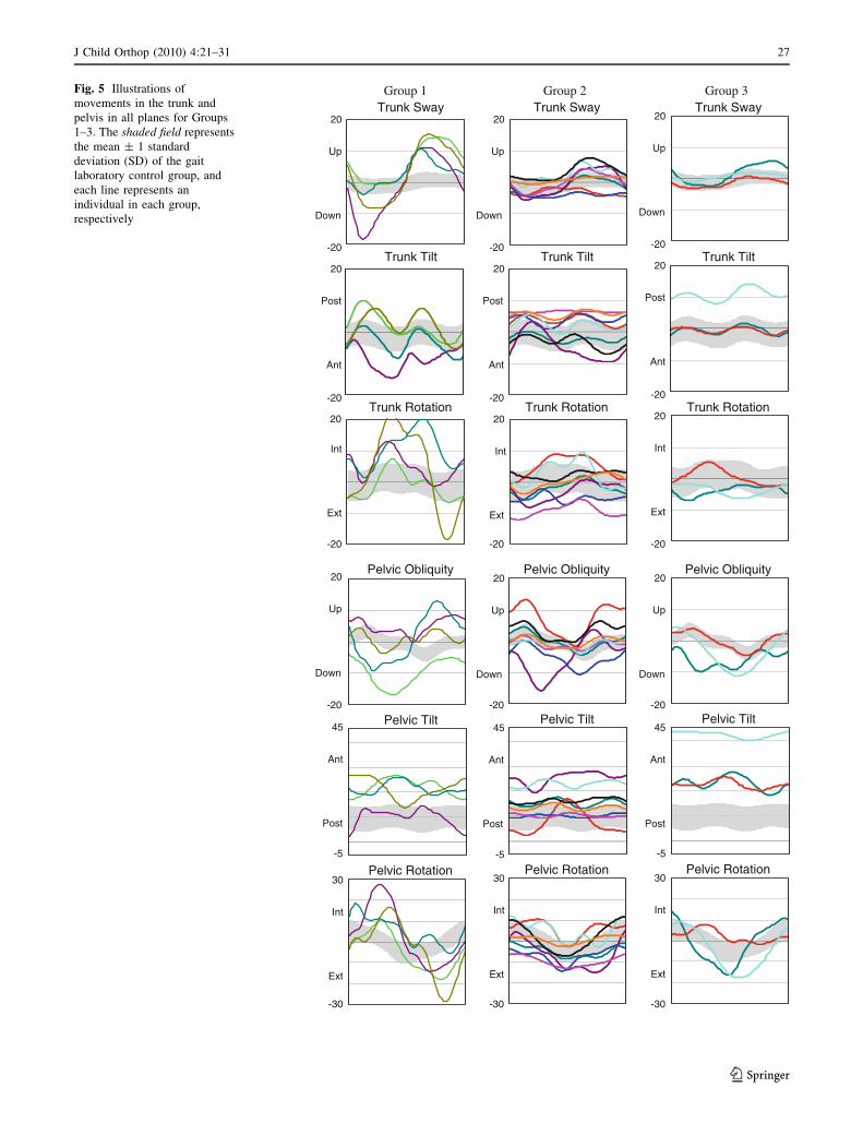
The results are presented with respect to the orthosis
groups. Reference kinematic data is illustrated by the gait
laboratory control group, consisting of 23 healthy children
aged 5–14 years.
Trunk kinematics
In Group 1, anterior-posterior trunk tilt movements tended
towards posterior in stance and swing, which was also
slightly pronounced in Group 2. In Groups 2 and 3, the
participants were relatively similar in all planes except one
participant in Group 3 who showed greater posterior trunk
tilt of approximately 11� during the entire gait cycle.
Between the groups, the trunk lateral sway range
(P = 0.015) and trunk rotation range (P = 0.007) differed
significantly (Fig. 5, Table 3).
Pelvis kinematics
In Group 1, internal rotation was more pronounced in
stance and external rotation in swing. In all groups, there
were various pelvic obliquity movements among the par-
ticipants, with pelvic lift in swing frequently observed in
Group 1. The mean anterior/posterior pelvic tilt values
were greatest in Group 3 (Table 3) and movements varied
among participants in all groups. Pelvis rotation range
differed significantly between the groups (P = 0.021)
(Fig. 5, Table 3).
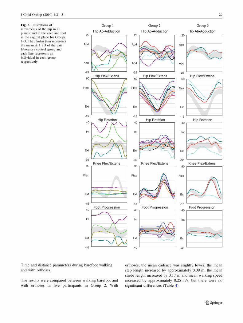
Hip kinematics
Group 1 showed an average of 2–3 times greater maximum
hip abduction than Groups 2 and 3 (Table 3), although
there were variations among participants in Groups 1 and 2.
The maximum hip extension was similar in Groups 1 and 2,
reaching almost neutral position, with reasonably similar
movements among participants. Group 3 had less hip
extension than Groups 1 and 2 (Table 3). The maximum
hip flexion was similar in all groups (Table 3), with rela-
tively similar movements among the participants in all
groups. The mean hip rotation at initial contact was similar
in the groups (Table 3), but large deviations among par-
ticipants were observed in Group 1 (Fig. 6).
Knee kinematics
The maximum knee flexion in swing as well as the mean
flexion/extension range were lowest in Group 1
(P = 0.048 and 0.015, respectively) (Table 3), as a
consequence of the locked orthotic knee joints. Despite
the locked orthoses in Group 1, corresponding to the
knee flexion contractures, there was greater knee flexion
at initial contact and greater knee flexion at mid stance
compared to Groups 2 and 3, with variation among
Group 1 (Table 3). In Group 2, the knee flexion/exten-
sion movements were similar among the participants.
Reduced knee flexion during swing was observed in one
participant in Group 3 due to restricted knee flexion
motion (Fig. 6).
Foot kinematics
Dorsiflexion and plantarflexion for Groups 1 and 2 were
not analysed due to various orthoses and footwear condi-
tions. Group 1 showed large variation among the partici-
pants, with both large internal as well as external foot
progression (Fig. 6, Table 3).
Time and distance parameters
Lower cadence and lower walking speed were observed in
Group 1 than in Groups 2 and 3 (P = 0.019 and 0.033,
respectively). The step length and stride length were sim-
ilar in the groups (P = 0.341 and 0.460, respectively).
Group 1 showed somewhat greater step width compared to
Groups 2 and 3, though the difference was not statistically
significant (P = 0.072) (Table 3).
Observations with respect to the gait laboratory
kinematic control data and time and distance
parameters reference values
Upon visual comparison of the AMC groups’ data with
respect to the gait laboratory control data, the greatest
deviations in trunk movements were seen in Group 1 in all
planes. In the pelvis, great deviations were observed in
Group 1 in all movements, in Group 2 in increased pelvic
obliquity in swing and in both Groups 2 and 3 in increased
anterior pelvic tilt. At the hip, obvious discrepancy was
observed in Group 1 as increased abduction and hip rota-
tion, and in Group 3 as less maximum hip extension. At the
knee, discrepancy was observed in Group 1 in knee flexion
extension movements during the entire gait cycle, and in
Group 3 as less flexion in swing in one participant. At the
foot, discrepancy was observed in Group 1 in variation
26 J Child Orthop (2010) 4:21–31
123
Trunk Sway20
-20
Up
Down
Trunk Tilt20
-20
Post
Ant
Trunk Rotation20
-20
Int
Ext
Pelvic Obliquity20
-20
Up
Down
Pelvic Tilt45
-5
Ant
Post
Pelvic Rotation30
-30
Int
Ext
Trunk Sway20
-20
Up
Down
Trunk Tilt20
-20
Post
Ant
Trunk Rotation20
-20
Int
Ext
Trunk Sway20
-20
Up
Down
Trunk Tilt20
-20
Post
Ant
Trunk Rotation20
-20
Int
Ext
Pelvic Obliquity20
-20
Up
Down
Pelvic Tilt45
-5
Ant
Post
Pelvic Rotation30
-30
Int
Ext
Pelvic Obliquity20
-20
Up
Down
Pelvic Tilt45
-5
Ant
Post
Pelvic Rotation30
-30
Int
Ext
Group 1 Group 2 Group 3 Fig. 5 Illustrations of
movements in the trunk and
pelvis in all planes for Groups
1–3. The shaded field represents
the mean ± 1 standard
deviation (SD) of the gait
laboratory control group, and
each line represents an
individual in each group,
respectively
J Child Orthop (2010) 4:21–31 27
123

among the participants and in Group 2 in increased internal
foot progression in swing (Figs. 5 and 6).
Compared to the control group’s average (standard
deviation) time and distances parameters, cadence 130
(14) steps/min, step length/leg length 0.79 (0.06), stride
length/leg length 1.59 (0.12), walking speed/leg length
1.72 (0.26) s-1 and step width 0.14 (0.02) m, it was
notable that Groups 2 and 3 had similar cadence, all
groups had step length relatively close to the control
group, Group 2 had longer stride length and very similar
walking speed, and Group 1 had almost twice the step
width.
Table 3 Mean (standard deviation [SD]) of the trunk, pelvis and lower limb joint angles, cadence, step length, stride length, walking speed and
step width in Groups 1–3
Group 1 (n = 4) Group 2 (n = 8) Group 3 (n = 3) P-valuek
Mean (SD) Mean (SD) Mean (SD)
Trunk (�)
Lateral sway (range) 21.4 (6.9) 6.5 (2.8) 5.2 (2.3) 0.015
Tilt ant/posta (average) -1.9 (5.0) 1.9 (4.4) 3.5 (6.6) 0.419
Rotation (range) 22.6 (12.5) 8.7 (2.1) 6.5 (1.5) 0.007
Pelvis
Elevation (range) 13.5 (6.2) 10.0 (5.1) 10.9 (4.1) 0.322
Tilt ant/postb (average) 18.8 (5.8) 15.6 (6.4) 29.5 (11.3) 0.126
Rotation (range) 34.7 (11.2) 13.9 (5.6) 22.5 (10.9) 0.021
Hip
Abductionc (max.) -18.1 (9.9) -8.7 (5.7) -5.8 (3.2) 0.109
Extensiond (max.) -4.4 (8.0) -1.5 (8.4) 17.2 (4.1) 0.055
Flexion (max.e) 44.0 (11.3) 41.6 (11.5) 52.9 (9.4) 0.347
Rotationf (ICg) -7.0 (34.3) -7.8 (10.1) -9.4 (13.5) 0.989
Knee
Flexion (ICg) 17.9 (7.4) 6.9 (6.5) 3.0 (7.7) 0.052
Flexion (MSh) 15.8 (9.7) 5.7 (9.4) 9.2 (4.8) 0.205
Flexion (max.e) 19.3 (7.4) 55.1 (9.2) 40.8 (27.3) 0.048
Flex/ext (range) 4.7 (2.3) 51.8 (11.6) 39.4 (23.2) 0.015
Ankle
Dorsiflexion (max.) – – 7.4 (5.0) –
Plantarflexioni (max.) – – -17.6 (4.2) –
Foot progressionf (averagej) -0.7 (24.8) -7.4 (7.1) -8.2 (5.0) 0.853
Time and distance parameters
Cadence (steps/min) 87 (6) 120 (16) 128 (5) 0.019
Step length/leg length 0.71 (0.05) 0.81 (0.14) 0.74 (0.03) 0.341
Stride length/leg length 1.46 (0.10) 1.63 (0.26) 1.43 (0.03) 0.46
Walking speed/leg length (s-1) 1.06 (0.13) 1.65 (0.45) 1.53 (0.06) 0.033
Step width (m) 0.24 (0.07) 0.16 (0.05) 0.12 (0.04) 0.072
a ? = posteriorb ? = anteriorc - = abductiond - = extensione Max in swing phasef - = externalg Initial contacth Mid stancei - = plantarflexionj Stancek Comparison between AMC groups
28 J Child Orthop (2010) 4:21–31
123
Time and distance parameters during barefoot walking
and with orthoses
The results were compared between walking barefoot and
with orthoses in five participants in Group 2. With
orthoses, the mean cadence was slightly lower, the mean
step length increased by approximately 0.09 m, the mean
stride length increased by 0.17 m and mean walking speed
increased by approximately 0.25 m/s, but there were no
significant differences (Table 4).
Hip Ab-Adduction20
-25
Add
Abd
Hip Flex/Extens60
-15
Flex
Ext
Hip Rotation40
-30
Int
Ext
Knee Flex/Extens90
-15
Flex
Ext
Foot Progression40
-40
Int
Ext
Hip Ab-Adduction20
-25
Add
Abd
Hip Flex/Extens60
-15
Flex
Ext
Hip Rotation40
-30
Int
Ext
Knee Flex/Extens90
-15
Flex
Ext
Foot Progression40
-40
Int
Ext
Hip Ab-Adduction20
-25
Add
Abd
Hip Flex/Extens60
-15
Flex
Ext
Hip Rotation40
-30
Int
Ext
Knee Flex/Extens90
-15
Flex
Ext
Foot Progression40
-40
Int
Ext
Group 1 Group 2 Group 3 Fig. 6 Illustrations of
movements of the hip in all
planes, and in the knee and foot
in the sagittal plane for Groups
1–3. The shaded field represents
the mean ± 1 SD of the gait
laboratory control group and
each line represents an
individual in each group,
respectively
J Child Orthop (2010) 4:21–31 29
123

Discussion
By subdividing the participants according to the orthoses
they had been prescribed, we identified different walking
patterns. This subject population is heterogeneous, and
orthosis prescription, while not an obvious criterion for
group definition, was indicative to a large extent of sub-
jects’ physical properties and body function. The partici-
pants in Group 1 who used KAFOs with locked knee joints
displayed more extensive trunk and pelvic movements, hip
abduction/adduction as well as hip rotation movements
compared to the other groups, including those three par-
ticipants who used no orthoses. A typically wide-based
walking pattern in children with AMC wearing KAFOs has
been described [20]. In Group 1, wider steps and more
abducted hip movements were found compared to the other
two groups, which may be interpreted as increasing their
support base to improve balance, but also as compensatory
mechanisms for the extended knees.
Hip flexion and extension movements in all groups were
similar to the gait laboratory control group, but less hip
extension in late stance was seen in all groups. In Group 3,
who did not require orthoses, two children had passive hip
extension range of motion to the neutral position, but did
not utilise their full hip extension during stance, possibly
due to previous hip deformities, and one child had hip
flexion contractures.
The step length was similar in all groups and to the gait
laboratory reference value, which may be attributable to
good hip extension strength. The importance of hip
extension strength grade 4 or better for functional ambu-
lation in patients with AMC has been reported [10] and
good correlation has been found between hip muscle
strength and motor function in patients with amyoplasia
[9]. Even if it is not possible to measure the muscle
strength over the entire range of motion, it is important to
grade the resistance throughout the available arc of motion,
since children with AMC may be strong in the midrange
[13].
All of the children in this study were able to perform
movements against gravity with or without manual resis-
tance in hip flexion, extension and abduction, and most of
them in knee extension and flexion. KAFOs with locked
knee joints have been recommended if knee extensor
weakness or a knee flexion contracture is present [5, 10].
KAFOs with open knee joints have frequently been used by
children with myelomeningocele to stabilise the knee joint
in the frontal and transverse planes [21]. In children with
AMC, both flexion contractures [10] and hyperextension
have frequently been reported, wherein flexion contrac-
tures, in contrast to hyperextension, have a negative
influence on walking ability [5, 22]. In our group, five knee
joints reached a neutral position, ten had knee flexion
contractures and 15 knees could hyperextend. In one child,
knee hyperextension was excessive, which made an
extension stop necessary. The thigh segment also improved
the control of the knee in the frontal and transverse planes.
In AMC, complete correction of clubfoot has been
described as difficult to achieve [23] and residual stiffness
has been reported [6], which can cause ambulatory diffi-
culties [6, 10]. In a recently published long-term follow-up
study [24], all independent or community walkers were
reported to have plantigrade feet, despite foot deformities
at birth. In children with joint contractures, improved
postural alignment of the body segments can be obtained
by adding wedges under the heels [13]. Such wedges were
used in one child in Group 3 (subject 14). Six of eight
participants in Group 2 used an energy-restoring carbon
fibre ankle joint in their orthoses. In four of six participants,
the stride length was longer with respect to the control
group data, which is in accordance with previous results
[15]. The greater step length and walking speed observed in
Group 2 compared to Group 3 may also be attributable to
the use of ankle carbon fibre springs.
Six of eight children in Group 2 were able to walk
independently without orthoses, of which one child could
only do so with difficulty. The results of the five partic-
ipants who performed gait analysis both barefoot and with
orthoses indicate that time and distance parameters
improved during walking with orthoses; however, no
significant differences could be found in such a small
number.
All participants used their orthoses regularly through-
out the day, which indicates high acceptance of the
orthoses. In Group 1, all children required orthoses to
achieve the ability to walk. With orthoses, they were able
to walk indoors and even short distances outdoors. The
children who used KAFO-L were able to lock and unlock
the knee joints independently, and were also able to
change their position from sitting to standing. Three
children required help putting on the orthoses due to
impaired hand function. Limited ambulation due to poor
protective responses of the upper extremity has been
reported [13] and that children wearing KAFOs often
require walkers [20]. In this study group, only one child
Table 4 Mean (SD) of cadence, step length, stride length and
walking speed in five patients in Group 2 comparing barefoot and
walking with orthoses
N = 5 Barefoot Orthoses P-value
Mean (SD) Mean (SD)
Cadence (steps/min) 132 (8) 127 (21) 0.5
Step length (m) 0.49 (0.06) 0.58 (0.09) 0.08
Stride length (m) 1.00 (0.17) 1.17 (0.17) 0.08
Walking speed (m/s) 1.09 (0.21) 1.34 (0.27) 0.138
30 J Child Orthop (2010) 4:21–31
123

occasionally used a walking aid, in the school yard for
safety reasons.
The ambulatory activity level in youths with AMC has
been reported as being lower compared to a control group
[25]. In our study, all four children in Group 1 were
household ambulators and of the total of seven children
who used a wheelchair, six also had a powered wheelchair,
four of whom had impaired hand function. Future studies
of energy consumption can provide more information
about the physical effort during walking in children with
AMC.
Conclusion
Gait pattern in children with arthrogryposis multiplex
congenita (AMC) was recorded with their orthoses, ranging
from locked knee joints to ankle foot orthoses and shoes
only. We have shown differences among the groups in the
trunk, pelvis and knee kinematics, and in cadence and
walking speed. In the children requiring locked knee joints,
the greatest trunk and pelvis movements and the lowest
knee flexion were observed, as well as the lowest cadence
and slowest walking speed.
The step length was similar in all groups and to the gait
laboratory reference values, which may be attributable to
good hip extension strength in all participants. Comparison
between barefoot and orthotic condition was performed in
only five participants, and indicated improved stride and
temporal parameters, though the small participant number
precludes conclusions on orthosis benefit.
Acknowledgments We would like to thank the children and their
parents for participating in the study. This study was supported by the
Norrbacka-Eugenia Foundation.
References
1. Hall JG (1997) Arthrogryposis multiplex congenita: etiology,
genetics, classification, diagnostic approach, and general aspects.
J Pediatr Orthop B 6:159–166
2. Thompson GH, Bilenker RM (1985) Comprehensive manage-
ment of arthrogryposis multiplex congenita. Clin Orthop Relat
Res 194:6–14
3. Bevan WP, Hall JG, Bamshad M, Staheli LT, Jaffe KM, Song K
(2007) Arthrogryposis multiplex congenita (amyoplasia): an
orthopaedic perspective. J Pediatr Orthop 27:594–600
4. Darin N, Kimber E, Kroksmark AK, Tulinius M (2002) Multiple
congenital contractures: birth prevalence, etiology, and outcome.
J Pediatr 140:61–67
5. Staheli LT (1998) Lower extremity management. In: Staheli LT,
Hall JG, Jaffe KM, Paholke DO (eds) Arthrogryposis: a text atlas.
Cambridge University Press, Cambridge, pp 55–73
6. Guidera KJ, Drennan JC (1985) Foot and ankle deformities in
arthrogryposis multiplex congenita. Clin Orthop Relat Res
194:93–98
7. Carlson WO, Speck GJ, Vicari V, Wenger DR (1985) Arthro-
gryposis multiplex congenita. A long-term follow-up study. Clin
Orthop Relat Res 194:115–123
8. Staheli LT (1998) Orthopedic management principles. In: Staheli
LT, Hall JG, Jaffe KM, Paholke DO (eds) Arthrogryposis: a text
atlas. Cambridge University Press, Cambridge, pp 27–43
9. Kroksmark AK, Kimber E, Jerre R, Beckung E, Tulinius M
(2006) Muscle involvement and motor function in amyoplasia.
Am J Med Genet A 140:1757–1767
10. Hoffer MM, Swank S, Eastman F, Clark D, Teitge R (1983)
Ambulation in severe arthrogryposis. J Pediatr Orthop 3:293–296
11. Hahn G (1985) Arthrogryposis. Pediatric review and habilitative
aspects. Clin Orthop Relat Res 140:104–114
12. Sells JM, Jaffe KM, Hall JG (1996) Amyoplasia, the most
common type of arthrogryposis: the potential for good outcome.
Pediatrics 97:225–231
13. Donohoe M (2006) Arthrogryposis multiplex congenita. In:
Campbell SK, Vander Linden DW, Palisano RJ (eds) Physical
therapy for children, 3rd edn. Saunders Elsevier Inc., St. Louis,
pp 381–400
14. Florence J (1977) The orthotic management of arthrogryphosis.
Prosthet Orthot Int 1:111–113
15. Bartonek A, Eriksson M, Gutierrez-Farewik EM (2007) Effects
of carbon fibre spring orthoses on gait in ambulatory children
with motor disorders and plantarflexor weakness. Dev Med Child
Neurol 49:615–620
16. Bartonek A, Saraste H (2001) Factors influencing ambulation in
myelomeningocele: a cross-sectional study. Dev Med Child
Neurol 43:253–260
17. Danielsson AJ, Bartonek A, Levey E, McHale K, Sponseller P,
Saraste H (2008) Associations between orthopaedic findings,
ambulation and health-related quality of life in children with
myelomeningocele. J Child Orthop 2:45–54
18. Hislop HJ (2007) Daniels and Worthingham’s muscle testing:
techniques of manual examination, 8th edn. Saunders Elsevier
Inc., St. Louis
19. Davis RB, Ounpuu S, Tyburski D, Gage JR (1991) A gait analysis
data collection and reduction technique. Hum Mov Sci 10:575–
587
20. Graubert CS, Chaplin DL, Jaffe KM (1998) Physical and occu-
pational therapy. In: Staheli LT, Hall JG, Jaffe KM, Paholke DO
(eds) Arthrogryposis: a text atlas. Cambridge University Press,
Cambridge, pp 87–113
21. Gutierrez EM, Bartonek A, Haglund-Akerlind Y, Saraste H
(2003) Characteristic gait kinematics in persons with lumbosacral
myelomeningocele. Gait Posture 18:170–177
22. Murray C, Fixsen JA (1997) Management of knee deformity in
classical arthrogryposis multiplex congenita (amyoplasia con-
genita). J Pediatr Orthop B 6:186–191
23. Ponseti IV (1996) Congenital clubfoot. Fundamentals of treat-
ment. Oxford University Press Inc., New York
24. Fassier A, Wicart P, Dubosset J, Seringe R (2009) Arthrogryposis
multiplex congenita. Long-term follow-up from birth until skel-
etal maturity. J Child Orthop 3:383–390. doi:10.1007/s11832-
009-0187-4
25. Dillon ER, Bjornson KF, Jaffe KM, Hall JG, Song K (2009)
Ambulatory activity in youth with arthrogryposis: a cohort study.
J Pediatr Orthop 29:214–217
J Child Orthop (2010) 4:21–31 31
123


