Embed Size (px)
Citation preview
Gatifloxacin Induces S and G2-Phase Cell Cycle Arrest inPancreatic Cancer Cells via p21/p27/p53Vikas Yadav1,2, Sarwat Sultana2, Jyoti Yadav1*, Neeru Saini1*
1 Institute of Genomics and Integrative Biology (CSIR), Mall Road, Delhi, India, 2Department of Medical Elementology and Toxicology, Jamia Hamdard (Hamdard
University), Hamdard Nagar, Delhi, India
Abstract
Pancreatic cancer, despite being the most dreadful among gastrointestinal cancers, is poorly diagnosed, and further, thesituation has been aggravated owing to acquired drug resistance against the single known drug therapy. While previousstudies have highlighted the growth inhibitory effects of older generation fluoroquinolones, the current study aims toevaluate the growth inhibitory effects of newer generation fluoroquinolone, Gatifloxacin, on pancreatic cancer cell lines MIAPaCa-2 and Panc-1 as well as to elucidate the underlying molecular mechanisms. Herein, we report that Gatifloxacinsuppresses the proliferation of MIA PaCa-2 and Panc-1 cells by causing S and G2-phase cell cycle arrest without induction ofapoptosis. Blockade in S-phase of the cell cycle was associated with increased TGF-b1 expression and translocation ofSmad3-4 complex to the nucleus with subsequent activation of p21 in MIA PaCa-2 cells, whereas TGF-b signallingattenuated Panc-1 cells showed S-phase arrest by direct activation of p27. However, Gatifloxacin mediated G2–phase cellcycle arrest was found to be p53 dependent in both the cell lines. Our study is of interest because fluoroquinolones havethe ability to penetrate pancreatic tissue which can be very effective in combating pancreatic cancers that are usuallyassociated with loss or downregulation of CDK inhibitors p21/p27 as well as mutational inactivation of p53. Additionally,Gatifloxacin was also found to synergize the effect of Gemcitabine, the only known drug against pancreatic cancer, as wellas the broad spectrum anticancer drug cisplatin. Taken together our results suggest that Gatifloxacin possesses anticanceractivities against pancreatic cancer and is a promising candidate to be repositioned from broad spectrum antibiotics toanticancer agent.
Citation: Yadav V, Sultana S, Yadav J, Saini N (2012) Gatifloxacin Induces S and G2-Phase Cell Cycle Arrest in Pancreatic Cancer Cells via p21/p27/p53. PLoSONE 7(10): e47796. doi:10.1371/journal.pone.0047796
Editor: Guenter Schneider, Technische Universitat Munchen, Germany
Received May 3, 2012; Accepted September 17, 2012; Published October 25, 2012
Copyright: � 2012 Yadav et al. This is an open-access article distributed under the terms of the Creative Commons Attribution License, which permitsunrestricted use, distribution, and reproduction in any medium, provided the original author and source are credited.
Funding: This work was supported by grants (NWP-13 and EXP0011) from the Council of Scientific and Industrial Research (CSIR), India. VY was supported withSenior Research Fellowship from CSIR. The funders had no role in study design, data collection and analysis, decision to publish, or preparation of the manuscript.
Competing Interests: The authors have declared that no competing interests exist.
* E-mail: [email protected] (JY); [email protected] (NS)
Introduction
Pancreatic cancer, the malignant neoplasm of the pancreas is
currently the fifth most common cause of cancer death [1]. Despite
improvements in diagnosis, the prognosis still remains poor
because of delayed symptom presentation, aggressive tumor
growth and profound desmoplastic reaction [2;3]. The 5-year
survival rate is approximately 15–20%, following pancreas re-
section in case of pancreatic cancer [4]. Apart from surgery,
adjuvant chemotherapy with Gemcitabine and erlotinib has been
shown to improve prognosis in resectable pancreatic cancer cases
[5]. So, there is an increase in survival rate with conventional
cytotoxic chemotherapy as compared to surgery [6], however still
there’s need to develop or identify the potential anticancer drug
with increased selectivity and reduced toxicity.
In recent years older generation fluoroquinolones such as
Moxifloxacin and Ciprofloxacin possessing broad spectrum
antibiotic activity, have shown growth inhibitory effects by
inducing apoptosis and cell cycle arrest in various cancer cell
lines [7–10]. These nonantimicrobial activities have rendered
them unique among other broad spectrum antibiotics. Gatiflox-
acin or 8-methoxy fluoroquinolones is the newer (fourth)
generation fluoroquinolone which shows similar antibiotic effects
by targeting bacterial DNA gyrase and Topoisomerase [11–13],
and is also a potent drug in several highly infectious diseases such
as, the Sexually Transmitted Diseases, Toxoplasmosis and
Tularaemia [14–16]. Like other fluoroquinolone family members
it is also known to have certain immunomodulatory effects in vitro
in various cell lines as well as it is under clinical trials for treatment
of pulmonary tuberculosis [17;18]. Having so much similar
implications with other fluoroquinolones family members, no
growth inhibitory activity against cancer cell lines has been
reported for Gatifloxacin. Moreover, the pancreatic tissue
penetrating efficiency is very well reported [19] in case of
fluoroquinolones which tempted us to explore the mechanism by
which Gatifloxacin suppresses pancreatic cancer cell growth. In
this study, we investigated the effect of Gatifloxacin on survival
and proliferation of pancreatic cancer cell lines (MIA PaCa-2 and
Panc-1) and found that Gatifloxacin was able to suppress
proliferation of both cell lines, with MIA PaCa-2 being more
sensitive than Panc-1. The differential outcome might be because
of difference in functional TGF-b receptors in the two cell lines
with MIA PaCa-2 being sensitive to TGF-b1 and Panc-1 being
resistant as they lack functional TGF-b type I receptor [20]. We
found that Gatifloxacin arrests cells in G2-phase via inactivation of
cdc2-cyclinB1 complex by phosphorylation of cdc2 on Tyr15
through p53 activation. Moreover we show that Gatifloxacin can
induce p21 and p27 levels in MIA PaCa-2 and Panc-1 cells
PLOS ONE | www.plosone.org 1 October 2012 | Volume 7 | Issue 10 | e47796
respectively thereby causing S-phase arrest. We further demon-
strated that Gatifloxacin was able to synergize the antiproliferative
activity of Gemcitabine and Cisplatin in both cell lines.
Results
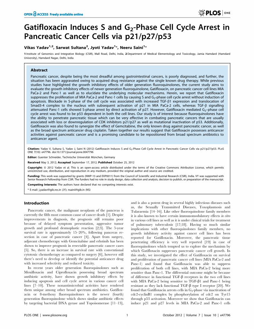
Gatifloxacin Suppresses Proliferation of PancreaticCarcinoma Cells In VitroTo evaluate the anti-proliferative effect of Gatifloxacin, we used
MIA PaCa-2 and Panc-1 pancreatic carcinoma cell lines. We first
studied the effect of different doses (0–400 mg/ml) of Gatifloxacin
on the viability of MIA PaCa-2 and Panc-1 cells for 24 h and 48 h
using MTT assay. As shown in Figure 1, GFX treatment resulted
in time and dose-dependent decrease in cell proliferation in MIA
PaCa-2 and Panc-1 cells albeit at different levels. Gatifloxacin
treatment resulted in 15–46% (p= 0.002) decrease in cell viability
in MIA PaCa-2 and 1–43% (p= 0.007) decrease in cell viability in
Panc-1 cells after 24 h respectively (Figure 1A i). We observed 19–
73% (p= 0.0016) decrease in cell viability in MIA PaCa-2 and 11–
72% (p= 0.00016) decrease in cell viability in Panc-1 cells at 48 h
respectively (Figure 1A ii). The above data clearly shows MIA
PaCa-2 to be more sensitive to Gatifloxacin than Panc-1 at all
doses. A striking observation from this data was the decrease in
viability, being more pronounced at higher doses (100, 200 and
400 mg/ml) of Gatifloxacin treatment after 24 h and 48 h
treatment in both cell lines and hence we took these concentra-
tions and time points to carry out further experiments.
Gatifloxacin Induces Cell Cycle Arrest at S and G2 - Phasein Pancreatic Carcinoma Cells without Induction ofApoptosisWe next investigated whether Gatifloxacin-mediated decrease
in viability of MIA PaCa-2 and Panc-1 cells is because of
apoptosis/necrosis. We first checked for the induction of apoptosis
by annexin assay. As shown in Figure 1B we didn’t find any
significant changes in apoptotic/necrotic population at all the
doses of Gatifloxacin as compared to vehicle treated cells in both
cell lines at 24 h as well as at 48 h respectively. To further cross
validate our annexin data, we next checked the expression level of
proapoptotic protein Bax by western blotting and activity of
caspase -3, -8 and -9 in a dose and time dependent manner under
the effect of Gatifloxacin. We did not find any significant change
either in Bax protein level (Figure 1C) or caspase -3, -8 and -9
activity (Figure 1D), which shows that Gatifloxacin does not hinder
the viability of cells. Results of Annexin V and Caspase activity
were also validated using a positive control, curcumin (60 mM for
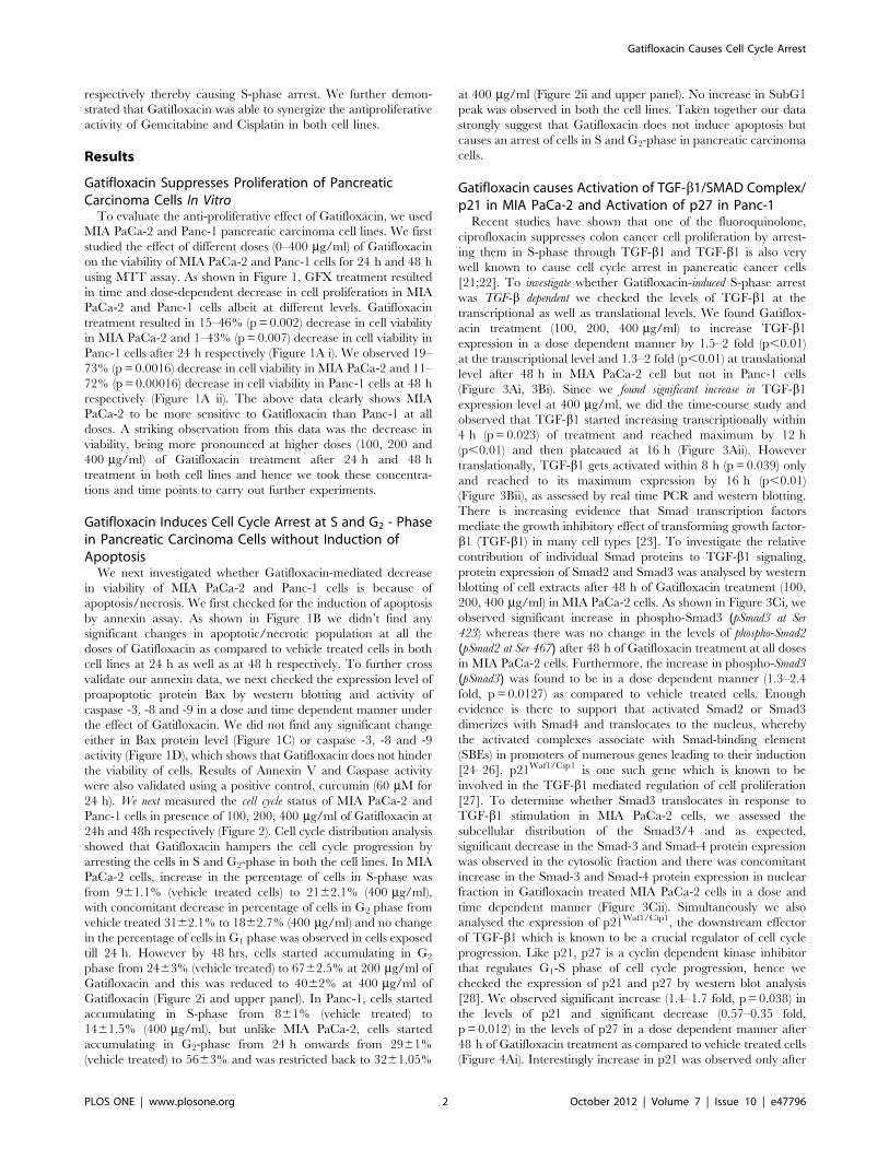
24 h). We next measured the cell cycle status of MIA PaCa-2 and
Panc-1 cells in presence of 100, 200, 400 mg/ml of Gatifloxacin at
24h and 48h respectively (Figure 2). Cell cycle distribution analysis
showed that Gatifloxacin hampers the cell cycle progression by
arresting the cells in S and G2-phase in both the cell lines. In MIA
PaCa-2 cells, increase in the percentage of cells in S-phase was
from 961.1% (vehicle treated cells) to 2162.1% (400 mg/ml),
with concomitant decrease in percentage of cells in G2 phase from
vehicle treated 3162.1% to 1862.7% (400 mg/ml) and no change
in the percentage of cells in G1 phase was observed in cells exposed
till 24 h. However by 48 hrs, cells started accumulating in G2
phase from 2463% (vehicle treated) to 6762.5% at 200 mg/ml of
Gatifloxacin and this was reduced to 4062% at 400 mg/ml of
Gatifloxacin (Figure 2i and upper panel). In Panc-1, cells started
accumulating in S-phase from 861% (vehicle treated) to
1461.5% (400 mg/ml), but unlike MIA PaCa-2, cells started
accumulating in G2-phase from 24 h onwards from 2961%
(vehicle treated) to 5663% and was restricted back to 3261.05%
at 400 mg/ml (Figure 2ii and upper panel). No increase in SubG1
peak was observed in both the cell lines. Taken together our data
strongly suggest that Gatifloxacin does not induce apoptosis but
causes an arrest of cells in S and G2-phase in pancreatic carcinoma
cells.
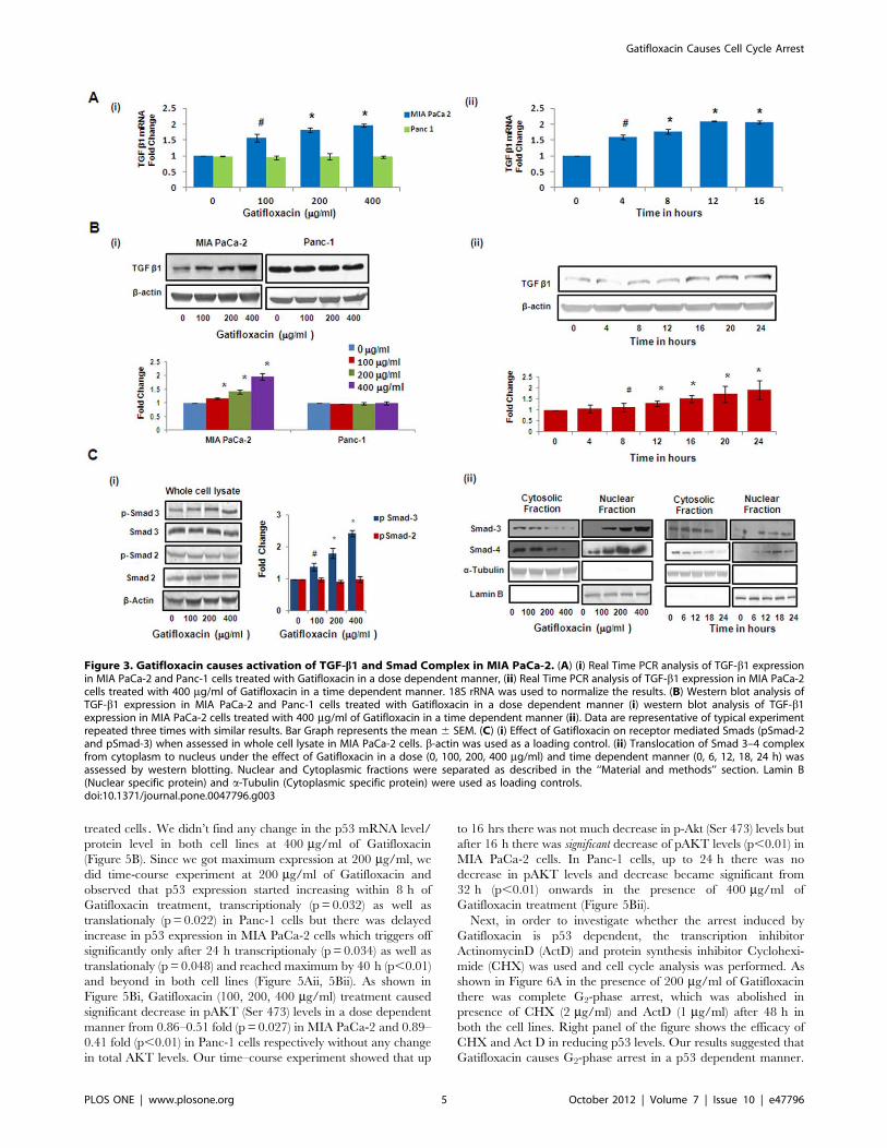
Gatifloxacin causes Activation of TGF-b1/SMAD Complex/p21 in MIA PaCa-2 and Activation of p27 in Panc-1Recent studies have shown that one of the fluoroquinolone,
ciprofloxacin suppresses colon cancer cell proliferation by arrest-
ing them in S-phase through TGF-b1 and TGF-b1 is also very
well known to cause cell cycle arrest in pancreatic cancer cells
[21;22]. To investigate whether Gatifloxacin-induced S-phase arrest
was TGF-b dependent we checked the levels of TGF-b1 at the
transcriptional as well as translational levels. We found Gatiflox-
acin treatment (100, 200, 400 mg/ml) to increase TGF-b1expression in a dose dependent manner by 1.5–2 fold (p,0.01)
at the transcriptional level and 1.3–2 fold (p,0.01) at translational
level after 48 h in MIA PaCa-2 cell but not in Panc-1 cells
(Figure 3Ai, 3Bi). Since we found significant increase in TGF-b1expression level at 400 mg/ml, we did the time-course study and
observed that TGF-b1 started increasing transcriptionally within
4 h (p= 0.023) of treatment and reached maximum by 12 h
(p,0.01) and then plateaued at 16 h (Figure 3Aii). However
translationally, TGF-b1 gets activated within 8 h (p = 0.039) only
and reached to its maximum expression by 16 h (p,0.01)
(Figure 3Bii), as assessed by real time PCR and western blotting.
There is increasing evidence that Smad transcription factors
mediate the growth inhibitory effect of transforming growth factor-
b1 (TGF-b1) in many cell types [23]. To investigate the relative
contribution of individual Smad proteins to TGF-b1 signaling,
protein expression of Smad2 and Smad3 was analysed by western
blotting of cell extracts after 48 h of Gatifloxacin treatment (100,
200, 400 mg/ml) in MIA PaCa-2 cells. As shown in Figure 3Ci, we
observed significant increase in phospho-Smad3 (pSmad3 at Ser
423) whereas there was no change in the levels of phospho-Smad2
(pSmad2 at Ser 467) after 48 h of Gatifloxacin treatment at all doses
in MIA PaCa-2 cells. Furthermore, the increase in phospho-Smad3
(pSmad3) was found to be in a dose dependent manner (1.3–2.4
fold, p = 0.0127) as compared to vehicle treated cells. Enough
evidence is there to support that activated Smad2 or Smad3
dimerizes with Smad4 and translocates to the nucleus, whereby
the activated complexes associate with Smad-binding element
(SBEs) in promoters of numerous genes leading to their induction
[24–26]. p21Waf1/Cip1 is one such gene which is known to be
involved in the TGF-b1 mediated regulation of cell proliferation
[27]. To determine whether Smad3 translocates in response to
TGF-b1 stimulation in MIA PaCa-2 cells, we assessed the
subcellular distribution of the Smad3/4 and as expected,
significant decrease in the Smad-3 and Smad-4 protein expression
was observed in the cytosolic fraction and there was concomitant
increase in the Smad-3 and Smad-4 protein expression in nuclear
fraction in Gatifloxacin treated MIA PaCa-2 cells in a dose and
time dependent manner (Figure 3Cii). Simultaneously we also
analysed the expression of p21Waf1/Cip1, the downstream effector
of TGF-b1 which is known to be a crucial regulator of cell cycle
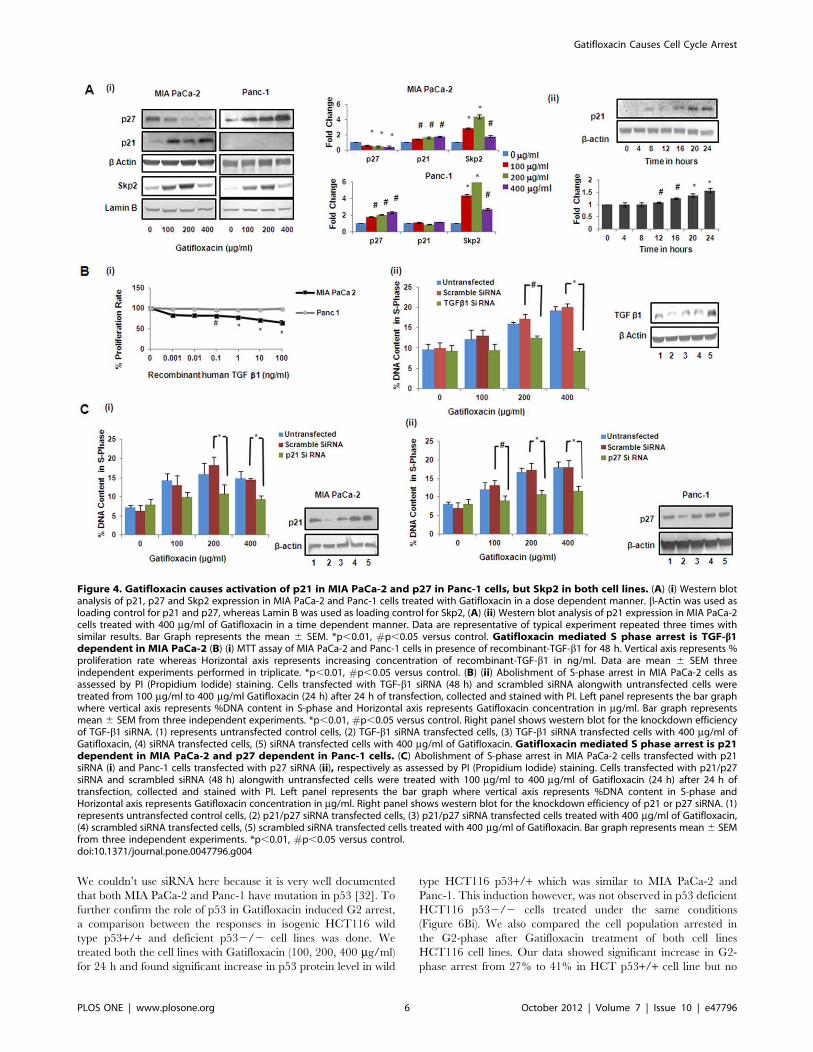
progression. Like p21, p27 is a cyclin dependent kinase inhibitor
that regulates G1-S phase of cell cycle progression, hence we
checked the expression of p21 and p27 by western blot analysis
[28]. We observed significant increase (1.4–1.7 fold, p = 0.038) in
the levels of p21 and significant decrease (0.57–0.35 fold,
p = 0.012) in the levels of p27 in a dose dependent manner after
48 h of Gatifloxacin treatment as compared to vehicle treated cells
(Figure 4Ai). Interestingly increase in p21 was observed only after
Gatifloxacin Causes Cell Cycle Arrest
PLOS ONE | www.plosone.org 2 October 2012 | Volume 7 | Issue 10 | e47796
TGF-b1 activation i.e after 12 h (p = 0.035), in MIA PaCa-2 cells
(Figure 4Aii). However in case of Panc-1 cells that lacks functional
TGF-b1 we didn’t find activation of p21, but we do find the
activation of p27. We observed significant increase (1.8–2.3 fold,
p = 0.021) in the levels of p27 in a dose dependent manner after
48 h of Gatifloxacin treatment as compared to vehicle treated cells
(Figure 4Ai). Simultaneously we also checked the levels of Skp2
which mediates ubiquitination and degradation of p27/p21 in
both the cell lines MIA PaCa-2 and Panc-1 in response to
Gatifloxacin treatment (0, 100, 200 and 400 mg/ml) [29]. We
found significant increase (2.7–4.3 fold, p= .008 in MIA PaCa-2
cells and 4.3–6.3 fold p= 0.005 in Panc-1) in Skp2 levels in both
the cell lines. As expected we found inverse correlation between
Skp2 and p27/p21 (Figure 4Ai). Taken together this study
indicates that TGF-b1/Smad3/p21, is one of the pathways that
leads to S-phase arrest in MIA PaCa-2 cells and p27 playing
crucial role in S-phase arrest of Panc-1 cells.
Gatifloxacin Mediated S-phase Arrest is TGF-b1/p21Dependent in MIA PaCa-2 and p27 Dependent in Panc-1CellsTo confirm the role of TGF-b1 in suppressing the proliferation,
recombinant TGF-b1 was transfected at varied doses (0–100 ng/
ml) in MIA PaCa-2 and Panc-1 cells for 48h and MTT assay was
performed. We observed that over expression of TGF-b1significantly suppressed MIA PaCa-2 cell proliferation in a dose
dependent manner. We observed significant decrease (35%,
p,0.05, Figure 4Bi) in cell proliferation at a concentration of
Figure 1. Gatifloxacin inhibits proliferation of cultured pancreatic cancer cells without induction of Apoptosis.MTT assay of MIA PaCa-2 and Panc-1 cells after treatment with Gatifloxacin (0–400 mg/ml) for 24 h (Ai) and 48 h (Aii). Cells were seeded in 96 well plates (16104 cells/well)which were allowed to adhere overnight and were subsequently treated with increasing concentration of Gatifloxacin for 24 and 48 h. Vertical axisrepresents % proliferation rate whereas Horizontal axis represents increasing concentration of Gatifloxacin in mg/ml. Data are mean 6 SEM threeindependent experiments performed in triplicate. * p,0.01 compared to vehicle control. (B) Annexin V-PE binding in MIA PaCa-2 (i-v) and Panc-1 (vi-x) after treatment with Gatifloxacin for 48 h as evaluated by 7-AAD and AnnexinV staining. (i) and (vi) Vehicle treated control cells, (ii) and (vii) cellstreated with 100 mg/ml, (iii) and (viii) cells treated with 200 mg/ml, (iv) and (ix) cells treated with 400 mg/ml of Gatifloxacin. (v) MIA PaCa-2 cells and (x)Panc-1 cells treated with curcumin 60 mM for 24 h as positive control. Vertical axis represents 7-AAD positive cells whereas horizontal axis representsAnnexin V-PE positive cells. Representative of three independent experiments has been shown with similar results. (C) Western blot analysis of theexpression of Bax protein under the effect of Gatifloxacin in a time (24, 48 h) and dose (0, 100, 200, 400 mg/ml) dependent manner. b-Actin was usedas a loading control. (D) Caspase 3, 8, 9 activity of MIA PaCa-2 and Panc-1 cells treated with Gatifloxacin in time (24, 48 h) and dose (0, 100, 200,400 mg/ml) dependent manner. Bar graph represents mean 6 SEM from three independent experiments.doi:10.1371/journal.pone.0047796.g001
Gatifloxacin Causes Cell Cycle Arrest
PLOS ONE | www.plosone.org 3 October 2012 | Volume 7 | Issue 10 | e47796
100 ng/ml in MIA PaCa-2 cells but not in Panc-1 cells. Our
findings are consistent to the findings of Francisco j nicolas et al
which suggests Panc-1 to be resistant to TGF-b1 induced growth
arrest. Further to confirm the role of TGF-b1 in mediating S-
phase arrest induced by Gatifloxacin in MIA PaCa-2, we silenced
the TGF-b1 expression by using siRNA and did cell cycle analysis.
As shown in Figure 4Bii, S-Phase arrest was totally abolished at all
the three doses of Gatifloxacin used in TGF-b1 siRNA transfected
cells as compared to scrambled siRNA transfected cells or
untransfected cells. Accordingly, we next sought to determine
the role of p21 and p27 on S-phase arrest in TGF-b1 sensitive
MIA PaCa-2 or resistant Panc-1 cells respectively. We found that
p21 siRNA transfected MIA PaCa-2 (Figure 4Ci) and p27 siRNA
transfected Panc-1 cells (Figure 4Cii) strongly inhibits the
Gatifloxacin induced S-phase arrest as compared to scrambled
siRNA at all the doses. siRNA mediated knock down of TGF-b1,p21 and p27 was also confirmed using western blot analysis
(Figure 4Bii, Ci and Cii). siRNA mediated knockdown results
suggest that S phase arrest of Gatifloxacin is TGF-b1/p21dependent in MIA PaCa-2 and p27 dependent in Panc-1 cells.
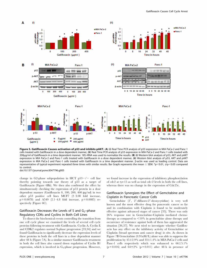
Gatifloxacin Inhibits pAKT and causes G2–phase Arrest inp53 Dependent Manner in Both Cell LinesVarious studies have shown that p53 can activate the expression
of the p21 which then inhibits cyclin dependent kinases and leads
to cell cycle arrest [30]. Similarly pAKT has also been shown to be
a critical regulator of cell survival and cell cycle progression apart
from negatively regulating p53 [31]. Hence, we next checked the
expression of p53, total AKT and pAKT in presence or absence of
varied doses of Gatifloxacin. Figure 5A and 5B showed activation
of p53 at both transcriptional and translational levels. We found
Gatifloxacin treatment at 100 and 200 mg/ml dose increases p53
expression by 1.4–2 fold (p,0.01) transcriptionaly and 1.8–2.45
fold (p,0.01) translationally in MIA PaCa-2 cells and 1.75–2.5
fold (p,0.013) transcriptionaly and 1.4–2.0 fold translationally in
Panc-1 cells after 48 h (Figure 5Ai, 5Bi) as compared to vehicle
Figure 2. Gatifloxacin induces S and G2 phase cell cycle arrest in pancreatic cancer cells. Effects of Gatifloxacin on cell cycle wereinvestigated using PI (Propidium Iodide) staining. Cells were treated with (0–400 mg/ml) Gatifloxacin for 24 and 48 h, collected and stained with PI.Here Pink peak represents G1-phase, Green peak represents S-phase and Blue peak represents G2-phase respectively. Upper panel showsrepresentative of three independent experiments with similar results and lower panel represents the bar diagram of cells in different phases. Bargraph represents mean 6 SEM from three independent experiments. (i) Representative bar graph for MIA PaCa-2, (ii) Representative bar graph forPanc-1.doi:10.1371/journal.pone.0047796.g002
Gatifloxacin Causes Cell Cycle Arrest
PLOS ONE | www.plosone.org 4 October 2012 | Volume 7 | Issue 10 | e47796
treated cells. We didn’t find any change in the p53 mRNA level/
protein level in both cell lines at 400 mg/ml of Gatifloxacin
(Figure 5B). Since we got maximum expression at 200 mg/ml, we
did time-course experiment at 200 mg/ml of Gatifloxacin and
observed that p53 expression started increasing within 8 h of
Gatifloxacin treatment, transcriptionaly (p = 0.032) as well as
translationaly (p = 0.022) in Panc-1 cells but there was delayed
increase in p53 expression in MIA PaCa-2 cells which triggers off
significantly only after 24 h transcriptionaly (p = 0.034) as well as
translationaly (p = 0.048) and reached maximum by 40 h (p,0.01)
and beyond in both cell lines (Figure 5Aii, 5Bii). As shown in
Figure 5Bi, Gatifloxacin (100, 200, 400 mg/ml) treatment caused
significant decrease in pAKT (Ser 473) levels in a dose dependent
manner from 0.86–0.51 fold (p = 0.027) in MIA PaCa-2 and 0.89–
0.41 fold (p,0.01) in Panc-1 cells respectively without any change
in total AKT levels. Our time–course experiment showed that up
to 16 hrs there was not much decrease in p-Akt (Ser 473) levels but
after 16 h there was significant decrease of pAKT levels (p,0.01) in
MIA PaCa-2 cells. In Panc-1 cells, up to 24 h there was no
decrease in pAKT levels and decrease became significant from
32 h (p,0.01) onwards in the presence of 400 mg/ml of
Gatifloxacin treatment (Figure 5Bii).
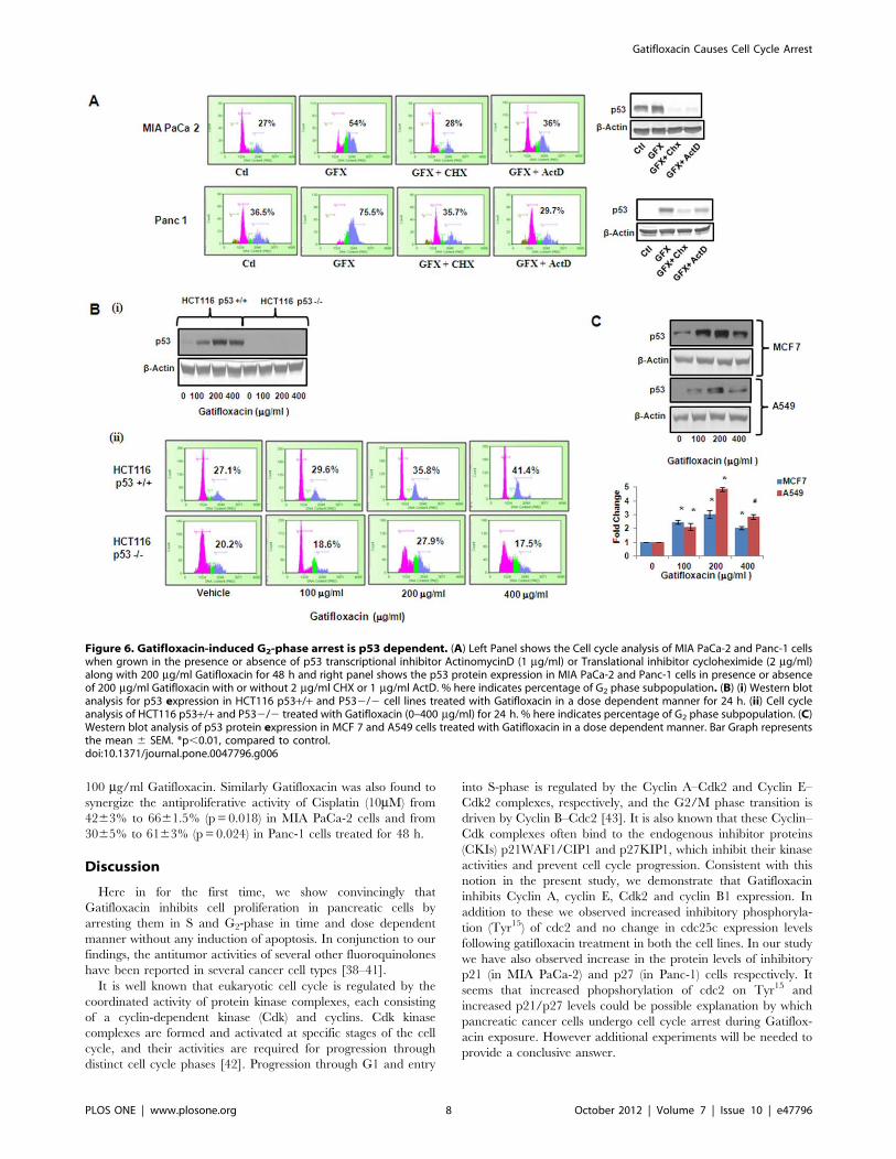
Next, in order to investigate whether the arrest induced by
Gatifloxacin is p53 dependent, the transcription inhibitor
ActinomycinD (ActD) and protein synthesis inhibitor Cyclohexi-
mide (CHX) was used and cell cycle analysis was performed. As
shown in Figure 6A in the presence of 200 mg/ml of Gatifloxacin
there was complete G2-phase arrest, which was abolished in
presence of CHX (2 mg/ml) and ActD (1 mg/ml) after 48 h in
both the cell lines. Right panel of the figure shows the efficacy of
CHX and Act D in reducing p53 levels. Our results suggested that
Gatifloxacin causes G2-phase arrest in a p53 dependent manner.
Figure 3. Gatifloxacin causes activation of TGF-b1 and Smad Complex in MIA PaCa-2. (A) (i) Real Time PCR analysis of TGF-b1 expressionin MIA PaCa-2 and Panc-1 cells treated with Gatifloxacin in a dose dependent manner, (ii) Real Time PCR analysis of TGF-b1 expression in MIA PaCa-2cells treated with 400 mg/ml of Gatifloxacin in a time dependent manner. 18S rRNA was used to normalize the results. (B) Western blot analysis ofTGF-b1 expression in MIA PaCa-2 and Panc-1 cells treated with Gatifloxacin in a dose dependent manner (i) western blot analysis of TGF-b1expression in MIA PaCa-2 cells treated with 400 mg/ml of Gatifloxacin in a time dependent manner (ii). Data are representative of typical experimentrepeated three times with similar results. Bar Graph represents the mean 6 SEM. (C) (i) Effect of Gatifloxacin on receptor mediated Smads (pSmad-2and pSmad-3) when assessed in whole cell lysate in MIA PaCa-2 cells. b-actin was used as a loading control. (ii) Translocation of Smad 3–4 complexfrom cytoplasm to nucleus under the effect of Gatifloxacin in a dose (0, 100, 200, 400 mg/ml) and time dependent manner (0, 6, 12, 18, 24 h) wasassessed by western blotting. Nuclear and Cytoplasmic fractions were separated as described in the ‘‘Material and methods’’ section. Lamin B(Nuclear specific protein) and a-Tubulin (Cytoplasmic specific protein) were used as loading controls.doi:10.1371/journal.pone.0047796.g003
Gatifloxacin Causes Cell Cycle Arrest
PLOS ONE | www.plosone.org 5 October 2012 | Volume 7 | Issue 10 | e47796
We couldn’t use siRNA here because it is very well documented
that both MIA PaCa-2 and Panc-1 have mutation in p53 [32]. To
further confirm the role of p53 in Gatifloxacin induced G2 arrest,
a comparison between the responses in isogenic HCT116 wild
type p53+/+ and deficient p532/2 cell lines was done. We
treated both the cell lines with Gatifloxacin (100, 200, 400 mg/ml)
for 24 h and found significant increase in p53 protein level in wild
type HCT116 p53+/+ which was similar to MIA PaCa-2 and
Panc-1. This induction however, was not observed in p53 deficient
HCT116 p532/2 cells treated under the same conditions
(Figure 6Bi). We also compared the cell population arrested in
the G2-phase after Gatifloxacin treatment of both cell lines
HCT116 cell lines. Our data showed significant increase in G2-
phase arrest from 27% to 41% in HCT p53+/+ cell line but no
Figure 4. Gatifloxacin causes activation of p21 in MIA PaCa-2 and p27 in Panc-1 cells, but Skp2 in both cell lines. (A) (i) Western blotanalysis of p21, p27 and Skp2 expression in MIA PaCa-2 and Panc-1 cells treated with Gatifloxacin in a dose dependent manner. b-Actin was used asloading control for p21 and p27, whereas Lamin B was used as loading control for Skp2, (A) (ii) Western blot analysis of p21 expression in MIA PaCa-2cells treated with 400 mg/ml of Gatifloxacin in a time dependent manner. Data are representative of typical experiment repeated three times withsimilar results. Bar Graph represents the mean 6 SEM. *p,0.01, #p,0.05 versus control. Gatifloxacin mediated S phase arrest is TGF-b1dependent in MIA PaCa-2 (B) (i) MTT assay of MIA PaCa-2 and Panc-1 cells in presence of recombinant-TGF-b1 for 48 h. Vertical axis represents %proliferation rate whereas Horizontal axis represents increasing concentration of recombinant-TGF-b1 in ng/ml. Data are mean 6 SEM threeindependent experiments performed in triplicate. *p,0.01, #p,0.05 versus control. (B) (ii) Abolishment of S-phase arrest in MIA PaCa-2 cells asassessed by PI (Propidium Iodide) staining. Cells transfected with TGF-b1 siRNA (48 h) and scrambled siRNA alongwith untransfected cells weretreated from 100 mg/ml to 400 mg/ml Gatifloxacin (24 h) after 24 h of transfection, collected and stained with PI. Left panel represents the bar graphwhere vertical axis represents %DNA content in S-phase and Horizontal axis represents Gatifloxacin concentration in mg/ml. Bar graph representsmean 6 SEM from three independent experiments. *p,0.01, #p,0.05 versus control. Right panel shows western blot for the knockdown efficiencyof TGF-b1 siRNA. (1) represents untransfected control cells, (2) TGF-b1 siRNA transfected cells, (3) TGF-b1 siRNA transfected cells with 400 mg/ml ofGatifloxacin, (4) siRNA transfected cells, (5) siRNA transfected cells with 400 mg/ml of Gatifloxacin. Gatifloxacin mediated S phase arrest is p21dependent in MIA PaCa-2 and p27 dependent in Panc-1 cells. (C) Abolishment of S-phase arrest in MIA PaCa-2 cells transfected with p21siRNA (i) and Panc-1 cells transfected with p27 siRNA (ii), respectively as assessed by PI (Propidium Iodide) staining. Cells transfected with p21/p27siRNA and scrambled siRNA (48 h) alongwith untransfected cells were treated with 100 mg/ml to 400 mg/ml of Gatifloxacin (24 h) after 24 h oftransfection, collected and stained with PI. Left panel represents the bar graph where vertical axis represents %DNA content in S-phase andHorizontal axis represents Gatifloxacin concentration in mg/ml. Right panel shows western blot for the knockdown efficiency of p21 or p27 siRNA. (1)represents untransfected control cells, (2) p21/p27 siRNA transfected cells, (3) p21/p27 siRNA transfected cells treated with 400 mg/ml of Gatifloxacin,(4) scrambled siRNA transfected cells, (5) scrambled siRNA transfected cells treated with 400 mg/ml of Gatifloxacin. Bar graph represents mean6 SEMfrom three independent experiments. *p,0.01, #p,0.05 versus control.doi:10.1371/journal.pone.0047796.g004
Gatifloxacin Causes Cell Cycle Arrest
PLOS ONE | www.plosone.org 6 October 2012 | Volume 7 | Issue 10 | e47796
change in G2-phase subpopulation in HCT p532/2 cell line
thereby pointing towards our theory of p53 as a target of
Gatifloxacin (Figure 6Bii). We then also confirmed the effect by
simultaneously checking the expression of p53 protein in a dose
dependent manner (Gatifloxacin 0, 100, 200, 400 mg/ml) in two
other p53 positive cell lines MCF7 (2–3.08 fold increase,
p = 0.0078) and A549 (2.1–4.8 fold increase, p = 0.0085) re-
spectively (Figure 6C).
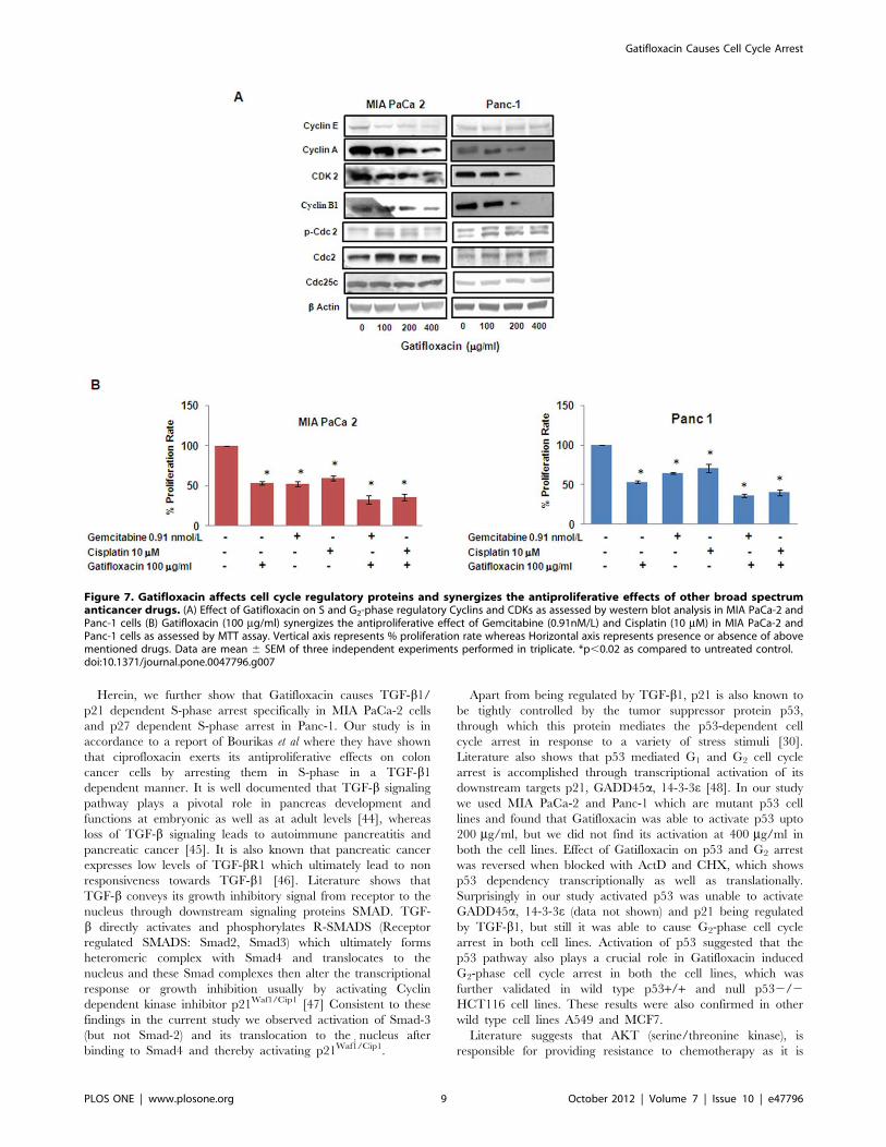
Gatifloxacin Decreases the Levels of S and G2–phaseRegulatory CDKs and Cyclins in Both Cell LinesTo dissect the biochemical events controlling the transition from
one cell cycle phase we examined the levels of several cell cycle
proteins following treatment with Gatifloxacin. Cyclin A, Cyclin E
and CDK2 regulates normal S-phase progression [33;34] and we
found Gatifloxacin to significantly decrease the expression levels of
these proteins in both the cell lines in a dose dependent manner
after 48 h (Figure 7A). In a similar manner Gatifloxacin treatment
in both the cell lines also caused down regulation of Cyclin B1
expression, which is involved in G2-phase progression. However,
we found increase in the expression of inhibitory phosphorylation
of cdc2 at tyr-15 as well as total cdc-2 levels in both the cell lines,
whereas there was no change in the expression of Cdc25c.
Gatifloxacin Synergizes the Effect of Gemcitabine andCisplatin in Pancreatic Cancer CellsGemcitabine (29, 29-difluoro-29-deoxycytidine) is very well
known and the most effective drug for pancreatic cancer so far
and its combination with Cisplatin is found to be moderately
effective against advanced stages of cancer [35]. There was only
26% response rate in Gemcitabine-Cisplatin mediated chemo-
therapy as compared to ,10% in gemcitabine alone therapy and
occurrence of resistance against both of them has aggravated the
situation [36,37]. We next tried to investigate whether Gatiflox-
acin has any effect on the inhibitory activity of Gemcitabine or
Cisplatin (broad spectrum anti cancer drug) in vitro. As shown in
Figure 7B Gemcitabine (0.91nM/L) alone was able to suppress the
proliferation by 4562.9% and 3361.4% in MIA PaCa-2 cells and
Panc-1 cells respectively which was enhanced to 6665.1%
(p= 0.016) and 6462% (p= 0.011) after 48 h in presence of
Figure 5. Gatifloxacin Causes activation of p53 and inhibits pAKT. (A) (i) Real Time PCR analysis of p53 expression in MIA PaCa-2 and Panc-1cells treated with Gatifloxacin in a dose dependent manner, (ii) Real Time PCR analysis of p53 expression in MIA PaCa-2 and Panc-1 cells treated with200mg/ml of Gatifloxacin in a time dependent manner. 18S rRNA was used to normalize the results. (B) (i) Western blot analysis of p53, AKT and pAKTexpression in MIA PaCa-2 and Panc-1 cells treated with Gatifloxacin in a dose dependent manner, (ii) Western blot analysis of p53, AKT and pAKTexpression in MIA PaCa-2 and Panc-1 cells treated with Gatifloxacin in a time dependent manner. b-actin was used as loading control. Data arerepresentative of typical experiment repeated three times with similar results. Bar Graph represents the mean 6 SEM. *p,0.01, #p,0.05 comparedto control.doi:10.1371/journal.pone.0047796.g005
Gatifloxacin Causes Cell Cycle Arrest
PLOS ONE | www.plosone.org 7 October 2012 | Volume 7 | Issue 10 | e47796
100 mg/ml Gatifloxacin. Similarly Gatifloxacin was also found to
synergize the antiproliferative activity of Cisplatin (10mM) from
4263% to 6661.5% (p= 0.018) in MIA PaCa-2 cells and from
3065% to 6163% (p= 0.024) in Panc-1 cells treated for 48 h.
Discussion
Here in for the first time, we show convincingly that
Gatifloxacin inhibits cell proliferation in pancreatic cells by
arresting them in S and G2-phase in time and dose dependent
manner without any induction of apoptosis. In conjunction to our
findings, the antitumor activities of several other fluoroquinolones
have been reported in several cancer cell types [38–41].
It is well known that eukaryotic cell cycle is regulated by the
coordinated activity of protein kinase complexes, each consisting
of a cyclin-dependent kinase (Cdk) and cyclins. Cdk kinase
complexes are formed and activated at specific stages of the cell
cycle, and their activities are required for progression through
distinct cell cycle phases [42]. Progression through G1 and entry
into S-phase is regulated by the Cyclin A–Cdk2 and Cyclin E–
Cdk2 complexes, respectively, and the G2/M phase transition is
driven by Cyclin B–Cdc2 [43]. It is also known that these Cyclin–
Cdk complexes often bind to the endogenous inhibitor proteins
(CKIs) p21WAF1/CIP1 and p27KIP1, which inhibit their kinase
activities and prevent cell cycle progression. Consistent with this
notion in the present study, we demonstrate that Gatifloxacin
inhibits Cyclin A, cyclin E, Cdk2 and cyclin B1 expression. In
addition to these we observed increased inhibitory phosphoryla-
tion (Tyr15) of cdc2 and no change in cdc25c expression levels
following gatifloxacin treatment in both the cell lines. In our study
we have also observed increase in the protein levels of inhibitory
p21 (in MIA PaCa-2) and p27 (in Panc-1) cells respectively. It
seems that increased phopshorylation of cdc2 on Tyr15 and
increased p21/p27 levels could be possible explanation by which
pancreatic cancer cells undergo cell cycle arrest during Gatiflox-
acin exposure. However additional experiments will be needed to
provide a conclusive answer.
Figure 6. Gatifloxacin-induced G2-phase arrest is p53 dependent. (A) Left Panel shows the Cell cycle analysis of MIA PaCa-2 and Panc-1 cellswhen grown in the presence or absence of p53 transcriptional inhibitor ActinomycinD (1 mg/ml) or Translational inhibitor cycloheximide (2 mg/ml)along with 200 mg/ml Gatifloxacin for 48 h and right panel shows the p53 protein expression in MIA PaCa-2 and Panc-1 cells in presence or absenceof 200 mg/ml Gatifloxacin with or without 2 mg/ml CHX or 1 mg/ml ActD. % here indicates percentage of G2 phase subpopulation. (B) (i) Western blotanalysis for p53 expression in HCT116 p53+/+ and P532/2 cell lines treated with Gatifloxacin in a dose dependent manner for 24 h. (ii) Cell cycleanalysis of HCT116 p53+/+ and P532/2 treated with Gatifloxacin (0–400 mg/ml) for 24 h. % here indicates percentage of G2 phase subpopulation. (C)Western blot analysis of p53 protein expression in MCF 7 and A549 cells treated with Gatifloxacin in a dose dependent manner. Bar Graph representsthe mean 6 SEM. *p,0.01, compared to control.doi:10.1371/journal.pone.0047796.g006
Gatifloxacin Causes Cell Cycle Arrest
PLOS ONE | www.plosone.org 8 October 2012 | Volume 7 | Issue 10 | e47796
Herein, we further show that Gatifloxacin causes TGF-b1/p21 dependent S-phase arrest specifically in MIA PaCa-2 cells
and p27 dependent S-phase arrest in Panc-1. Our study is in
accordance to a report of Bourikas et al where they have shown
that ciprofloxacin exerts its antiproliferative effects on colon
cancer cells by arresting them in S-phase in a TGF-b1dependent manner. It is well documented that TGF-b signaling
pathway plays a pivotal role in pancreas development and
functions at embryonic as well as at adult levels [44], whereas
loss of TGF-b signaling leads to autoimmune pancreatitis and
pancreatic cancer [45]. It is also known that pancreatic cancer
expresses low levels of TGF-bR1 which ultimately lead to non
responsiveness towards TGF-b1 [46]. Literature shows that
TGF-b conveys its growth inhibitory signal from receptor to the
nucleus through downstream signaling proteins SMAD. TGF-
b directly activates and phosphorylates R-SMADS (Receptor
regulated SMADS: Smad2, Smad3) which ultimately forms
heteromeric complex with Smad4 and translocates to the
nucleus and these Smad complexes then alter the transcriptional
response or growth inhibition usually by activating Cyclin
dependent kinase inhibitor p21Waf1/Cip1 [47] Consistent to these
findings in the current study we observed activation of Smad-3
(but not Smad-2) and its translocation to the nucleus after
binding to Smad4 and thereby activating p21Waf1`/Cip1.
Apart from being regulated by TGF-b1, p21 is also known to
be tightly controlled by the tumor suppressor protein p53,
through which this protein mediates the p53-dependent cell
cycle arrest in response to a variety of stress stimuli [30].
Literature also shows that p53 mediated G1 and G2 cell cycle
arrest is accomplished through transcriptional activation of its
downstream targets p21, GADD45a, 14-3-3e [48]. In our study
we used MIA PaCa-2 and Panc-1 which are mutant p53 cell
lines and found that Gatifloxacin was able to activate p53 upto
200 mg/ml, but we did not find its activation at 400 mg/ml in
both the cell lines. Effect of Gatifloxacin on p53 and G2 arrest
was reversed when blocked with ActD and CHX, which shows
p53 dependency transcriptionally as well as translationally.
Surprisingly in our study activated p53 was unable to activate
GADD45a, 14-3-3e (data not shown) and p21 being regulated
by TGF-b1, but still it was able to cause G2-phase cell cycle
arrest in both cell lines. Activation of p53 suggested that the
p53 pathway also plays a crucial role in Gatifloxacin induced
G2-phase cell cycle arrest in both the cell lines, which was
further validated in wild type p53+/+ and null p532/2
HCT116 cell lines. These results were also confirmed in other
wild type cell lines A549 and MCF7.
Literature suggests that AKT (serine/threonine kinase), is
responsible for providing resistance to chemotherapy as it is
Figure 7. Gatifloxacin affects cell cycle regulatory proteins and synergizes the antiproliferative effects of other broad spectrumanticancer drugs. (A) Effect of Gatifloxacin on S and G2-phase regulatory Cyclins and CDKs as assessed by western blot analysis in MIA PaCa-2 andPanc-1 cells (B) Gatifloxacin (100 mg/ml) synergizes the antiproliferative effect of Gemcitabine (0.91nM/L) and Cisplatin (10 mM) in MIA PaCa-2 andPanc-1 cells as assessed by MTT assay. Vertical axis represents % proliferation rate whereas Horizontal axis represents presence or absence of abovementioned drugs. Data are mean 6 SEM of three independent experiments performed in triplicate. *p,0.02 as compared to untreated control.doi:10.1371/journal.pone.0047796.g007
Gatifloxacin Causes Cell Cycle Arrest
PLOS ONE | www.plosone.org 9 October 2012 | Volume 7 | Issue 10 | e47796
found to be hyperactive in pancreatic cancer and reports have
shown that inhibition of AKT signaling increases the chemo-
sensitisation of drugs [49]. Our data clearly shows that
Gatifloxacin not only downregulates AKT but also synergizes
the effect of Gemcitabine and Cisplatin in Pancreatic Cancer
cells. According to above results it was tempting to speculate
a model for the action of Gatifloxacin in pancreatic cancer cells
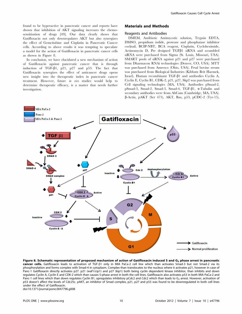
as shown in Figure 8.
In conclusion, we have elucidated a new mechanism of action
of Gatifloxacin against pancreatic cancer that is through
induction of TGF-b1, p21, p27 and p53. The fact that
Gatifloxacin synergizes the effect of anticancer drugs opens
new insight into the therapeutic index in pancreatic cancer
treatment. However, future in vivo studies would help to
determine therapeutic efficacy, is a matter that needs further
investigation.
Materials and Methods
Reagents and AntibodiesDMEM, Antibiotic Antimycotic solution, Trypsin EDTA,
DMSO, propidium iodide, protease and phosphatase inhibitor
cocktail, BCIP-NBT, BCA reagent, Cisplatin, Cycloheximide,
Actinomycin D, Pre designed TGFb1 siRNA and scrambled
siRNA were purchased from Sigma (St. Louis, Missouri, USA).
SMART pools of siRNA against p21 and p27 were purchased
from Dharmacon RNAi technologies (Denver, CO, USA). MTT
was purchased from Amresco (Ohio, USA). Fetal bovine serum
was purchased from Biological Industries (Kibbutz Beit Haemek,
Israel). Human recombinant TGF-b1 and antibodies Cyclin A,
Cyclin E, Cyclin B1, CDK-2, p21, p27, Skp2 was purchased from
Cell signaling technologies (MA, USA). Antibodies pSmad-2,
pSmad-3, Smad-2, Smad-3, Smad-4, TGF-b1, a-Tubulin and
secondary antibodies were from AbCam (Cambridge, MA, USA).
b-Actin, pAKT (Ser 473), AKT, Bax, p53, pCDC-2 (Tyr-15),
Figure 8. Schematic representation of proposed mechanism of action of Gatifloxacin induced S and G2 phase arrest in pancreaticcancer cells. Gatifloxacin leads to activation of TGF-b1 only in MIA PaCa-2 cell line which than activates Smad-3 but not Smad-2 via itsphosphorylation and forms complex with Smad-4 in cytoplasm. Complex than translocates to the nucleus where it activates p21, however in case ofPanc-1 Gatifloxacin directly activates p27. p21 (waf1/cip1) and p27 (kip1) both being cyclin dependent kinase inhibitor, than inhibits and downregulates Cyclin A, Cyclin E and CDK-2 which than causes S-phase arrest in both the cell lines. Gatifloxacin also activates p53 in both MIA PaCa-2 andPanc-1 cell lines which than down regulates Cyclin B1, upregulates inhibitory pCdc2 and Cdc2 which than leads to G2 arrest. However, activation ofp53 doesn’t affect the levels of Cdc25c. pAKT, an inhibitor of Smad complex, p21, p27 and p53 was found to be downregulated in both cell linesunder the effect of Gatifloxacin.doi:10.1371/journal.pone.0047796.g008
Gatifloxacin Causes Cell Cycle Arrest
PLOS ONE | www.plosone.org 10 October 2012 | Volume 7 | Issue 10 | e47796
CDC-2, CDC25c and Lamin-B antibodies were purchased from
Santacruz biotechnology (Santa Cruz, CA, USA). Caspase 3,8,9
activity kit was purchased from G-Biosciences (St Louis, MO,
USA). Gatifloxacin was obtained from Cipla (India) and
Gemcitabine was obtained from Eli Lily (India).
Cell Culture and TransfectionMIA PaCa-2 and Panc-1 cells were obtained from National
Center for Cell Science, Pune, India and maintained in DMEM
medium containing 10%(v/v) FBS, 100 units/ml penicillin,
100 mg/mlstreptomycin, 0.25 mg/ml, amphotericin B in a humid-
ified 5% CO2 atmosphere. Cells growing in logarithmic phase
were used in all experiments. Overnight serum starved cells were
used to study TGF -b1, p53 and pAKT expression. Synchronized
and growth arrested cultures were than subjected to Gatifloxacin
(0–400 mg/ml) treatment in complete media for 24h and 48h
respectively. Wherever indicated siRNA transfections were done
in 6 well plate using lipofectamine 2000 (Invitrogen, CA, USA)
according to manufacturer’s protocol. Cells were treated with
Gatifloxacin, 24 h post transfection and were harvested after
trypsinization for cell cycle experiment.
Cell Viability AssayCell viability assay was performed using MTT [3-(4,5-dimethyl
thiazol-2yl)-2,5-diphenyltetrazolium bromide]. 10,000 cells per
well were seeded in 96 well plates and treated with different
concentrations (0–400 mg/ml) of Gatifloxacin in triplicates. As
controls, Dextrose 5% (w/v) treated cells (Vehicle) were included
in each experiments. Following treatments for 24h and 48 h,
10 mL of MTT (5 mg/ml) was added to each well and incubated
for 3 h at 37uC in dark. Formazan crystals formed were dissolved
in 100ml DMSO and the absorbance was measured at 570 nM
using an ELISA reader. Cell viability was calculated as reported
earlier [50].
Flow Cytometric Analysis and Caspase Activity forApoptosis and Cell Cycle AnalysisApoptosis was assessed using Guava Nexin kit and Guava PCA
system according to the manufacturer’s protocol (Guava Tech-
nologies, Hayward, California, USA). Annexin-PE fluorescence
was analyzed by cytosoft software (Guava Technologies, Hayward.
California, USA). For analysis of cell cycle distribution, 1610 6
cells were harvested by centrifugation, washed in phosphate-buffer
saline (PBS), fixed with ice cold 70% ethanol and treated with
1 mg/ml RNAse for 30 min. Intracellular DNA was labeled with
propidium iodide (50 mg/ml) and incubated at 4uC in dark.
Samples were than analyzed using flow cytometer (Guava
Technologies, Hayward, California, USA) and cytosoft software
(Guava Technologies, Hayward, California, USA). A minimum of
5,000 events were counted [51]. Caspase activity was measured
using G-Biosciences Caspase 3, 8, 9 kit according to manufac-
turer’s protocol.
Real Time PCRTotal RNA was extracted using Trizol reagent (Invitrogen, CA,
USA). Reverse transcription was carried out using M-MuLV
reverse trancriptase (MBI Fermentas, USA) according to the
manufacturer’s protocol using 1 mg of total RNA. Real Time PCR
for TGF-b1 and p53 was performed using SYBR Green PCR
master mix (Applied Biosystems, Foster city, CA, USA) in an ABI
Prism 7000 sequence detection system (Applied Biosystems), and
amplification were performed in triplicate and repeated thrice.
Results were normalized with 18S rRNA and analysis was done
using pfaffl’s method [52]. Primers sequences used for real time
PCR were as follows: TGF-b1 FP: 59GCCCTGGACACCAAC-
TATTG3’ RP: 59CGTGTCCAGGCTCCAAATG3’, p53 FP:
59TTGCAATAGGTGTGCGTCAGA3’ RP: 59AGTG-
CAGGCCAACTTGTTCAG3’ and 18S FP:
59GTAACCCGTTGAACCCCATT3’ RP: 59CCATC-CAATCGGTAGTAGTAGCG3’.
Preparation of Cell Lysates and Immunoblot AnalysisCell pellets obtained after treatment with Gatifloxacin (0–
400 mg/ml) were lysed with cell lytic buffer containing protease/
phosphatase inhibitor cocktail purchased from Sigma (St. Louis,
Missouri, USA). Protein concentration was determined using BCA
(Sigma, StLouis, Missouri, USA) protein estimation kit. Equal
amount of sample lysate (90 mg for p21, p27 and 50 mg for rest of
the proteins) were separated by SDS-PAGE and transferred to
PVDF membrane. The membrane was blocked with 5% skim milk
(3% BSA in case of phospho-protein) in TBST and probed with
primary antibody overnight followed by incubation with appro-
priate secondary antibody (ALP or HRP linked). After washing,
blots were developed using enzyme based chemiluminescence
assays (alkaline phosphatase) by BCIP-NBT (Sigma, Missouri,
USA) or enhanced chemiluminescence ECL western blot de-
tection system (Pierce, Illinois, USA). Measurement of signal
intensity of protein expression on PVDF membrane was done
using alphaimager 3400 (Alpha Innotech Corporation, San
Leandro, California, USA) and normalized using b-Actin as
whole cell loading control and Lamin as nuclear control [53]. All
data were expressed as fold change. All the experiments were
repeated three times; representative results are presented.
Subcellular FractionationGatifloxacin treated MIA PaCa-2 cells were washed twice with
PBS, pelleted at 5000 rpm for 10 min. The Pellets were
resuspended by gentle pipetting in 100 ml of ice-cold Cytoplasmic
lysis buffer (Pierce, Illinois, USA) containing protease inhibitor and
was subjected to vigorous vortexing for 10 sec followed by
incubation in ice for 1 min. Cycle was repeated 5 times and
supernatant was isolated by centrifugation at 7500 rpm for 5 min,
remaining pellet was resuspended in 20 ml of Nuclear lysis buffer
(Pierce, Illinois, USA) and vortexed vigorously at 4uC for 30 min.
The suspension was centrifuged at 12,000 rpm for 20 min. a-Tubulin and LaminB was used to check the purity of Cytoplasmic
and Nuclear fractions respectively.
Statistical AnalysisResults are given as mean of three independent experiments 6
SEM. Statistical analysis was performed with student’s two tailed t-
test using SPSS (windows version 7.5); values of p#0.05 were
considered statistically significant.
Acknowledgments
Authors would like to acknowledge Anita Goel for her help in carrying out
FACS experiments and would also like to thanks Dr. Sanjeev Das from NII
(National Institute of Immunology, India) for gifting HCT116 p53+/+ and
p532/2 cell lines which he obtained from Dr. Bert Vogelstein (Johns
Hopkins University, USA) [54].
Author Contributions
Conceived and designed the experiments: VY NS. Performed the
experiments: VY. Analyzed the data: VY JY SS NS. Contributed
reagents/materials/analysis tools: JY NS. Wrote the paper: VY NS.
Gatifloxacin Causes Cell Cycle Arrest
PLOS ONE | www.plosone.org 11 October 2012 | Volume 7 | Issue 10 | e47796
References
1. Lillemoe KD (1995) Current management of pancreatic carcinoma. Ann Surg
221: 133–148.2. Duffy JP, Eibl G, Reber HA, Hines OJ (2003) Influence of hypoxia and
neoangiogenesis on the growth of pancreatic cancer. Mol.Cancer 2: 12.3. Apte MV, Park S, Phillips PA, Santucci N, Goldstein D, et al. (2004)
Desmoplastic reaction in pancreatic cancer: role of pancreatic stellate cells.
Pancreas 29: 179–187.4. Riall TS, Cameron JL, Lillemoe KD, Winter JM, Campbell KA, et al. (2006)
Resected periampullary adenocarcinoma: 5-year survivors and their 6- to 10-year follow-up. Surgery 140: 764–772.
5. Bao PQ, Ramanathan RK, Krasinkas A, Bahary N, Lembersky BC, et al. (2011)
III, Erratum to: Phase II Study of Gemcitabine and Erlotinib as AdjuvantTherapy for Patients with Resected Pancreatic Cancer Ann Surg Oncol.
6. Palmer KR, Kerr M, Knowles G, Cull A, Carter DC, et al. (1994)Chemotherapy prolongs survival in inoperable pancreatic carcinoma. Br J
Surg. 81: 882–885.7. Aranha O, Wood DP Jr, Sarkar FH (2000) Ciprofloxacin mediated cell growth
inhibition, S/G2-M cell cycle arrest, and apoptosis in a human transitional cell
carcinoma of the bladder cell line. Clin.Cancer Res. 6: 891–900.8. Fabian I, Reuveni D, Levitov A, Halperin D, Priel E, et al. (2006) Moxifloxacin
enhances antiproliferative and apoptotic effects of etoposide but inhibits itsproinflammatory effects in THP-1 and Jurkat cells. Br J Cancer 95: 1038–1046.
9. Herold C, Ocker M, Ganslmayer M, Gerauer H, Hahn EG, et al. (2002)
Ciprofloxacin induces apoptosis and inhibits proliferation of human colorectalcarcinoma cells. Br J Cancer 86: 443–448.
10. Jun YT, Kim HJ, Song MJ, Lim JH, Lee DG, et al. (2003) In vitro effects ofciprofloxacin and roxithromycin on apoptosis of jurkat T lymphocytes.
Antimicrob Agents Chemother. 47: 1161–1164.11. Drlica K (1999) Mechanism of fluoroquinolone action. Curr Opin Microbiol. 2:
504–508.
12. Khodursky AB, ZechiedrichEL, Cozzarelli NR (1995) Topoisomerase IV isa target of quinolones in Escherichia coli. Proc Natl Acad Sci U.S.A 92: 11801–
11805.13. Shen LL, Baranowski J, Pernet AG (1989) Mechanism of inhibition of DNA
gyrase by quinolone antibacterials: specificity and cooperativity of drug binding
to DNA. Biochemistry 28: 3879–3885.14. Khan AA, Slifer TR, Araujo FG, Remington JS (2001) Activity of gatifloxacin
alone or in combination with pyrimethamine or gamma interferon againstToxoplasma gondii. Antimicrob Agents Chemother. 45: 48–51.
15. Maeda S, Tamaki M, Kubota Y, Nguyen PB, Yasuda M, et al. (2007) Treatmentof men with urethritis negative for Neisseria gonorrhoeae, Chlamydia
trachomatis, Mycoplasma genitalium, Mycoplasma hominis, Ureaplasma
parvum and Ureaplasma urealyticum. Int J Urol. 14: 422–425.16. Piercy T, Steward J, Lever MS, Brooks TJ (2005) In vivo efficacy of
fluoroquinolones against systemic tularaemia infection in mice. J AntimicrobChemother. 56: 1069–1073.
17. Rustomjee R, Lienhardt C, Kanyok T, Davies GR, Levin J, et al. (2008) A Phase
II study of the sterilising activities of ofloxacin, gatifloxacin and moxifloxacin inpulmonary tuberculosis. Int.J.Tuberc.Lung Dis. 12: 128–138.
18. Takeyama K, Mitsuzawa H, Nishitani C, Shimizu T, Sano H, et al. (2007) The6-fluoro-8-methoxy quinolone gatifloxacin down-regulates interleukin-8 pro-
duction in prostate cell line PC-3. Antimicrob.Agents Chemother. 51: 162–168.19. Adam U, Herms S, Werner U, Strubelt H, Makowiec F, et al. (2001) The
penetration of ciprofloxacin into human pancreatic and peripancreatic necroses
in acute necrotizing pancreatitis. Infection 29: 326–331.20. Nicolas FJ, Hill CS (2003) Attenuation of the TGF-beta-Smad signaling pathway
in pancreatic tumor cells confers resistance to TGF-beta-induced growth arrest.Oncogene 22: 3698–3711.
21. Bourikas LA, Kolios G, Valatas V, Notas G, Drygiannakis I, et al. (2009)
Ciprofloxacin decreases survival in HT-29 cells via the induction of TGF-beta1secretion and enhances the anti-proliferative effect of 5-fluorouracil. Br J
Pharmacol. 157: 362–370.22. Truty MJ, Urrutia R (2007) Basics of TGF-beta and pancreatic cancer.
Pancreatology. 7: 423–435.
23. Kretschmer A, Moepert K, Dames S, Sternberger M, Kaufmann J, et al. (2003)Differential regulation of TGF-beta signaling through Smad2, Smad3 and
Smad4. Oncogene 22: 6748–6763.24. Heldin CH, Miyazono K, Ten DP (1997) TGF-beta signalling from cell
membrane to nucleus through SMAD proteins. Nature 390: 465–471.25. Lagna G, Hata A, Hemmati-Brivanlou A, Massague J (1996) Partnership
between DPC4 and SMAD proteins in TGF-beta signalling pathways. Nature
383: 832–836.26. Liu F, Pouponnot C, Massague J (1997) Dual role of the Smad4/DPC4 tumor
suppressor in TGFbeta-inducible transcriptional complexes. Genes Dev. 11(1997) 3157–3167.
27. Moustakas A, Kardassis D (1998) Regulation of the human p21/WAF1/Cip1
promoter in hepatic cells by functional interactions between Sp1 and Smadfamily members. Proc Natl Acad Sci U.S.A 95: 6733–6738.
28. Chu IM, Hengst L, Slingerland JM (2008) The Cdk inhibitor p27 in human
cancer: prognostic potential and relevance to anticancer therapy. Nat RevCancer 8: 253–267.
29. Reed SI (2003) Ratchets and clocks: the cell cycle, ubiquitylation and proteinturnover. Nat. Rev. Mol. Cell. Biol. 11: 855–864.
30. Oren M (2003) Decision making by p53: life, death and cancer. Cell Death
Differ. 10: 431–442.31. Gottlieb TM, Leal JF, Seger R, Taya Y, Oren M (2002) Cross-talk between Akt,
p53 and Mdm2: possible implications for the regulation of apoptosis. Oncogene21: 1299–1303.
32. Barton CM, Staddon SL, Hughes CM, Hall PA, Sullivan CO, et al. (1991)
Abnormalities of the p53 tumour suppressor gene in human pancreatic cancer.Br J Cancer 64: 1076–1082.
33. Malumbres M, Barbacid M (2009) Cell cycle, CDKs and cancer: a changingparadigm. Nat Rev Cancer 9: 153–166.
34. Vermeulen K, Van Bockstaele DR, Berneman ZN (2003) The cell cycle: a reviewof regulation, deregulation and therapeutic targets in cancer. Cell Prolif. 36:
131–149.
35. Heinemann V, Wilke H, Mergenthaler HG, Clemens M, Konig H, et al. (2000)Gemcitabine and cisplatin in the treatment of advanced or metastatic pancreatic
cancer. Ann Oncol 11: 1399–1403.36. Colucci G, Giuliani F, Gebbia V, Biglietto M, Rabitti P, et al. (2002)
Gemcitabine alone or with cisplatin for the treatment of patients with locally
advanced and/or metastatic pancreatic carcinoma: a prospective, randomizedphase III study of the Gruppo Oncologia dell’Italia Meridionale. Cancer 94:
902–910.37. Kim MP, Gallick GE (2008) Gemcitabine resistance in pancreatic cancer:
picking the key players. Clin Cancer Res. 14: 1284–1285.38. Aranha O, Grignon R, Fernandes N, McDonnell TJ, Wood DP Jr, et al. (2003)
Suppression of human prostate cancer cell growth by ciprofloxacin is associated
with cell cycle arrest and apoptosis. Int J Oncol. 22: 787–794.39. Mondal ER, Das SK, Mukherjee P (2004) Comparative evaluation of
antiproliferative activity and induction of apoptosis by some fluoroquinoloneswith a human non-small cell lung cancer cell line in culture. Asian Pac J Cancer
Prev. 5: 196–204.
40. Reuveni D, Halperin D, Fabian I, Tsarfaty G, Askenasy N, et al. (2010)Moxifloxacin increases anti-tumor and anti-angiogenic activity of irinotecan in
human xenograft tumors. Biochem Pharmacol. 79: 1100–1107.41. Reuveni D, Halperin D, Shalit I, Priel E, Fabian I (2010) Moxifloxacin enhances
etoposide-induced cytotoxic, apoptotic and anti-topoisomerase II effects ina human colon carcinoma cell line. Int J Oncol. 37: 463–471.
42. Jin YH, Choi J, Shin S, Lee KY, Park JH, et al. (2003) Panaxadiol selectively
inhibits cyclin A-associated Cdk2 activity by elevating p21WAF1/CIP1 proteinlevels in mammalian cells. Carcinogenesis 24: 1767–1772.
43. Grana X, Reddy EP (1995) Cell cycle control in mammalian cells: role of cyclins,cyclin dependent kinases (CDKs), growth suppressor genes and cyclin-dependent
kinase inhibitors (CKIs). Oncogene 11: 211–219.
44. Miralles F, Battelino T, Czernichow P, Scharfmann R (1998) TGF-beta playsa key role in morphogenesis of the pancreatic islets of Langerhans by controlling
the activity of the matrix metalloproteinase MMP-2. J Cell Biol. 143: 827–836.45. Hahm KB, Im YH, Lee C, Parks WT, Bang YJ, et al. (2000) Loss of TGF-beta
signaling contributes to autoimmune pancreatitis. J Clin Invest 105: 1057–1065.46. Wagner M, Kleeff J, Lopez ME, Bockman I, Massaque J, et al. (1998)
Transfection of the type I TGF-beta receptor restores TGF-beta responsiveness
in pancreatic cancer. Int J Cancer 78: 255–260.47. Pardali K, Kurisaki A, Moren A, Ten DP, Kardassis D, et al. (2000) Role of
Smad proteins and transcription factor Sp1 in p21(Waf1/Cip1) regulation bytransforming growth factor-beta. J Biol Chem. 275: 29244–29256.
48. Johnson DG,Walker CL (1999) Cyclins and cell cycle checkpoints. Annu.R-
ev.Pharmacol.Toxicol. 39: 295–312.49. Fahy BN, Schlieman M, Virudachalam S, Bold RJ (2003) AKT inhibition is
associated with chemosensitisation in the pancreatic cancer cell line MIA-PaCa-2. Br J Cancer 89: 391–397.
50. Goel A, Prasad AK, Parmar VS, Ghosh B, Saini N (2007) 7,8-Dihydroxy-4-
methylcoumarin induces apoptosis of human lung adenocarcinoma cells byROS-independent mitochondrial pathway through partial inhibition of ERK/
MAPK signaling. FEBS Lett. 581: 2447–2454.51. Goel A, Prasad AK, Parmar VS, Ghosh B, Saini N (2009) Apoptogenic effect of
7,8-diacetoxy-4-methylcoumarin and 7,8-diacetoxy-4-methylthiocoumarin inhuman lung adenocarcinoma cell line: role of NF-kappaB, Akt, ROS and
MAP kinase pathway. Chem.Biol.Interact. 179: 363–374.
52. Pfaffl MW (2001) A new mathematical model for relative quantification in real-time RT-PCR. Nucleic Acids Res. 29 e45.
53. Chhabra R, Adlakha YK, Hariharan M, Scaria V, et al. (2009) Upregulation ofmiR-23a-27a-24–2 cluster induces caspase-dependent and -independent apo-
ptosis in human embryonic kidney cells. PLoS One. 4: e5848.
54. Bunz F, Dutriaux A, Lengauer C, Waldman T, Zhou S, et al. (1998) Requirmentfor p53 and p21 to sustain G2 arrest after DNA damage. Science 282: 1497–501.
Gatifloxacin Causes Cell Cycle Arrest
PLOS ONE | www.plosone.org 12 October 2012 | Volume 7 | Issue 10 | e47796


