Embed Size (px)
Citation preview

The autoantigen Pso p27 – a posttranslational modification of
SCCA molecules
Ole-Jan Iversen, Hilde Lysvand and Lars Hagena , Department of Laboratory Medicine
Children’s and Women’s Health and Department of Cancer research and Molecular Medicinea,
Faculty of Medicine, Norwegian University of Science and Technology, Trondheim, Norway.
Corresponding author:
Prof. Ole-Jan Iversen
Norwegian University of Science and Technology, Faculty of Medicine
Department of Laboratory Medicine, Children’s and Women’s Health,
Postbox 8905
N-7491 Trondheim
Phone: +47 72573066
Fax: +47 72576416
email: [email protected]
Running title: The Autoantigen Pso p27
Key words: Psoriasis, Pso p27, Autoantigen, SCCA

INTRODUCTION
Psoriasis is a chronic inflammatory skin disease which afflicts about 2% of the
population. The significance of immune reactions in the pathogenesis of psoriasis has
been verified through several studies as recently reviewed by Bowcock and Krueger [1]
and Lowes et al. [2]. Family studies have clearly shown the presence of inherited
predisposition of psoriasis [3-5] while twin studies demonstrate the significance of
environmental factors [6-8].
The infiltration of inflammatory cells as an initial event in the development of new skin
lesions suggests that immune reactions play a crucial role in the pathogenesis of
psoriasis [9].
There are two characteristic features of psoriasis which remain to be clarified.
1. The limited skin lesions – suggesting a local production of antigens
responsible for the immune reactions.
2. The permanence of the skin lesions - suggesting antigen variability and
failure with respect to development of immunological tolerance.
The psoriasis associated antigen, Pso p27, is to our knowledge the only antigen in
psoriatic lesions recognized by antibodies obtained from psoriatic scale [10].
The Pso p27 antigen is primarily found in mast cells in psoriatic lesions and is not
present in uninvolved psoriatic skin or skin biopsies from healthy controls [11].
Suppression of Pso p27 antigen is found to coincide with the remission of disease
activity both spontaneously and as a consequence of treatment [11-13]. In this way Pso
p27 fulfil the criteria as a candidate for a localised causal antigen.
2

In this study we perform a protein sequencing of Pso p27 and argue for the suggestion
that Pso p27 is a posttranslational modification of various representatives of the SCCA
family. The protein heterogeneity and antigenic variability is demonstrated through 2-
dimensional gel-electrophoresis and immunoblot using specific monoclonal antibodies.
This heterogeneity and the structural deviations from the native SCCA molecules may
contribute to the failure of immunologic tolerance.
3

MATRIALS AND METHODS
Purification of Pso 27 Protein from Psoriatic Scale
Scale (150 mg) was homogenized in 4 ml 0.1 M Na-carbonatebuffer pH 10.8 containing
0.5 M NaCl by using an UltraTurrax T25 (Rose Scientific Ltd.).The crude extract was
centrifuged at 16.000 x g and immunoglobulins were removed from supernatant by
filtering through Nanosep 100K Omega (Pall Corporation). The extract was neutralized
with 0.1 M HCl and applied on CnBr-act Sepharose 4B (Amersham Biosciences)
coupled with monoclonal anti-Pso p27 antibodies. After washing with PBS the protein
was eluted with 0.1 M Glycin-HCl pH 2,6 containing 0.5 M NaCl.
Sample Preparation for MALDI Analysis
Purified Pso p27 protein was run on a 10 % NuPage Novex Bis-Tris acrylamid gel
(Invitrogen Life Science) using MOPS running buffer. The gel was further stained with
Simply Blue Safe Stain (Invitrogen Life Science). The proteinband containing about 1
µg protein was excised and in-gel digested [14] with Sequence Grade Modified Trypsin
(Promega corp), Endoproteinase Lys-C (Sigma) or Endoproteinase Glu-C (New
England Biolabs). The peptides were further extracted from the gel and desalted using
Stage Tip Purification [15]. The purified material was mixed with an equal volume of 10
mg/ml 2,5-dihydroxybenzoic acid (DHB), air dried on a stainless steel sample stage and
analysed on a Ultraflex III TOF/TOF (Bruker Daltonics) mass spectrometer.
2D Gel-electrophoresis and Immunoblot
4

For 2D PAGE the Pso p27 protein was focused in an immobilized pH gradient pH 4-7
using an IPGphor II unit (GE Healthcare). In the second dimension the protein was
separated in a 12% polyacrylamide gel using the Hoefer SE600 unit (GE Healthcare).
Finally the gel was stained using the Proteosilver stain kit (SIGMA), or transferred to
nitrocellulose for detection of Pso p27 antigen. The nitrocellulose membrane was
blocked with 5% fat-free dry milk in PBS-T followed by incubation with either of the
monoclonal antibodies against Pso p27; 3A3D10 and 2C7D10 [16]. The binding of the
monoclonal antibodies was visualized by incubation with Horseradish persoxidase
conjugated rabbit anti-mouse immunoglobulines (DAKO) and SuperSignal West Femto
(Thermo scientific) as substrate.
5

RESULTS
MS Spectrum and MS/MS Analysis
The MS spectrum of the tryptic digest of Pso p27 is shown in figure 1a. The peptide
mass fingerprint analysis using the Mascot software (Matrix Science) showed good
correlations to SCCA molecules.
MALDI-MS/MS analysis of the various fragments confirmed the relationship between
Pso p27 and SCCA. Furthermore, the sequencing also demonstrated the presence of
various SCCA molecules as for example peak 1871 and 1887 (fig 1b) which revealed
QYTSFHFASLEDVQAK and QYTSFHFALLEDVQAK, respectively, suggesting the
presence of both SCCA1 and SCCA2 molecules.
6

849.464
2177.964
1181.542
1742.862
1022.608 1434.660
1870.877
2306.0731562.752
2100.111 2414.2922599.4341337.645
0.0
0.5
1.0
1.5
2.0
4x10In
tens
. [a.u
.]
800 1000 1200 1400 1600 1800 2000 2200 2400 2600m/z
Figure 1a. MS-spectrum of trypsin digested Pso p27
7

1742.862
1790.8351870.877
1886.905
1913.984
1759.900
1853.8341927.892
0.00
0.25
0.50
0.75
1.00
1.25
1.50
4x10In
tens
. [a.u
.]
1700 1750 1800 1850 1900 1950 2000m/z
Figure 1b. MS-spectrum of trypsin digested of Pso p27 – selected area
8

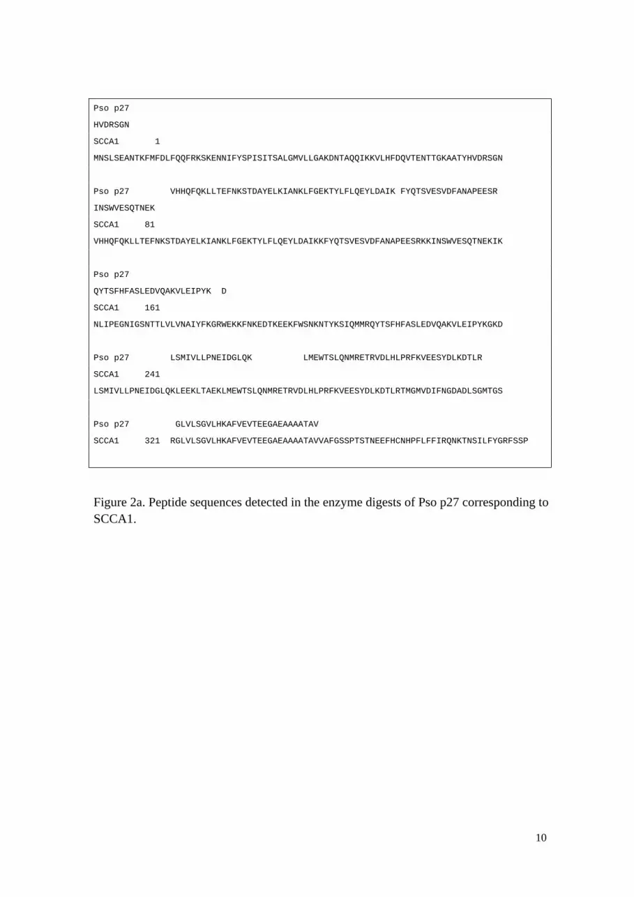
Peptide Sequences in Pso p27.
MALDI-MS/MS analysis of the m/z = 1689 fragment of the Lys-C digest showed
HVDRSGNVHHQFQK. No peptide fragments between this sequence and the N-
terminal ends of the SCCA molecules were detected (Fig. 2a, b). This indicates that
HVDRSGNVHHQFQK represent the N-terminal end of the Pso p27 antigen.
MALDI-MS/MS analysis of the m/z =1758 fragment of the trypsic digest showed the
sequence AFVEVTEEGVEAAAATAV. No peptide fragments between this sequence
and the C-terminal end of any SCCA molecules were detected. Based on this it seems
reasonable to suggesting that AFVEVTEEGVEAAAATAV represent the C-terminal end
of the Pso p27 antigen.
The results obtained using MALDI-MS/MS after digestion with the three
endoproteases; trypsin, Glu-C and Lys-C showed significant sequence homology with
both SCCA1 and SCCA2 as shown in fig. 2 a and b.
9
Pso p27
HVDRSGN
SCCA1 1
MNSLSEANTKFMFDLFQQFRKSKENNIFYSPISITSALGMVLLGAKDNTAQQIKKVLHFDQVTENTTGKAATYHVDRSGN
Pso p27 VHHQFQKLLTEFNKSTDAYELKIANKLFGEKTYLFLQEYLDAIK FYQTSVESVDFANAPEESR
INSWVESQTNEK
SCCA1 81
VHHQFQKLLTEFNKSTDAYELKIANKLFGEKTYLFLQEYLDAIKKFYQTSVESVDFANAPEESRKKINSWVESQTNEKIK
Pso p27
QYTSFHFASLEDVQAKVLEIPYK D
SCCA1 161
NLIPEGNIGSNTTLVLVNAIYFKGRWEKKFNKEDTKEEKFWSNKNTYKSIQMMRQYTSFHFASLEDVQAKVLEIPYKGKD
Pso p27 LSMIVLLPNEIDGLQK LMEWTSLQNMRETRVDLHLPRFKVEESYDLKDTLR
SCCA1 241
LSMIVLLPNEIDGLQKLEEKLTAEKLMEWTSLQNMRETRVDLHLPRFKVEESYDLKDTLRTMGMVDIFNGDADLSGMTGS
Pso p27 GLVLSGVLHKAFVEVTEEGAEAAAATAV
SCCA1 321 RGLVLSGVLHKAFVEVTEEGAEAAAATAVVAFGSSPTSTNEEFHCNHPFLFFIRQNKTNSILFYGRFSSP
Figure 2a. Peptide sequences detected in the enzyme digests of Pso p27 corresponding to SCCA1.
10

Pso p27
HVDRSGN
SCCA2 1
MNSLSEANTKFMFGLFQQFRKSKENNIFYSPISITSALGMVLLGAKDNTAQQISKILHFDQVTENTTEKAATYHVDRSGN
Pso p27 VHHQFQKLLTEFNKSTDAYELKIANKLFGEKTYQFLQEYLDAIK FYQTSVESTDFANAPEESR
INSWVESQTNEK
SCCA2 81
VHHQFQKLLTEFNKSTDAYELKIANKLFGEKTYQFLQEYLDAIKKFYQTSVESTDFANAPEESRKKINSWVESQTNEKIK
Pso p27
QYNSFNFALLEDVQAKVLEIPYK D
SCCA2 161
NLFPDGTIGNDTTLVLVNAIYFKGQWENKFKKENTKEEKFWPNKNTYKSVQMMRQYNSFNFALLEDVQAKVLEIPYKGKD
Pso p27 LSMIVLLPNEIDGLQK LMEWTSLQNMRETCVDLHLPR
SCCA2 241
LSMIVLLPNEIDGLQKLEEKLTAEKLMEWTSLQNMRETCVDLHLPRFKMEESYDLKDTLRTMGTVNIFNGDADLSGMTWS
Pso p27 AFVEVTEEGVEAAAATAV
SCCA2 321 HGLSVSKVLHKAFVEVTEEGVEAAAATAVVVVELSSPSTNEEFCCNHPFLFFIRQNKTNSILFYGRFSPP
Figure 2b. Peptide sequences detected in the enzyme digests of Pso p27 corresponding to SCCA2.
11

2D Gel-electrophoresis and Immunoblot.
Protein staining of Pso p27 after 2-dimensional gel-electrophoresis revealed several
proteins with iso-electric points between 4.7 and 5.3 (Fig 3).
Immunoblot analyses were performed using two murine monoclonal antibodies against
Pso p27 as primary antibodies. The antibodies recognize two different epitopes, and as
demonstrated in Fig 3, both epitopes were recognized in the different variants of the
protein. The modest correlation between the intensity of the silver staining and the
immunoblot is most likely due to variation with respect to exposure of the epitopes.
12

Figure 3. 2D- gel-electrophoresis with protein staining and immunoblot using two
different monoclonal antibodies against Pso p27
pH 4.7 pH 5.3
13
Silver
3A3D10
2C7D10

DISCUSSION
Much effort has been put forward in the search for etiological agents associated with
chronic inflammatory - or autoimmune diseases. During the last decades we have
focused on a protein, Pso p27, associated with psoriasis [10-13, 17]. Pso p27 is
expressed in psoriatic lesions and is not detected in uninvolved psoriatic skin or skin
biopsies from healthy controls [10, 13]. Through analysis of antibodies obtained from
psoriatic scale, we have demonstrated the potential role of Pso p27 as an antigen in
psoriasis [10]. The presence of Pso p27 in mast cells in the skin lesions [11] is of
particular interest in view of the potential role of mast cells as immune potentiating cells
[18].
The N-terminal amino acid sequence of Pso p27 described fifteen years ago [19] shows
a clear homology to SCCA [20]. In this paper the relationship between Pso p27 and
SCCA molecules is substantiated. Surprisingly, the Pso p27 antigen seems to represent
various SCCA molecules but with N-terminal and C-terminal ends deviating from the
SCCA molecules. The terminal ends of the Pso p27 antigens are most likely due to a
posttranslational digestion of SCCA molecules with highly specific endoproteases.
The 2-dimensional gel-electrophoresis demonstrates heterogeneity of the Pso p27
antigen with respect to pI. This heterogeneity may be due to variation in amino acid
sequences, but also variation with respect of oxidation of amino acids which was
observed through the mass spectrometry analysis. The two murine monoclonal
antibodies against Pso p27 used in the immunoblot analysis recognize two different
14

epitopes on Pso p27 as they function in sandwich ELISA with Pso p27 as antigen [16].
The fact that all the variants of Pso p27 detected in the 2-dimensional gel-
electrophoresis are recognized by both monoclonal antibodies, demonstrate at least two
common antigenic structures among the variants. The monoclonal antibodies used for
isolation of Pso p27, does not cross-react with the native forms of SCCA 1 or SCCA 2
[21]. Moreover, when the monoclonal antibodies are used in indirect
immunofluorescence analysis of psoriatic skin lesions the antibodies bind primarily to
mast cells and scales, and scarcely to epidermal cells [13] in contrast to anti-SCCA
antibodies [22].
Based on the observations presented in this study it seems reasonable to suggest that a
posttranslational modification of SCCA molecules make them immunogenic and that
the great variability with respect to protein structure is of importance with respect to
failure in immunologic tolerance.
Pso p27 antigen has been shown to participate in the generation of complement
activating immune complexes, both in the psoriatic plaques and in synovial fluid from
patients with psoriasis arthritis [17, 23]. Furthermore, Pso p27 antigen has been detected
in patients with various inflammatory diseases as for example ankylosing spondylitis
[23], sarcoidosis [16] and chronic inflammatory bowel diseases [24].
A highly relevant question is whether the Pso p27 antigen is a common antigenic
principle in chronic inflammatory diseases.
15

ACKNOWLEDGEMENT
The work has been supported by grants from The Research Council of Norway. Thanks
to the FUGE Proteomics Laboratory, Norwegian University of Science and Technology
for assistance with methods and instruments.
16

REFERENCES
[1] Bowcock AM, Krueger JG.. Getting under the skin: The immunogenetics of
psoriasis. Nature Reviews Immunology. 2005; 5: 699-711.
[2] Lowes MA, Bowcock AM, Krueger G.. Pathogenesis and therapy of psoriasis.
Nature. 2007; 445: 866-73.
[3] Swanbeck G, Inerot A, Martinsson T, Enerbäck C, Enlund F, Samuelson L, et al.
Genetic councelling in psoriasis: empirical data on psoriasis among first-degree
relatives of 3095 psoriatic probands. British Journal of Dermatology. 1997;137: 939-42.
[4] Henseler T. Genetics of psoriasis. Arch Dermatol Res. 1998; 290: 463-76.
[5] Rahman P, Elder JT. Genetic epidemiology of psoriasis and psoriatic arthritis.
Annals of the Rheumatic Diseases 2005; 64: 37-9.
[6] Brandrup F, Holm N, Grunnet N, Henningsen K, Hansen HE. Psoriasis in
monozygotic twins – variations in expression in individuals with identical genetic
constitution. Acta dermato-venerologica. 1982; 62: 229-36.
[7] Duffy DL, Spelman LS, Martin NG.. Psoriasis in Australian Twins. Journal of the
American Academy of dermatology. 1993; 29: 428-34.
[8] Krueger G, Ellis CN. Psoriasis-recent advances in understanding its pathogenesis
and treatment. Journal of the American Academy of Dermatology. 2005; 53: 94-100.
[9] Christophers E, Parzefal R, Braun-Falc O. Initial events in Psoriasis – quantitative
assessments. British J of Dermatol 1973; 89: 327-34.
[10] Iversen O-J, Bergh K, Lysvand H.Use of scale antibodies in the detection of
antigens in psoriatic lesions. Acta Derm Venereol. 1993; 73: 31-4.
[11] Iversen O-J, Lysvand H, Jacobsen T, Bergh K, Lie BA. The psoriasis associated
17

antigen, pso p27, is expressed by tryptase positive cells in psoriatic lesions. Arch.
Dermatol. Res. 1995; 287: 503-5.
[12] Dalaker M, Jacobsen T, Lysvand H, Iversen O-J. The expression of the psoriasis
associated antigen pso p27 is inhibited by Cyclosporin A. Acta dermatol Venerol. 1999;
79: 281-4.
[13] Song P, Lysvand H, Yan Y, Liu W, Iversen O-J. Expression of the psoriasis-
associated protein, Pso p27, is inhibited by Traditional Chinese Medicine. Journal of
Ethnopharmacology 2010; 127: 171-4.
[14] Shevchenko A, Wilm M, Vorm O, Mann M. Mass Spectrometric Sequencing of
Proteins from Silver-Stained Polyacrylamide Gels. Anal. Chem. 1996; 68: 850-8.
[15] Rappsilber J, Mann M, Ishihama Y. Protocol for micro-purification, enrichment,
pre-fractionation and storage of peptides for proteomics using StageTips. Nat Protoc.
2007; 2(8):1896-906.
[16] Jacobsen T, Lie BA, Lysvand H, Wiik M, Pettersen HB, Iversen, O-J. Detection of
psoriasis associated antigen, pso p27, in sarcoidosis bronchoalveolar lavage using
monoclonal antibodies. Clin. Immunol. Immunopathol. 1996; 81: 82-7.
[17] Asbakk K, Bergh K, Iversen O-J. The psoriasis associated antigen, pso p27
participates in the formation of complement activating immune complexes in psoriatic
scale. APMIS. 1990; 98: 143-9.
[18] Gaudenzio N, Espagnolle N, Mars LT, Liblau R, Valitutti S, Espinosa E. Cell-cell
cooperation at the T helper cell/mast cell immunological synapse. Blood. 2009; 114:
4979-88.
[19] Iversen O-J, Lysvand H, Bergh K, Eriksen J, Elsayed S. The N-terminal amino-acid
18

sequence of the psoriasis associated antigen, pso p27. Arch Dermatol. Res 1995; 287:
761-3.
[20] Schneider SS, Schick C, Fish KE, Miller E, Pena JC, Treter SD, Hui SM,
Silverman GA. A serine proteinase inhibitor locus at 18q21.3 contains a tandem
duplication of the human squamous cell carcinoma antigen gene. Proc Natl Acad Sci.
1995; 92: 3147-51.
[21] Yu X, Ikeda S, Yaguchi H, Ogawa H, Uchida T, Lysvand H, Iversen O-J. An anti-
pso p27 monoclonal antibody reacts with skin and peripheral blood leukocytes from
Japanese psoriatic patients and shows cross-reactivity with SCCA2b. Arch of dermatol
res 2005; 296: 372-4.
[22] Takeda A, Higuchi D, Takahashi T, Ogo M, Baciu P, Goetinck PF, Hibino T.
Overexpression of Serpin Squamous Cell Carcinoma Antigens in Psoriatic Skin. Journal
of Investigative Dermatology 2002; 118: 147-54.
[23] Rødahl E, Åsbakk K, Iversen O-J. Participation of antigens related to the psoriasis
associated antigen, pso p27, in immune complex formation in patients with ankylosing
spondylitis. Ann Rheum Dis. 1988; 47: 628-33.
[24] Iversen O-J, Jacobsen T. Chronic inflammatory diseases. Sarcoidosis, Vasculitis
and diffuse lung diseases 1996;13: 66-9.
19


