Embed Size (px)
Citation preview
Gene Repertoire Evolution of Streptococcus pyogenesInferred from Phylogenomic Analysis with Streptococcuscanis and Streptococcus dysgalactiaeTristan Lefebure¤, Vince P. Richards, Ping Lang, Paulina Pavinski-Bitar, Michael J. Stanhope*
Department of Population Medicine and Diagnostic Sciences, College of Veterinary Medicine, Cornell University, Ithaca, New York, United States of America
Abstract
Streptococcus pyogenes, is an important human pathogen classified within the pyogenic group of streptococci, exclusivelyadapted to the human host. Our goal was to employ a comparative evolutionary approach to better understand thegenomic events concomitant with S. pyogenes human adaptation. As part of ascertaining these events, we sequenced thegenome of one of the potential sister species, the agricultural pathogen S. canis, and combined it in a comparativegenomics reconciliation analysis with two other closely related species, Streptococcus dysgalactiae and Streptococcus equi, todetermine the genes that were gained and lost during S. pyogenes evolution. Genome wide phylogenetic analyses involving15 Streptococcus species provided convincing support for a clade of S. equi, S. pyogenes, S. dysgalactiae, and S. canis andsuggested that the most likely S. pyogenes sister species was S. dysgalactiae. The reconciliation analysis identified 113 genesthat were gained on the lineage leading to S. pyogenes. Almost half (46%) of these gained genes were phage associated and14 showed significant matches to experimentally verified bacteria virulence factors. Subsequent to the origin of S. pyogenes,over half of the phage associated genes were involved in 90 different LGT events, mostly involving different strains of S.pyogenes, but with a high proportion involving the horse specific pathogen S. equi subsp. equi, with the directionality almostexclusively (86%) in the S. pyogenes to S. equi direction. Streptococcus agalactiae appears to have played an important role inthe evolution of S. pyogenes with a high proportion of LGTs originating from this species. Overall the analysis suggests thatS. pyogenes adaptation to the human host was achieved in part by (i) the integration of new virulence factors (e.g. speB, andthe sal locus) and (ii) the construction of new regulation networks (e.g. rgg, and to some extent speB).
Citation: Lefebure T, Richards VP, Lang P, Pavinski-Bitar P, Stanhope MJ (2012) Gene Repertoire Evolution of Streptococcus pyogenes Inferred from PhylogenomicAnalysis with Streptococcus canis and Streptococcus dysgalactiae. PLoS ONE 7(5): e37607. doi:10.1371/journal.pone.0037607
Editor: Michael Chaussee, University of South Dakota, United States of America
Received January 18, 2012; Accepted April 24, 2012; Published May 30, 2012
Copyright: � 2012 Lefebure et al. This is an open-access article distributed under the terms of the Creative Commons Attribution License, which permitsunrestricted use, distribution, and reproduction in any medium, provided the original author and source are credited.
Funding: This work was supported by the National Institute of Allergy and Infectious Disease, U.S. National Institutes of Health, under Grant No. AI073368-01A2awarded to M.J.S. The funders had no role in study design, data collection and analysis, decision to publish, or preparation of the manuscript.
Competing Interests: The authors have declared that no competing interests exist.
* E-mail: [email protected]
¤ Current address: Universite de Lyon, Universite Lyon 1, Centre National de la Recherche Scientifique, Ecologie des Hydrosystemes Naturels et Anthropises,Villeurbanne, France
Introduction
Streptococcus pyogenes is a leading human pathogen responsible for
illness ranging from mild skin and respiratory infections (e.g.
pharyngitis and impetigo) to life-threatening invasive (e.g. pneu-
monia, septicemia, streptococcal toxic shock syndrome, necrotiz-
ing fasciitis), and post-infection diseases (e.g. acute rheumatic fever,
paediatric autoimmune neuropsychiatric disorders). Many differ-
ent serotypes and strains have been described, with some being
linked to particular disease. For example, strains causing
necrotizing fasciitis are largely serotype M1 and M3 [1], while
M18 is often linked to acute rheumatic fever [2], and M28 to
puerperal sepsis [3]. As part of attempts to understand the nature
of this diversity, 12 complete and one draft genome have been
sequenced (11 complete at the beginning of this study)
[2,4,5,6,7,8,9,10,11,12]. The publications associated with these
genomes have suggested links between lisogenic phages, and the
virulence factors they are carrying, to specific diseases. A long and
detailed list of S. pyogenes virulence factors is now available (e.g.
[13]). Information is now available regarding the genomic
repertoire of S. pyogenes (e.g. [14], the link between some virulence
factors and disease [15,16], and the history of lateral gene transfer
for some of the loci (e.g. [6,17,18]. Nonetheless, many of the
molecular details related to the adaptive specifics of this organism
remain unknown. S. pyogenes is classified within the pyogenic group,
which is currently composed of 12 species of Streptococcus [19],
which inhabit various species of mammals (e.g. bovine, dogs, cats,
horse, swine, humans). Most species of the pyogenic group are
found in a range of different hosts. S. pyogenes is unusual in that it is
only found in humans.
The putative sister group to S. pyogenes is uncertain. There is
phylogenetic evidence suggesting it could be S. canis [19], or
Streptococcus dysgalactiae [20]. Whatever the precise evolutionary
history, with ribosomal sequence divergence of around 3%, these
three taxa are clearly very closely related [19]. S. canis colonizes a
variety of hosts including dogs, cats, and cows, with few reported
human infections [21,22]. In addition to causing bovine mastitis
[23], S. canis shares with S. pyogenes the potential to cause similar
disease, such as respiratory tract infections [24], streptococcal toxic
shock syndrome [25], endocarditis [26], and necrotizing fasciitis
[25]. Streptococcus dysgalactiae includes two subspecies, Streptococcus
PLoS ONE | www.plosone.org 1 May 2012 | Volume 7 | Issue 5 | e37607
dysgalactiae subsp dysgalactiae and Streptococcus dysgalactiae subsp
equisimilis. S. dysgalactiae subsp. equisimilis, was primarily regarded
as a human commensal organism [27] but is now recognized as an
increasingly important human pathogen, linked to a spectrum of
human diseases including cellulitis, peritonitis, septic arthritis,
pneumonia, endocarditis, acute pharyngitis, bacteremia, and toxic
shock syndrome [28], which, like S. canis, includes several
infections similar to those caused by S. pyogenes. S. dysgalactiae
subsp. dysgalactiae on the other hand, is strictly an animal pathogen
and a major cause of bovine mastitis. Given the overall lack of host
specific adaptation of the taxa within the pyogenic group,
concomitant with the characteristics of S. canis and S. dysgalactiae,
it is likely that the ancestor to S. pyogenes was not a strict human
pathogen, if not a human pathogen at all. This suggests that one of
the principal factors in the evolution of S. pyogenes was its strict
adaptation to the human host.
The sequenced S. pyogenes genomes have facilitated the
identification of many of the molecular features associated with
strain and serotype differentiation, but it remains unclear what
makes S. pyogenes a strict human pathogen compared to many of
the host generalists typical of the pyogenic group. An improved
understanding of this issue is important in any attempt to develop
a broad medical strategy, such as a GAS vaccine (GAS: group A
streptococcus). In this study, we describe the genomic features that
evolved since the divergence of S. pyogenes from its closest relatives,
in an attempt to understand the molecular details associated with
S. pyogenes development as a strict human pathogen. For this
purpose we sequenced the S. canis genome and combined it in
comparative analysis with genome sequence data from the closely
related taxa S. pyogenes, S. dysgalactiae, and S. equi. A closely related
taxon provides the ability to ascribe to the S. pyogenes branch the
specific features of S. pyogenes evolution. The use of a less closely
related taxon as a reference (e.g. one of the publicly available S.
agalactiae genomes) would yield a less accurate description because
it would merge the evolutionary history of several lineages (e.g. S.
canis, S. agalactiae, S. iniae and S. equi). More specifically, our
purpose was to (1) provide a rigorous genome based phylogenetic
perspective on identifying the S. pyogenes sister group and (2)
identify the genes that were gained and lost along the S. pyogenes
lineage after the divergence of S. pyogenes from its closest relatives.
Materials and Methods
Genome sequencing and annotationStreptococcus canis strain FSL Z3-227 was isolated in New York
State in 1999 from the milk of dairy cows associated with an
outbreak of mastitis [23]. Based on results from bacterial culture
and ribotyping, a farm cat with chronic sinusitus was the likely
source of the outbreak [23]. The S. canis genome was sequenced
using 454 pyrosequencing [29] on a FLX sequencer. A total of
128,749 single end reads and 140,788 paired-end reads assembled
into 91 contigs (.200 bp) and 8 scaffolds, representing an average
236 site coverage. A physical map of the genome was determined
by OpGen Technologies, Inc. (Madison, WI) using restriction
enzyme BgIII and the optical mapping technique. The order and
orientation of the scaffolds was determined by aligning the scaffold
on the optical map using Opgen Mapviewer. Small inter and
intra-scaffold gaps were closed by PCR and sequenced using
Sanger sequencing, while 7 large gaps were amplified with long
range PCR and sequenced on the Illumina GA2 sequencer. The
Illumina reads were assembled with Velvet [30] using a large
range of parameters and the best assembly was selected using the
N50 statistic. Genome annotation was done by NCBI Prokaryotic
Genomes Automatic Annotation. This pipeline is composed of
HMM-based gene prediction methods and employs a sequence
similarity-based approach involving comparison of the predicted
gene products to the non-redundant protein database, Entrez
Protein Clusters, the Conserved Domain Database, and the COGs
(Clusters of Orthologous Groups). This Whole Genome Shotgun
project has been deposited at DDBJ/EMBL/GenBank under the
accession AIDX00000000. The version described in this paper is
the first version, AIDX01000000.
Orthologous genesAll the complete Streptococcus genomes available at the time of
this study were collected from NCBI (Table 1). Orthologs were
delimited using OrthoMCL2 [31], with post-processing as detailed
elsewhere [32,33]. Briefly, reciprocal BLASTp was performed
within and between all genome pairs (e-value cut-off = 1E-5). The
resulting e-values were then used to build a normalized similarity
matrix, which was analyzed using a Markov Cluster algorithm to
delineate proteins into clusters containing sets of orthologs and
recent paralogs [34]. Proteins were considered recent paralogs if
they were more similar to each other than to any protein from
another genome. Fragmented protein sequences, such as those
that span separate contigs or insertion sequences, can be
erroneously categorized as distinct orthologs. To correct for this,
clusters containing single proteins were not considered distinct
orthologs (rather fragments of the same protein) if they met the
following criteria: (i) showed strong homology with another cluster
(i.e. could potentially group together to form a single orthologous
cluster), (ii) failed to group together because the protein clustering
independently, showed no reciprocal BLASTp hit with one of the
proteins in the second cluster, (iii) the two proteins showing no
reciprocal BLASTp hit originated from the same genome. Proteins
that were larger than 30 amino acids and had no BLASTp hit with
any other protein were considered strain specific (E-value#1e-10).
Clusters were annotated by merging annotation of sequences from
the same cluster. Potential virulence factors were searched via
BLASTp against the VFDB database [35] using the longest
sequence of each cluster. Clusters were aligned as described
elsewhere [36]. Briefly, sequences are translated into proteins,
aligned with Probalign [37], backtranslated into DNA, and sites
with low posterior probability masked. Clusters with more than
50% of their sites masked are disregarded from any downstream
analysis.
Gene trees and species tree reconstructionGene trees were reconstructed for all the clusters composed of
more than 2 sequences using PhyML [38] with a GTR+G+I
model of evolution and the SPR tree search heuristic, with 500
pseudo bootstrap replicates. Total evidence (concatenation of the
core genes) and core-gene tree consensus approaches were first
used to tentatively infer a species tree. Level of concordance
between core gene trees was also assessed using Bucky [39] on a
reduced data-set: S. canis, S. dysgalactiae, S. equi and two S. pyogenes
(spy1 and spy2, Table 1). For each gene independently, MrBayes
[40] was used to obtain gene tree posterior probabilities, then
Bucky takes into account gene tree concordance, providing a
revised posterior probability distribution for each gene and
estimates the proportion of the core genes for which any given
clade is true.
ReconciliationTo reconstruct the history of genomic events associated with the
emergence of the different pyogenic species, we used the
reconciliation approach AnGST [41]. Using a species tree,
AnGST reconciles a gene tree with the species tree using a
Gene Content Evolution of Streptococcus pyogenes
PLoS ONE | www.plosone.org 2 May 2012 | Volume 7 | Issue 5 | e37607
generalized parsimony criterion to infer a minimal set of
evolutionary events including gene birth, speciation, gene loss,
gene duplication and lateral gene transfer. All the clusters
composed of more than two sequences were analyzed by AnGST
using the three most common gene trees as species tree and default
parameters. These three trees involved different relationships for S.
pyogenes, S. canis, and S. dysgalactiae: (spy,sde)sca; (spy,sca)sde;
(sca,sde)spy. Inference errors due to phylogenetic uncertainty were
minimized by incorporating 500 pseudo-bootstrap replicates per
gene. The focus of our examination is the S. pyogenes branch, and if
an evolutionary event (gene gain, loss etc.) for this branch was
judged the same irrespective of which of the three topologies was
considered, it was evaluated as robust. Despite the fact there is one
topology that is more likely (described below), we adopted this
conservative approach, since there is, nonetheless, some uncer-
tainty in the sister group relationship to S. pyogenes involving these
taxa. A significant proportion of the genes gained on the S. pyogenes
branch were phage associated. Using the tree with the most likely
topology - (spy,sde)sca - we further investigated, using this same
AnGST approach, the history of these phage associated genes after
they were gained on the spy branch.
Results and Discussion
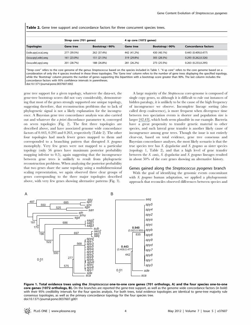
Gene trees and species treeIndependent gene tree reconstruction for the Streptococcus core
genome (701 genes) displayed an overall consensus within the
pyogenic clade (S. agalactiae, S. uberis, S. equi, S. canis, S. dysgalactiae
and S. pyogenes) with the exception of the relationship between
three species: S. canis, S. dysgalactiae and S. pyogenes (Table 2 and
Fig. 1). The most common topology was S. dysgalactiae and S.
pyogenes as sister groups (topology 1, 39.5% of the gene trees),
followed by the monophyly of S. canis and S. dysgalactiae (topology 3:
28.7%) and finally the monophyly of S. canis and S. pyogenes
(topology 2: 23%). The majority rule topology was also supported
by the total evidence approach (ie. concatenation of the genes
prior to phylogenetic reconstruction, Fig. 1). Focusing on a
reduced number of taxa (four species) to increase the core-gene
sample size (1072 genes), resulted in the same pattern whether
using consensus or total evidence approaches (Table 2). Incon-
gruences between gene trees can result from phylogenetic
reconstruction problems, incomplete lineage sorting, hidden
paralogy and lateral gene transfer (e.g. [42]). If one considers
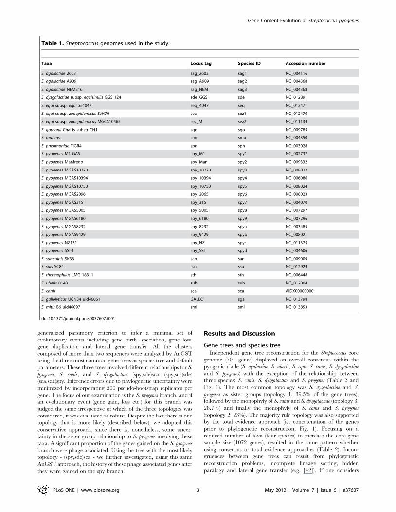
Table 1. Streptococcus genomes used in the study.
Taxa Locus tag Species ID Accession number
S. agalactiae 2603 sag_2603 sag1 NC_004116
S. agalactiae A909 sag_A909 sag2 NC_004368
S. agalactiae NEM316 sag_NEM sag3 NC_004368
S. dysgalactiae subsp. equisimilis GGS 124 sde_GGS sde NC_012891
S. equi subsp. equi Se4047 seq_4047 seq NC_012471
S. equi subsp. zooepidemicus SzH70 sez sez1 NC_012470
S. equi subsp. zooepidemicus MGCS10565 sez_M sez2 NC_011134
S. gordonii Challis substr CH1 sgo sgo NC_009785
S. mutans smu smu NC_004350
S. pneumoniae TIGR4 spn spn NC_003028
S. pyogenes M1 GAS spy_M1 spy1 NC_002737
S. pyogenes Manfredo spy_Man spy2 NC_009332
S. pyogenes MGAS10270 spy_10270 spy3 NC_008022
S. pyogenes MGAS10394 spy_10394 spy4 NC_006086
S. pyogenes MGAS10750 spy_10750 spy5 NC_008024
S. pyogenes MGAS2096 spy_2065 spy6 NC_008023
S. pyogenes MGAS315 spy_315 spy7 NC_004070
S. pyogenes MGAS5005 spy_5005 spy8 NC_007297
S. pyogenes MGAS6180 spy_6180 spy9 NC_007296
S. pyogenes MGAS8232 spy_8232 spya NC_003485
S. pyogenes MGAS9429 spy_9429 spyb NC_008021
S. pyogenes NZ131 spy_NZ spyc NC_011375
S. pyogenes SSI-1 spy_SSI spyd NC_004606
S. sanguinis SK36 san san NC_009009
S. suis SC84 ssu ssu NC_012924
S. thermophilus LMG 18311 sth sth NC_006448
S. uberis 0140J sub sub NC_012004
S. canis sca sca AIDX00000000
S. gallolyticus UCN34 uid46061 GALLO sga NC_013798
S. mitis B6 uid46097 smi smi NC_013853
doi:10.1371/journal.pone.0037607.t001
Gene Content Evolution of Streptococcus pyogenes
PLoS ONE | www.plosone.org 3 May 2012 | Volume 7 | Issue 5 | e37607
gene tree support for a given topology, whatever the data-set, the
gene-tree bootstrap scores did not vary considerably, demonstrat-
ing that most of the genes strongly supported one unique topology,
suggesting therefore, that reconstruction problems due to lack of
phylogenetic signal is not a likely explanation for the incongru-
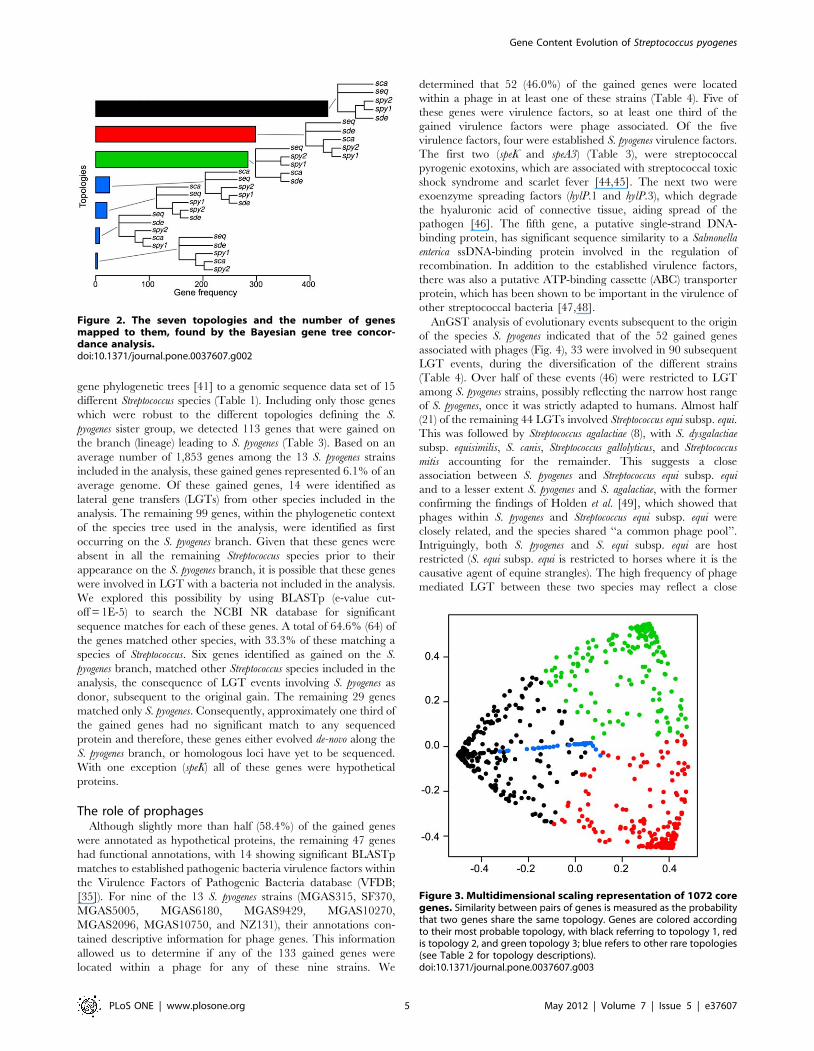
ence. A Bayesian gene tree concordance analysis was also carried
out and whatever the a priori discordance parameter a, converged
on seven topologies (Fig. 2). The first three topologies are
described above, and have associated genome wide concordance
factors of 0.443, 0.293 and 0.263, respectively (Table 2). The other
four topologies had much fewer genes mapped to them and
corresponded to a branching pattern that disrupted S. pyogenes
monophyly. Very few genes were not mapped to a particular
topology (only 56 genes have maximum posterior probability
mapping inferior to 0.5), again suggesting that the incongruence
between gene trees is unlikely to result from phylogenetic
reconstruction problems. When analyzing the posterior probability
that two genes share the same topology using a multidimensional
scaling representation, we again observed three clear groups of
genes corresponding to the three major topologies described
above, with very few genes showing alternative patterns (Fig. 3).
A large majority of the Streptococcus core-genome is composed of
single copy genes, so although it is difficult to rule out instances of
hidden paralogy, it is unlikely to be the cause of the high frequency
of incongruence we observe. Incomplete lineage sorting (also
called deep coalescence), is more frequent when divergence time
between two speciation events is shorter and population size is
larger [42,43], which both seem plausible in our example. Bacteria
have a great propensity to transfer genetic material to other
species, and such lateral gene transfer is another likely cause of
incongruence among gene trees. Though the issue is not entirely
clear-cut, based on total evidence, gene tree consensus and
Bayesian concordance analyses, the most likely scenario is that the
true species tree has S. dysgalactiae and S. pyogenes as sister species
(topology 1, Table 2), and that a high level of gene transfer
between the S. canis, S. dysgalactiae and S. pyogenes lineages resulted
in about 50% of the core genes showing an alternative history.
Genes gained along the Streptococcus pyogenes branchWith the goal of identifying the genomic events concomitant
with S. pyogenes human adaptation, we applied a phylogenomic
approach that reconciles observed differences between species and
Figure 1. Total evidence trees using the Streptococcus one-to-one core genes (701 orthologs, A) and the four species one-to-onecore genes (1072 orthologs, B). On the branches are reported the gene-tree support, as well as the genome wide concordance factors (in bold)with their 95% credibility intervals for the four species analysis. For both trees, total evidence topologies are identical to gene-tree majority ruleconsensus topologies, as well as the primary concordance topology for the four species tree.doi:10.1371/journal.pone.0037607.g001
Table 2. Gene tree support and concordance factors for three concurrent species trees.
Strep core (701 genes) 4 sp core (1072 genes)
Topologies Gene tree Bootstrap.90% Gene tree Bootstrap.90% Concordance factors
((sde,spy),sca),seq; 277 (39.5%) 262 (37.4%) 442 (41.2%) 430 (40.1%) 0.443 (0.409,0.477)
((sca,spy),sde),seq; 161 (23.0%) 151 (21.5%) 319 (29.8%) 305 (28.5%) 0.293 (0.262,0.326)
((sca,sde),spy),seq; 201 (28.7%) 188 (26.8%) 281 (26.2%) 273 (25.5%) 0.263 (0.233,0.295)
‘‘Strep core’’ refers to the core genome of the genus Streptococcus based on the species included in Table 1. ‘‘4 sp core’’ refers to the core genome based on aconsideration of only the 4 species involved in these three topologies. The ‘Gene tree’ column refers to the number of gene trees displaying the specified topology,while the ‘Bootstrap’ column presents the number of genes supporting this bipartition with a bootstrap score greater than 90%. The last column includes theconcordance factors with 95% confidence intervals in parentheses.doi:10.1371/journal.pone.0037607.t002
Gene Content Evolution of Streptococcus pyogenes
PLoS ONE | www.plosone.org 4 May 2012 | Volume 7 | Issue 5 | e37607
gene phylogenetic trees [41] to a genomic sequence data set of 15
different Streptococcus species (Table 1). Including only those genes
which were robust to the different topologies defining the S.
pyogenes sister group, we detected 113 genes that were gained on
the branch (lineage) leading to S. pyogenes (Table 3). Based on an
average number of 1,853 genes among the 13 S. pyogenes strains
included in the analysis, these gained genes represented 6.1% of an
average genome. Of these gained genes, 14 were identified as
lateral gene transfers (LGTs) from other species included in the
analysis. The remaining 99 genes, within the phylogenetic context
of the species tree used in the analysis, were identified as first
occurring on the S. pyogenes branch. Given that these genes were
absent in all the remaining Streptococcus species prior to their
appearance on the S. pyogenes branch, it is possible that these genes
were involved in LGT with a bacteria not included in the analysis.
We explored this possibility by using BLASTp (e-value cut-
off = 1E-5) to search the NCBI NR database for significant
sequence matches for each of these genes. A total of 64.6% (64) of
the genes matched other species, with 33.3% of these matching a
species of Streptococcus. Six genes identified as gained on the S.
pyogenes branch, matched other Streptococcus species included in the
analysis, the consequence of LGT events involving S. pyogenes as
donor, subsequent to the original gain. The remaining 29 genes
matched only S. pyogenes. Consequently, approximately one third of
the gained genes had no significant match to any sequenced
protein and therefore, these genes either evolved de-novo along the
S. pyogenes branch, or homologous loci have yet to be sequenced.
With one exception (speK) all of these genes were hypothetical
proteins.
The role of prophagesAlthough slightly more than half (58.4%) of the gained genes
were annotated as hypothetical proteins, the remaining 47 genes
had functional annotations, with 14 showing significant BLASTp
matches to established pathogenic bacteria virulence factors within
the Virulence Factors of Pathogenic Bacteria database (VFDB;
[35]). For nine of the 13 S. pyogenes strains (MGAS315, SF370,
MGAS5005, MGAS6180, MGAS9429, MGAS10270,
MGAS2096, MGAS10750, and NZ131), their annotations con-
tained descriptive information for phage genes. This information
allowed us to determine if any of the 133 gained genes were
located within a phage for any of these nine strains. We
determined that 52 (46.0%) of the gained genes were located
within a phage in at least one of these strains (Table 4). Five of
these genes were virulence factors, so at least one third of the
gained virulence factors were phage associated. Of the five
virulence factors, four were established S. pyogenes virulence factors.
The first two (speK and speA3) (Table 3), were streptococcal
pyrogenic exotoxins, which are associated with streptococcal toxic
shock syndrome and scarlet fever [44,45]. The next two were
exoenzyme spreading factors (hylP.1 and hylP.3), which degrade
the hyaluronic acid of connective tissue, aiding spread of the
pathogen [46]. The fifth gene, a putative single-strand DNA-
binding protein, has significant sequence similarity to a Salmonella
enterica ssDNA-binding protein involved in the regulation of
recombination. In addition to the established virulence factors,
there was also a putative ATP-binding cassette (ABC) transporter
protein, which has been shown to be important in the virulence of
other streptococcal bacteria [47,48].
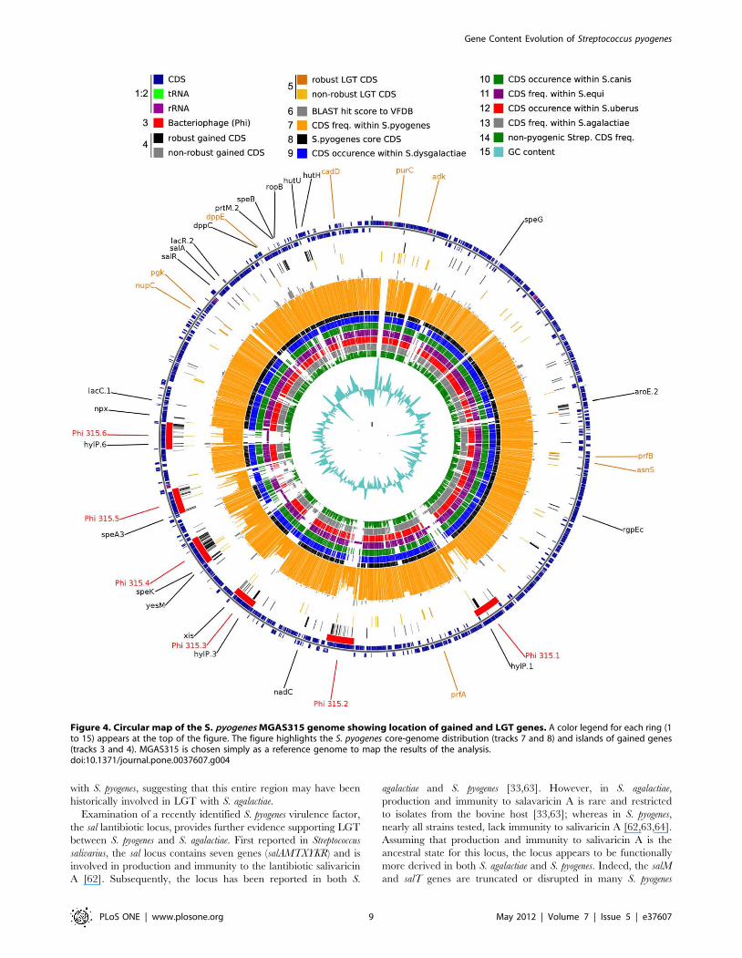
AnGST analysis of evolutionary events subsequent to the origin
of the species S. pyogenes indicated that of the 52 gained genes
associated with phages (Fig. 4), 33 were involved in 90 subsequent
LGT events, during the diversification of the different strains
(Table 4). Over half of these events (46) were restricted to LGT
among S. pyogenes strains, possibly reflecting the narrow host range
of S. pyogenes, once it was strictly adapted to humans. Almost half
(21) of the remaining 44 LGTs involved Streptococcus equi subsp. equi.
This was followed by Streptococcus agalactiae (8), with S. dysgalactiae
subsp. equisimilis, S. canis, Streptococcus gallolyticus, and Streptococcus
mitis accounting for the remainder. This suggests a close
association between S. pyogenes and Streptococcus equi subsp. equi
and to a lesser extent S. pyogenes and S. agalactiae, with the former
confirming the findings of Holden et al. [49], which showed that
phages within S. pyogenes and Streptococcus equi subsp. equi were
closely related, and the species shared ‘‘a common phage pool’’.
Intriguingly, both S. pyogenes and S. equi subsp. equi are host
restricted (S. equi subsp. equi is restricted to horses where it is the
causative agent of equine strangles). The high frequency of phage
mediated LGT between these two species may reflect a close
Figure 3. Multidimensional scaling representation of 1072 coregenes. Similarity between pairs of genes is measured as the probabilitythat two genes share the same topology. Genes are colored accordingto their most probable topology, with black referring to topology 1, redis topology 2, and green topology 3; blue refers to other rare topologies(see Table 2 for topology descriptions).doi:10.1371/journal.pone.0037607.g003
Figure 2. The seven topologies and the number of genesmapped to them, found by the Bayesian gene tree concor-dance analysis.doi:10.1371/journal.pone.0037607.g002
Gene Content Evolution of Streptococcus pyogenes
PLoS ONE | www.plosone.org 5 May 2012 | Volume 7 | Issue 5 | e37607
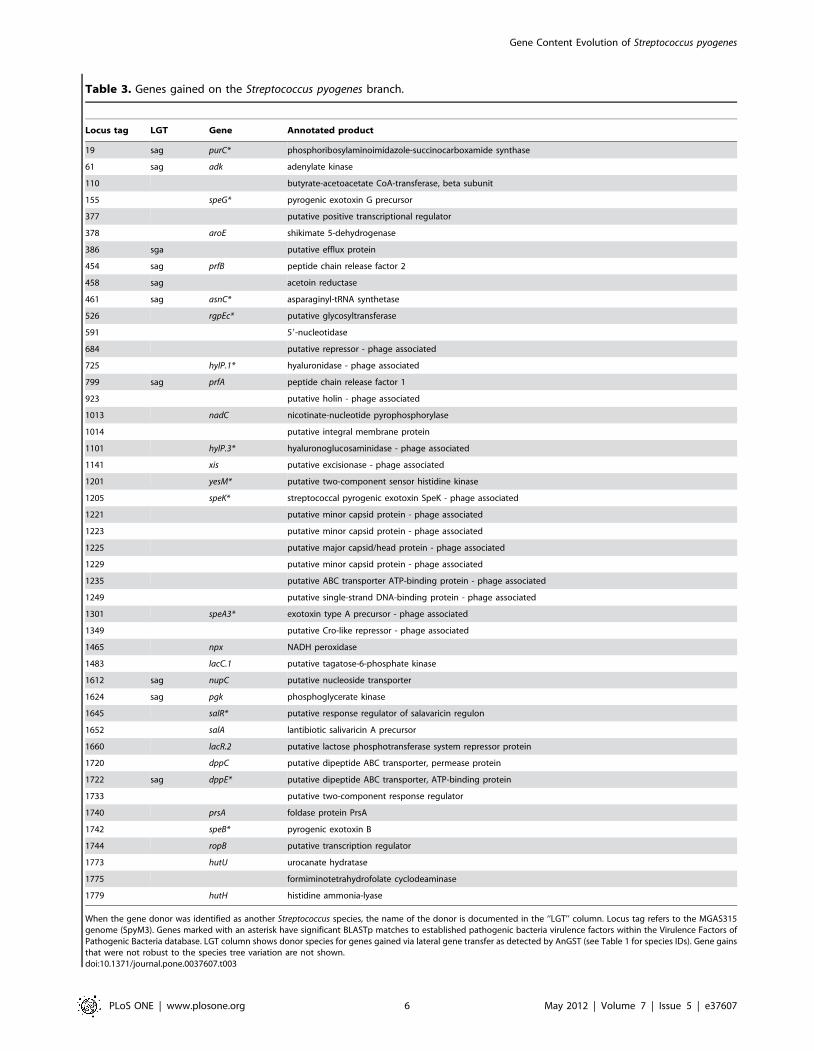
Table 3. Genes gained on the Streptococcus pyogenes branch.
Locus tag LGT Gene Annotated product
19 sag purC* phosphoribosylaminoimidazole-succinocarboxamide synthase
61 sag adk adenylate kinase
110 butyrate-acetoacetate CoA-transferase, beta subunit
155 speG* pyrogenic exotoxin G precursor
377 putative positive transcriptional regulator
378 aroE shikimate 5-dehydrogenase
386 sga putative efflux protein
454 sag prfB peptide chain release factor 2
458 sag acetoin reductase
461 sag asnC* asparaginyl-tRNA synthetase
526 rgpEc* putative glycosyltransferase
591 59-nucleotidase
684 putative repressor - phage associated
725 hylP.1* hyaluronidase - phage associated
799 sag prfA peptide chain release factor 1
923 putative holin - phage associated
1013 nadC nicotinate-nucleotide pyrophosphorylase
1014 putative integral membrane protein
1101 hylP.3* hyaluronoglucosaminidase - phage associated
1141 xis putative excisionase - phage associated
1201 yesM* putative two-component sensor histidine kinase
1205 speK* streptococcal pyrogenic exotoxin SpeK - phage associated
1221 putative minor capsid protein - phage associated
1223 putative minor capsid protein - phage associated
1225 putative major capsid/head protein - phage associated
1229 putative minor capsid protein - phage associated
1235 putative ABC transporter ATP-binding protein - phage associated
1249 putative single-strand DNA-binding protein - phage associated
1301 speA3* exotoxin type A precursor - phage associated
1349 putative Cro-like repressor - phage associated
1465 npx NADH peroxidase
1483 lacC.1 putative tagatose-6-phosphate kinase
1612 sag nupC putative nucleoside transporter
1624 sag pgk phosphoglycerate kinase
1645 salR* putative response regulator of salavaricin regulon
1652 salA lantibiotic salivaricin A precursor
1660 lacR.2 putative lactose phosphotransferase system repressor protein
1720 dppC putative dipeptide ABC transporter, permease protein
1722 sag dppE* putative dipeptide ABC transporter, ATP-binding protein
1733 putative two-component response regulator
1740 prsA foldase protein PrsA
1742 speB* pyrogenic exotoxin B
1744 ropB putative transcription regulator
1773 hutU urocanate hydratase
1775 formiminotetrahydrofolate cyclodeaminase
1779 hutH histidine ammonia-lyase
When the gene donor was identified as another Streptococcus species, the name of the donor is documented in the ‘‘LGT’’ column. Locus tag refers to the MGAS315genome (SpyM3). Genes marked with an asterisk have significant BLASTp matches to established pathogenic bacteria virulence factors within the Virulence Factors ofPathogenic Bacteria database. LGT column shows donor species for genes gained via lateral gene transfer as detected by AnGST (see Table 1 for species IDs). Gene gainsthat were not robust to the species tree variation are not shown.doi:10.1371/journal.pone.0037607.t003
Gene Content Evolution of Streptococcus pyogenes
PLoS ONE | www.plosone.org 6 May 2012 | Volume 7 | Issue 5 | e37607
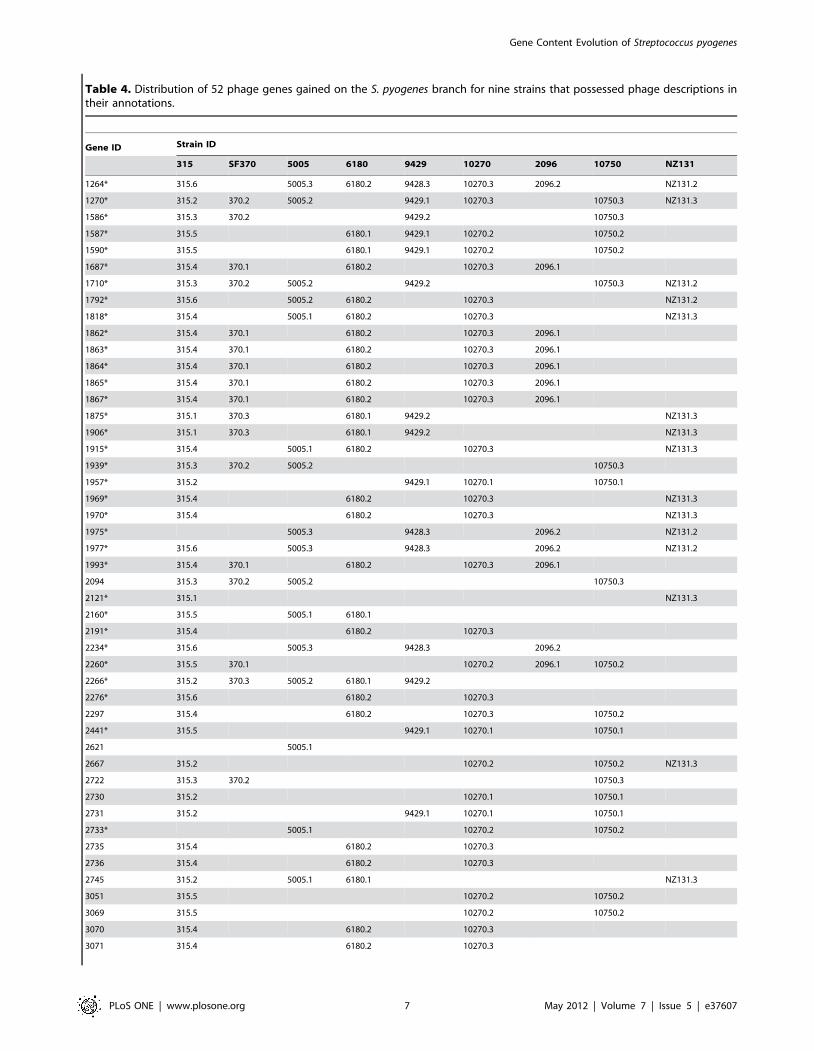
Table 4. Distribution of 52 phage genes gained on the S. pyogenes branch for nine strains that possessed phage descriptions intheir annotations.
Gene ID Strain ID
315 SF370 5005 6180 9429 10270 2096 10750 NZ131
1264* 315.6 5005.3 6180.2 9428.3 10270.3 2096.2 NZ131.2
1270* 315.2 370.2 5005.2 9429.1 10270.3 10750.3 NZ131.3
1586* 315.3 370.2 9429.2 10750.3
1587* 315.5 6180.1 9429.1 10270.2 10750.2
1590* 315.5 6180.1 9429.1 10270.2 10750.2
1687* 315.4 370.1 6180.2 10270.3 2096.1
1710* 315.3 370.2 5005.2 9429.2 10750.3 NZ131.2
1792* 315.6 5005.2 6180.2 10270.3 NZ131.2
1818* 315.4 5005.1 6180.2 10270.3 NZ131.3
1862* 315.4 370.1 6180.2 10270.3 2096.1
1863* 315.4 370.1 6180.2 10270.3 2096.1
1864* 315.4 370.1 6180.2 10270.3 2096.1
1865* 315.4 370.1 6180.2 10270.3 2096.1
1867* 315.4 370.1 6180.2 10270.3 2096.1
1875* 315.1 370.3 6180.1 9429.2 NZ131.3
1906* 315.1 370.3 6180.1 9429.2 NZ131.3
1915* 315.4 5005.1 6180.2 10270.3 NZ131.3
1939* 315.3 370.2 5005.2 10750.3
1957* 315.2 9429.1 10270.1 10750.1
1969* 315.4 6180.2 10270.3 NZ131.3
1970* 315.4 6180.2 10270.3 NZ131.3
1975* 5005.3 9428.3 2096.2 NZ131.2
1977* 315.6 5005.3 9428.3 2096.2 NZ131.2
1993* 315.4 370.1 6180.2 10270.3 2096.1
2094 315.3 370.2 5005.2 10750.3
2121* 315.1 NZ131.3
2160* 315.5 5005.1 6180.1
2191* 315.4 6180.2 10270.3
2234* 315.6 5005.3 9428.3 2096.2
2260* 315.5 370.1 10270.2 2096.1 10750.2
2266* 315.2 370.3 5005.2 6180.1 9429.2
2276* 315.6 6180.2 10270.3
2297 315.4 6180.2 10270.3 10750.2
2441* 315.5 9429.1 10270.1 10750.1
2621 5005.1
2667 315.2 10270.2 10750.2 NZ131.3
2722 315.3 370.2 10750.3
2730 315.2 10270.1 10750.1
2731 315.2 9429.1 10270.1 10750.1
2733* 5005.1 10270.2 10750.2
2735 315.4 6180.2 10270.3
2736 315.4 6180.2 10270.3
2745 315.2 5005.1 6180.1 NZ131.3
3051 315.5 10270.2 10750.2
3069 315.5 10270.2 10750.2
3070 315.4 6180.2 10270.3
3071 315.4 6180.2 10270.3
Gene Content Evolution of Streptococcus pyogenes
PLoS ONE | www.plosone.org 7 May 2012 | Volume 7 | Issue 5 | e37607
human-horse association and/or that S. pyogenes was an important
factor in the evolution of S. equi subsp. equi as it split from S. equi
subsp. zooepidemicus to become a strict horse pathogen [49]. The
direction of phage mediated LGT between S. pyogenes and S. equi
subsp. equi lends support to the latter hypothesis, as 85.7% of the
LGTs between these two species were from S. pyogenes to S. equi
subsp. equi. The taxon S. equi. subsp. equi must be of relatively
recent age, since the clone is very homogeneous, with sequence
divergence of housekeeping loci across diverse collections of strains
extremely minimal [50] and microarray data of ours confirming
this across the genome, while concomitantly indicating that
relatively few genes comprise the dispensable component of the
genome compared to other Streptococcus taxa ([14] and Stanhope
unpublished data). S. pyogenes on the other hand, is clearly of much
older origin. We suggest that phage mediated LGTs from S.
pyogenes to one or a few S. equi subsp. zooepidemicus strains were
instrumental in creating the progenitor or founder of S. equi subsp.
equi, which then developed into the current version of this clonal
organism. Such a scenario could be a rare example of reverse
zoonosis, although it is also possible that this transpired within the
human host, involving an instance of co-infection involving S.
pyogenes and S. equi subsp. zooepidemicus. Cases of human infection
by S. equi subsp. zooepidemicus, although not common, are
nonetheless reported, involving both zoonotic transmission from
domesticated animals [51], and the consumption of inadequately
pasteurized milk products [52]. The genes involved in this pyogenes-
equi LGT included hyaluronoglucosaminidase (hylP) and strepto-
coccal pyrogenic exotoxin (speK) with the remainder annotated as
hypotheticals or phage associated proteins. The majority (more
than 75%) of these LGTs originated from serotype 5 and 49
strains. This is not to say, that we found no evidence of the reverse
directionality in LGT – from equi to pyogenes – AnGST simply
identified the majority of the LGT between these two taxa in the
pyogenes-equi direction (17 vs 3).
M-protein islandThe M-protein pathogenicity island is a region of 35 genes
present in all sequenced S. pyogenes genomes [53]. Three genes
showing homology to the VFDB were located within this island:
dppE, speB, and a putative two-component response regulator.
DppE (gained via LGT from S. agalactiae) is one of six ABC
transporter proteins present in the island, and speB is another
streptococcal pyrogenic exotoxin. In addition to these three genes,
the island contained five additional loci gained along the S. pyogenes
branch (Table 3). One of these, ropB (also known as rgg) is of
particular interest, due to its interaction with speB. Both rgg and
speB have recently been shown to have greater expression in
pharyngal conditions as opposed to invasive conditions [54]. Rgg
regulates the expression of several virulence factors (including
speB), as well as activates the utilization of non-glucose carbohy-
drates [55]. Transcriptome analyses have shown that rgg is over-
expressed in saliva conditions [56] and in the adherence phase
[57]. SpeB leads to the cleavage or inactivation of many bacterial
proteins, including virulence factors involved in invasive disease
that contribute to host-pathogen interaction [54]. Differential
expression of speB may lead to different levels of lethality, because
decreased production of speB results in the preservation of S.
pyogenes virulence factors. Thus, both genes are playing an essential
role for survival in human saliva. SpeB-rgg interaction may have
contributed to S. pyogenes colonization of the human pharynx as its
main habitat, without generating invasive disease that would kill
the host and thereby reduce possibilities of dispersion. In that
sense, the gain of speB and rgg might have been a critical
component in S. pyogenes adaptation to the human host.
The M-protein, a key S. pyogenes virulence factor [16], was not
gained along the S. pyogenes branch and is shared with several
Streptococcus species; for example, S. dysgalactiae subsp. equisimilis, S.
dysgalactiae subsp. dysgalactiae, and S. agalactiae [58]. Indeed, a large
proportion of the M-protein island genes are shared with other
Streptococcus species [58]. However, it is only in S. pyogenes that the
genes form a contiguous island [53,58]. Consequently, it appears
that while the majority (77%) of the M-protein island genes were
present in the S. pyogenes ancestor, it was here that they clustered to
form a contiguous island, highlighting the importance of gene
rearrangement in addition to gene gain as an important
evolutionary factor in the emergence of new species.
The role of Streptococcus agalactiaeStreptococcus agalactiae appears to have played an important role in
the evolution of S. pyogenes with 10 of the 14 LGTs having a
Streptococcus donor, originating from this species (Table 3). Three of
these laterally transferred genes (prfB, asnC, and an acetoin
reductase) are clustered together within S. pyogenes genome
sequences. Two of these genes (asnC and acetoin reductase)
showed significant BLASTp matches to Escherichia coli virulence
factors. asnC was similar to LysU, a heat shock inducible lysyl-
rRNA synthase that enables pathogen survival at elevated
temperatures [59]. Acetoin reductase was similar to entA, which
is involved in the biosynthesis and excretion of the siderophore
enterobactin that enables survival in iron poor environments such
as the urinary tract [60,61]. Comparison to S. agalactiae (NEM316)
showed these three genes to be contained within an approximately
11 kbp region that shared approximately 76% sequence identity
Table 4. Cont.
Gene ID Strain ID
315 SF370 5005 6180 9429 10270 2096 10750 NZ131
3072 315.4 6180.2 10270.3
3080 315.3 10750.3
3081 315.3 10750.3
3087 315.2 6180.1 9429.1
3088 315.4 6180.2
Columns under strain IDs contain phage IDs. Asterisks mark genes that experienced LGT subsequent to being gained on the S. pyogenes branch. The strain ID prefixMGAS was omitted to save space.doi:10.1371/journal.pone.0037607.t004
Gene Content Evolution of Streptococcus pyogenes
PLoS ONE | www.plosone.org 8 May 2012 | Volume 7 | Issue 5 | e37607
with S. pyogenes, suggesting that this entire region may have been
historically involved in LGT with S. agalactiae.
Examination of a recently identified S. pyogenes virulence factor,
the sal lantibiotic locus, provides further evidence supporting LGT
between S. pyogenes and S. agalactiae. First reported in Streptococcus
salivarius, the sal locus contains seven genes (salAMTXYKR) and is
involved in production and immunity to the lantibiotic salivaricin
A [62]. Subsequently, the locus has been reported in both S.
agalactiae and S. pyogenes [33,63]. However, in S. agalactiae,
production and immunity to salavaricin A is rare and restricted
to isolates from the bovine host [33,63]; whereas in S. pyogenes,
nearly all strains tested, lack immunity to salivaricin A [62,63,64].
Assuming that production and immunity to salivaricin A is the
ancestral state for this locus, the locus appears to be functionally
more derived in both S. agalactiae and S. pyogenes. Indeed, the salM
and salT genes are truncated or disrupted in many S. pyogenes
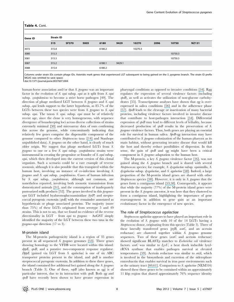
Figure 4. Circular map of the S. pyogenes MGAS315 genome showing location of gained and LGT genes. A color legend for each ring (1to 15) appears at the top of the figure. The figure highlights the S. pyogenes core-genome distribution (tracks 7 and 8) and islands of gained genes(tracks 3 and 4). MGAS315 is chosen simply as a reference genome to map the results of the analysis.doi:10.1371/journal.pone.0037607.g004
Gene Content Evolution of Streptococcus pyogenes
PLoS ONE | www.plosone.org 9 May 2012 | Volume 7 | Issue 5 | e37607
strains [63]. Similarly, the salM gene is truncated and the salA gene
missing in bovine adapted S. agalactiae (strain FSL S3-026) [33].
Phelps and Neely [65] demonstrated that for S. pyogenes, the locus
had shifted its immunity function from salivaricin to the host
immune system, with the salY gene of the locus now required for
survival within macrophages. Furthermore, alignment of S. pyogenes
(strain MGAS315) (all genes of the locus are intact in this strain) to
the bovine S. agalactiae strain, and S. salivarius (strain 20P3), showed
very high sequence identity (97.6%) between S. pyogenes and S.
agalactiae, but somewhat lower identity between these two species
and S. salivarius (92.9% and 93.7% respectively).
Given that the lantibiotic operon of S. pyogenes is adapted to an
alternate function that allows the bacteria to colonize intra-cellular
environments, and in particular, provides it the possibility to
survive within macrophages, this may have had an important
impact on the success of S. pyogenes, not only because it makes S.
pyogenes resistant to phagocytosis, but also because phagocytic cells
may serve as a reservoir of infection and a refuge to antibiotic
treatment [66], as well as facilitating asymptomatic carriage of S.
pyogenes [67]. The gain and adaptation of this sal locus has
probably allowed S. pyogenes to colonize new and sterile tissues,
which in turn are conditions that could trigger an adaptive habitat
shift, accelerating the differentiation of S. pyogenes.
Isolated genesSeveral genes gained on the S. pyogenes branch not contained
within phages or gene clusters are also implicated in virulence. For
example, in addition to the three pyrogenic exotoxin genes already
mentioned, a fourth (speG) was also gained on the S. pyogenes
branch. This gene, along with speK, was involved in subsequent
LGT events after the origin of the species S. pyogenes. SpeG was
transferred to S. dysgalactiae subsp. equisimilis and speK was
transferred to S. equi subsp. equi. Other genes gained on the S.
pyogenes branch that showed significant sequence similarity to
established virulence factors were yesM and purC. YesM shows
similarity to algZ of Pseudomonas aeruginosa, which has been
implicated in alginate (mucoid) production in P. aeruginosa strains
in cystic fibrosis patients [68], whereas purC has similarity to purC
of Mycobacterium tuberculosis, and disruption of this gene has been
shown to attenuate the ability of M. tuberculosis and Mycobacterium
bovis to multiply within mouse bone marrow macrophages [69].
ConclusionThe emergence of S. pyogenes was ultimately linked to its strict
adaptation to the human host and particularly to the human saliva
and pharynx environment. We have shown that adaptation to this
new habitat was achieved in part by (i) the integration of new
virulence factors (e.g. speB, and the sal locus) and (ii) the
construction of new regulation networks (e.g. rgg, and to some
extent speB). While the virulence factors were undoubtedly
important in allowing S. pyogenes to survive and compete with
human host defenses, it is also apparent that the regulation of
newly acquired or already existing virulence factors was a
fundamental issue. Two recent studies have shown that a single
frameshift mutation in a regulatory gene (covRS), causes S. pyogenes
to switch from a local to an invasive infection [54,70]. This shift is
due to the transition to a fundamentally different transcriptome
[54], where the expression of speB is abolished, preventing the
degradation of other virulence factors and allowing them to reach
host tissues [70]. An extreme invasive disease phenotype would be
an evolutionary dead end for any pathogen, as it would kill the
host and reduce success of dispersal and colonization of new hosts.
Thus, it would appear that during the evolution of S. pyogenes, new
regulation networks (e.g. rgg) were integrated with already existing
ones (e.g. the covRS), providing a more sensitive global regulation
network, which in turn was instrumental in the adaptation of the
species as a long term strict human pathogen. This highlights the
fundamental role of regulation in the host/pathogen relationship,
and suggests the need for further comparative analysis that would
integrate non-coding functional elements and their role in
virulence regulation (e.g. [71])
Acknowledgments
We would like to thank Ruth N. Zadoks for the Streptococcus canis strain used
for genome sequencing and helpful information about S. canis. We
gratefully acknowledge TIDRA (http://www.tidra.org) for providing a
significant amount of the computing resources and services needed for this
work.
Author Contributions
Conceived and designed the experiments: TL VR MJS. Performed the
experiments: PL PPB. Analyzed the data: TL VR. Wrote the paper: TL
VR MJS.
References
1. Musser JM, Hauser AR, Kim MH, Schlievert PM, Nelson K, et al. (1991)
Streptococcus pyogenes causing toxic-shock-like syndrome and other invasive
diseases: clonal diversity and pyrogenic exotoxin expression. Proc Natl Acad
Sci U S A 88: 2668–2672.
2. Smoot JC, Barbian KD, Van Gompel JJ, Smoot LM, Chaussee MS, et al. (2002)
Genome sequence and comparative microarray analysis of serotype M18 group
A Streptococcus strains associated with acute rheumatic fever outbreaks. Proc Natl
Acad Sci U S A 99: 4668–4673.
3. Colman G, Tanna A, Efstratiou A, Gaworzewska ET (1993) The serotypes of
Streptococcus pyogenes present in Britain during 1980–1990 and their association
with disease. J Med Microbiol 39: 165–178.
4. Beres SB, Richter EW, Nagiec MJ, Sumby P, Porcella SF, et al. (2006)
Molecular genetic anatomy of inter- and intraserotype variation in the human
bacterial pathogen group A Streptococcus. Proc Natl Acad Sci U S A 103:
7059–7064.
5. Ferretti JJ, McShan WM, Ajdic D, Savic DJ, Savic G, et al. (2001) Complete
genome sequence of an M1 strain of Streptococcus pyogenes. Proc Natl Acad Sci U S A
98: 4658–4663.
6. Sumby P, Porcella SF, Madrigal AG, Barbian KD, Virtaneva K, et al. (2005)
Evolutionary origin and emergence of a highly successful clone of serotype M1
group a Streptococcus involved multiple horizontal gene transfer events. The
Journal of Infectious Diseases 192: 771–782.
7. Beres SB, Sylva GL, Barbian KD, Lei B, Hoff JS, et al. (2002) Genome sequence
of a serotype M3 strain of group A Streptococcus: phage-encoded toxins, the high-
virulence phenotype, and clone emergence. Proc Natl Acad Sci U S A 99:
10078–10083.
8. Green NM, Zhang S, Porcella SF, Nagiec MJ, Barbian KD, et al. (2005)
Genome sequence of a serotype M28 strain of group a Streptococcus: potential new
insights into puerperal sepsis and bacterial disease specificity. The Journal of
Infectious Diseases 192: 760–770.
9. Nakagawa I, Kurokawa K, Yamashita A, Nakata M, Tomiyasu Y, et al. (2003)
Genome sequence of an M3 strain of Streptococcus pyogenes reveals a large-scale
genomic rearrangement in invasive strains and new insights into phage
evolution. Genome Res 13: 1042–1055.
10. Banks DJ, Porcella SF, Barbian KD, Beres SB, Philips LE, et al. (2004) Progress
toward characterization of the group A Streptococcus metagenome: complete
genome sequence of a macrolide-resistant serotype M6 strain. The Journal of
Infectious Diseases 190: 727–738.
11. Holden MT, Scott A, Cherevach I, Chillingworth T, Churcher C, et al. (2007)
Complete genome of acute rheumatic fever-associated serotype M5 Streptococcus
pyogenes strain manfredo. J Bacteriol 189: 1473–1477.
12. McShan WM, Ferretti JJ, Karasawa T, Suvorov AN, Lin S, et al. (2008)
Genome sequence of a nephritogenic and highly transformable M49 strain of
Streptococcus pyogenes. J Bacteriol 190: 7773–7785.
13. McCormick JK, Peterson ML, Schlievert PM (2006) Toxins and superantigens
of group A streptococci. Gram-positive pathogens. Washington: ASM press. pp
47–58.
14. Lefebure T, Stanhope MJ (2007) Evolution of the core and pan-genome of
Streptococcus: positive selection, recombination, and genome composition.
Genome Biol 8: R71.
Gene Content Evolution of Streptococcus pyogenes
PLoS ONE | www.plosone.org 10 May 2012 | Volume 7 | Issue 5 | e37607
15. Musser JM, DeLeo FR (2005) Toward a genome-wide systems biology analysis
of host-pathogen interactions in group A Streptococcus. The American Journal ofPathology 167: 1461–1472.
16. Cunningham MW (2000) Pathogenesis of group A streptococcal infections. Clin
Microbiol Rev 13: 470–511.17. Aziz RK, Edwards RA, Taylor WW, Low DE, McGeer A, et al. (2005) Mosaic
prophages with horizontally acquired genes account for the emergence anddiversification of the globally disseminated M1T1 clone of Streptococcus pyogenes.
J Bacteriol 187: 3311–3318.
18. Beres SB, Musser JM (2007) Contribution of exogenous genetic elements to thegroup A Streptococcus metagenome. PLoS One 2: e800.
19. Tapp J, Thollesson M, Herrmann B (2003) Phylogenetic relationships andgenotyping of the genus Streptococcus by sequence determination of the RNase P
RNA gene, rnpB. Int J Syst Evol Microbiol 53: 1861–1871.20. Jensen A, Kilian M (2012) Delineation of Streptococcus dysgalactiae, Its Subspecies,
and Its Clinical and Phylogenetic Relationship to Streptococcus pyogenes. J Clin
Microbiol 50: 113–126.21. Lam MM, Clarridge JE, 3rd, Young EJ, Mizuki S (2007) The other group G
Streptococcus: increased detection of Streptococcus canis ulcer infections in dogowners. J Clin Microbiol 45: 2327–2329.
22. Galperine T, Cazorla C, Blanchard E, Boineau F, Ragnaud JM, et al. (2007)
Streptococcus canis infections in humans: retrospective study of 54 patients. J Infect55: 23–26.
23. Tikofsky LL, Zadoks RN (2005) Cross-infection between cats and cows: originand control of Streptococcus canis mastitis in a dairy herd. J Dairy Sci 88:
2707–2713.24. Murase T, Morita T, Sunagawa Y, Sawada M, Shimada A, et al. (2003)
Isolation of Streptococcus canis from a Japanese raccoon dog with fibrinous
pleuropneumonia. Vet Rec 153: 471–472.25. DeWinter LM, Prescott JF (1999) Relatedness of Streptococcus canis from canine
streptococcal toxic shock syndrome and necrotizing fasciitis. Can J Vet Res 63:90–95.
26. Sykes JE, Kittleson MD, Pesavento PA, Byrne BA, MacDonald KA, et al. (2006)
Evaluation of the relationship between causative organisms and clinicalcharacteristics of infective endocarditis in dogs: 71 cases (1992–2005). J Am
Vet Med Assoc 228: 1723–1734.27. Rolston KV (1986) Group G streptococcal infections. Arch Intern Med 146:
857–858.28. Brandt CM, Spellerberg B (2009) Human infections due to Streptococcus
dysgalactiae subspecies equisimilis. Clin Infect Dis 49: 766–772.
29. Margulies M, Egholm M, Altman WE, Attiya S, Bader JS, et al. (2005) Genomesequencing in microfabricated high-density picolitre reactors. Nature 437:
376–380.30. Zerbino DR, Birney E (2008) Velvet: algorithms for de novo short read assembly
using de Bruijn graphs. Genome Res 18: 821–829.
31. Li L, Stoeckert C, Jr., Roos DS (2003) OrthoMCL: identification of orthologgroups for eukaryotic genomes. Genome Res 13: 2178–2189.
32. Lefebure T, Bitar PD, Suzuki H, Stanhope MJ (2010) Evolutionary dynamics ofcomplete Campylobacter pan-genomes and the bacterial species concept. Genome
Biol Evol 2: 646–655.33. Richards VP, Lang P, Bitar PD, Lefebure T, Schukken YH, et al. (2011)
Comparative genomics and the role of lateral gene transfer in the evolution of
bovine adapted Streptococcus agalactiae. Infect Genet Evol 11: 1263–1275.34. Enright AJ, Van Dongen S, Ouzounis CA (2002) An efficient algorithm for
large-scale detection of protein families. Nucleic Acids Res 30: 1575–1584.35. Yang J, Chen L, Sun L, Yu J, Jin Q (2008) VFDB 2008 release: an enhanced
web-based resource for comparative pathogenomics. Nucleic Acids Res 36:
D539–542.36. Lefebure T, Stanhope MJ (2009) Pervasive, genome-wide positive selection
leading to functional divergence in the bacterial genus Campylobacter. GenomeRes 19: 1224–1232.
37. Roshan U, Livesay DR (2006) Probalign: multiple sequence alignment using
partition function posterior probabilities. Bioinformatics 22: 2715–2721.38. Guindon S, Dufayard JF, Lefort V, Anisimova M, Hordijk W, et al. (2010) New
algorithms and methods to estimate maximum-likelihood phylogenies: assessingthe performance of PhyML 3.0. Syst Biol 59: 307–321.
39. Ane C, Larget B, Baum DA, Smith SD, Rokas A (2007) Bayesian estimation ofconcordance among gene trees. Mol Biol Evol 24: 412–426.
40. Ronquist F, Huelsenbeck JP (2003) MrBayes 3: Bayesian phylogenetic inference
under mixed models. Bioinformatics 19: 1572–1574.41. David LA, Alm EJ (2011) Rapid evolutionary innovation during an Archaean
genetic expansion. Nature 469: 93–96.42. Maddison W (1997) Gene Trees in Species Trees. Systematic Biology 46:
523–536.
43. Hudson RR (1992) Gene trees, species trees and the segregation of ancestralalleles. Genetics 131: 509–513.
44. Talkington DF, Schwartz B, Black CM, Todd JK, Elliott J, et al. (1993)Association of phenotypic and genotypic characteristics of invasive Streptococcus
pyogenes isolates with clinical components of streptococcal toxic shock syndrome.Infect Immun 61: 3369–3374.
45. Muller-Alouf H, Carnoy C, Simonet M, Alouf JE (2001) Superantigen bacterial
toxins: state of the art. Toxicon 39: 1691–1701.
46. Starr CR, Engleberg NC (2006) Role of hyaluronidase in subcutaneous spread
and growth of group A streptococcus. Infect Immun 74: 40–48.
47. Basavanna S, Khandavilli S, Yuste J, Cohen JM, Hosie AH, et al. (2009)
Screening of Streptococcus pneumoniae ABC transporter mutants demonstrates that
LivJHMGF, a branched-chain amino acid ABC transporter, is necessary for
disease pathogenesis. Infect Immun 77: 3412–3423.
48. Jones AL, Knoll KM, Rubens CE (2000) Identification of Streptococcus agalactiae
virulence genes in the neonatal rat sepsis model using signature-tagged
mutagenesis. Mol Microbiol 37: 1444–1455.
49. Holden MT, Heather Z, Paillot R, Steward KF, Webb K, et al. (2009) Genomic
evidence for the evolution of Streptococcus equi: host restriction, increased
virulence, and genetic exchange with human pathogens. PLoS Pathog 5:
e1000346.
50. Webb K, Jolley KA, Mitchell Z, Robinson C, Newton JR, et al. (2008)
Development of an unambiguous and discriminatory multilocus sequence typing
scheme for the Streptococcus zooepidemicus group. Microbiology 154: 3016–3024.
51. Eyre DW, Kenkre JS, Bowler IC, McBride SJ (2010) Streptococcus equi subspecies
zooepidemicus meningitis–a case report and review of the literature. Eur J Clin
Microbiol Infect Dis 29: 1459–1463.
52. Kuusi M, Lahti E, Virolainen A, Hatakka M, Vuento R, et al. (2006) An
outbreak of Streptococcus equi subspecies zooepidemicus associated with consump-
tion of fresh goat cheese. BMC Infect Dis 6: 36.
53. Panchaud A, Guy L, Collyn F, Haenni M, Nakata M, et al. (2009) M-protein
and other intrinsic virulence factors of Streptococcus pyogenes are encoded on an
ancient pathogenicity island. BMC Genomics 10: 198.
54. Sumby P, Whitney AR, Graviss EA, DeLeo FR, Musser JM (2006) Genome-
wide analysis of group A streptococci reveals a mutation that modulates global
phenotype and disease specificity. PLoS Pathogens 2: e5.
55. Dmitriev AV, McDowell EJ, Kappeler KV, Chaussee MA, Rieck LD, et al.
(2006) The Rgg regulator of Streptococcus pyogenes influences utilization of
nonglucose carbohydrates, prophage induction, and expression of the NAD-
glycohydrolase virulence operon. J Bacteriol 188: 7230–7241.
56. Shelburne SA, Granville C, Tokuyama M, Sitkiewicz I, Patel P, et al. (2005)
Growth characteristics of and virulence factor production by group A
Streptococcus during cultivation in human saliva. Infect Immun 73: 4723–4731.
57. Ryan PA, Kirk BW, Euler CW, Schuch R, Fischetti VA (2007) Novel algorithms
reveal streptococcal transcriptomes and clues about undefined genes. PLoS
Computational Biology 3: e132.
58. Suzuki H, Lefebure T, Hubisz MJ, Pavinski Bitar P, Lang P, et al. (2011)
Comparative genomic analysis of the Streptococcus dysgalactiae species group: gene
content, molecular adaptation, and promoter evolution. Genome Biol Evol 3:
168–185.
59. Rudolph B, Gebendorfer KM, Buchner J, Winter J (2010) Evolution of
Escherichia coli for growth at high temperatures. J Biol Chem 285: 19029–19034.
60. Khalil S, Pawelek PD (2011) Enzymatic adenylation of 2,3-dihydroxybenzoate is
enhanced by a protein-protein interaction between Escherichia coli 2,3-dihydro-
2,3-dihydroxybenzoate dehydrogenase (EntA) and 2,3-dihydroxybenzoate-AMP
ligase (EntE). Biochemistry (Mosc) 50: 533–545.
61. Wiles TJ, Kulesus RR, Mulvey MA (2008) Origins and virulence mechanisms of
uropathogenic Escherichia coli. Exp Mol Pathol 85: 11–19.
62. Ross KF, Ronson CW, Tagg JR (1993) Isolation and characterization of the
lantibiotic salivaricin A and its structural gene salA from Streptococcus salivarius
20P3. Appl Environ Microbiol 59: 2014–2021.
63. Wescombe PA, Upton M, Dierksen KP, Ragland NL, Sivabalan S, et al. (2006)
Production of the lantibiotic salivaricin A and its variants by oral streptococci
and use of a specific induction assay to detect their presence in human saliva.
Appl Environ Microbiol 72: 1459–1466.
64. Simpson WJ, Ragland NL, Ronson CW, Tagg JR (1995) A lantibiotic gene
family widely distributed in Streptococcus salivarius and Streptococcus pyogenes. Dev
Biol Stand 85: 639–643.
65. Phelps HA, Neely MN (2007) SalY of the Streptococcus pyogenes lantibiotic locus is
required for full virulence and intracellular survival in macrophages. Infect
Immun 75: 4541–4551.
66. Thulin P, Johansson L, Low DE, Gan BS, Kotb M, et al. (2006) Viable group A
streptococci in macrophages during acute soft tissue infection. PLoS Medicine 3:
e53.
67. Tart AH, Walker MJ, Musser JM (2007) New understanding of the group A
Streptococcus pathogenesis cycle. Trends Microbiol 15: 318–325.
68. Wozniak DJ, Sprinkle AB, Baynham PJ (2003) Control of Pseudomonas aeruginosa
algZ expression by the alternative sigma factor AlgT. J Bacteriol 185:
7297–7300.
69. Jackson M, Phalen SW, Lagranderie M, Ensergueix D, Chavarot P, et al. (1999)
Persistence and protective efficacy of a Mycobacterium tuberculosis auxotroph
vaccine. Infect Immun 67: 2867–2873.
70. Walker MJ, Hollands A, Sanderson-Smith ML, Cole JN, Kirk JK, et al. (2007)
DNase Sda1 provides selection pressure for a switch to invasive group A
streptococcal infection. Nat Med 13: 981–985.
71. Bejerano G, Siepel AC, Kent WJ, Haussler D (2005) Computational screening
of conserved genomic DNA in search of functional noncoding elements. Nature
Methods 2: 535–545.
Gene Content Evolution of Streptococcus pyogenes
PLoS ONE | www.plosone.org 11 May 2012 | Volume 7 | Issue 5 | e37607


