Embed Size (px)
Citation preview

BioMed CentralBMC Medical Genetics
ss
Open AcceResearch articleGenetic polymorphisms are associated with serum levels of sex hormone binding globulin in postmenopausal womenJosé A Riancho*1, Carmen Valero1, María T Zarrabeitia2, María T García-Unzueta3, José A Amado3 and Jesús González-Macías1Address: 1Department of Internal Medicine, Hospital UM Valdecilla-IFIMAV, University of Cantabria, Santander, Spain, 2Unit of Legal Medicine, University of Cantabria, Santander, Spain and 3Laboratory of Endocrinology, Hospital UM Valdecilla, University of Cantabria, Santander, Spain
Email: José A Riancho* - [email protected]; Carmen Valero - [email protected]; María T Zarrabeitia - [email protected]; María T García-Unzueta - [email protected]; José A Amado - [email protected]; Jesús González-Macías - [email protected]
* Corresponding author
AbstractBackground: Estrogen activity plays a critical role in bone homeostasis. The serum levels of sexhormone binding globulin (SHBG) influence free estrogen levels and activity on target tissues. Theobjective of this study was to analyze the influence of common polymorphisms of the SHBG geneon serum SHBG, bone mineral density (BMD), and osteoporotic fractures.
Methods: Four biallelic polymorphisms of the SHBG gene were studied by means of Taqman assaysin 753 postmenopausal women. BMD was measured by DXA and serum SHBG was measured byELISA.
Results: Age, body weight, and two polymorphisms of the SHBG gene (rs6257 and rs1799941 [A/G]) were significantly associated with serum SHBG in unadjusted and age- and weight-adjustedmodels. Alleles at the rs1799941 locus showed the strongest association with serum SHBG (p =0.0004). The difference in SHBG levels between women with AA and GG genotypes at thers1799941 locus was 39%. There were no significant differences in BMD across SHBG genotypes.The genotypes showed similar frequency distributions in control women and women withvertebral or hip fractures.
Conclusion: Some common genetic variants of the SHBG gene, and particularly an A/Gpolymorphism situated in the 5' region, influence serum SHBG levels. However, a significantassociation with BMD or osteoporotic fractures has not been demonstrated.
BackgroundOsteoporosis is a complex disease characterized byreduced bone mineral density (BMD) and a propensity forfractures that results from the interaction of genetic andenvironmental factors. It has been estimated that the her-itability of BMD is about 50–80% [1,2]. Estrogens play acritical role in bone homeostasis, and are essential for the
acquisition and maintenance of bone mass. Estrogen defi-ciency accounts for the marked decrease in BMD whengonadal function ceases at the menopause, leading to thehigh incidence of osteoporotic fractures in postmenopau-sal women. However, it must be stressed that estrogensmay still play a role after the menopause. In fact, the aro-matization of androgenic precursors in the adipose tissue
Published: 17 December 2008
BMC Medical Genetics 2008, 9:112 doi:10.1186/1471-2350-9-112
Received: 1 August 2008Accepted: 17 December 2008
This article is available from: http://www.biomedcentral.com/1471-2350/9/112
© 2008 Riancho et al; licensee BioMed Central Ltd. This is an Open Access article distributed under the terms of the Creative Commons Attribution License (http://creativecommons.org/licenses/by/2.0), which permits unrestricted use, distribution, and reproduction in any medium, provided the original work is properly cited.
Page 1 of 6(page number not for citation purposes)

BMC Medical Genetics 2008, 9:112 http://www.biomedcentral.com/1471-2350/9/112
and other extragonadal tissues continues during life andan association between serum estradiol and certain poly-morphisms of the aromatase gene has been reported [3].Similar to other lipophilic hormones, estradiol circulatesin the blood bound to proteins, specifically to sex hor-mone binding globulin (SHBG). The free fraction of thehormone is the fraction usually considered to be the bio-logically important form. As a consequence, the levels ofSHBG may influence estradiol bioavailability and cellulareffects. Thus, it is important to understand the factors thatmodulate SHBG levels. Some of the factors are well-known, including pregnancy, exogenous sex steroids, andbody weight [4]. In recent years, some studies have sug-gested that certain genetic variants of the SHBG genecould also influence SHBG levels and estrogen activity onboth normal and neoplastic target tissues. However, theavailable data are limited and the reported results are con-troversial [5-13]. Therefore, the objective of this study wasto analyze the influence of several polymorphisms of theSHBG gene on SHBG serum levels, BMD, and oste-oporotic fractures
MethodsIndividualsWe studied a group of 753 postmenopausal women over60 years of age. They were volunteers recruited by voiceand written announcements or sent to our outpatientclinic to be studied for possible osteoporosis (n = 463;age, 70 ± 6 yr), and women admitted to the hospital withhip fractures due to low-energy trauma (n = 290; age, 78± 6 yr). All participants were living in Cantabria, a regionin Northern Spain with a population of about 530,000.They were interviewed by one of the investigators in orderto check the absence of exclusion criteria. Subjects takingbisphosphonates, corticosteroids, antiepileptics, estro-gens, or other drugs known to modify bone mass, or withnon-Spanish ancestry, were excluded. The study wasapproved by the Institutional Committee on Ethics inClinical Research.
TechniquesBMD was determined in control women and women withspine osteoporosis by anterior-posterior DXA scans at thelumbar spine (L2–L4) and the total hip region using anHologic QDR 4500 densitometer (Hologic, Waltham,MA, USA). Serum SHBG was measured by ELISA (IBL,Hamburg, Germany) with a sensitivity of 0.2 nmol/l anda coefficient of variation of 9%.
Genetic analysisWe used PupaSuite (available at http://pupasuite.bioinfo.cipf.es) and FastSNP/VisualSNP (available at http://genepipe.ngc.sinica.edu.tw/visualsnp/input.do) to iden-tify potentially functional single nucleotide polymor-phisms in the SHBG gene. Those web-based tools utilize
several algorithms to identify sequences likely to influ-ence gene transcription or the function of the gene prod-uct based on a number of features, such as the homologyto transcription factor binding sites, the coincidence withsplicing sites, or amino acid changes [14-16]. We chosevariations with a minor allelic frequency > 5% in Cauca-sian populations and thus selected rs6260, rs6257, andrs6259. We also included rs1799941 based on some liter-ature reports suggesting an association with SHBG levels[8]. Genomic DNA was obtained from the peripheralblood using a commercial kit, according to the manufac-turer's instructions (Qiagen, Hilden, Germany). SHBGgenotyping was performed by means of a procedure basedon the exonuclease activity of Taq DNA-polymerase, usingallele-specific Taqman probes labelled with VIC and FAM.Primers and probes were designed by the manufacturerwith Primer Express software (Applied Biosystems, FosterCity, CA, USA). After amplification in an ABI9700 ther-mal cycler (Applied Biosystems), the fluorescence wasread in an ABI7300 sequence detector. Random sampleswere analysed twice to check for consistency of results.
StatisticsHardy-Weinberg equilibrium (HWE) was checked withHWSIM software (available at http://krunch.med.yale.edu/hwsim/). Linkage disequilibrium measures were esti-mated with Haploview software [17]. SHBG values didnot follow a normal distribution, thus their natural loga-rithms were computed prior to statistical analysis.Between-genotype comparisons were done by means ofgeneral linear models, with and without adjustment forpossible confounders, and by linear regression, assumingan additive genetic model by coding genotypes numeri-cally as 1,2 (heterozygotes) and 3, and using SPSS soft-ware (SPSS Inc., Chicago, IL, USA). Power calculationswere done with Quanto software (Gauderman WJ, Morri-son JM. QUANTO 1.2: A computer program for powerand sample size calculations for genetic-epidemiologystudies, http://hydra.usc.edu/gxe, 2006).
ResultsThe rs6260 locus had very little variation in our popula-tion, with a minor allele frequency of 0.5% and was thusnot considered further in the analysis. The minor allelicfrequencies for rs6257 (C), rs6259 (A), and rs1799941(A) were 16%, 15%, and 27%, respectively. There was noevidence for deviation from the HWE. The three polymor-phisms exhibited significant linkage disequilibrium(rs1799941-rs6259: D' = 1.0, r2 = 0.05, p < 0.001;rs1799941-rs6257: D' = 0.81, r2 = 0.03, p = 0.03; andrs6257-rs6259: D' = 1.0, r2 = 0.01, p = 0.01).
Based on univariate regression analysis, age, body weight,and the polymorphisms were significantly associated withserum SHBG levels measured in 307 randomly selected
Page 2 of 6(page number not for citation purposes)

BMC Medical Genetics 2008, 9:112 http://www.biomedcentral.com/1471-2350/9/112
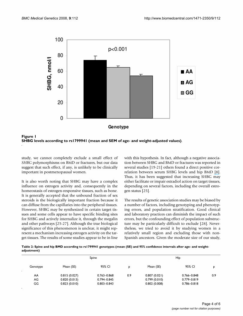
women of the control and osteoporotic subgroups.Among the polymorphisms, rs1799941 and rs6257showed a stronger association with serum SHBG thanrs6259 (Table 1). When the results were adjusted by ageand weight, the associations of rs1799941 and rs6257genotypes with serum SHBG remained statistically signif-icant (p = 0.0004 and 0.009, respectively). In Figure 1, theage and weight-adjusted SHBG levels across rs1799941genotypes are shown. The SHBG levels in AA homozy-gotes were 39% higher than in GG homozygotes. Basedon multiple linear regression models, age and weighttogether accounted for 10.5% of the variance of serumSHBG; the inclusion of the rs1799941 genotype explaineda further 3.7% of the SHBG variance. The inclusion ofother polymorphisms did not further increase the propor-tion of variance explained.
Serum SHBG showed a negative association with BMD (p= 0.040 at the spine and 0.014 at the hip). Thus, we nextdetermined the association between the rs1799941 alleleand BMD and osteoporotic fractures; however, there wereno genotype-related significant differences in the spine orhip BMD (Table 2), nor in the distribution of genotypesin women with and without fractures (Table 3). The studyhad 87% power to detect an association betweenrs1799941 polymorphisms and BMD if at least 2% of theBMD variance was explained. Likewise, the power todetect an association with vertebral and hip fractures was87% and 96%, respectively, assuming a risk ratio of 1.6.
DiscussionThe SHBG gene is located on chromosome 17 and themajor transcript is encoded by 8 exons, spanning approx-imately 3.2 Kb. In the present study, we report that severalpolymorphisms of the SHBG gene are strongly associatedwith serum SHBG levels in postmenopausal women. Inparticular, women with the AA genotype at the rs1799941locus had 39% higher SHBG levels than those with theGG genotype. This polymorphism is located in the 5'region of the gene, 8 nucleotides upstream of the tran-scription start site. Thus, it is well situated to influencegene transcription. In fact, higher SHBG serum levels have
also been recently reported in men and women bearing Aalleles, in comparison with those with G alleles. Dunninget al. [18] found that SHBG serum levels were 28% higherin women with the AA genotype than in those with theGG genotype. Similarly, Eriksson et al. [8] found 22% and26% higher SHBG levels in individuals with the AA geno-type in two cohorts of Swedish men. The C/T rs6257 pol-ymorphism was also associated with SHBG levels in thepresent study. The mechanism is unclear, as it is located inan intronic region (intron 1). A search of potentially func-tional polymorphisms with the PupaSuite web-based toolrevealed that it is located in a potential binding site for thetranscription factor hepatocyte nuclear factor 3/Fox. TheA/G rs6259 polymorphism is located in exon 8 and causesa change in the amino acid sequence of SHBG (Asn>Asp).It was also associated with SHBG levels, but to a lesserextent than the other polymorphisms. It has been sug-gested that the Asn>Asp modification affects a potentialglycosylation site and increases the half-life of the proteincoded for the less common allele [6]. Previous studies ofthe relationship of this SNP with serum SHBG in womenhave given conflicting results [6,9,18].
Our results show a clear association between severalSHBG gene polymorphisms and serum SHBG levels, butsince they are in linkage disequilibrium, we cannot estab-lish with certainty which polymorphism is truly responsi-ble for the association. On the other hand, we cannotexclude the possibility that other polymorphisms in link-age disequilibrium with those studied herein are actuallyresponsible for the association with SHBG levels [6,8].Serum SHBG showed a stronger association withrs1799941 than with other loci. However, allelic frequen-cies of rs1799941 were more balanced than those of otherpolymorphisms, which may favour the detection of asso-ciations between genotypes and SHBG levels.
Several authors have reported an association betweenserum SHBG and BMD or osteoporotic fractures [19-24].In the present study, we did not find an associationbetween SHBG alleles and BMD, despite the fact that theywere strongly associated with serum SHBG levels, whichin turn was associated with BMD. This lack of associationbetween SHBG genotype and BMD may be due merely tothe multiple factors influencing these traits and introduc-ing noise in the relationship between genotype, serumSHBG levels, and BMD. Although the association betweengenotypes and serum SHBG was statistically significantand quantitatively important (39% difference between AAand GG genotypes), there was wide inter-individual vari-ation, and thus rs1799941 alleles explained < 4% ofserum SHBG variance. Similarly, the contribution ofserum SHBG to BMD variance was rather small (SHBGexplained 2% of the BMD variance at the spine and 3% atthe hip). Thus, given the moderate sample size of this
Table 1: Factors associated with serum SHBG levels (Beta standarized regression coefficients and p values from univariate and multivariate regression analyses)
Univariate Age- and weight-adjusted
Beta p Beta p
Age 0.17 0.002Weight 0.28 0.00006rs6257 0.15 0.010 0.14 0.009rs1799941 0.14 0.0015 0.19 0.0004rs6259 0.11 0.048 0.06 0.27
Page 3 of 6(page number not for citation purposes)
BMC Medical Genetics 2008, 9:112 http://www.biomedcentral.com/1471-2350/9/112
study, we cannot completely exclude a small effect ofSHBG polymorphisms on BMD or fractures, but our datasuggest that such effect, if any, is unlikely to be clinicallyimportant in postmenopausal women.
It is also worth noting that SHBG may have a complexinfluence on estrogen activity and, consequently in thehomeostasis of estrogen-responsive tissues, such as bone.It is generally accepted that the unbound fraction of sexsteroids is the biologically important fraction because itcan diffuse from the capillaries into the peripheral tissues.However, SHBG may be synthesized in certain target tis-sues and some cells appear to have specific binding sitesfor SHBG and actively internalize it, through the megalinand other pathways [25-27]. Although the true biologicalsignificance of this phenomenon is unclear, it might rep-resent a mechanism increasing estrogen activity on the tar-get tissues. The results of some studies appear to be in line
with this hypothesis. In fact, although a negative associa-tion between SHBG and BMD or fractures was reported inseveral studies [19-21] others found a direct positive cor-relation between serum SHBG levels and hip BMD [8].Thus, it has been suggested that increasing SHBG mayeither facilitate or impair estradiol action on target tissues,depending on several factors, including the overall estro-gen status [25].
The results of genetic association studies may be biased bya number of factors, including genotyping and phenotyp-ing errors, and population stratification. Good clinicaland laboratory practices can diminish the impact of sucherrors, but the confounding effect of population substruc-ture may be particularly difficult to exclude [28]. Never-theless, we tried to avoid it by studying women in arelatively small region and excluding those with non-Spanish ancestors. Given the moderate size of our study,
SHBG levels according to rs1799941 (mean and SEM of age- and weight-adjusted values)Figure 1SHBG levels according to rs1799941 (mean and SEM of age- and weight-adjusted values).
0
20
40
60
80
100
Genotype
AA
AG
GG
p<0.001S
HB
G, n
mo
l/l
Table 2: Spine and hip BMD according to rs1799941 genotypes (mean (SE) and 95% confidence intervals after age- and weight-adjustment)
Spine Hip
Genotype Mean (SE) 95% CI p Mean (SE) 95% CI p
AA 0.815 (0.027) 0.763–0.868 0.9 0.807 (0.021) 0.766–0.848 0.9AG 0.820 (0.013) 0.794–0.845 0.799 (0.010) 0.779–0.819GG 0.823 (0.010) 0.803–0.843 0.802 (0.008) 0.786–0.818
Page 4 of 6(page number not for citation purposes)
BMC Medical Genetics 2008, 9:112 http://www.biomedcentral.com/1471-2350/9/112
a relatively small, but still clinically significant effect (i.e.,a 1.2 odds ratio for fracture) cannot be excluded. On theother hand, we cannot exclude the possibility that otherpolymorphisms in the SHBG gene region might have aninfluence on BMD or fracture risk. The SHBG gene is smalland only a few SNPs are included in the Hapmap data-base. Therefore, we focused on potentially functionalSNPs, rather than in haplotype-tagging SNPs.
ConclusionOur results show that apart from other factors, such as ageand body weight, some common genetic variants of theSHBG gene, and particularly an A/G polymorphism situ-ated in the 5' region, influence serum SHBG, but we havenot been able to demonstrate its association with eitherBMD or osteoporotic fractures.
Competing interestsThe authors declare that they have no competing interests.
Authors' contributionsJAR conceived and coordinated the study, carried out thestatistical analysis and wrote the manuscript draft. CV wasresponsible for the bone densitometry studies. MTZ wasresponsible for the genetic studies. MTGU carried out thebiochemical analyses. JAA participated in the design ofstudy and supervised the biochemical analyses. JGMhelped in the design of the study and made substantialcontributions to the interpretation of results. All authorsreviewed the first draft and read and approved the finalmanuscript.
AcknowledgementsWe acknowledge the technical assistance of Blanca Paule, Carolina Sañudo, Jana Arozamena, and Verónica Mijares, and the review of the manuscript style by David Cushley (International Science Editing).
Supported by FIS grant 06/34 and IFIMAV.
References1. Peacock M, Turner CH, Econs MJ, Foroud T: Genetics of oste-
oporosis. Endocr Rev 2002, 23:303-326.
2. Ralston SH, de Crombrugghe B: Genetic regulation of bone massand susceptibility to osteoporosis. Genes Dev 2006,20:2492-2506.
3. Riancho JA: Polymorphisms in the CYP19 gene that influencebone mineral density. Pharmacogenomics 2007, 8:339-352.
4. Strauss R, Barbieri J: Yen and Jaffe's reproductive endocrinology: physiol-ogy, pathophysiology and clinical management Philadelphia: Saunders;2004.
5. Abrahamson PE, Tworoger SS, Aiello EJ, Bernstein L, Ulrich CM, Gil-liland FD, Stanczyk FZ, Baumgartner R, Baumgartner K, Sorensen B,et al.: Associations between the CYP17, CYPIB1, COMT andSHBG polymorphisms and serum sex hormones in post-menopausal breast cancer survivors. Breast Cancer Res Treat2007, 105:45-54.
6. Cousin P, Calemard-Michel L, Lejeune H, Raverot G, Yessaad N, Emp-toz-Bonneton A, Morel Y, Pugeat M: Influence of SHBG genepentanucleotide TAAAA repeat and D327N polymorphismon serum sex hormone-binding globulin concentration inhirsute women. J Clin Endocrinol Metab 2004, 89:917-924.
7. Cui Y, Shu XO, Cai Q, Jin F, Cheng JR, Cai H, Gao YT, Zheng W:Association of breast cancer risk with a common functionalpolymorphism (Asp327Asn) in the sex hormone-bindingglobulin gene. Cancer Epidemiology, Biomarkers & Prevention 2005,14:1096-1101.
8. Eriksson AL, Lorentzon M, Mellstrom D, Vandenput L, Swanson C,Andersson N, Hammond GL, Jakobsson J, Rane A, Orwoll ES, et al.:SHBG gene promoter polymorphisms in men are associatedwith serum sex hormone-binding globulin, androgen andandrogen metabolite levels, and hip bone mineral density. JClin Endocrinol Metab 2006, 91:5029-5037.
9. Ferk P, Teran N, Gersak K: The (TAAAA)n microsatellite poly-morphism in the SHBG gene influences serum SHBG levelsin women with polycystic ovary syndrome. Hum Reprod 2007,22:1031-1036.
10. Haiman CA, Riley SE, Freedman ML, Setiawan VW, Conti DV, LeMarchand L: Common genetic variation in the sex steroid hor-mone-binding globulin (SHBG) gene and circulating shbglevels among postmenopausal women: the MultiethnicCohort. J Clin Endocrinol Metab 2005, 90:2198-2204.
11. Hogeveen KN, Talikka M, Hammond GL: Human sex hormone-binding globulin promoter activity is influenced by a(TAAAA)n repeat element within an Alu sequence. J BiolChem 2001, 276:36383-36390.
12. Kataoka N, Cai Q, Xu WH, Xiang YB, Cai H, Zheng W, Shu XO:Association of endometrial cancer risk with a functional pol-ymorphism (Asp(327)Asn) in the sex hormone-binding glob-ulin gene. Cancer 2007, 109:1296-1302.
13. Xita N, Tsatsoulis A, Chatzikyriakidou A, Georgiou I: Association ofthe (TAAAA)n repeat polymorphism in the sex hormone-binding globulin (SHBG) gene with polycystic ovary syn-drome and relation to SHBG serum levels. J Clin EndocrinolMetab 2003, 88:5976-5980.
14. Conde L, Vaquerizas JM, Dopazo H, Arbiza L, Reumers J, Rousseau F,Schymkowitz J, Dopazo J: PupaSuite: finding functional singlenucleotide polymorphisms for large-scale genotyping pur-poses. Nucleic Acids Res 2006, 34:W621-W625.
15. Reumers J, Conde L, Medina I, Maurer-Stroh S, Van Durme J, DopazoJ, Rousseau F, Schymkowitz J: Joint annotation of coding and
Table 3: Distributions of rs1799941 genotypes in control women and women with osteoporotic fractures (numbers (%) and odds ratios considering the most frequent genotype as the reference)
Controls Vertebral fractures Hip fractures
Genotype n (%) n (%) OR (95% CI) n (%) OR (95% CI)
AA 24 (7) 9 (6) 1.0 (0.5–2.3) 19 (6) 0.8 (0.4–1.5)AG 125 (40) 54 (37) 1.2 (0.8–1.8) 107 (37) 0.9 (0.6–1.2)GG 169 (53) 62 (57) reference 164 (57) reference
Total 318 (100) 145 (100) 290 (100)
Page 5 of 6(page number not for citation purposes)

BMC Medical Genetics 2008, 9:112 http://www.biomedcentral.com/1471-2350/9/112
Publish with BioMed Central and every scientist can read your work free of charge
"BioMed Central will be the most significant development for disseminating the results of biomedical research in our lifetime."
Sir Paul Nurse, Cancer Research UK
Your research papers will be:
available free of charge to the entire biomedical community
peer reviewed and published immediately upon acceptance
cited in PubMed and archived on PubMed Central
yours — you keep the copyright
Submit your manuscript here:http://www.biomedcentral.com/info/publishing_adv.asp
BioMedcentral
non-coding single nucleotide polymorphisms and mutationsin the SNPeffect and PupaSuite databases. Nucleic Acids Res2008, 36:D825-D829.
16. Yuan HY, Chiou JJ, Tseng WH, Liu CH, Liu CK, Lin YJ, Wang HH, YaoA, Chen YT, Hsu CN: FASTSNP: an always up-to-date andextendable service for SNP function analysis and prioritiza-tion. Nucleic Acids Res 2006, 34:W635-W641.
17. Barrett JC, Fry B, Maller J, Daly MJ: Haploview: analysis and visu-alization of LD and haplotype maps. Bioinformatics 2005,21:263-265.
18. Dunning AM, Dowsett M, Healey CS, Tee L, Luben RN, Folkerd E,Novik KL, Kelemen L, Ogata S, Pharoah PD, et al.: Polymorphismsassociated with circulating sex hormone levels in postmeno-pausal women. J Natl Cancer Inst 2004, 96:936-945.
19. Goderie-Plomp HW, van der KM, de Ronde W, Hofman A, de JongFH, Pols HA: Endogenous sex hormones, sex hormone-bind-ing globulin, and the risk of incident vertebral fractures inelderly men and women: the Rotterdam Study. J Clin Endocri-nol Metab 2004, 89:3261-3269.
20. Rapuri PB, Gallagher JC, Haynatzki G: Endogenous levels of serumestradiol and sex hormone binding globulin determine bonemineral density, bone remodeling, the rate of bone loss, andresponse to treatment with estrogen in elderly women. J ClinEndocrinol Metab 2004, 89:4954-4962.
21. Lormeau C, Soudan B, d'Herbomez M, Pigny P, Duquesnoy B, CortetB: Sex hormone-binding globulin, estradiol, and bone turno-ver markers in male osteoporosis. Bone 2004, 34:933-939.
22. Bjornerem A, Ahmed LA, Joakimsen RM, Berntsen GK, Fonnebo V,Jorgensen L, Oian P, Seeman E, Straume B: A prospective study ofsex steroids, sex hormone-binding globulin, and non-verte-bral fractures in women and men: the Tromso Study. Eur JEndocrinol 2007, 157:119-125.
23. Bjornerem A, Emaus N, Berntsen GK, Joakimsen RM, Fonnebo V,Wilsgaard T, Oian P, Seeman E, Straume B: Circulating sex ster-oids, sex hormone-binding globulin, and longitudinal changesin forearm bone mineral density in postmenopausal womenand men: the tromso study. Calcif Tissue Int 2007, 81:65-72.
24. Araujo AB, Travison TG, Leder BZ, McKinlay JB: Correlationsbetween serum testosterone, estradiol, and sex hormone-binding globulin and bone mineral density in a diverse sam-ple of men. J Clin Endocrinol Metab 2008, 93:2135-2141.
25. Khosla S: Sex hormone binding globulin: inhibitor, facilitator(or both) of sex steroid action? J Clin Endocrinol Metab 2006,91:4764-4766.
26. Kahn SM, Hryb DJ, Nakhla AM, Romas NA, Rosner W: Sex hor-mone-binding globulin is synthesized in target cells. J Endocri-nol 2002, 175:113-120.
27. Caldwell JD, Suleman F, Jirikowski GF: Internalization of sex hor-mone binding globulin into fibroblast 3T3 cells. Horm MetabRes 2007, 39:620-622.
28. Cardon LR, Palmer LJ: Population stratification and spuriousallelic association. Lancet 2003, 361:598-604.
Pre-publication historyThe pre-publication history for this paper can be accessedhere:
http://www.biomedcentral.com/1471-2350/9/112/prepub
Page 6 of 6(page number not for citation purposes)


