Embed Size (px)
Citation preview

1. Introduction
Legumes are the most important group of crop plants nextto cereals and much effort has been devoted to develop effi-cient in vitro regeneration systems because of their recalci-trance to tissue culture regeneration. Among them, peanut isan important cash crop for farmers particularly in the semi-
arid tropics. Asian land races of peanut have a narrow genet-ic base and low genetic diversity because of which they lackresistance to many pests and diseases. These can causemajor losses in terms of quality and quantity. Genetic trans-formation offers a solution to these problems by making thetransfer of genes from alien sources feasible for generatingtransgenic plants possessing resistance to biotic and abiotic
Genetic transformation of peanut (Arachis hypogaea L.) usingcotyledonary node as explant and a promoterless gus::nptII
fusion gene based vector
T SWATHI ANURADHA, S K JAMI, R S DATLA† and P B KIRTI*Department of Plant Sciences, University of Hyderabad, Hyderabad 500 046, India
†Plant Biotechnology Institute, Saskatoon, SK S7N 0W9, Canada
*Corresponding author (Fax, 91-40-23010120; Email: [email protected])
We have generated putative promoter tagged transgenic lines in Arachis hypogaea cv JL-24 using cotyledonary node(CN) as an explant and a promoterless gus::nptII bifunctional fusion gene mediated by Agrobacterium transformation.MS medium fortified with 6-benzylaminopurine (BAP) at 4 mg/l in combination with 0.1 mg/l α-napthaleneacetic acid(NAA) was the most effective out of the various BAP and NAA combinations tested in multiple shoot bud formation.Parameters enhancing genetic transformation viz. seedling age, Agrobacterium genetic background and co-cultivationperiods were studied by using the binary vector p35SGUSINT. Genetic transformation with CN explants from 6-day-old seedlings co-cultivated with Agrobacterium GV2260 strain for 3 days resulted in high kanamycin resistant shootinduction percentage (45%); approximately 31% transformation frequency was achieved with p35S GUSINT in β-glu-curonidase (GUS) assays. Among the in vivo GUS fusions studied with promoterless gus::nptII construct, GUS-posi-tive sectors occupied 38% of the total transient GUS percentage. We have generated over 141 putative T0 plants byusing the promoterless construct and transferred them to the field. Among these, 82 plants survived well in the greenhouse and 5 plants corresponding to 3.54% showed stable integration of the fusion gene as evidenced by GUS,polymerase chain reaction (PCR) and Southern blot analyses. Twenty-four plants were positive for GUS showingeither tissue-specific expression or blue spots in at least one plant part. The progeny of 15 T0 plants indicatedMendelian inheritance pattern of segregation for single-copy integration. The tissue-specific GUS expression patternswere more or less similar in both T0 and corresponding T1 progeny plants. We present the differential patterns of GUSexpression identified in the putative promoter-tagged transgenic lines in the present communication.
[Anuradha T S, Jami S K, Datla R S and Kirti P B 2006 Genetic transformation of peanut (Arachis hypogaea L.) using cotyledonary node asexplant and a promoterless gus::nptII fusion gene based vector; J. Biosci. 31 235–246]
http://www.ias.ac.in/jbiosci
Abbreviations used: AMV, Alfalfa mosaic virus; BAP, 6-benzylaminophrine; CN, cotyledonary node; GUS, β-glucuronidase;NAA, α-naphthaleneacetic acid ; PCR, polymerase chain reaction; RIM, root induction medium.
Keywords. Agrobacterium tumefaciens; cotyledonary node; gus::nptII bifunctional fusion gene; peanut; promoter tagged lines
J. Biosci. 31(2), June 2006, 235–246, © Indian Academy of Sciences 235

stresses. Improving transformation frequency remains themost important factor in plant transgene technology(Gheysen et al 1998). Genetic modification of plants isdependent on tissue-specific promoters, as the targetedexpression of transgenes is needed in many cases to avoidundesirable ectopic expression with widely used constitu-tive promoters. Promoters offer a fundamental control ingene expression and there is a great deal of interest in iso-lating and characterizing plant promoters worldwide (Datlaet al 1997). The present study involves a promoter taggingapproach with a promoterless gus::nptII bifunctional fusiongene (Datla et al 1991). This strategy based on T-DNAinsertional mutagenesis was exploited earlier to identifyplant promoters in different plants like tobacco (Andre et al1986; Fobert et al 1994), Arabidopsis (Koncz et al 1989;Kertbundit et al 1991; Topping et al 1994) and potato(Lindsey et al 1993). Vector constructs containing either areporter gene or a marker gene lacking promoter elementshave been used for tagging (Teeri et al 1986). T-DNA inser-tional mutagenesis acts as a powerful tool in the isolationand characterization of plant regulatory sequences / pro-moters (Walden 2002). A similar tagging approach in tobac-co using a promoterless lacZ::nptII fusion gene was carriedout (Suntio and Teeri 1994). However, expression analysiswas difficult because of the complexity of lacZ:: nptIIfusion gene. Recently, by employing hypocotyls as explantsand a promoterless gus::nptII fusion gene, promoter taggedlines were produced and genomic sequences upstream ofthis construct were isolated via plasmid rescue (Bade et al2003). Among forage legumes, T-DNA insertional mutage-nesis programme in the model legume Medicago truncatu-la revealed that several genes were tagged in the transgeniclines and in vivo GUS fusions were also obtained (Scholteet al 2002). A promoter tagging approach in the legume,Lotus japonicus was initiated to identify plant genesinvolved in the nitrogen fixation (Webb et al 2000). A highfrequency transformation protocol is required for generatingT-DNA tagged lines. The recalcitrance of many legumes totissue culture response and plant regeneration has drivenresearchers to develop alternate transformation systems thattarget axillary meristem in the cotyledonary nodes (Somerset al 2003). We report here for the first time the mode ofgenetic transformation using cotyledonary node (CN) as anexplant in peanut mediated through Agrobacterium tumefa-ciens. The main objective of this work is to create tran-scriptional gene fusions between the upstream promoter ele-ments in the plant genome and the downstream fusion geneand thus the reporter gene expression can be assayed. Byusing CN as an explant and a promoterless gus::nptII fusionvector, we have generated T-DNA tagged lines with a widerange of β-glucuronidase (GUS) expression patterns. Theintegration was confirmed by polymerase chain reaction(PCR) and Southern analyses in some of these plants. The
inheritance of the gus::nptII bifunctional fusion gene in theT1 generation was confirmed by GUS and PCR analysesand followed Mendelian pattern of segregation. To the bestof our knowledge this is the first report on promoter taggingin grain legumes.
2. Materials and methods
2.1 Plant material and explant preparation
Mature Arachis hypogaea cv. JL-24 seeds were surface ster-ilized by rinsing with 70% (v/v) ethanol for 1 min and 7 minwith 0.1% (w/v) aqueous mercuric chloride followed byseveral washes with sterile double distilled water. Theywere then imbibed in sterile double distilled water for 4-6 hand germinated on autoclaved filter paper wicks soaked insterile double distilled water. Explants were prepared from1-6-day-old seedlings in the initial experiments to evaluatethe influence of explant age on transformation frequency. Inthe subsequent experiments, 6-day-old seedlings were used.The seed coat and the radicle were removed and the cotyle-donary nodes were excised by cutting both epicotyls andhypocotyls approximately 2 mm above and below the nodalregion and the embryo axis was bisected along the longitu-dinal plane. The meristematic region present in the nodalregion was macerated by 6–8 diagonal shallow cuts by asterile surgical blade and the cotyledons along with theirnodes were embedded into the shoot induction medium(SIM) in such a way that the wounded meristematic nodalregion and the adaxial surface of the cotyledon were indirect contact with the medium. From each seed, twoexplants were obtained. The explant is now designated asCN. The method of explanting has been adapted fromTownsend and Thomas (1993).
2.2 Regeneration
Regeneration with CN explant was standardized by testingvarious combinations of growth regulators like 6-benzy-laminopurine (BAP), kinetin (KN), 2,4-dichloro-phenoxy-acetic acid (2,4-D) and α-napthaleneacetic acid (NAA) withMS basal salts (Murashige and Skoog 1962), pH 5.6–5.8before autoclaving and 3% sucrose as a carbon source.Explants were cultured on agar (0.8%) solidified SIM con-taining 4 mg/l BAP and 0.1 mg/l NAA that induced maxi-mum number of shoots per explant. The cultureswere maintained at 28 ± 1°C under a continuous 16/8 h(light/dark) photoperiod with light supplied by cool whitefluorescent lamps at an intensity of about 1600 lux.Subcultures were done at 15-day interval with 2-week dura-tion each on SIM for the development of adventitious shootbuds. Explants producing multiple shoots were transferred
T Swathi Anuradha et al236
J. Biosci. 31(2), June 2006

to shoot elongation medium (SEM) comprising 2 mg/l BAPand 0.1 mg/l NAA. Elongated shoots were cut at the inter-nodal region and transferred to root induction medium(RIM) solidified with 5% agargel (Sigma, USA) comprisingof 0.8 mg/l NAA with MS basal salts.
2.3 Plasmid construct and Agrobacterium strain
A derivative of binary vector pRD400, harbouring a pro-moterless synthetic gus::nptII bifunctional fusion gene (Datlaet al 1991) with an alfala mosaic virus (AMV) translationalenhancer (Datla et al 1993) at the 5′ end and nos terminatorwas used to transform peanut through a disarmed rifampicinand carbenicillin resistant Agrobacterium tumefaciensstrain, GV2260. The strain was maintained on solidifiedLuria agar plates with 100 mg/l rifampicin, 100 mg/l car-benicillin and 50 mg/l kanamycin monosulphate. A freshovernight culture of the Agrobacterium, obtained by inocu-lating single colony in Luria broth containing appropriateantibiotics with an OD600nm 0.8, was pelleted by centrifu-gation at 5000 rpm for 5 min, resuspended in sterile doubledistilled water and stored at 4°C for 2 h before infection. Abinary vector p35SGUSINT (Vancanneyt et al 1990) wasused initially to optimize the transformation conditions.
2.4 Transformation
Freshly cut CN explants were infected by dipping in thebacterial suspension by their proximal cut ends and incu-bated for 5–10 min in 90 × 15 mm sterile petridishes. Theywere co-cultivated on SIM for 72 h in 16/8 h photoperiod at25 ± 2°C. After a 4-day recovery period on SIM containing250 mg/l cefotaxime, explants were transferred to a freshSIM supplemented with 175 mg/l kanamycin monosulphate(Sigma, USA) and 250 mg/l cefotaxime. Cultures with com-pletely green shoots were maintained on SEM with 175mg/l for two selections of two-week duration each. Cultureswith green and white shoots intermingled were transferredto medium without kanamycin and there after maintainedon MS medium without any selection agent. Shoots withtwo internodes were cut and transferred to RIM for rootinduction. Plants with well-developed roots were trans-planted to autoclaved soil and vermiculite mixture (1:1ratio) in plastic pots and were hardened under culture con-ditions for 2 weeks prior to their transfer to greenhouse.Later they were transferred to bigger pots and allowed toflower and set seed in the greenhouse.
2.5 Selection
An effective kanamycin concentration for the selection oftransformed shoots was initially standardized by culturing
control untransformed CN explants on SIM containing dif-ferent concentrations of kanamycin (100, 125, 150, 175,200, 250 mg/l). Two subcultures were carried out on freshSIM having the same level of antibiotic and then scored forthe regeneration percentage and number of shoots perexplant. Similarly, kanamycin concentration that inhibitedroot formation was determined by transferring controlshoots (2–3 cm in length) regenerated from non-trans-formed explants to RIM supplemented with differentkanamycin levels (20, 30, 40, 50, 60, 70 mg/l etc.).
2.6 GUS analysis
Phenotypic GUS expression was determined by stainingunfixed leaflets, roots, flower and shoot parts of over 141putative T0 plants analysed so far. GUS assay was carriedout by incubating the above plant tissues from the putativetransformants in a buffer comprising 1 mM X-Gluc (5-bromo-4-chloro-3-indolyl β-D-glucuronic acid, Biosynth,Switzerland), 100 mM sodium phosphate (pH 7.2), 0.1%Triton X-100, 0.5 mM potassium ferricyanide and 10%methanol (Jefferson et al 1987) overnight at 37°C andstained tissues were cleared of chlorophyll by soaking in70% ethanol. GUS assays were also performed with theprogeny of 15 T0 plants. Transient GUS assays were done intriplicate to study in vivo GUS fusions, by staining youngleaflets and shoot parts, which were picked up randomlyfrom the cultures.
2.7 DNA isolation and PCR analysis
Total genomic DNA was extracted from young leaves ofputative transformants and control plants using standardCTAB method (Doyle and Doyle 1987). Plants were keptunder shade for 2 days prior to harvesting leaves from them.The PCR was performed to screen putative T0 transfor-mants and T1 generation plants for the presence of thefusion gene by using specific primers for nptII and uidAgenes. The PCR reactions were carried out using 100 ng ofpurified genomic DNA as template and 2.5 U of recombi-nant Taq DNA Polymerase (Invitrogen Corporation, SaoPaulo, Brazil). The 700 bp of the nptII fragment was ampli-fied by using 21-mer oligonucleotide primers (nptIIF5′-GAGGCTATTCGGCTATGACTG-3′ and nptIIR 5′-ATCGGGAGCGGCGATACGTA-3′). The cycling condi-tions comprised an initial denaturation at 94°C for 4 min,followed by 30 cycles of 94°C for 1 min, 61°C for 45 s,72°C for 1 min and a final extension of 10 min at 72°C. The469 bp GUS fragment was amplified by using 22-meroligonucleotide primers (GUSF 5′-TACCTCGCATTAC-CATTACGCG-3′ and GUSR 5′-CTTCTCTGCCGTTTC-CAAATCG-3′). Cycling conditions were similar as in PCR
Promoter tagging in peanut 237
J. Biosci. 31(2), June 2006

using nptII primers except for the annealing temperature at63°C for 55 s. The amplified products were electrophoresedon 1.2% agarose gels and visualized with ethidium bromide.The fragments resolved on agarose gels were transferred toHybond N+ by Southern blotting and the blots werehybridized with GUS fragment, PCR amplified from therespective plasmid labelled with α–32P.
2.8 Southern hybridization
Genomic DNA (15 µg) from T1 plants was separatelydigested to completion with HindIII that does not cut with-in the T-DNA region and the restriction fragments wereresolved by electrophoresis on 0.8% agarose gels and blot-ted by capillary method onto Hybond N+ membrane(Amersham Pharmacia, UK) using 20X SSC as a transferbuffer. Membranes were probed with α-32P dATP labelled700 bp nptII fragment amplified from the plasmid havinggus::nptII gene. Following 16 h of hybridization at 65°C,membranes were washed for 20 min each at 65°C in 2XSSC, 0.1% SDS, 1X SSC, 0.1% SDS and finally with 0.1XSSC, 0.1% SDS for 10 min. The washed membranes werewrapped in saran wrap and subjected to autoradiography(Sambrook et al 1989).
2.9 Statistical methods
Standard statistical methods were used. Since the number ofseeds obtained in transgenic peanut plants would be insuffi-cient to conduct Chi-square test on individual progeniesexcept three, Brandt and Snedecor’s rows and columnsmethod was used in Chi-square analysis (Bailey 1965).For the three progenies with a minimum population of 12,Chi-square test was conducted individually also.
3. Results
3.1 Regeneration
Cotyledonary node explants excised from 1-8-day-oldseedlings were cultured on different media with variousgrowth regulator combinations for multiple shoot induction(data not presented). Six-day old seedlings responded wellin terms of regeneration and transformation (figure 1). Thepercentage regeneration and average number of shoots werehighest on MS media fortified with BAP and NAA. Amongthe various BAP and NAA combinations tested, high fre-quency regeneration was obtained following culture ofexplants on MS medium supplemented with 3, 4 and 5 mg/lBAP along with 0.1 and 0.2 mg/l NAA (table 1). BAP at 4mg/l and 0.1 mg/l NAA was most effective for multipleshoot bud formation. Both the percent regeneration as well
as the average number of shoot buds per explant was foundto be higher (82% regeneration, 28 ± 1.5), when the adaxi-al side of the cotyledon was in direct contact with themedium compared to the abaxial side (58% regeneration,8.75 ± 2.4). Elongated shoots when cultured on RIM devel-oped adventitious roots within 15 days of culture. An aver-age of 6 plants were recovered from each explant.
3.2 Genetic transformation
To evaluate the transient GUS frequency as well as the stabletransformation efficiency, a number of parameters enhanc-ing genetic transformation were studied by using binaryvector p35SGUSINT. Optimized conditions determined werefollowed in the subsequent experiments. Parameters that
T Swathi Anuradha et al238
J. Biosci. 31(2), June 2006
Effect of age of explant
Age of the explant-Days
0 1 2 3 4 5 6 7 8
0
10
20
30
40
50
Kanamycin resistant shoot induction
percentage
Uid A percentage
Kan
am
yci
n r
esis
tan
t sh
oot
ind
uct
ion
p
erce
nta
ge
& Uid
A p
erce
nta
ge
Figure 1. Effect of explant age in days on regeneration and uidApercentage.
Table 1. Effect of different concentrations of BAP and NAA onregeneration from 6-day-old CN explants of A. hypogaea cv JL-24.
MS mediumsupplemented with Average number
BAP and Regeneration of shoot buds / NAA (mg/l) (%) explant ± SE
3 0.1 69 19 ± 2.14 0.1 82 28 ± 1.55 0.1 76 21 ± 23 0.2 64 16 ± 2.54 0.2 73 23 ± 1.755 0.2 71 14 ± 3.4
Each mean value was an average calculated from three experi-ments ± SE.Optimum growth regulator combination has been shown in boldface.

were tested included the seedling age, Agrobacterium strainsand co-cultivation periods. The total number of GUS spotsand GUS positive sectors on different leaf and shoot partswere scored. The GUS positive sectors are the deeply stainedblue regions on different plant parts such as leaves, roots,stem etc. Kanamycin resistant shoot induction percentagewas calculated at the end of first subculture as the percent-age of shoots growing on medium containing kanamycin tothe total number of explants cultured. Transient transforma-tion frequency was determined 72 h after co-cultivation inorder to assess the efficiency of transformation and is thepercentage of explants showing at least one discrete darkblue GUS positive sector or GUS spot, whereas stable trans-formation efficiency is the percentage of transformed shootsshowing positive for GUS staining, PCR and Southernanalysis (Egnin et al 1998). For each factor tested, at leastthree experiments were performed and a minimum of 20explants was used in each experiment. Kanamycin resistantshoot induction percentage as well as transient transforma-tion frequencies were studied in these experiments.
3.2a Effect of age of explant: Cotyledonary nodes obtainedfrom 1-8-day-old seedlings were used as explants and twotransformation experiments were carried out in order todetermine the transient GUS expression as well askanamycin resistant shoot regeneration percentage.Transient transformation frequency or GUS staining per-centage in 1, 2 and 3-day-old CN explants after co-cultiva-tion was low when compared with that of 6-day-old CNexplants. The possible fact could be that the surface area ofthe existing meristematic cells might be more in the 6-day-old CN explants facilitating efficient T-DNA transfer.
The transient GUS percentage in both 5- and 6-day-oldexplants after co-cultivation was more or less similar but theydiffered in shoot regeneration response; hence the latter wasemployed for further experiments (figure 1). The possible rea-son can be attributed to the actively dividing state of explantcells during 5–6 days of development whereas, the low uidAand regeneration response of 8-day-old CN explants could bedue to the further differentiation of primary meristem and theavailability of less number of actively dividing cells.
3.2b Effect of Agrobacterium genetic background: The tran-sient GUS percentage was studied with two disarmed Agro-bacterium strains, GV2260 and LBA4404. Of the two strains,GV2260 was found to enhance transient transformationefficiency of 6-day-old nodes by at least 3 times compared toLBA4404. Also, more dark blue GUS positive sectors wereobserved in cultures with strain GV2260. Hence, GV2260 wasused in subsequent experiments (data not shown).
3.2c Effect of co-cultivation period: When 6-day-oldexplants were co-cultivated with GV2260 for 1, 2 and 3days, kanamycin resistant shoot regeneration percentage
was high with 1-day-old co-cultivated explants and decreasedgradually within 2 and 3 days (figure 2). Transient GUSstaining percentage increased considerably when explantswere co-cultivated for 1–3 days. When the co-cultivationperiod exceeded 3 days, it resulted in necrosis of theexplants. The high shoot induction percentage of 1- and 2-day-old co-cultivated explants is likely due to the reduceddamage caused by Agrobacterium during co-cultivation.Approximately, 31% of transformation frequency wasobserved with p35SGUSINT based on transient GUS stain-ing. The data obtained so far was collected based ontransient GUS analyses.
3.3 Selection
Further, we determined the optimal concentration ofkanamycin for the selection of transformed shoots by cul-turing the uninfected control CN explants on SIM contain-ing kanamycin in dose-dependent concentrations (100, 125,150, 175, 200 and 250 mg/l). Kanamycin resistant shootinduction percentage decreased with increasing kanamycinconcentration (data not shown). At 175 mg/l kanamycin,regeneration percentage decreased drastically and theshoots produced were chlorotic. Hence, this concentrationwas chosen for the selection of putative transformants.Explants showed necrosis and shoot induction diminishedcompletely at kanamycin concentrations 200 and 250 mg/l.Similarly, a kanamycin concentration of 60 mg/l causedcomplete inhibition of root induction from control shoots(data not shown). It is well known that root induction ismore sensitive to kanamycin than shoot organogenesis andfurther explains the fact that the utility of any antibioticdepends on both plant species as well as the explantinvolved (Saini et al 2003).
Promoter tagging in peanut 239
J. Biosci. 31(2), June 2006
Effect of cocultivation period
Cocultivation period in days
0 1 2 3 4
0
10
20
30
40
50
60
70Kanamycin resistant shoot induction percentage
Uid A percentage
Kan
am
yci
n r
esis
tan
t sh
oot
ind
uct
ion
p
erce
nta
ge
& Uid
A p
erce
nta
ge
Figure 2. Effect of co-cultivation period on regeneration anduidA percentage.

3.4 Regeneration of transformants
Genetic transformation experiments with 6-day-old CNexplants co-cultivated with GV2260 carrying p35SGUSINTfor 3 days resulted in more number of kanamycin resistantshoots and higher frequency of GUS positive sectors intransient GUS analysis. Further, transformation experi-ments were conducted using a promoterless gus::nptIIbifunctional fusion gene construct following the parametersinvestigated thus far. After co-cultivation and a short recov-ery period, explants were cultured on SIM supplementedwith 175 mg/l kanamycin until two selections and latersome of the cultures were maintained on medium free ofany selection agent. To study the transient GUS expression,different tissues were collected randomly from the culturesand stained in the X-Gluc solution. Among in vivo GUSfusions, the range of GUS expression patterns include GUSpositive sectors on leaves, shoot parts, brown calli, deepblue staining along the midrib of leaves, cut regions ofleaves and also GUS spots on various plant parts. The GUSpositive sectors on the leaves were found to occupy maxi-mum (38%) of the total transient GUS percentage. The totalnumber of GUS positive sectors on shoot parts and browncalli were found to be less than the number of GUS spots onleaves and petiole (figure 3). No staining was observed inany tissue of the untransformed shoots studied. Theseresults suggest that the GUS expression observed wasonly because of the productive transcriptional fusionsbetween the upstream regulatory elements in the plantgenome and the downstream gus::nptII fusion gene devoidof promoter in the vector. However, the appearance ofGUS spots on leaves clearly infers that some of the shoots
were chimeric and does not necessarily indicate tissue-spe-cific expression.
We have generated over 141 putative T0 plants by usingthe promoterless construct (figure 4A) and transferred themto green house. Among these, 82 plants survived well in thegreenhouse and 5 plants corresponding to 3.54% showedstable integration of the fusion gene as evidenced by GUS,PCR and Southern analyses. Three out of thirty three shootsrooted well on RIM with kanamycin. These plants werephenotypically similar to normal control plants. Figure 4Brepresents different regeneration stages of peanut fromexplant stage to plant development.
3.5 GUS expression in individual transgenic plants
In order to investigate the uidA gene expression patterns invarious plant tissues, GUS assays were carried out withleaves, roots, shoot parts and floral organs of over 141 T0putative transformants. Twenty-four plants were found to bepositive for GUS showing either tissue-specific expressionor blue spots in at least one plant part. GUS studies in flow-ers revealed that 3 putative transformants out of 24 wereGUS positive exhibiting differential uidA expression pat-terns. These plants did not reveal any detectable GUS stain-ing in any other plant parts studied. Relatively high fre-quency of GUS staining was observed in leaves of putativetransformants (75%; 18/24), while in roots it was low(41.66%; 10/24). Among these, only 16.66% of the trans-formants (4/24) exhibited GUS expression in shoot regions.Plants that have been rooted on RIM supplemented withkanamycin were found to be positive for GUS, PCR andSouthern hybridization. Faint blue colouration wasobserved in many of the plant parts that were analysed forGUS. Deep blue sectors were found on the leaves of plantC-53 (figure 4C.). Plants C-23, C-27, C-28, C-31, C-33, C-44 and C-52 did not show any GUS staining in the subse-quent assays and the blue spots observed in these plants canbe considered as artifacts. The cultures, from which theseplants were obtained, were initially maintained on SIM con-taining 175 mg/l kanamycin for up to two selections andlater transferred on to a medium free of any selective agent.Some of these GUS positive plants can be considered asputative promoter tagged lines. These results indicate that,within a population of transformed plants, expression of apromoterless gus::nptII gene occurs at high frequency in awide range of plant parts.
3.6 Segregation analysis of the uidA expression inthe progeny
It is necessary to characterize the inheritance pattern offusion gene in the T1 generation in order to understand the
T Swathi Anuradha et al240
J. Biosci. 31(2), June 2006
% Gus +ve sectors on leaves
% Gus +ve sectors on shootparts
% Gus spots on leaves and petiole
% Gus expression observed along the midrib region
% Gus +ve sectors on brown calli
21
12
38
11
18
Figure 3. Distribution of in vivo GUS fusions among cultures.

Promoter tagging in peanut 241
J. Biosci. 31(2), June 2006
2.5 kb
(A)
(B)
(C)
BamHI EcoRI BglII
Not I
RB AMV GUS:NPT II NOS TER MCS LB
Figure 4. (A) Synthetic T-DNA region showing the AMV enhancer sequence, promoterless gus:: nptII bifunctional fusion gene, NOS-TER, Nopaline synthase terminator; RB, right border; LB, left border and unique restriction sites. (B) Regeneration and Agrobacterium-mediated transformation with CN in A. hypogaea CV JL-24. (a) CN explants showing multiple bud induction after three weeks on SIMcontaining 175 mg/l kanamycin. (b) Cultures with complete green multiple shoots after six weeks on SIM containing 175 mg/l kanamycin.(c) Completely bleached control cultures on SIM containing 175 mg/l kanamycin. (d) Profusely rooted shoot on RIM. (e) Hardening ofplant in soil: vermiculite mixture. (f) Acclimatization of plant in greenhouse conditions. (C) GUS expression observed in various plantparts of different T0 plants. (a) Control JL-24 roots. (b) GUS expression in root tip. (c) GUS expression in roots. (d) Control JL-24 flower.(e and f) GUS expression in complete flower of C-1 plant showing stained floral parts. (g) GUS stained keel petal and pedicel of plantC-2. (h) GUS expression only in keel petal of plant C-13. (i) GUS expression in leaves in contrast to bleached control leaf.

stability of foreign gene integration. The progeny of 15GUS positive plants were analysed to study the segregationpattern of the fusion gene. For undertaking the Chi-squaretest for segregation, the minimum population size is 12.However, we have the minimum population size only inthree samples and they showed good fit for monogenicsegregation suggesting single copy integration. However,for none of these three lines, Southern data were available.Chi-square test was also conducted on pooled data from allthe lines following the rows and columns method, whichindicated a fit for monohybrid ratio indicating single copyintegration. However, plant No. C-43 has been observed tohave two copies of the T-DNA in Southern analysis. Rootand leaf-specific expression was found to be predominantamong the progeny of plants C-12 and C-17 (table 2). Thetissue-specific GUS expression patterns were more or lesssimilar in both parent and progeny plants. Segregation ofgus and nptII genes in the progenies was confirmed throughPCR.
3.7 PCR analysis and Southern hybridization
The presence of the fusion gene was confirmed in about 10T0 putative transformants by PCR using specific primers foruidA and nptII genes. To check the fidelity of theamplicons in PCR reaction, the PCR products were
transferred to nylon membranes for Southern hybridizationand probed with radiolabelled GUS fragment. This experi-ment confirmed the transgenic nature of the T0 plants.Figure 5A shows the Southern of PCR products from 12 T0plants. The PCR screening was carried out in the T1 proge-nies in order to ascertain the inheritance pattern of theintegrated fusion gene. Figure 5B shows the amplificationof nptII and uidA genes in the progeny of 5 PCR confirmedT0 plants.
Southern hybridization analysis revealed the integrationand copy number of nptII gene among the progeny of 5 indi-vidual T0 plants analysed. Digestion of genomic DNA ofT1 plants with HindIII that does not cut internally within theT-DNA region of pRD400 gus::nptII fusion gene shouldyield unique restriction patterns when probed with a GUS ornptII fragment and the number of bands should correspondto the copy number.
Southern analysis of the progeny of the 5 GUS or PCRconfirmed T0 plants showed differential banding patternsupon probing with nptII fragment. Plants C-1a, C-17a, C-4aand C-53a were shown to contain independent single copyinsertions whereas; C-43a plant showed two copy insertionsrespectively (figure 6). The hybridized bands were greaterthan the size of T-DNA and this confirms the integrationof T-DNA into plant genome suggesting the origin ofplants as a result of independent transformation events.
T Swathi Anuradha et al242
J. Biosci. 31(2), June 2006
Table 2. Segregation pattern of the GUS expression in T1 generation.
Copies of the fusion
gene integrated Total No.PCR (Southern of seeds GUS and GUS and Chi-square Probability Segregation
Plant amplification analysis) collected PCR +ve PCR -ve value (P) ratio
C-53 + +/1 9 8 1C-4 + +/1 6 5 1C-17 + +/1 12 9 3 0 0.001 3:1C-12 + _ 5 4 1C-25 + _ 21 17 4 0.209 0.05 3:1C-43 + +/2 7 3 4C-47 + _ 2 2 0C-11 + _ 7 6 1C-1 + +/1 7 7 0C-13 + _ 11 7 4C-51 + _ 18 15 3 0.15 0.05 3:1C-2 + _ 4 1 3C-50 + _ 11 5 6C-48 + _ 6 3 3C-49 + _ 6 2 4
Minimum sample size for Chi-square test is 12. Only three samples have the minimum size of twelve and Chi-square test was conductedon these samples.Brandt and Snedecor’s rows and columns method was also used in Chi-square analysis (Bailey 1965) on the pooled data of GUS positiveand negative segregation with a Chi-square value =24.9887, degrees of freedom 14, and probability P ≤ 0.05 indicating a fit for monohy-brid ratio for all lines. Plant C-43 has been observed to have two copies of the transgene.
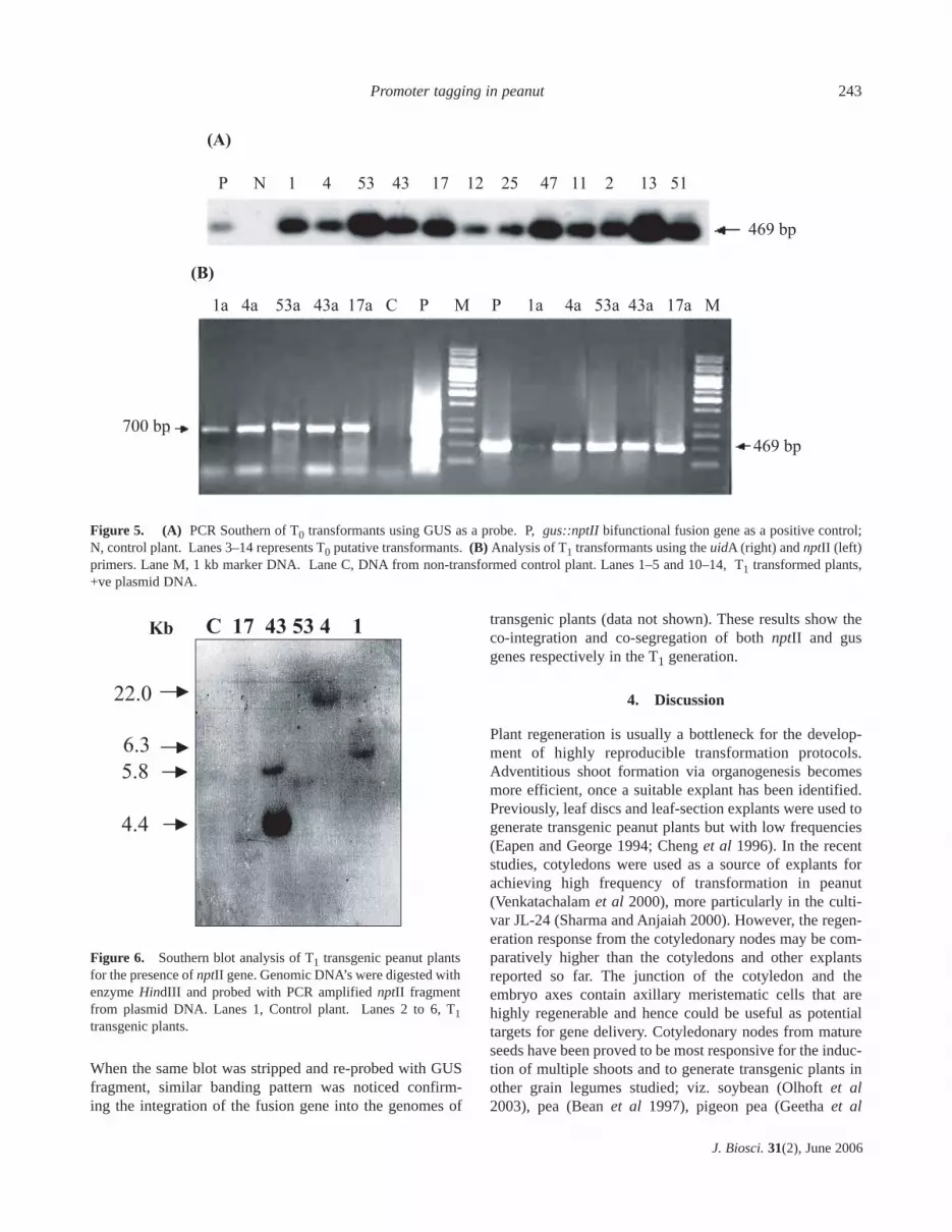
When the same blot was stripped and re-probed with GUSfragment, similar banding pattern was noticed confirm-ing the integration of the fusion gene into the genomes of
transgenic plants (data not shown). These results show theco-integration and co-segregation of both nptII and gusgenes respectively in the T1 generation.
4. Discussion
Plant regeneration is usually a bottleneck for the develop-ment of highly reproducible transformation protocols.Adventitious shoot formation via organogenesis becomesmore efficient, once a suitable explant has been identified.Previously, leaf discs and leaf-section explants were used togenerate transgenic peanut plants but with low frequencies(Eapen and George 1994; Cheng et al 1996). In the recentstudies, cotyledons were used as a source of explants forachieving high frequency of transformation in peanut(Venkatachalam et al 2000), more particularly in the culti-var JL-24 (Sharma and Anjaiah 2000). However, the regen-eration response from the cotyledonary nodes may be com-paratively higher than the cotyledons and other explantsreported so far. The junction of the cotyledon and theembryo axes contain axillary meristematic cells that arehighly regenerable and hence could be useful as potentialtargets for gene delivery. Cotyledonary nodes from matureseeds have been proved to be most responsive for the induc-tion of multiple shoots and to generate transgenic plants inother grain legumes studied; viz. soybean (Olhoft et al2003), pea (Bean et al 1997), pigeon pea (Geetha et al
Promoter tagging in peanut 243
J. Biosci. 31(2), June 2006
(A)
(B)
P N 1 4 53 43 17 12 25 47 11 2 13 51
469 bp
1a 4a 53a 43a 17a C P M P 1a 4a 53a 43a 17a M
469 bp
700 bp
Figure 5. (A) PCR Southern of T0 transformants using GUS as a probe. P, gus::nptII bifunctional fusion gene as a positive control;N, control plant. Lanes 3–14 represents T0 putative transformants. (B) Analysis of T1 transformants using the uidA (right) and nptII (left)primers. Lane M, 1 kb marker DNA. Lane C, DNA from non-transformed control plant. Lanes 1–5 and 10–14, T1 transformed plants,+ve plasmid DNA.
C 17 43 53 4 1
22.0
6.3
5.8
4.4
Kb
22.0
6.3
5.8
4.4
Figure 6. Southern blot analysis of T1 transgenic peanut plantsfor the presence of nptII gene. Genomic DNA’s were digested withenzyme HindIII and probed with PCR amplified nptII fragmentfrom plasmid DNA. Lanes 1, Control plant. Lanes 2 to 6, T1transgenic plants.

1999), mungbean (Jaiwal et al 2001), black gram (Sainiet al 2003). Agrobacterium-based genetic transformationleads to the production of large number of stable transgenicplants and more preferred to other gene transfer mechanisms.
Promoter tagging system with gus::nptII bifunctionalfusion gene imparts both kanamycin resistance and GUSactivity (Datla et al 1991). As proven earlier, AMV transla-tional enhancer sequence elevates the expression levels ofthe reporter gene (Datla et al 1993) and thereby facilitatingthe recovery of weak promoters also. Insertion of the pro-moterless reporter gene downstream to a promoter by chancenot only disrupts normal gene function but also activates theexpression of the reporter gene i.e., it sequentially comesinto the transcriptional control of the upstream plant promot-er. The inserted element acts as a tag for gene identification.This approach is based on the assumption that even highlyregulated genes are expressed to some extent in the undiffer-entiated cells of the explant. Productive fusion events renderplant cells resistant to kanamycin and are thus selected(Datla et al 1997). Selection agent kanamycin played a sig-nificant role in recovering transformed shoots. Some of thecontrol explants did not bleach completely, but shoot budformation was suppressed to a maximum possible extent.However, shoots in which promoterless gene inserted down-stream to weak promoters showing minimal activity can alsobe recovered by culturing them on medium without anyselective agent. Apparent chlorosis of some of these shootsenabled us to separate these shoots from normal green shootsand thereafter maintained on media free of kanamycin. Someof the chlorotic shoots later cultured on media free ofkanamycin were GUS-positive showing expression in thefloral parts that subsequently confirmed Southern positive.
GUS analysis revealed a wide variety of expression pat-terns. The GUS data of independent transformants is inaccordance with transient GUS expression profile. The totalnumber of GUS hits was more in the randomly stained leafparts than in the other shoot parts. Deeply stained GUS pos-itive sectors on explants among the cultures were more innumber, which indicates that the shoots arising from thoseareas could be transformed. Our studies revealed thatamong the 141 T0 plants analysed so far, expression ofGUS in leaves was more frequent compared to that inroot, stem, and flower (P ≤ 0.05), the hypothesis testedbeing that all plants expressing in leaf do express in root orstem also. Lindsey et al (1993) reported high frequencyuidA expression in roots of tobacco plants when comparedto other plant parts. The possible reason for which integra-tion is more in leaves might be due to the occurrence ofmore number of recombinational hot spots forming lociclose to genes that are active within leaf tissues. Foreigngene integration into the transcriptionally active chromatinof dividing meristematic leaf cells may enhance potentialreporter gene expression.
The different patterns of GUS expression indicate thatdifferent regulatory sequences were tagged in each plant.The results indicate that the type of promoter that getshooked onto the fusion gene influenced transient expressionof the GUS gene. Intense blue staining observed was morein young compared to older leaves. Chi-square analysis andsegregation ratio’s for the introduced genes indicates thatGUS characters are following 3:1 ratio as P ≤ 0.05. Hence,the hypothesis tested being that this segregation pattern dueto chance is low and the distribution is significant. Theplants, in which T1 progeny followed Mendelian inheritancepattern, were found to have single copy of exogenous gene.The GUS expression segregated in non-Mendelian fashionin the progeny of the plant C-43 accounting for the 2 copiesof the integrated transgene.
In plants transformed with constructs having constitutivepromoters, a negative correlation can be observed betweencopy number and the quantum of expression due to homol-ogy-dependent transcriptional silencing. In case of plantstransformed with promoterless construct, no correlationbetween low expression and elevated copy number could beobserved and the range of GUS expression patterns and theT-DNA copy number cannot be interlinked (Datla et al1993; Lindsey et al 1993). The level of expression andspecificity also varied among different plants having similarcopy number and this might depend on the type of theupstream plant regulatory sequence that gets hooked on topromoterless fusion gene (Datla et al 1991). For example, inplant C-43a GUS expression was confined more to leaves,but it was shown to have 2 copies of fusion gene. The thickhybridization signal observed at size 4.4 kb in plant C-43amight correspond to two copies, and this plant needs to bestudied further. In contrast, GUS staining in leaf lamina androot tip was noticed in plant C-53 showing single copyinsertion. Plant C-1, which had single T-DNA copy showedmore restricted GUS expression pattern than did plants C-4,C-53, C-17, which also had single copy of integrated GUSgene indicating that the tagged regulatory sequences mayhave varied activities. The genes identified by techniquessuch as cDNA library screening represent those that aremore transcriptionally active, producing more abundant orstable mRNAs (Lindsey et al 1993). Some of the promotertagged plants in which fusion gene inserted downstream toa tissue-specific promoters were selected for further pro-moter analysis.
To the best of our knowledge, this is the first report ofefforts aimed at tagging regulatory elements in peanut,which is a crop of worldwide importance. Selected promot-er tagged plants are being analysed at present to clone theupstream unknown sequences flanking the T-DNA region.This has implications for the spectrum of promoters thatwill be discovered by this method. Our future plan of workfocuses on the reintroduction of the isolated regulatory
T Swathi Anuradha et al244
J. Biosci. 31(2), June 2006

elements into peanut and other heterologous systems to testthe specificity of the tagged promoter sequences. Thus, thisapproach offers new opportunities to generate a bank oftissue-specific promoters for potential applications in thefield of plant genetic engineering for improvement of agro-nomic traits.
Acknowledgements
The authors thank the National Agricultural TechnologyProject (NATP)-Indian Council of Agricultural Research(ICAR), New Delhi, for a research grant to PBK. TSA isgrateful to NATP-ICAR and Institute of Life Sciences(ILS) grant for the award of junior and senior researchFellowship. Financial support to SKJ by the Council ofScientific and Industrial Research, New Delhi is gratefullyacknowledged.
References
Andre D, Colau D, Schell J, Van Montagu M and Hernalsteens J P1986 Gene tagging in plants by a T-DNA insertion thatgenerates APH (3′) II plant gene fusions; Mol. Gen. Genet. 204512–518
Bade J, Grinsven E, Custers J, Hoekstra S and Ponstein A 2003T-DNA tagging in Brassica napus as an efficient tool for theisolation of new promoters for selectable marker genes; PlantMol. Biol. 52 53–68
Bailey N T J 1965 Statistical methods in biology (London: EnglishUniversities Press)
Bean S J, Gooding P S and Mullineaux P M 1997 A simple systemfor pea transformation; Plant Cell Rep. 16 513–519
Cheng M, Jarret R L, Li Z, Xing A and Demski J W 1996Production of fertile transgenic peanut (Arachis hypogaea L.)plants using Agrobacterium tumefaciens; Plant Cell Rep. 15653–657
Datla R S S, Bekkaoui F, Hammerlindl J K, Pilate G, Dunstan Dand Crosby W 1993 Improved high-level constitutive foreigngene expression in plants using an AMV RNA4 untranslatedleader sequence; Plant Sci. 94 139–149
Datla R S S, Hammerlindl J, Pelcher L, Crosby W and Selvaraj G1991 A bifunctional fusion between β-glucuronidase andneomycin phosphotransferase: a broad spectrum markerenzyme for plants; Gene 101 2139–2246
Datla R, Anderson W and Selvaraj G 1997 Plant promoters fortransgene expression; in Biotechnology annual review (ed)M R El-Gewely (Elsevier Science B V) pp 269–296
Doyle J J and Doyle J L 1987 A rapid DNA isolation procedurefor small quantities of fresh leaf tissue; Phytochem. Bull. 1911–15
Eapen S and George L 1994 Agrobacterium tumefaciens-mediatedgene transfer in peanut (Arachis hypogaea L.); Plant Cell Rep.13 582–586
Egnin M, Mora A and Prakash C S 1998 Factors enhancingAgrobacterium tumefaciens-mediated gene transfer in Peanut
(Arachis hypogaea L.); In Vitro Cell Dev. Biol. Plant 34310–318
Fobert P, Labbe H, Cosmopulos J, McHugh S, Ouellet T, Hattori J,Sunohara G, Iyer V and Miki B 1994 T-DNA tagging of a seedcoat-specific cryptic promoter in tobacco; Plant J. 6 567–577
Geetha N, Venkatachalam P and LakshmiSita G 1999Agrobacterium-mediated genetic transformation of Pigeonpea(Cajanus cajan L.) and development of transgenic plants viaDirect Organogenesis; Plant Biotechnol. 16 213–218
Gheysen G, Angenon G and Van Montagu M 1998 Agrobacteriummediated plant transformation: a scientifically intriguing storywith significant applications; in Transgenic plant research(ed.) K Lindsey (Amsterdam: Harwood Academic Publishers)pp 1–33
Jaiwal P K, Kumari R, Ignacimuthu S, Potrykus I and Sautter C2001 Agrobacterium tumefaciens-mediated genetic transforma-tion of mungbean (Vigna radiata L.Wilczek): a recalcitrantgrain legume; Plant Sci. 161 239–247
Jefferson R A, Kavanagh T A and Bevan M W 1987 GUS fusion:β-glucuronidase as a sensitive and versatile gene fusion markerin higher plants; EMBO J. 6 3901–3907
Kertbundit S, De DeGreve H, Deboeck B, Montagu M V andHernalsteens J 1991 In Vivo random β-glucuronidase genefusions in Arabidopsis thaliana; Proc. Natl. Acad. Sci. USA 885212–5216
Koncz C, Martini N, Mayerhoffer B, Koncz-Kalman Z, Korber H,Redei G and Schell J 1989 High-frequency T-DNA tagging inplants; Proc. Natl. Acad. Sci. USA 86 8467–8471
Lindsey K, Wei W, Clarke M C, McArdle H F, Rooke L M andTopping J F 1993 Tagging genomic sequences that direct trans-gene expression by activation of a promoter trap in plants;Transgenic Res. 2 33–47
Murashige T and Skoog F A 1962 A revised medium for rapidgrowth and bioassays with tobacco tissue cultures; PlantPhysiol. 15 473–497
Olhoft P M, Flagel L E, Donovan C M and Somers D A 2003Efficient soybean transformation using hygromycin B selectionin the cotyledonary-node method; Planta 216 723–735
Saini R, Jaiwal S and Jaiwal P K 2003 Stable genetic transforma-tion of Vigna mungo L.Hepper via Agrobacterium tumefaciens;Plant Cell Rep. 21 851–859
Sambrook J, Fritsch E F and Maniatis T 1989 Molecular cloning:A laboratory manual, 2nd edition (New York: Cold SpringHarbor Laboratory Press)
Scholte M, d’Erfurth I, Rippa S, Mondy S, Cosson V, Durand P,Breda C, Trinh H, Ignacio-Rodriguez-Llorente and KondorosiE 2002 T-DNA tagging in the model legume Medicagotruncatula allows efficient gene discovery; Mol. Breed. 10203–215
Sharma K K and Anjaiah V 2000 An efficient method for the pro-duction of transgenic plants of peanut (Arachis hypogaea L.)through Agrobacterium tumefaciens-mediated genetic transfor-mation; Plant Sci. 159 7–19
Somers D A, Samac D A and Olhoft P M 2003 Recent advances inlegume transformation; Plant Physiol. 131 892–899
Suntio T M and Teeri T 1994 A new bifunctional reporter genefor in vitro tagging of plant promoters; Plant Mol. Biol.Rep. 1243–57
Promoter tagging in peanut 245
J. Biosci. 31(2), June 2006

Teeri T, Estrella H, Depicker A, Van Montagu M and Palva E1986 Identification of plant promoters in situ by T-DNAmediated transcriptional fusions to the nptII gene; EMBO J. 81755–1760
Townsend J A and Thomas L A 1993 An improved method ofAgrobacterium mediated Transformation of cultured soybeancells, Patent W094/02620
Topping J F, Agyeman F, Henricot B and Lindsey K 1994Identification of molecular markers of embryogenesis inArabidopsis thaliana by promoter trapping; Plant J. 5 895–903
Vancanneyt G, Schmidt R and O’ Connor-Sanchez A, Willmitzer Land Rocha-Sosa M 1990 Construction of an intron-containingmarker gene: splicing of the intron in transgenic and its use in
monitoring early events in Agrobacterium-mediated plant trans-formation; Mol. Gen.Genet. 220 24–250
Venkatachalam P, Geetha N, Khandelwal A, Shaila M S andLakshmiSita G 2000 Agrobacterium-mediated genetic transfor-mation and regeneration of transgenic plants from cotyledonexplants of groundnut (Arachis hypogaea L.) via somaticembryogenesis; Curr. Sci. 78 1130–1136
Walden R 2002 T-DNA tagging in a genomics era; Crit. Rev. PlantSci. 21 143–165
Webb K J, Skot L, Nicolson M N, Jorgenson B and Mizen S2000 Mesorhizobium loti increases root-specific expression ofa calcium-binding protein homologue identified by promotertagging in Lotus japonicus. Mol.Plant Microbe Interact. 13606–616
T Swathi Anuradha et al246
J. Biosci. 31(2), June 2006
MS received 17 September 2005; accepted 17 February 2006
ePublication: 24 April 2006
Corresponding editor: IMRAN SIDDIQI


