Embed Size (px)
Citation preview
cancers
Review
Genome Instability in Multiple Myeloma: Facts and Factors
Anna Y. Aksenova 1,* , Anna S. Zhuk 2 , Artem G. Lada 3, Irina V. Zotova 4,5 , Elena I. Stepchenkova 4,5 ,Ivan I. Kostroma 6 , Sergey V. Gritsaev 6 and Youri I. Pavlov 7,8,*
�����������������
Citation: Aksenova, A.Y.; Zhuk, A.S.;
Lada, A.G.; Zotova, I.V.;
Stepchenkova, E.I.; Kostroma, I.I.;
Gritsaev, S.V.; Pavlov, Y.I. Genome
Instability in Multiple Myeloma:
Facts and Factors. Cancers 2021, 13,
5949. https://doi.org/10.3390/
cancers13235949
Academic Editors: Ellen Leich and
Mary Frances McMullin
Received: 31 August 2021
Accepted: 22 November 2021
Published: 26 November 2021
Publisher’s Note: MDPI stays neutral
with regard to jurisdictional claims in
published maps and institutional affil-
iations.
Copyright: © 2021 by the authors.
Licensee MDPI, Basel, Switzerland.
This article is an open access article
distributed under the terms and
conditions of the Creative Commons
Attribution (CC BY) license (https://
creativecommons.org/licenses/by/
4.0/).
1 Laboratory of Amyloid Biology, St. Petersburg State University, 199034 St. Petersburg, Russia2 International Laboratory “Computer Technologies”, ITMO University, 197101 St. Petersburg, Russia;
[email protected] Department of Microbiology and Molecular Genetics, University of California, Davis, CA 95616, USA;
[email protected] Department of Genetics and Biotechnology, St. Petersburg State University, 199034 St. Petersburg, Russia;
[email protected] (I.V.Z.); [email protected] (E.I.S.)5 Vavilov Institute of General Genetics, St. Petersburg Branch, Russian Academy of Sciences,
199034 St. Petersburg, Russia6 Russian Research Institute of Hematology and Transfusiology, 191024 St. Petersburg, Russia;
[email protected] (I.I.K.); [email protected] (S.V.G.)7 Eppley Institute for Research in Cancer, Fred and Pamela Buffett Cancer Center, University of Nebraska
Medical Center, Omaha, NE 68198, USA8 Departments of Biochemistry and Molecular Biology, Microbiology and Pathology, Genetics Cell Biology and
Anatomy, University of Nebraska Medical Center, Omaha, NE 68198, USA* Correspondence: [email protected] (A.Y.A.); [email protected] (Y.I.P.)
Simple Summary: Multiple myeloma is an incurable blood cancer caused by the malignant trans-formation of immunoglobulin-producing plasma cells. The mechanisms leading to the origin ofcancerous cells and the evolution of myeloma disease are not understood. The development ofmyeloma is accompanied by genetic changes affecting various cellular pathways. This reviewdescribes current progress in understanding the etiology of the disease that might stimulate thedevelopment of new therapies.
Abstract: Multiple myeloma (MM) is a malignant neoplasm of terminally differentiated immunoglobulin-producing B lymphocytes called plasma cells. MM is the second most common hematologic malignancy,and it poses a heavy economic and social burden because it remains incurable and confers a profounddisability to patients. Despite current progress in MM treatment, the disease invariably recurs, even afterthe transplantation of autologous hematopoietic stem cells (ASCT). Biological processes leading to apathological myeloma clone and the mechanisms of further evolution of the disease are far from completeunderstanding. Genetically, MM is a complex disease that demonstrates a high level of heterogeneity.Myeloma genomes carry numerous genetic changes, including structural genome variations andchromosomal gains and losses, and these changes occur in combinations with point mutationsaffecting various cellular pathways, including genome maintenance. MM genome instability in itsextreme is manifested in mutation kataegis and complex genomic rearrangements: chromothripsis,templated insertions, and chromoplexy. Chemotherapeutic agents used to treat MM add anotherlevel of complexity because many of them exacerbate genome instability. Genome abnormalities aredriver events and deciphering their mechanisms will help understand the causes of MM and play apivotal role in developing new therapies.
Keywords: multiple myeloma; genome instability; translocations; chromothripsis; kataegis; editingdeaminases; DNA repair
Cancers 2021, 13, 5949. https://doi.org/10.3390/cancers13235949 https://www.mdpi.com/journal/cancers
Cancers 2021, 13, 5949 2 of 63
1. Clinical Manifestation of Multiple Myeloma and Recent Research Approaches1.1. Clinical Characteristics of Pre-MM and MM: Heterogeneity and Clonal Evolution ofCancer Cells
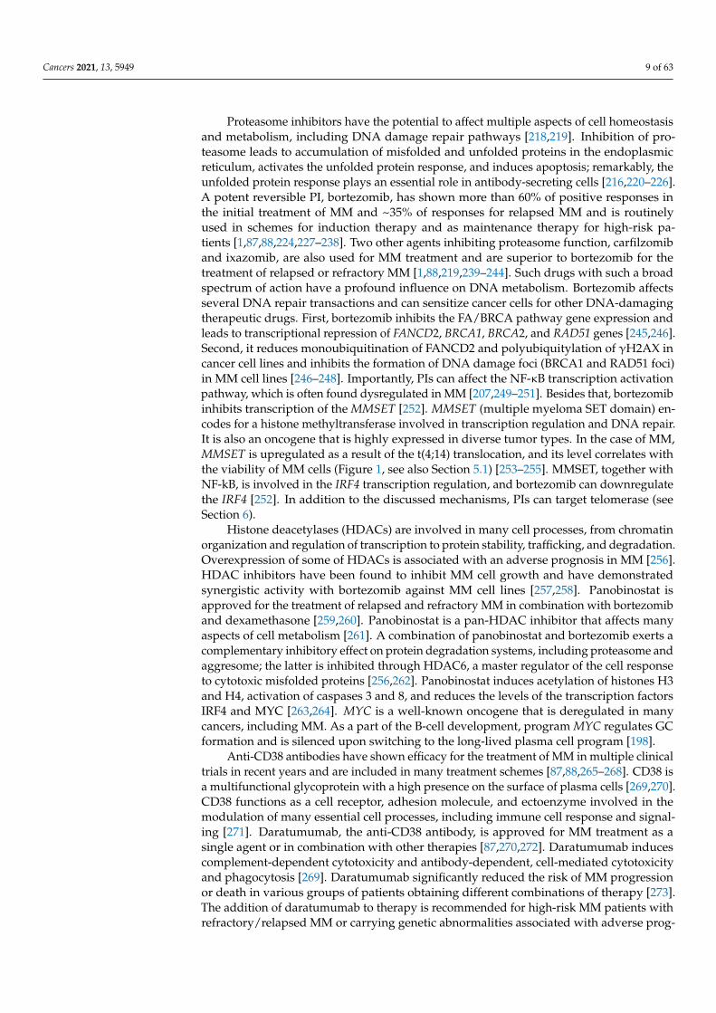
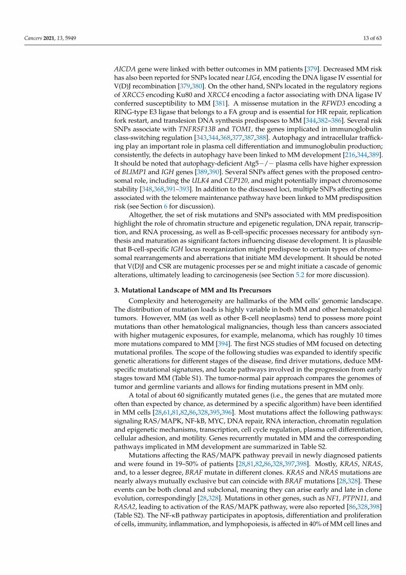
MM is characterized by aberrant expansion of terminally differentiated monoclonalplasma cells resulting in symptoms described by the acronym “CRAB”: hypercalcemia,renal failure, anemia, and bone lesions. In addition to that, diagnostic criteria includethree biomarkers of malignancy: the presence of excessive clonal bone marrow plasmacells, elevated serum free light chain ratio (ratio of κ to λ free light chains), or focal bonelesions [1–3] (Figure 1). These symptoms and biomarkers are called myeloma-definingevents (MDE) [1]. At least one MDE in addition to a biopsy-proven plasmacytoma or≥10% of plasma cells in bone marrow is required for the MM diagnosis [1]. Developmentof MM is a multi-stage process beginning from a premalignant stage termed monoclonalgammopathy of undetermined significance (MGUS) [4–6]. MGUS can be accidentallyfound years to decades before the actual diagnosis of MM. It could be present in ~3% ofthe normal human population over 50 years old [7]. MGUS does not necessarily developinto MM; further progression of MGUS into the active MM has linear risk and occurs witha rate of ~1% per year [1,8–11].
An additional, more advanced stage is observed in some patients, which is referred toas smoldering multiple myeloma (SMM) [1,11–14]. MGUS and SMM are usually asymp-tomatic stages characterized by different levels of M-protein production and different ratiosof clonal plasma cells in bone marrow (see Figure 1). SMM may represent asymptomaticMM rather than being an MM precursor [15]. Patients with SMM follow a declining log-arithmic progression curve to symptomatic MM: 10% risk per year for the first 5 yearsfollowing diagnosis, 3% risk per year for the following 5 years, and a subsequent 1% riskper year [1,8,13]. Both MGUS and SMM have specific diagnostic criteria (Figure 1). MMand its precursors can produce different types of monoclonal proteins. For instance, theproduction of different isotypes of the immunoglobulin heavy chain has been describedin MGUS [7]. The two clinically significant entities are IgM MGUS and non-IgM MGUSdemonstrating different chances of progression into the MM. While the non-IgM MGUS isassociated with a risk of progression to MM, IgM MGUS most frequently progresses intonon-Hodgkin lymphoma and its subtype Waldenström macroglobulinemia [10,16,17]. Inaddition, no immunoglobulin heavy chain production can be seen in some cases in patientswith abnormal serum free light chain ratio, which is attributed to the light chain MGUSand the light chain MM [6,18]. An uncommon subtype of MM is non-secretory myeloma,found in 2–3% of all MM cases [1,18,19].
The biological processes leading to the appearance of a pathological myeloma cloneand the mechanisms of the disease evolution are not yet deciphered. Factors responsiblefor the emergence of MM include a combination of genetic predisposition, alterationsin genomes of the lymphoid cells (such as IGH translocations or hyperdiploidy), and avariety of secondary changes, which include accumulation of mutations, chromosomalrearrangements, and complex genetic events (Figure 1). Apparent inefficiency of the initialtreatment, fast recurrences, resistance to the earlier prescribed drugs, and clonal behaviorof the disease implies that MM represents a heterogeneous entity. Multiple studies suggestthat newly diagnosed MM represents an aggregate of the main pathological clone withseveral subclones that acquire proliferative priority when the main clone is suppressedduring specific therapy [20–34].
The classic clonal evolution implies the sequential acquisition of mutations with aconcomitant sequential selection of successive subclones, their expansion, and mutualinterference [35,36]. Each tumor cell can carry many genetic abnormalities, includingmutations that provide a selective growth advantage: “driver” mutations, selectivelyneutral “passenger” mutations, and deleterious mutations affecting fitness [37]. In additionto that, there are “mutator” mutations that increase the rate of genetic changes. Thedynamics of tumor evolution is a function of mutation rate elevation and clonal expansionthat relies on “driver” mutations. Natural selection provides “selective sweeps” when one
Cancers 2021, 13, 5949 3 of 63
or several clones grow to dominate the neoplasm [36]. These dominant clones accumulatenew genetic changes in addition to the mutational landscape of the original tumor as theevolution proceeds. The complexity of this process is augmented by epigenetic changesthat, similar to changes in the DNA, can confer either growth advantage or be selectivelyneutral or deleterious. Epigenetic changes can also affect mutation rates. On top of thiscomplexity is the impact of therapeutic drugs, which can modify DNA, affect DNA repairprocesses, and modulate the growth advantage of the cancer cells [38–40].
MGUS
• M-protein <3 g/dL
• Clonal plasma
cells in BM <10%
Smoldering
Myeloma
• M-protein ≥ 3 g/dL
(serum) OR ≥500
mg/ 24 hrs (urine)
• Clonal plasma cells
in BM 10-60%
Multiple
Myeloma
• CRAB
• Clonal plasma cells
in BM ≥60%
• Serum free light
chain ratio ≥100
• >1 MRI focal lesion
≥5mm
Remission
• Stringent complete
response (sCR)
• Complete response
(CR)
• Very good partial
response (VGPR)
• Partial response (PR)
• Minimal response
(MR)
Relapse /
Progression• CRAB
• M-protein increase
• New bone lesions
• Increase of
plasma cells %
• Hypercalcaemia
• Decrease in
hemoglobin
• Rise in serum
creatinine
▪ IGH translocations:
t(4;14)
t(6;14)
t(11;14)
t(14;16)
t(14;20)
▪ Hyperdiploidy:
Chr. 3, 5, 7, 9, 11, 15,
19, 21
▪ Structural
aberrations: gain(1q),
del(13q) and del(17p)
▪ Mutations in:
KRAS, NRAS, DIS3,
HIST1H1E, EGR1,
LTB, CCND1
▪ Accumulation of various structural
genome rearrangements including
MYC translocations
▪ Complex structural rearrangements
(partially)
▪ Accumulation of mutations in genes
belonging to various pathways:
RAS/MAPK, NF-kB, MYC, DNA-repair,
RNA interaction, chromatin regulation
and epigenetic mechanisms,
transcription, cell cycle regulation,
plasma cell differentiation, cellular
adhesion and motility
▪ High mutation burden associated with
deregulation of AID/APOBEC enzymes
Normal cells
▪ Mutations in:
LSD1/KDM1A
ARID1A
USP45
DIS3
CDKN2A
EP300
▪ Alteration in
telomere
maintenance
pathway
▪ Risk SNPs
▪ DNA lesions and mutations induced by
chemotherapeutic alkylating drugs: DNA
alkylation, DNA crosslinks, mutations in
transcribed strand (melphalan signature)
▪ Modulation of DNA repair by proteasome
inhibitors
▪ Altering specificity of CRL4CRBN E3 ligase by
IMiDs, degradation and dysregulation of
some key for MM transcription factors
▪ Mutation kataegis and catastrophic genome
rearrangements induced by combined action
of accumulated genetic changes and action
of therapeutic drugs
Predisposition Primary events Secondary events Treatment-induced events
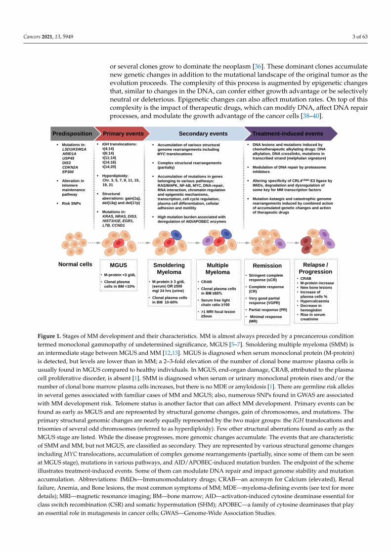
Figure 1. Stages of MM development and their characteristics. MM is almost always preceded by a precancerous conditiontermed monoclonal gammopathy of undetermined significance, MGUS [5–7]. Smoldering multiple myeloma (SMM) isan intermediate stage between MGUS and MM [12,13]. MGUS is diagnosed when serum monoclonal protein (M-protein)is detected, but levels are lower than in MM; a 2–3-fold elevation of the number of clonal bone marrow plasma cells isusually found in MGUS compared to healthy individuals. In MGUS, end-organ damage, CRAB, attributed to the plasmacell proliferative disorder, is absent [1]. SMM is diagnosed when serum or urinary monoclonal protein rises and/or thenumber of clonal bone marrow plasma cells increases, but there is no MDE or amyloidosis [1]. There are germline risk allelesin several genes associated with familiar cases of MM and MGUS; also, numerous SNPs found in GWAS are associatedwith MM development risk. Telomere status is another factor that can affect MM development. Primary events can befound as early as MGUS and are represented by structural genome changes, gain of chromosomes, and mutations. Theprimary structural genomic changes are nearly equally represented by the two major groups: the IGH translocations andtrisomies of several odd chromosomes (referred to as hyperdiploidy). Few other structural aberrations found as early as theMGUS stage are listed. While the disease progresses, more genomic changes accumulate. The events that are characteristicof SMM and MM, but not MGUS, are classified as secondary. They are represented by various structural genome changesincluding MYC translocations, accumulation of complex genome rearrangements (partially, since some of them can be seenat MGUS stage), mutations in various pathways, and AID/APOBEC-induced mutation burden. The endpoint of the schemeillustrates treatment-induced events. Some of them can modulate DNA repair and impact genome stability and mutationaccumulation. Abbreviations: IMiDs—Immunomodulatory drugs; CRAB—an acronym for Calcium (elevated), Renalfailure, Anemia, and Bone lesions, the most common symptoms of MM; MDE—myeloma-defining events (see text for moredetails); MRI—magnetic resonance imaging; BM—bone marrow; AID—activation-induced cytosine deaminase essential forclass switch recombination (CSR) and somatic hypermutation (SHM); APOBEC—a family of cytosine deaminases that playan essential role in mutagenesis in cancer cells; GWAS—Genome-Wide Association Studies.
Cancers 2021, 13, 5949 4 of 63
The clonal development of the tumor may follow the linear model if driver changesprovide a strong selective advantage that outcompetes all previous clones, be branched ifseveral clones expand simultaneously, or neutral if there is no selective advantage betweenmultiple clones’ appearance and co-existence. Alternatively, most essential changes mayoccur simultaneously or near-simultaneously early in tumor development, establishingseveral dominant clones that grow stably, a characteristic of punctuated evolution [36,41].It is generally recognized that the MM development follows the rules of branched evo-lution [27,42,43]. However, patterns of clones’ phylogeny consistent with linear, neutral,and punctuated models of evolution have also been described during MM developmentand progression [22,27,29,44,45]. Moreover, recent advances in studies of MM and itsprecursors have centered the punctuated evolution model as characterizing best the earlystages of the MM development [20,46–49]. For instance, significant heterogeneity is presentbefore the disease manifestation at the MGUS and SMM stages [20,23,30,47,48,50–52], andfurther progression from MGUS/SMM to MM is characterized by clonal stability in sometumors [20,46–48]. Importantly, clonal evolution in MM seems to be closely interrelatedwith treatment strategies [22–26,28,34,45,53,54]. As the disease proceeds, the variability ofthe clones increases, which creates the ground for further clonal diversification and evolu-tion [23,45,47,51–53,55–63]. One or several such clones will ultimately lead to the recurrenceof myeloma. It is possible that the change from the main clone to subclones during MMtreatment causes a change in the clinical and hematological phenotype of MM and, thus,explains the inefficiency of the previously conducted therapy. Noteworthy, patients withMM or its precursors have an increased risk of developing secondary primary malignancies(SPMs), such as myelodysplastic syndrome (MDS), acute myeloid leukemia (AML), andothers [64–67]. The origin of these tumors is likely related to the genotoxic action of sometherapeutic agents [65,68–74], although an excess risk for hematopoietic neoplasms otherthan MM in MGUS patients supports an idea that endogenous factors play an essentialrole in SPMs’ development [9,75–77]. Therefore, it is possible that the genetic landscapeof hematopoietic stem cells in MM patients may predispose them to different malignantprograms that can unfold spontaneously or as a result of therapeutic intervention.
Recent developments and advances of next-generation sequencing technologies (NGS)help to produce a large amount of genomic data that are invaluable for MM diagnosis,choices of possible treatment, assessment of drug response, and understanding of diseaseevolution [78–80]. During the last decade, dozens of large-scale NGS studies have beenundertaken to find peculiarities of the MM genomes and pinpoint genetic drivers of MM(Table S1) [28,29,61,81–83]. Whole-exome (WES) or whole-genome (WGS) sequencing ofMM genomes allows for detecting point mutations, small insertions or deletions, andstructural variations. Many studies combine their own NGS data with the publicly avail-able datasets from Multiple Myeloma Research Foundation (MMRF) CoMMpass Study(Clinicaltrials.gov identification number: NCT01454297) [84]. Sequencing data for MMsamples are deposited in the European Genome Archive or at the Genotype and Phenotypedatabase (dbGaP). The Multiple Myeloma Genome Project (MGP) attempts to assembleand analyze NGS data for MM, improve clinical testing, and define treatment strategies forMM patients [85,86]. MGP provides the repository of WES, WGS, and RNA-Seq data forpatients with MM derived from different sources, such as the Multiple Myeloma ResearchFoundation, the Myeloma XI trial, and others [85].
1.2. Current Treatment Algorithms of MM: Mechanisms of Action of Anti-MM Agents
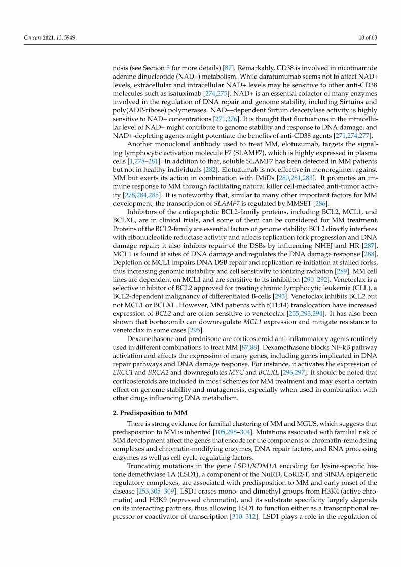
MM patient management strategy differs significantly depending on age, comorbidi-ties, cytogenetic parameters, disease stage, risk stratification, and other factors [87–91](Figure 2). For the treatment of primary patients under 70 years of age without seriouscomorbidities, high-dose chemotherapy (HDCT) with autologous hematopoietic stem celltransplantation (ASCT) is included in the treatment program (Figure 2). Several recentstudies show that ASCT can be a safe choice for patients older than 70 years [92–96].Despite significant toxicity, ASCT remains the standard of care and is associated with
Cancers 2021, 13, 5949 5 of 63
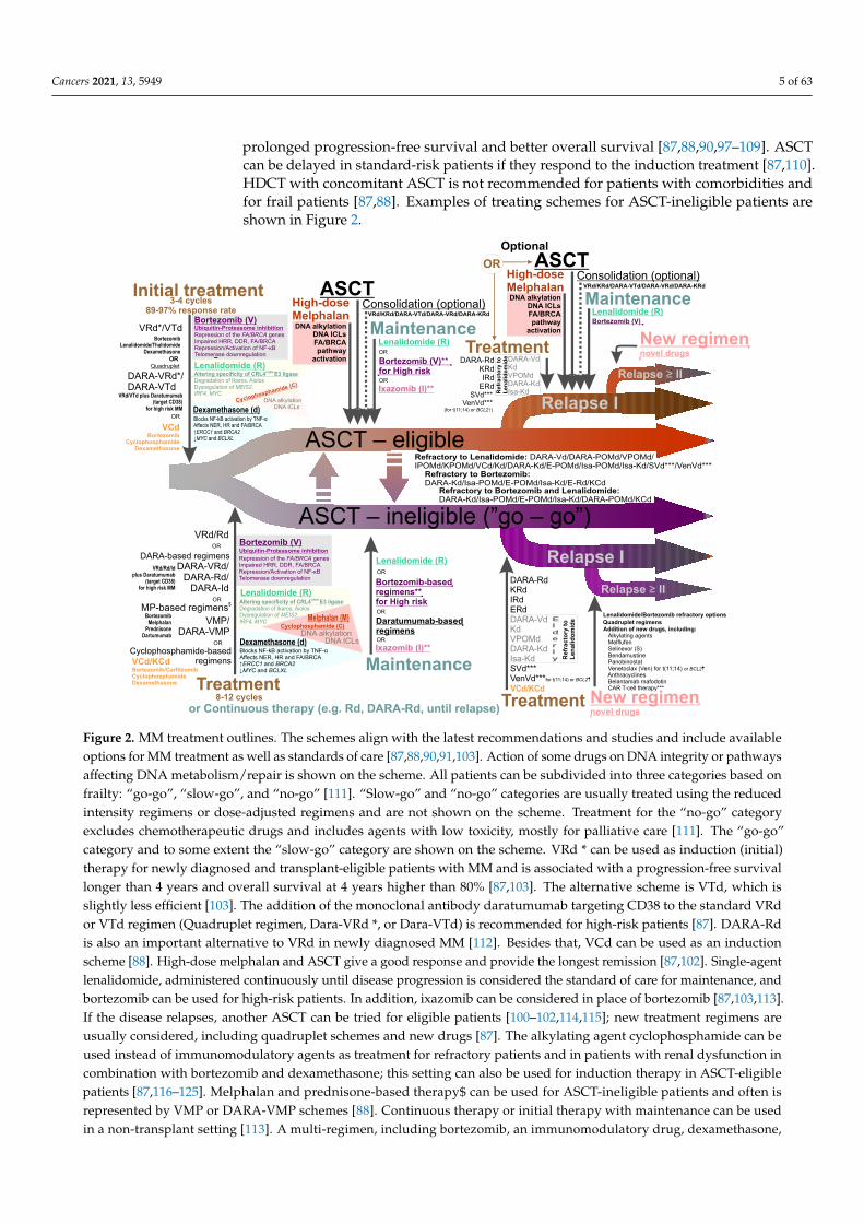
prolonged progression-free survival and better overall survival [87,88,90,97–109]. ASCTcan be delayed in standard-risk patients if they respond to the induction treatment [87,110].HDCT with concomitant ASCT is not recommended for patients with comorbidities andfor frail patients [87,88]. Examples of treating schemes for ASCT-ineligible patients areshown in Figure 2.
Initial treatment
ASCT – eligible
VRd*/VTd Ubiquitin-Proteasome inhibition
BortezomibLenalidomide/Thalidomide
Dexamethasone
DARA-VRd*/DARA-VTd
VRd/VTd plus Daratumumab (target CD38)
for high risk MM
Repression of the FA/BRCA genesImpaired HRR, DDR, FA/BRCARepression/Activation of NF-κBTelomerase downregulation
Bortezomib (V)
CRBN Altering specificity of CRL4 E3 ligaseDegradation of Ikaros, Aiolos Dysregulation of MEIS2,IRF4, MYC
Lenalidomide (R)
3-4 cycles89-97% response rate
MaintenanceLenalidomide (R)
Bortezomib (V)** for High risk
Relapse I
Relapse ILenalidomide (R)
Bortezomib-basedregimens**for High risk
Treatment
DARA-based regimens
OR
ORUbiquitin-Proteasome inhibition
Repression of the FA/BRCA genesImpaired HRR, DDR, FA/BRCARepression/Activation of NF-κBTelomerase downregulation
Bortezomib (V)
Lenalidomide (R)
ASCTHigh-doseMelphalan
or Continuous therapy (e.g. Rd, DARA-Rd, until relapse)
Maintenance
Optional
Refractory to Lenalidomide: DARA-Vd/DARA-POMd/VPOMd/IPOMd/KPOMd/VCd/Kd/DARA-Kd/E-POMd/Isa-POMd/Isa-Kd/SVd***/VenVd***
Treatment
VRd/Rd
DARA-RdKRdIRdERdDARA-VdKdVPOMdDARA-KdIsa-KdSVd***VenVd***for t(11;14) or BCL2
Relapse ≥ II
Addition of new drugs, including: Alkylating agents Melflufen Selinexor (S) Bendamustine Panobinostat Venetoclax (Ven) for t(11;14) or BCL2
Anthracyclines Belantamab mafodotin CAR T-cell therapy***
Relapse ≥ II
Dexamethasone (d)
New regimennovel drugs
New regimennovel drugs
VRd/Rd/Id plus Daratumumab
(target CD38)for high risk MM
Quadruplet
OR
VCd/KCd
Consolidation (optional)
Lenalidomide (R)Bortezomib (V)
DARA-VRd/DARA-Rd/
DARA-Id
VMP/DARA-VMP
BortezomibMelphalan
PrednisoneDartumumab
8-12 cycles
Melphalan (M)
Blocks NF-kB activation by TNF-αAffects NER, HR and FA/BRCA ↑ERCC1 and BRCA2 ↓MYC and BCLXL
Blocks NF-kB activation by TNF-αAffects NER, HR and FA/BRCA ↑ERCC1 and BRCA2 ↓MYC and BCLXL
Cyclophosphamide (C)
DNA alkylation DNA ICLs
DNA alkylationDNA ICLsFA/BRCApathway
activation
Refractory to Bortezomib:DARA-Kd/Isa-POMd/E-POMd/Isa-Kd/E-Rd/KCd
$MP-based regimens
ASCT – ineligible (”go – go”)
VRd/KRd/DARA-VTd/DARA-VRd/DARA-KRd
VCd Bortezomib
Cyclophosphamide Dexamethasone
OR
OR
VCd/KCd Bortezomib/CarfilzomibCyclophosphamide Dexamethasone
Maintenance
Daratumumab-basedregimens
Cyclophosphamide (C)
DNA alkylation DNA ICLs
CRBN Altering specificity of CRL4 E3 ligase
Degradation of Ikaros, Aiolos Dysregulation of MEIS2,IRF4, MYC
Dexamethasone (d)
Treatment
ASCTHigh-doseMelphalan
Consolidation (optional)
DNA alkylationDNA ICLsFA/BRCApathway
activation
VRd/KRd/DARA-VTd/DARA-VRd/DARA-KRd
OR
DARA-RdKRdIRd
ERdIxazomib (I)**
OROR
OR
OR
OR
Cyclophosphamide-based regimens
OR
Ixazomib (I)**
Refractory to Bortezomib and Lenalidomide:DARA-Kd/Isa-POMd/E-POMd/Isa-Kd/DARA-POMd/KCd
Lenalidomide/Bortezomib refractory optionsQuadruplet regimens
Elderly R
efr
acto
ry t
oL
en
alid
om
ide
DARA-VdKdVPOMdDARA-KdIsa-KdR
efr
acto
ry t
oL
en
alid
om
ide
SVd***VenVd***
(for t(11;14) or BCL2↑)
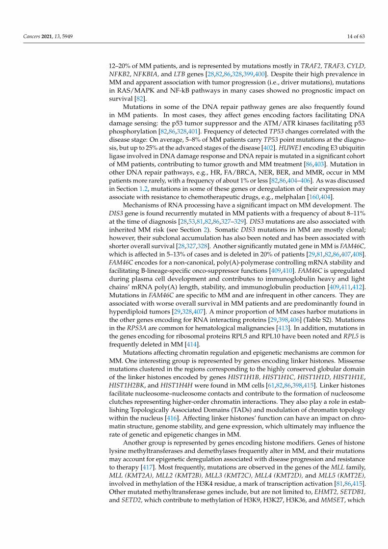
Figure 2. MM treatment outlines. The schemes align with the latest recommendations and studies and include availableoptions for MM treatment as well as standards of care [87,88,90,91,103]. Action of some drugs on DNA integrity or pathwaysaffecting DNA metabolism/repair is shown on the scheme. All patients can be subdivided into three categories based onfrailty: “go-go”, “slow-go”, and “no-go” [111]. “Slow-go” and “no-go” categories are usually treated using the reducedintensity regimens or dose-adjusted regimens and are not shown on the scheme. Treatment for the “no-go” categoryexcludes chemotherapeutic drugs and includes agents with low toxicity, mostly for palliative care [111]. The “go-go”category and to some extent the “slow-go” category are shown on the scheme. VRd * can be used as induction (initial)therapy for newly diagnosed and transplant-eligible patients with MM and is associated with a progression-free survivallonger than 4 years and overall survival at 4 years higher than 80% [87,103]. The alternative scheme is VTd, which isslightly less efficient [103]. The addition of the monoclonal antibody daratumumab targeting CD38 to the standard VRdor VTd regimen (Quadruplet regimen, Dara-VRd *, or Dara-VTd) is recommended for high-risk patients [87]. DARA-Rdis also an important alternative to VRd in newly diagnosed MM [112]. Besides that, VCd can be used as an inductionscheme [88]. High-dose melphalan and ASCT give a good response and provide the longest remission [87,102]. Single-agentlenalidomide, administered continuously until disease progression is considered the standard of care for maintenance, andbortezomib can be used for high-risk patients. In addition, ixazomib can be considered in place of bortezomib [87,103,113].If the disease relapses, another ASCT can be tried for eligible patients [100–102,114,115]; new treatment regimens areusually considered, including quadruplet schemes and new drugs [87]. The alkylating agent cyclophosphamide can beused instead of immunomodulatory agents as treatment for refractory patients and in patients with renal dysfunction incombination with bortezomib and dexamethasone; this setting can also be used for induction therapy in ASCT-eligiblepatients [87,116–125]. Melphalan and prednisone-based therapy$ can be used for ASCT-ineligible patients and often isrepresented by VMP or DARA-VMP schemes [88]. Continuous therapy or initial therapy with maintenance can be usedin a non-transplant setting [113]. A multi-regimen, including bortezomib, an immunomodulatory drug, dexamethasone,
Cancers 2021, 13, 5949 6 of 63
cytotoxic cisplatin, doxorubicin, cyclophosphamide, and etoposide, may be used for plasma cell leukemia or extramedullarydisease [87]. Schemes that can be used for refractory to lenalidomide and/or bortezomib patients are also given in thecenter of the figure. Abbreviations: V—bortezomib, DARA—daratumumab, R—lenalidomide, T—thalidomide,POM —pomalidomide, d—dexamethasone; K—carfilzomib, I—ixazomib, E—elotuzumab, Isa—isatuximab,C—cyclophosphamide, P—prednisone; S—selinexor, Ven—venetoclax, ICL—intermolecular crosslinks in the DNA,FA/BRCA—pathway responsible for the repair of intermolecular crosslinks in the DNA, HR—homologous recom-bination, HRR—homologous recombination repair, DDR—DNA damage response, NER—nucleotide excision repair.*—Lenalidomide-containing regimens (e.g., VRd and Dara-VRd) are not yet approved by European Medicines Agency(EMA, EU) as induction for ASCT-eligible patients, although they offer good risk–benefit profiles and are widely used in theUSA [87,88]. **—Bortezomib and ixazomib have not yet been approved by the EMA for maintenance after ASCT [88,113].***—Awaiting EMA approval. $—melphalan-containing regimens in this setting are not recommended in the USA due toconcerns about SPMs and stem cells damage.
The therapeutic arsenal for MM treatment includes DNA damaging agents (mel-phalan, cyclophosphamide, etc.), immunomodulatory agents (IMiDs, e.g., thalidomide,lenalidomide, pomalidomide), proteasome inhibitors (PIs, e.g., bortezomib, carfilzomib,ixazomib), monoclonal antibodies (daratumumab, isatuximab, elotuzumab), and corti-costeroids [1,87,91]. In addition to that, several drugs can be used in combination withother therapies as they are not considered efficient alone in most cases. These includeinhibitors of histone deacetylase 6 (panobinostat), an inhibitor of Exportin-1 (selinexor,considered efficient in combination with dexamethazone), the DNA intercalating drugsanthracyclines (doxorubicin), and the BCL2 inhibitor venetoclax, which does not yet haveapproval for treatment of MM but appears to be efficient against MM with t(11;14) rear-rangement [87,126,127]. Besides that, new agents and therapies have been approved orare about to be approved for the treatment of MM. They include an immunoconjugatedrug targeting B-cell maturation antigen, BCMA (Belantamab Mafodotin), an inhibitor ofkinesin spindle protein, KSP (Filanesib), chimeric antigen receptor (CAR) T-cell therapy(e.g., bb2121 therapy that targets BCMA), and bispecific antibodies [128–134].
Many agents used in MM treatment are genotoxic and can further elevate the ge-netic variability of MM, which can be harmful and potentiate disease progression andrelapse [25,135]. The consequences of using one or another agent for the genome dif-fer depending on the preexisting genomic alterations and mutations. Drugs within thechemotherapeutic arsenal with potential genotoxicity include alkylating DNA agents(melphalan, cyclophosphamide, bendamustine, busulfan), intercalating DNA agents (dox-orubicin), microtubule-depolymerizing drugs (vincristine), agents inactivating topoiso-merase II (etoposide, doxorubucin), and crosslinking agents (cisplatin). Melphalan is aphenylalanine-substituted derivative of nitrogen mustard that alkylates adenine and gua-nine in the DNA. It has two highly reactive chloroethylamine groups and thus induces intra-or intermolecular crosslinks in the DNA and between DNA and proteins [136–138]. Thehigh-dose melphalan is a standard regimen before ASCT [87,103,136,139,140]. Cyclophos-phamide also belongs to a family of mustard-alkylating agents and induces alkylationand crosslinking of the DNA [141–143]. At low doses, cyclophosphamide demonstratesimmunomodulatory activity [144]. Notably, a significant increase in mutation burden andspecific mutational signature have been reported in MM patients exposed to high-dosemelphalan [54,59,63,145,146]. Melphalan (and to a lesser extent cyclophosphamide) is alsoassociated with an increased risk of SPMs in MM patients [64,68,69,71,72,147–150]. The lineof chemotherapeutic drugs has been consistently elaborated upon with novel therapeuticagents in recent years. One of them, a peptide-conjugate alkylator drug, melphalan flufe-namide (melflufen), is currently in clinical trials [151–153]. Alkylating and crosslinkingagents induce modifications to DNA that are a subject of DNA repair to allow for DNAreplication and transcription [154]. The vulnerability of cancer cells to these drugs dependson the competing processes of cell cycle progression and repair. Base excision repair (BER),nucleotide excision repair (NER), and mismatch repair (MMR) remove monoadducts in
Cancers 2021, 13, 5949 7 of 63
the DNA, intra-strand crosslinks, and other lesions that affect only one strand of the twostrands of the duplex DNA [155]. The DNA double-strand breaks (DSBs) interrupt thecontinuity of the DNA molecule and are repaired via two major repair routes: homologousrecombination repair (HRR) and nonhomologous end-joining (NHEJ) [155]. The first oneuses the homologous sequence as an instruction to recover the lost information, and thesecond one just joins the two ends together and seals the break. Despite their provisorydivision into error-free and error-prone pathways, correspondingly, both HRR and NHEJcan lead to point mutations and chromosome rearrangements. Inter-strand crosslinks con-stitute a physical barrier to the progression of both RNA and DNA polymerases since theyimpede the unwinding of the double helix. This type of lesion requires the action of multi-ple players belonging to the BER, NER, MMR, and HRR pathways and are grouped into aspecialized FA/BRCA pathway [156,157]. FA/BRCA was named after Fanconi anemia (FA),a genetic disorder leading to developmental abnormalities and predisposition to cancerdevelopment [158]. Mutations in the FA/BRCA pathway genes make cells hypersensitiveto agents inducing DNA crosslinks [156,159]. Upregulation of the FA/BRCA pathway con-tributes to acquired resistance to melphalan in MM cell lines [160]. Human myeloma celllines frequently acquire mutations in the genes of the FA/BRCA pathway [161]. Selinexor,an inhibitor of the nuclear Exportin 1 (XPO1), decreases the expression of the FA/BRCAand NF-kB pathway genes, reduces melphalan-induced monoubiquitination of FANCD2,and overcomes the resistance of the MM cell lines to melphalan [162]. In addition to that,several other key factors of HR and NHEJ are found upregulated in MM cell lines, whichcan have implications to MM resistance to DNA-damaging drugs [163–166]. Bendamus-tine is a derivative of mechlorethamine that acts both as an alkylating agent and purineanalog [167,168]. It has anti-MM activity and can overcome resistance to melphalan in MMcell lines [169]. It is most often used for the treatment of relapsed or refractory-to-other-regimens MM and patients with renal impairment not eligible for ASCT but it can be usedas a conditioning therapy before ASCT when combined with melphalan [140,170–176].Bendamustine efficiently activates the DNA damage response and has a synergistic effectwith alkylating agents and pyrimidine analogs in killing MM cell lines [168].
IMiDs are derivatives of the teratogenic drug thalidomide that are approved by theUS FDA for the treatment of MM [87,120,177–182]. Lenalidomide is the standard-of-care maintenance therapy for patients with standard risk and after ASCT [87,97,183–188].Thalidomide, lenalidomide, and pomalidomide bind to Cereblon (CRBN) [177,189–192].CRBN, together with DNA Damage Binding Protein-1 (DDB1), Cullin-4A (Cul4A), andRegulator of Cullins-1 (Roc1), forms a complex called Cullin-RING ligase 4 (CRL4CRBN),an E3 ubiquitin ligase; CRBN functions as a substrate receptor [191]. The drugs inhibit thebinding of CRL4CRBN to its endogenous substrate, transcription factor MEIS2, which regu-lates MM cell survival and sensitivity to anti-MM drugs [191,193]. CRBN in the presence ofdrugs acquires the ability to target two specific B cell transcription factors, Ikaros (IKZF1)and Aiolos (IKZF3), for proteasomal degradation [194–196] (Figures 2 and 3). Ikaros andAiolos regulate the expression of the key plasma cell differentiation transcription factor,IRF4, which plays a central role in the pathogenesis of MM [197,198] (Figure 3). IRF4controls B-cell-to-plasma-cell program transition and is essential for Germinal Centers (GC)formation, CSR, and SHM [199]. The AICDA gene encoding AID cytosine deaminase essen-tial for CSR and SHM (Figure 3) is among the IRF4 targets [200]. IRF4 is often overexpressedin MM as a result of mutations, translocations, or other events [201–203]. It is of interestthat lenalidomide can partially affect the IRF4 level [194,195,197,204]. In addition to that,thalidomide and its derivatives can affect the level of cytokine production since they caninhibit TNF-α and IL-1b production and increase IL-2 and IL-10 production [194,205,206].Inhibition of NF- κB activation has been reported for IMiDs [207,208]. Lenalidomide canaffect the levels of other factors, such as KPNA2 and SALL4 [196,209,210]. It is noteworthythat levels of some of these factors, e.g., KPNA2, can be linked to the overall survival andprogression-free survival rates in MM patients [209]. It should be noted that some studieshave reported increased risk of SPMs associated with lenalidomide [64,65,74,186,211].
Cancers 2021, 13, 5949 8 of 63
Pre-B-cell
SLCμH Igαβ
Pro-B-cell
Pre-B-cell receptor (Pre-BCR)
Igα
Fetal liverBone marrow
antigen-independent development
Stem cell
antigen selection
PAX5
Igα and SLC expression
IGH locus contraction
Immature B-cell
Mature naïve B-cell
D→J and V→DJ rearrangements at IGH
IgM BCR
RAG1/2, HMGB1/2, TdT IGL or IGK rearrangement
SLC
ZFP318
Alternative splicingof Cμ and Cδ exons
IgM BCR IgD BCR
Activated B-cell
IgM BCR
EBF1PU.1IkarosE2A
Plasma cell
RAG1/2, HMGB1/2
antigen-dependent development
Peripheral lymphoid organs (spleen, lymph nodes)
Class Switch Recombination (CSR)
AID
Germinal centers
Somatic Hypermutation
Plasmablast Memory B-cell
Short-livedCycling
All isotypes Ig
Short-lived IgM>IgG Long-lived IgG>IgA>IgM
Quiescent
Long-livedQuiescent
IRF4(hi)XBP1
BLIMP1(mid)
IRF4(hi)XBP1
BLIMP1(high)
PAX5BACH2
PU.1-IRF8
PAX5BCL6
BACH2PAX5BACH2
PU.1-IRF8
PAX5BACH2
PU.1-IRF8
BloodPeripheral lymphoid organsBone marrow (Plasma cells)
IRF4(low)PAX5
BACH2PU.1-IRF8
PAX5BACH2
PU.1-IRF8
PAX5BACH2
PU.1-IRF8IRF4 IRF4(low)
IRF4(hi) IRF4(hi)
IkarosAiolos
Silencing of SLC expression
FOXO1
AID
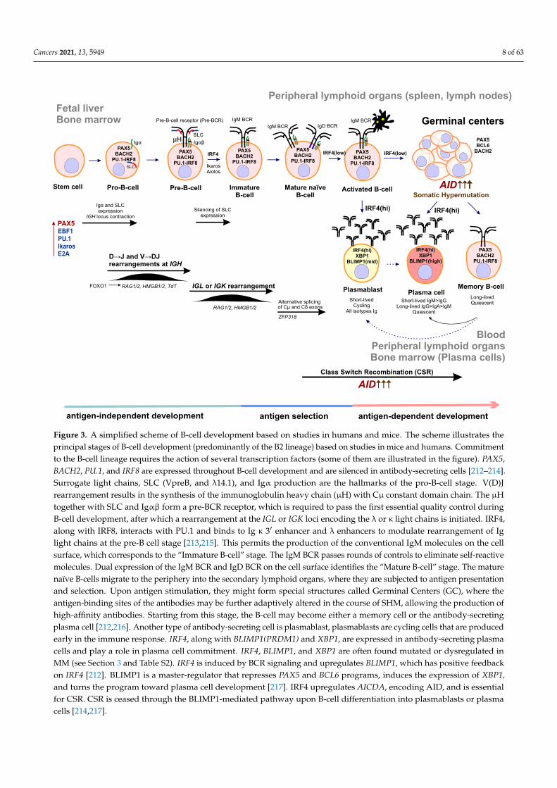
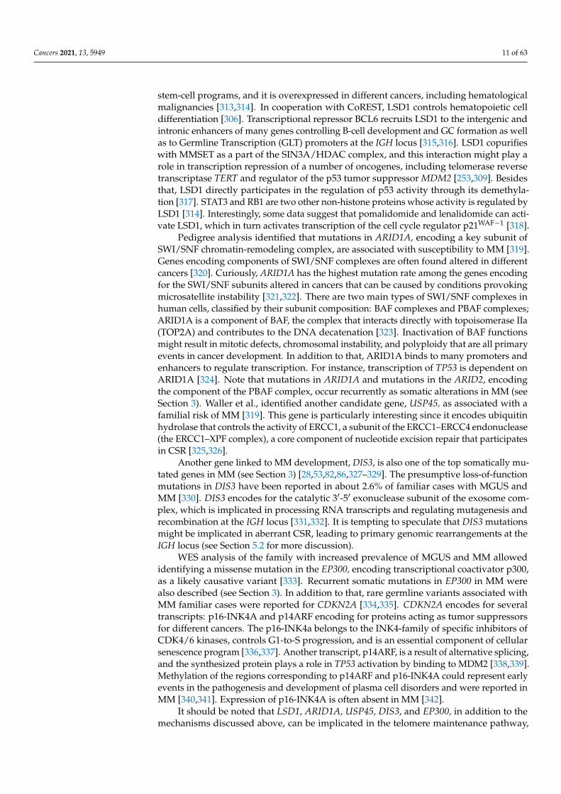
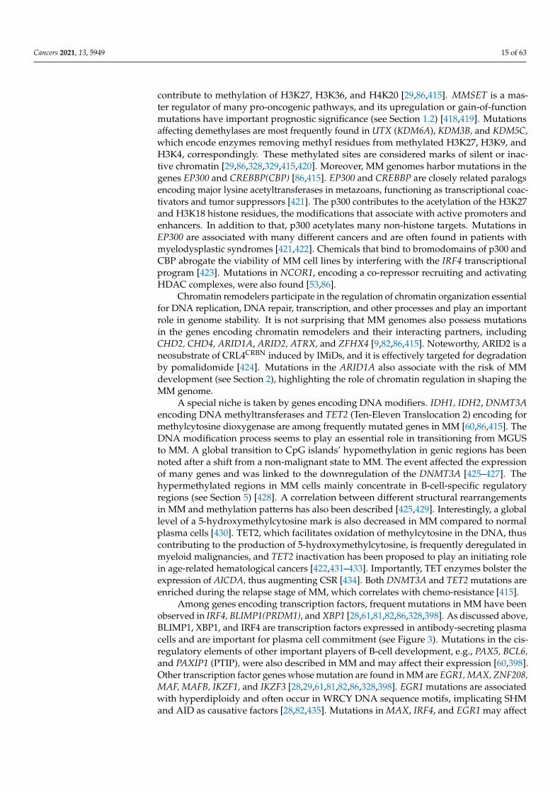
Figure 3. A simplified scheme of B-cell development based on studies in humans and mice. The scheme illustrates theprincipal stages of B-cell development (predominantly of the B2 lineage) based on studies in mice and humans. Commitmentto the B-cell lineage requires the action of several transcription factors (some of them are illustrated in the figure). PAX5,BACH2, PU.1, and IRF8 are expressed throughout B-cell development and are silenced in antibody-secreting cells [212–214].Surrogate light chains, SLC (VpreB, and λ14.1), and Igα production are the hallmarks of the pro-B-cell stage. V(D)Jrearrangement results in the synthesis of the immunoglobulin heavy chain (µH) with Cµ constant domain chain. The µHtogether with SLC and Igαβ form a pre-BCR receptor, which is required to pass the first essential quality control duringB-cell development, after which a rearrangement at the IGL or IGK loci encoding the λ or κ light chains is initiated. IRF4,along with IRF8, interacts with PU.1 and binds to Ig κ 3′ enhancer and λ enhancers to modulate rearrangement of Iglight chains at the pre-B cell stage [213,215]. This permits the production of the conventional IgM molecules on the cellsurface, which corresponds to the “Immature B-cell” stage. The IgM BCR passes rounds of controls to eliminate self-reactivemolecules. Dual expression of the IgM BCR and IgD BCR on the cell surface identifies the “Mature B-cell” stage. The maturenaïve B-cells migrate to the periphery into the secondary lymphoid organs, where they are subjected to antigen presentationand selection. Upon antigen stimulation, they might form special structures called Germinal Centers (GC), where theantigen-binding sites of the antibodies may be further adaptively altered in the course of SHM, allowing the production ofhigh-affinity antibodies. Starting from this stage, the B-cell may become either a memory cell or the antibody-secretingplasma cell [212,216]. Another type of antibody-secreting cell is plasmablast, plasmablasts are cycling cells that are producedearly in the immune response. IRF4, along with BLIMP1(PRDM1) and XBP1, are expressed in antibody-secreting plasmacells and play a role in plasma cell commitment. IRF4, BLIMP1, and XBP1 are often found mutated or dysregulated inMM (see Section 3 and Table S2). IRF4 is induced by BCR signaling and upregulates BLIMP1, which has positive feedbackon IRF4 [212]. BLIMP1 is a master-regulator that represses PAX5 and BCL6 programs, induces the expression of XBP1,and turns the program toward plasma cell development [217]. IRF4 upregulates AICDA, encoding AID, and is essentialfor CSR. CSR is ceased through the BLIMP1-mediated pathway upon B-cell differentiation into plasmablasts or plasmacells [214,217].
Cancers 2021, 13, 5949 9 of 63
Proteasome inhibitors have the potential to affect multiple aspects of cell homeostasisand metabolism, including DNA damage repair pathways [218,219]. Inhibition of pro-teasome leads to accumulation of misfolded and unfolded proteins in the endoplasmicreticulum, activates the unfolded protein response, and induces apoptosis; remarkably, theunfolded protein response plays an essential role in antibody-secreting cells [216,220–226].A potent reversible PI, bortezomib, has shown more than 60% of positive responses inthe initial treatment of MM and ~35% of responses for relapsed MM and is routinelyused in schemes for induction therapy and as maintenance therapy for high-risk pa-tients [1,87,88,224,227–238]. Two other agents inhibiting proteasome function, carfilzomiband ixazomib, are also used for MM treatment and are superior to bortezomib for thetreatment of relapsed or refractory MM [1,88,219,239–244]. Such drugs with such a broadspectrum of action have a profound influence on DNA metabolism. Bortezomib affectsseveral DNA repair transactions and can sensitize cancer cells for other DNA-damagingtherapeutic drugs. First, bortezomib inhibits the FA/BRCA pathway gene expression andleads to transcriptional repression of FANCD2, BRCA1, BRCA2, and RAD51 genes [245,246].Second, it reduces monoubiquitination of FANCD2 and polyubiquitylation of γH2AX incancer cell lines and inhibits the formation of DNA damage foci (BRCA1 and RAD51 foci)in MM cell lines [246–248]. Importantly, PIs can affect the NF-κB transcription activationpathway, which is often found dysregulated in MM [207,249–251]. Besides that, bortezomibinhibits transcription of the MMSET [252]. MMSET (multiple myeloma SET domain) en-codes for a histone methyltransferase involved in transcription regulation and DNA repair.It is also an oncogene that is highly expressed in diverse tumor types. In the case of MM,MMSET is upregulated as a result of the t(4;14) translocation, and its level correlates withthe viability of MM cells (Figure 1, see also Section 5.1) [253–255]. MMSET, together withNF-kB, is involved in the IRF4 transcription regulation, and bortezomib can downregulatethe IRF4 [252]. In addition to the discussed mechanisms, PIs can target telomerase (seeSection 6).
Histone deacetylases (HDACs) are involved in many cell processes, from chromatinorganization and regulation of transcription to protein stability, trafficking, and degradation.Overexpression of some of HDACs is associated with an adverse prognosis in MM [256].HDAC inhibitors have been found to inhibit MM cell growth and have demonstratedsynergistic activity with bortezomib against MM cell lines [257,258]. Panobinostat isapproved for the treatment of relapsed and refractory MM in combination with bortezomiband dexamethasone [259,260]. Panobinostat is a pan-HDAC inhibitor that affects manyaspects of cell metabolism [261]. A combination of panobinostat and bortezomib exerts acomplementary inhibitory effect on protein degradation systems, including proteasome andaggresome; the latter is inhibited through HDAC6, a master regulator of the cell responseto cytotoxic misfolded proteins [256,262]. Panobinostat induces acetylation of histones H3and H4, activation of caspases 3 and 8, and reduces the levels of the transcription factorsIRF4 and MYC [263,264]. MYC is a well-known oncogene that is deregulated in manycancers, including MM. As a part of the B-cell development, program MYC regulates GCformation and is silenced upon switching to the long-lived plasma cell program [198].
Anti-CD38 antibodies have shown efficacy for the treatment of MM in multiple clinicaltrials in recent years and are included in many treatment schemes [87,88,265–268]. CD38 isa multifunctional glycoprotein with a high presence on the surface of plasma cells [269,270].CD38 functions as a cell receptor, adhesion molecule, and ectoenzyme involved in themodulation of many essential cell processes, including immune cell response and signal-ing [271]. Daratumumab, the anti-CD38 antibody, is approved for MM treatment as asingle agent or in combination with other therapies [87,270,272]. Daratumumab inducescomplement-dependent cytotoxicity and antibody-dependent, cell-mediated cytotoxicityand phagocytosis [269]. Daratumumab significantly reduced the risk of MM progressionor death in various groups of patients obtaining different combinations of therapy [273].The addition of daratumumab to therapy is recommended for high-risk MM patients withrefractory/relapsed MM or carrying genetic abnormalities associated with adverse prog-
Cancers 2021, 13, 5949 10 of 63
nosis (see Section 5 for more details) [87]. Remarkably, CD38 is involved in nicotinamideadenine dinucleotide (NAD+) metabolism. While daratumumab seems not to affect NAD+levels, extracellular and intracellular NAD+ levels may be sensitive to other anti-CD38molecules such as isatuximab [274,275]. NAD+ is an essential cofactor of many enzymesinvolved in the regulation of DNA repair and genome stability, including Sirtuins andpoly(ADP-ribose) polymerases. NAD+-dependent Sirtuin deacetylase activity is highlysensitive to NAD+ concentrations [271,276]. It is thought that fluctuations in the intracellu-lar level of NAD+ might contribute to genome stability and response to DNA damage, andNAD+-depleting agents might potentiate the benefits of anti-CD38 agents [271,274,277].
Another monoclonal antibody used to treat MM, elotuzumab, targets the signal-ing lymphocytic activation molecule F7 (SLAMF7), which is highly expressed in plasmacells [1,278–281]. In addition to that, soluble SLAMF7 has been detected in MM patientsbut not in healthy individuals [282]. Elotuzumab is not effective in monoregimen againstMM but exerts its action in combination with IMiDs [280,281,283]. It promotes an im-mune response to MM through facilitating natural killer cell-mediated anti-tumor activ-ity [278,284,285]. It is noteworthy that, similar to many other important factors for MMdevelopment, the transcription of SLAMF7 is regulated by MMSET [286].
Inhibitors of the antiapoptotic BCL2-family proteins, including BCL2, MCL1, andBCLXL, are in clinical trials, and some of them can be considered for MM treatment.Proteins of the BCL2-family are essential factors of genome stability. BCL2 directly interfereswith ribonucleotide reductase activity and affects replication fork progression and DNAdamage repair; it also inhibits repair of the DSBs by influencing NHEJ and HR [287].MCL1 is found at sites of DNA damage and regulates the DNA damage response [288].Depletion of MCL1 impairs DNA DSB repair and replication re-initiation at stalled forks,thus increasing genomic instability and cell sensitivity to ionizing radiation [289]. MM celllines are dependent on MCL1 and are sensitive to its inhibition [290–292]. Venetoclax is aselective inhibitor of BCL2 approved for treating chronic lymphocytic leukemia (CLL), aBCL2-dependent malignancy of differentiated B-cells [293]. Venetoclax inhibits BCL2 butnot MCL1 or BCLXL. However, MM patients with t(11;14) translocation have increasedexpression of BCL2 and are often sensitive to venetoclax [255,293,294]. It has also beenshown that bortezomib can downregulate MCL1 expression and mitigate resistance tovenetoclax in some cases [295].
Dexamethasone and prednisone are corticosteroid anti-inflammatory agents routinelyused in different combinations to treat MM [87,88]. Dexamethasone blocks NF-kB pathwayactivation and affects the expression of many genes, including genes implicated in DNArepair pathways and DNA damage response. For instance, it activates the expression ofERCC1 and BRCA2 and downregulates MYC and BCLXL [296,297]. It should be noted thatcorticosteroids are included in most schemes for MM treatment and may exert a certaineffect on genome stability and mutagenesis, especially when used in combination withother drugs influencing DNA metabolism.
2. Predisposition to MM
There is strong evidence for familial clustering of MM and MGUS, which suggests thatpredisposition to MM is inherited [105,298–304]. Mutations associated with familial risk ofMM development affect the genes that encode for the components of chromatin-remodelingcomplexes and chromatin-modifying enzymes, DNA repair factors, and RNA processingenzymes as well as cell cycle-regulating factors.
Truncating mutations in the gene LSD1/KDM1A encoding for lysine-specific his-tone demethylase 1A (LSD1), a component of the NuRD, CoREST, and SIN3A epigeneticregulatory complexes, are associated with predisposition to MM and early onset of thedisease [253,305–309]. LSD1 erases mono- and dimethyl groups from H3K4 (active chro-matin) and H3K9 (repressed chromatin), and its substrate specificity largely dependson its interacting partners, thus allowing LSD1 to function either as a transcriptional re-pressor or coactivator of transcription [310–312]. LSD1 plays a role in the regulation of
Cancers 2021, 13, 5949 11 of 63
stem-cell programs, and it is overexpressed in different cancers, including hematologicalmalignancies [313,314]. In cooperation with CoREST, LSD1 controls hematopoietic celldifferentiation [306]. Transcriptional repressor BCL6 recruits LSD1 to the intergenic andintronic enhancers of many genes controlling B-cell development and GC formation as wellas to Germline Transcription (GLT) promoters at the IGH locus [315,316]. LSD1 copurifieswith MMSET as a part of the SIN3A/HDAC complex, and this interaction might play arole in transcription repression of a number of oncogenes, including telomerase reversetranscriptase TERT and regulator of the p53 tumor suppressor MDM2 [253,309]. Besidesthat, LSD1 directly participates in the regulation of p53 activity through its demethyla-tion [317]. STAT3 and RB1 are two other non-histone proteins whose activity is regulated byLSD1 [314]. Interestingly, some data suggest that pomalidomide and lenalidomide can acti-vate LSD1, which in turn activates transcription of the cell cycle regulator p21WAF−1 [318].
Pedigree analysis identified that mutations in ARID1A, encoding a key subunit ofSWI/SNF chromatin-remodeling complex, are associated with susceptibility to MM [319].Genes encoding components of SWI/SNF complexes are often found altered in differentcancers [320]. Curiously, ARID1A has the highest mutation rate among the genes encodingfor the SWI/SNF subunits altered in cancers that can be caused by conditions provokingmicrosatellite instability [321,322]. There are two main types of SWI/SNF complexes inhuman cells, classified by their subunit composition: BAF complexes and PBAF complexes;ARID1A is a component of BAF, the complex that interacts directly with topoisomerase IIa(TOP2A) and contributes to the DNA decatenation [323]. Inactivation of BAF functionsmight result in mitotic defects, chromosomal instability, and polyploidy that are all primaryevents in cancer development. In addition to that, ARID1A binds to many promoters andenhancers to regulate transcription. For instance, transcription of TP53 is dependent onARID1A [324]. Note that mutations in ARID1A and mutations in the ARID2, encodingthe component of the PBAF complex, occur recurrently as somatic alterations in MM (seeSection 3). Waller et al., identified another candidate gene, USP45, as associated with afamilial risk of MM [319]. This gene is particularly interesting since it encodes ubiquitinhydrolase that controls the activity of ERCC1, a subunit of the ERCC1–ERCC4 endonuclease(the ERCC1–XPF complex), a core component of nucleotide excision repair that participatesin CSR [325,326].
Another gene linked to MM development, DIS3, is also one of the top somatically mu-tated genes in MM (see Section 3) [28,53,82,86,327–329]. The presumptive loss-of-functionmutations in DIS3 have been reported in about 2.6% of familiar cases with MGUS andMM [330]. DIS3 encodes for the catalytic 3′-5′ exonuclease subunit of the exosome com-plex, which is implicated in processing RNA transcripts and regulating mutagenesis andrecombination at the IGH locus [331,332]. It is tempting to speculate that DIS3 mutationsmight be implicated in aberrant CSR, leading to primary genomic rearrangements at theIGH locus (see Section 5.2 for more discussion).
WES analysis of the family with increased prevalence of MGUS and MM allowedidentifying a missense mutation in the EP300, encoding transcriptional coactivator p300,as a likely causative variant [333]. Recurrent somatic mutations in EP300 in MM werealso described (see Section 3). In addition to that, rare germline variants associated withMM familiar cases were reported for CDKN2A [334,335]. CDKN2A encodes for severaltranscripts: p16-INK4A and p14ARF encoding for proteins acting as tumor suppressorsfor different cancers. The p16-INK4a belongs to the INK4-family of specific inhibitors ofCDK4/6 kinases, controls G1-to-S progression, and is an essential component of cellularsenescence program [336,337]. Another transcript, p14ARF, is a result of alternative splicing,and the synthesized protein plays a role in TP53 activation by binding to MDM2 [338,339].Methylation of the regions corresponding to p14ARF and p16-INK4A could represent earlyevents in the pathogenesis and development of plasma cell disorders and were reported inMM [340,341]. Expression of p16-INK4A is often absent in MM [342].
It should be noted that LSD1, ARID1A, USP45, DIS3, and EP300, in addition to themechanisms discussed above, can be implicated in the telomere maintenance pathway,
Cancers 2021, 13, 5949 12 of 63
and thus might influence the predisposition to MM through telomere structure and lengthregulation (see discussion in Section 6).
More than 100 different SNPs were also reported in GWAS studies to associate withthe risk of MM development [343–351] or MGUS [352,353]. Genes and pathways affectedby these SNPs are proposed to be susceptibility candidates based on linkage disequilibriumdistance and/or altered gene expression. In many cases, they affect non-coding regions orlie thousands of base pairs away from the candidate gene. In total, genes affecting severalbiological pathways have been tightened to MM development risk, including histonemodification and chromatin remodeling, transcription, and co-transcriptional RNA matu-ration, IRF4-MYC regulatory network, B-cell differentiation, genome stability, and telomeremaintenance (see Table S3). A recent approach taking advantage of a transcriptome-wideassociation study allowed for the expansion of GWAS analysis and identified new MMrisk genes, including DNA/RNA-editing cytosine deaminases APOBEC3C, APOBEC3D,APOBEC3F, APOBEC3G, and APOBEC3H at 22q13.1, responsible for immunity, and RNF40at 16p11.2, involved in DSB-repair [354].
It should be noted that some SNPs identified in GWAS studies were linked to chro-matin remodeling and epigenetic regulation that, in connection with the risk variantsobserved for family cases, highlights the importance of chromatin organization and reg-ulation for MM development. One example is SNPs at 7q36.1, presumably affecting theexpression of SMARCD3, encoding a component of the BAF complex [344,346,347,355].Several other nucleotide variants affect components of the PRC1 and PRC2, the two ma-jor Polycomb group (PcG) repressive complexes controlling cell-specific transcriptionalprograms via histone modifications [356,357]. Thus, SNPs at 22q13.1 localize to the CBX7locus encoding a component of PRC1, the complex that facilitates H2AK119 ubiquitina-tion and plays a role in lymphogenesis and hematopoietic malignancies [345,358–360].H2AK119 ubiquitination at sites of DNA breaks has been linked to DSB repair [357,361].Overexpression of BMI1, encoding for another subunit of the PRC1 complex that stimulatesthe ubiquitinase activity of PRC1 toward H2AK119 and promotes DSB repair, has beenobserved in myeloid malignancies [361,362]. BMI1 is essential for MM cell growth, and itsdepletion sensitizes MM cells to bortezomib [363,364]. It worth noting that another SNP,rs34229995, associated with MM localizes to the regulatory region of JARID2 encodingthe subunit of the PRC2 complex [344,365]. JARID2 binds to H2AK119Ub and facilitatestrimethylation of H3K27 by the PRC2, thus promoting transcriptional repression [357].Curiously, PcG complexes are known negative regulators of CDKN2A expression, the genethat has been linked to familiar MM cases [366,367]. The involvement of CDKN2A inMM development is additionally confirmed by the association of rs2811710 located in theregulatory region of the CDKN2A with MM risk [344]. Several SNPs at 7p15.3 are linkedto the increased expression of CDCA7L [352,368–370]. CDCA7L affects the genome-widemethylation level, and it is a target gene of MYC; it is also involved in CCND1 upregulationin glioma [371,372].
Another group of SNPs associated with MM development is linked with transcriptionregulation and RNA processing. Thus, rs4325816 is located in the intron of the SP3 gene en-coding transcription regulator, which is among the bortezomib targets [373]. SNPs locatedat 19p13.11 are located near the KLF2 gene, which is listed among hotspots for structuralvariations in MM [83]. KLF2 is a transcription factor that plays a role in maintaining B-cellquiescence and regulating IRF4, BLIMP1, and AID levels; its expression is induced afterpre-BCR signaling and is maintained until B-cell activation [374]. Bortezomib upregu-lates KLF2 [375]. SNPs at 5q15 are linked with ELL2 encoding for the elongation factorof RNA polymerase II, which regulates immunoglobulin mRNA processing in plasmacells [376–378]. ELL2 travels with the RNA polymerase II across the IGH µ- and γ-genesegments and mediates the association of polyadenylation-cleavage factor CstF-64 with theRNA polymerase II and switching to a secretory-specific poly(A) site [378].
Regulation of DNA repair and B-cell-specific genome rearrangements, such as CSRand V(D)J recombination, seem to associate with MM development. Polymorphisms in the
Cancers 2021, 13, 5949 13 of 63
AICDA gene were linked with better outcomes in MM patients [379]. Decreased MM riskhas also been reported for SNPs located near LIG4, encoding the DNA ligase IV essential forV(D)J recombination [379,380]. On the other hand, SNPs located in the regulatory regionsof XRCC5 encoding Ku80 and XRCC4 encoding a factor associating with DNA ligase IVconferred susceptibility to MM [381]. A missense mutation in the RFWD3 encoding aRING-type E3 ligase that belongs to a FA group and is essential for HR repair, replicationfork restart, and translesion DNA synthesis predisposes to MM [344,382–386]. Several riskSNPs associate with TNFRSF13B and TOM1, the genes implicated in immunoglobulinclass-switching regulation [343,344,368,377,387,388]. Autophagy and intracellular traffick-ing play an important role in plasma cell differentiation and immunoglobulin production;consistently, the defects in autophagy have been linked to MM development [216,344,389].It should be noted that autophagy-deficient Atg5−/− plasma cells have higher expressionof BLIMP1 and IGH genes [389,390]. Several SNPs affect genes with the proposed centro-somal role, including the ULK4 and CEP120, and might potentially impact chromosomestability [348,368,391–393]. In addition to the discussed loci, multiple SNPs affecting genesassociated with the telomere maintenance pathway have been linked to MM predispositionrisk (see Section 6 for discussion).
Altogether, the set of risk mutations and SNPs associated with MM predispositionhighlight the role of chromatin structure and epigenetic regulation, DNA repair, transcrip-tion, and RNA processing, as well as B-cell-specific processes necessary for antibody syn-thesis and maturation as significant factors influencing disease development. It is plausiblethat B-cell-specific IGH locus reorganization might predispose to certain types of chromo-somal rearrangements and aberrations that initiate MM development. It should be notedthat V(D)J and CSR are mutagenic processes per se and might initiate a cascade of genomicalterations, ultimately leading to carcinogenesis (see Section 5.2 for more discussion).
3. Mutational Landscape of MM and Its Precursors
Complexity and heterogeneity are hallmarks of the MM cells’ genomic landscape.The distribution of mutation loads is highly variable in both MM and other hematologicaltumors. However, MM (as well as other B-cell neoplasms) tend to possess more pointmutations than other hematological malignancies, though less than cancers associatedwith higher mutagenic exposures, for example, melanoma, which has roughly 10 timesmore mutations compared to MM [394]. The first NGS studies of MM focused on detectingmutational profiles. The scope of the following studies was expanded to identify specificgenetic alterations for different stages of the disease, find driver mutations, deduce MM-specific mutational signatures, and locate pathways involved in the progression from earlystages toward MM (Table S1). The tumor-normal pair approach compares the genomes oftumor and germline variants and allows for finding mutations present in MM only.
A total of about 60 significantly mutated genes (i.e., the genes that are mutated moreoften than expected by chance, as determined by a specific algorithm) have been identifiedin MM cells [28,61,81,82,86,328,395,396]. Most mutations affect the following pathways:signaling RAS/MAPK, NF-kB, MYC, DNA repair, RNA interaction, chromatin regulationand epigenetic mechanisms, transcription, cell cycle regulation, plasma cell differentiation,cellular adhesion, and motility. Genes recurrently mutated in MM and the correspondingpathways implicated in MM development are summarized in Table S2.
Mutations affecting the RAS/MAPK pathway prevail in newly diagnosed patientsand were found in 19–50% of patients [28,81,82,86,328,397,398]. Mostly, KRAS, NRAS,and, to a lesser degree, BRAF mutate in different clones. KRAS and NRAS mutations arenearly always mutually exclusive but can coincide with BRAF mutations [28,328]. Theseevents can be both clonal and subclonal, meaning they can arise early and late in cloneevolution, correspondingly [28,328]. Mutations in other genes, such as NF1, PTPN11, andRASA2, leading to activation of the RAS/MAPK pathway, were also reported [86,328,398](Table S2). The NF-κB pathway participates in apoptosis, differentiation and proliferationof cells, immunity, inflammation, and lymphopoiesis, is affected in 40% of MM cell lines and
Cancers 2021, 13, 5949 14 of 63
12–20% of MM patients, and is represented by mutations mostly in TRAF2, TRAF3, CYLD,NFKB2, NFKBIA, and LTB genes [28,82,86,328,399,400]. Despite their high prevalence inMM and apparent association with tumor progression (i.e., driver mutations), mutationsin RAS/MAPK and NF-kB pathways in many cases showed no prognostic impact onsurvival [82].
Mutations in some of the DNA repair pathway genes are also frequently foundin MM patients. In most cases, they affect genes encoding factors facilitating DNAdamage sensing: the p53 tumor suppressor and the ATM/ATR kinases facilitating p53phosphorylation [82,86,328,401]. Frequency of detected TP53 changes correlated with thedisease stage: On average, 5–8% of MM patients carry TP53 point mutations at the diagno-sis, but up to 25% at the advanced stages of the disease [402]. HUWE1 encoding E3 ubiquitinligase involved in DNA damage response and DNA repair is mutated in a significant cohortof MM patients, contributing to tumor growth and MM treatment [86,403]. Mutation inother DNA repair pathways, e.g., HR, FA/BRCA, NER, BER, and MMR, occur in MMpatients more rarely, with a frequency of about 1% or less [82,86,404–406]. As was discussedin Section 1.2, mutations in some of these genes or deregulation of their expression mayassociate with resistance to chemotherapeutic drugs, e.g., melphalan [160,404].
Mechanisms of RNA processing have a significant impact on MM development. TheDIS3 gene is found recurrently mutated in MM patients with a frequency of about 8–11%at the time of diagnosis [28,53,81,82,86,327–329]. DIS3 mutations are also associated withinherited MM risk (see Section 2). Somatic DIS3 mutations in MM are mostly clonal;however, their subclonal accumulation has also been noted and has been associated withshorter overall survival [28,327,328]. Another significantly mutated gene in MM is FAM46C,which is affected in 5–13% of cases and is deleted in 20% of patients [29,81,82,86,407,408].FAM46C encodes for a non-canonical, poly(A)-polymerase controlling mRNA stability andfacilitating B-lineage-specific onco-suppressor functions [409,410]. FAM46C is upregulatedduring plasma cell development and contributes to immunoglobulin heavy and lightchains’ mRNA poly(A) length, stability, and immunoglobulin production [409,411,412].Mutations in FAM46C are specific to MM and are infrequent in other cancers. They areassociated with worse overall survival in MM patients and are predominantly found inhyperdiploid tumors [29,328,407]. A minor proportion of MM cases harbor mutations inthe other genes encoding for RNA interacting proteins [29,398,406] (Table S2). Mutationsin the RPS3A are common for hematological malignancies [413]. In addition, mutations inthe genes encoding for ribosomal proteins RPL5 and RPL10 have been noted and RPL5 isfrequently deleted in MM [414].
Mutations affecting chromatin regulation and epigenetic mechanisms are common forMM. One interesting group is represented by genes encoding linker histones. Missensemutations clustered in the regions corresponding to the highly conserved globular domainof the linker histones encoded by genes HIST1H1B, HIST1H1C, HIST1H1D, HIST1H1E,HIST1H2BK, and HIST1H4H were found in MM cells [61,82,86,398,415]. Linker histonesfacilitate nucleosome–nucleosome contacts and contribute to the formation of nucleosomeclutches representing higher-order chromatin interactions. They also play a role in estab-lishing Topologically Associated Domains (TADs) and modulation of chromatin topologywithin the nucleus [416]. Affecting linker histones’ function can have an impact on chro-matin structure, genome stability, and gene expression, which ultimately may influence therate of genetic and epigenetic changes in MM.
Another group is represented by genes encoding histone modifiers. Genes of histonelysine methyltransferases and demethylases frequently alter in MM, and their mutationsmay account for epigenetic deregulation associated with disease progression and resistanceto therapy [417]. Most frequently, mutations are observed in the genes of the MLL family,MLL (KMT2A), MLL2 (KMT2B), MLL3 (KMT2C), MLL4 (KMT2D), and MLL5 (KMT2E),involved in methylation of the H3K4 residue, a mark of transcription activation [81,86,415].Other mutated methyltransferase genes include, but are not limited to, EHMT2, SETDB1,and SETD2, which contribute to methylation of H3K9, H3K27, H3K36, and MMSET, which
Cancers 2021, 13, 5949 15 of 63
contribute to methylation of H3K27, H3K36, and H4K20 [29,86,415]. MMSET is a mas-ter regulator of many pro-oncogenic pathways, and its upregulation or gain-of-functionmutations have important prognostic significance (see Section 1.2) [418,419]. Mutationsaffecting demethylases are most frequently found in UTX (KDM6A), KDM3B, and KDM5C,which encode enzymes removing methyl residues from methylated H3K27, H3K9, andH3K4, correspondingly. These methylated sites are considered marks of silent or inac-tive chromatin [29,86,328,329,415,420]. Moreover, MM genomes harbor mutations in thegenes EP300 and CREBBP(CBP) [86,415]. EP300 and CREBBP are closely related paralogsencoding major lysine acetyltransferases in metazoans, functioning as transcriptional coac-tivators and tumor suppressors [421]. The p300 contributes to the acetylation of the H3K27and H3K18 histone residues, the modifications that associate with active promoters andenhancers. In addition to that, p300 acetylates many non-histone targets. Mutations inEP300 are associated with many different cancers and are often found in patients withmyelodysplastic syndromes [421,422]. Chemicals that bind to bromodomains of p300 andCBP abrogate the viability of MM cell lines by interfering with the IRF4 transcriptionalprogram [423]. Mutations in NCOR1, encoding a co-repressor recruiting and activatingHDAC complexes, were also found [53,86].
Chromatin remodelers participate in the regulation of chromatin organization essentialfor DNA replication, DNA repair, transcription, and other processes and play an importantrole in genome stability. It is not surprising that MM genomes also possess mutationsin the genes encoding chromatin remodelers and their interacting partners, includingCHD2, CHD4, ARID1A, ARID2, ATRX, and ZFHX4 [9,82,86,415]. Noteworthy, ARID2 is aneosubstrate of CRL4CRBN induced by IMiDs, and it is effectively targeted for degradationby pomalidomide [424]. Mutations in the ARID1A also associate with the risk of MMdevelopment (see Section 2), highlighting the role of chromatin regulation in shaping theMM genome.
A special niche is taken by genes encoding DNA modifiers. IDH1, IDH2, DNMT3Aencoding DNA methyltransferases and TET2 (Ten-Eleven Translocation 2) encoding formethylcytosine dioxygenase are among frequently mutated genes in MM [60,86,415]. TheDNA modification process seems to play an essential role in transitioning from MGUSto MM. A global transition to CpG islands’ hypomethylation in genic regions has beennoted after a shift from a non-malignant state to MM. The event affected the expressionof many genes and was linked to the downregulation of the DNMT3A [425–427]. Thehypermethylated regions in MM cells mainly concentrate in B-cell-specific regulatoryregions (see Section 5) [428]. A correlation between different structural rearrangementsin MM and methylation patterns has also been described [425,429]. Interestingly, a globallevel of a 5-hydroxymethylcytosine mark is also decreased in MM compared to normalplasma cells [430]. TET2, which facilitates oxidation of methylcytosine in the DNA, thuscontributing to the production of 5-hydroxymethylcytosine, is frequently deregulated inmyeloid malignancies, and TET2 inactivation has been proposed to play an initiating rolein age-related hematological cancers [422,431–433]. Importantly, TET enzymes bolster theexpression of AICDA, thus augmenting CSR [434]. Both DNMT3A and TET2 mutations areenriched during the relapse stage of MM, which correlates with chemo-resistance [415].
Among genes encoding transcription factors, frequent mutations in MM have beenobserved in IRF4, BLIMP1(PRDM1), and XBP1 [28,61,81,82,86,328,398]. As discussed above,BLIMP1, XBP1, and IRF4 are transcription factors expressed in antibody-secreting plasmacells and are important for plasma cell commitment (see Figure 3). Mutations in the cis-regulatory elements of other important players of B-cell development, e.g., PAX5, BCL6,and PAXIP1 (PTIP), were also described in MM and may affect their expression [60,398].Other transcription factor genes whose mutation are found in MM are EGR1, MAX, ZNF208,MAF, MAFB, IKZF1, and IKZF3 [28,29,61,81,82,86,328,398]. EGR1 mutations are associatedwith hyperdiploidy and often occur in WRCY DNA sequence motifs, implicating SHMand AID as causative factors [28,82,435]. Mutations in MAX, IRF4, and EGR1 may affect
Cancers 2021, 13, 5949 16 of 63
the expression level of the MYC oncogene. Despite these facts, IRF4 and EGR1 mutationspositively impact the survival of MM patients [82].
Several cell cycle pathway genes mutate recurrently in MM. The most essentialare CCND1, CCND3, RB1, CDKN2C, and CDKN1B [28,60,61,82,86,161,395]. Mutationsin CCND1 are an early event in the development of MM and associate with t(11;14) translo-cation (see Section 5.1). CDKNK2C and RB1 are potent cell-cycle regulators, and theirinactivation correlates with a high proliferation index.
Dynamic changes and mutational processes involved in MM pathogenesis are revealedby analysis of genomes at different stages of the disease, starting at diagnosis, progression,and relapses or post-treatment time points [29,61,63]. The genetic determinants of the MMevolution from MGUS and SMM stages (Figure 1) are much less understood. Several WESand WGS studies characterized the spectrum of mutations and structural rearrangementsof MGUS and SMM to find the genetic patterns of progression from premalignant stage toMM [20,48,51,62,436–439]. The genomic dataset of both MGUS and SMM contains fewermutations than MM, but mutations of KRAS, NRAS, DIS3, HIST1H1E, EGR1, LTB, andCCND1 occur both in MGUS and MM [20,51,52,62]. The mutational landscape of SMM issimilar to newly diagnosed MM, although it varies in groups with different progressionrisks to MM [48,62]. Mutations of NRAS, KRAS, BRAF, DIS3, FAM46C, TRAF3, TP53, ATM,LTB, EGR1, RB1, MAX, CDKN2A, and other genes were found in SMM patients [47,48,51,56].Importantly, no mutations in the DNA repair genes such as TP53, ATM, and ATR wereidentified in stable MGUS patients, and biallelic inactivation of TP53, RB1, DIS3, MAX,and CDKN2A were rare events for low-risk SMM, suggesting that these abnormalities areassociated with tumor progression [48,51,52,62].
Several studies have revealed genomic changes in response to drug therapy [25,53,54,60,440–444].As was discussed in Section 1.2, the CRBN protein is the main target of IMiDs, suchas thalidomide, pomalidomide, and lenalidomide, used in MM therapy. Mutations inthe CRBN and in the genes acting in this pathway, such as IKZF1, CUL4A, and IRF4,were more frequently found in patients refractory to IMiDs and associated with worseoutcomes [329,441,443]. A shorter duration of therapy with IMiDs was proposed to resultin fewer mutations in cereblon pathway genes [209,443]. PIs are widely used in many MMtreatment regimens, but almost all patients develop resistance to these drugs over time. Ithas been shown that treatment with PIs may cause accumulation of mutations in the genesencoding components of proteasome such as PSMB5, PSMB8, PSMD1, and PSMG2 [442].Recent studies suggest that MM at relapse has a more complex genetic landscape comparedto primary tumors and highlights the biological role of TP53 inactivation and certainstructural genome rearrangements (e.g., 1q gain, del(17p), and MYC translocations, seeSection 5.1) rather than mutations in genes associated with resistance to PIs or IMiDs inacquired resistance to therapy [25,54,60]. Notably, the response to IMiDs could also beinfluenced by the naturally occurring polymorphism in the CRBN gene region and by theepigenetic modifications of the CRBN regulatory elements [445,446].
4. Mutational Signatures in MM Genomes
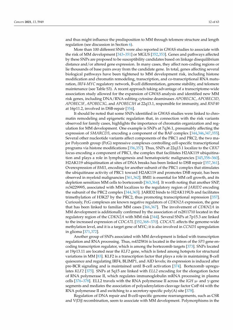
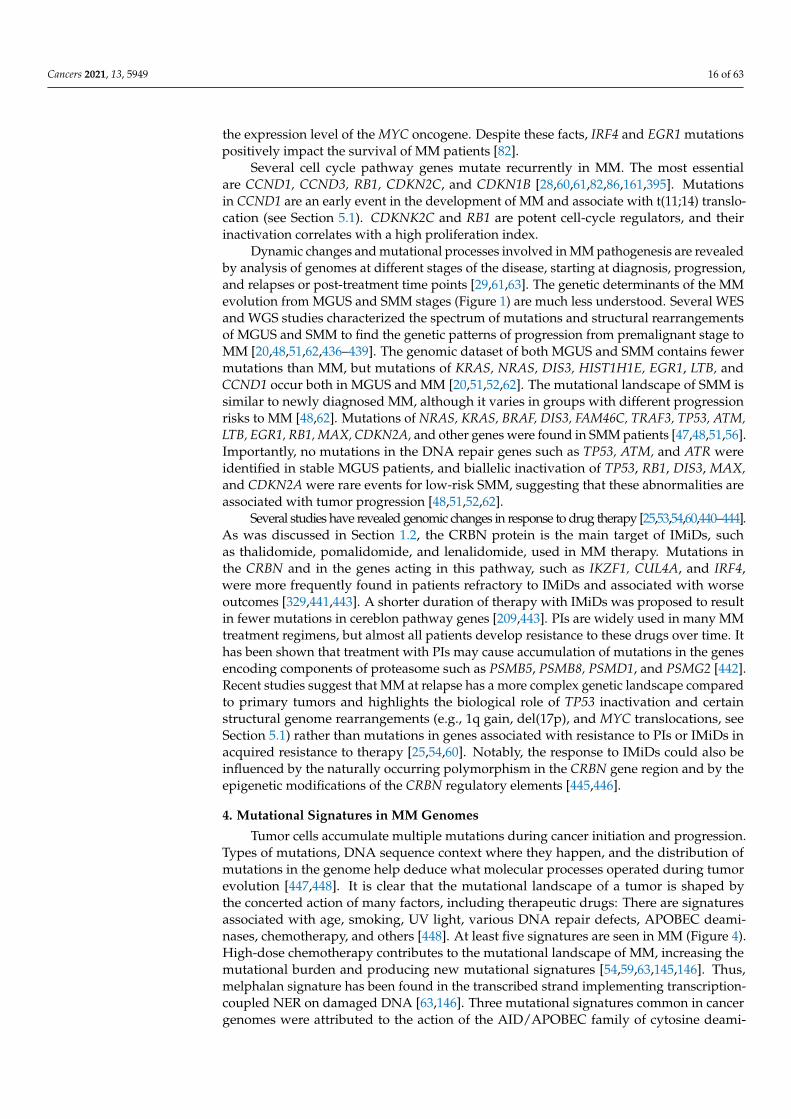
Tumor cells accumulate multiple mutations during cancer initiation and progression.Types of mutations, DNA sequence context where they happen, and the distribution ofmutations in the genome help deduce what molecular processes operated during tumorevolution [447,448]. It is clear that the mutational landscape of a tumor is shaped bythe concerted action of many factors, including therapeutic drugs: There are signaturesassociated with age, smoking, UV light, various DNA repair defects, APOBEC deami-nases, chemotherapy, and others [448]. At least five signatures are seen in MM (Figure 4).High-dose chemotherapy contributes to the mutational landscape of MM, increasing themutational burden and producing new mutational signatures [54,59,63,145,146]. Thus,melphalan signature has been found in the transcribed strand implementing transcription-coupled NER on damaged DNA [63,146]. Three mutational signatures common in cancergenomes were attributed to the action of the AID/APOBEC family of cytosine deami-
Cancers 2021, 13, 5949 17 of 63
nases [449]. AID/APOBEC is a superfamily of enzymes that convert cytosine to uraciland consists of two subfamilies, AID and APOBEC3 [450]. AID (activation-induced cy-tosine deaminase) edits DNA in immunoglobulin genes in the process of SHM and CSRin the maturating B-cells. Proteins of the APOBEC3 subfamily mediate the restriction ofviruses by deaminating viral cDNA. AID/APOBEC proteins are active on ssDNA and canact processively, thus generating clustered mutations [451]. Individual APOBEC proteinfamily members possess intrinsic deamination sequence specificity, thus allowing for at-tributing the mutations found in cancers to the action of specific APOBEC based on thesequence context of the changes. It has been proposed that AID/APOBEC processivity andavailability of long ssDNA regions in tumor evolution give rise to a phenomenon calledkataegis–localized hypermutated regions [452–454].
t(14, 8)
IGH MYC
t(other)
Intact chromosome
P/TSS
gene ‘Z’
ATG
IGH MYC
A
B
C
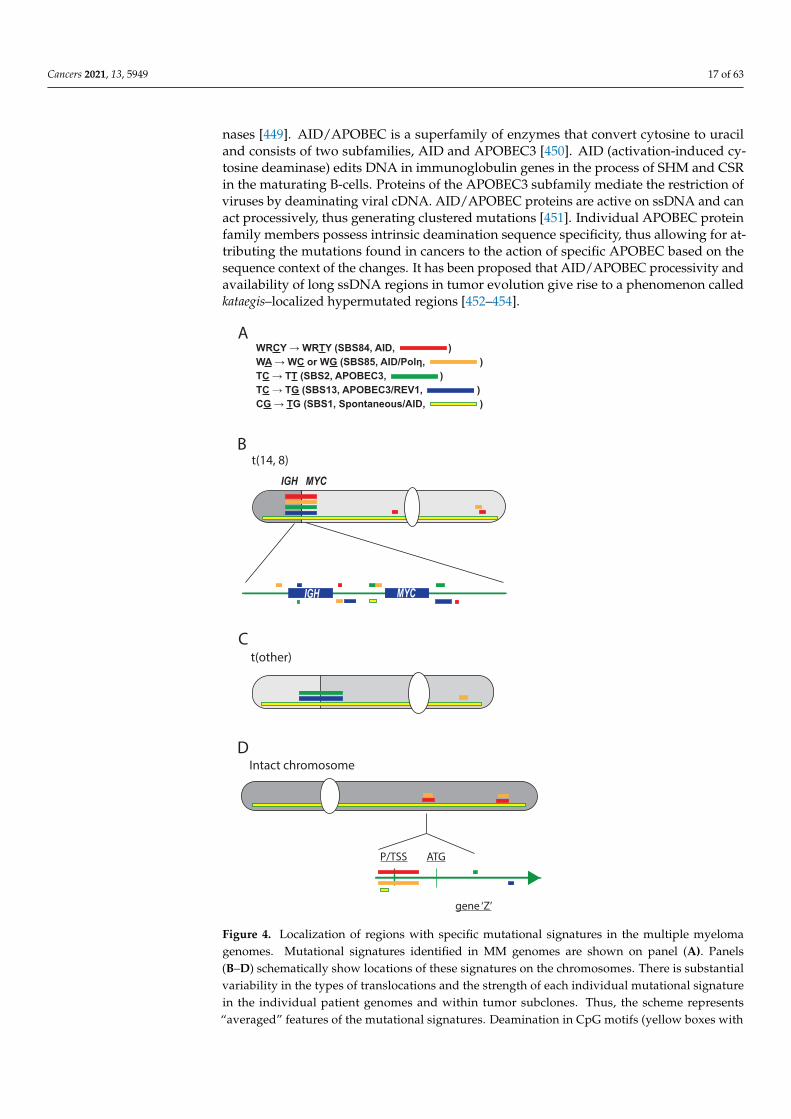
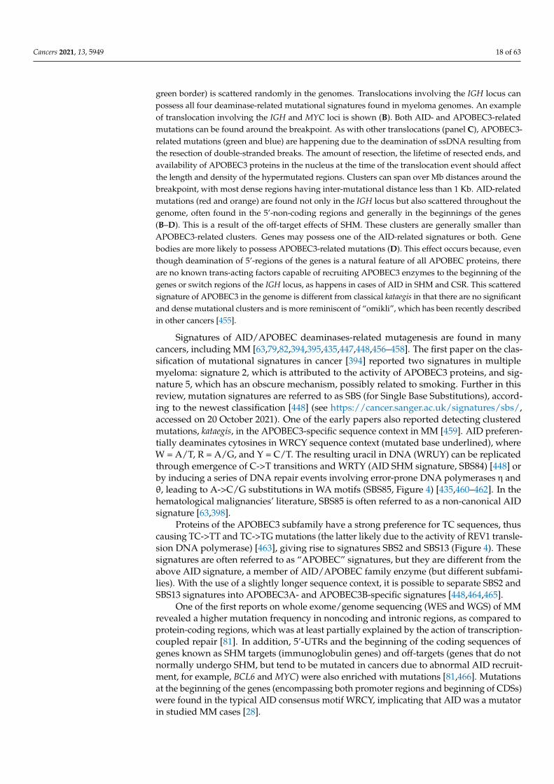
WRCY → WRTY (SBS84, AID, )WA → WC or WG (SBS85, AID/Polη, )TC → TT (SBS2, APOBEC3, )TC → TG (SBS13, APOBEC3/REV1, )CG → TG (SBS1, Spontaneous/AID, )
D
Figure 4. Localization of regions with specific mutational signatures in the multiple myelomagenomes. Mutational signatures identified in MM genomes are shown on panel (A). Panels(B–D) schematically show locations of these signatures on the chromosomes. There is substantialvariability in the types of translocations and the strength of each individual mutational signaturein the individual patient genomes and within tumor subclones. Thus, the scheme represents“averaged” features of the mutational signatures. Deamination in CpG motifs (yellow boxes with
Cancers 2021, 13, 5949 18 of 63
green border) is scattered randomly in the genomes. Translocations involving the IGH locus canpossess all four deaminase-related mutational signatures found in myeloma genomes. An exampleof translocation involving the IGH and MYC loci is shown (B). Both AID- and APOBEC3-relatedmutations can be found around the breakpoint. As with other translocations (panel C), APOBEC3-related mutations (green and blue) are happening due to the deamination of ssDNA resulting fromthe resection of double-stranded breaks. The amount of resection, the lifetime of resected ends, andavailability of APOBEC3 proteins in the nucleus at the time of the translocation event should affectthe length and density of the hypermutated regions. Clusters can span over Mb distances around thebreakpoint, with most dense regions having inter-mutational distance less than 1 Kb. AID-relatedmutations (red and orange) are found not only in the IGH locus but also scattered throughout thegenome, often found in the 5’-non-coding regions and generally in the beginnings of the genes(B–D). This is a result of the off-target effects of SHM. These clusters are generally smaller thanAPOBEC3-related clusters. Genes may possess one of the AID-related signatures or both. Genebodies are more likely to possess APOBEC3-related mutations (D). This effect occurs because, eventhough deamination of 5’-regions of the genes is a natural feature of all APOBEC proteins, thereare no known trans-acting factors capable of recruiting APOBEC3 enzymes to the beginning of thegenes or switch regions of the IGH locus, as happens in cases of AID in SHM and CSR. This scatteredsignature of APOBEC3 in the genome is different from classical kataegis in that there are no significantand dense mutational clusters and is more reminiscent of “omikli”, which has been recently describedin other cancers [455].
Signatures of AID/APOBEC deaminases-related mutagenesis are found in manycancers, including MM [63,79,82,394,395,435,447,448,456–458]. The first paper on the clas-sification of mutational signatures in cancer [394] reported two signatures in multiplemyeloma: signature 2, which is attributed to the activity of APOBEC3 proteins, and sig-nature 5, which has an obscure mechanism, possibly related to smoking. Further in thisreview, mutation signatures are referred to as SBS (for Single Base Substitutions), accord-ing to the newest classification [448] (see https://cancer.sanger.ac.uk/signatures/sbs/,accessed on 20 October 2021). One of the early papers also reported detecting clusteredmutations, kataegis, in the APOBEC3-specific sequence context in MM [459]. AID preferen-tially deaminates cytosines in WRCY sequence context (mutated base underlined), whereW = A/T, R = A/G, and Y = C/T. The resulting uracil in DNA (WRUY) can be replicatedthrough emergence of C->T transitions and WRTY (AID SHM signature, SBS84) [448] orby inducing a series of DNA repair events involving error-prone DNA polymerases η andθ, leading to A->C/G substitutions in WA motifs (SBS85, Figure 4) [435,460–462]. In thehematological malignancies’ literature, SBS85 is often referred to as a non-canonical AIDsignature [63,398].
Proteins of the APOBEC3 subfamily have a strong preference for TC sequences, thuscausing TC->TT and TC->TG mutations (the latter likely due to the activity of REV1 transle-sion DNA polymerase) [463], giving rise to signatures SBS2 and SBS13 (Figure 4). Thesesignatures are often referred to as “APOBEC” signatures, but they are different from theabove AID signature, a member of AID/APOBEC family enzyme (but different subfami-lies). With the use of a slightly longer sequence context, it is possible to separate SBS2 andSBS13 signatures into APOBEC3A- and APOBEC3B-specific signatures [448,464,465].
One of the first reports on whole exome/genome sequencing (WES and WGS) of MMrevealed a higher mutation frequency in noncoding and intronic regions, as compared toprotein-coding regions, which was at least partially explained by the action of transcription-coupled repair [81]. In addition, 5’-UTRs and the beginning of the coding sequences ofgenes known as SHM targets (immunoglobulin genes) and off-targets (genes that do notnormally undergo SHM, but tend to be mutated in cancers due to abnormal AID recruit-ment, for example, BCL6 and MYC) were also enriched with mutations [81,466]. Mutationsat the beginning of the genes (encompassing both promoter regions and beginning of CDSs)were found in the typical AID consensus motif WRCY, implicating that AID was a mutatorin studied MM cases [28].
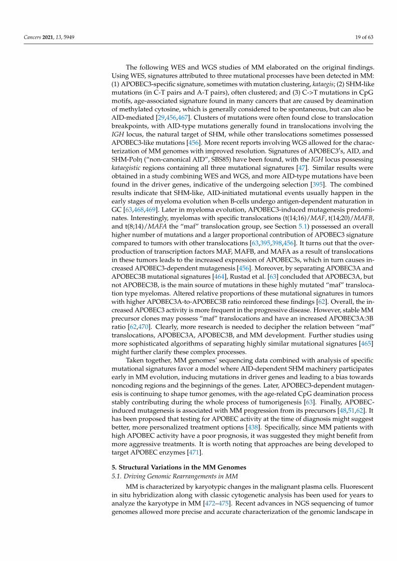
Cancers 2021, 13, 5949 19 of 63
The following WES and WGS studies of MM elaborated on the original findings.Using WES, signatures attributed to three mutational processes have been detected in MM:(1) APOBEC3-specific signature, sometimes with mutation clustering, kataegis; (2) SHM-likemutations (in C-T pairs and A-T pairs), often clustered; and (3) C->T mutations in CpGmotifs, age-associated signature found in many cancers that are caused by deaminationof methylated cytosine, which is generally considered to be spontaneous, but can also beAID-mediated [29,456,467]. Clusters of mutations were often found close to translocationbreakpoints, with AID-type mutations generally found in translocations involving theIGH locus, the natural target of SHM, while other translocations sometimes possessedAPOBEC3-like mutations [456]. More recent reports involving WGS allowed for the charac-terization of MM genomes with improved resolution. Signatures of APOBEC3′s, AID, andSHM-Polη (“non-canonical AID”, SBS85) have been found, with the IGH locus possessingkataegistic regions containing all three mutational signatures [47]. Similar results wereobtained in a study combining WES and WGS, and more AID-type mutations have beenfound in the driver genes, indicative of the undergoing selection [395]. The combinedresults indicate that SHM-like, AID-initiated mutational events usually happen in theearly stages of myeloma evolution when B-cells undergo antigen-dependent maturation inGC [63,468,469]. Later in myeloma evolution, APOBEC3-induced mutagenesis predomi-nates. Interestingly, myelomas with specific translocations (t(14;16)/MAF, t(14;20)/MAFB,and t(8;14)/MAFA the “maf” translocation group, see Section 5.1) possessed an overallhigher number of mutations and a larger proportional contribution of APOBEC3 signaturecompared to tumors with other translocations [63,395,398,456]. It turns out that the over-production of transcription factors MAF, MAFB, and MAFA as a result of translocationsin these tumors leads to the increased expression of APOBEC3s, which in turn causes in-creased APOBEC3-dependent mutagenesis [456]. Moreover, by separating APOBEC3A andAPOBEC3B mutational signatures [464], Rustad et al. [63] concluded that APOBEC3A, butnot APOBEC3B, is the main source of mutations in these highly mutated “maf” transloca-tion type myelomas. Altered relative proportions of these mutational signatures in tumorswith higher APOBEC3A-to-APOBEC3B ratio reinforced these findings [62]. Overall, the in-creased APOBEC3 activity is more frequent in the progressive disease. However, stable MMprecursor clones may possess “maf” translocations and have an increased APOBEC3A:3Bratio [62,470]. Clearly, more research is needed to decipher the relation between “maf”translocations, APOBEC3A, APOBEC3B, and MM development. Further studies usingmore sophisticated algorithms of separating highly similar mutational signatures [465]might further clarify these complex processes.
Taken together, MM genomes’ sequencing data combined with analysis of specificmutational signatures favor a model where AID-dependent SHM machinery participatesearly in MM evolution, inducing mutations in driver genes and leading to a bias towardsnoncoding regions and the beginnings of the genes. Later, APOBEC3-dependent mutagen-esis is continuing to shape tumor genomes, with the age-related CpG deamination processstably contributing during the whole process of tumorigenesis [63]. Finally, APOBEC-induced mutagenesis is associated with MM progression from its precursors [48,51,62]. Ithas been proposed that testing for APOBEC activity at the time of diagnosis might suggestbetter, more personalized treatment options [438]. Specifically, since MM patients withhigh APOBEC activity have a poor prognosis, it was suggested they might benefit frommore aggressive treatments. It is worth noting that approaches are being developed totarget APOBEC enzymes [471].
5. Structural Variations in the MM Genomes5.1. Driving Genomic Rearrangements in MM
MM is characterized by karyotypic changes in the malignant plasma cells. Fluorescentin situ hybridization along with classic cytogenetic analysis has been used for years toanalyze the karyotype in MM [472–475]. Recent advances in NGS sequencing of tumorgenomes allowed more precise and accurate characterization of the genomic landscape in
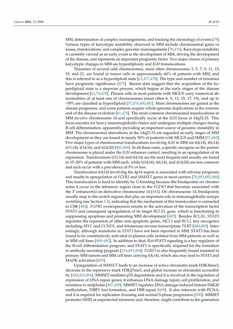
Cancers 2021, 13, 5949 20 of 63
MM, determination of complex rearrangements, and tracking the chronology of events [79].Various types of karyotype instability observed in MM include chromosomal gains orlosses, translocations, and complex genomic rearrangements [78,476]. Karyotype instabilityis currently viewed as an early event in the development of MM, driving the developmentof the disease, and represents an important prognostic factor. Two major classes of primarykaryotypic changes in MM are hyperdiploidy and IGH-translocations.
Trisomies of several odd chromosomes, most often chromosomes 3, 5, 7, 9, 11, 15,19, and 21, are found in tumor cells in approximately 60% of patients with MM, andthis is referred to as a hyperdiploid state [61,477,478]. The type and number of trisomieshave prognostic significance [479]. Recent data suggest that the acquisition of the hy-perdiploid state is a stepwise process, which begins at the early stages of the diseasedevelopment [61,79,478]. Plasma cells in most patients with MGUS carry numerical ab-normalities of at least one of chromosomes (most often 6, 9, 13, 15, 17, 19), and up to~50% are classified as hyperdiploid [57,478,480,481]. More chromosomes are gained as thedisease progresses, and some patients acquire whole-genome duplications at the extremeend of the disease evolution [61,478]. The most common chromosomal translocations inMM involve chromosome 14 and specifically occur at the IGH locus at 14q32.33. Thislocus encodes for heavy immunoglobulin chains and undergoes multiple changes duringB cell differentiation, apparently providing an important source of genomic instability inMM. The chromosomal aberrations at the 14q23.33 are regarded as early stages of MMdevelopment as they are found in nearly 50% of patients with MGUS and SMM [57,482].Five major types of chromosomal translocations involving IGH in MM are t(4;14), t(6;14),t(11;14), t(14;16), and t(14;20) [483,484]. In all these cases, a specific oncogene on the partnerchromosome is placed under the IGH enhancer control, resulting in an upregulation of itsexpression. Translocations t(11;14) and t(4;14) are the most frequent and usually are foundin 15–20% of patients with MM each, while t(14;16), t(6;14), and t(14;20) are less commonand each occur with a prevalence of 5% or less.
Translocation t(4;14) involving the 4p16 region is associated with adverse prognosisand results in upregulation of FGFR3 and MMSET genes in most carriers [78,483,485,486].This translocation is hard to identify by G-banding because the breakpoints on chromo-some 4 occur in the telomeric region close to the FGFR3 that becomes associated withthe 3′ enhancer(s) on derivative chromosome 14 [484]. On chromosome 14, breakpointsusually map to the switch regions that play an important role in immunoglobulin isotypeswitching (see Section 5.2), indicating that the mechanism of this translocation is connectedto CSR [484]. FGFR3 overexpression results in the activation of the transcription factorSTAT3 and consequent upregulation of its target BCLXL gene, which is functioning insuppressing apoptosis and promoting MM development [487]. Besides BCLXL, STAT3regulates the expression of other anti-apoptotic genes, MCL1 and BCL2, key oncogenesincluding MYC and CCND1, and telomerase reverse transcriptase TERT [488,489]. Inter-estingly, although mutations in STAT3 have not been reported in MM, STAT3 has beenfound to be constitutively activated in plasma cells isolated from MM patients as well asin MM cell lines [490–492]. In addition to that, IL6-STAT3 signaling is a key regulator ofthe B-cell differentiation program, and STAT3 is specifically required for the transitionto antibody-secreting program [214,493,494]. FGRF3 is also frequently found mutated inprimary MM tumors and MM cell lines carrying t(4;14), which also may lead to STAT3 andMAPK activation [495]
Upregulation of MMSET leads to an increase of active chromatin mark H3K36me2,decrease in the repressive mark H3K27me3, and global increase in chromatin accessibil-ity [418,419,496]. MMSET mediates p53 degradation and it is involved in the regulation ofexpression of DNA repair genes; it enhances DNA damage repair, cell proliferation, andresistance to melphalan [497,498]. MMSET regulates DNA damage-induced histone H4K20methylation, 53BP1 foci formation, and DSB repair [499]. It also interacts with PCNA,and it is required for replication licensing and normal S-phase progression [500]. MMSETpromotes NHEJ at unprotected telomeres and, therefore, might contribute to the generation
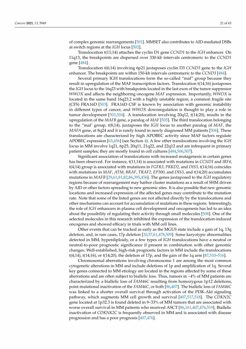
Cancers 2021, 13, 5949 21 of 63
of complex genomic rearrangements [501]. MMSET also contributes to AID-mediated DSBsat switch regions at the IGH locus [502].
Translocation t(11;14) attaches the cyclin D1 gene CCND1 to the IGH enhancer. On11q13, the breakpoints are dispersed over 330-kb intervals centromeric to the CCND1gene [484].
Translocation t(6;14) involving 6p21 juxtaposes cyclin D3 CCND3 gene to the IGHenhancer. The breakpoints are within 150-kb intervals centromeric to the CCND3 [484].
Several primary IGH translocations form the so-called “maf” group because theyresult in upregulation of the MAF transcription factors. Translocation t(14;16) juxtaposesthe IGH locus to the 16q23 with breakpoints located in the last exon of the tumor suppressorWWOX and affects the neighboring oncogene MAF expression. Importantly, WWOX islocated in the same band 16q23.2 with a highly unstable region, a common fragile site(CFS) FRA16D [503]. FRA16D CSF is known by association with genomic instabilityin different types of cancer, and WWOX downregulation is thought to play a role intumor development [503,504]. A translocation involving 20q12, t(14;20), results in theupregulation of the MAFB gene, a paralog of MAF [505]. The third translocation belongingto the “maf’ group, t(8;14), juxtaposes the IGH locus to another paralog of MAF, theMAFA gene, at 8q24 and it is rarely found in newly diagnosed MM patients [506]. Thesetranslocations are characterized by high APOBEC activity since MAF factors regulateAPOBEC expression [63,456] (see Section 4). A few other translocations involving the IGHlocus in MM involve 1q21, 6p25, 20q11, 21q22, and 22q12 and are infrequent in primarypatient samples; they are mostly found in cell cultures [484,506,507].
Significant association of translocations with increased mutagenesis in certain geneshas been observed. For instance, t(11;14) is associated with mutations in CCND1 and IRF4,t(4;14) group is associated with mutations in FGFR3, PRKD2, and DIS3, t(14;16) coincideswith mutations in MAF, ATM, BRAF, TRAF2, EP300, and DIS3, and t(14;20) accumulatesmutations in MAFB [29,61,81,82,86,395,456]. The genes juxtaposed to the IGH regulatoryregions because of rearrangement may harbor cluster mutations as a result of SHM drivenby AID or other factors spreading to new genomic sites. It is also possible that new genomiclocations and increased expression of the affected genes may contribute to the mutationrate. Note that some of the listed genes are not affected directly by the translocations andother mechanisms can account for accumulation of mutations in these regions. Interestingly,the role of IGH enhancers in plasma cell development and oncogenesis has led to an ideaabout the possibility of regulating their activity through small molecules [508]. One of theselected molecules in this research inhibited the expression of the translocation-inducedoncogenes and showed efficacy in trials with MM cell lines.
Other events that can be tracked as early as the MGUS state include a gain of 1q, 13qdeletion, and, in rare cases, 17p deletion [20,57,61,478,509]. Some karyotypic abnormalitiesdetected in MM, hyperdiploidy, or a few types of IGH translocations have a neutral orneutral-to-poor prognostic significance if present in combination with other genomicchanges. Well-established, high-risk prognostic factors in MM include the translocationst(4;14), t(14;16), or t(14;20), the deletion of 17p, and the gain of the 1q arm [87,510–516].
Chromosomal aberrations involving chromosome 1 are among the most commoncytogenetic alterations in MM and include deletions of 1p and amplification of 1q. Severalkey genes connected to MM etiology are located in the regions affected by some of theseaberrations and are often subject to biallelic loss. Thus, tumors in ~4% of MM patients arecharacterized by a biallelic loss of FAM46C resulting from homozygous 1p12 deletions,point mutational inactivation of the FAM46C, or both [86,407]. The biallelic loss of FAM46Cwas linked to a shorter overall survival through activation of the PI3K-Akt signalingpathway, which augments MM cell growth and survival [407,517,518]. The CDKN2Cgene located at 1p32.3 is found deleted in 9–33% of MM tumors that are associated withworse overall survival in MM patients who received ASCT [86,161,407,476,519]. Biallelicinactivation of CDKNK2C is frequently observed in MM and is associated with diseaseprogression and has a poor prognosis [407,476].
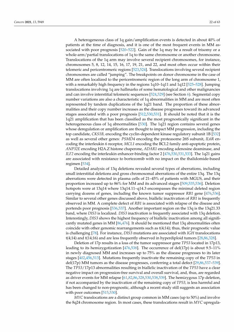
Cancers 2021, 13, 5949 22 of 63
A heterogeneous class of 1q gain/amplification events is detected in about 40% ofpatients at the time of diagnosis, and it is one of the most frequent events in MM as-sociated with poor prognosis [520–522]. Gain of the 1q may be a result of trisomy or awhole-arm/partial translocations of 1q to the same chromosome or another chromosome.Translocations of the 1q arm may involve several recipient chromosomes, for instance,chromosomes 5, 8, 12, 14, 15, 16, 17, 19, 21, and 22, and most often occur within theirtelomeric and pericentromeric regions [523,524]. Translocations involving several recipientchromosomes are called “jumping”. The breakpoints on donor chromosome in the case ofMM are often localized to the pericentromeric region of the long arm of chromosome 1,with a remarkably high frequency in the regions 1q10–1q11 and 1q12 [525–528]. Jumpingtranslocations involving 1q are hallmarks of some hematological and other malignanciesand can involve interstitial telomeric sequences [524,529] (see Section 6). Segmental copynumber variations are also a characteristic of 1q abnormalities in MM and are most oftenrepresented by tandem duplications of the 1q21 band. The proportion of these abnor-malities and their copy number increases as the disease progresses toward its advancedstages associated with a poor prognosis [512,530,531]. It should be noted that it is the1q21 amplification that has been classified as the most prognostically significant in theheterogeneous class of 1q abnormalities [530]. The 1q21 region contains several geneswhose deregulation or amplification are thought to impact MM progression, including thetop candidate, CKS1B, encoding the cyclin-dependent kinase regulatory subunit 1B [532]as well as several other genes: PSMD4 encoding the proteasome 26S subunit, IL6R en-coding the interleukin 6 receptor, MCL1 encoding the BCL2 family anti-apoptotic protein,ANP32E encoding H2A.Z histone chaperone, ADAR1 encoding adenosine deaminase, andILF2 encoding the interleukin enhancer-binding factor 2 [476,530,531,533]. The 1q21 gainsare associated with resistance to bortezomib with no impact on the thalidomide-basedregimen [534].
Detailed analysis of 13q deletions revealed several types of aberrations, includingsmall interstitial deletions and gross chromosomal aberrations of the entire 13q. The 13qaberrations were detected in plasma cells of 21–45% of patients with MGUS, and theirproportion increased up to 86% for MM and its advanced stages [509,535,536]. Deletionhotspots were at 13q14 where 13q14.11–q14.3 encompasses the minimal deleted regioncarrying dozens of genes, including the known tumor suppressor RB1 gene [476,536].Similar to several other genes discussed above, biallelic inactivation of RB1 is frequentlyobserved in MM. A complete defect of RB1 is associated with relapse of the disease andportends poor prognosis [536,537]. Another important region on the 13q is the 13q21.33band, where DIS3 is localized. DIS3 inactivation is frequently associated with 13q deletion.Interestingly, DIS3 shows the highest frequency of biallelic inactivation among all signifi-cantly mutated genes in MM [86,476]. It should be mentioned that 13q abnormalities oftencoincide with other genomic rearrangements such as t(4;14); thus, their prognostic valueis challenging [78]. For instance, DIS3 mutations are associated with IGH translocationst(4;14) and t(14;16) and are less frequently observed in hyperdiploid tumors [28,86,328].
Deletion of 17p results in a loss of the tumor suppressor gene TP53 located in 17p13,leading to its hemizygotization [476,538]. The occurrence of del(17p) is about 9.5–11%in newly diagnosed MM and increases up to 75% as the disease progresses to its laterstages [402,456,513]. Mutations frequently inactivate the remaining copy of the TP53 indel(17p) MM tumors as the disease progresses, conferring a total defect [29,86,537–539].The TP53/17p13 abnormalities resulting in biallelic inactivation of the TP53 have a clearnegative impact on progression-free survival and overall survival, and, thus, are regardedas driver events for MM relapse [61,82,86,328,530,538,539]. The hemizygous 17p deletion,if not accompanied by the inactivation of the remaining copy of TP53, is less harmful andhas been changed to non-prognostic, although a recent study still suggests an associationwith poor outcomes [515,530].
MYC translocations are a distinct group common in MM cases (up to 50%) and involvethe 8q24 chromosome region. In most cases, these translocations result in MYC upregula-
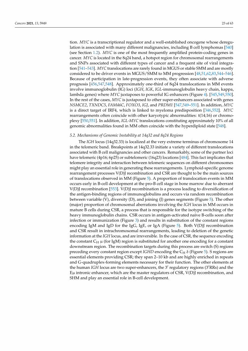
Cancers 2021, 13, 5949 23 of 63
tion. MYC is a transcriptional regulator and a well-established oncogene whose deregu-lation is associated with many different malignancies, including B cell lymphomas [540](see Section 1.2). MYC is one of the most frequently amplified protein-coding genes incancer. MYC is located in the 8q24 band, a hotspot region for chromosomal rearrangementsand SNPs associated with different types of cancer and a frequent site of viral integra-tion [541–543]. MYC translocations are rarely found in MGUS or stable SMM and are mostlyconsidered to be driver events in MGUS/SMM to MM progression [48,51,62,83,544–546].Because of participation in late-progression events, they often associate with adverseprognosis [456,547,548]. Approximately one-third of 8q24 translocations in MM eventsinvolve immunoglobulin (IG) loci (IGH, IGK, IGL-immunoglobulin heavy chain, kappa,lambda genes) where MYC juxtaposes to powerful IG enhancers (Figure 4). [545,549,550].In the rest of the cases, MYC is juxtaposed to other super-enhancers associated with genesNSMCE2, TXNDC5, FAM46C, FOXO3, IGJ, and PRDM1 [547,549–551]. In addition, MYCis a direct target of IRF4, which is linked to myeloma predisposition [346,552]. MYCrearrangements often coincide with other karyotypic abnormalities: t(14;16) or chromo-plexy [550,551]. In addition, IGL-MYC translocations constituting approximately 10% of allgenomic abnormalities found in MM often coincide with the hyperdiploid state [548].
5.2. Mechanisms of Genomic Instability at 14q32 and 8q24 Regions
The IGH locus (14q32.33) is localized at the very extreme terminus of chromosome 14in the telomeric band. Breakpoints at 14q32.33 initiate a variety of different translocationsassociated with B cell malignancies and other cancers. Remarkably, some of the partner locihave telomeric (4p16; 6p25) or subtelomeric (16q23) locations [484]. This fact implicates thattelomere integrity and interaction between telomeric sequences on different chromosomesmight play an essential role in generating these rearrangements. Lymphoid-specific genomerearrangement processes V(D)J recombination and CSR are thought to be the main sourcesof translocations observed in MM (Figure 5). A proportion of translocation events in MMoccurs early in B-cell development at the pro-B cell stage in bone marrow due to aberrantV(D)J recombination [553]. V(D)J recombination is a process leading to diversification ofthe antigen-binding regions of immunoglobulins and occurs via random recombinationbetween variable (V), diversity (D), and joining (J) genes segments (Figure 5). The other(major) proportion of chromosomal aberrations involving the IGH locus in MM occurs inmature B cells during CSR, a process that is responsible for the isotype switching of theheavy immunoglobulin chains. CSR occurs in antigen-activated naïve B-cells soon afterinfection or immunization (Figure 3) and results in substitution of the constant regionsencoding IgM and IgD for the IgG, IgE, or IgA (Figure 5). Both V(D)J recombinationand CSR result in intrachromosomal rearrangements, leading to deletion of the geneticinformation at the IGH locus, and are irreversible. In the case of CSR, the sequence encodingthe constant CH µ (for IgM) region is substituted for another one encoding for a constantdownstream region. The recombination targets during this process are switch (S) regionspreceding every constant region except IGHD encoding the CH δ (Figure 5). S regions areessential elements providing CSR; they span 2–10 kb and are highly enriched in repeatsand G-quadruplex-forming elements necessary for their function. The other elements atthe human IGH locus are two super-enhancers, the 3′ regulatory regions (3′RRs) and theEµ intronic enhancer, which are the master regulators of CSR, V(D)J recombination, andSHM and play an essential role in B-cell development.
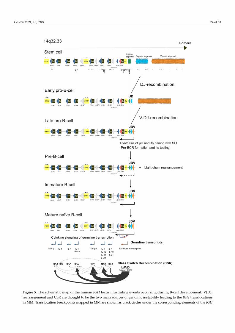
Cancers 2021, 13, 5949 24 of 63
V-gene segmentD-gene segmentJ-gene segment
ATP6VG1P1
IGHG3IGHG1IGHEP1IGHA1 IGHMIGHDIGHGPIGHG2IGHG4IGHEIGHA2
Eμ3’RR1Sα2 Sε Sγ4 Sγ2 Sα1 Sγ1 Sγ3 Sμ
JD
Telomere
DJ-recombination
JDV
V-DJ-recombination
Early pro-B-cell
Late pro-B-cell
Synthesis of μH and its pairing with SLC
Pre-BCR formation and its testing
Pre-B-cell
14q32.33
JDV
Stem cell
3’RR2
ATP6VG1P1
IGHG3IGHG1IGHEP1IGHA1 IGHMIGHDIGHGPIGHG2IGHG4IGHEIGHA2
Eμ3’RR1Sα2 Sε Sγ4 Sγ2 Sα1 Sγ1 Sγ3 Sμ3’RR2
ATP6VG1P1
IGHG3IGHG1IGHEP1IGHA1 IGHMIGHDIGHGPIGHG2IGHG4IGHEIGHA2
Eμ3’RR1Sα2 Sε Sγ4 Sγ2 Sα1 Sγ1 Sγ3 Sμ3’RR2
ATP6VG1P1
IGHG3IGHG1IGHEP1IGHA1 IGHMIGHDIGHGPIGHG2IGHG4IGHEIGHA2
Eμ3’RR1Sα2 Sε Sγ4 Sγ2 Sα1 Sγ1 Sγ3 Sμ3’RR2 Light chain rearrangement
Eμ-driven transcription
+
Immature B-cell
JDV
ATP6VG1P1
IGHG3IGHG1IGHEP1IGHA1 IGHMIGHDIGHGPIGHG2IGHG4IGHEIGHA2
Eμ3’RR1Sα2 Sε Sγ4 Sγ2 Sα1 Sγ1 Sγ3 Sμ3’RR2
Mature naïve B-cell
JDV
ATP6VG1P1
IGHG3IGHG1IGHEP1IGHA1 IGHMIGHDIGHGPIGHG2IGHG4IGHEIGHA2
Eμ3’RR1Sα2 Sε Sγ4 Sγ2 Sα1 Sγ1 Sγ3 Sμ3’RR2
IFN-γ
IL-4IL-4 TGF-β1
IL-27
IL-4 IL-4
Germline transcripts
IL-4
IL-10
IL-21 IL-21
IL-10
TGF-β1
IgM/DIgG1IgA1IgG2
Primary CSR
IgA2
Cytokine signaling of germline transcription
IgG3IgG4IgE Class Switch Recombination (CSR)
Figure 5. The schematic map of the human IGH locus illustrating events occurring during B-cell development. V(D)Jrearrangement and CSR are thought to be the two main sources of genomic instability leading to the IGH translocationsin MM. Translocation breakpoints mapped in MM are shown as black circles under the corresponding elements of the IGH
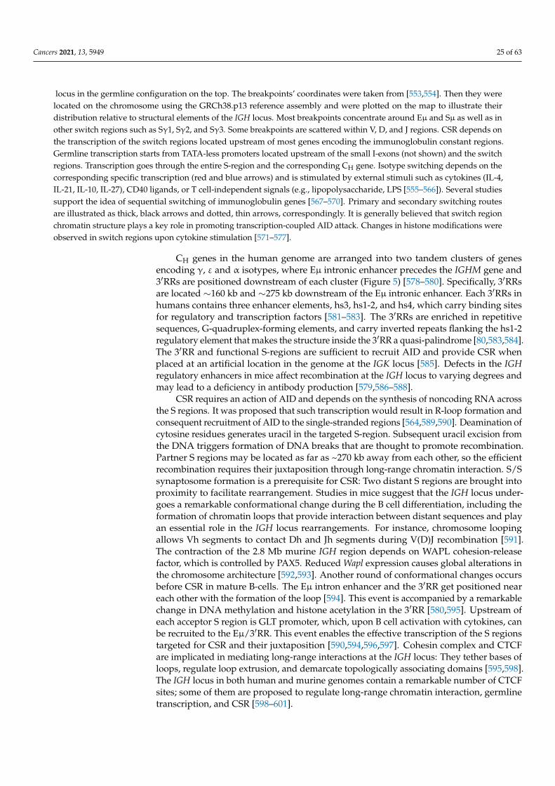
Cancers 2021, 13, 5949 25 of 63
locus in the germline configuration on the top. The breakpoints’ coordinates were taken from [553,554]. Then they werelocated on the chromosome using the GRCh38.p13 reference assembly and were plotted on the map to illustrate theirdistribution relative to structural elements of the IGH locus. Most breakpoints concentrate around Eµ and Sµ as well as inother switch regions such as Sγ1, Sγ2, and Sγ3. Some breakpoints are scattered within V, D, and J regions. CSR depends onthe transcription of the switch regions located upstream of most genes encoding the immunoglobulin constant regions.Germline transcription starts from TATA-less promoters located upstream of the small I-exons (not shown) and the switchregions. Transcription goes through the entire S-region and the corresponding CH gene. Isotype switching depends on thecorresponding specific transcription (red and blue arrows) and is stimulated by external stimuli such as cytokines (IL-4,IL-21, IL-10, IL-27), CD40 ligands, or T cell-independent signals (e.g., lipopolysaccharide, LPS [555–566]). Several studiessupport the idea of sequential switching of immunoglobulin genes [567–570]. Primary and secondary switching routesare illustrated as thick, black arrows and dotted, thin arrows, correspondingly. It is generally believed that switch regionchromatin structure plays a key role in promoting transcription-coupled AID attack. Changes in histone modifications wereobserved in switch regions upon cytokine stimulation [571–577].
CH genes in the human genome are arranged into two tandem clusters of genesencoding γ, ε and α isotypes, where Eµ intronic enhancer precedes the IGHM gene and3′RRs are positioned downstream of each cluster (Figure 5) [578–580]. Specifically, 3′RRsare located ∼160 kb and ∼275 kb downstream of the Eµ intronic enhancer. Each 3′RRs inhumans contains three enhancer elements, hs3, hs1-2, and hs4, which carry binding sitesfor regulatory and transcription factors [581–583]. The 3′RRs are enriched in repetitivesequences, G-quadruplex-forming elements, and carry inverted repeats flanking the hs1-2regulatory element that makes the structure inside the 3′RR a quasi-palindrome [80,583,584].The 3′RR and functional S-regions are sufficient to recruit AID and provide CSR whenplaced at an artificial location in the genome at the IGK locus [585]. Defects in the IGHregulatory enhancers in mice affect recombination at the IGH locus to varying degrees andmay lead to a deficiency in antibody production [579,586–588].
CSR requires an action of AID and depends on the synthesis of noncoding RNA acrossthe S regions. It was proposed that such transcription would result in R-loop formation andconsequent recruitment of AID to the single-stranded regions [564,589,590]. Deamination ofcytosine residues generates uracil in the targeted S-region. Subsequent uracil excision fromthe DNA triggers formation of DNA breaks that are thought to promote recombination.Partner S regions may be located as far as ~270 kb away from each other, so the efficientrecombination requires their juxtaposition through long-range chromatin interaction. S/Ssynaptosome formation is a prerequisite for CSR: Two distant S regions are brought intoproximity to facilitate rearrangement. Studies in mice suggest that the IGH locus under-goes a remarkable conformational change during the B cell differentiation, including theformation of chromatin loops that provide interaction between distant sequences and playan essential role in the IGH locus rearrangements. For instance, chromosome loopingallows Vh segments to contact Dh and Jh segments during V(D)J recombination [591].The contraction of the 2.8 Mb murine IGH region depends on WAPL cohesion-releasefactor, which is controlled by PAX5. Reduced Wapl expression causes global alterations inthe chromosome architecture [592,593]. Another round of conformational changes occursbefore CSR in mature B-cells. The Eµ intron enhancer and the 3′RR get positioned neareach other with the formation of the loop [594]. This event is accompanied by a remarkablechange in DNA methylation and histone acetylation in the 3′RR [580,595]. Upstream ofeach acceptor S region is GLT promoter, which, upon B cell activation with cytokines, canbe recruited to the Eµ/3′RR. This event enables the effective transcription of the S regionstargeted for CSR and their juxtaposition [590,594,596,597]. Cohesin complex and CTCFare implicated in mediating long-range interactions at the IGH locus: They tether bases ofloops, regulate loop extrusion, and demarcate topologically associating domains [595,598].The IGH locus in both human and murine genomes contain a remarkable number of CTCFsites; some of them are proposed to regulate long-range chromatin interaction, germlinetranscription, and CSR [598–601].
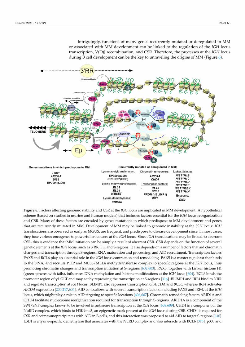
Cancers 2021, 13, 5949 26 of 63
Intriguingly, functions of many genes recurrently mutated or deregulated in MMor associated with MM development can be linked to the regulation of the IGH locustranscription, V(D)J recombination, and CSR. Therefore, the processes at the IGH locusduring B cell development can be the key to unraveling the origins of MM (Figure 6).
PAX5
MLL3MLL4
PAX5
PAX5
RNA Pol II
TELOMERE
Exosome
RNA Pol II
Exosome
Eμ
3’RR
DNA methylation
Histone modification
PAX5
DNA methylation
Sμ
Sγ1
KD
M6A
p300/CBP
RNA Pol II
RNA Pol II
Exosome
CC
C
CC
CC
AID
AID
AID
AID
SWI/SNFARID1A
PAX5
H1
CTCFCohesin
BCL6
NuRDLSD1
MMSETPTIP
Genes mutations in which predispose to MM: Recurrently mutated or deregulated in MM:
LSD1ARID1A
DIS3EP300 (p300)
DIS3
MLL3MLL4
MMSET
HIST1H1BHIST1H1CHIST1H1DHIST1H1E
HIST1H2BKHIST1H4H
Linker histones
CHD4
Chromatin remodelers:
ARID1ACHD4
Transcription factors:
PAX5BCL6
PRDM1 (BLIMP1)IRF4
Lysine acetyltransferases:
EP300 (p300)CREBBP (CBP)
Exosome:
Lysine methyltransferases:
Lysine demethylases:
KDM6A
BLIMP1
Chromatin remodelers
Histone modifiers
PAX5IRF4
IRF4
IRF4
IRF4
Figure 6. Factors affecting genomic stability and CSR at the IGH locus are implicated in MM development. A hypotheticalscheme (based on studies in murine and human models) that includes factors essential for the IGH locus reorganizationand CSR. Many of these factors are encoded by genes mutations in which predispose to MM development and genesthat are recurrently mutated in MM. Development of MM may be linked to genomic instability at the IGH locus: IGHtranslocations are observed as early as MGUS, are frequent, and predispose to disease development since, in most cases,they fuse various oncogenes to powerful enhancers at the IGH locus. Since IGH translocations may be linked to aberrantCSR, this is evidence that MM initiation can be simply a result of aberrant CSR. CSR depends on the function of severalgenetic elements at the IGH locus, such as 3’RR, Eµ, and S-regions. It also depends on a number of factors that aid chromatinchanges and transcription through S-regions, RNA maturation and processing, and AID recruitment. Transcription factorsPAX5 and BCL6 play an essential role in the IGH locus contraction and remodeling. PAX5 is a master regulator that bindsto the DNA, and recruits PTIP and MLL3/MLL4 methyltransferase complex to specific regions at the IGH locus, thuspromoting chromatin changes and transcription initiation at S-regions [602,603]. PAX5, together with Linker histone H1(green spheres with tails), influences DNA methylation and histone modifications at the IGH locus [604]. BCL6 binds thepromoter region of γ1 GLT and may act by repressing the transcription at S-regions [316]. BLIMP1 and IRF4 bind to 3′RRand regulate transcription at IGH locus; BLIMP1 also represses transcription of AICDA and BCL6, whereas IRF4 activatesAICDA expression [200,217,605]. AID co-localizes with several transcription factors, including PAX5 and IRF4, at the IGHlocus, which might play a role in AID targeting to specific locations [606,607]. Chromatin-remodeling factors ARID1A andCHD4 facilitate nucleosome reorganization required for transcription through S-regions. ARID1A is a component of theSWI/SNF complex known to be involved in antisense transcription at the IGH locus [608,609]. CHD4 is a component of theNuRD complex, which binds to H3K9me3, an epigenetic mark present at the IGH locus during CSR. CHD4 is required forCSR and coimmunoprecipitates with AID in B-cells, and this interaction was proposed to aid AID to target S-regions [610].LSD1 is a lysine-specific demethylase that associates with the NuRD complex and also interacts with BCL6 [315]. p300 and
Cancers 2021, 13, 5949 27 of 63
CBP are closely related acetyltransferases that play a role in transcription activation and enhancer regulation [611,612].KDM6A acts in concert with MLL3/MLL4 and p300/CBP in the mediation of transcription initiation. MMSET is lysinemethyltransferase, which is required for GLT transcription at S-regions [613]. In addition, MMSET promotes AID-mediatedDNA breaks at the donor switch region [502]. DIS3 is a component of the exosome, the complex that facilitates 3′-5′ RNAprocessing and aids in AID recruitment to both DNA strands [331,332,614]. In addition to these mechanisms, the telomericlocation of the IGH locus suggests that any events that affect telomeric length and structure or telomeric chromatin mayinfluence the CSR and provoke chromosomal aberrations.
PAX5 is a major transcription regulator involved in B-cell development, and itsmutational inactivation or deregulation is associated with B-cell malignancies [615–617].Mutations in the cis-regulatory elements affecting PAX5 expression and structural rear-rangements involving PAX5 were identified in MM [60,398,618,619]. Interestingly, PAX5regulatory regions, as well regulatory regions of some other B-cell specific oncogenes, getin close proximity and undergo antisense transcription and are targeted by AID, whichmay explain translocations between these loci and the IGH locus and mutagenesis in theseregions [614,620]. Differences in PAX5 expression profiles in B-cells between healthy peopleand MM patients and unique PAX5 isoforms have been noted in MM [621]. PAX5 hasmultiple sites of binding at the IGH locus and participates in spatial locus contractionnecessary for its rearrangement and V-DJ recombination [622–626]. The 3′RRs in mice areinterspersed with multiple PAX5 binding sites and undergo dynamic changes in PAX5-binding during CSR [596]. PAX5, together with Linker histone H1 is involved in B-cellspecific DNA methylation and histone modifications at the 3’RR in mice [604]. DNA is hy-permethylated at B-cell specific intronic enhancer regions in MM but not in normal plasmacells, and these sites overlap with binding sites of some transcription factors, includingPAX5 [428]. PAX5 binding to the DNA recruits PTIP and promotes binding of MLL3/MLL4histone lysine methyltransferase complex to specific regions at the IGH locus in mice,which promotes chromatin changes and transcription initiation at switch-regions criticalfor CSR [602,603]. MLL3/4 containing COMPASS complex includes PTIP and KDM6Aand functions with p300/CBP in the de novo “commissioning” of enhancers [611]. As wasdiscussed in Section 3, mutations in MLL3, MLL4, KDM6A, EP300, CBP, and, to a lesserextent, PTIP were all found in MM.
Interestingly, the IGH locus is one of the most heavily transcribed in plasma cells.Antisense transcription has been observed from variable region exons and from S regionsand is thought to be implicated in AID-targeting [608,614]. SWI/SNF chromatin remod-eling complexes are recruited to IGH locus and facilitate antisense IGH transcription andaccessibility of IGH for recombination in B cell precursors [609]. As was discussed earlier(Sections 2 and 3), genes encoding SWI/SNF complexes are altered in many cancers, in-cluding MM, and mutations in the genes encoding components of SWI/SNF complex arelinked to MM predisposition. In hematopoietic progenitors and non-B lineage cells, IGHis localized to the nuclear periphery [627]. More specifically, in fibroblasts, the IGH locuswas shown to be tethered to the nuclear lamina, where its transcription is repressed [628].Relocalization of the IGH locus from the periphery to the nuclear interior occurs in pro-B cells that, along with intergenic and antisense transcription and action of chromatinremodelers, facilitates genes shuffling during V(D)J recombination and further matura-tion of the antigenic repertoire [627,629,630]. It has been proposed that in cells, thereis a special perinucleolar recombination compartment where the IGH locus undergoesrearrangements during B-cell maturation and where the activity of AID and RAG1/2 canbe sequestered [631].
AID recruitment to the transcribed S-regions at the IGH locus is aided by the exosome.Exosome interacts with the RNA Polymerase II and may facilitate exonucleolytic degra-dation of a 3′ free RNA end on backtracked complexes, thus helping to generate ssDNAsubstrate for AID. The DIS3 gene that we discussed in Sections 2 and 3 is a catalytic com-ponent of the exosome and is implicated in regulating of AID-dependent mutagenesis and
Cancers 2021, 13, 5949 28 of 63
recombination at the IGH locus [331,332]. Other genes involved in transcription regulation,RNA processing, and maturation might also impact the complex process of the IGH locusreorganization. More research is required to understand the interrelation between variousprocesses at the IGH locus and MM development.
Similar to IGH translocations, some types of MYC translocations are thought to origi-nate from the aberrant, B-cell specific processes: V(D)J recombination, IGH switch recombi-nation, and SHM [632]. Studies in a murine model with AID-deficient mice have suggestedthat AID plays a crucial role in MYC/IGH translocations [633]. However, the localization ofMYC translocations breakpoints in the IGH locus in newly diagnosed MM patients showedthat they are spread out over the constant regions with no apparent association with AIDmotif clusters, pointing to a different mechanism of their generation [551]. 8q24 is a verypeculiar region – it is a so-called “gene desert” apparently representing an important sourceof genomic instability and translocation events. This region is almost devoid of protein-coding genes but contains multiple regulatory elements, and it is a source of noncodingDNA transcription [542]. Remarkably, the breakpoints at 8q24 found in MM surround aregion of active chromatin characterized by H3K27Ac, H3K36me3, and H3K4me1 marks,as well as DNaseI hypersensitivity [551]. Human Plasmacytoma Variant Translocation 1(PVT1) gene is located in the same chromosome band 8q24 just 54Kb away from MYC, andis frequently involved in structural rearrangements observed in MM, primarily in MYCrearrangements [634]. PVT1 encodes 52 noncoding RNAs, including circular ncRNAs,linear ncRNAs, and microRNAs functioning as negative regulators of immune responseand associating with hematological malignancies and other types of cancer [635]. 8q24translocations result in overexpression of MYC as well as PVT1 [551]. Besides that, thePVT1 promoter can inhibit MYC expression by outcompeting intragenic enhancers locatedin the PVT1 locus [636]. Interestingly, in mice, MYC is recruited to the same transcriptionfactory as IGH, which might facilitate spatial closure of these regions and consequently spe-cific chromosomal translocations involving IGH and MYC [637]. MYC and IGH loci in micehave been shown to interact physically with each other and are tethered to the nucleolusvia the nucleolar organizing regions at their chromosomes [638,639]. Colocalization of IGH,MYC, and CCND1 loci at a time when B cells undergo V(D)J recombination or SHM/CSRmight favor certain types of translocations [640,641].
5.3. Complex Chromosomal Rearrangements in MM and Their Mechanisms
Several types of complex chromosomal rearrangements have been observed in MM.They include chromoplexy, templated insertions, and chromothripsis [61,83,619,642–644].Chromoplexy represents a complex genomic alteration leading to balanced rearrangements(i.e., without apparent cytogenetically detectable loss of genetic material) at multiple break-points of several chromosomes that are believed to occur in a coordinated fashion [645,646].In chromoplexy, the rearrangements often occur as nearly precise junctions without largedeletions; however, small deletions may occur involving those affecting function of on-cosuppressors [647]. NHEJ is thought to be a primary mechanism of the generation ofsuch rearrangements. Chromoplexy is considered a late-progression event in MM, possiblylinked to positive selection and drug resistance development [61].
Templated insertions are a distinct class of structural rearrangements characterizedby DNA segments copied from different areas of the genome, joined and inserted as onecontiguous sequence into a single derivative chromosome [645,648]. These insertions arefound in about 20% of MM cases and are clonal events, suggesting their early origin inMM development [61,83,619]. Templated insertions in MM can occur in one or severalsteps and lead to amplification of oncogenes (e.g., MYC) or their juxtaposition to powerfulenhancers [61,547,619,645]. The term “chromoanasynthesis” is often used in literature todescribe a replication-based process leading to complex rearrangements that are templatedand characterized by gain or amplification of chromosomal segments [647]. Break-inducedreplication (BIR) is a specific HR mechanism responsible for the repair of DNA strandbreaks independently of each other that involves template switching and replication, and
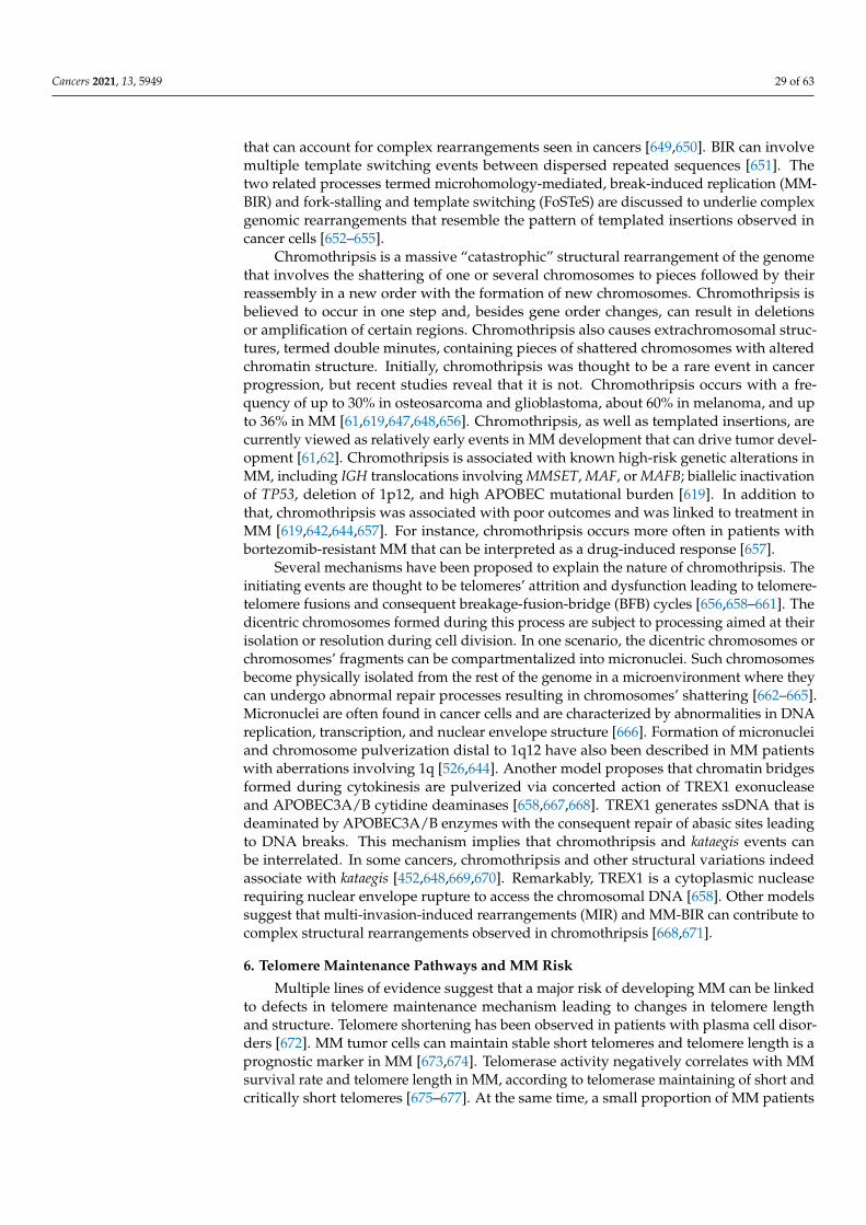
Cancers 2021, 13, 5949 29 of 63
that can account for complex rearrangements seen in cancers [649,650]. BIR can involvemultiple template switching events between dispersed repeated sequences [651]. Thetwo related processes termed microhomology-mediated, break-induced replication (MM-BIR) and fork-stalling and template switching (FoSTeS) are discussed to underlie complexgenomic rearrangements that resemble the pattern of templated insertions observed incancer cells [652–655].
Chromothripsis is a massive “catastrophic” structural rearrangement of the genomethat involves the shattering of one or several chromosomes to pieces followed by theirreassembly in a new order with the formation of new chromosomes. Chromothripsis isbelieved to occur in one step and, besides gene order changes, can result in deletionsor amplification of certain regions. Chromothripsis also causes extrachromosomal struc-tures, termed double minutes, containing pieces of shattered chromosomes with alteredchromatin structure. Initially, chromothripsis was thought to be a rare event in cancerprogression, but recent studies reveal that it is not. Chromothripsis occurs with a fre-quency of up to 30% in osteosarcoma and glioblastoma, about 60% in melanoma, and upto 36% in MM [61,619,647,648,656]. Chromothripsis, as well as templated insertions, arecurrently viewed as relatively early events in MM development that can drive tumor devel-opment [61,62]. Chromothripsis is associated with known high-risk genetic alterations inMM, including IGH translocations involving MMSET, MAF, or MAFB; biallelic inactivationof TP53, deletion of 1p12, and high APOBEC mutational burden [619]. In addition tothat, chromothripsis was associated with poor outcomes and was linked to treatment inMM [619,642,644,657]. For instance, chromothripsis occurs more often in patients withbortezomib-resistant MM that can be interpreted as a drug-induced response [657].
Several mechanisms have been proposed to explain the nature of chromothripsis. Theinitiating events are thought to be telomeres’ attrition and dysfunction leading to telomere-telomere fusions and consequent breakage-fusion-bridge (BFB) cycles [656,658–661]. Thedicentric chromosomes formed during this process are subject to processing aimed at theirisolation or resolution during cell division. In one scenario, the dicentric chromosomes orchromosomes’ fragments can be compartmentalized into micronuclei. Such chromosomesbecome physically isolated from the rest of the genome in a microenvironment where theycan undergo abnormal repair processes resulting in chromosomes’ shattering [662–665].Micronuclei are often found in cancer cells and are characterized by abnormalities in DNAreplication, transcription, and nuclear envelope structure [666]. Formation of micronucleiand chromosome pulverization distal to 1q12 have also been described in MM patientswith aberrations involving 1q [526,644]. Another model proposes that chromatin bridgesformed during cytokinesis are pulverized via concerted action of TREX1 exonucleaseand APOBEC3A/B cytidine deaminases [658,667,668]. TREX1 generates ssDNA that isdeaminated by APOBEC3A/B enzymes with the consequent repair of abasic sites leadingto DNA breaks. This mechanism implies that chromothripsis and kataegis events canbe interrelated. In some cancers, chromothripsis and other structural variations indeedassociate with kataegis [452,648,669,670]. Remarkably, TREX1 is a cytoplasmic nucleaserequiring nuclear envelope rupture to access the chromosomal DNA [658]. Other modelssuggest that multi-invasion-induced rearrangements (MIR) and MM-BIR can contribute tocomplex structural rearrangements observed in chromothripsis [668,671].
6. Telomere Maintenance Pathways and MM Risk
Multiple lines of evidence suggest that a major risk of developing MM can be linkedto defects in telomere maintenance mechanism leading to changes in telomere lengthand structure. Telomere shortening has been observed in patients with plasma cell disor-ders [672]. MM tumor cells can maintain stable short telomeres and telomere length is aprognostic marker in MM [673,674]. Telomerase activity negatively correlates with MMsurvival rate and telomere length in MM, according to telomerase maintaining of short andcritically short telomeres [675–677]. At the same time, a small proportion of MM patients
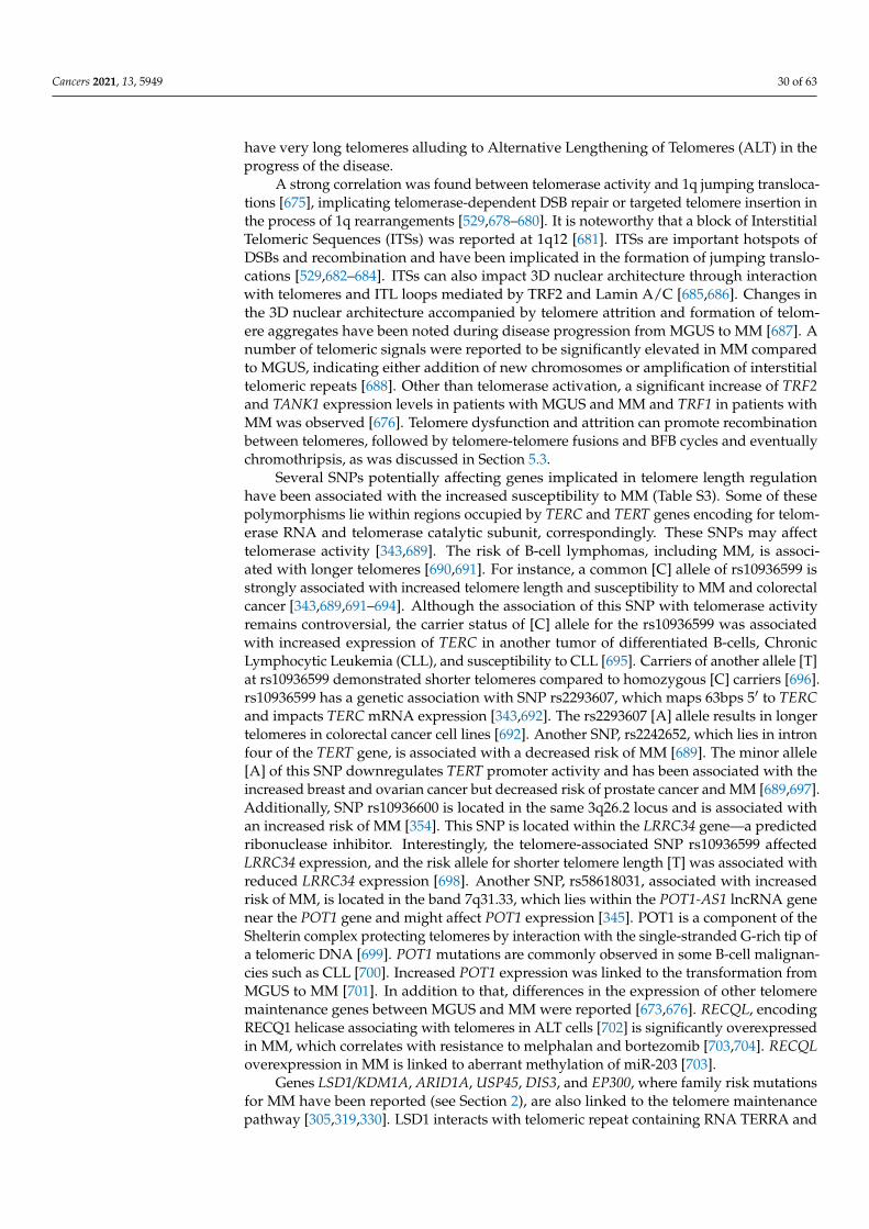
Cancers 2021, 13, 5949 30 of 63
have very long telomeres alluding to Alternative Lengthening of Telomeres (ALT) in theprogress of the disease.
A strong correlation was found between telomerase activity and 1q jumping transloca-tions [675], implicating telomerase-dependent DSB repair or targeted telomere insertion inthe process of 1q rearrangements [529,678–680]. It is noteworthy that a block of InterstitialTelomeric Sequences (ITSs) was reported at 1q12 [681]. ITSs are important hotspots ofDSBs and recombination and have been implicated in the formation of jumping translo-cations [529,682–684]. ITSs can also impact 3D nuclear architecture through interactionwith telomeres and ITL loops mediated by TRF2 and Lamin A/C [685,686]. Changes inthe 3D nuclear architecture accompanied by telomere attrition and formation of telom-ere aggregates have been noted during disease progression from MGUS to MM [687]. Anumber of telomeric signals were reported to be significantly elevated in MM comparedto MGUS, indicating either addition of new chromosomes or amplification of interstitialtelomeric repeats [688]. Other than telomerase activation, a significant increase of TRF2and TANK1 expression levels in patients with MGUS and MM and TRF1 in patients withMM was observed [676]. Telomere dysfunction and attrition can promote recombinationbetween telomeres, followed by telomere-telomere fusions and BFB cycles and eventuallychromothripsis, as was discussed in Section 5.3.
Several SNPs potentially affecting genes implicated in telomere length regulationhave been associated with the increased susceptibility to MM (Table S3). Some of thesepolymorphisms lie within regions occupied by TERC and TERT genes encoding for telom-erase RNA and telomerase catalytic subunit, correspondingly. These SNPs may affecttelomerase activity [343,689]. The risk of B-cell lymphomas, including MM, is associ-ated with longer telomeres [690,691]. For instance, a common [C] allele of rs10936599 isstrongly associated with increased telomere length and susceptibility to MM and colorectalcancer [343,689,691–694]. Although the association of this SNP with telomerase activityremains controversial, the carrier status of [C] allele for the rs10936599 was associatedwith increased expression of TERC in another tumor of differentiated B-cells, ChronicLymphocytic Leukemia (CLL), and susceptibility to CLL [695]. Carriers of another allele [T]at rs10936599 demonstrated shorter telomeres compared to homozygous [C] carriers [696].rs10936599 has a genetic association with SNP rs2293607, which maps 63bps 5′ to TERCand impacts TERC mRNA expression [343,692]. The rs2293607 [A] allele results in longertelomeres in colorectal cancer cell lines [692]. Another SNP, rs2242652, which lies in intronfour of the TERT gene, is associated with a decreased risk of MM [689]. The minor allele[A] of this SNP downregulates TERT promoter activity and has been associated with theincreased breast and ovarian cancer but decreased risk of prostate cancer and MM [689,697].Additionally, SNP rs10936600 is located in the same 3q26.2 locus and is associated withan increased risk of MM [354]. This SNP is located within the LRRC34 gene—a predictedribonuclease inhibitor. Interestingly, the telomere-associated SNP rs10936599 affectedLRRC34 expression, and the risk allele for shorter telomere length [T] was associated withreduced LRRC34 expression [698]. Another SNP, rs58618031, associated with increasedrisk of MM, is located in the band 7q31.33, which lies within the POT1-AS1 lncRNA genenear the POT1 gene and might affect POT1 expression [345]. POT1 is a component of theShelterin complex protecting telomeres by interaction with the single-stranded G-rich tip ofa telomeric DNA [699]. POT1 mutations are commonly observed in some B-cell malignan-cies such as CLL [700]. Increased POT1 expression was linked to the transformation fromMGUS to MM [701]. In addition to that, differences in the expression of other telomeremaintenance genes between MGUS and MM were reported [673,676]. RECQL, encodingRECQ1 helicase associating with telomeres in ALT cells [702] is significantly overexpressedin MM, which correlates with resistance to melphalan and bortezomib [703,704]. RECQLoverexpression in MM is linked to aberrant methylation of miR-203 [703].
Genes LSD1/KDM1A, ARID1A, USP45, DIS3, and EP300, where family risk mutationsfor MM have been reported (see Section 2), are also linked to the telomere maintenancepathway [305,319,330]. LSD1 interacts with telomeric repeat containing RNA TERRA and
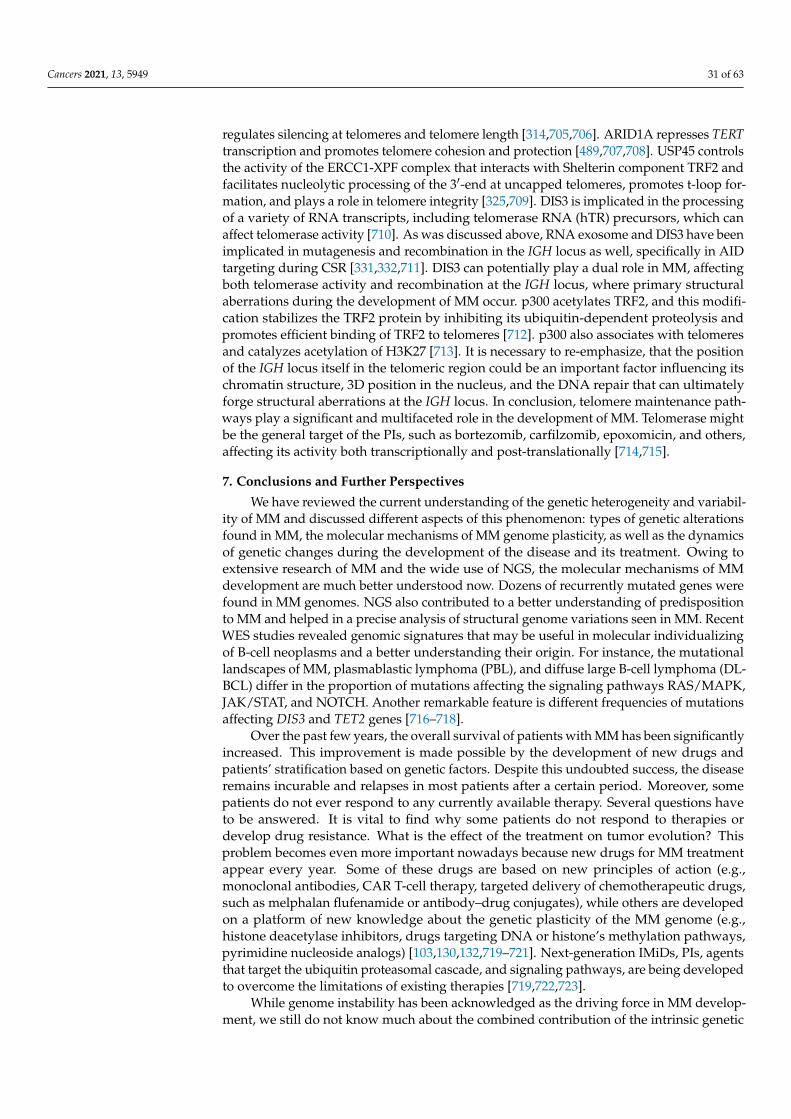
Cancers 2021, 13, 5949 31 of 63
regulates silencing at telomeres and telomere length [314,705,706]. ARID1A represses TERTtranscription and promotes telomere cohesion and protection [489,707,708]. USP45 controlsthe activity of the ERCC1-XPF complex that interacts with Shelterin component TRF2 andfacilitates nucleolytic processing of the 3′-end at uncapped telomeres, promotes t-loop for-mation, and plays a role in telomere integrity [325,709]. DIS3 is implicated in the processingof a variety of RNA transcripts, including telomerase RNA (hTR) precursors, which canaffect telomerase activity [710]. As was discussed above, RNA exosome and DIS3 have beenimplicated in mutagenesis and recombination in the IGH locus as well, specifically in AIDtargeting during CSR [331,332,711]. DIS3 can potentially play a dual role in MM, affectingboth telomerase activity and recombination at the IGH locus, where primary structuralaberrations during the development of MM occur. p300 acetylates TRF2, and this modifi-cation stabilizes the TRF2 protein by inhibiting its ubiquitin-dependent proteolysis andpromotes efficient binding of TRF2 to telomeres [712]. p300 also associates with telomeresand catalyzes acetylation of H3K27 [713]. It is necessary to re-emphasize, that the positionof the IGH locus itself in the telomeric region could be an important factor influencing itschromatin structure, 3D position in the nucleus, and the DNA repair that can ultimatelyforge structural aberrations at the IGH locus. In conclusion, telomere maintenance path-ways play a significant and multifaceted role in the development of MM. Telomerase mightbe the general target of the PIs, such as bortezomib, carfilzomib, epoxomicin, and others,affecting its activity both transcriptionally and post-translationally [714,715].
7. Conclusions and Further Perspectives
We have reviewed the current understanding of the genetic heterogeneity and variabil-ity of MM and discussed different aspects of this phenomenon: types of genetic alterationsfound in MM, the molecular mechanisms of MM genome plasticity, as well as the dynamicsof genetic changes during the development of the disease and its treatment. Owing toextensive research of MM and the wide use of NGS, the molecular mechanisms of MMdevelopment are much better understood now. Dozens of recurrently mutated genes werefound in MM genomes. NGS also contributed to a better understanding of predispositionto MM and helped in a precise analysis of structural genome variations seen in MM. RecentWES studies revealed genomic signatures that may be useful in molecular individualizingof B-cell neoplasms and a better understanding their origin. For instance, the mutationallandscapes of MM, plasmablastic lymphoma (PBL), and diffuse large B-cell lymphoma (DL-BCL) differ in the proportion of mutations affecting the signaling pathways RAS/MAPK,JAK/STAT, and NOTCH. Another remarkable feature is different frequencies of mutationsaffecting DIS3 and TET2 genes [716–718].
Over the past few years, the overall survival of patients with MM has been significantlyincreased. This improvement is made possible by the development of new drugs andpatients’ stratification based on genetic factors. Despite this undoubted success, the diseaseremains incurable and relapses in most patients after a certain period. Moreover, somepatients do not ever respond to any currently available therapy. Several questions haveto be answered. It is vital to find why some patients do not respond to therapies ordevelop drug resistance. What is the effect of the treatment on tumor evolution? Thisproblem becomes even more important nowadays because new drugs for MM treatmentappear every year. Some of these drugs are based on new principles of action (e.g.,monoclonal antibodies, CAR T-cell therapy, targeted delivery of chemotherapeutic drugs,such as melphalan flufenamide or antibody–drug conjugates), while others are developedon a platform of new knowledge about the genetic plasticity of the MM genome (e.g.,histone deacetylase inhibitors, drugs targeting DNA or histone’s methylation pathways,pyrimidine nucleoside analogs) [103,130,132,719–721]. Next-generation IMiDs, PIs, agentsthat target the ubiquitin proteasomal cascade, and signaling pathways, are being developedto overcome the limitations of existing therapies [719,722,723].
While genome instability has been acknowledged as the driving force in MM develop-ment, we still do not know much about the combined contribution of the intrinsic genetic
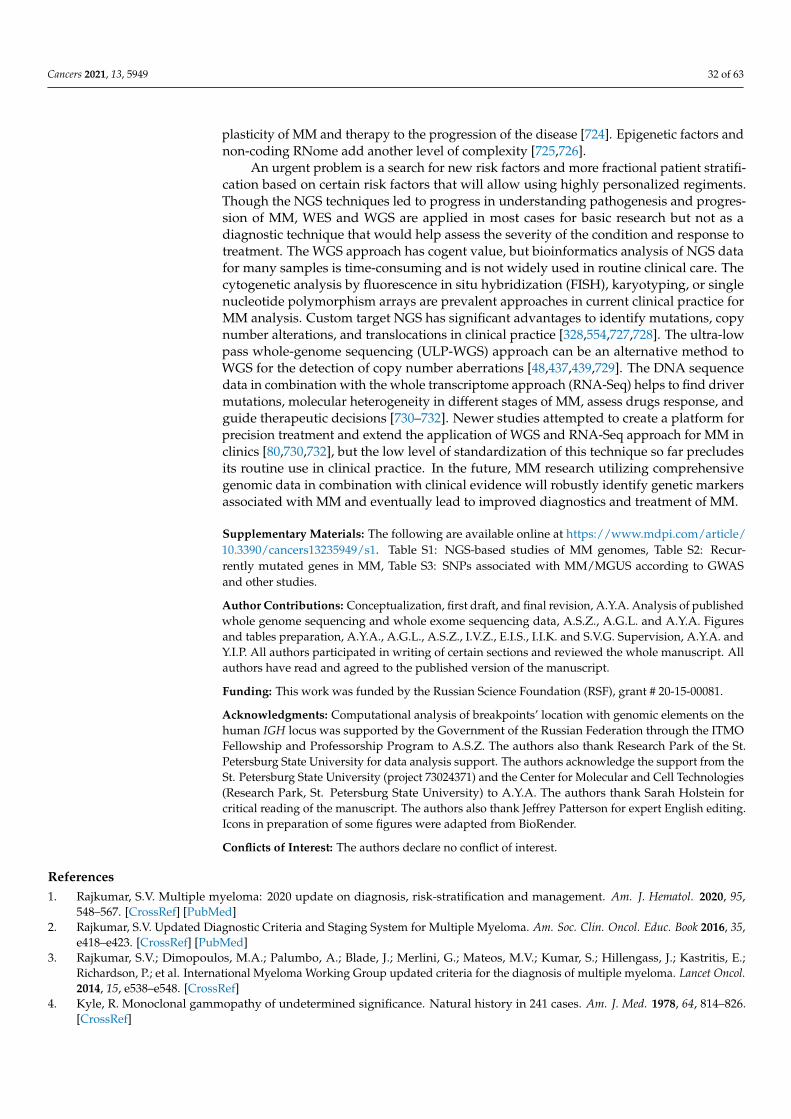
Cancers 2021, 13, 5949 32 of 63
plasticity of MM and therapy to the progression of the disease [724]. Epigenetic factors andnon-coding RNome add another level of complexity [725,726].
An urgent problem is a search for new risk factors and more fractional patient stratifi-cation based on certain risk factors that will allow using highly personalized regiments.Though the NGS techniques led to progress in understanding pathogenesis and progres-sion of MM, WES and WGS are applied in most cases for basic research but not as adiagnostic technique that would help assess the severity of the condition and response totreatment. The WGS approach has cogent value, but bioinformatics analysis of NGS datafor many samples is time-consuming and is not widely used in routine clinical care. Thecytogenetic analysis by fluorescence in situ hybridization (FISH), karyotyping, or singlenucleotide polymorphism arrays are prevalent approaches in current clinical practice forMM analysis. Custom target NGS has significant advantages to identify mutations, copynumber alterations, and translocations in clinical practice [328,554,727,728]. The ultra-lowpass whole-genome sequencing (ULP-WGS) approach can be an alternative method toWGS for the detection of copy number aberrations [48,437,439,729]. The DNA sequencedata in combination with the whole transcriptome approach (RNA-Seq) helps to find drivermutations, molecular heterogeneity in different stages of MM, assess drugs response, andguide therapeutic decisions [730–732]. Newer studies attempted to create a platform forprecision treatment and extend the application of WGS and RNA-Seq approach for MM inclinics [80,730,732], but the low level of standardization of this technique so far precludesits routine use in clinical practice. In the future, MM research utilizing comprehensivegenomic data in combination with clinical evidence will robustly identify genetic markersassociated with MM and eventually lead to improved diagnostics and treatment of MM.
Supplementary Materials: The following are available online at https://www.mdpi.com/article/10.3390/cancers13235949/s1. Table S1: NGS-based studies of MM genomes, Table S2: Recur-rently mutated genes in MM, Table S3: SNPs associated with MM/MGUS according to GWASand other studies.
Author Contributions: Conceptualization, first draft, and final revision, A.Y.A. Analysis of publishedwhole genome sequencing and whole exome sequencing data, A.S.Z., A.G.L. and A.Y.A. Figuresand tables preparation, A.Y.A., A.G.L., A.S.Z., I.V.Z., E.I.S., I.I.K. and S.V.G. Supervision, A.Y.A. andY.I.P. All authors participated in writing of certain sections and reviewed the whole manuscript. Allauthors have read and agreed to the published version of the manuscript.
Funding: This work was funded by the Russian Science Foundation (RSF), grant # 20-15-00081.
Acknowledgments: Computational analysis of breakpoints’ location with genomic elements on thehuman IGH locus was supported by the Government of the Russian Federation through the ITMOFellowship and Professorship Program to A.S.Z. The authors also thank Research Park of the St.Petersburg State University for data analysis support. The authors acknowledge the support from theSt. Petersburg State University (project 73024371) and the Center for Molecular and Cell Technologies(Research Park, St. Petersburg State University) to A.Y.A. The authors thank Sarah Holstein forcritical reading of the manuscript. The authors also thank Jeffrey Patterson for expert English editing.Icons in preparation of some figures were adapted from BioRender.
Conflicts of Interest: The authors declare no conflict of interest.
References1. Rajkumar, S.V. Multiple myeloma: 2020 update on diagnosis, risk-stratification and management. Am. J. Hematol. 2020, 95,
548–567. [CrossRef] [PubMed]2. Rajkumar, S.V. Updated Diagnostic Criteria and Staging System for Multiple Myeloma. Am. Soc. Clin. Oncol. Educ. Book 2016, 35,
e418–e423. [CrossRef] [PubMed]3. Rajkumar, S.V.; Dimopoulos, M.A.; Palumbo, A.; Blade, J.; Merlini, G.; Mateos, M.V.; Kumar, S.; Hillengass, J.; Kastritis, E.;
Richardson, P.; et al. International Myeloma Working Group updated criteria for the diagnosis of multiple myeloma. Lancet Oncol.2014, 15, e538–e548. [CrossRef]
4. Kyle, R. Monoclonal gammopathy of undetermined significance. Natural history in 241 cases. Am. J. Med. 1978, 64, 814–826.[CrossRef]
Cancers 2021, 13, 5949 33 of 63
5. Dhodapkar, M.V. MGUS to myeloma: A mysterious gammopathy of underexplored significance. Blood 2016, 128, 2599–2606.[CrossRef]
6. Dispenzieri, A.; Katzmann, J.A.; Kyle, R.A.; Larson, D.R.; Melton, L.J.; Colby, C.L.; Therneau, T.M.; Clark, R.; Kumar, S.K.;Bradwell, A.; et al. Prevalence and risk of progression of light-chain monoclonal gammopathy of undetermined significance: Aretrospective population-based cohort study. Lancet 2010, 375, 1721–1728. [CrossRef]
7. Kyle, R.A.; Therneau, T.M.; Rajkumar, S.V.; Larson, D.R.; Plevak, M.F.; Offord, J.R.; Dispenzieri, A.; Katzmann, J.A.; Melton,L.J. Prevalence of Monoclonal Gammopathy of Undetermined Significance. N. Engl. J. Med. 2006, 354, 1362–1369. [CrossRef][PubMed]
8. Chudasama, R.; Barth, P. Risk Stratification of Precursors to Multiple Myeloma in 2020. Rhode Isl. Med. J. 2020, 103, 46–47.9. Kyle, R.A.; Therneau, T.M.; Rajkumar, S.V.; Offord, J.R.; Larson, D.R.; Plevak, M.F.; Melton, L.J. A Long-Term Study of Prognosis
in Monoclonal Gammopathy of Undetermined Significance. N. Engl. J. Med. 2002, 346, 564–569. [CrossRef] [PubMed]10. Kyle, R.A.; Larson, D.R.; Therneau, T.M.; Dispenzieri, A.; Kumar, S.; Cerhan, J.R.; Rajkumar, S.V. Long-Term Follow-up of
Monoclonal Gammopathy of Undetermined Significance. N. Engl. J. Med. 2018, 378, 241–249. [CrossRef]11. Landgren, O.; Kyle, R.A.; Pfeiffer, R.M.; Katzmann, J.A.; Caporaso, N.E.; Hayes, R.B.; Dispenzieri, A.; Kumar, S.; Clark, R.J.;
Baris, D.; et al. Monoclonal gammopathy of undetermined significance (MGUS) consistently precedes multiple myeloma: Aprospective study. Blood 2009, 113, 5412–5417. [CrossRef] [PubMed]
12. Kyle, R.; Greipp, P. Smoldering multiple myeloma. N. Engl. J. Med. 1980, 302, 1347–1349. [CrossRef]13. Kyle, R.A.; Remstein, E.D.; Therneau, T.M.; Dispenzieri, A.; Kurtin, P.J.; Hodnefield, J.M.; Larson, D.R.; Plevak, M.F.; Jelinek, D.F.;
Fonseca, R.; et al. Clinical Course and Prognosis of Smoldering (Asymptomatic) Multiple Myeloma. N. Engl. J. Med. 2007, 356,2582–2590. [CrossRef]
14. Weiss, B.M.; Abadie, J.; Verma, P.; Howard, R.S.; Kuehl, W.M. A monoclonal gammopathy precedes multiple myeloma in mostpatients. Blood 2009, 113, 5418–5422. [CrossRef]
15. Lonial, S.; Dhodapkar, M.V.; Rajkumar, S.V. Smoldering myeloma and the art of war. J. Clin. Oncol. 2020, 38, 2363–2365. [CrossRef][PubMed]
16. Kyle, R.A.; Dispenzieri, A.; Kumar, S.; Larson, D.; Therneau, T.; Vincent Rajkumar, S. IgM monoclonal gammopathy ofundetermined significance (MGUS) and smoldering Waldenström’s macroglobulinemia (SWM). Clin. Lymphoma Myeloma Leuk.2011, 11, 74–76. [CrossRef] [PubMed]
17. Girard, L.P.; Soekojo, C.Y.; Ooi, M.; Poon, L.M.; Chng, W.J.; de Mel, S. Immunoglobulin M paraproteinaemias. Cancers 2020,12, 1688. [CrossRef]
18. Kyle, R.A.; Gertz, M.A.; Witzig, T.E.; Lust, J.A.; Lacy, M.Q.; Dispenzieri, A.; Fonseca, R.; Rajkumar, S.V.; Offord, J.R.;Larson, D.R.; et al. Review of 1027 patients with newly diagnosed multiple myeloma. Mayo Clin. Proc. 2003, 78, 21–33. [CrossRef]
19. Chawla, S.S.; Kumar, S.K.; Dispenzieri, A.; Greenberg, A.J.; Larson, D.R.; Kyle, R.A.; Lacy, M.Q.; Gertz, M.A.; Rajkumar, S.V.Clinical course and prognosis of non-secretory multiple myeloma. Eur. J. Haematol. 2015, 95, 57–64. [CrossRef] [PubMed]
20. Dutta, A.K.; Fink, J.L.; Grady, J.P.; Morgan, G.J.; Mullighan, C.G.; To, L.B.; Hewett, D.R.; Zannettino, A.C.W. Subclonal evolutionin disease progression from MGUS/SMM to multiple myeloma is characterised by clonal stability. Leukemia 2018, 33, 457–468.[CrossRef] [PubMed]
21. Rasche, L.; Chavan, S.S.; Stephens, O.W.; Patel, P.; Tytarenko, R.; Ashby, C.; Bauer, M.; Stein, C.; Deshpande, S.; Wardell, C.; et al.Spatial genomic heterogeneity in multiple myeloma revealed by multi-region sequencing. Nat. Commun. 2017, 8, 268. [CrossRef][PubMed]
22. Keats, J.J.; Chesi, M.; Egan, J.B.; Garbitt, V.M.; Palmer, S.E.; Braggio, E.; Van Wier, S.; Blackburn, P.R.; Baker, A.S.;Dispenzieri, A.; et al. Clonal competition with alternating dominance in multiple myeloma. Blood 2012, 120, 1067–1076.[CrossRef]
23. Walker, B.A.; Wardell, C.P.; Melchor, L.; Brioli, A.; Johnson, D.C.; Kaiser, M.F.; Mirabella, F.; Lopez-Corral, L.; Humphray, S.;Murray, L.; et al. Intraclonal heterogeneity is a critical early event in the development of myeloma and precedes the developmentof clinical symptoms. Leukemia 2014, 28, 384–390. [CrossRef]
24. Kortüm, K.M.; Langer, C.; Monge, J.; Bruins, L.; Zhu, Y.X.; Shi, C.X.; Jedlowski, P.; Egan, J.B.; Ojha, J.; Bullinger, L.; et al.Longitudinal analysis of 25 sequential sample-pairs using a custom multiple myeloma mutation sequencing panel (M3P). Ann.Hematol. 2015, 94, 1205–1211. [CrossRef] [PubMed]
25. Weinhold, N.; Ashby, C.; Rasche, L.; Chavan, S.S.; Stein, C.; Stephens, O.W.; Tytarenko, R.; Bauer, M.A.; Meissner, T.;Deshpande, S.; et al. Clonal selection and double-hit events involving tumor suppressor genes underlie relapse in myeloma.Blood 2016, 128, 1735–1744. [CrossRef]
26. Egan, J.B.; Shi, C.X.; Tembe, W.; Christoforides, A.; Kurdoglu, A.; Sinari, S.; Middha, S.; Asmann, Y.; Schmidt, J.; Braggio, E.; et al.Whole-genome sequencing of multiple myeloma from diagnosis to plasma cell leukemia reveals genomic initiating events,evolution, and clonal tides. Blood 2012, 120, 1060–1066. [CrossRef]
27. Melchor, L.; Brioli, A.; Wardell, C.P.; Murison, A.; Potter, N.E.; Kaiser, M.F.; Fryer, R.A.; Johnson, D.C.; Begum, D.B.;Wilson, S.H.; et al. Single-cell genetic analysis reveals the composition of initiating clones and phylogenetic patterns of branchingand parallel evolution in myeloma. Leukemia 2014, 28, 1705–1715. [CrossRef] [PubMed]
Cancers 2021, 13, 5949 34 of 63
28. Lohr, J.G.; Stojanov, P.; Carter, S.L.; Cruz-Gordillo, P.; Lawrence, M.S.; Auclair, D.; Sougnez, C.; Knoechel, B.; Gould, J.;Saksena, G.; et al. Widespread genetic heterogeneity in multiple myeloma: Implications for targeted therapy. Cancer Cell 2014, 25,91–101. [CrossRef]
29. Bolli, N.; Avet-Loiseau, H.; Wedge, D.C.; Van Loo, P.; Alexandrov, L.B.; Martincorena, I.; Dawson, K.J.; Iorio, F.; Nik-Zainal, S.;Bignell, G.R.; et al. Heterogeneity of genomic evolution and mutational profiles in multiple myeloma. Nat. Commun. 2014, 5, 1–13.[CrossRef]
30. Ledergor, G.; Weiner, A.; Zada, M.; Wang, S.Y.; Cohen, Y.C.; Gatt, M.E.; Snir, N.; Magen, H.; Koren-Michowitz, M.;Herzog-Tzarfati, K.; et al. Single cell dissection of plasma cell heterogeneity in symptomatic and asymptomatic myeloma. Nat.Med. 2018, 24, 1867–1876. [CrossRef]
31. Bochtler, T.; Merz, M.; Hielscher, T.; Granzow, M.; Hoffmann, K.; Krämer, A.; Raab, M.S.; Hillengass, J.; Seckinger, A.;Kimmich, C.; et al. Cytogenetic intraclonal heterogeneity of plasma cell dyscrasia in AL amyloidosis as compared with multiplemyeloma. Blood Adv. 2018, 2, 2607–2618. [CrossRef] [PubMed]
32. Paíno, T.; Paiva, B.; Sayagués, J.M.; Mota, I.; Carvalheiro, T.; Corchete, L.A.; Aires-Mejía, I.; Pérez, J.J.; Sanchez, M.L.;Barcena, P.; et al. Phenotypic identification of subclones in multiple myeloma with different chemoresistant, cytogenetic andclonogenic potential. Leukemia 2015, 29, 1186–1194. [CrossRef] [PubMed]
33. Morgan, G.J.; Walker, B.A.; Davies, F.E. The genetic architecture of multiple myeloma. Nat. Rev. Cancer 2012, 12, 335–348.[CrossRef] [PubMed]
34. Walker, B.A.; Wardell, C.P.; Melchor, L.; Hulkki, S.; Potter, N.E.; Johnson, D.; Fenwick, K.; Kozarewa, I.; Gonzalez, D.;Lord, C.; et al. Intraclonal heterogeneity and distinct molecular mechanisms characterize the development of t(4;14) and t(11;14)myeloma. Blood 2012, 120, 1077–1086. [CrossRef] [PubMed]
35. Nowell, P.C. The clonal evolution of tumor cell populations. Science 1976, 194, 23–28. [CrossRef] [PubMed]36. Greaves, M.; Maley, C.C. Clonal evolution in cancer. Nature 2012, 481, 306–313. [CrossRef]37. Pon, J.R.; Marra, M.A. Driver and passenger mutations in cancer. Annu. Rev. Pathol. Mech. Dis. 2015, 10, 25–50. [CrossRef]
[PubMed]38. Baylin, S.B.; Jones, P.A. Epigenetic determinants of cancer. Cold Spring Harb. Perspect. Biol. 2016, 8, a019505. [CrossRef] [PubMed]39. Venkatesan, S.; Swanton, C.; Taylor, B.S.; Costello, J.F. Treatment-Induced Mutagenesis and Selective Pressures Sculpt Cancer
Evolution. Cold Spring Harb. Perspect. Med. 2017, 7, a026617. [CrossRef]40. Chatterjee, A.; Rodger, E.J.; Eccles, M.R. Epigenetic drivers of tumourigenesis and cancer metastasis. Semin. Cancer Biol. 2018, 51,
149–159. [CrossRef] [PubMed]41. Davis, A.; Gao, R.; Navin, N. Tumor evolution: Linear, branching, neutral or punctuated? Biochim. Biophys. Acta 2017, 1867, 151.
[CrossRef]42. Furukawa, Y.; Kikuchi, J. Molecular basis of clonal evolution in multiple myeloma. Int. J. Hematol. 2020, 111, 496–511. [CrossRef]
[PubMed]43. Bahlis, N.J. Darwinian evolution and tiding clones in multiple myeloma. Blood 2012, 120, 927–928. [CrossRef]44. Magrangeas, F.; Avet-Loiseau, H.; Gouraud, W.; Lodé, L.; Decaux, O.; Godmer, P.; Garderet, L.; Voillat, L.; Facon, T.;
Stoppa, A.M.; et al. Minor clone provides a reservoir for relapse in multiple myeloma. Leukemia 2013, 27, 473–481. [CrossRef][PubMed]
45. Jones, J.R.; Weinhold, N.; Ashby, C.; Walker, B.A.; Wardell, C.; Pawlyn, C.; Rasche, L.; Melchor, L.; Cairns, D.A.;Gregory, W.M.; et al. Clonal evolution in myeloma: The impact of maintenance lenalidomide and depth of response onthe genetics and sub-clonal structure of relapsed disease in uniformly treated newly diagnosed patients. Haematologica 2019, 104,1440–1450. [CrossRef] [PubMed]
46. Diamond, B.; Yellapantula, V.; Rustad, E.H.; Maclachlan, K.H.; Mayerhoefer, M.; Kaiser, M.; Morgan, G.; Landgren, O.; Maura, F.Positive selection as the unifying force for clonal evolution in multiple myeloma. Leukemia 2021, 35, 1511–1515. [CrossRef]
47. Bolli, N.; Maura, F.; Minvielle, S.; Gloznik, D.; Szalat, R.; Fullam, A.; Martincorena, I.; Dawson, K.J.; Samur, M.K.; Zamora, J.; et al.Genomic patterns of progression in smoldering multiple myeloma. Nat. Commun. 2018, 9, 1–10. [CrossRef] [PubMed]
48. Bustoros, M.; Sklavenitis-Pistofidis, R.; Park, J.; Redd, R.; Zhitomirsky, B.; Dunford, A.J.; Salem, K.; Tai, Y.T.; Anand, S.;Mouhieddine, T.H.; et al. Genomic profiling of smoldering multiple myeloma identifies patients at a high risk of diseaseprogression. J. Clin. Oncol. 2020, 38, 2380–2389. [CrossRef]
49. Johnson, D.C.; Lenive, O.; Mitchell, J.; Jackson, G.; Owen, R.; Drayson, M.; Cook, G.; Jones, J.R.; Pawlyn, C.; Davies, F.E.; et al.Neutral tumor evolution in myeloma is associated with poor prognosis. Blood 2017, 130, 1639–1643. [CrossRef]
50. Merz, M.; Hielscher, T.; Schult, D.; Mai, E.K.; Raab, M.S.; Hillengass, J.; Seckinger, A.; Hose, D.; Granzow, M.; Jauch, A.; et al.Cytogenetic subclone formation and evolution in progressive smoldering multiple myeloma. Leukemia 2020, 34, 1192–1196.[CrossRef]
51. Boyle, E.M.; Deshpande, S.; Tytarenko, R.; Ashby, C.; Wang, Y.; Bauer, M.A.; Johnson, S.K.; Wardell, C.P.; Thanendrarajan, S.;Zangari, M.; et al. The molecular make up of smoldering myeloma highlights the evolutionary pathways leading to multiplemyeloma. Nat. Commun. 2021, 12, 293. [CrossRef]
52. Mikulasova, A.; Wardell, C.P.; Murison, A.; Boyle, E.M.; Jackson, G.H.; Smetana, J.; Kufova, Z.; Pour, L.; Sandecka, V.;Almasi, M.; et al. The spectrum of somatic mutations in monoclonal gammopathy of undetermined significance indicatesa less complex genomic landscape than that in multiple myeloma. Haematologica 2017, 102, 1617–1625. [CrossRef] [PubMed]
Cancers 2021, 13, 5949 35 of 63
53. Corre, J.; Cleynen, A.; Robiou du Pont, S.; Buisson, L.; Bolli, N.; Attal, M.; Munshi, N.; Avet-Loiseau, H. Multiple myeloma clonalevolution in homogeneously treated patients. Leukemia 2018, 32, 2636–2647. [CrossRef] [PubMed]
54. Ziccheddu, B.; Biancon, G.; Bagnoli, F.; De Philippis, C.; Maura, F.; Rustad, E.H.; Dugo, M.; Devecchi, A.; De Cecco, L.;Sensi, M.; et al. Integrative analysis of the genomic and transcriptomic landscape of double-refractory multiple myeloma. BloodAdv. 2020, 4, 830–844. [CrossRef] [PubMed]
55. López-Corral, L.; Gutiérrez, N.C.; Vidriales, M.B.; Mateos, M.V.; Rasillo, A.; García-Sanz, R.; Paiva, B.; San Miguel, J.F. Theprogression from MGUS to smoldering myeloma and eventually to multiple myeloma involves a clonal expansion of geneticallyabnormal plasma cells. Clin. Cancer Res. 2011, 17, 1692–1700. [CrossRef]
56. Mailankody, S.; Kazandjian, D.; Korde, N.; Roschewski, M.; Manasanch, E.; Bhutani, M.; Tageja, N.; Kwok, M.; Zhang, Y.;Zingone, A.; et al. Baseline mutational patterns and sustained MRD negativity in patients with high-risk smoldering myeloma.Blood Adv. 2017, 1, 1911–1918. [CrossRef] [PubMed]
57. Maura, F.; Bolli, N.; Rustad, E.H.; Hultcrantz, M.; Munshi, N.; Landgren, O. Moving from Cancer Burden to Cancer Genomics forSmoldering Myeloma: A Review. JAMA Oncol. 2020, 6, 425–432. [CrossRef] [PubMed]
58. Samur, M.K.; Samur, A.A.; Fulciniti, M.; Szalat, R.; Han, T.; Shammas, M.; Richardson, P.; Magrangeas, F.; Minvielle, S.;Corre, J.; et al. Genome-wide somatic alterations in multiple myeloma reveal a superior outcome group. J. Clin. Oncol. 2020, 38,3107–3118. [CrossRef]
59. Samur, M.K.; Roncador, M.; Aktas-Samur, A.; Fulciniti, M.; Bazarbachi, A.H.; Szalat, R.; Shammas, M.A.; Sperling, A.S.;Richardson, P.G.; Magrangeas, F.; et al. High-Dose Melphalan Significantly Increases Mutational Burden in Multiple MyelomaCells at Relapse: Results from a Randomized Study in Multiple Myeloma. Blood 2020, 136, 4–5. [CrossRef]
60. Hoang, P.H.; Cornish, A.J.; Sherborne, A.L.; Chubb, D.; Kimber, S.; Jackson, G.; Morgan, G.J.; Cook, G.; Kinnersley, B.;Kaiser, M.; et al. An enhanced genetic model of relapsed IGH-translocated multiple myeloma evolutionary dynamics. BloodCancer J. 2020, 10, 101. [CrossRef]
61. Maura, F.; Bolli, N.; Angelopoulos, N.; Dawson, K.J.; Leongamornlert, D.; Martincorena, I.; Mitchell, T.J.; Fullam, A.; Gonzalez, S.;Szalat, R.; et al. Genomic landscape and chronological reconstruction of driver events in multiple myeloma. Nat. Commun. 2019,10, 3835. [CrossRef]
62. Oben, B.; Froyen, G.; Maclachlan, K.H.; Leongamornlert, D.; Abascal, F.; Zheng-Lin, B.; Yellapantula, V.; Derkach, A.; Geerdens, E.;Diamond, B.T.; et al. Whole-genome sequencing reveals progressive versus stable myeloma precursor conditions as two distinctentities. Nat. Commun. 2021, 12, 1861. [CrossRef] [PubMed]
63. Rustad, E.H.; Yellapantula, V.; Leongamornlert, D.; Bolli, N.; Ledergor, G.; Nadeu, F.; Angelopoulos, N.; Dawson, K.J.;Mitchell, T.J.; Osborne, R.J.; et al. Timing the initiation of multiple myeloma. Nat. Commun. 2020, 11, 1917. [CrossRef][PubMed]
64. Poh, C.; Keegan, T.; Rosenberg, A.S. Second primary malignancies in multiple myeloma: A review. Blood Rev. 2021, 46, 100757.[CrossRef]
65. Musto, P.; Anderson, K.C.; Attal, M.; Richardson, P.G.; Badros, A.; Hou, J.; Comenzo, R.; Du, J.; Durie, B.G.M.; San Miguel, J.; et al.Second primary malignancies in multiple myeloma: An overview and IMWG consensus. Ann. Oncol. 2017, 28, 228–245.[CrossRef]
66. Maclachlan, K.; Diamond, B.; Maura, F.; Hillengass, J.; Turesson, I.; Landgren, C.O.; Kazandjian, D. Second malignancies inmultiple myeloma; emerging patterns and future directions. Best Pract. Res. Clin. Haematol. 2020, 33, 101144. [CrossRef]
67. Engelhardt, M.; Ihorst, G.; Landgren, O.; Pantic, M.; Reinhardt, H.; Waldschmidt, J.; May, A.M.; Schumacher, M.; Kleber, M.;Wäsch, R. Large registry analysis to accurately define second malignancy rates and risks in a well-characterized cohort of 744consecutive multiple myeloma patients followed-up for 25 years. Haematologica 2015, 100, 1340–1349. [CrossRef] [PubMed]
68. Govindarajan, R.; Jagannath, S.; Flick, J.T.; Vesole, D.H.; Sawyer, J.; Barlogie, B.; Tricot, G. Preceding standard therapy is the likelycause of MDS after autotransplants for multiple myeloma. Br. J. Haematol. 1996, 95, 349–353. [CrossRef]
69. Kyle, R.A.; Pierre, R.V.; Bayrd, E.D. Multiple Myeloma and Acute Myelomonocytic Leukemia. N. Engl. J. Med. 1970, 283,1121–1125. [CrossRef]
70. Pemmaraju, N.; Shah, D.; Kantarjian, H.; Orlowski, R.Z.; Nogueras González, G.M.; Baladandayuthapani, V.; Jain, N.; Wagner, V.;Garcia-Manero, G.; Shah, J.; et al. Characteristics and outcomes of patients with multiple myeloma who develop therapy-relatedmyelodysplastic syndrome, chronic myelomonocytic leukemia, or acute myeloid leukemia. Clin. Lymphoma Myeloma Leuk. 2015,15, 110–114. [CrossRef]
71. Palumbo, A.; Bringhen, S.; Kumar, S.K.; Lupparelli, G.; Usmani, S.; Waage, A.; Larocca, A.; van der Holt, B.; Musto, P.;Offidani, M.; et al. Second primary malignancies with lenalidomide therapy for newly diagnosed myeloma: A meta-analysis ofindividual patient data. Lancet Oncol. 2014, 15, 333–342. [CrossRef]
72. Finnish Leukaemia Group. Acute leukaemia and other secondary neoplasms in patients treated with conventional chemotherapyfor multiple myeloma: A Finnish Leukaemia Group study. Eur. J. Haematol. 2000, 65, 123–127. [CrossRef]
73. Usmani, S.Z.; Sawyer, J.; Rosenthal, A.; Cottler-Fox, M.; Epstein, J.; Yaccoby, S.; Sexton, R.; Hoering, A.; Singh, Z.; Heuck, C.J.; et al.Risk factors for MDS and acute leukemia following total therapy 2 and 3 for multiple myeloma. Blood 2013, 121, 4753–4757.[CrossRef]

Cancers 2021, 13, 5949 36 of 63
74. Dimopoulos, M.A.; Richardson, P.G.; Brandenburg, N.; Yu, Z.; Weber, D.M.; Niesvizky, R.; Morgan, G.J. A review of secondprimary malignancy in patients with relapsed or refractory multiple myeloma treated with lenalidomide. Blood 2012, 119,2764–2767. [CrossRef]
75. Mailankody, S.; Pfeiffer, R.M.; Kristinsson, S.Y.; Korde, N.; Bjorkholm, M.; Goldin, L.R.; Turesson, I.; Landgren, O. Risk of acutemyeloid leukemia and myelodysplastic syndromes after multiple myeloma and its precursor disease (MGUS). Blood 2011, 118,4086–4092. [CrossRef]
76. Roeker, L.E.; Larson, D.R.; Kyle, R.A.; Kumar, S.; Dispenzieri, A.; Rajkumar, S.V. Risk of acute leukemia and myelodysplasticsyndromes in patients with monoclonal gammopathy of undetermined significance (MGUS): A population-based study of 17 315patients. Leukemia 2013, 27, 1391–1393. [CrossRef]
77. Turesson, I.; Kovalchik, S.A.; Pfeiffer, R.M.; Kristinsson, S.Y.; Goldin, L.R.; Drayson, M.T.; Landgren, O. Monoclonal gammopathyof undetermined significance and risk of lymphoid and myeloid malignancies: 728 cases followed up to 30 years in Sweden.Blood 2014, 123, 338–345. [CrossRef] [PubMed]
78. Manier, S.; Salem, K.Z.; Park, J.; Landau, D.A.; Getz, G.; Ghobrial, I.M. Genomic complexity of multiple myeloma and its clinicalimplications. Nat. Rev. Clin. Oncol. 2017, 14, 100–113. [CrossRef]
79. Maura, F.; Rustad, E.H.; Boyle, E.M.; Morgan, G.J. Reconstructing the evolutionary history of multiple myeloma. Best Pract. Res.Clin. Haematol. 2020, 33, 101145. [CrossRef] [PubMed]
80. Bolli, N.; Genuardi, E.; Ziccheddu, B.; Martello, M.; Oliva, S.; Terragna, C. Next-Generation Sequencing for Clinical Managementof Multiple Myeloma: Ready for Prime Time? Front. Oncol. 2020, 10, 189. [CrossRef]
81. Chapman, M.A.; Lawrence, M.S.; Keats, J.J.; Cibulskis, K.; Sougnez, C.; Schinzel, A.C.; Harview, C.L.; Brunet, J.P.; Ahmann, G.J.;Adli, M.; et al. Initial genome sequencing and analysis of multiple myeloma. Nature 2011, 471, 467–472. [CrossRef]
82. Walker, B.A.; Boyle, E.M.; Wardell, C.P.; Murison, A.; Begum, D.B.; Dahir, N.M.; Proszek, P.Z.; Johnson, D.C.; Kaiser, M.F.;Melchor, L.; et al. Mutational Spectrum, Copy Number Changes, and Outcome: Results of a Sequencing Study of Patients WithNewly Diagnosed Myeloma. J. Clin. Oncol. 2015, 33, 3911–3920. [CrossRef]
83. Rustad, E.H.; Yellapantula, V.; Glodzik, D.; Gundem, G.; Leongamornlert, D.A.; Campbell, P.J.; Papaemmanuil, E.; Landgren, O.;Maura, F. Revealing the Impact of Recurrent and Rare Structural Variations in Multiple Myeloma. Blood 2019, 134, 576. [CrossRef]
84. Keats, J.J.; Craig, D.W.; Liang, W.; Venkata, Y.; Kurdoglu, A.; Aldrich, J.; Auclair, D.; Allen, K.; Harrison, B.; Jewell, S.; et al. InterimAnalysis Of The MMRF CoMMpass Trial, a Longitudinal Study In Multiple Myeloma Relating Clinical Outcomes To Genomicand Immunophenotypic Profiles. Blood 2013, 122, 532. [CrossRef]
85. Walker, B.A.; Samur, K.M.; Mavrommatis, K.; Ashby, C.; Wardell, P.C.; Ortiz, M.; Towfic, F.; Stein, K.C.; Bauer, A.M. The MultipleMyeloma Genome Project: Development of a Molecular Segmentation Strategy for the Clinical Classification of Multiple Myeloma.Blood 2016, 128, 196. [CrossRef]
86. Walker, B.A.; Mavrommatis, K.; Wardell, C.P.; Ashby, T.C.; Bauer, M.; Davies, F.E.; Rosenthal, A.; Wang, H.; Qu, P.;Hoering, A.; et al. Identification of novel mutational drivers reveals oncogene dependencies in multiple myeloma. Blood 2018,132, 587–597. [CrossRef]
87. Rajkumar, S.V.; Kumar, S. Multiple myeloma current treatment algorithms. Blood Cancer J. 2020, 10, 94. [CrossRef] [PubMed]88. Dimopoulos, M.A.; Moreau, P.; Terpos, E.; Mateos, M.-V.; Zweegman, S.; Cook, G.; Delforge, M.; Hájek, R.; Schjesvold, F.;
Cavo, M.; et al. Multiple Myeloma: EHA-ESMO Clinical Practice Guidelines for Diagnosis, Treatment and Follow-up. HemaSphere2021, 5, e528. [CrossRef] [PubMed]
89. Mohty, M.; Knauf, W.; Romanus, D.; Corman, S.; Verleger, K.; Kwon, Y.; Cherepanov, D.; Cambron-Mellott, M.J.; Vikis, H.G.;Gonzalez, F.; et al. Real-world treatment patterns and outcomes in non-transplant newly diagnosed multiple Myeloma in France,Germany, Italy, and the United Kingdom. Eur. J. Haematol. 2020, 105, 308–325. [CrossRef]
90. Mateos, M.V.; Ludwig, H.; Bazarbachi, A.; Beksac, M.; Bladé, J.; Boccadoro, M.; Cavo, M.; Delforge, M.; Dimopoulos, M.A.;Facon, T.; et al. Insights on Multiple Myeloma Treatment Strategies. HemaSphere 2019, 3, e163. [CrossRef]
91. Bobin, A.; Liuu, E.; Moya, N.; Gruchet, C.; Sabirou, F.; Lévy, A.; Gardeney, H.; Nsiala, L.; Cailly, L.; Guidez, S.; et al. Multiplemyeloma: An overview of the current and novel therapeutic approaches in 2020. Cancers 2020, 12, 2885. [CrossRef]
92. Muchtar, E.; Dingli, D.; Kumar, S.; Buadi, F.K.; Dispenzieri, A.; Hayman, S.R.; Wolf, R.C.; Gastineau, D.A.; Chakraborty, R.;Hogan, W.J.; et al. Autologous stem cell transplant for multiple myeloma patients 70 years or older. Bone Marrow Transplant. 2016,51, 1449–1455. [CrossRef]
93. Fiala, M.A.; King, J.; Feinberg, D.; Goldsmith, S.R.; Schroeder, M.A.; Ghobadi, A.; Stockerl-Goldstein, K.E.; Vij, R.; Wildes, T.M.Autologous stem cell transplant for patients with multiple myeloma between ages 75 and 78. Bone Marrow Transplant. 2021, 56,2016–2018. [CrossRef] [PubMed]
94. Vaxman, I.; Visram, A.; Kumar, S.; Dispenzieri, A.; Buadi, F.; Dingli, D.; Lacy, M.; Muchtar, E.; Kapoor, P.; Hogan, W.; et al.Autologous stem cell transplantation for multiple myeloma patients aged ≥ 75 treated with novel agents. Bone Marrow Transplant.2021, 56, 1144–1150. [CrossRef] [PubMed]
95. Dong, N.; McKiernan, P.; Siegel, D.S.D.; Vesole, D.H.; Rowley, S.D.; Andrews, T.; Ortega, A.; Skarbnik, A.P.; Biran, N.;Richter, J.R.; et al. Autologous stem cell transplantation in multiple myeloma patients over age 75. J. Clin. Oncol. 2018, 36, 8025.[CrossRef]
Cancers 2021, 13, 5949 37 of 63
96. Josepht, N.S.; Gupta, V.A.; Wyman, S.; Graiser, M.; Kaufman, J.L.; Almaula, D.; Andrews, J.; Hofmeister, C.; Dhodapkar, M.;Heffner, L.T.; et al. Benefits of autologous stem cell transplant for elderly myeloma patients in the last quarter of life. Transplant.Cell. Ther. 2021, in press.
97. Palumbo, A.; Cavallo, F.; Gay, F.; Di Raimondo, F.; Ben Yehuda, D.; Petrucci, M.T.; Pezzatti, S.; Caravita, T.; Cerrato, C.;Ribakovsky, E.; et al. Autologous Transplantation and Maintenance Therapy in Multiple Myeloma. N. Engl. J. Med. 2014, 371,895–905. [CrossRef] [PubMed]
98. Attal, M.; Harousseau, J.-L.; Stoppa, A.-M.; Sotto, J.-J.; Fuzibet, J.-G.; Rossi, J.-F.; Casassus, P.; Maisonneuve, H.; Facon, T.;Ifrah, N.; et al. A Prospective, Randomized Trial of Autologous Bone Marrow Transplantation and Chemotherapy in MultipleMyeloma. N. Engl. J. Med. 1996, 335, 91–97. [CrossRef]
99. Ntanasis-Stathopoulos, I.; Gavriatopoulou, M.; Kastritis, E.; Terpos, E.; Dimopoulos, M.A. Multiple myeloma: Role of autologoustransplantation. Cancer Treat. Rev. 2020, 82, 101929. [CrossRef]
100. Cavo, M.; Gay, F.; Beksac, M.; Pantani, L.; Petrucci, M.T.; Dimopoulos, M.A.; Dozza, L.; van der Holt, B.; Zweegman, S.;Oliva, S.; et al. Autologous haematopoietic stem-cell transplantation versus bortezomib–melphalan–prednisone, with or withoutbortezomib–lenalidomide–dexamethasone consolidation therapy, and lenalidomide maintenance for newly diagnosed multiplemyeloma (EMN02/HO95): A mult. Lancet Haematol. 2020, 7, e456–e468. [CrossRef]
101. Stadtmauer, E.A.; Pasquini, M.C.; Blackwell, B.; Hari, P.; Bashey, A.; Devine, S.; Efebera, Y.; Ganguly, S.; Gasparetto, C.;Geller, N.; et al. Autologous transplantation, consolidation, and maintenance therapy in multiple myeloma: Results of the BMTCTN 0702 trial. J. Clin. Oncol. 2019, 37, 589–597. [CrossRef]
102. Gonsalves, W.I.; Buadi, F.K.; Ailawadhi, S.; Bergsagel, P.L.; Chanan Khan, A.A.; Dingli, D.; Dispenzieri, A.; Fonseca, R.; Hayman,S.R.; Kapoor, P.; et al. Utilization of hematopoietic stem cell transplantation for the treatment of multiple myeloma: A MayoStratification of Myeloma and Risk-Adapted Therapy (mSMART) consensus statement. Bone Marrow Transplant. 2019, 54, 353–367.[CrossRef]
103. Legarda, M.A.; Cejalvo, M.J.; de la Rubia, J. Recent advances in the treatment of patients with multiple myeloma. Cancers 2020,12, 3576. [CrossRef] [PubMed]
104. Kumar, S.K.; Buadi, F.K.; Vincent Rajkumar, S. Pros and cons of frontline autologous transplant in multiple myeloma: The debateover timing. Blood 2019, 133, 652–659. [CrossRef] [PubMed]
105. Greenberg, A.J.; Rajkumar, S.V.; Vachon, C.M. Familial monoclonal gammopathy of undetermined significance and multiplemyeloma: Epidemiology, risk factors, and biological characteristics. Blood 2012, 119, 5359–5366. [CrossRef]
106. Alexanian, R.; Haut, A.; Khan, A.U.; Lane, M.; McKelvey, E.M.; Migliore, P.J.; Stuckey, W.J., Jr.; Wilson, H.E. Treatment for MultipleMyeloma: Combination Chemotherapy With Different Melphalan Dose Regimens. JAMA 1969, 208, 1680–1685. [CrossRef]
107. Bensinger, W.I.; Becker, P.S.; Gooley, T.A.; Chauncey, T.R.; Maloney, D.G.; Gopal, A.K.; Green, D.J.; Press, O.W.; Lill, M.;Ifthikharuddin, J.J.; et al. A randomized study of melphalan 200 mg/m2 vs 280 mg/m2 as a preparative regimen for patientswith multiple myeloma undergoing auto-SCT. Bone Marrow Transplant. 2016, 51, 67–71. [CrossRef]
108. Palumbo, A.; Bringhen, S.; Bruno, B.; Falcone, A.P.; Liberati, A.M.; Grasso, M.; Ria, R.; Pisani, F.; Cangialosi, C.; Caravita, T.; et al.Melphalan 200 mg/m2 versus melphalan 100 mg/m2 in newly diagnosed myeloma patients: A prospective, multicenter phase 3study. Blood 2010, 115, 1873–1879. [CrossRef]
109. Gay, F.; Oliva, S.; Petrucci, M.T.; Conticello, C.; Catalano, L.; Corradini, P.; Siniscalchi, A.; Magarotto, V.; Pour, L.; Carella, A.; et al.Chemotherapy plus lenalidomide versus autologous transplantation, followed by lenalidomide plus prednisone versus lenalido-mide maintenance, in patients with multiple myeloma: A randomised, multicentre, phase 3 trial. Lancet Oncol. 2015, 16, 1617–1629.[CrossRef]
110. Kazandjian, D.; Mo, C.C.; Landgren, O.; Richardson, P.G. The role of high-dose melphalan with autologous stem-cell transplantin multiple myeloma: Is it time for a paradigm shift? Br. J. Haematol. 2020, 191, 692–703. [CrossRef]
111. Derudas, D.; Capraro, F.; Martinelli, G.; Cerchione, C. How i manage frontline transplant-ineligible multiple myeloma. Hematol.Rep. 2020, 12 (Suppl. 1), 8956. [CrossRef]
112. Facon, T.; Kumar, S.; Plesner, T.; Orlowski, R.Z.; Moreau, P.; Bahlis, N.; Basu, S.; Nahi, H.; Hulin, C.; Quach, H.; et al. Daratumumabplus Lenalidomide and Dexamethasone for Untreated Myeloma. N. Engl. J. Med. 2019, 380, 2104–2115. [CrossRef]
113. Dimopoulos, M.A.; Jakubowiak, A.J.; McCarthy, P.L.; Orlowski, R.Z.; Attal, M.; Bladé, J.; Goldschmidt, H.; Weisel, K.C.;Ramasamy, K.; Zweegman, S.; et al. Developments in continuous therapy and maintenance treatment approaches for patientswith newly diagnosed multiple myeloma. Blood Cancer J. 2020, 10, 1–19. [CrossRef]
114. Cook, G.; Ashcroft, A.J.; Cairns, D.A.; Williams, C.D.; Brown, J.M.; Cavenagh, J.D.; Snowden, J.A.; Parrish, C.; Yong, K.;Cavet, J.; et al. The effect of salvage autologous stem-cell transplantation on overall survival in patients with relapsed multiplemyeloma (final results from BSBMT/UKMF Myeloma X Relapse [Intensive]): A randomised, open-label, phase 3 trial. LancetHaematol. 2016, 3, e340–e351. [CrossRef]
115. Cavo, M.; Tosi, P.; Zamagni, E.; Cellini, C.; Tacchetti, P.; Patriarca, F.; Di Raimondo, F.; Volpe, E.; Ronconi, S.; Cangini, D.; et al.Prospective, randomized study of single compared with double autologous stem-cell transplantation for multiple myeloma:Bologna 96 clinical study. J. Clin. Oncol. 2007, 25, 2434–2441. [CrossRef]
116. Kropff, M.; Vogel, M.; Bisping, G.; Schlag, R.; Weide, R.; Knauf, W.; Fiechtner, H.; Kojouharoff, G.; Kremers, S.; Berdel, W.E.Bortezomib and low-dose dexamethasone with or without continuous low-dose oral cyclophosphamide for primary refractory orrelapsed multiple myeloma: A randomized phase III study. Ann. Hematol. 2017, 96, 1857–1866. [CrossRef]
Cancers 2021, 13, 5949 38 of 63
117. Reeder, C.B.; Reece, D.E.; Kukreti, V.; Mikhael, J.R.; Chen, C.; Trudel, S.; Laumann, K.; Vohra, H.; Fonseca, R.; Bergsagel, P.L.; et al.Long-term survival with cyclophosphamide, bortezomib and dexamethasone induction therapy in patients with newly diagnosedmultiple myeloma. Br. J. Haematol. 2014, 167, 563–565. [CrossRef]
118. Garderet, L.; Kuhnowski, F.; Berge, B.; Roussel, M.; Escoffre-Barbe, M.; Lafon, I.; Facon, T.; Leleu, X.; Karlin, L.; Perrot, A.; et al.Pomalidomide, cyclophosphamide, and dexamethasone for relapsed multiple myeloma. Blood 2018, 132, 2555–2563. [CrossRef][PubMed]
119. Morgan, G.J.; Davies, F.E.; Gregory, W.M.; Russell, N.H.; Bell, S.E.; Szubert, A.J.; Coy, N.N.; Cook, G.; Feyler, S.; Byrne, J.L.; et al.Cyclophosphamide, thalidomide, and dexamethasone (CTD) as initial therapy for patients with multiple myeloma unsuitable forautologous transplantation. Blood 2011, 118, 1231–1238. [CrossRef] [PubMed]
120. Kumar, S.; Flinn, I.; Richardson, P.G.; Hari, P.; Callander, N.; Noga, S.J.; Stewart, A.K.; Turturro, F.; Rifkin, R.; Wolf, J.; et al.Randomized, multicenter, phase 2 study (EVOLUTION) of combinations of bortezomib, dexamethasone, cyclophosphamide, andlenalidomide in previously untreated multiple myeloma. Blood 2012, 119, 4375–4382. [CrossRef] [PubMed]
121. Yimer, H.; Melear, J.; Faber, E.; Bensinger, W.I.; Burke, J.M.; Narang, M.; Stevens, D.; Gunawardena, S.; Lutska, Y.; Qi, K.; et al.Daratumumab, bortezomib, cyclophosphamide and dexamethasone in newly diagnosed and relapsed multiple myeloma: LYRAstudy. Br. J. Haematol. 2019, 185, 492–502. [CrossRef]
122. Jimenez Zepeda, V.H.; Duggan, P.; Neri, P.E.; Bahlis, N.J. Cyclophosphamide, Bortezomib and Dexamethasone (CyBORD) Is aFeasible and Active Regimen for Non-Transplant Eligible Multiple Myeloma Patients. Blood 2014, 124, 5751. [CrossRef]
123. Reeder, C.B.; Reece, D.E.; Kukreti, V.; Chen, C.; Trudel, S.; Hentz, J.; Noble, B.; Pirooz, N.A.; Spong, J.E.; Piza, J.G.; et al.Cyclophosphamide, bortezomib and dexamethasone induction for newly diagnosed multiple myeloma: High response rates in aphase II clinical trial. Leukemia 2009, 23, 1337–1341. [CrossRef]
124. Areethamsirikul, N.; Masih-Khan, E.; Chu, C.M.; Jimenez-Zepeda, V.; Reece, D.E.; Trudel, S.; Kukreti, V.; Tiedemann, R.; Chen, C.CyBorD induction therapy in clinical practice. Bone Marrow Transplant. 2015, 50, 375–379. [CrossRef] [PubMed]
125. Khan, M.L.; Reeder, C.B.; Kumar, S.; Lacy, M.; Reece, D.E.; Laumann, K.M.; Mikhael, J.; Fonseca, R.; Rajkumar, V.; Stewart, A.K.A comparison of lenalidomide/dexamethasone (RD) versus cyclophosphamide/lenalidomide/dexamethasone (CRD) versuscyclophosphamide/bortezomib/dexamethasone (CyborD) in newly diagnosed multiple myeloma (MM). J. Clin. Oncol. 2010,28, 8131. [CrossRef]
126. Pinto, V.; Bergantim, R.; Caires, H.R.; Seca, H.; Guimarães, J.E.; Vasconcelos, M.H. Multiple myeloma: Available therapies andcauses of drug resistance. Cancers 2020, 12, 407. [CrossRef] [PubMed]
127. Chari, A.; Vogl, D.T.; Gavriatopoulou, M.; Nooka, A.K.; Yee, A.J.; Huff, C.A.; Moreau, P.; Dingli, D.; Cole, C.; Lonial, S.; et al.Oral Selinexor–Dexamethasone for Triple-Class Refractory Multiple Myeloma. N. Engl. J. Med. 2019, 381, 727–738. [CrossRef][PubMed]
128. Lonial, S.; Lee, H.C.; Badros, A.; Trudel, S.; Nooka, A.K.; Chari, A.; Abdallah, A.O.; Callander, N.; Lendvai, N.; Sborov, D.; et al.Belantamab mafodotin for relapsed or refractory multiple myeloma (DREAMM-2): A two-arm, randomised, open-label, phase 2study. Lancet Oncol. 2020, 21, 207–221. [CrossRef]
129. Shah, J.J.; Kaufman, J.L.; Zonder, J.A.; Cohen, A.D.; Bensinger, W.I.; Hilder, B.W.; Rush, S.A.; Walker, D.H.; Tunquist, B.J.;Litwiler, K.S.; et al. A Phase 1 and 2 study of Filanesib alone and in combination with low-dose dexamethasone in re-lapsed/refractory multiple myeloma. Cancer 2017, 123, 4617–4630. [CrossRef]
130. Mikkilineni, L.; Kochenderfer, J.N. CAR T cell therapies for patients with multiple myeloma. Nat. Rev. Clin. Oncol. 2021, 18, 71–84.[CrossRef]
131. van de Donk, N.W.C.J.; Usmani, S.Z.; Yong, K. CAR T-cell therapy for multiple myeloma: State of the art and prospects. LancetHaematol. 2021, 8, e446–e461. [CrossRef]
132. Offidani, M.; Corvatta, L.; Morè, S.; Olivieri, A. Novel experimental drugs for treatment of multiple myeloma. J. Exp. Pharmacol.2021, 13, 245–264. [CrossRef] [PubMed]
133. Lassiter, G.; Bergeron, C.; Guedry, R.; Cucarola, J.; Kaye, A.M.; Cornett, E.M.; Kaye, A.D.; Varrassi, G.; Viswanath, O.; Urits, I.Belantamab mafodotin to treat multiple myeloma: A comprehensive review of disease, drug efficacy and side effects. Curr. Oncol.2021, 28, 63. [CrossRef]
134. Raje, N.; Berdeja, J.; Lin, Y.; Siegel, D.; Jagannath, S.; Madduri, D.; Liedtke, M.; Rosenblatt, J.; Maus, M.V.; Turka, A.; et al.Anti-BCMA CAR T-Cell Therapy bb2121 in Relapsed or Refractory Multiple Myeloma. N. Engl. J. Med. 2019, 380, 1726–1737.[CrossRef] [PubMed]
135. Gourzones, C.; Bret, C.; Moreaux, J. Treatment May Be Harmful: Mechanisms/Prediction/Prevention of Drug-Induced DNADamage and Repair in Multiple Myeloma. Front. Genet. 2019, 10, 861. [CrossRef]
136. Esma, F.; Salvini, M.; Troia, R.; Boccadoro, M.; Larocca, A.; Pautasso, C. Melphalan hydrochloride for the treatment of multiplemyeloma. Expert Opin. Pharmacother. 2017, 18, 1127–1136. [CrossRef]
137. Osborne, M.R.; Lawley, P.D. Alkylation of DNA by melphalan with special reference to adenine derivatives and adenine-guaninecross-linking. Chem. Biol. Interact. 1993, 89, 49–60. [CrossRef]
138. Osborne, M.R.; Lawley, P.D. The reaction of melphalan with deoxyguanosine and deoxyguanylic acid. Chem. Biol. Interact. 1992,84, 189–198. [CrossRef]
139. Musto, P.; D’Auria, F. Melphalan: Old and new uses of a still master drug for multiple myeloma. Expert Opin. Investig. Drugs2007, 16, 1467–1487. [CrossRef]
Cancers 2021, 13, 5949 39 of 63
140. Schjesvold, F.; Oriol, A. Current and novel alkylators in multiple myeloma. Cancers 2021, 13, 2465. [CrossRef]141. Mehta, J.R.; Przybylski, M.; Ludlum, D.B. Alkylation of Guanosine and Deoxyguanosine by Phosphoramide Mustard. Cancer Res.
1980, 40, 4183–4186.142. Maccubbin, A.E.; C’aballes, L.; Gurtoo, H.L. A Cyclophosphamide/DNA Phosphoester Adduct Formed in Vitro and in Vivo.
Cancer Res. 1991, 51, 886–892.143. Benson, A.J.; Martin, C.N.; Garner, R.C. N-(2-hydroxyethyl)-N-[2-(7-guaninyl)ethyl]amine, the putative major DNA adduct of
cyclophosphamide in vitro and in vivo in the rat. Biochem. Pharmacol. 1988, 37, 2979–2985. [CrossRef]144. Swan, D.; Gurney, M.; Krawczyk, J.; Ryan, A.E.; O’Dwyer, M. Beyond DNA Damage: Exploring the Immunomodulatory Effects
of Cyclophosphamide in Multiple Myeloma. HemaSphere 2020, 4, e350. [CrossRef]145. Landau, H.J.; Yellapantula, V.; Diamond, B.T.; Rustad, E.H.; Maclachlan, K.H.; Gundem, G.; Medina-Martinez, J.; Ossa, J.A.;
Levine, M.F.; Zhou, Y.; et al. Accelerated single cell seeding in relapsed multiple myeloma. Nat. Commun. 2020, 11, 3617.[CrossRef] [PubMed]
146. Maura, F.; Weinhold, N.; Diamond, B.; Kazandjian, D.; Rasche, L.; Morgan, G.; Landgren, O. The mutagenic impact of melphalanin multiple myeloma. Leukemia 2021, 35, 2145–2150. [CrossRef]
147. Gonzalez, F.; Trujillo, J.M.; Alexanian, R. Acute leukemia in multiple myeloma. Ann. Intern. Med. 1977, 86, 440–443. [CrossRef][PubMed]
148. Law, I.P.; Blom, J. Second malignancies in patients with multiple myeloma. Oncology 1977, 34, 20–24. [CrossRef] [PubMed]149. Bergsagel, D.E.; Bailey, A.J.; Langley, G.R.; MacDonald, R.N.; White, D.F.; Miller, A.B. The Chemotherapy of Plasma-Cell Myeloma
and the Incidence of Acute Leukemia. N. Engl. J. Med. 1979, 301, 743–748. [CrossRef] [PubMed]150. Rosner, F.; Grünwald, H. Multiple myeloma terminating in acute leukemia. Report of 12 cases and review of the literature. Am. J.
Med. 1974, 57, 927–939. [CrossRef]151. Richardson, P.G.; Bringhen, S.; Voorhees, P.; Plesner, T.; Mellqvist, U.H.; Reeves, B.; Paba-Prada, C.; Zubair, H.; Byrne, C.;
Chauhan, D.; et al. Melflufen plus dexamethasone in relapsed and refractory multiple myeloma (O-12-M1): A multicentre,international, open-label, phase 1–2 study. Lancet Haematol. 2020, 7, e395–e407. [CrossRef]
152. Wickström, M.; Nygren, P.; Larsson, R.; Harmenberg, J.; Lindberg, J.; Sjöberg, P.; Jerling, M.; Lehmann, F.; Richardson, P.;Anderson, K.; et al. Melflufen-a peptidase-potentiated alkylating agent in clinical trials. Oncotarget 2017, 8, 66641–66655. [Cross-Ref]
153. Mateos, M.V.; González-Calle, V. Is there a role for new drugs with alkylating properties in multiple myeloma? Lancet Haematol.2020, 7, e357–e359. [CrossRef]
154. Soll, J.M.; Sobol, R.W.; Mosammaparast, N. Regulation of DNA Alkylation Damage Repair: Lessons and Therapeutic Opportuni-ties. Trends Biochem. Sci. 2017, 42, 206–218. [CrossRef]
155. Friedberg, E.C.; Walker, G.C.; Siede, W.; Wood, R.D.; Schultz, R.A.; Ellenberger, T. DNA Repair and Mutagenesis, 2nd ed.; ASMPress: Washington, DC, USA, 2005.
156. Datta, A.; Brosh, R.M. Holding all the cards—how fanconi anemia proteins deal with replication stress and preserve genomicstability. Genes 2019, 10, 170. [CrossRef] [PubMed]
157. Ceccaldi, R.; Sarangi, P.; D’Andrea, A.D. The Fanconi anaemia pathway: New players and new functions. Nat. Rev. Mol. Cell Biol.2016, 17, 337–349. [CrossRef] [PubMed]
158. Renaudin, X.; Rosselli, F. The FANC/BRCA Pathway Releases Replication Blockades by Eliminating DNA Interstrand Cross-Links.Genes 2020, 11, 585. [CrossRef] [PubMed]
159. Lyakhovich, A.; Surralles, J. Disruption of the Fanconi anemia/BRCA pathway in sporadic cancer. Cancer Lett. 2006, 232, 99–106.[CrossRef] [PubMed]
160. Chen, Q.; Van Der Sluis, P.C.; Boulware, D.; Hazlehurst, L.A.; Dalton, W.S. The FA/BRCA pathway is involved in melphalan-induced DNA interstrand cross-link repair and accounts for melphalan resistance in multiple myeloma cells. Blood 2005, 106,698–705. [CrossRef]
161. Tessoulin, B.; Moreau-Aubry, A.; Descamps, G.; Gomez-Bougie, P.; Maïga, S.; Gaignard, A.; Chiron, D.; Ménoret, E.; Le Gouill, S.;Moreau, P.; et al. Whole-exon sequencing of human myeloma cell lines shows mutations related to myeloma patients at relapsewith major hits in the DNA regulation and repair pathways. J. Hematol. Oncol. 2018, 11, 137. [CrossRef]
162. Turner, J.G.; Cui, Y.; Bauer, A.A.; Dawson, J.L.; Gomez, J.A.; Kim, J.; Cubitt, C.L.; Nishihori, T.; Dalton, W.S.; Sullivan, D.M.Melphalan and Exportin 1 Inhibitors Exert Synergistic Antitumor Effects in Preclinical Models of Human Multiple Myeloma.Cancer Res. 2020, 80, 5344–5354. [CrossRef] [PubMed]
163. Herrero, A.B.; SanMiguel, J.; Gutierrez, N.C. Deregulation of DNA double-strand break repair in multiple myeloma: Implicationsfor genome stability. PLoS ONE 2015, 10, e0121581. [CrossRef]
164. Herrero, A.B.; Gutiérrez, N.C. Targeting ongoing DNA damage in multiple myeloma: Effects of DNA damage response inhibitorson plasma cell survival. Front. Oncol. 2017, 7, 98. [CrossRef] [PubMed]
165. Shammas, M.A.; Reis, R.J.S.; Koley, H.; Batchu, R.B.; Li, C.; Munshi, N.C. Dysfunctional homologous recombination mediatesgenomic instability and progression in myeloma. Blood 2009, 113, 2290–2297. [CrossRef]
166. Spanswick, V.J.; Lowe, H.L.; Newton, C.; Bingham, J.P.; Bagnobianchi, A.; Kiakos, K.; Craddock, C.; Ledermann, J.A.;Hochhauser, D.; Hartley, J.A. Evidence for different mechanisms of “unhooking” for melphalan and cisplatin-induced DNAinterstrand cross-links in vitro and in clinical acquired resistant tumour samples. BMC Cancer 2012, 12, 436. [CrossRef]
Cancers 2021, 13, 5949 40 of 63
167. Tageja, N.; Nagi, J. Bendamustine: Something old, something new. Cancer Chemother. Pharmacol. 2010, 66, 413–423. [CrossRef][PubMed]
168. Hiraoka, N.; Kikuchi, J.; Yamauchi, T.; Koyama, D.; Wada, T.; Uesawa, M.; Akutsu, M.; Mori, S.; Nakamura, Y.; Ueda, T.; et al.Purine analog-like properties of bendamustine underlie rapid activation of DNA damage response and synergistic effects withpyrimidine analogues in lymphoid malignancies. PLoS ONE 2014, 9, e90675.
169. Cives, M.; Ciavarella, S.; Rizzo, F.M.; De Matteo, M.; Dammacco, F.; Silvestris, F. Bendamustine overcomes resistance to melphalanin myeloma cell lines by inducing cell death through mitotic catastrophe. Cell. Signal. 2013, 25, 1108–1117. [CrossRef]
170. Gomez-Arteaga, A.; Mark, T.M.; Guarneri, D.; Christos, P.J.; Gergis, U.; Greenberg, J.D.; Hsu, J.; Mayer, S.A.; Niesvizky, R.;Pearse, R.N.; et al. High-dose bendamustine and melphalan conditioning for autologous stem cell transplantation for patientswith multiple myeloma. Bone Marrow Transplant. 2019, 54, 2027–2038. [CrossRef]
171. Gentile, M.; Vigna, E.; Recchia, A.G.; Morabito, L.; Mendicino, F.; Giagnuolo, G.; Morabito, F. Bendamustine in multiple myeloma.Eur. J. Haematol. 2015, 95, 377–388. [CrossRef]
172. Mateos, M.V.; Orio, A.; Rosiño, L.; Arriba, F.D.; Puig, N.; Martín, J.; Martínez-López, J.; Echeveste, M.A.; Sarrá, J.; Ocio, E.; et al.Bendamustine, bortezomib and prednisone for the treatment of patients with newly diagnosed multiple myeloma: Results of aprospective phase 2 Spanish/PETHEMA trial. Haematologica 2015, 100, 1096–1102. [CrossRef]
173. Rodon, P.; Hulin, C.; Pegourie, B.; Tiab, M.; Anglaret, B.; Benboubker, L.; Jardel, H.; Decaux, O.; Kolb, B.; Roussel, M.; et al. Phaseii study of bendamustine, bortezomib and dexamethasone as Second-Line treatment for elderly patients with multiple myeloma:The intergroupe francophone du myelome 2009-01 trial. Haematologica 2015, 100, e56–e59. [CrossRef]
174. Knauf, W.; Dingeldein, G.; Schlag, R.; Welslau, M.; Moehler, T.; Terzer, T.; Walter, S.; Habermehl, C.; Kunz, C.; Goldschmidt, H.; et al.First-line therapy with bendamustine/prednisone/bortezomib—A GMMG trial for non-transplant eligible symptomatic multiplemyeloma patients. Eur. J. Haematol. 2020, 105, 116–125. [CrossRef]
175. Pönisch, W.; Mitrou, P.S.; Merkle, K.; Herold, M.; Assmann, M.; Wilhelm, G.; Dachselt, K.; Richter, P.; Schirmer, V.; Schulze, A.; et al.Treatment of Bendamustine and Prednisone in patients with newly diagnosed multiple myeloma results in superior completeresponse rate, prolonged time to treatment failure and improved quality of life compared to treatment with Melphalan andPrednisone—A randomized phase III study of the East German Study Group of Hematology and Oncology (OSHO). J. CancerRes. Clin. Oncol. 2006, 132, 205–212. [PubMed]
176. Mark, T.M.; Reid, W.; Niesvizky, R.; Gergis, U.; Pearse, R.; Mayer, S.; Greenberg, J.; Coleman, M.; Van Besien, K.; Shore, T. A phase1 study of bendamustine and melphalan conditioning for autologous stem cell transplantation in multiple myeloma. Biol. BloodMarrow Transplant. 2013, 19, 831–837. [CrossRef] [PubMed]
177. Asatsuma-Okumura, T.; Ito, T.; Handa, H. Molecular mechanisms of cereblon-based drugs. Pharmacol. Ther. 2019, 202, 132–139.[CrossRef]
178. Moreau, P.; Hulin, C.; Macro, M.; Caillot, D.; Chaleteix, C.; Roussel, M.; Garderet, L.; Royer, B.; Brechignac, S.; Tiab, M.; et al. VTDis superior to VCD prior to intensive therapy in multiple myeloma: Results of the prospective IFM2013-04 trial. Blood 2016, 127,2569–2574. [CrossRef]
179. Richardson, P.G.; Weller, E.; Lonial, S.; Jakubowiak, A.J.; Jagannath, S.; Raje, N.S.; Avigan, D.E.; Xie, W.; Ghobrial, I.M.;Schlossman, R.L.; et al. Lenalidomide, bortezomib, and dexamethasone combination therapy in patients with newly diagnosedmultiple myeloma. Blood 2010, 116, 679–686. [CrossRef] [PubMed]
180. Miguel, J.S.; Weisel, K.; Moreau, P.; Lacy, M.; Song, K.; Delforge, M.; Karlin, L.; Goldschmidt, H.; Banos, A.; Oriol, A.; et al.Pomalidomide plus low-dose dexamethasone versus high-dose dexamethasone alone for patients with relapsed and refractorymultiple myeloma (MM-003): A randomised, open-label, phase 3 trial. Lancet Oncol. 2013, 14, 1055–1066. [CrossRef]
181. Siegel, D.S.; Schiller, G.J.; Song, K.W.; Agajanian, R.; Stockerl-Goldstein, K.; Kaya, H.; Sebag, M.; Samaras, C.; Malek, E.;Talamo, G.; et al. Pomalidomide plus low-dose dexamethasone in relapsed refractory multiple myeloma after lenalidomidetreatment failure. Br. J. Haematol. 2020, 188, 501–510. [CrossRef]
182. Terpos, E.; Repousis, P.; Lalayanni, C.; Hatjiharissi, E.; Assimakopoulou, T.; Vassilopoulos, G.; Pouli, A.; Spanoudakis, E.;Michalis, E.; Pangalis, G.; et al. Pomalidomide Plus Low-Dose Dexamethasone in Relapsed/Refractory Multiple Myeloma Pa-tients: Results of the Real-World “POWERFUL” Study. J. Clin. Med. 2021, 10, 1509. [CrossRef]
183. Barosi, G.; Gale, R.P. Is lenalidomide the standard-of-care after an autotransplant for plasma cell myeloma? Leukemia 2019, 33,588–596. [CrossRef]
184. Attal, M.; Lauwers-Cances, V.; Marit, G.; Caillot, D.; Moreau, P.; Facon, T.; Stoppa, A.M.; Hulin, C.; Benboubker, L.;Garderet, L.; et al. Lenalidomide Maintenance after Stem-Cell Transplantation for Multiple Myeloma. N. Engl. J. Med. 2012, 366,1782–1791. [CrossRef] [PubMed]
185. Jackson, G.H.; Davies, F.E.; Pawlyn, C.; Cairns, D.A.; Striha, A.; Collett, C.; Hockaday, A.; Jones, J.R.; Kishore, B.; Garg, M.; et al.Lenalidomide maintenance versus observation for patients with newly diagnosed multiple myeloma (Myeloma XI): A multicentre,open-label, randomised, phase 3 trial. Lancet Oncol. 2019, 20, 57–73. [CrossRef]
186. McCarthy, P.L.; Holstein, S.A.; Petrucci, M.T.; Richardson, P.G.; Hulin, C.; Tosi, P.; Bringhen, S.; Musto, P.; Anderson, K.C.;Caillot, D.; et al. Lenalidomide maintenance after autologous stem-cell transplantation in newly diagnosed multiple myeloma: Ameta-analysis. J. Clin. Oncol. 2017, 35, 3279–3289. [CrossRef]
Cancers 2021, 13, 5949 41 of 63
187. Palumbo, A.; Hajek, R.; Delforge, M.; Kropff, M.; Petrucci, M.T.; Catalano, J.; Gisslinger, H.; Wiktor-Jedrzejczak, W.; Zodelava, M.;Weisel, K.; et al. Continuous Lenalidomide Treatment for Newly Diagnosed Multiple Myeloma. N. Engl. J. Med. 2012, 366,1759–1769. [CrossRef] [PubMed]
188. McCarthy, P.L.; Owzar, K.; Hofmeister, C.C.; Hurd, D.D.; Hassoun, H.; Richardson, P.G.; Giralt, S.; Stadtmauer, E.A.; Weisdorf, D.J.;Vij, R.; et al. Lenalidomide after Stem-Cell Transplantation for Multiple Myeloma. N. Engl. J. Med. 2012, 366, 1770–1781. [CrossRef]
189. Ito, T.; Ando, H.; Suzuki, T.; Ogura, T.; Hotta, K.; Imamura, Y.; Yamaguchi, Y.; Handa, H. Identification of a primary target ofthalidomide teratogenicity. Science 2010, 327, 1345–1350. [CrossRef] [PubMed]
190. Lopez-Girona, A.; Mendy, D.; Ito, T.; Miller, K.; Gandhi, A.K.; Kang, J.; Karasawa, S.; Carmel, G.; Jackson, P.; Abbasian, M.; et al.Cereblon is a direct protein target for immunomodulatory and antiproliferative activities of lenalidomide and pomalidomide.Leukemia 2012, 26, 2326–2335. [CrossRef]
191. Fischer, E.S.; Böhm, K.; Lydeard, J.R.; Yang, H.; Stadler, M.B.; Cavadini, S.; Nagel, J.; Serluca, F.; Acker, V.; Lingaraju, G.M.; et al.Structure of the DDB1-CRBN E3 ubiquitin ligase in complex with thalidomide. Nature 2014, 512, 49–53. [CrossRef]
192. Zhu, Y.X.; Braggio, E.; Shi, C.X.; Bruins, L.A.; Schmidt, J.E.; Van Wier, S.; Chang, X.B.; Bjorklund, C.C.; Fonseca,R.; Bergsagel, P.L.; et al. Cereblon expression is required for the antimyeloma activity of lenalidomide and pomalidomide. Blood2011, 118, 4771–4779. [CrossRef]
193. Abruzzese, M.P.; Bilotta, M.T.; Fionda, C.; Zingoni, A.; Soriani, A.; Petrucci, M.T.; Ricciardi, M.R.; Molfetta, R.; Paolini, R.;Santoni, A.; et al. The homeobox transcription factor MEIS2 is a regulator of cancer cell survival and IMiDs activity in MultipleMyeloma: Modulation by Bromodomain and Extra-Terminal (BET) protein inhibitors. Cell Death Dis. 2019, 10, 324. [CrossRef]
194. Krönke, J.; Udeshi, N.D.; Narla, A.; Grauman, P.; Hurst, S.N.; McConkey, M.; Svinkina, T.; Heckl, D.; Comer, E.; Li, X.; et al.Lenalidomide causes selective degradation of IKZF1 and IKZF3 in multiple myeloma cells. Science 2014, 343, 301–305. [CrossRef][PubMed]
195. Lu, G.; Middleton, R.E.; Sun, H.; Naniong, M.V.; Ott, C.J.; Mitsiades, C.S.; Wong, K.K.; Bradner, J.E.; Kaelin, W.G. The myelomadrug lenalidomide promotes the cereblon-dependent destruction of ikaros proteins. Science 2014, 343, 305–309. [CrossRef]
196. Zhu, Y.X.; Braggio, E.; Shi, C.X.; Kortuem, K.M.; Bruins, L.A.; Schmidt, J.E.; Chang, X.B.; Langlais, P.; Luo, M.; Jedlowski, P.; et al.Identification of cereblon-binding proteins and relationship with response and survival after IMiDs in multiple myeloma. Blood2014, 124, 536–545. [CrossRef] [PubMed]
197. Agnarelli, A.; Chevassut, T.; Mancini, E.J. IRF4 in multiple myeloma—Biology, disease and therapeutic target. Leuk. Res. 2018, 72,52–58. [CrossRef]
198. Jovanovic, K.K.; Roche-Lestienne, C.; Ghobrial, I.M.; Facon, T.; Quesnel, B.; Manier, S. Targeting MYC in multiple myeloma.Leukemia 2018, 32, 1295–1306. [CrossRef] [PubMed]
199. Shaffer, A.L.; Tolga Emre, N.C.; Romesser, P.B.; Staudt, L.M. IRF4: Immunity. Malignancy! Therapy? Clin. Cancer Res. 2009, 15,2954–2961. [CrossRef]
200. Klein, U.; Casola, S.; Cattoretti, G.; Shen, Q.; Lia, M.; Mo, T.; Ludwig, T.; Rajewsky, K.; Dalla-Favera, R. Transcription factor IRF4controls plasma cell differentiation and class-switch recombination. Nat. Immunol. 2006, 7, 773–782. [CrossRef]
201. Iida, S.; Rao, P.H.; Butler, M.; Corradini, P.; Boccadoro, M.; Klein, B.; Chaganti, R.S.K.; Dalla-Favera, R. Deregulation ofMUM1/IRF4 by chromosomal translocation in multiple myeloma. Nat. Genet. 1997, 17, 230. [CrossRef]
202. Yoshida, S.; Nakazawa, N.; Iida, S.; Hayami, Y.; Sato, S.; Wakita, A.; Shimizu, S.; Taniwaki, M.; Ueda, R. Detection of MUM1/IRF4-IgH fusion in multiple myeloma. Leukemia 1999, 13, 1812–1816. [CrossRef]
203. Wang, L.; Yao, Z.Q.; Moorman, J.P.; Xu, Y.; Ning, S. Gene expression profiling identifies IRF4-Associated molecular signatures inhematological malignancies. PLoS ONE 2014, 9, e106788. [CrossRef] [PubMed]
204. Misiewicz-Krzeminska, I.; de Ramón, C.; Corchete, L.A.; Krzeminski, P.; Rojas, E.A.; Isidro, I.; García-Sanz, R.; Martínez-López, J.;Oriol, A.; Bladé, J.; et al. Quantitative expression of Ikaros, IRF4, and PSMD10 proteins predicts survival in VRD-treated patientswith multiple myeloma. Blood Adv. 2020, 4, 6023–6033. [CrossRef]
205. Moreira, A.L.; Sampaio, E.P.; Zmuidzinas, A.; Frindt, P.; Smith, K.A.; Kaplan, G. Thalidomide exerts its inhibitory action on tumornecrosis factor α by enhancing mrna degradation. J. Exp. Med. 1993, 177, 1675–1680. [CrossRef] [PubMed]
206. Payvandi, F.; Wu, L.; Haley, M.; Schafer, P.H.; Zhang, L.H.; Chen, R.S.; Muller, G.W.; Stirling, D.I. Immunomodulatory drugsinhibit expression of cyclooxygenase-2 from TNF-α, IL-1β, and LPS-stimulated human PBMC in a partially IL-10-dependentmanner. Cell. Immunol. 2004, 230, 81–88. [CrossRef]
207. Hideshima, T.; Chauhan, D.; Richardson, P.; Mitsiades, C.; Mitsiades, N.; Hayashi, T.; Munshi, N.; Dang, L.; Castro, A.;Palombella, V.; et al. NF-κB as a therapeutic target in multiple myeloma. J. Biol. Chem. 2002, 277, 16639–16647. [CrossRef][PubMed]
208. Keifer, J.A.; Guttridge, D.C.; Ashburner, B.P.; Baldwin, A.S. Inhibition of NF-κB Activity by Thalidomide through Suppression ofIκB Kinase Activity. J. Biol. Chem. 2001, 276, 22382–22387. [CrossRef]
209. Tachita, T.; Kinoshita, S.; Ri, M.; Aoki, S.; Asano, A.; Kanamori, T.; Yoshida, T.; Totani, H.; Ito, A.; Kusumoto, S.; et al. Expression,mutation, and methylation of cereblon-pathway genes at pre- and post-lenalidomide treatment in multiple myeloma. Cancer Sci.2020, 111, 1333–1343. [CrossRef]
210. Donovan, K.A.; An, J.; Nowak, R.P.; Yuan, J.C.; Fink, E.C.; Berry, B.C.; Ebert, B.L.; Fischer, E.S. Thalidomide promotes degradationof SALL4, a transcription factor implicated in Duane radial ray syndrome. Elife 2018, 7, e38430. [CrossRef]

Cancers 2021, 13, 5949 42 of 63
211. Aguiar, P.M.; de Mendonça Lima, T.; Colleoni, G.W.B.; Storpirtis, S. Efficacy and safety of bortezomib, thalidomide, andlenalidomide in multiple myeloma: An overview of systematic reviews with meta-analyses. Crit. Rev. Oncol. Hematol. 2017, 113,195–212. [CrossRef]
212. Nutt, S.L.; Hodgkin, P.D.; Tarlinton, D.M.; Corcoran, L.M. The generation of antibody-secreting plasma cells. Nat. Rev. Immunol.2015, 15, 160–171. [CrossRef]
213. Wang, H.; Morse, H.C.; Bolland, S. Transcriptional Control of Mature B Cell Fates. Trends Immunol. 2020, 41, 601–613. [CrossRef]214. Laidlaw, B.J.; Cyster, J.G. Transcriptional regulation of memory B cell differentiation. Nat. Rev. Immunol. 2021, 21, 209–220.
[CrossRef]215. Ma, S.; Turetsky, A.; Trinh, L.; Lu, R. IFN Regulatory Factor 4 and 8 Promote Ig Light Chain κ Locus Activation in Pre-B Cell
Development. J. Immunol. 2006, 177, 7898–7904. [CrossRef] [PubMed]216. Tellier, J.; Nutt, S.L. Plasma cells: The programming of an antibody-secreting machine. Eur. J. Immunol. 2019, 49, 30–37. [CrossRef]
[PubMed]217. Shaffer, A.L.; Lin, K.I.; Kuo, T.C.; Yu, X.; Hurt, E.M.; Rosenwald, A.; Giltnane, J.M.; Yang, L.; Zhao, H.; Calame, K.; et al. Blimp-1
orchestrates plasma cell differentiation by extinguishing the mature B cell gene expression program. Immunity 2002, 17, 51–62.[CrossRef]
218. Fricker, L.D. Proteasome Inhibitor Drugs. Annu. Rev. Pharmacol. Toxicol. 2020, 60, 457–476. [CrossRef]219. Ito, S. Proteasome inhibitors for the treatment of multiple myeloma. Cancers 2020, 12, 265. [CrossRef] [PubMed]220. Hershko, A. The ubiquitin system for protein degradation and some of its roles in the control of the cell division cycle. Cell Death
Differ. 2005, 12, 1191–1197. [CrossRef]221. Adams, J.; Palombella, V.J.; Sausville, E.A.; Johnson, J.; Destree, A.; Lazarus, D.D.; Maas, J.; Pien, C.S.; Prakash, S.; Elliott, P.J.
Proteasome inhibitors: A novel class of potent and effective antitumor agents. Cancer Res. 1999, 59, 2615–2622.222. Drexler, H.C.A. Activation of the cell death program by inhibition of proteasome function. Proc. Natl. Acad. Sci. USA 1997, 94,
855–860. [CrossRef] [PubMed]223. Gandolfi, S.; Laubach, J.P.; Hideshima, T.; Chauhan, D.; Anderson, K.C.; Richardson, P.G. The proteasome and proteasome
inhibitors in multiple myeloma. Cancer Metastasis Rev. 2017, 36, 561–584. [CrossRef] [PubMed]224. Manasanch, E.E.; Orlowski, R.Z. Proteasome inhibitors in cancer therapy. Nat. Rev. Clin. Oncol. 2017, 14, 417–433. [CrossRef]
[PubMed]225. Obeng, E.A.; Carlson, L.M.; Gutman, D.M.; Harrington, W.J.; Lee, K.P.; Boise, L.H. Proteasome inhibitors induce a terminal
unfolded protein response in multiple myeloma cells. Blood 2006, 107, 4907–4916. [CrossRef]226. Meister, S.; Schubert, U.; Neubert, K.; Herrmann, K.; Burger, R.; Gramatzki, M.; Hahn, S.; Schreiber, S.; Wilhelm, S.;
Herrmann, M.; et al. Extensive immunoglobulin production sensitizes myeloma cells for proteasome inhibition. Cancer Res. 2007,67, 1783–1792. [CrossRef]
227. Cavo, M.; Tacchetti, P.; Patriarca, F.; Petrucci, M.T.; Pantani, L.; Galli, M.; Di Raimondo, F.; Crippa, C.; Zamagni, E.; Palumbo, A.;et al. Bortezomib with thalidomide plus dexamethasone compared with thalidomide plus dexamethasone as induction therapybefore, and consolidation therapy after, double autologous stem-cell transplantation in newly diagnosed multiple myeloma: Arandomised phase 3. Lancet 2010, 376, 2075–2085. [CrossRef]
228. Barlogie, B.; Anaissie, E.; Van Rhee, F.; Haessler, J.; Hollmig, K.; Pineda-Roman, M.; Cottler-Fox, M.; Mohiuddin, A.; Alsayed, Y.;Tricot, G.; et al. Incorporating bortezomib into upfront treatment for multiple myeloma: Early results of total therapy 3. Br. J.Haematol. 2007, 138, 176–185. [CrossRef]
229. Richardson, P.G.; Xie, W.; Jagannath, S.; Jakubowiak, A.; Lonial, S.; Raje, N.S.; Alsina, M.; Ghobrial, I.M.; Schlossman, R.L.;Munshi, N.C.; et al. A phase 2 trial of lenalidomide, bortezomib, and dexamethasone in patients with relapsed and re-lapsed/refractory myeloma. Blood 2014, 123, 1461–1469. [CrossRef]
230. Sonneveld, P.; Schmidt-Wolf, I.G.H.; Van Der Holt, B.; El Jarari, L.; Bertsch, U.; Salwender, H.; Zweegman, S.; Vellenga, E.;Broyl, A.; Blau, I.W.; et al. Bortezomib induction and maintenance treatment in patients with newly diagnosed multiple myeloma:Results of the randomized phase III HOVON-65/ GMMG-HD4 trial. J. Clin. Oncol. 2012, 30, 2946–2955. [CrossRef]
231. Robak, P.; Robak, T. Bortezomib for the Treatment of Hematologic Malignancies: 15 Years Later. Drugs RD 2019, 19, 73–92.[CrossRef]
232. Richardson, P.G.; Sonneveld, P.; Schuster, M.; Irwin, D.; Stadtmauer, E.; Facon, T.; Harousseau, J.L.; Ben-Yehuda, D.; Lonial, S.;Goldschmidt, H.; et al. Extended follow-up of a phase 3 trial in relapsed multiple myeloma: Final time-to-event results of theAPEX trial. Blood 2007, 110, 3557–3560. [CrossRef] [PubMed]
233. Richardson, P.G.; Barlogie, B.; Berenson, J.; Singhal, S.; Jagannath, S.; Irwin, D.; Rajkumar, S.V.; Srkalovic, G.; Alsina, M.;Alexanian, R.; et al. A Phase 2 Study of Bortezomib in Relapsed, Refractory Myeloma. N. Engl. J. Med. 2003, 348, 2609–2617.[CrossRef] [PubMed]
234. Durie, B.G.M.; Hoering, A.; Abidi, M.H.; Rajkumar, S.V.; Epstein, J.; Kahanic, S.P.; Thakuri, M.; Reu, F.; Reynolds, C.M.;Sexton, R.; et al. Bortezomib with lenalidomide and dexamethasone versus lenalidomide and dexamethasone alone in patientswith newly diagnosed myeloma without intent for immediate autologous stem-cell transplant (SWOG S0777): A randomised,open-label, phase 3 trial. Lancet 2017, 389, 519–527. [CrossRef]
Cancers 2021, 13, 5949 43 of 63
235. San Miguel, J.F.; Schlag, R.; Khuageva, N.K.; Dimopoulos, M.A.; Shpilberg, O.; Kropff, M.; Spicka, I.; Petrucci, M.T.; Palumbo, A.;Samoilova, O.S.; et al. Bortezomib plus Melphalan and Prednisone for Initial Treatment of Multiple Myeloma. N. Engl. J. Med.2008, 359, 906–917. [CrossRef] [PubMed]
236. Attal, M.; Lauwers-Cances, V.; Hulin, C.; Facon, T.; Caillot, D.; Escoffre, M.; Arnulf, B.; MACRO, M.; Belhadj, K.; Garderet, L.; et al.Autologous Transplantation for Multiple Myeloma in the Era of New Drugs: A Phase III Study of the Intergroupe FrancophoneDu Myelome (IFM/DFCI 2009 Trial). Blood 2015, 126, 391. [CrossRef]
237. Goldschmidt, H.; Lokhorst, H.M.; Mai, E.K.; Van Der Holt, B.; Blau, I.W.; Zweegman, S.; Weisel, K.C.; Vellenga, E.;Pfreundschuh, M.; Kersten, M.J.; et al. Bortezomib before and after high-dose therapy in myeloma: Long-term results from thephase III HOVON-65/GMMG-HD4 trial. Leukemia 2018, 32, 383–390. [CrossRef]
238. Richardson, P.G.; Sonneveld, P.; Schuster, M.W.; Irwin, D.; Stadtmauer, E.A.; Facon, T.; Harousseau, J.-L.; Ben-Yehuda, D.;Lonial, S.; Goldschmidt, H.; et al. Bortezomib or High-Dose Dexamethasone for Relapsed Multiple Myeloma. N. Engl. J. Med.2005, 352, 2487–2498. [CrossRef]
239. Stewart, A.K.; Rajkumar, S.V.; Dimopoulos, M.A.; Masszi, T.; Špicka, I.; Oriol, A.; Hájek, R.; Rosiñol, L.; Siegel, D.S.;Mihaylov, G.G.; et al. Carfilzomib, Lenalidomide, and Dexamethasone for Relapsed Multiple Myeloma. N. Engl. J. Med. 2015,372, 142–152. [CrossRef]
240. Dimopoulos, M.A.; Moreau, P.; Palumbo, A.; Joshua, D.; Pour, L.; Hájek, R.; Facon, T.; Ludwig, H.; Oriol, A.; Goldschmidt, H.; et al.Carfilzomib and dexamethasone versus bortezomib and dexamethasone for patients with relapsed or refractory multiple myeloma(ENDEAVOR): And randomised, phase 3, open-label, multicentre study. Lancet Oncol. 2016, 17, 27–38. [CrossRef]
241. Moreau, P.; Masszi, T.; Grzasko, N.; Bahlis, N.J.; Hansson, M.; Pour, L.; Sandhu, I.; Ganly, P.; Baker, B.W.; Jackson, S.R.; et al. OralIxazomib, Lenalidomide, and Dexamethasone for Multiple Myeloma. N. Engl. J. Med. 2016, 374, 1621–1634. [CrossRef]
242. Dimopoulos, M.A.; Gay, F.; Schjesvold, F.; Beksac, M.; Hajek, R.; Weisel, K.C.; Goldschmidt, H.; Maisnar, V.; Moreau, P.;Min, C.K.; et al. Oral ixazomib maintenance following autologous stem cell transplantation (TOURMALINE-MM3): A double-blind, randomised, placebo-controlled phase 3 trial. Lancet 2019, 393, 253–264. [CrossRef]
243. Hari, P.; Mateos, M.-V.; Abonour, R.; Knop, S.; Bensinger, W.; Ludwig, H.; Song, K.; Hajek, R.; Moreau, P.; Siegel, D.S.; et al.Efficacy and safety of carfilzomib regimens in multiple myeloma patients relapsing after autologous stem cell transplant: ASPIREand ENDEAVOR outcomes. Leukemia 2017, 31, 2630–2641. [CrossRef] [PubMed]
244. Dimopoulos, M.A.; Goldschmidt, H.; Niesvizky, R.; Joshua, D.; Chng, W.J.; Oriol, A.; Orlowski, R.Z.; Ludwig, H.; Facon, T.;Hajek, R.; et al. Carfilzomib or bortezomib in relapsed or refractory multiple myeloma (ENDEAVOR): An interim overall survivalanalysis of an open-label, randomised, phase 3 trial. Lancet Oncol. 2017, 18, 1327–1337. [CrossRef]
245. Yarde, D.N.; Oliveira, V.; Mathews, L.; Wang, X.; Villagra, A.; Boulware, D.; Shain, K.H.; Hazlehurst, L.A.; Alsina, M.;Chen, D.T.; et al. Targeting the Fanconi anemia/BRCA pathway circumvents drug resistance in multiple myeloma. CancerRes. 2009, 69, 9367–9375. [CrossRef] [PubMed]
246. Neri, P.; Ren, L.; Gratton, K.; Stebner, E.; Johnson, J.; Klimowicz, A.; Duggan, P.; Tassone, P.; Mansoor, A.; Stewart, D.A.; et al.Bortezomib-induced “BRCAness” sensitizes multiple myeloma cells to PARP inhibitors. Blood 2011, 118, 6368–6379. [CrossRef]
247. Jacquemont, C.; Taniguchi, T. Proteasome function is required for DNA damage response and fanconi anemia pathway activation.Cancer Res. 2007, 67, 7395–7405. [CrossRef]
248. Murakawa, Y.; Sonoda, E.; Barber, L.J.; Zeng, W.; Yokomori, K.; Kimura, H.; Niimi, A.; Lehmann, A.; Guang, Y.Z.; Hochegger, H.;et al. Inhibitors of the proteasome suppress homologous DNA recombination in mammalian cells. Cancer Res. 2007, 67, 8536–8543.[CrossRef]
249. Vrábel, D.; Pour, L.; Ševcíková, S. The impact of NF-κB signaling on pathogenesis and current treatment strategies in multiplemyeloma. Blood Rev. 2019, 34, 56–66. [CrossRef]
250. Hideshima, T.; Ikeda, H.; Chauhan, D.; Okawa, Y.; Raje, N.; Podar, K.; Mitsiades, C.; Munshi, N.C.; Richardson, P.G.;Carrasco, R.D.; et al. Bortezomib induces canonical nuclear factor-κB activation in multiple myeloma cells. Blood 2009, 114,1046–1052. [CrossRef]
251. Li, C.; Chen, S.; Yue, P.; Deng, X.; Lonial, S.; Khuri, F.R.; Sun, S.Y. Proteasome inhibitor PS-341 (Bortezomib) induces calpain-dependent IκBα degradation. J. Biol. Chem. 2010, 285, 16096–16104. [CrossRef]
252. Xie, Z.; Bi, C.; Chooi, J.Y.; Chan, Z.L.; Mustafa, N.; Chng, W.J. MMSET regulates expression of IRF4 in t(4;14) myeloma and itssilencing potentiates the effect of bortezomib. Leukemia 2015, 29, 2347–2354. [CrossRef]
253. Marango, J.; Shimoyama, M.; Nishio, H.; Meyer, J.A.; Min, D.J.; Sirulnik, A.; Martinez-Martinez, Y.; Chesi, M.; Bergsagel, P.L.;Zhou, M.M.; et al. The MMSET protein is a histone methyltransferase with characteristics of a transcriptional corepressor. Blood2008, 111, 3145–3154. [CrossRef] [PubMed]
254. Xie, Z.; Chng, W.J. MMSET: Role and therapeutic opportunities in multiple myeloma. Biomed. Res. Int. 2014, 2014, 636514.[CrossRef]
255. Barwick, B.G.; Gupta, V.A.; Vertino, P.M.; Boise, L.H. Cell of origin and genetic alterations in the pathogenesis of multiplemyeloma. Front. Immunol. 2019, 10, 1121. [CrossRef] [PubMed]
256. Imai, Y.; Hirano, M.; Kobayashi, M.; Futami, M.; Tojo, A. HDAC Inhibitors Exert Anti-Myeloma Effects through Multiple Modesof Action. Cancers 2019, 11, 475. [CrossRef] [PubMed]
Cancers 2021, 13, 5949 44 of 63
257. Hideshima, T.; Bradner, J.E.; Wong, J.; Chauhan, D.; Richardson, P.; Schreiber, S.L.; Anderson, K.C. Small-molecule inhibition ofproteasome and aggresome function induces synergistic antitumor activity in multiple myeloma. Proc. Natl. Acad. Sci. USA 2005,102, 8567–8572. [CrossRef]
258. Pei, X.Y.; Dai, Y.; Grant, S. Synergistic induction of oxidative injury and apoptosis in human multiple myeloma cells by theproteasome inhibitor bortezomib and histone deacetylase inhibitors. Clin. Cancer Res. 2004, 10, 3839–3852. [CrossRef]
259. Laubach, J.P.; Schjesvold, F.; Mariz, M.; Dimopoulos, M.A.; Lech-Maranda, E.; Spicka, I.; Hungria, V.T.M.; Shelekhova, T.;Abdo, A.; Jacobasch, L.; et al. Efficacy and safety of oral panobinostat plus subcutaneous bortezomib and oral dexamethasone inpatients with relapsed or relapsed and refractory multiple myeloma (PANORAMA 3): An open-label, randomised, phase 2 study.Lancet Oncol. 2021, 22, 142–154. [CrossRef]
260. San-Miguel, J.F.; Hungria, V.T.M.; Yoon, S.S.; Beksac, M.; Dimopoulos, M.A.; Elghandour, A.; Jedrzejczak, W.W.; Günther, A.;Nakorn, T.N.; Siritanaratkul, N.; et al. Overall survival of patients with relapsed multiple myeloma treated with panobinostat orplacebo plus bortezomib and dexamethasone (the PANORAMA 1 trial): A randomised, placebo-controlled, phase 3 trial. LancetHaematol. 2016, 3, e506–e515. [CrossRef]
261. Wang, P.; Wang, Z.; Liu, J. Role of HDACs in normal and malignant hematopoiesis. Mol. Cancer 2020, 19, 5. [CrossRef]262. Boyault, C.; Zhang, Y.; Fritah, S.; Caron, C.; Gilquin, B.; So, H.K.; Garrido, C.; Yao, T.P.; Vourc’h, C.; Matthias, P.; et al. HDAC6
controls major cell response pathways to cytotoxic accumulation of protein aggregates. Genes Dev. 2007, 21, 2172–2181. [CrossRef][PubMed]
263. Tang, S.; Ma, D.; Cheng, B.; Fang, Q.; Kuang, X.; Yu, K.; Wang, W.; Hu, B.; Wang, J. Crucial role of HO-1/IRF4-dependentapoptosis induced by panobinostat and lenalidomide in multiple myeloma. Exp. Cell Res. 2018, 363, 196–207. [CrossRef]
264. Bat-Erdene, A.; Miki, H.; Oda, A.; Nakamura, S.; Teramachi, J.; Amachi, R.; Tenshin, H.; Hiasa, M.; Iwasa, M.; Harada, T.; et al.Synergistic targeting of Sp1, a critical transcription factor for myeloma cell growth and survival, by panobinostat and proteasomeinhibitors. Oncotarget 2016, 7, 79064–79075. [CrossRef] [PubMed]
265. Ntanasis-Stathopoulos, I.; Gavriatopoulou, M.; Terpos, E. Antibody therapies for multiple myeloma. Expert Opin. Biol. Ther. 2020,20, 295–303. [CrossRef]
266. Dimopoulos, M.A.; San-Miguel, J.; Belch, A.; White, D.; Benboubker, L.; Cook, G.; Leiba, M.; Morton, J.; Joy Ho, P.; Kim, K.; et al.Daratumumab plus lenalidomide and dexamethasone versus lenalidomide and dexamethasone in relapsed or refractory multiplemyeloma: Updated analysis of POLLUX. Haematologica 2018, 103, 2088–2096. [CrossRef] [PubMed]
267. Spencer, A.; Lentzsch, S.; Weisel, K.; Avet-Loiseau, H.; Mark, T.M.; Spicka, I.; Masszi, T.; Lauri, B.; Levin, M.D.; Bosi, A.; et al.Daratumumab plus bortezomib and dexamethasone versus bortezomib and dexamethasone in relapsed or refractory multiplemyeloma: Updated analysis of CASTOR. Haematologica 2018, 103, 2079–2087. [CrossRef] [PubMed]
268. Attal, M.; Richardson, P.G.; Rajkumar, S.V.; San-Miguel, J.; Beksac, M.; Spicka, I.; Leleu, X.; Schjesvold, F.; Moreau,P.; Dimopoulos, M.A.; et al. Isatuximab plus pomalidomide and low-dose dexamethasone versus pomalidomide and low-dosedexamethasone in patients with relapsed and refractory multiple myeloma (ICARIA-MM): A randomised, multicentre, open-label,phase 3 study. Lancet 2019, 394, 2096–2107. [CrossRef]
269. Van De Donk, N.W.C.J.; Richardson, P.G.; Malavasi, F. CD38 antibodies in multiple myeloma: Back to the future. Blood 2018, 131,13–29. [CrossRef] [PubMed]
270. Van De Donk, N.W.C.J.; Usmani, S.Z. CD38 antibodies in multiple myeloma: Mechanisms of action and modes of resistance.Front. Immunol. 2018, 9, 2134. [CrossRef] [PubMed]
271. Covarrubias, A.J.; Perrone, R.; Grozio, A.; Verdin, E. NAD+ metabolism and its roles in cellular processes during ageing. Nat. Rev.Mol. Cell Biol. 2020, 22, 119–141. [CrossRef]
272. D’agostino, M.; Innorcia, S.; Boccadoro, M.; Bringhen, S. Monoclonal antibodies to treat multiple myeloma: A dream come true.Int. J. Mol. Sci. 2020, 21, 8192. [CrossRef] [PubMed]
273. Dima, D.; Dower, J.; Comenzo, R.L.; Varga, C. Evaluating daratumumab in the treatment of multiple myeloma: Safety, efficacyand place in therapy. Cancer Manag. Res. 2020, 12, 7891–7903. [CrossRef]
274. Kennedy, B.E.; Sadek, M.; Elnenaei, M.O.; Reiman, A.; Gujar, S.A. Targeting NAD+ Synthesis to Potentiate CD38-BasedImmunotherapy of Multiple Myeloma. Trends Cancer 2020, 6, 9–12. [CrossRef]
275. Baum, N.; Fliegert, R.; Bauche, A.; Hambach, J.; Menzel, S.; Haag, F.; Bannas, P.; Koch-Nolte, F. Daratumumab and nanobody-based heavy chain antibodies inhibit the adpr cyclase but not the nad+ hydrolase activity of cd38-expressing multiple myelomacells. Cancers 2021, 13, 76. [CrossRef] [PubMed]
276. Katsyuba, E.; Romani, M.; Hofer, D.; Auwerx, J. NAD+ homeostasis in health and disease. Nat. Metab. 2020, 2, 9–31. [CrossRef][PubMed]
277. Saville, K.M.; Clark, J.; Wilk, A.; Rogers, G.D.; Andrews, J.F.; Koczor, C.A.; Sobol, R.W. NAD+-mediated regulation of mammalianbase excision repair. DNA Repair (Amst) 2020, 93, 102930. [CrossRef] [PubMed]
278. Campbell, K.S.; Cohen, A.D.; Pazina, T. Mechanisms of NK cell activation and clinical activity of the therapeutic SLAMF7antibody, elotuzumab in multiple myeloma. Front. Immunol. 2018, 9, 2551. [CrossRef] [PubMed]
279. Lonial, S.; Dimopoulos, M.; Palumbo, A.; White, D.; Grosicki, S.; Spicka, I.; Walter-Croneck, A.; Moreau, P.; Mateos, M.-V.;Magen, H.; et al. Elotuzumab Therapy for Relapsed or Refractory Multiple Myeloma. N. Engl. J. Med. 2015, 373, 621–631.[CrossRef]
Cancers 2021, 13, 5949 45 of 63
280. Dimopoulos, M.A.; Lonial, S.; White, D.; Moreau, P.; Weisel, K.; San-Miguel, J.; Shpilberg, O.; Grosicki, S.; Špicka, I.; Walter-Croneck, A.; et al. Elotuzumab, lenalidomide, and dexamethasone in RRMM: Final overall survival results from the phase 3randomized ELOQUENT-2 study. Blood Cancer J. 2020, 10, 91. [CrossRef]
281. Dimopoulos, M.A.; Dytfeld, D.; Grosicki, S.; Moreau, P.; Takezako, N.; Hori, M.; Leleu, X.; LeBlanc, R.; Suzuki, K.; Raab, M.S.; et al.Elotuzumab plus Pomalidomide and Dexamethasone for Multiple Myeloma. N. Engl. J. Med. 2018, 379, 1811–1822. [CrossRef]
282. Suzuki, A.; Kakugawa, S.; Miyoshi, M.; Hori, M.; Suzuki, K.; Furukawa, Y.; Ohta, K. Soluble SLAMF7 is a predictive biomarkerfor elotuzumab therapy. Leukemia 2020, 34, 3088–3090. [CrossRef] [PubMed]
283. Trudel, S.; Moreau, P.; Touzeau, C. Update on elotuzumab for the treatment of relapsed/refractory multiple myeloma: Patients’selection and perspective. Onco. Targets. Ther. 2019, 12, 5813–5822. [CrossRef]
284. Guo, H.; Cruz-Munoz, M.-E.; Wu, N.; Robbins, M.; Veillette, A. Immune Cell Inhibition by SLAMF7 Is Mediated by a MechanismRequiring Src Kinases, CD45, and SHIP-1 That Is Defective in Multiple Myeloma Cells. Mol. Cell. Biol. 2015, 35, 41–51. [CrossRef]
285. Balasa, B.; Yun, R.; Belmar, N.A.; Fox, M.; Chao, D.T.; Robbins, M.D.; Starling, G.C.; Rice, A.G. Elotuzumab enhances naturalkiller cell activation and myeloma cell killing through interleukin-2 and TNF-α pathways. Cancer Immunol. Immunother. 2015, 64,61–73. [CrossRef] [PubMed]
286. Xie, Z.; Gunaratne, J.; Cheong, L.L.; Liu, S.C.; Koh, T.L.; Huang, G.; Blackstock, W.P.; Chng, W.J. Plasma membrane proteomicsidentifies biomarkers associated with MMSET overexpression in t(4;14) multiple myeloma. Oncotarget 2013, 4, 1008–1018.[CrossRef]
287. Gabellini, C.; Trisciuoglio, D.; Bufalo, D. Del Non-canonical roles of Bcl-2 and Bcl-xL proteins: Relevance of BH4 domain.Carcinogenesis 2017, 38, 579–587. [CrossRef] [PubMed]
288. Jamil, S.; Stoica, C.; Hackett, T.L.; Duronio, V. MCL-1 localizes to sites of DNA damage and regulates DNA damage response. CellCycle 2010, 9, 2915–2927. [CrossRef]
289. Mattoo, A.R.; Pandita, R.K.; Chakraborty, S.; Charaka, V.; Mujoo, K.; Hunt, C.R.; Pandita, T.K. MCL-1 Depletion Impairs DNADouble-Strand Break Repair and Reinitiation of Stalled DNA Replication Forks. Mol. Cell. Biol. 2017, 37, e00535-16. [CrossRef]
290. Gong, J.N.; Khong, T.; Segal, D.; Yao, Y.; Riffkin, C.D.; Garnier, J.M.; Lin Khaw, S.; Lessene, G.; Spencer, A.; Herold, M.J.; et al.Hierarchy for targeting prosurvival BCL2 family proteins in multiple myeloma: Pivotal role of MCL1. Blood 2016, 128, 1834–1844.[CrossRef]
291. Tron, A.E.; Belmonte, M.A.; Adam, A.; Aquila, B.M.; Boise, L.H.; Chiarparin, E.; Cidado, J.; Embrey, K.J.; Gangl, E.;Gibbons, F.D.; et al. Discovery of Mcl-1-specific inhibitor AZD5991 and preclinical activity in multiple myeloma and acutemyeloid leukemia. Nat. Commun. 2018, 9, 5341. [CrossRef]
292. Kotschy, A.; Szlavik, Z.; Murray, J.; Davidson, J.; Maragno, A.L.; Le Toumelin-Braizat, G.; Chanrion, M.; Kelly, G.L.; Gong, J.N.;Moujalled, D.M.; et al. The MCL1 inhibitor S63845 is tolerable and effective in diverse cancer models. Nature 2016, 538, 477–482.[CrossRef]
293. Touzeau, C.; Maciag, P.; Amiot, M.; Moreau, P. Targeting Bcl-2 for the treatment of multiple myeloma. Leukemia 2018, 32,1899–1907. [CrossRef] [PubMed]
294. Paner, A.; Patel, P.; Dhakal, B. The evolving role of translocation t(11;14) in the biology, prognosis, and management of multiplemyeloma. Blood Rev. 2020, 41, 100643. [CrossRef] [PubMed]
295. Punnoose, E.A.; Leverson, J.D.; Peale, F.; Boghaert, E.R.; Belmont, L.D.; Tan, N.; Young, A.; Mitten, M.; Ingalla, E.; Darbonne,W.C.; et al. Expression profile of BCL-2, BCL-XL, and MCL-1 predicts pharmacological response to the BCL-2 selective antagonistvenetoclax in multiple myeloma models. Mol. Cancer Ther. 2016, 15, 1132–1144. [CrossRef]
296. Chauhan, D.; Auclair, D.; Robinson, E.K.; Hideshima, T.; Li, G.; Podar, K.; Gupta, D.; Richardson, P.; Schlossma,R.L.; Krett, N.; et al. Identification of genes regulated by Dexamethasone in multiple myeloma cells using oligonucleotide arrays.Oncogene 2002, 21, 1346–1358. [CrossRef]
297. Burington, B.; Barlogie, B.; Zhan, F.; Crowley, J.; Shaughnessy, J.D. Tumor cell gene expression changes following short-termin vivo exposure to single agent chemotherapeutics are related to survival in multiple myeloma. Clin. Cancer Res. 2008, 14, 4821.[CrossRef]
298. Cannon-Albright, L.A.; Thomas, A.; Goldgar, D.E.; Gholami, K.; Rowe, K.; Jacobsen, M.; McWhorter, W.P.; Skolnick, M.H.Familiality of Cancer in Utah. Cancer Res. 1994, 54, 2378–2385. [PubMed]
299. Landgren, O.; Linet, M.S.; McMaster, M.L.; Gridley, G.; Hemminki, K.; Goldin, L.R. Familial characteristics of autoimmuneand hematologic disorders in 8,406 multiple myeloma patients: A population-based case-control study. Int. J. Cancer 2006, 118,3095–3098. [CrossRef]
300. Albright, F.; Teerlink, C.; Werner, T.L.; Cannon-Albright, L.A. Significant evidence for a heritable contribution to cancer predispo-sition: A review of cancer familiality by site. BMC Cancer 2012, 12, 138. [CrossRef]
301. Schinasi, L.H.; Brown, E.E.; Camp, N.J.; Wang, S.S.; Hofmann, J.N.; Chiu, B.C.; Miligi, L.; Beane Freeman, L.E.; de Sanjose, S.;Bernstein, L.; et al. Multiple myeloma and family history of lymphohaematopoietic cancers: Results from the InternationalMultiple Myeloma Consortium. Br. J. Haematol. 2016, 175, 87–101. [CrossRef]
302. Landgren, O.; Kristinsson, S.Y.; Goldin, L.R.; Caporaso, N.E.; Blimark, C.; Mellqvist, U.H.; Wahlin, A.; Bjorkholm, M.; Turesson, I.Risk of plasma cell and lymphoproliferative disorders among 14 621 first-degree relatives of 4458 patients with monoclonalgammopathy of undetermined significance in Sweden. Blood 2009, 114, 791–795. [CrossRef]
Cancers 2021, 13, 5949 46 of 63
303. Greenberg, A.J.; Vincent Rajkumar, S.; Larson, D.R.; Dispenzieri, A.; Therneau, T.M.; Colby, C.L.; Phelps, T.K.; Kumar, S.K.;Katzmann, J.A.; Kyle, R.A.; et al. Increased prevalence of light chain monoclonal gammopathy of undetermined significance(LC-MGUS) in first-degree relatives of individuals with multiple myeloma. Br. J. Haematol. 2012, 157, 472–475. [CrossRef][PubMed]
304. Vachon, C.M.; Kyle, R.A.; Therneau, T.M.; Foreman, B.J.; Larson, D.R.; Colby, C.L.; Phelps, T.K.; Dispenzieri, A.; Kumar, S.K.;Katzmann, J.A.; et al. Increased risk of monoclonal gammopathy in first-degree relatives of patients with multiple myeloma ormonoclonal gammopathy of undetermined significance. Blood 2009, 114, 785–790. [CrossRef]
305. Wei, X.; Calvo-Vidal, M.N.; Chen, S.; Wu, G.; Revuelta, M.V.; Sun, J.; Zhang, J.; Walsh, M.F.; Nichols, K.E.; Joseph, V.; et al.Germline lysine-specific demethylase 1 (lsd1/kdm1a) mutations confer susceptibility to multiple myeloma. Cancer Res. 2018, 78,2747–2759. [CrossRef] [PubMed]
306. Saleque, S.; Kim, J.; Rooke, H.M.; Orkin, S.H. Epigenetic Regulation of Hematopoietic Differentiation by Gfi-1 and Gfi-1b IsMediated by the Cofactors CoREST and LSD1. Mol. Cell 2007, 27, 562–572. [CrossRef]
307. De Smedt, E.; Lui, H.; Maes, K.; De Veirman, K.; Menu, E.; Vanderkerken, K.; De Bruyne, E. The epigenome in multiple myeloma:Impact on tumor cell plasticity and drug response. Front. Oncol. 2018, 8, 566. [CrossRef]
308. Shi, Y.J.; Matson, C.; Lan, F.; Iwase, S.; Baba, T.; Shi, Y. Regulation of LSD1 histone demethylase activity by its associated factors.Mol. Cell 2005, 19, 857–864. [CrossRef] [PubMed]
309. Yang, Y.; Huang, W.; Qiu, R.; Liu, R.; Zeng, Y.; Gao, J.; Zheng, Y.; Hou, Y.; Wang, S.; Yu, W.; et al. LSD1 coordinates with theSIN3A/HDAC complex and maintains sensitivity to chemotherapy in breast cancer. J. Mol. Cell Biol. 2018, 10, 285–301. [CrossRef]
310. Gu, F.; Lin, Y.; Wang, Z.; Wu, X.; Ye, Z.; Wang, Y.; Lan, H. Biological roles of LSD1 beyond its demethylase activity. Cell. Mol. LifeSci. 2020, 77, 3341–3350. [CrossRef]
311. Hyun, K.; Jeon, J.; Park, K.; Kim, J. Writing, erasing and reading histone lysine methylations. Exp. Mol. Med. 2017, 49, 324.[CrossRef]
312. Nanou, A.; Toumpeki, C.; Lavigne, M.D.; Lazou, V.; Demmers, J.; Paparountas, T.; Thanos, D.; Katsantoni, E. The dual roleof LSD1 and HDAC3 in STAT5-dependent transcription is determined by protein interactions, binding affinities, motifs andgenomic positions. Nucleic Acids Res. 2017, 45, 142–154. [CrossRef]
313. Karakaidos, P.; Verigos, J.; Magklara, A. Lsd1/kdm1a, a gate-keeper of cancer stemness and a promising therapeutic target.Cancers 2019, 11, 1821. [CrossRef] [PubMed]
314. Majello, B.; Gorini, F.; Saccà, C.D.; Amente, S. Expanding the role of the histone lysine-specific demethylase lsd1 in cancer. Cancers2019, 11, 324. [CrossRef]
315. Hatzi, K.; Geng, H.; Doane, A.S.; Meydan, C.; LaRiviere, R.; Cardenas, M.; Duy, C.; Shen, H.; Vidal, M.N.C.; Baslan, T.; et al.Histone demethylase LSD1 is required for germinal center formation and BCL6-driven lymphomagenesis. Nat. Immunol. 2019,20, 86–96. [CrossRef]
316. Roco, J.A.; Mesin, L.; Binder, S.C.; Nefzger, C.; Gonzalez-Figueroa, P.; Canete, P.F.; Ellyard, J.; Shen, Q.; Robert, P.A.;Cappello, J.; et al. Class-Switch Recombination Occurs Infrequently in Germinal Centers. Immunity 2019, 51, 337–350.e7.[CrossRef] [PubMed]
317. Huang, J.; Sengupta, R.; Espejo, A.B.; Lee, M.G.; Dorsey, J.A.; Richter, M.; Opravil, S.; Shiekhattar, R.; Bedford, M.T.;Jenuwein, T.; et al. p53 is regulated by the lysine demethylase LSD1. Nature 2007, 449, 105–108. [CrossRef]
318. Escoubet-Lozach, L.; Lin, I.L.; Jensen-Pergakes, K.; Brady, H.A.; Gandhi, A.K.; Schafer, P.H.; Muller, G.W.; Worland, P.J.;Chan, K.W.H.; Verhelle, D. Pomalidomide and lenalidomide induce p21WAF-1 expression in both lymphoma and multiplemyeloma through a LSD1-mediated epigenetic mechanism. Cancer Res. 2009, 69, 7347–7356. [CrossRef]
319. Waller, R.G.; Darlington, T.M.; Wei, X.; Madsen, M.J.; Thomas, A.; Curtin, K.; Coon, H.; Rajamanickam, V.; Musinsky, J.;Jayabalan, D.; et al. Novel pedigree analysis implicates DNA repair and chromatin remodeling in multiple myeloma risk. PLOSGenet. 2018, 14, e1007111. [CrossRef]
320. Masliah-Planchon, J.; Bièche, I.; Guinebretière, J.-M.; Bourdeaut, F.; Delattre, O. SWI/SNF Chromatin Remodeling and HumanMalignancies. Annu. Rev. Pathol. Mech. Dis. 2015, 10, 145–171. [CrossRef]
321. Mathur, R. ARID1A loss in cancer: Towards a mechanistic understanding. Pharmacol. Ther. 2018, 190, 15–23. [CrossRef] [PubMed]322. Yamamoto, H.; Imai, K. Microsatellite instability: An update. Arch. Toxicol. 2015, 89, 899–921. [CrossRef]323. Dykhuizen, E.C.; Hargreaves, D.C.; Miller, E.L.; Cui, K.; Korshunov, A.; Kool, M.; Pfister, S.; Cho, Y.J.; Zhao, K.; Crabtree, G.R.
BAF complexes facilitate decatenation of DNA by topoisomerase IIα. Nature 2013, 497, 624–627. [CrossRef]324. Trizzino, M.; Barbieri, E.; Petracovici, A.; Wu, S.; Welsh, S.A.; Owens, T.A.; Licciulli, S.; Zhang, R.; Gardini, A. The Tumor
Suppressor ARID1A Controls Global Transcription via Pausing of RNA Polymerase II. Cell Rep. 2018, 23, 3933–3945. [CrossRef][PubMed]
325. Perez-Oliva, A.B.; Lachaud, C.; Szyniarowski, P.; Muñoz, I.; Macartney, T.; Hickson, I.; Rouse, J.; Alessi, D.R. USP 45 deubiquitylasecontrols ERCC 1– XPF endonuclease-mediated DNA damage responses. EMBO J. 2015, 34, 326–343. [CrossRef]
326. Schrader, C.E.; Vardo, J.; Linehan, E.; Twarog, M.Z.; Niedernhofer, L.J.; Hoeijmakers, J.H.J.; Stavnezer, J. Deletion of the nucleotideexcision repair gene Ercc1 reduces immunoglobulin class switching and alters mutations near switch recombination junctions. J.Exp. Med. 2004, 200, 321–330. [CrossRef] [PubMed]
327. Weißbach, S.; Langer, C.; Puppe, B.; Nedeva, T.; Bach, E.; Kull, M.; Bargou, R.; Einsele, H.; Rosenwald, A.; Knop, S.; et al. Themolecular spectrum and clinical impact of DIS3 mutations in multiple myeloma. Br. J. Haematol. 2015, 169, 57–70. [CrossRef]
Cancers 2021, 13, 5949 47 of 63
328. Bolli, N.; Biancon, G.; Moarii, M.; Gimondi, S.; Li, Y.; de Philippis, C.; Maura, F.; Sathiaseelan, V.; Tai, Y.T.Y.T.; Mudie, L.; et al.Analysis of the genomic landscape of multiple myeloma highlights novel prognostic markers and disease subgroups. Leukemia2018, 32, 2604–2616. [CrossRef] [PubMed]
329. Ruiz-Heredia, Y.; Sánchez-Vega, B.; Onecha, E.; Barrio, S.; Alonso, R.; Martínez-ávila, J.C.; Cuenca, I.; Agirre, X.; Braggio, E.;Hernández, M.T.; et al. Mutational screening of newly diagnosed multiple myeloma patients by deep targeted sequencing.Haematologica 2018, 103, e544–e548. [CrossRef]
330. Pertesi, M.; Vallée, M.; Wei, X.; Revuelta, M.V.; Galia, P.; Demangel, D.; Oliver, J.; Foll, M.; Chen, S.; Perrial, E.; et al. Exomesequencing identifies germline variants in DIS3 in familial multiple myeloma. Leukemia 2019, 33, 2324–2330. [CrossRef]
331. Laffleur, B.; Basu, U.; Lim, J. RNA Exosome and Non-coding RNA-Coupled Mechanisms in AID-Mediated Genomic Alterations.J. Mol. Biol. 2017, 429, 3230–3241. [CrossRef]
332. Basu, U.; Meng, F.L.; Keim, C.; Grinstein, V.; Pefanis, E.; Eccleston, J.; Zhang, T.; Myers, D.; Wasserman, C.R.; Wesemann, D.R.; et al.The RNA exosome targets the AID cytidine deaminase to both strands of transcribed duplex DNA substrates. Cell 2011, 144,353–363. [CrossRef]
333. Bolli, N.; Barcella, M.; Salvi, E.; D’Avila, F.; Vendramin, A.; De Philippis, C.; Munshi, N.C.; Avet-Loiseau, H.; Campbell, P.J.;Mussetti, A.; et al. Next-generation sequencing of a family with a high penetrance of monoclonal gammopathies for theidentification of candidate risk alleles. Cancer 2017, 123, 3701–3708. [CrossRef]
334. Dilworth, D.; Liu, L.; Stewart, A.K.; Berenson, J.R.; Lassam, N.; Hogg, D. Germline CDKN2A mutation implicated in predispositionto multiple myeloma. Blood 2000, 95, 1869–1871. [CrossRef]
335. Shah, V.; Boyd, K.D.; Houlston, R.S.; Kaiser, M.F. Constitutional mutation in CDKN2A is associated with long term survivorshipin multiple myeloma: A case report. BMC Cancer 2017, 17, 718. [CrossRef]
336. Rayess, H.; Wang, M.B.; Srivatsan, E.S. Cellular senescence and tumor suppressor gene p16. Int. J. Cancer 2012, 130, 1715–1725.[CrossRef] [PubMed]
337. He, S.; Sharpless, N.E. Senescence in Health and Disease. Cell 2017, 169, 1000–1011. [CrossRef] [PubMed]338. Sherr, C.J. The INK4a/ARF network in tumour suppression. Nat. Rev. Mol. Cell Biol. 2001, 2, 731–737. [CrossRef] [PubMed]339. Tessoulin, B.; Eveillard, M.; Lok, A.; Chiron, D.; Moreau, P.; Amiot, M.; Moreau-Aubry, A.; Le Gouill, S.; Pellat-Deceunynck, C.
p53 dysregulation in B-cell malignancies: More than a single gene in the pathway to hell. Blood Rev. 2017, 31, 251–259. [CrossRef]340. Stanganelli, C.; Arbelbide, J.; Fantl, D.B.; Corrado, C.; Slavutsky, I. DNA methylation analysis of tumor suppressor genes in
monoclonal gammopathy of undetermined significance. Ann. Hematol. 2010, 89, 191–199. [CrossRef] [PubMed]341. Ng, M.H.L.; Chung, Y.F.; Lo, K.W.; Wickham, N.W.R.; Lee, J.C.K.; Huang, D.P. Frequent hypermethylation of p16 and p15 genes
in multiple myeloma. Blood 1997, 89, 2500–2506. [CrossRef] [PubMed]342. Taniguchi, T.; Chikatsu, N.; Takahashi, S.; Fujita, A.; Uchimaru, K.; Asano, S.; Fujita, T.; Motokura, T. Expression of p16(INK4A)
and p14(ARF) in hematological malignancies. Leukemia 1999, 13, 1760–1769. [CrossRef]343. Chubb, D.; Weinhold, N.; Broderick, P.; Chen, B.; Johnson, D.C.; Försti, A.; Vijayakrishnan, J.; Migliorini, G.; Dobbins, S.E.;
Holroyd, A.; et al. Common variation at 3q26.2, 6p21.33, 17p11.2 and 22q13.1 influences multiple myeloma risk. Nat. Genet. 2013,45, 1221–1225. [CrossRef]
344. Mitchell, J.S.; Li, N.; Weinhold, N.; Försti, A.; Ali, M.; Van Duin, M.; Thorleifsson, G.; Johnson, D.C.; Chen, B.;Halvarsson, B.M.; et al. Genome-wide association study identifies multiple susceptibility loci for multiple myeloma. Nat.Commun. 2016, 7, 12050. [CrossRef]
345. Went, M.; Sud, A.; Försti, A.; Halvarsson, B.M.; Weinhold, N.; Kimber, S.; van Duin, M.; Thorleifsson, G.; Holroyd, A.;Johnson, D.C.; et al. Identification of multiple risk loci and regulatory mechanisms influencing susceptibility to multiple myeloma.Nat. Commun. 2018, 9, 3707. [CrossRef] [PubMed]
346. Pertesi, M.; Went, M.; Hansson, M.; Hemminki, K.; Houlston, R.S.; Nilsson, B. Genetic predisposition for multiple myeloma.Leukemia 2020, 34, 697–708. [CrossRef]
347. Janz, S.; Zhan, F.; Sun, F.; Cheng, Y.; Pisano, M.; Yang, Y.; Goldschmidt, H.; Hari, P. Germline risk contribution to genomicinstability in multiple myeloma. Front. Genet. 2019, 10, 424. [CrossRef]
348. Broderick, P.; Chubb, D.; Johnson, D.C.; Weinhold, N.; Försti, A.; Lloyd, A.; Olver, B.; Ma, Y.P.; Dobbins, S.E.; Walker, B.A.; et al.Common variation at 3p22.1 and 7p15.3 influences multiple myeloma risk. Nat. Genet. 2012, 44, 58–61. [CrossRef] [PubMed]
349. Weinhold, N.; Johnson, D.C.; Chubb, D.; Chen, B.; Försti, A.; Hosking, F.J.; Broderick, P.; Ma, Y.P.; Dobbins, S.E.; Hose, D.; et al.The CCND1 c.870G>A polymorphism is a risk factor for t(11;14)(q13;q32) multiple myeloma. Nat. Genet. 2013, 45, 522–525.[CrossRef]
350. Martino, A.; Campa, D.; Jamroziak, K.; Reis, R.M.; Sainz, J.; Buda, G.; García-Sanz, R.; Lesueur, F.; Marques, H.; Moreno, V.; et al.Impact of polymorphic variation at 7p15.3, 3p22.1 and 2p23.3 loci on risk of multiple myeloma. Br. J. Haematol. 2012, 158, 805–809.[CrossRef]
351. Erickson, S.W.; Raj, V.R.; Stephens, O.W.; Dhakal, I.; Chavan, S.S.; Sanathkumar, N.; Coleman, E.A.; Lee, J.Y.; Goodwin, J.A.;Apewokin, S.; et al. Genome-wide scan identifies variant in 2q12.3 associated with risk for multiple myeloma. Blood 2014, 124,2001–2003. [CrossRef] [PubMed]
352. Weinhold, N.; Johnson, D.C.; Rawstron, A.C.; Försti, A.; Doughty, C.; Vijayakrishnan, J.; Broderick, P.; Dahir, N.B.; Begum, D.B.;Hosking, F.J.; et al. Inherited genetic susceptibility to monoclonal gammopathy of unknown significance. Blood 2014, 123,2513–2517. [CrossRef] [PubMed]
Cancers 2021, 13, 5949 48 of 63
353. Thomsen, H.; Campo, C.; Weinhold, N.; da Silva Filho, M.I.; Pour, L.; Gregora, E.; Vodicka, P.; Vodickova, L.; Hoffmann, P.;Nöthen, M.M.; et al. Genomewide association study on monoclonal gammopathy of unknown significance (MGUS). Eur. J.Haematol. 2017, 99, 70–79. [CrossRef] [PubMed]
354. Went, M.; Kinnersley, B.; Sud, A.; Johnson, D.C.; Weinhold, N.; Försti, A.; van Duin, M.; Orlando, G.; Mitchell, J.S.; Kuiper, R.; et al.Transcriptome-wide association study of multiple myeloma identifies candidate susceptibility genes. Hum. Genomics 2019, 13, 37.[CrossRef] [PubMed]
355. Clay-Gilmour, A.I.; Hildebrandt, M.A.T.; Brown, E.E.; Hofmann, J.N.; Spinelli, J.J.; Giles, G.G.; Cozen, W.; Bhatti, P.; Wu, X.;Waller, R.G.; et al. Coinherited genetics of multiple myeloma and its precursor, monoclonal gammopathy of undeterminedsignificance. Blood Adv. 2020, 4, 2789–2797. [CrossRef] [PubMed]
356. Piunti, A.; Shilatifard, A. Epigenetic balance of gene expression by polycomb and compass families. Science 2016, 352. [CrossRef]357. Barbour, H.; Daou, S.; Hendzel, M.; Affar, E.B. Polycomb group-mediated histone H2A monoubiquitination in epigenome
regulation and nuclear processes. Nat. Commun. 2020, 11, 5947. [CrossRef] [PubMed]358. Scott, C.L.; Gil, J.; Hernando, E.; Teruya-Feldstein, J.; Narita, M.; Martínez, D.; Visakorpi, T.; Mu, D.; Cordon-Cardo, C.;
Peters, G.; et al. Role of the chromobox protein CBX7 in lymphomagenesis. Proc. Natl. Acad. Sci. USA 2007, 104, 5389–5394.[CrossRef]
359. Hu, D.; Shilatifard, A. Epigenetics of hematopoiesis and hematological malignancies. Genes Dev. 2016, 30, 2021–2041. [CrossRef]360. Di Carlo, V.; Mocavini, I.; Di Croce, L. Polycomb complexes in normal and malignant hematopoiesis. J. Cell Biol. 2019, 218, 55–69.
[CrossRef]361. Ginjala, V.; Nacerddine, K.; Kulkarni, A.; Oza, J.; Hill, S.J.; Yao, M.; Citterio, E.; van Lohuizen, M.; Ganesan, S. BMI1 Is Recruited
to DNA Breaks and Contributes to DNA Damage-Induced H2A Ubiquitination and Repair. Mol. Cell. Biol. 2011, 31, 1972–1982.[CrossRef]
362. Ismail, H.; Andrin, C.; McDonald, D.; Hendzel, M.J. BMI1-mediated histone ubiquitylation promotes DNA double-strand breakrepair. J. Cell Biol. 2010, 191, 45–60. [CrossRef] [PubMed]
363. Jagani, Z.; Wiederschain, D.; Loo, A.; He, D.; Mosher, R.; Fordjour, P.; Monahan, J.; Morrissey, M.; Yao, Y.M.; Lengauer, C.; et al.The polycomb group protein Bmi-1 is essential for the growth of multiple myeloma cells. Cancer Res. 2010, 70, 5528–5538.[CrossRef]
364. Wu, S.Q.; Xu, Z.Z.; Niu, W.Y.; Huang, H.B.; Zhan, R. shRNA-mediated Bmi-1 silencing sensitizes multiple myeloma cells tobortezomib. Int. J. Mol. Med. 2014, 34, 616–623. [CrossRef] [PubMed]
365. Laugesen, A.; Højfeldt, J.W.; Helin, K. Role of the polycomb repressive complex 2 (PRC2) in transcriptional regulation and cancer.Cold Spring Harb. Perspect. Med. 2016, 6, a026575. [CrossRef] [PubMed]
366. Bracken, A.P.; Kleine-Kohlbrecher, D.; Dietrich, N.; Pasini, D.; Gargiulo, G.; Beekman, C.; Theilgaard-Mönch, K.; Minucci, S.;Porse, B.T.; Marine, J.C.; et al. The Polycomb group proteins bind throughout the INK4A-ARF locus and are disassociated insenescent cells. Genes Dev. 2007, 21, 525–530. [CrossRef] [PubMed]
367. Agherbi, H.; Gaussmann-Wenger, A.; Verthuy, C.; Chasson, L.; Serrano, M.; Djabali, M. Polycomb mediated epigenetic silencingand replication timing at the INK4a/ARF locus during senescence. PLoS ONE 2009, 4, e5622. [CrossRef]
368. Rand, K.A.; Song, C.; Dean, E.; Serie, D.J.; Curtin, K.; Sheng, X.; Hu, D.; Huff, C.A.; Bernal-Mizrachi, L.; Tomasson, H.M.; et al. Ameta-analysis of multiple myeloma risk regions in African and European ancestry populations identifies putatively functionalloci. Cancer Epidemiol. Biomarkers Prev. 2016, 25, 1609–1618. [CrossRef]
369. Weinhold, N.; Meissner, T.; Johnson, D.C.; Seckinger, A.; Moreaux, J.; Försti, A.; Chen, B.; Nickel, J.; Chubb, D.;Rawstron, A.C.; et al. The 7p15.3 (rs4487645) association for multiple myeloma shows strong allele-specific regulation ofthe myc-interacting gene cdca7l in malignant plasma cells. Haematologica 2015, 100, e110–e113. [CrossRef] [PubMed]
370. Li, N.; Johnson, D.C.; Weinhold, N.; Studd, J.B.; Orlando, G.; Mirabella, F.; Mitchell, J.S.; Meissner, T.; Kaiser, M.;Goldschmidt, H.; et al. Multiple myeloma risk variant at 7p15.3 creates an IRF4-binding site and interferes with CDCA7Lexpression. Nat. Commun. 2016, 7, 13656. [CrossRef]
371. Ji, Q.K.; Ma, J.W.; Liu, R.H.; Li, X.S.; Shen, F.Z.; Huang, L.Y.; Hui, L.; Ma, Y.J.; Jin, B.Z. CDCA7L promotes glioma proliferation bytargeting CCND1 and predicts an unfavorable prognosis. Mol. Med. Rep. 2019, 20, 1149–1156. [CrossRef]
372. Hop, P.J.; Luijk, R.; Daxinger, L.; Iterson, M.V.; Dekkers, K.F.; Jansen, R.; Meurs, J.B.J.V.; Hoen, P.A.C.T.; Ikram, M.A.; Greevenbroek,M.M.J.V.; et al. Genome-wide identification of genes regulating DNA methylation using genetic anchors for causal inference.Genome Biol. 2020, 21, 220. [CrossRef]
373. Karki, K.; Harishchandra, S.; Safe, S. Bortezomib targets SP transcription factors in cancer cells s. Mol. Pharmacol. 2018, 94,1187–1196. [CrossRef]
374. Winkelmann, R.; Sandrock, L.; Kirberg, J.; Jäck, H.M.; Schuh, W. KLF2- a negative regulator of pre-B cell clonal expansion and Bcell activation. PLoS ONE 2014, 9, e97953.
375. Salama, Y.; Heida, A.H.; Yokoyama, K.; Takahashi, S.; Hattori, K.; Heissig, B. The EGFL7-ITGB3-KLF2 axis enhances survival ofmultiple myeloma in preclinical models. Blood Adv. 2020, 4, 1021–1037. [CrossRef] [PubMed]
376. Ali, M.; Ajore, R.; Wihlborg, A.K.; Niroula, A.; Swaminathan, B.; Johnsson, E.; Stephens, O.W.; Morgan, G.; Meissner, T.;Turesson, I.; et al. The multiple myeloma risk allele at 5q15 lowers ELL2 expression and increases ribosomal gene expression. Nat.Commun. 2018, 9, 1649. [CrossRef]
Cancers 2021, 13, 5949 49 of 63
377. Swaminathan, B.; Thorleifsson, G.; Jöud, M.; Ali, M.; Johnsson, E.; Ajore, R.; Sulem, P.; Halvarsson, B.M.; Eyjolfsson, G.;Haraldsdottir, V.; et al. Variants in ELL2 influencing immunoglobulin levels associate with multiple myeloma. Nat. Commun.2015, 6, 12. [CrossRef]
378. Martincic, K.; Alkan, S.A.; Cheatle, A.; Borghesi, L.; Milcarek, C. Transcription elongation factor ELL2 directs immunoglobulinsecretion in plasma cells by stimulating altered RNA processing. Nat. Immunol. 2009, 10, 1102–1109. [CrossRef] [PubMed]
379. Campa, D.; Martino, A.; Macauda, A.; Dudzinski, M.; Suska, A.; Druzd-Sitek, A.; Raab, M.S.; Moreno, V.; Huhn, S.;Butrym, A.; et al. Genetic polymorphisms in genes of class switch recombination and multiple myeloma risk and survival: AnIMMEnSE study. Leuk. Lymphoma 2019, 60, 1803–1811. [CrossRef]
380. Roddam, P.L.; Rollinson, S.; O’Driscoll, M.; Jeggo, P.A.; Jack, A.; Morgan, G.J. Genetic variants of NHEJ DNA ligase IV can affectthe risk of developing multiple myeloma, a tumour characterised by aberrant class switch recombination. J. Med. Genet. 2002, 39,900–905. [CrossRef]
381. Hayden, P.J.; Tewari, P.; Morris, D.W.; Staines, A.; Crowley, D.; Nieters, A.; Becker, N.; De sanjosé, S.; Foretova, L.;Maynadié, M.; et al. Variation in DNA repair genes XRCC3, XRCC4, XRCC5 and susceptibility to myeloma. Hum. Mol. Genet.2007, 16, 3117–3127. [CrossRef]
382. Elia, A.E.H.; Wang, D.C.; Willis, N.A.; Boardman, A.P.; Hajdu, I.; Adeyemi, R.O.; Lowry, E.; Gygi, S.P.; Scully, R.; Elledge, S.J.RFWD3-Dependent Ubiquitination of RPA Regulates Repair at Stalled Replication Forks. Mol. Cell 2015, 60, 280–293. [CrossRef][PubMed]
383. Inano, S.; Sato, K.; Katsuki, Y.; Kobayashi, W.; Tanaka, H.; Nakajima, K.; Nakada, S.; Miyoshi, H.; Knies, K.; Takaori-Kondo, A.; et al.RFWD3-Mediated Ubiquitination Promotes Timely Removal of Both RPA and RAD51 from DNA Damage Sites to FacilitateHomologous Recombination. Mol. Cell 2017, 66, 622–634.e8. [CrossRef] [PubMed]
384. Duan, H.; Pathania, S. RPA, RFWD3 and BRCA2 at stalled forks: A balancing act. Mol. Cell. Oncol. 2020, 7, 1801089. [CrossRef]385. Gallina, I.; Hendriks, I.A.; Hoffmann, S.; Larsen, N.B.; Johansen, J.; Colding-Christensen, C.S.; Schubert, L.; Sellés-Baiget, S.;
Fábián, Z.; Kühbacher, U.; et al. The ubiquitin ligase RFWD3 is required for translesion DNA synthesis. Mol. Cell 2021, 81,442–458.e9. [CrossRef]
386. Feeney, L.; Muñoz, I.M.; Lachaud, C.; Toth, R.; Appleton, P.L.; Schindler, D.; Rouse, J. RPA-Mediated Recruitment of the E3 LigaseRFWD3 Is Vital for Interstrand Crosslink Repair and Human Health. Mol. Cell 2017, 66, 610–621.e4. [CrossRef]
387. Salzer, U.; Jennings, S.; Grimbacher, B. To switch or not to switch—The opposing roles of TACI in terminal B cell differentiation.Eur. J. Immunol. 2007, 37, 17–20. [CrossRef]
388. Wang, T.; Liu, N.S.; Seet, L.F.; Hong, W. The emerging role of VHS domain-containing Tom1, Tom1L1 and Tom1L2 in membranetrafficking. Traffic 2010, 11, 1119–1128. [CrossRef]
389. Pengo, N.; Scolari, M.; Oliva, L.; Milan, E.; Mainoldi, F.; Raimondi, A.; Fagioli, C.; Merlini, A.; Mariani, E.; Pasqualetto, E.; et al.Plasma cells require autophagy for sustainable immunoglobulin production. Nat. Immunol. 2013, 14, 298–305. [CrossRef][PubMed]
390. Cenci, S. Autophagy, a new determinant of plasma cell differentiation and antibody responses. Mol. Immunol. 2014, 62, 289–295.[CrossRef]
391. Greenberg, A.J.; Lee, A.M.; Serie, D.J.; McDonnell, S.K.; Cerhan, J.R.; Liebow, M.; Larson, D.R.; Colby, C.L.; Norman, A.D.;Kyle, R.A.; et al. Single-nucleotide polymorphism rs1052501 associated with monoclonal gammopathy of undetermined signifi-cance and multiple myeloma. Leukemia 2013, 27, 515–516. [CrossRef]
392. Preuss, F.; Chatterjee, D.; Mathea, S.; Shrestha, S.; St-Germain, J.; Saha, M.; Kannan, N.; Raught, B.; Rottapel, R.; Knapp, S.Nucleotide Binding, Evolutionary Insights, and Interaction Partners of the Pseudokinase Unc-51-like Kinase 4. Structure 2020, 28,1184–1196.e6. [CrossRef] [PubMed]
393. Kumar, A.; Rajendran, V.; Sethumadhavan, R.; Purohit, R. CEP proteins: The knights of centrosome dynasty. Protoplasma 2013,250, 965–983. [CrossRef] [PubMed]
394. Alexandrov, L.B.; Nik-Zainal, S.; Wedge, D.C.; Aparicio, S.A.J.R.; Behjati, S.; Biankin, A.V.; Bignell, G.R.; Bolli, N.; Borg, A.;Børresen-Dale, A.L.; et al. Signatures of mutational processes in human cancer. Nature 2013, 500, 415–421. [CrossRef] [PubMed]
395. Hoang, P.H.; Cornish, A.J.; Dobbins, S.E.; Kaiser, M.; Houlston, R.S. Mutational processes contributing to the development ofmultiple myeloma. Blood Cancer J. 2019, 9, 60. [CrossRef]
396. Lawrence, M.S.; Stojanov, P.; Polak, P.; Kryukov, G.V.; Cibulskis, K.; Sivachenko, A.; Carter, S.L.; Stewart, C.; Mermel, C.H.;Roberts, S.A.; et al. Mutational heterogeneity in cancer and the search for new cancer-associated genes. Nature 2013, 499, 214–218.[CrossRef] [PubMed]
397. Liu, P.; Leong, T.; Quam, L.; Billadeau, D.; Kay, N.E.; Greipp, P.; Kyle, R.A.; Oken, M.M.; Van Ness, B. Activating Mutations of N-And K-ras in Multiple Myeloma Show Different Clinical Associations: Analysis of the Eastern Cooperative Oncology GroupPhase III Trial. Blood 1996, 88, 2699–2706. [CrossRef]
398. Hoang, P.H.; Dobbins, S.E.; Cornish, A.J.; Chubb, D.; Law, P.J.; Kaiser, M.; Houlston, R.S. Whole-genome sequencing of multiplemyeloma reveals oncogenic pathways are targeted somatically through multiple mechanisms. Leukemia 2018, 32, 2459–2470.[CrossRef]
399. Annunziata, C.M.; Davis, R.E.; Demchenko, Y.; Bellamy, W.; Gabrea, A.; Zhan, F.; Lenz, G.; Hanamura, I.; Wright, G.; Xiao, W.; et al.Frequent Engagement of the Classical and Alternative NF-κB Pathways by Diverse Genetic Abnormalities in Multiple Myeloma.Cancer Cell 2007, 12, 115–130. [CrossRef]
Cancers 2021, 13, 5949 50 of 63
400. Keats, J.J.; Fonseca, R.; Chesi, M.; Schop, R.; Baker, A.; Chng, W.J.; Van Wier, S.; Tiedemann, R.; Shi, C.X.; Sebag, M.; et al.Promiscuous Mutations Activate the Noncanonical NF-κB Pathway in Multiple Myeloma. Cancer Cell 2007, 12, 131–144.[CrossRef]
401. Mithraprabhu, S.; Hocking, J.; Ramachandran, M.; Choi, K.; Klarica, D.; Khong, T.; Reynolds, J.; Spencer, A. DNA-repair genemutations are highly prevalent in circulating tumour dna from multiple myeloma patients. Cancers 2019, 11, 917. [CrossRef]
402. Jovanovic, K.K.; Escure, G.; Demonchy, J.; Willaume, A.; Van De Wyngaert, Z.; Farhat, M.; Chauvet, P.; Facon, T.; Quesnel, B.;Manier, S. Deregulation and targeting of TP53 pathway in multiple myeloma. Front. Oncol. 2019, 9, 665. [CrossRef]
403. Kunz, V.; Bommert, K.S.; Kruk, J.; Schwinning, D.; Chatterjee, M.; Stühmer, T.; Bargou, R.; Bommert, K. Targeting of the E3ubiquitin-protein ligase HUWE1 impairs DNA repair capacity and tumor growth in preclinical multiple myeloma models. Sci.Rep. 2020, 10, 18419. [CrossRef]
404. Dumontet, C.; Landi, S.; Reiman, T.; Perry, T.; Plesa, A.; Bellini, I.; Barale, R.; Pilarski, L.M.; Troncy, J.; Tavtigian, S.; et al. Geneticpolymorphisms associated with outcome in multiple myeloma patients receiving high-dose melphalan. Bone Marrow Transplant.2010, 45, 1316–1324. [CrossRef]
405. Kumar, S.; Talluri, S.; Pal, J.; Yuan, X.; Lu, R.; Nanjappa, P.; Samur, M.K.; Munshi, N.C.; Shammas, M.A. Role ofapurinic/apyrimidinic nucleases in the regulation of homologous recombination in myeloma: Mechanisms and transla-tional significance. Blood Cancer J. 2018, 8, 92. [CrossRef]
406. Keats, J.J.; Speyer, G.; Christofferson, A.; Legendre, C.; Aldrich, J.; Russell, M.; Cuyugan, L.; Adkins, J.; Blanski, A.; Hodges, M.;et al. Molecular Predictors of Outcome and Drug Response in Multiple Myeloma: An Interim Analysis of the Mmrf CoMMpassStudy. Blood 2016, 128, 194. [CrossRef]
407. Boyd, K.D.; Ross, F.M.; Walker, B.A.; Wardell, C.P.; Tapper, W.J.; Chiecchio, L.; Dagrada, G.P.; Konn, Z.J.; Gregory, W.M.; Jackson,G.H.; et al. Mapping of chromosome 1p deletions in myeloma identifies FAM46C at 1p12 and CDKN2C at 1p32.3 as being genesin regions associated with adverse survival. Clin. Cancer Res. 2011, 17, 7776–7784. [CrossRef]
408. Barbieri, M.; Manzoni, M.; Fabris, S.; Ciceri, G.; Todoerti, K.; Simeon, V.; Musto, P.; Cortelezzi, A.; Baldini, L.; Neri, A.; et al.Compendium of FAM46C gene mutations in plasma cell dyscrasias. Br. J. Haematol. 2016, 174, 642–645. [CrossRef]
409. Mroczek, S.; Chlebowska, J.; Kulinski, T.M.; Gewartowska, O.; Gruchota, J.; Cysewski, D.; Liudkovska, V.; Borsuk, E.; Nowis,D.; Dziembowski, A. The non-canonical poly(A) polymerase FAM46C acts as an onco-suppressor in multiple myeloma. Nat.Commun. 2017, 8, 619. [CrossRef] [PubMed]
410. Zhang, H.; Zhang, S.; Hu, J.; Wu, Y.; Ma, X.; Chen, Y.; Yu, B.; Liao, S.; Huang, H.; Gao, S. Structural and functional characterizationof multiple myeloma associated cytoplasmic poly(A) polymerase FAM46C. Cancer Commun. 2021, 41, 615. [CrossRef] [PubMed]
411. Herrero, A.B.; Quwaider, D.; Corchete, L.A.; Mateos, M.V.; García-Sanz, R.; Gutiérrez, N.C. FAM46C controls antibody productionby the polyadenylation of immunoglobulin mRNAs and inhibits cell migration in multiple myeloma. J. Cell. Mol. Med. 2020, 24,4171–4182. [CrossRef] [PubMed]
412. Kuchta, K.; Muszewska, A.; Knizewski, L.; Steczkiewicz, K.; Wyrwicz, L.S.; Pawlowski, K.; Rychlewski, L.; Ginalski, K. FAM46proteins are novel eukaryotic non-canonical poly(A) polymerases. Nucleic Acids Res. 2016, 44, 3534–3548. [CrossRef] [PubMed]
413. Zhou, Z.; Gong, Q.; Wang, Y.; Li, M.; Wang, L.; Ding, H.; Li, P. The biological function and clinical significance of SF3B1 mutationsin cancer. Biomark. Res. 2020, 8, 38. [CrossRef]
414. Hofman, I.J.F.; Patchett, S.; Van Duin, M.; Geerdens, E.; Verbeeck, J.; Michaux, L.; Delforge, M.; Sonneveld, P.; Johnson, A.W.; DeKeersmaecker, K. Low frequency mutations in ribosomal proteins RPL10 and RPL5 in multiple myeloma. Haematologica 2017, 102,e317–e320. [CrossRef] [PubMed]
415. Pawlyn, C.; Kaiser, M.F.; Heuck, C.; Melchor, L.; Wardell, C.P.; Murison, A.; Chavan, S.S.; Johnson, D.C.; Begum, D.B.; Dahir,N.M.; et al. The spectrum and clinical impact of epigenetic modifier mutations in Myeloma. Clin. Cancer Res. 2016, 22, 5783–5794.[CrossRef]
416. Fyodorov, D.V.; Zhou, B.R.; Skoultchi, A.I.; Bai, Y. Emerging roles of linker histones in regulating chromatin structure and function.Nat. Rev. Mol. Cell Biol. 2018, 19, 192–206. [CrossRef] [PubMed]
417. Dupéré-Richer, D.; Licht, J.D. Epigenetic regulatory mutations and epigenetic therapy for multiple myeloma. Curr. Opin. Hematol.2017, 24, 336–344. [CrossRef] [PubMed]
418. Martinez-Garcia, E.; Popovic, R.; Min, D.J.; Sweet, S.M.M.; Thomas, P.M.; Zamdborg, L.; Heffner, A.; Will, C.; Lamy, L.; Staudt,L.M.; et al. The MMSET histone methyl transferase switches global histone methylation and alters gene expression in t(4;14)multiple myeloma cells. Blood 2011, 117, 211–220. [CrossRef]
419. Popovic, R.; Martinez-Garcia, E.; Giannopoulou, E.G.; Zhang, Q.; Zhang, Q.; Ezponda, T.; Shah, M.Y.; Zheng, Y.; Will, C.M.; Small,E.C.; et al. Histone Methyltransferase MMSET/NSD2 Alters EZH2 Binding and Reprograms the Myeloma Epigenome throughGlobal and Focal Changes in H3K36 and H3K27 Methylation. PLoS Genet. 2014, 10, e1004566. [CrossRef]
420. Van Haaften, G.; Dalgliesh, G.L.; Davies, H.; Chen, L.; Bignell, G.; Greenman, C.; Edkins, S.; Hardy, C.; O’Meara, S.; Teague, J.; et al.Somatic mutations of the histone H3K27 demethylase gene UTX in human cancer. Nat. Genet. 2009, 41, 521–523. [CrossRef]
421. Attar, N.; Kurdistani, S.K. Exploitation of EP300 and CREBBP lysine acetyltransferases by cancer. Cold Spring Harb. Perspect. Med.2017, 7, a026534. [CrossRef]
422. Heuser, M.; Yun, H.; Thol, F. Epigenetics in myelodysplastic syndromes. Semin. Cancer Biol. 2018, 51, 170–179. [CrossRef][PubMed]
Cancers 2021, 13, 5949 51 of 63
423. Conery, A.R.; Centore, R.C.; Neiss, A.; Keller, P.J.; Joshi, S.; Spillane, K.L.; Sandy, P.; Hatton, C.; Pardo, E.; Zawadzke, L.; et al.Bromodomain inhibition of the transcriptional coactivators CBP/EP300 as a therapeutic strategy to target the IRF4 network inmultiple myeloma. Elife 2016, 5, e10483. [CrossRef] [PubMed]
424. Yamamoto, J.; Suwa, T.; Murase, Y.; Tateno, S.; Mizutome, H.; Asatsuma-Okumura, T.; Shimizu, N.; Kishi, T.; Momose, S.;Kizaki, M.; et al. ARID2 is a pomalidomide-dependent CRL4CRBN substrate in multiple myeloma cells. Nat. Chem. Biol. 2020,16, 1208–1217. [CrossRef] [PubMed]
425. Walker, B.A.; Wardell, C.P.; Chiecchio, L.; Smith, E.M.; Boyd, K.D.; Neri, A.; Davies, F.E.; Ross, F.M.; Morgan, G.J. Aberrant globalmethylation patterns affect the molecular pathogenesis and prognosis of multiple myeloma. Blood 2011, 117, 553–562. [CrossRef][PubMed]
426. Salhia, B.; Baker, A.; Ahmann, G.; Auclair, D.; Fonseca, R.; Carpten, J.D. DNA methylation analysis determines the high frequencyof genic hypomethylation and low frequency of hypermethylation events in plasma cell tumors. Cancer Res. 2010, 70, 6934–6944.[CrossRef] [PubMed]
427. Heuck, C.J.; Mehta, J.; Bhagat, T.; Gundabolu, K.; Yu, Y.; Khan, S.; Chrysofakis, G.; Schinke, C.; Tariman, J.; Vickrey, E.; et al.Myeloma Is Characterized by Stage-Specific Alterations in DNA Methylation That Occur Early during Myelomagenesis. J.Immunol. 2013, 190, 2966–2975. [CrossRef]
428. Agirre, X.; Castellano, G.; Pascual, M.; Heath, S.; Kulis, M.; Segura, V.; Bergmann, A.; Esteve, A.; Merkel, A.; Raineri, E.; et al.Whole-epigenome analysis in multiple myeloma reveals DNA hypermethylation of B cell-specific enhancers. Genome Res. 2015,25, 478–487. [CrossRef]
429. Choudhury, S.R.; Ashby, C.; Tytarenko, R.; Bauer, M.; Wang, Y.; Deshpande, S.; Den, J.; Schinke, C.; Zangari, M.;Thanendrarajan, S.; et al. The functional epigenetic landscape of aberrant gene expression in molecular subgroups ofnewly diagnosed multiple myeloma. J. Hematol. Oncol. 2020, 13, 108. [CrossRef]
430. Alberge, J.B.; Magrangeas, F.; Wagner, M.; Denié, S.; Guérin-Charbonnel, C.; Campion, L.; Attal, M.; Avet-Loiseau, H.; Carell, T.;Moreau, P.; et al. DNA hydroxymethylation is associated with disease severity and persists at enhancers of oncogenic regions inmultiple myeloma. Clin. Epigenetics 2020, 12, 163. [CrossRef]
431. Mullighan, C.G. TET2 mutations in myelodysplasia and myeloid malignancies. Nat. Genet. 2009, 41, 766–767. [CrossRef]432. Duployez, N.; Goursaud, L.; Fenwarth, L.; Bories, C.; Marceau-Renaut, A.; Boyer, T.; Fournier, E.; Nibourel, O.; Roche-Lestienne,
C.; Huet, G.; et al. Familial myeloid malignancies with germline TET2 mutation. Leukemia 2020, 34, 1450–1453. [CrossRef]433. Solary, E.; Bernard, O.A.; Tefferi, A.; Fuks, F.; Vainchenker, W. The Ten-Eleven Translocation-2 (TET2) gene in hematopoiesis and
hematopoietic diseases. Leukemia 2014, 28, 485–496. [CrossRef] [PubMed]434. Lio, C.W.J.; Shukla, V.; Samaniego-Castruita, D.; González-Avalos, E.; Chakraborty, A.; Yue, X.; Schatz, D.G.; Ay, F.; Rao, A. TET
enzymes augment activation-induced deaminase (AID) expression via 5-hydroxymethylcytosine modifications at the Aicdasuperenhancer. Sci. Immunol. 2019, 4, eaau7523. [CrossRef]
435. Rogozin, I.B.; Pavlov, Y.I.; Bebenek, K.; Matsuda, T.; Kunkel, T.A. Somatic mutation hotspots correlate with DNA polymerase etaerror spectrum. Nat. Immunol. 2001, 2, 530–536. [CrossRef] [PubMed]
436. Zhao, S.; Choi, M.; Heuck, C.; Mane, S.; Barlogie, B.; Lifton, R.P.; Dhodapkar, M. V Serial exome analysis of disease progression inpremalignant gammopathies. Leukemia 2014, 28, 1548–1552. [CrossRef] [PubMed]
437. Bustoros, M.; Park, J.; Salem, K.Z.; Liu, C.-J.; Capelletti, M.; Huynh, D.; Tai, Y.-T.; Mouhieddine, T.H.; Freeman, S.; Ha, G.; et al.Next Generation Sequencing Identifies Smoldering Multiple Myeloma Patients with a High Risk of Disease Progression. Blood2017, 130, 392.
438. Maura, F.; Petljak, M.; Lionetti, M.; Cifola, I.; Liang, W.; Pinatel, E.; Alexandrov, L.B.; Fullam, A.; Martincorena, I.;Dawson, K.J.; et al. Biological and prognostic impact of APOBEC-induced mutations in the spectrum of plasma cell dyscrasiasand multiple myeloma cell lines. Leukemia 2018, 32, 1043–1047. [CrossRef]
439. Manzoni, M.; Marchica, V.; Storti, P.; Ziccheddu, B.; Sammarelli, G.; Todaro, G.; Pelizzoni, F.; Salerio, S.; Notarfranchi, L.;Pompa, A.; et al. Application of next-generation sequencing for the genomic characterization of patients with smolderingmyeloma. Cancers 2020, 12, 1332. [CrossRef]
440. Egan, J.B.; Kortuem, K.M.; Kurdoglu, A.; Izatt, T.; Aldrich, J.; Reiman, R.; Phillips, L.; Baker, A.; Shi, C.X.; Schmidt, J.; et al.Extramedullary myeloma whole genome sequencing reveals novel mutations in Cereblon, proteasome subunit G2 and theglucocorticoid receptor in multi drug resistant disease. Br. J. Haematol. 2013, 161, 748–751. [CrossRef]
441. Kortüm, K.M.; Mai, E.K.; Hanafiah, N.H.; Shi, C.X.; Zhu, Y.X.; Bruins, L.; Barrio, S.; Jedlowski, P.; Merz, M.; Xu, J.; et al. Targetedsequencing of refractory myeloma reveals a high incidence of mutations in CRBN and Ras pathway genes. Blood 2016, 128,1226–1233. [CrossRef]
442. Barrio, S.; Stühmer, T.; Da-Viá, M.; Barrio-Garcia, C.; Lehners, N.; Besse, A.; Cuenca, I.; Garitano-Trojaola, A.; Fink, S.;Leich, E.; et al. Spectrum and functional validation of PSMB5 mutations in multiple myeloma. Leukemia 2019, 33, 447–456.[CrossRef]
443. Gooding, S.; Ansari-Pour, N.; Towfic, F.; Estévez, M.O.; Chamberlain, P.P.; Tsai, K.T.; Flynt, E.; Hirst, M.; Rozelle, D.;Dhiman, P.; et al. Multiple cereblon genetic changes are associated with acquired resistance to lenalidomide or pomalidomide inmultiple myeloma. Blood 2021, 137, 232–237. [CrossRef]
Cancers 2021, 13, 5949 52 of 63
444. Cohen, Y.C.; Zada, M.; Wang, S.Y.; Bornstein, C.; David, E.; Moshe, A.; Li, B.; Shlomi-Loubaton, S.; Gatt, M.E.; Gur, C.; et al.Identification of resistance pathways and therapeutic targets in relapsed multiple myeloma patients through single-cell sequencing.Nat. Med. 2021, 27, 491–503. [CrossRef]
445. Iskierka-Jazdzewska, E.; Canzian, F.; Stepien, A.; Martino, A.; Campa, D.; Stein, A.; Krawczyk-Kulis, M.; Rybicka-Ramos, M.;Kyrcz-Krzemien, S.; Butrym, A.; et al. Cereblon (CRBN) gene polymorphisms predict clinical response and progression-freesurvival in relapsed/refractory multiple myeloma patients treated with lenalidomide: A pharmacogenetic study from theIMMEnSE consortium. Leuk. Lymphoma 2020, 61, 699–706. [CrossRef]
446. Haertle, L.; Barrio, S.; Munawar, U.; Han, S.; Zhou, X.; Vogt, C.; Alonso, R.; Bittrich, M.; Ruiz-Heredia, Y.; Da-Via, M.; et al.Cereblon Enhancer Methylation and IMiD Resistance in Multiple Myeloma. Blood 2021, in press. [CrossRef] [PubMed]
447. Rogozin, I.B.; Pavlov, Y.I.; Goncearenco, A.; De, S.; Lada, A.G.; Poliakov, E.; Panchenko, A.R.; Cooper, D.N. Mutational signaturesand mutable motifs in cancer genomes. Brief. Bioinform. 2017, 19, 1085–1101. [CrossRef] [PubMed]
448. Alexandrov, L.B.; Kim, J.; Haradhvala, N.J.; Huang, M.N.; Tian Ng, A.W.; Wu, Y.; Boot, A.; Covington, K.R.; Gordenin, D.A.;Bergstrom, E.N.; et al. The repertoire of mutational signatures in human cancer. Nature 2020, 578, 94–101. [CrossRef]
449. Roberts, S.A.; Gordenin, D.A. Hypermutation in human cancer genomes: Footprints and mechanisms. Nat. Rev. Cancer 2014, 14,786–800. [CrossRef]
450. Conticello, S.G. The AID/APOBEC family of nucleic acid mutators. Genome Biol. 2008, 9, 229. [CrossRef] [PubMed]451. Pham, P.; Bransteitter, R.; Petruska, J.; Goodman, M.F. Processive AID-catalysed cytosine deamination on single-stranded DNA
simulates somatic hypermutation. Nature 2003, 424, 103–107. [CrossRef]452. Nik-Zainal, S.; Alexandrov, L.B.; Wedge, D.C.; Van Loo, P.; Greenman, C.D.; Raine, K.; Jones, D.; Hinton, J.; Marshall, J.;
Stebbings, L.A.; et al. Mutational processes molding the genomes of 21 breast cancers. Cell 2012, 149, 979–993. [CrossRef]453. Lada, A.G.; Dhar, A.; Boissy, R.J.; Hirano, M.; Rubel, A.A.; Rogozin, I.B.; Pavlov, Y.I. AID/APOBEC cytosine deaminase induces
genome-wide kataegis. Biol. Direct 2012, 7, 47. [CrossRef] [PubMed]454. Saini, N.; Gordenin, D.A. Hypermutation in single-stranded DNA. DNA Repair (Amst) 2020, 91–92, 102868. [CrossRef]455. Mas-Ponte, D.; Supek, F. DNA mismatch repair promotes APOBEC3-mediated diffuse hypermutation in human cancers. Nat.
Genet. 2020, 52, 958–968. [CrossRef]456. Walker, B.A.; Wardell, C.P.; Murison, A.; Boyle, E.M.; Begum, D.B.; Dahir, N.M.; Proszek, P.Z.; Melchor, L.; Pawlyn, C.;
Kaiser, M.F.; et al. APOBEC family mutational signatures are associated with poor prognosis translocations in multiple myeloma.Nat. Commun. 2015, 6, 6997. [CrossRef]
457. Maura, F.; Degasperi, A.; Nadeu, F.; Leongamornlert, D.; Davies, H.; Moore, L.; Royo, R.; Ziccheddu, B.; Puente, X.S.;Avet-Loiseau, H.; et al. A practical guide for mutational signature analysis in hematological malignancies. Nat. Commun.2019, 10, 2969. [CrossRef]
458. Maura, F.; Landgren, O.; Morgan, G.J. Designing Evolutionary-based Interception Strategies to Block the Transition from PrecursorPhases to Multiple Myeloma. Clin. Cancer Res. 2021, 27, 15–23. [CrossRef] [PubMed]
459. Roberts, S.A.; Sterling, J.; Thompson, C.; Harris, S.; Mav, D.; Shah, R.; Klimczak, L.J.; Kryukov, G.V.; Malc, E.; Mieczkowski, P.A.; et al.Clustered Mutations in Yeast and in Human Cancers Can Arise from Damaged Long Single-Strand DNA Regions. Mol. Cell 2012,46, 424–435. [CrossRef]
460. Rogozin, I.B.; Kolchanov, N.A. Somatic hypermutagenesis in immunoglobulin genes. II. Influence of neighbouring base sequenceson mutagenesis. BBA-Gene Struct. Expr. 1992, 1171, 11–18. [CrossRef]
461. Pavlov, Y.I.; Rogozin, I.B.; Galkin, A.P.; Aksenova, A.Y.; Hanaoka, F.; Rada, C.; Kunkel, T.A. Correlation of somatic hypermutationspecificity and A-T base pair substitution errors by DNA polymerase eta during copying of a mouse immunoglobulin kappalight chain transgene. Proc. Natl. Acad. Sci. USA 2002, 99, 9954–9959. [CrossRef] [PubMed]
462. Rogozin, I.B.; Roche-Lima, A.; Tyryshkin, K.; Carrasquillo-Carrión, K.; Lada, A.G.; Poliakov, L.Y.; Schwartz, E.; Saura, A.;Yurchenko, V.; Cooper, D.N.; et al. DNA Methylation, Deamination, and Translesion Synthesis Combine to Generate FootprintMutations in Cancer Driver Genes in B-Cell Derived Lymphomas and Other Cancers. Front. Genet. 2021, 12, 783. [CrossRef]
463. Taylor, B.J.M.; Nik-Zainal, S.; Wu, Y.L.; Stebbings, L.A.; Raine, K.; Campbell, P.J.; Rada, C.; Stratton, M.R.; Neuberger, M.S. DNAdeaminases induce break-associated mutation showers with implication of APOBEC3B and 3A in breast cancer kataegis. Elife2013, 2, e00534. [CrossRef]
464. Chan, K.; Roberts, S.A.; Klimczak, L.J.; Sterling, J.F.; Saini, N.; Malc, E.P.; Kim, J.; Kwiatkowski, D.J.; Fargo, D.C.;Mieczkowski, P.A.; et al. An APOBEC3A hypermutation signature is distinguishable from the signature of backgroundmutagenesis by APOBEC3B in human cancers. Nat. Genet. 2015, 47, 1067–1072. [CrossRef]
465. Rogozin, I.; Roche-Lima, A.; Lada, A.; Belinky, F.; Sidorenko, I.; Glazko, G.; Babenko, V.; Cooper, D.; Pavlov, Y. NucleotideWeight Matrices Reveal Ubiquitous Mutational Footprints of AID/APOBEC Deaminases in Human Cancer Genomes. Cancers2019, 11, 211. [CrossRef]
466. Álvarez-Prado, Á.F.; Pérez-Durán, P.; Pérez-García, A.; Benguria, A.; Torroja, C.; de Yébenes, V.G.; Ramiro, A.R. A broad atlas ofsomatic hypermutation allows prediction of activation-induced deaminase targets. J. Exp. Med. 2018, 215, 761–771. [CrossRef]
467. Rogozin, I.B.; Lada, A.G.; Goncearenco, A.; Green, M.R.; De, S.; Nudelman, G.; Panchenko, A.R.; Koonin, E.V.; Pavlov, Y.I.Activation induced deaminase mutational signature overlaps with CpG methylation sites in follicular lymphoma and othercancers. Sci. Rep. 2016, 6, 38133. [CrossRef] [PubMed]
Cancers 2021, 13, 5949 53 of 63
468. Maura, F.; Rustad, E.H.; Yellapantula, V.; Łuksza, M.; Hoyos, D.; Maclachlan, K.H.; Diamond, B.T.; Greenbaum, B.D.; Morgan, G.;Lesokhin, A.; et al. Role of AID in the temporal pattern of acquisition of driver mutations in multiple myeloma. Leukemia 2020, 34,1476–1480. [CrossRef] [PubMed]
469. Maul, R.W.; Gearhart, P.J. AID and somatic hypermutation. In Advances in Immunology; Academic Press Inc.: AEAmsterdam, TheNetherlands, 2010; Volume 105, pp. 159–191.
470. Ross, F.M.; Chiecchio, L.; Dagrada, G.; Protheroe, R.K.M.; Stockley, D.M.; Harrison, C.J.; Cross, N.C.; Szubert, A.J.; Drayson, M.T.;Morgan, G.J. The t(14;20) is a poor prognostic factor in myeloma but is associated with long-term stable disease in monoclonalgammopathies of undetermined significance. Haematologica 2010, 95, 1221–1225. [CrossRef] [PubMed]
471. Rouf Banday, A.; Onabajo, O.O.; Lin, S.H.Y.; Obajemu, A.; Vargas, J.M.; Delviks-Frankenberry, K.A.; Lamy, P.; Bayanjargal, A.;Zettelmeyer, C.; Florez-Vargas, O.; et al. Targeting natural splicing plasticity of APOBEC3B restricts its expression and mutagenicactivity. Commun. Biol. 2021, 4, 386. [CrossRef]
472. Flactif, M.; Zandecki, M.; Laï, J.L.; Bernardi, F.; Obein, V.; Bauters, F.; Facon, T. Interphase fluorescence in situ hybridization (FISH)as a powerful tool for the detection of aneuploidy in multiple myeloma. Leukemia 1995, 9, 2109–2114.
473. Drach, J.; Schuster, J.; Nowotny, H.; Angerler, J.; Rosenthal, F.; Fiegl, M.; Rothermundt, C.; Gsur, A.; Jager, U.; Heinz, R.; et al.Multiple myeloma: High incidence of chromosomal aneuploidy as detected by interphase fluorescence in situ hybridization.Cancer Res. 1995, 55, 3854–3859.
474. Lee, W.; Han, K.; Drut, R.M.; Harris, C.P.; Meisner, L.F. Use of fluorescence in situ hybridization for retrospective detection ofaneuploidy in multiple myeloma. Genes Chromosom. Cancer 1993, 7, 137–143. [CrossRef]
475. Pratt, G. Molecular aspects of multiple myeloma. J. Clin. Pathol.-Mol. Pathol. 2002, 55, 273–283. [CrossRef]476. Walker, B.A.; Leone, P.E.; Chiecchio, L.; Dickens, N.J.; Jenner, M.W.; Boyd, K.D.; Johnson, D.C.; Gonzalez, D.; Dagrada, G.P.;
Protheroe, R.K.M.; et al. A compendium of myeloma-associated chromosomal copy number abnormalities and their prognosticvalue. Blood 2010, 116, e56–e65. [CrossRef] [PubMed]
477. Chretien, M.L.; Corre, J.; Lauwers-Cances, V.; Magrangeas, F.; Cleynen, A.; Yon, E.; Hulin, C.; Leleu, X.; Orsini-Piocelle, F.;Blade, J.S.; et al. Understanding the role of hyperdiploidy in myeloma prognosis: Which trisomies really matter? Blood 2015, 126,2713–2719. [CrossRef]
478. Aktas Samur, A.; Minvielle, S.; Shammas, M.; Fulciniti, M.; Magrangeas, F.; Richardson, P.G.; Moreau, P.; Attal, M.; Anderson,K.C.; Parmigiani, G.; et al. Deciphering the chronology of copy number alterations in Multiple Myeloma. Blood Cancer J. 2019,9, 39. [CrossRef]
479. Barilà, G.; Bonaldi, L.; Grassi, A.; Martines, A.; Liço, A.; Macrì, N.; Nalio, S.; Pavan, L.; Berno, T.; Branca, A.; et al. Identification ofthe true hyperdiploid multiple myeloma subset by combining conventional karyotyping and FISH analysis. Blood Cancer J. 2020,10, 18. [CrossRef] [PubMed]
480. Rasillo, A.; Tabernero, M.D.; Sánchez, M.L.; Pérez, D.A.M.; Martín, A.M.; Hernández, J.; Moro, M.J.; Fernández-Calvo, J.; Sayagués,J.M.; Bortoluci, A.; et al. Fluorescence in situ hybridization analysis of aneuploidization patterns in monoclonal gammopathy ofundetermined significance versus multiple myeloma and plasma cell leukemia. Cancer 2003, 97, 601–609. [PubMed]
481. Brousseau, M.; Leleu, X.; Gerard, J.; Gastinne, T.; Godon, A.; Genevieve, F.; Dib, M.; Lai, J.L.; Facon, T.; Zandecki, M. Hyper-diploidy is a common finding in monoclonal gammopathy of undetermined significance and monosomy 13 is restricted to thesehyperdiploid patients. Clin. Cancer Res. 2007, 13, 6026–6031. [CrossRef] [PubMed]
482. Fonseca, R.; Barlogie, B.; Bataille, R.; Bastard, C.; Bergsagel, P.L.; Chesi, M.; Davies, F.E.; Drach, J.; Greipp, P.R.; Kirsch, I.R.; et al.Genetics and Cytogenetics of Multiple Myeloma: A Workshop Report. Cancer Res. 2004, 64, 1546–1558. [CrossRef]
483. Bergsagel, P.L.; Chesi, M.; Nardini, E.; Brents, L.A.; Kirby, S.L.; Kuehl, W.M. Promiscuous translocations into immunoglobulinheavy chain switch regions in multiple myeloma. Proc. Natl. Acad. Sci. USA 1996, 93, 13931–13936. [CrossRef] [PubMed]
484. Leif Bergsagel, P.; Michael Kuehl, W. Chromosome translocations in multiple myeloma. Oncogene 2001, 20, 5611–5622. [CrossRef][PubMed]
485. Chesi, M.; Nardini, E.; Lim, R.S.C.; Smith, K.D.; Michael Kuehl, W.; Bergsagel, P.L. The t(4;14) translocation in myelomadysregulates both FGFR3 and a novel gene, MMSET, resulting in IgH/MMSET hybrid transcripts. Blood 1998, 92, 3025–3034.[CrossRef] [PubMed]
486. Kalff, A.; Spencer, A. The t(4;14) translocation and FGFR3 overexpression in multiple myeloma: Prognostic implications andcurrent clinical strategies. Blood Cancer J. 2012, 2, e89. [CrossRef]
487. Plowright, E.E.; Li, Z.; Bergsagel, P.L.; Chesi, M.; Barber, D.L.; Branch, D.R.; Hawley, R.G.; Stewart, A.K. Ectopic expressionof fibroblast growth factor receptor 3 promotes myeloma cell proliferation and prevents apoptosis. Blood 2000, 95, 992–998.[CrossRef]
488. Alvarez, J.V.; Frank, D.A. Genome-wide analysis of STAT target genes: Elucidating the mechanism of STAT-mediated oncogenesis.Cancer Biol. Ther. 2004, 3, 1045–1050. [CrossRef]
489. Ramlee, M.K.; Wang, J.; Toh, W.X.; Li, S. Transcription regulation of the human telomerase reverse transcriptase (hTERT) gene.Genes 2016, 7, 50. [CrossRef]
490. Bharti, A.C.; Shishodia, S.; Reuben, J.M.; Weber, D.; Alexanian, R.; Raj-Vadhan, S.; Estrov, Z.; Talpaz, M.; Aggarwal, B.B. Nuclearfactor-κB and STAT3 are constitutively active in CD138 + cells derived from multiple myeloma patients, and suppression of thesetranscription factors leads to apoptosis. Blood 2004, 103, 3175–3184. [CrossRef]
Cancers 2021, 13, 5949 54 of 63
491. Catlett-Falcone, R.; Landowski, T.H.; Oshiro, M.M.; Turkson, J.; Levitzki, A.; Savino, R.; Ciliberto, G.; Moscinski, L.; Fernández-Luna, J.L.; Nuñez, G.; et al. Constitutive activation of Stat3 signaling confers resistance to apoptosis in human U266 myelomacells. Immunity 1999, 10, 105–115. [CrossRef]
492. Brocke-Heidrich, K.; Kretzschmar, A.K.; Pfeifer, G.; Henze, C.; Löffler, D.; Koczan, D.; Thiesen, H.J.; Burger, R.; Gramatzki, M.;Horn, F. Interleukin-6-dependent gene expression profiles in multiple myeloma INA-6 cells reveal a Bcl-2 family-independentsurvival pathway closely associated with Stat3 activation. Blood 2004, 103, 242–251. [CrossRef]
493. Avery, D.T.; Deenick, E.K.; Ma, C.S.; Suryani, S.; Simpson, N.; Chew, G.Y.; Chan, T.D.; Palendira, U.; Bustamante, J.; Boisson-Dupuis, S.; et al. B cell-intrinsic signaling through IL-21 receptor and STAT3 is required for establishing long-lived antibodyresponses in humans. J. Exp. Med. 2010, 207, 155–171. [CrossRef] [PubMed]
494. Faris, M.; Kokot, N.; Stahl, N.; Nel, A.E. Involvement of Stat3 in interleukin-6-induced IgM production in a human B-cell line.Immunology 1997, 90, 350–357. [CrossRef] [PubMed]
495. Ronchetti, D.; Greco, A.; Compasso, S.; Colombo, G.; Dell’Era, P.; Otsuki, T.; Lombardi, L.; Neri, A. Deregulated FGFR3 mutantsin multiple myeloma cell lines with t(4;14): Comparative analysis of Y373C, K650E and the novel G384D mutations. Oncogene2001, 20, 3553–3562. [CrossRef] [PubMed]
496. Kuo, A.J.; Cheung, P.; Chen, K.; Zee, B.M.; Kioi, M.; Lauring, J.; Xi, Y.; Park, B.H.; Shi, X.; Garcia, B.A.; et al. NSD2 LinksDimethylation of Histone H3 at Lysine 36 to Oncogenic Programming. Mol. Cell 2011, 44, 609–620. [CrossRef]
497. Shah, M.Y.; Martinez-Garcia, E.; Phillip, J.M.; Chambliss, A.B.; Popovic, R.; Ezponda, T.; Small, E.C.; Will, C.; Phillip, M.P.;Neri, P.; et al. MMSET/WHSC1 enhances DNA damage repair leading to an increase in resistance to chemotherapeutic agents.Oncogene 2016, 35, 5905–5915. [CrossRef]
498. Park, J.W.; Chae, Y.C.; Kim, J.Y.; Oh, H.; Seo, S.B. Methylation of Aurora kinase A by MMSET reduces p53 stability and regulatescell proliferation and apoptosis. Oncogene 2018, 37, 6212–6224. [CrossRef]
499. Pei, H.; Zhang, L.; Luo, K.; Qin, Y.; Chesi, M.; Fei, F.; Bergsagel, P.L.; Wang, L.; You, Z.; Lou, Z. MMSET regulates histone H4K20methylation and 53BP1 accumulation at DNA damage sites. Nature 2011, 470, 124–129. [CrossRef]
500. Evans, D.L.; Zhang, H.; Ham, H.; Pei, H.; Lee, S.B.; Kim, J.J.; Billadeau, D.D.; Lou, Z. MMSET is dynamically regulated duringcell-cycle progression and promotes normal DNA replication. Cell Cycle 2016, 15, 95–105. [CrossRef]
501. de Krijger, I.; van der Torre, J.; Peuscher, M.H.; Eder, M.; Jacobs, J.J.L. H3K36 dimethylation by MMSET promotes classicalnon-homologous end-joining at unprotected telomeres. Oncogene 2020, 39, 4814–4827. [CrossRef]
502. Nguyen, H.V.; Dong, J.; Panchakshari, R.A.; Kumar, V.; Alt, F.W.; Bories, J.C. Histone methyltransferase MMSET promotesAID-mediated DNA breaks at the donor switch region during class switch recombination. Proc. Natl. Acad. Sci. USA 2017, 114,E10560–E10567. [CrossRef]
503. Gao, G.; Smith, D.I. WWOX, large common fragile site genes, and cancer. Exp. Biol. Med. 2015, 240, 285–295. [CrossRef]504. Mangelsdorf, M.; Ried, K.; Woollatt, E.; Dayan, S.; Eyre, H.; Finnis, M.; Hobson, L.; Nancarrow, J.; Venter, D.; Baker, E.; et al.
Chromosomal fragile site FRA16D and DNA instability in cancer. Cancer Res. 2000, 60, 1683–1689.505. Boersma-Vreugdenhil, G.R.; Kuipers, J.; Van Stralen, E.; Peeters, T.; Michaux, L.; Hagemeijer, A.; Pearson, P.L.; Clevers, H.C.; Bast,
B.J.E.G. The recurrent translocation t(14;20)(q32;q12) in multiple myeloma results in aberrant expression of MAFB: A molecularand genetic analysis of the chromosomal breakpoint. Br. J. Haematol. 2004, 126, 355–363. [CrossRef]
506. Chesi, M.; Bergsagel, P.L. Molecular pathogenesis of multiple myeloma: Basic and clinical updates. Int. J. Hematol. 2013, 97,313–323. [CrossRef]
507. Bergsagel, P.L.; Kuehl, W.M. Molecular pathogenesis and a consequent classification of multiple myeloma. J. Clin. Oncol. 2005, 23,6333–6338. [CrossRef] [PubMed]
508. Dolloff, N.G. Discovery platform for inhibitors of IgH gene enhancer activity. Cancer Biol. Ther. 2019, 20, 571–581. [CrossRef]509. Königsberg, R.; Ackermann, J.; Kaufmann, H.; Zojer, N.; Urbauer, E.; Krömer, E.; Jäger, U.; Gisslinger, H.; Schreiber, S.;
Heinz, R.; et al. Deletions of chromosome 13q in monoclonal gammopathy of undetermined significance. Leukemia 2000, 14,1975–1979. [CrossRef] [PubMed]
510. Gertz, M.A.; Lacy, M.Q.; Dispenzieri, A.; Greipp, P.R.; Litzow, M.R.; Henderson, K.J.; Van Wier, S.A.; Ahmann, G.J.; Fonseca, R.Clinical implications of t(11;14)(q13;q32), t(4;14)(p16.3;q32), and -17p13 in myeloma patients treated with high-dose therapy. Blood2005, 106, 2837–2840. [CrossRef] [PubMed]
511. Fonseca, R.; Blood, E.; Rue, M.; Harrington, D.; Oken, M.M.; Kyle, R.A.; Dewald, G.W.; Van Ness, B.; Van Wier, S.A.; Henderson,K.J.; et al. Clinical and biologic implications of recurrent genomic aberrations in myeloma. Blood 2003, 101, 4569–4575. [CrossRef]
512. Hanamura, I.; Stewart, J.P.; Huang, Y.; Zhan, F.; Santra, M.; Sawyer, J.R.; Hollmig, K.; Zangarri, M.; Pineda-Roman, M.; VanRhee, F.; et al. Frequent gain of chromosome band 1q21 in plasma-cell dyscrasias detected by fluorescence in situ hybridization:Incidence increases from MGUS to relapsed myeloma and is related to prognosis and disease progression following tandemstem-cell transplantatio. Blood 2006, 108, 1724–1732. [CrossRef]
513. Avet-Loiseau, H.; Attal, M.; Moreau, P.; Charbonnel, C.; Garban, F.; Hulin, C.; Leyvraz, S.; Michallet, M.; Yakoub-Agha, I.;Garderet, L.; et al. Genetic abnormalities and survival in multiple myeloma: The experience of the Intergroupe Francophone duMyélome. Blood 2007, 109, 3489–3495. [CrossRef] [PubMed]
514. Avet-Loiseau, H.; Attal, M.; Campion, L.; Caillot, D.; Hulin, C.; Marit, G.; Stoppa, A.M.; Voillat, L.; Wetterwald, M.;Pegourie, B.; et al. Long-term analysis of the ifm 99 trials for myeloma: Cytogenetic abnormalities [t(4;14), del(17p), 1q gains]play a major role in defining long-term survival. J. Clin. Oncol. 2012, 30, 1949–1952. [CrossRef]
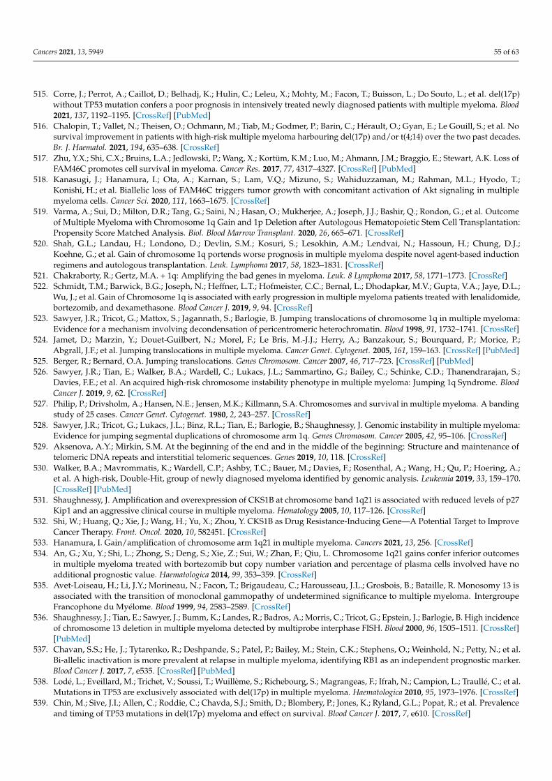
Cancers 2021, 13, 5949 55 of 63
515. Corre, J.; Perrot, A.; Caillot, D.; Belhadj, K.; Hulin, C.; Leleu, X.; Mohty, M.; Facon, T.; Buisson, L.; Do Souto, L.; et al. del(17p)without TP53 mutation confers a poor prognosis in intensively treated newly diagnosed patients with multiple myeloma. Blood2021, 137, 1192–1195. [CrossRef] [PubMed]
516. Chalopin, T.; Vallet, N.; Theisen, O.; Ochmann, M.; Tiab, M.; Godmer, P.; Barin, C.; Hérault, O.; Gyan, E.; Le Gouill, S.; et al. Nosurvival improvement in patients with high-risk multiple myeloma harbouring del(17p) and/or t(4;14) over the two past decades.Br. J. Haematol. 2021, 194, 635–638. [CrossRef]
517. Zhu, Y.X.; Shi, C.X.; Bruins, L.A.; Jedlowski, P.; Wang, X.; Kortüm, K.M.; Luo, M.; Ahmann, J.M.; Braggio, E.; Stewart, A.K. Loss ofFAM46C promotes cell survival in myeloma. Cancer Res. 2017, 77, 4317–4327. [CrossRef] [PubMed]
518. Kanasugi, J.; Hanamura, I.; Ota, A.; Karnan, S.; Lam, V.Q.; Mizuno, S.; Wahiduzzaman, M.; Rahman, M.L.; Hyodo, T.;Konishi, H.; et al. Biallelic loss of FAM46C triggers tumor growth with concomitant activation of Akt signaling in multiplemyeloma cells. Cancer Sci. 2020, 111, 1663–1675. [CrossRef]
519. Varma, A.; Sui, D.; Milton, D.R.; Tang, G.; Saini, N.; Hasan, O.; Mukherjee, A.; Joseph, J.J.; Bashir, Q.; Rondon, G.; et al. Outcomeof Multiple Myeloma with Chromosome 1q Gain and 1p Deletion after Autologous Hematopoietic Stem Cell Transplantation:Propensity Score Matched Analysis. Biol. Blood Marrow Transplant. 2020, 26, 665–671. [CrossRef]
520. Shah, G.L.; Landau, H.; Londono, D.; Devlin, S.M.; Kosuri, S.; Lesokhin, A.M.; Lendvai, N.; Hassoun, H.; Chung, D.J.;Koehne, G.; et al. Gain of chromosome 1q portends worse prognosis in multiple myeloma despite novel agent-based inductionregimens and autologous transplantation. Leuk. Lymphoma 2017, 58, 1823–1831. [CrossRef]
521. Chakraborty, R.; Gertz, M.A. + 1q: Amplifying the bad genes in myeloma. Leuk. 8 Lymphoma 2017, 58, 1771–1773. [CrossRef]522. Schmidt, T.M.; Barwick, B.G.; Joseph, N.; Heffner, L.T.; Hofmeister, C.C.; Bernal, L.; Dhodapkar, M.V.; Gupta, V.A.; Jaye, D.L.;
Wu, J.; et al. Gain of Chromosome 1q is associated with early progression in multiple myeloma patients treated with lenalidomide,bortezomib, and dexamethasone. Blood Cancer J. 2019, 9, 94. [CrossRef]
523. Sawyer, J.R.; Tricot, G.; Mattox, S.; Jagannath, S.; Barlogie, B. Jumping translocations of chromosome 1q in multiple myeloma:Evidence for a mechanism involving decondensation of pericentromeric heterochromatin. Blood 1998, 91, 1732–1741. [CrossRef]
524. Jamet, D.; Marzin, Y.; Douet-Guilbert, N.; Morel, F.; Le Bris, M.-J.J.; Herry, A.; Banzakour, S.; Bourquard, P.; Morice, P.;Abgrall, J.F.; et al. Jumping translocations in multiple myeloma. Cancer Genet. Cytogenet. 2005, 161, 159–163. [CrossRef] [PubMed]
525. Berger, R.; Bernard, O.A. Jumping translocations. Genes Chromosom. Cancer 2007, 46, 717–723. [CrossRef] [PubMed]526. Sawyer, J.R.; Tian, E.; Walker, B.A.; Wardell, C.; Lukacs, J.L.; Sammartino, G.; Bailey, C.; Schinke, C.D.; Thanendrarajan, S.;
Davies, F.E.; et al. An acquired high-risk chromosome instability phenotype in multiple myeloma: Jumping 1q Syndrome. BloodCancer J. 2019, 9, 62. [CrossRef]
527. Philip, P.; Drivsholm, A.; Hansen, N.E.; Jensen, M.K.; Killmann, S.A. Chromosomes and survival in multiple myeloma. A bandingstudy of 25 cases. Cancer Genet. Cytogenet. 1980, 2, 243–257. [CrossRef]
528. Sawyer, J.R.; Tricot, G.; Lukacs, J.L.; Binz, R.L.; Tian, E.; Barlogie, B.; Shaughnessy, J. Genomic instability in multiple myeloma:Evidence for jumping segmental duplications of chromosome arm 1q. Genes Chromosom. Cancer 2005, 42, 95–106. [CrossRef]
529. Aksenova, A.Y.; Mirkin, S.M. At the beginning of the end and in the middle of the beginning: Structure and maintenance oftelomeric DNA repeats and interstitial telomeric sequences. Genes 2019, 10, 118. [CrossRef]
530. Walker, B.A.; Mavrommatis, K.; Wardell, C.P.; Ashby, T.C.; Bauer, M.; Davies, F.; Rosenthal, A.; Wang, H.; Qu, P.; Hoering, A.;et al. A high-risk, Double-Hit, group of newly diagnosed myeloma identified by genomic analysis. Leukemia 2019, 33, 159–170.[CrossRef] [PubMed]
531. Shaughnessy, J. Amplification and overexpression of CKS1B at chromosome band 1q21 is associated with reduced levels of p27Kip1 and an aggressive clinical course in multiple myeloma. Hematology 2005, 10, 117–126. [CrossRef]
532. Shi, W.; Huang, Q.; Xie, J.; Wang, H.; Yu, X.; Zhou, Y. CKS1B as Drug Resistance-Inducing Gene—A Potential Target to ImproveCancer Therapy. Front. Oncol. 2020, 10, 582451. [CrossRef]
533. Hanamura, I. Gain/amplification of chromosome arm 1q21 in multiple myeloma. Cancers 2021, 13, 256. [CrossRef]534. An, G.; Xu, Y.; Shi, L.; Zhong, S.; Deng, S.; Xie, Z.; Sui, W.; Zhan, F.; Qiu, L. Chromosome 1q21 gains confer inferior outcomes
in multiple myeloma treated with bortezomib but copy number variation and percentage of plasma cells involved have noadditional prognostic value. Haematologica 2014, 99, 353–359. [CrossRef]
535. Avet-Loiseau, H.; Li, J.Y.; Morineau, N.; Facon, T.; Brigaudeau, C.; Harousseau, J.L.; Grosbois, B.; Bataille, R. Monosomy 13 isassociated with the transition of monoclonal gammopathy of undetermined significance to multiple myeloma. IntergroupeFrancophone du Myélome. Blood 1999, 94, 2583–2589. [CrossRef]
536. Shaughnessy, J.; Tian, E.; Sawyer, J.; Bumm, K.; Landes, R.; Badros, A.; Morris, C.; Tricot, G.; Epstein, J.; Barlogie, B. High incidenceof chromosome 13 deletion in multiple myeloma detected by multiprobe interphase FISH. Blood 2000, 96, 1505–1511. [CrossRef][PubMed]
537. Chavan, S.S.; He, J.; Tytarenko, R.; Deshpande, S.; Patel, P.; Bailey, M.; Stein, C.K.; Stephens, O.; Weinhold, N.; Petty, N.; et al.Bi-allelic inactivation is more prevalent at relapse in multiple myeloma, identifying RB1 as an independent prognostic marker.Blood Cancer J. 2017, 7, e535. [CrossRef] [PubMed]
538. Lodé, L.; Eveillard, M.; Trichet, V.; Soussi, T.; Wuillème, S.; Richebourg, S.; Magrangeas, F.; Ifrah, N.; Campion, L.; Traullé, C.; et al.Mutations in TP53 are exclusively associated with del(17p) in multiple myeloma. Haematologica 2010, 95, 1973–1976. [CrossRef]
539. Chin, M.; Sive, J.I.; Allen, C.; Roddie, C.; Chavda, S.J.; Smith, D.; Blombery, P.; Jones, K.; Ryland, G.L.; Popat, R.; et al. Prevalenceand timing of TP53 mutations in del(17p) myeloma and effect on survival. Blood Cancer J. 2017, 7, e610. [CrossRef]
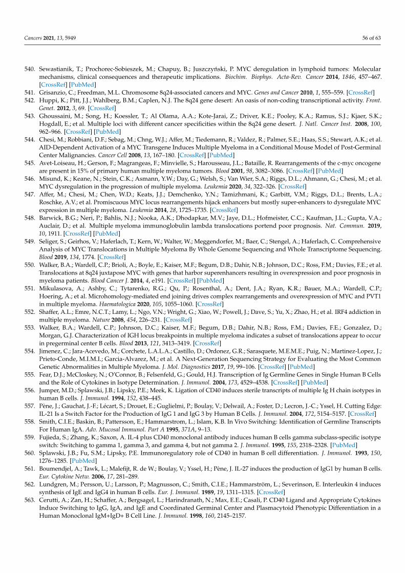
Cancers 2021, 13, 5949 56 of 63
540. Sewastianik, T.; Prochorec-Sobieszek, M.; Chapuy, B.; Juszczynski, P. MYC deregulation in lymphoid tumors: Molecularmechanisms, clinical consequences and therapeutic implications. Biochim. Biophys. Acta-Rev. Cancer 2014, 1846, 457–467.[CrossRef] [PubMed]
541. Grisanzio, C.; Freedman, M.L. Chromosome 8q24-associated cancers and MYC. Genes and Cancer 2010, 1, 555–559. [CrossRef]542. Huppi, K.; Pitt, J.J.; Wahlberg, B.M.; Caplen, N.J. The 8q24 gene desert: An oasis of non-coding transcriptional activity. Front.
Genet. 2012, 3, 69. [CrossRef]543. Ghoussaini, M.; Song, H.; Koessler, T.; Al Olama, A.A.; Kote-Jarai, Z.; Driver, K.E.; Pooley, K.A.; Ramus, S.J.; Kjaer, S.K.;
Hogdall, E.; et al. Multiple loci with different cancer specificities within the 8q24 gene desert. J. Natl. Cancer Inst. 2008, 100,962–966. [CrossRef] [PubMed]
544. Chesi, M.; Robbiani, D.F.; Sebag, M.; Chng, W.J.; Affer, M.; Tiedemann, R.; Valdez, R.; Palmer, S.E.; Haas, S.S.; Stewart, A.K.; et al.AID-Dependent Activation of a MYC Transgene Induces Multiple Myeloma in a Conditional Mouse Model of Post-GerminalCenter Malignancies. Cancer Cell 2008, 13, 167–180. [CrossRef] [PubMed]
545. Avet-Loiseau, H.; Gerson, F.; Magrangeas, F.; Minvielle, S.; Harousseau, J.L.; Bataille, R. Rearrangements of the c-myc oncogeneare present in 15% of primary human multiple myeloma tumors. Blood 2001, 98, 3082–3086. [CrossRef] [PubMed]
546. Misund, K.; Keane, N.; Stein, C.K.; Asmann, Y.W.; Day, G.; Welsh, S.; Van Wier, S.A.; Riggs, D.L.; Ahmann, G.; Chesi, M.; et al.MYC dysregulation in the progression of multiple myeloma. Leukemia 2020, 34, 322–326. [CrossRef]
547. Affer, M.; Chesi, M.; Chen, W.D.; Keats, J.J.; Demchenko, Y.N.; Tamizhmani, K.; Garbitt, V.M.; Riggs, D.L.; Brents, L.A.;Roschke, A.V.; et al. Promiscuous MYC locus rearrangements hijack enhancers but mostly super-enhancers to dysregulate MYCexpression in multiple myeloma. Leukemia 2014, 28, 1725–1735. [CrossRef]
548. Barwick, B.G.; Neri, P.; Bahlis, N.J.; Nooka, A.K.; Dhodapkar, M.V.; Jaye, D.L.; Hofmeister, C.C.; Kaufman, J.L.; Gupta, V.A.;Auclair, D.; et al. Multiple myeloma immunoglobulin lambda translocations portend poor prognosis. Nat. Commun. 2019,10, 1911. [CrossRef] [PubMed]
549. Seliger, S.; Geirhos, V.; Haferlach, T.; Kern, W.; Walter, W.; Meggendorfer, M.; Baer, C.; Stengel, A.; Haferlach, C. ComprehensiveAnalysis of MYC Translocations in Multiple Myeloma By Whole Genome Sequencing and Whole Transcriptome Sequencing.Blood 2019, 134, 1774. [CrossRef]
550. Walker, B.A.; Wardell, C.P.; Brioli, A.; Boyle, E.; Kaiser, M.F.; Begum, D.B.; Dahir, N.B.; Johnson, D.C.; Ross, F.M.; Davies, F.E.; et al.Translocations at 8q24 juxtapose MYC with genes that harbor superenhancers resulting in overexpression and poor prognosis inmyeloma patients. Blood Cancer J. 2014, 4, e191. [CrossRef] [PubMed]
551. Mikulasova, A.; Ashby, C.; Tytarenko, R.G.; Qu, P.; Rosenthal, A.; Dent, J.A.; Ryan, K.R.; Bauer, M.A.; Wardell, C.P.;Hoering, A.; et al. Microhomology-mediated end joining drives complex rearrangements and overexpression of MYC and PVT1in multiple myeloma. Haematologica 2020, 105, 1055–1060. [CrossRef]
552. Shaffer, A.L.; Emre, N.C.T.; Lamy, L.; Ngo, V.N.; Wright, G.; Xiao, W.; Powell, J.; Dave, S.; Yu, X.; Zhao, H.; et al. IRF4 addiction inmultiple myeloma. Nature 2008, 454, 226–231. [CrossRef]
553. Walker, B.A.; Wardell, C.P.; Johnson, D.C.; Kaiser, M.F.; Begum, D.B.; Dahir, N.B.; Ross, F.M.; Davies, F.E.; Gonzalez, D.;Morgan, G.J. Characterization of IGH locus breakpoints in multiple myeloma indicates a subset of translocations appear to occurin pregerminal center B cells. Blood 2013, 121, 3413–3419. [CrossRef]
554. Jimenez, C.; Jara-Acevedo, M.; Corchete, L.A.L.A.; Castillo, D.; Ordonez, G.R.; Sarasquete, M.E.M.E.; Puig, N.; Martinez-Lopez, J.;Prieto-Conde, M.I.M.I.; Garcia-Alvarez, M.; et al. A Next-Generation Sequencing Strategy for Evaluating the Most CommonGenetic Abnormalities in Multiple Myeloma. J. Mol. Diagnostics 2017, 19, 99–106. [CrossRef] [PubMed]
555. Fear, D.J.; McCloskey, N.; O’Connor, B.; Felsenfeld, G.; Gould, H.J. Transcription of Ig Germline Genes in Single Human B Cellsand the Role of Cytokines in Isotype Determination. J. Immunol. 2004, 173, 4529–4538. [CrossRef] [PubMed]
556. Jumper, M.D.; Splawski, J.B.; Lipsky, P.E.; Meek, K. Ligation of CD40 induces sterile transcripts of multiple Ig H chain isotypes inhuman B cells. J. Immunol. 1994, 152, 438–445.
557. Pène, J.; Gauchat, J.-F.; Lécart, S.; Drouet, E.; Guglielmi, P.; Boulay, V.; Delwail, A.; Foster, D.; Lecron, J.-C.; Yssel, H. Cutting Edge:IL-21 Is a Switch Factor for the Production of IgG 1 and IgG 3 by Human B Cells. J. Immunol. 2004, 172, 5154–5157. [CrossRef]
558. Smith, C.I.E.; Baskin, B.; Pattersson, E.; Hammarstrom, L.; Islam, K.B. In Vivo Switching: Identification of Germline TranscriptsFor Human IgA. Adv. Mucosal Immunol. Part A 1995, 371A, 9–13.
559. Fujieda, S.; Zhang, K.; Saxon, A. IL-4 plus CD40 monoclonal antibody induces human B cells gamma subclass-specific isotypeswitch: Switching to gamma 1, gamma 3, and gamma 4, but not gamma 2. J. Immunol. 1995, 155, 2318–2328. [PubMed]
560. Splawski, J.B.; Fu, S.M.; Lipsky, P.E. Immunoregulatory role of CD40 in human B cell differentiation. J. Immunol. 1993, 150,1276–1285. [PubMed]
561. Boumendjel, A.; Tawk, L.; Malefijt, R. de W.; Boulay, V.; Yssel, H.; Pène, J. IL-27 induces the production of IgG1 by human B cells.Eur. Cytokine Netw. 2006, 17, 281–289.
562. Lundgren, M.; Persson, U.; Larsson, P.; Magnusson, C.; Smith, C.I.E.; Hammarström, L.; Severinson, E. Interleukin 4 inducessynthesis of IgE and IgG4 in human B cells. Eur. J. Immunol. 1989, 19, 1311–1315. [CrossRef]
563. Cerutti, A.; Zan, H.; Schaffer, A.; Bergsagel, L.; Harindranath, N.; Max, E.E.; Casali, P. CD40 Ligand and Appropriate CytokinesInduce Switching to IgG, IgA, and IgE and Coordinated Germinal Center and Plasmacytoid Phenotypic Differentiation in aHuman Monoclonal IgM+IgD+ B Cell Line. J. Immunol. 1998, 160, 2145–2157.
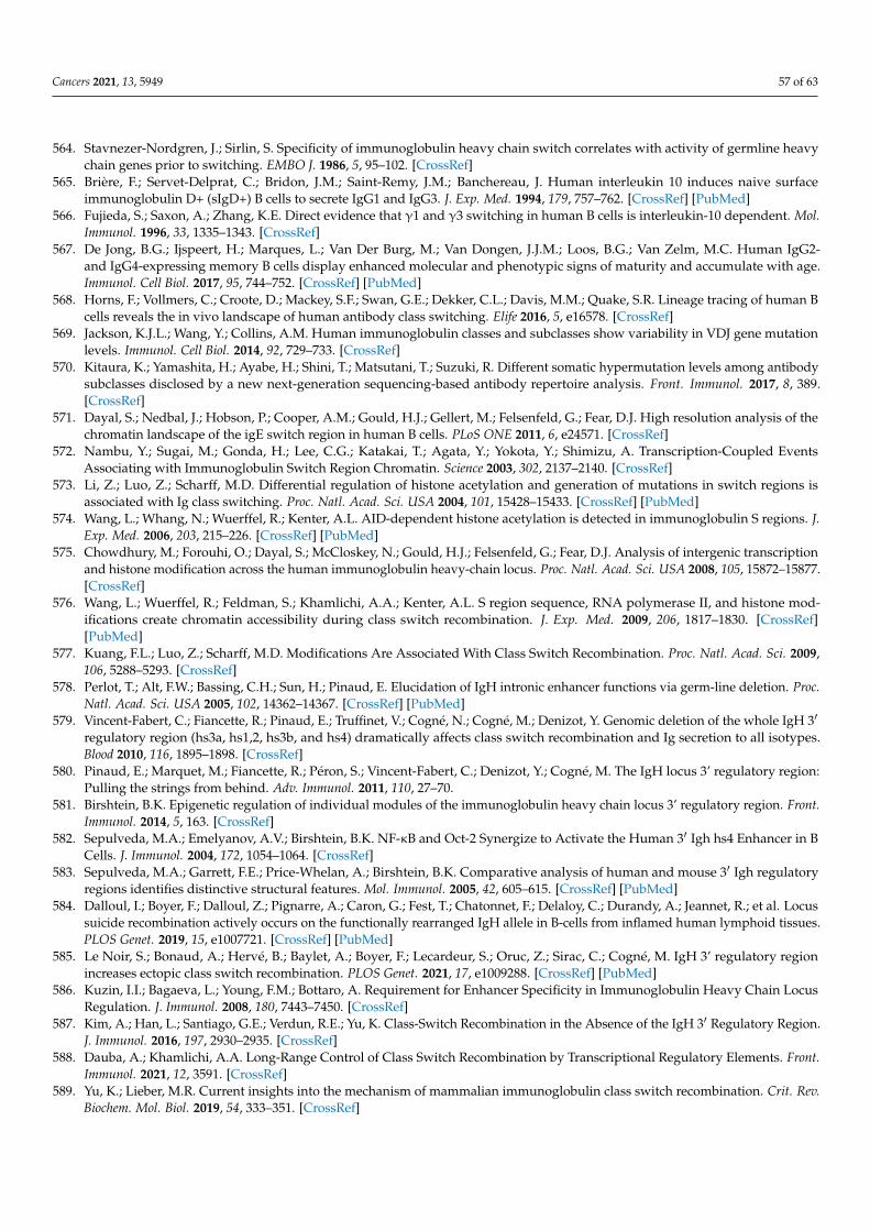
Cancers 2021, 13, 5949 57 of 63
564. Stavnezer-Nordgren, J.; Sirlin, S. Specificity of immunoglobulin heavy chain switch correlates with activity of germline heavychain genes prior to switching. EMBO J. 1986, 5, 95–102. [CrossRef]
565. Brière, F.; Servet-Delprat, C.; Bridon, J.M.; Saint-Remy, J.M.; Banchereau, J. Human interleukin 10 induces naive surfaceimmunoglobulin D+ (sIgD+) B cells to secrete IgG1 and IgG3. J. Exp. Med. 1994, 179, 757–762. [CrossRef] [PubMed]
566. Fujieda, S.; Saxon, A.; Zhang, K.E. Direct evidence that γ1 and γ3 switching in human B cells is interleukin-10 dependent. Mol.Immunol. 1996, 33, 1335–1343. [CrossRef]
567. De Jong, B.G.; Ijspeert, H.; Marques, L.; Van Der Burg, M.; Van Dongen, J.J.M.; Loos, B.G.; Van Zelm, M.C. Human IgG2-and IgG4-expressing memory B cells display enhanced molecular and phenotypic signs of maturity and accumulate with age.Immunol. Cell Biol. 2017, 95, 744–752. [CrossRef] [PubMed]
568. Horns, F.; Vollmers, C.; Croote, D.; Mackey, S.F.; Swan, G.E.; Dekker, C.L.; Davis, M.M.; Quake, S.R. Lineage tracing of human Bcells reveals the in vivo landscape of human antibody class switching. Elife 2016, 5, e16578. [CrossRef]
569. Jackson, K.J.L.; Wang, Y.; Collins, A.M. Human immunoglobulin classes and subclasses show variability in VDJ gene mutationlevels. Immunol. Cell Biol. 2014, 92, 729–733. [CrossRef]
570. Kitaura, K.; Yamashita, H.; Ayabe, H.; Shini, T.; Matsutani, T.; Suzuki, R. Different somatic hypermutation levels among antibodysubclasses disclosed by a new next-generation sequencing-based antibody repertoire analysis. Front. Immunol. 2017, 8, 389.[CrossRef]
571. Dayal, S.; Nedbal, J.; Hobson, P.; Cooper, A.M.; Gould, H.J.; Gellert, M.; Felsenfeld, G.; Fear, D.J. High resolution analysis of thechromatin landscape of the igE switch region in human B cells. PLoS ONE 2011, 6, e24571. [CrossRef]
572. Nambu, Y.; Sugai, M.; Gonda, H.; Lee, C.G.; Katakai, T.; Agata, Y.; Yokota, Y.; Shimizu, A. Transcription-Coupled EventsAssociating with Immunoglobulin Switch Region Chromatin. Science 2003, 302, 2137–2140. [CrossRef]
573. Li, Z.; Luo, Z.; Scharff, M.D. Differential regulation of histone acetylation and generation of mutations in switch regions isassociated with Ig class switching. Proc. Natl. Acad. Sci. USA 2004, 101, 15428–15433. [CrossRef] [PubMed]
574. Wang, L.; Whang, N.; Wuerffel, R.; Kenter, A.L. AID-dependent histone acetylation is detected in immunoglobulin S regions. J.Exp. Med. 2006, 203, 215–226. [CrossRef] [PubMed]
575. Chowdhury, M.; Forouhi, O.; Dayal, S.; McCloskey, N.; Gould, H.J.; Felsenfeld, G.; Fear, D.J. Analysis of intergenic transcriptionand histone modification across the human immunoglobulin heavy-chain locus. Proc. Natl. Acad. Sci. USA 2008, 105, 15872–15877.[CrossRef]
576. Wang, L.; Wuerffel, R.; Feldman, S.; Khamlichi, A.A.; Kenter, A.L. S region sequence, RNA polymerase II, and histone mod-ifications create chromatin accessibility during class switch recombination. J. Exp. Med. 2009, 206, 1817–1830. [CrossRef][PubMed]
577. Kuang, F.L.; Luo, Z.; Scharff, M.D. Modifications Are Associated With Class Switch Recombination. Proc. Natl. Acad. Sci. 2009,106, 5288–5293. [CrossRef]
578. Perlot, T.; Alt, F.W.; Bassing, C.H.; Sun, H.; Pinaud, E. Elucidation of IgH intronic enhancer functions via germ-line deletion. Proc.Natl. Acad. Sci. USA 2005, 102, 14362–14367. [CrossRef] [PubMed]
579. Vincent-Fabert, C.; Fiancette, R.; Pinaud, E.; Truffinet, V.; Cogné, N.; Cogné, M.; Denizot, Y. Genomic deletion of the whole IgH 3′
regulatory region (hs3a, hs1,2, hs3b, and hs4) dramatically affects class switch recombination and Ig secretion to all isotypes.Blood 2010, 116, 1895–1898. [CrossRef]
580. Pinaud, E.; Marquet, M.; Fiancette, R.; Péron, S.; Vincent-Fabert, C.; Denizot, Y.; Cogné, M. The IgH locus 3’ regulatory region:Pulling the strings from behind. Adv. Immunol. 2011, 110, 27–70.
581. Birshtein, B.K. Epigenetic regulation of individual modules of the immunoglobulin heavy chain locus 3’ regulatory region. Front.Immunol. 2014, 5, 163. [CrossRef]
582. Sepulveda, M.A.; Emelyanov, A.V.; Birshtein, B.K. NF-κB and Oct-2 Synergize to Activate the Human 3′ Igh hs4 Enhancer in BCells. J. Immunol. 2004, 172, 1054–1064. [CrossRef]
583. Sepulveda, M.A.; Garrett, F.E.; Price-Whelan, A.; Birshtein, B.K. Comparative analysis of human and mouse 3′ Igh regulatoryregions identifies distinctive structural features. Mol. Immunol. 2005, 42, 605–615. [CrossRef] [PubMed]
584. Dalloul, I.; Boyer, F.; Dalloul, Z.; Pignarre, A.; Caron, G.; Fest, T.; Chatonnet, F.; Delaloy, C.; Durandy, A.; Jeannet, R.; et al. Locussuicide recombination actively occurs on the functionally rearranged IgH allele in B-cells from inflamed human lymphoid tissues.PLOS Genet. 2019, 15, e1007721. [CrossRef] [PubMed]
585. Le Noir, S.; Bonaud, A.; Hervé, B.; Baylet, A.; Boyer, F.; Lecardeur, S.; Oruc, Z.; Sirac, C.; Cogné, M. IgH 3’ regulatory regionincreases ectopic class switch recombination. PLOS Genet. 2021, 17, e1009288. [CrossRef] [PubMed]
586. Kuzin, I.I.; Bagaeva, L.; Young, F.M.; Bottaro, A. Requirement for Enhancer Specificity in Immunoglobulin Heavy Chain LocusRegulation. J. Immunol. 2008, 180, 7443–7450. [CrossRef]
587. Kim, A.; Han, L.; Santiago, G.E.; Verdun, R.E.; Yu, K. Class-Switch Recombination in the Absence of the IgH 3′ Regulatory Region.J. Immunol. 2016, 197, 2930–2935. [CrossRef]
588. Dauba, A.; Khamlichi, A.A. Long-Range Control of Class Switch Recombination by Transcriptional Regulatory Elements. Front.Immunol. 2021, 12, 3591. [CrossRef]
589. Yu, K.; Lieber, M.R. Current insights into the mechanism of mammalian immunoglobulin class switch recombination. Crit. Rev.Biochem. Mol. Biol. 2019, 54, 333–351. [CrossRef]
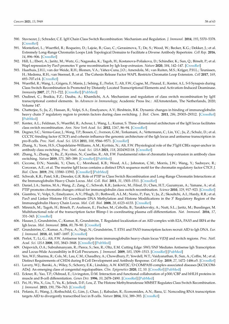
Cancers 2021, 13, 5949 58 of 63
590. Stavnezer, J.; Schrader, C.E. IgH Chain Class Switch Recombination: Mechanism and Regulation. J. Immunol. 2014, 193, 5370–5378.[CrossRef]
591. Montefiori, L.; Wuerffel, R.; Roqueiro, D.; Lajoie, B.; Guo, C.; Gerasimova, T.; De, S.; Wood, W.; Becker, K.G.; Dekker, J.; et al.Extremely Long-Range Chromatin Loops Link Topological Domains to Facilitate a Diverse Antibody Repertoire. Cell Rep. 2016,14, 896–906. [CrossRef]
592. Hill, L.; Ebert, A.; Jaritz, M.; Wutz, G.; Nagasaka, K.; Tagoh, H.; Kostanova-Poliakova, D.; Schindler, K.; Sun, Q.; Bönelt, P.; et al.Wapl repression by Pax5 promotes V gene recombination by Igh loop extrusion. Nature 2020, 584, 142–147. [CrossRef]
593. Haarhuis, J.H.I.; van der Weide, R.H.; Blomen, V.A.; Yáñez-Cuna, J.O.; Amendola, M.; van Ruiten, M.S.; Krijger, P.H.L.; Teunissen,H.; Medema, R.H.; van Steensel, B.; et al. The Cohesin Release Factor WAPL Restricts Chromatin Loop Extension. Cell 2017, 169,693–707.e14. [CrossRef]
594. Wuerffel, R.; Wang, L.; Grigera, F.; Manis, J.; Selsing, E.; Perlot, T.; Alt, F.W.; Cogne, M.; Pinaud, E.; Kenter, A.L. S-S Synapsis duringClass Switch Recombination Is Promoted by Distantly Located Transcriptional Elements and Activation-Induced Deaminase.Immunity 2007, 27, 711–722. [CrossRef] [PubMed]
595. Oudinet, C.; Braikia, F.Z.; Dauba, A.; Khamlichi, A.A. Mechanism and regulation of class switch recombination by IgHtranscriptional control elements. In Advances in Immunology; Academic Press Inc.: AEAmsterdam, The Netherlands, 2020;Volume 147.
596. Chatterjee, S.; Ju, Z.; Hassan, R.; Volpi, S.A.; Emelyanov, A.V.; Birshtein, B.K. Dynamic changes in binding of immunoglobulinheavy chain 3′ regulatory region to protein factors during class switching. J. Biol. Chem. 2011, 286, 29303–29312. [CrossRef][PubMed]
597. Kenter, A.L.; Feldman, S.; Wuerffel, R.; Achour, I.; Wang, L.; Kumar, S. Three-dimensional architecture of the IgH locus facilitatesclass switch recombination. Ann. New York Acad. Sci. 2012, 1267, 86–94. [CrossRef]
598. Degner, S.C.; Verma-Gaur, J.; Wong, T.P.; Bossen, C.; Iverson, G.M.; Torkamani, A.; Vettermann, C.; Lin, Y.C.; Ju, Z.; Schulz, D.; et al.CCCTC-binding factor (CTCF) and cohesin influence the genomic architecture of the Igh locus and antisense transcription inpro-B cells. Proc. Natl. Acad. Sci. USA 2011, 108, 9566–9571. [CrossRef]
599. Zhang, X.; Yoon, H.S.; Chapdelaine-Williams, A.M.; Kyritsis, N.; Alt, F.W. Physiological role of the 3′IgH CBEs super-anchor inantibody class switching. Proc. Natl. Acad. Sci. USA 2021, 118, 2024392118. [CrossRef]
600. Zhang, X.; Zhang, Y.; Ba, Z.; Kyritsis, N.; Casellas, R.; Alt, F.W. Fundamental roles of chromatin loop extrusion in antibody classswitching. Nature 2019, 575, 385–389. [CrossRef] [PubMed]
601. Ciccone, D.N.; Namiki, Y.; Chen, C.; Morshead, K.B.; Wood, A.L.; Johnston, C.M.; Morris, J.W.; Wang, Y.; Sadreyev, R.;Corcoran, A.E.; et al. The murine IgH locus contains a distinct DNA sequence motif for the chromatin regulatory factor CTCF. J.Biol. Chem. 2019, 294, 13580–13592. [CrossRef] [PubMed]
602. Schwab, K.R.; Patel, S.R.; Dressler, G.R. Role of PTIP in Class Switch Recombination and Long-Range Chromatin Interactions atthe Immunoglobulin Heavy Chain Locus. Mol. Cell. Biol. 2011, 31, 1503–1511. [CrossRef]
603. Daniel, J.A.; Santos, M.A.; Wang, Z.; Zang, C.; Schwab, K.R.; Jankovic, M.; Filsuf, D.; Chen, H.T.; Gazumyan, A.; Yamane, A.; et al.PTIP promotes chromatin changes critical for immunoglobulin class switch recombination. Science 2010, 329, 917–923. [CrossRef]
604. Giambra, V.; Volpi, S.; Emelyanov, A.V.; Pflugh, D.; Bothwell, A.L.M.; Norio, P.; Fan, Y.; Ju, Z.; Skoultchi, A.I.; Hardy, R.R.; et al.Pax5 and Linker Histone H1 Coordinate DNA Methylation and Histone Modifications in the 3′ Regulatory Region of theImmunoglobulin Heavy Chain Locus. Mol. Cell. Biol. 2008, 28, 6123–6133. [CrossRef]
605. Minnich, M.; Tagoh, H.; Bönelt, P.; Axelsson, E.; Fischer, M.; Cebolla, B.; Tarakhovsky, A.; Nutt, S.L.; Jaritz, M.; Busslinger, M.Multifunctional role of the transcription factor Blimp-1 in coordinating plasma cell differentiation. Nat. Immunol. 2016, 17,331–343. [CrossRef]
606. Hauser, J.; Grundström, C.; Kumar, R.; Grundström, T. Regulated localization of an AID complex with E2A, PAX5 and IRF4 at theIgh locus. Mol. Immunol. 2016, 80, 78–90. [CrossRef]
607. Grundström, C.; Kumar, A.; Priya, A.; Negi, N.; Grundström, T. ETS1 and PAX5 transcription factors recruit AID to Igh DNA. Eur.J. Immunol. 2018, 48, 1687–1697. [CrossRef]
608. Perlot, T.; Li, G.; Alt, F.W. Antisense transcripts from immunoglobulin heavy-chain locus V(D)J and switch regions. Proc. Natl.Acad. Sci. USA 2008, 105, 3843–3848. [CrossRef] [PubMed]
609. Osipovich, O.A.; Subrahmanyam, R.; Pierce, S.; Sen, R.; Oltz, E.M. Cutting Edge: SWI/SNF Mediates Antisense Igh Transcriptionand Locus-Wide Accessibility in B Cell Precursors. J. Immunol. 2009, 183, 1509–1513. [CrossRef] [PubMed]
610. Yen, W.F.; Sharma, R.; Cols, M.; Lau, C.M.; Chaudhry, A.; Chowdhury, P.; Yewdell, W.T.; Vaidyanathan, B.; Sun, A.; Coffre, M.; et al.Distinct Requirements of CHD4 during B Cell Development and Antibody Response. Cell Rep. 2019, 27, 1472–1486.e5. [CrossRef]
611. Lavery, W.J.; Barski, A.; Wiley, S.; Schorry, E.K.; Lindsley, A.W. KMT2C/D COMPASS complex-associated diseases [KCDCOM-ADs]: An emerging class of congenital regulopathies. Clin. Epigenetics 2020, 12, 10. [CrossRef] [PubMed]
612. Eckner, R.; Yao, T.P.; Oldread, E.; Livingston, D.M. Interaction and functional collaboration of p300/CBP and bHLH proteins inmuscle and B-cell differentiation. Genes Dev. 1996, 10, 2478–2490. [CrossRef] [PubMed]
613. Pei, H.; Wu, X.; Liu, T.; Yu, K.; Jelinek, D.F.; Lou, Z. The Histone Methyltransferase MMSET Regulates Class Switch Recombination.J. Immunol. 2013, 190, 756–763. [CrossRef]
614. Pefanis, E.; Wang, J.; Rothschild, G.; Lim, J.; Chao, J.; Rabadan, R.; Economides, A.N.; Basu, U. Noncoding RNA transcriptiontargets AID to divergently transcribed loci in B cells. Nature 2014, 514, 389–393. [CrossRef]
Cancers 2021, 13, 5949 59 of 63
615. Medvedovic, J.; Ebert, A.; Tagoh, H.; Busslinger, M. Pax5: A Master Regulator of B Cell Development and Leukemogenesis. InAdvances in Immunology; Academic Press Inc.: AEAmsterdam, The Netherlands, 2011; Volume 111, pp. 179–206.
616. O’Brien, P.; Morin, P.; Ouellette, R.J.; Robichaud, G.A. The Pax-5 gene: A pluripotent regulator of B-cell differentiation and cancerdisease. Cancer Res. 2011, 71, 7345–7350. [CrossRef]
617. Okuyama, K.; Strid, T.; Kuruvilla, J.; Somasundaram, R.; Cristobal, S.; Smith, E.; Prasad, M.; Fioretos, T.; Lilljebjörn, H.; Soneji, S.;et al. PAX5 is part of a functional transcription factor network targeted in lymphoid leukemia. PLoS Genet. 2019, 15, e1008280.[CrossRef] [PubMed]
618. Foltz, S.M.; Gao, Q.; Yoon, C.J.; Sun, H.; Yao, L.; Li, Y.; Jayasinghe, R.G.; Cao, S.; King, J.; Kohnen, D.R.; et al. Evolution andstructure of clinically relevant gene fusions in multiple myeloma. Nat. Commun. 2020, 11, 2666. [CrossRef]
619. Rustad, E.H.; Yellapantula, V.D.; Glodzik, D.; Maclachlan, K.H.; Diamond, B.; Boyle, E.M.; Ashby, C.; Blaney, P.; Gundem, G.;Hultcrantz, M.; et al. Revealing the Impact of Structural Variants in Multiple Myeloma. Blood Cancer Discov. 2020, 1, 258–273.[CrossRef] [PubMed]
620. Qian, J.; Wang, Q.; Dose, M.; Pruett, N.; Kieffer-Kwon, K.R.; Resch, W.; Liang, G.; Tang, Z.; Mathé, E.; Benner, C.; et al. B cellsuper-enhancers and regulatory clusters recruit AID tumorigenic activity. Cell 2014, 159, 1524–1537. [CrossRef] [PubMed]
621. Borson, N.D.; Lacy, M.Q.; Wettstein, P.J. Altered mRNA expression of Pax5 and Blimp-1 in B cells in multiple myeloma. Blood2002, 100, 4629–4639. [CrossRef]
622. Fuxa, M.; Skok, J.; Souabni, A.; Salvagiotto, G.; Roldan, E.; Busslinger, M. Pax5 induces V-to-DJ rearrangements and locuscontraction of the immunoglobulin heavy-chain gene. Genes Dev. 2004, 18, 411–422. [CrossRef] [PubMed]
623. Nutt, S.L.; Urbánek, P.; Relink, A.; Busslinger, M. Essential functions of Pax5 (BSAP) in pro-B cell development: Differencebetween fetal and adult B lymphopoiesis and reduced V-to-DJ recombination at the IgH locus. Genes Dev. 1997, 11, 476–491.[CrossRef]
624. Waters, S.H.; Saikh, K.U.; Stavnezer, J. A B-cell-specific nuclear protein that binds to DNA sites 5’ to immunoglobulin S alphatandem repeats is regulated during differentiation. Mol. Cell. Biol. 1989, 9, 5594–5601.
625. Liao, F.; Giannini, S.L.; Birshtein, B.K. A nuclear DNA-binding protein expressed during early stages of B cell differentationinteracts with diverse segments within and 3’ of the Ig H chain gene cluster. J. Immunol. 1992, 148, 2909–2917. [PubMed]
626. Xu, L.; Kim, M.G.; Marcu, K.B. Properties of B cell stage specific and ubiquitous nuclear factors binding to immunoglobulin heavychain gene switch regions. Int. Immunol. 1992, 4, 875–887. [CrossRef]
627. Kosak, S.T.; Skok, J.A.; Medina, K.L.; Riblet, R.; Le Beau, M.M.; Fisher, A.G.; Singh, H. Subnuclear compartmentalization ofimmunoglobulin loci during lymphocyte development. Science 2002, 296, 158–162. [CrossRef]
628. Zullo, J.M.; Demarco, I.A.; Piqué-Regi, R.; Gaffney, D.J.; Epstein, C.B.; Spooner, C.J.; Luperchio, T.R.; Bernstein, B.E.; Pritchard,J.K.; Reddy, K.L.; et al. DNA sequence-dependent compartmentalization and silencing of chromatin at the nuclear lamina. Cell2012, 149, 1474–1487. [CrossRef] [PubMed]
629. Bowen, A.J.; Corcoran, A.E. How chromatin remodelling allows shuffling of immunoglobulin heavy chain genes. Mol. Biosyst.2008, 4, 790–798. [CrossRef] [PubMed]
630. Corcoran, A.E. The epigenetic role of non-coding RNA transcription and nuclear organization in immunoglobulin repertoiregeneration. Semin. Immunol. 2010, 22, 353–361. [CrossRef]
631. Pichugin, A.; Iarovaia, O.V.; Gavrilov, A.; Sklyar, I.; Barinova, N.; Barinov, A.; Ivashkin, E.; Caron, G.; Aoufouchi, S.;Razin, S.V.; et al. The IGH locus relocalizes to a “recombination compartment” in the perinucleolar region of differentiatingB-lymphocytes. Oncotarget 2017, 8, 40079–40089. [CrossRef]
632. Seifert, M.; Scholtysik, R.; Küppers, R. Origin and pathogenesis of B cell lymphomas. In Methods in Molecular Biology; HumanaPress Inc.: Totowa, NJ, USA, 2019; Volume 1956, pp. 1–33.
633. Ramiro, A.R.; Jankovic, M.; Eisenreich, T.; Difilippantonio, S.; Chen-Kiang, S.; Muramatsu, M.; Honjo, T.; Nussenzweig, A.;Nussenzweig, M.C. AID is required for c-myc/IgH chromosome translocations in vivo. Cell 2004, 118, 431–438. [CrossRef]
634. Ghetti, M.; Vannini, I.; Storlazzi, C.T.; Martinelli, G.; Simonetti, G. Linear and circular PVT1 in hematological malignancies andimmune response: Two faces of the same coin. Mol. Cancer 2020, 19, 69. [CrossRef] [PubMed]
635. Derderian, C.; Orunmuyi, A.T.; Oluwabunmi Olapade-Olaopa, E.; Ogunwobi, O.O. PVT1 signaling is a mediator of cancerprogression. Front. Oncol. 2019, 9, 502. [CrossRef]
636. Cho, S.W.; Xu, J.; Sun, R.; Mumbach, M.R.; Carter, A.C.; Chen, Y.G.; Yost, K.E.; Kim, J.; He, J.; Nevins, S.A.; et al. Promoter oflncRNA Gene PVT1 Is a Tumor-Suppressor DNA Boundary Element. Cell 2018, 173, 1398–1412.e22. [CrossRef]
637. Osborne, C.S.; Chakalova, L.; Mitchell, J.A.; Horton, A.; Wood, A.L.; Bolland, D.J.; Corcoran, A.E.; Fraser, P. Myc dynamically andpreferentially relocates to a transcription factory occupied by Igh. PLoS Biol. 2007, 5, 1763–1772. [CrossRef]
638. Strongin, D.E.; Groudine, M.; Politz, J.C.R. Nucleolar tethering mediates pairing between the IgH and Myc loci. Nucleus 2014, 5,474–481. [CrossRef]
639. Iarovaia, O.V.; Ioudinkova, E.S.; Razin, S.V.; Vassetzky, Y.S. Role of the Nucleolus in Rearrangements of the IGH Locus. Mol. Biol.(Mosk) 2018, 52, 210–219. [CrossRef]
640. Sklyar, I.V.; Pichugin, A.M.; Razin, S.V.; Vassetzky, E.S.; Iarovaia, O.V. Nuclear localization of translocation partners in differentiat-ing B-cells. Dokl. Biochem. Biophys. 2015, 464, 312–314. [CrossRef]
Cancers 2021, 13, 5949 60 of 63
641. Sklyar, I.; Iarovaia, O.V.; Gavrilov, A.A.; Pichugin, A.; Germini, D.; Tsfasman, T.; Caron, G.; Fest, T.; Lipinski, M.; Razin, S.V.; et al.Distinct Patterns of Colocalization of the CCND1 and CMYC Genes with Their Potential Translocation Partner IGH at SuccessiveStages of B-Cell Differentiation. J. Cell. Biochem. 2016, 117, 1506–1510. [CrossRef]
642. Kaur, G.; Gupta, R.; Mathur, N.; Rani, L.; Kumar, L.; Sharma, A.; Singh, V.; Gupta, A.; Sharma, O.D. Clinical impact ofchromothriptic complex chromosomal rearrangements in newly diagnosed multiple myeloma. Leuk. Res. 2019, 76, 58–64.[CrossRef]
643. Smetana, J.; Oppelt, J.; Štork, M.; Pour, L.; Kuglík, P. Chromothripsis 18 in multiple myeloma patient with rapid extramedullaryrelapse. Mol. Cytogenet. 2018, 11, 7. [CrossRef] [PubMed]
644. Magrangeas, F.; Avet-Loiseau, H.; Munshi, N.C.; Minvielle, S. Chromothripsis identifies a rare and aggressive entity amongnewly diagnosed multiple myeloma patients. Blood 2011, 118, 675–678. [CrossRef] [PubMed]
645. Li, Y.; Roberts, N.D.; Wala, J.A.; Shapira, O.; Schumacher, S.E.; Kumar, K.; Khurana, E.; Waszak, S.; Korbel, J.O.; Haber, J.E.; et al.Patterns of somatic structural variation in human cancer genomes. Nature 2020, 578, 112–121. [CrossRef] [PubMed]
646. Baca, S.C.; Prandi, D.; Lawrence, M.S.; Mosquera, J.M.; Romanel, A.; Drier, Y.; Park, K.; Kitabayashi, N.; MacDonald, T.Y.;Ghandi, M.; et al. Punctuated evolution of prostate cancer genomes. Cell 2013, 153, 666–677. [CrossRef] [PubMed]
647. Marcozzi, A.; Pellestor, F.; Kloosterman, W.P. The genomic characteristics and origin of chromothripsis. In Methods in MolecularBiology; Humana Press Inc.: Totowa, NJ, USA, 2018; Volume 1769, pp. 3–19.
648. Campbell, P.J.; Getz, G.; Korbel, J.O.; Stuart, J.M.; Jennings, J.L.; Stein, L.D.; Perry, M.D.; Nahal-Bose, H.K.; Ouellette,B.F.F.; Li, C.H.; et al. Pan-cancer analysis of whole genomes. Nature 2020, 578, 82–93.
649. Kramara, J.; Osia, B.; Malkova, A. Break-Induced Replication: The Where, The Why, and The How. Trends Genet. 2018, 34, 518–531.[CrossRef]
650. Kockler, Z.W.; Osia, B.; Lee, R.; Musmaker, K.; Malkova, A. Repair of DNA Breaks by Break-Induced Replication. Annu. Rev.Biochem. 2021, 90, 165–191. [CrossRef] [PubMed]
651. Anand, R.P.; Tsaponina, O.; Greenwell, P.W.; Lee, C.S.; Du, W.; Petes, T.D.; Haber, J.E. Chromosome rearrangements via templateswitching between diverged repeated sequences. Genes Dev. 2014, 28, 2394–2406. [CrossRef]
652. Rodgers, K.; McVey, M. Error-Prone Repair of DNA Double-Strand Breaks. J. Cell. Physiol. 2016, 231, 15–24. [CrossRef]653. Zhang, F.; Khajavi, M.; Connolly, A.M.; Towne, C.F.; Batish, S.D.; Lupski, J.R. The DNA replication FoSTeS/MMBIR mechanism
can generate genomic, genic and exonic complex rearrangements in humans. Nat. Genet. 2009, 41, 849–853. [CrossRef] [PubMed]654. Holland, A.J.; Cleveland, D.W. Chromoanagenesis and cancer: Mechanisms and consequences of localized, complex chromosomal
rearrangements. Nat. Med. 2012, 18, 1630–1638. [CrossRef]655. Hastings, P.J.; Ira, G.; Lupski, J.R. A Microhomology-Mediated Break-Induced Replication Model for the Origin of Human Copy
Number Variation. PLoS Genet. 2009, 5, e1000327. [CrossRef]656. Stephens, P.J.; Greenman, C.D.; Fu, B.; Yang, F.; Bignell, G.R.; Mudie, L.J.; Pleasance, E.D.; Lau, K.W.; Beare, D.;
Stebbings, L.A.; et al. Massive genomic rearrangement acquired in a single catastrophic event during cancer development. Cell2011, 144, 27–40. [CrossRef]
657. Lee, K.J.; Lee, K.H.; Yoon, K.-A.; Sohn, J.Y.; Lee, E.; Lee, H.; Eom, H.-S.; Kong, S.-Y. Chromothripsis in Treatment Resistance inMultiple Myeloma. Genom. Inform. 2017, 15, 87–97. [CrossRef]
658. Maciejowski, J.; Li, Y.; Bosco, N.; Campbell, P.J.; De Lange, T. Chromothripsis and Kataegis Induced by Telomere Crisis. Cell 2015,163, 1641–1654. [CrossRef]
659. Mardin, B.R.; Drainas, A.P.; Waszak, S.M.; Weischenfeldt, J.; Isokane, M.; Stütz, A.M.; Raeder, B.; Efthymiopoulos, T.; Buccitelli,C.; Segura-Wang, M.; et al. A cell-based model system links chromothripsis with hyperploidy. Mol. Syst. Biol. 2015, 11, 828.[CrossRef]
660. Li, Y.; Schwab, C.; Ryan, S.L.; Papaemmanuil, E.; Robinson, H.M.; Jacobs, P.; Moorman, A.V.; Dyer, S.; Borrow, J.; Griffiths, M.; et al.Constitutional and somatic rearrangement of chromosome 21 in acute lymphoblastic leukaemia. Nature 2014, 508, 98–102.[CrossRef]
661. Garsed, D.W.; Marshall, O.J.; Corbin, V.D.A.; Hsu, A.; DiStefano, L.; Schröder, J.; Li, J.; Feng, Z.P.; Kim, B.W.; Kowarsky, M.; et al.The Architecture and Evolution of Cancer Neochromosomes. Cancer Cell 2014, 26, 653–667. [CrossRef]
662. Liu, S.; Kwon, M.; Mannino, M.; Yang, N.; Renda, F.; Khodjakov, A.; Pellman, D. Nuclear envelope assembly defects link mitoticerrors to chromothripsis. Nature 2018, 561, 551–555. [CrossRef] [PubMed]
663. Zhang, C.Z.; Spektor, A.; Cornils, H.; Francis, J.M.; Jackson, E.K.; Liu, S.; Meyerson, M.; Pellman, D. Chromothripsis from DNAdamage in micronuclei. Nature 2015, 522, 179–184. [CrossRef] [PubMed]
664. Crasta, K.; Ganem, N.J.; Dagher, R.; Lantermann, A.B.; Ivanova, E.V.; Pan, Y.; Nezi, L.; Protopopov, A.; Chowdhury, D.; Pellman,D. DNA breaks and chromosome pulverization from errors in mitosis. Nature 2012, 482, 53–58. [CrossRef]
665. Ly, P.; Brunner, S.F.; Shoshani, O.; Kim, D.H.; Lan, W.; Pyntikova, T.; Flanagan, A.M.; Behjati, S.; Page, D.C.; Campbell, P.J.; et al.Chromosome segregation errors generate a diverse spectrum of simple and complex genomic rearrangements. Nat. Genet. 2019,51, 705–715. [CrossRef] [PubMed]
666. Ye, C.J.; Sharpe, Z.; Alemara, S.; Mackenzie, S.; Liu, G.; Abdallah, B.; Horne, S.; Regan, S.; Heng, H.H. Micronuclei and genomechaos: Changing the system inheritance. Genes 2019, 10, 366. [CrossRef] [PubMed]
667. Maciejowski, J.; De Lange, T. Telomeres in cancer: Tumour suppression and genome instability. Nat. Rev. Mol. Cell Biol. 2017, 18,175–186. [CrossRef] [PubMed]
Cancers 2021, 13, 5949 61 of 63
668. Cleal, K.; Jones, R.E.; Grimstead, J.W.; Hendrickson, E.A.; Baird, D.M. Chromothripsis during telomere crisis is independent ofNHEJ, and consistent with a replicative origin. Genome Res. 2019, 29, 737–749. [CrossRef]
669. Cheng, C.; Zhou, Y.; Li, H.; Xiong, T.; Li, S.; Bi, Y.; Kong, P.; Wang, F.; Cui, H.; Li, Y.; et al. Whole-Genome Sequencing RevealsDiverse Models of Structural Variations in Esophageal Squamous Cell Carcinoma. Am. J. Hum. Genet. 2016, 98, 256–274.[CrossRef]
670. Nik-Zainal, S.; Van Loo, P.; Wedge, D.C.; Alexandrov, L.B.; Greenman, C.D.; Lau, K.W.; Raine, K.; Jones, D.; Marshall, J.;Ramakrishna, M.; et al. The life history of 21 breast cancers. Cell 2012, 149, 994–1007. [CrossRef] [PubMed]
671. Piazza, A.; Wright, W.D.; Heyer, W.D. Multi-invasions Are Recombination Byproducts that Induce Chromosomal Rearrangements.Cell 2017, 170, 760–773.e15. [CrossRef]
672. Cottliar, A.; Pedrazzini, E.; Corrado, C.; Engelberger, M.I.; Narbaitz, M.; Slavutsky, I. Telomere shortening in patients with plasmacell disorders. Eur. J. Haematol. 2003, 71, 334–340. [CrossRef]
673. de la Guardia, R.D.; Catalina, P.; Panero, J.; Elosua, C.; Pulgarin, A.; López, M.B.; Ayllón, V.; Ligero, G.; Slavutsky, I.; Leone, P.E.Expression profile of telomere-associated genes in multiple myeloma. J. Cell. Mol. Med. 2012, 16, 3009–3021. [CrossRef]
674. Hyatt, S.; Jones, R.E.; Heppel, N.H.; Grimstead, J.W.; Fegan, C.; Jackson, G.H.; Hills, R.; Allan, J.M.; Pratt, G.; Pepper, C.; et al.Telomere length is a critical determinant for survival in multiple myeloma. Br. J. Haematol. 2017, 178, 94–98. [CrossRef]
675. Wu, K.D.; Orme, L.M.; Shaughnessy, J.; Jacobson, J.; Barlogie, B.; Moore, M.A.S. Telomerase and telomere length in multiplemyeloma: Correlations with disease heterogeneity, cytogenetic status, and overall survival. Blood 2003, 101, 4982–4989. [CrossRef]
676. Panero, J.; Arbelbide, J.; Fantl, D.B.; Rivello, H.G.; Kohan, D.; Slavutsky, I. Altered mRNA expression of telomere-associatedgenes in monoclonal gammopathy of undetermined significance and multiple myeloma. Mol. Med. 2010, 16, 471–478. [CrossRef][PubMed]
677. Shiratsuchi, M.; Muta, K.; Abe, Y.; Motomura, S.; Taguchi, F.; Takatsuki, H.; Uike, N.; Umemura, T.; Nawata, H.; Nishimura, J.Clinical significance of telomerase activity in multiple myeloma. Cancer 2002, 94, 2232–2238. [CrossRef] [PubMed]
678. Nergadze, S.G.; Santagostino, M.A.; Salzano, A.; Mondello, C.; Giulotto, E. Contribution of telomerase RNA retrotranscription toDNA double-strand break repair during mammalian genome evolution. Genome Biol. 2007, 8, R260. [CrossRef]
679. Marzec, P.; Armenise, C.; Pérot, G.; Roumelioti, F.-M.M.; Basyuk, E.; Gagos, S.; Chibon, F.; Déjardin, J. Nuclear-Receptor-MediatedTelomere Insertion Leads to Genome Instability in ALT Cancers. Cell 2015, 160, 913–927. [CrossRef]
680. Onozawa, M.; Zhang, Z.; Kim, Y.J.; Goldberg, L.; Varga, T.; Bergsagel, P.L.; Kuehl, W.M.; Aplan, P.D. Repair of DNA double-strandbreaks by templated nucleotide sequence insertions derived from distant regions of the genome. Proc. Natl. Acad. Sci. USA 2014,111, 7729–7734. [CrossRef] [PubMed]
681. Azzalin, C.M.; Mucciolo, E.; Bertoni, L.; Giulotto, E. Fluorescence in situ hybridization with a synthetic (T2AG3)n polynucleotidedetects several intrachromosomal telomere-like repeats on human chromosomes. Cytogenet. Cell Genet. 1997, 78, 112–115.[CrossRef]
682. Aksenova, A.Y.; Greenwell, P.W.; Dominska, M.; Shishkin, A.A.; Kim, J.C.; Petes, T.D.; Mirkin, S.M. Genome rearrangementscaused by interstitial telomeric sequences in yeast. Proc. Natl. Acad. Sci. USA 2013, 110, 19866–19871. [CrossRef] [PubMed]
683. Kilburn, A.E.; Shea, M.J.; Sargent, R.G.; Wilson, J.H. Insertion of a telomere repeat sequence into a mammalian gene causeschromosome instability. Mol. Cell. Biol. 2001, 21, 126–135. [CrossRef]
684. Bosco, N.; de Lange, T. A TRF1-controlled common fragile site containing interstitial telomeric sequences. Chromosoma 2012, 121,465–474. [CrossRef]
685. Wood, A.M.; Danielsen, J.M.R.; Lucas, C.A.; Rice, E.L.; Scalzo, D.; Shimi, T.; Goldman, R.D.; Smith, E.D.; Le Beau, M.M.; Kosak,S.T. TRF2 and lamin A/C interact to facilitate the functional organization of chromosome ends. Nat. Commun. 2014, 5, 5467.[CrossRef]
686. Wood, A.M.; Laster, K.; Rice, E.L.; Kosak, S.T. A beginning of the end: New insights into the functional organization of telomeres.Nucleus 2015, 6, 172–178. [CrossRef]
687. Klewes, L.; Vallente, R.; Dupas, E.; Brand, C.; Grün, D.; Guffei, A.; Sathitruangsak, C.; Awe, J.A.; Kuzyk, A.; Lichtensztejn, D.; et al.Three-dimensional nuclear telomere organization in multiple myeloma. Transl. Oncol. 2013, 6, 749–756. [CrossRef] [PubMed]
688. Yu, P.L.I.; Wang, R.R.; Johnston, G.; Wang, Y.; Tammur, P.; Tamm, A.; Punab, M.; Rangel-Pozzo, A.; Mai, S. Distinct NuclearOrganization of Telomeresand Centromeres in Monoclonal Gammopathyof Undetermined Significance and Multiple Myeloma.Cells 2019, 8, 723. [CrossRef] [PubMed]
689. Campa, D.; Martino, A.; Varkonyi, J.; Lesueur, F.; Jamroziak, K.; Landi, S.; Jurczyszyn, A.; Marques, H.; Andersen, V.;Jurado, M.; et al. Risk of multiple myeloma is associated with polymorphisms within telomerase genes and telomere length. Int.J. Cancer 2015, 136, E351–E358. [CrossRef] [PubMed]
690. Hosnijeh, F.S.; Matullo, G.; Russo, A.; Guarrera, S.; Modica, F.; Nieters, A.; Overvad, K.; Guldberg, P.; Tjønneland, A.;Canzian, F.; et al. Prediagnostic telomere length and risk of B-cell lymphoma-Results from the EPIC cohort study. Int. J.Cancer 2014, 135, 2910–2917. [CrossRef]
691. Went, M.; Cornish, A.J.; Law, P.J.; Kinnersley, B.; van Duin, M.; Weinhold, N.; Försti, A.; Hansson, M.; Sonneveld, P.;Goldschmidt, H.; et al. Search for multiple myeloma risk factors using Mendelian randomization. Blood Adv. 2020, 4, 2172–2179.[CrossRef]
Cancers 2021, 13, 5949 62 of 63
692. Jones, A.M.; Beggs, A.D.; Carvajal-Carmona, L.; Farrington, S.; Tenesa, A.; Walker, M.; Howarth, K.; Ballereau, S.; Hodgson, S.V.;Zauber, A.; et al. TERC polymorphisms are associated both with susceptibility to colorectal cancer and with longer telomeres.Gut 2012, 61, 248–254. [CrossRef] [PubMed]
693. Houlston, R.S.; Cheadle, J.; Dobbins, S.E.; Tenesa, A.; Jones, A.M.; Howarth, K.; Spain, S.L.; Broderick, P.; Domingo, E.;Farrington, S.; et al. Meta-analysis of three genome-wide association studies identifies susceptibility loci for colorectal cancer at1q41, 3q26.2, 12q13.13 and 20q13.33. Nat. Genet. 2010, 42, 973–979. [CrossRef] [PubMed]
694. Codd, V.; Nelson, C.P.; Albrecht, E.; Mangino, M.; Deelen, J.; Buxton, J.L.; Hottenga, J.J.; Fischer, K.; Esko, T.; Surakka, I.; et al.Identification of seven loci affecting mean telomere length and their association with disease. Nat. Genet. 2013, 45, 422–427.[CrossRef]
695. Speedy, H.E.; Di Bernardo, M.C.; Sava, G.P.; Dyer, M.J.S.; Holroyd, A.; Wang, Y.; Sunter, N.J.; Mansouri, L.; Juliusson, G.; Smedby,K.E.; et al. A genome-wide association study identifies multiple susceptibility loci for chronic lymphocytic leukemia. Nat. Genet.2014, 46, 56–60. [CrossRef]
696. Michalek, J.E.; Kepa, A.; Vincent, J.; Frissa, S.; Goodwin, L.; Hotopf, M.; Hatch, S.L.; Breen, G.; Powell, T.R. Genetic predispositionto advanced biological ageing increases risk for childhood-onset recurrent major depressive disorder in a large UK sample. J.Affect. Disord. 2017, 213, 207–213. [CrossRef]
697. Bojesen, S.E.; Pooley, K.A.; Johnatty, S.E.; Beesley, J.; Michailidou, K.; Tyrer, J.P.; Edwards, S.L.; Pickett, H.A.; Shen, H.C.; Smart,C.E.; et al. Multiple independent variants at the TERT locus are associated with telomere length and risks of breast and ovariancancer. Nat. Genet. 2013, 45, 371–384. [CrossRef] [PubMed]
698. Coutts, F.; Palmos, A.B.; Duarte, R.R.R.; de Jong, S.; Lewis, C.M.; Dima, D.; Powell, T.R. The polygenic nature of telomere lengthand the anti-ageing properties of lithium. Neuropsychopharmacology 2019, 44, 757–765. [CrossRef]
699. Palm, W.; de Lange, T. How shelterin protects mammalian telomeres. Annu. Rev. Genet. 2008, 42, 301–334. [CrossRef] [PubMed]700. Speedy, H.E.; Kinnersley, B.; Chubb, D.; Broderick, P.; Law, P.J.; Litchfield, K.; Jayne, S.; Dyer, M.J.S.; Dearden, C.;
Follows, G.A.; et al. Germ line mutations in shelterin complex genes are associated with familial chronic lymphocytic leukemia.Blood 2016, 128, 2319–2326. [CrossRef] [PubMed]
701. Panero, J.; Stanganelli, C.; Arbelbide, J.; Fantl, D.B.; Kohan, D.; García Rivello, H.; Rabinovich, G.A.; Slavutsky, I. Expressionprofile of shelterin components in plasma cell disorders. Clinical significance of POT1 overexpression. Blood Cells Mol. Dis. 2014,52, 134–139. [CrossRef]
702. Popuri, V.; Hsu, J.; Khadka, P.; Horvath, K.; Liu, Y.; Croteau, D.L.; Bohr, V.A. Human RECQL1 participates in telomere maintenance.Nucleic Acids Res. 2014, 42, 5671–5688. [CrossRef]
703. Viziteu, E.; Klein, B.; Basbous, J.; Lin, Y.L.; Hirtz, C.; Gourzones, C.; Tiers, L.; Bruyer, A.; Vincent, L.; Grandmougin, C.; et al.RECQ1 helicase is involved in replication stress survival and drug resistance in multiple myeloma. Leukemia 2017, 31, 2104–2113.[CrossRef]
704. Viziteu, E.; Kassambara, A.; Pasero, P.; Klein, B.; Moreaux, J. RECQ helicases are deregulated in hematological malignancies inassociation with a prognostic value. Biomark. Res. 2016, 4, 3. [CrossRef]
705. Porro, A.; Feuerhahn, S.; Lingner, J. TERRA-reinforced association of LSD1 with MRE11 promotes processing of uncappedtelomeres. Cell Rep. 2014, 6, 765–776. [CrossRef]
706. Hirschi, A.; Martin, W.J.; Luka, Z.; Loukachevitch, L.V.; Reiter, N.J. G-quadruplex RNA binding and recognition by the lysine-specific histone demethylase-1 enzyme. RNA 2016, 22, 1250–1260. [CrossRef]
707. Rahmanto, Y.S.; Jung, J.G.; Wu, R.C.; Kobayashi, Y.; Heaphy, C.M.; Meeker, A.K.; Wang, T.L.; Shih, I.M. Inactivating ARID1Atumor suppressor enhances TERT transcription and maintains telomere length in cancer cells. J. Biol. Chem. 2016, 291, 9690–9699.[CrossRef]
708. Zhao, B.; Lin, J.; Rong, L.; Wu, S.; Deng, Z.; Fatkhutdinov, N.; Zundell, J.; Fukumoto, T.; Liu, Q.; Kossenkov, A.; et al. ARID1Apromotes genomic stability through protecting telomere cohesion. Nat. Commun. 2019, 10, 4067. [CrossRef] [PubMed]
709. Zhu, X.D.; Niedernhofer, L.; Kuster, B.; Mann, M.; Hoeijmakers, J.H.J.; De Lange, T. ERCC1/XPF Removes the 3′ Overhang fromUncapped Telomeres and Represses Formation of Telomeric DNA-Containing Double Minute Chromosomes. Mol. Cell 2003, 12,1489–1498. [CrossRef]
710. Tseng, C.K.; Wang, H.F.; Burns, A.M.M.; Schroeder, M.R.R.; Gaspari, M.; Baumann, P. Human Telomerase RNA Processing andQuality Control. Cell Rep. 2015, 13, 2232–2243. [CrossRef] [PubMed]
711. Robinson, S.R.; Oliver, A.W.; Chevassut, T.J.; Newbury, S.F. The 3’ to 5’ exoribonuclease DIS3: From structure and mechanisms tobiological functions and role in human disease. Biomolecules 2015, 5, 1515–1539. [CrossRef] [PubMed]
712. Her, Y.R.; Chung, I.K. P300-mediated acetylation of TRF2 is required for maintaining functional telomeres. Nucleic Acids Res.2013, 41, 2267–2283. [CrossRef]
713. Cubiles, M.D.; Barroso, S.; Vaquero-Sedas, M.I.; Enguix, A.; Aguilera, A.; Vega-Palas, M.A. Epigenetic features of human telomeres.Nucleic Acids Res. 2018, 46, 2347–2355. [CrossRef]
714. Shalem-Cohavi, N.; Beery, E.; Nordenberg, J.; Rozovski, U.; Raanani, P.; Lahav, M.; Uziel, O. The effects of proteasome inhibitorson telomerase activity and regulation in multiple myeloma cells. Int. J. Mol. Sci. 2019, 20, 2509. [CrossRef]
715. Weiss, C.; Uziel, O.; Wolach, O.; Nordenberg, J.; Beery, E.; Bulvick, S.; Kanfer, G.; Cohen, O.; Ram, R.; Bakhanashvili, M.; et al.Differential downregulation of telomerase activity by bortezomib in multiple myeloma cells-multiple regulatory pathwaysin vitro and ex vivo. Br. J. Cancer 2012, 107, 1844–1852. [CrossRef]
Cancers 2021, 13, 5949 63 of 63
716. Frontzek, F.; Staiger, A.M.; Zapukhlyak, M.; Xu, W.; Bonzheim, I.; Borgmann, V.; Sander, P.; Baptista, M.J.; Heming, J.N.;Berning, P.; et al. Molecular and functional profiling identifies therapeutically targetable vulnerabilities in plasmablastic lym-phoma. Nat. Commun. 2021, 12, 1–14. [CrossRef]
717. Liu, Z.; Filip, I.; Gomez, K.; Engelbrecht, D.; Meer, S.; Lalloo, P.N.; Patel, P.; Perner, Y.; Zhao, J.; Wang, J.; et al. GenomicCharacterization of HIV-Associated Plasmablastic Lymphoma Identifies Pervasive Mutations in the JAK–STAT Pathway. BloodCancer Discov. 2020, 1, 112–125. [CrossRef]
718. Leeman-Neill, R.J.; Soderquist, C.R.; Montanari, F.; Raciti, P.; Park, D.; Radeski, D.; Mansukhani, M.M.; Murty, V.V.; Hsiao,S.; Alobeid, B.; et al. Phenogenomic heterogeneity of post-transplant plasmablastic lymphomas. Haematologica 2020. onlineahead of print.
719. Paradzik, T.; Bandini, C.; Mereu, E.; Labrador, M.; Taiana, E.; Amodio, N.; Neri, A.; Piva, R. The landscape of signaling pathwaysand proteasome inhibitors combinations in multiple myeloma. Cancers 2021, 13, 1235. [CrossRef]
720. Yang, Y.; Li, Y.; Gu, H.; Dong, M.; Cai, Z. Emerging agents and regimens for multiple myeloma. J. Hematol. Oncol. 2020, 13, 150.[CrossRef] [PubMed]
721. Tamura, H.; Ishibashi, M.; Sunakawa, M.; Inokuchi, K. Immunotherapy for Multiple Myeloma. Cancers 2019, 11, 2009. [CrossRef]722. Tian, Z.; D’Arcy, P.; Wang, X.; Ray, A.; Tai, Y.T.; Hu, Y.; Carrasco, R.D.; Richardson, P.; Linder, S.; Chauhan, D.; et al. A novel
small molecule inhibitor of deubiquitylating enzyme USP14 and UCHL5 induces apoptosis in multiple myeloma and overcomesbortezomib resistance. Blood 2014, 123, 706–716. [CrossRef]
723. John, L.; Krauth, M.T.; Podar, K.; Raab, M.S. Pathway-directed therapy in multiple myeloma. Cancers 2021, 13, 1668. [CrossRef]724. Alagpulinsa, D.A.; Szalat, R.E.; Poznansky, M.C.; Shmookler Reis, R.J. Genomic Instability in Multiple Myeloma. Trends in Cancer
2020, 6, 858–873. [CrossRef]725. Taiana, E.; Cantafio, M.E.G.; Favasuli, V.K.; Bandini, C.; Viglietto, G.; Piva, R.; Neri, A.; Amodio, N. Genomic instability in
multiple myeloma: A “non-coding rna” perspective. Cancers 2021, 13, 2127. [CrossRef]726. Caprio, C.; Sacco, A.; Giustini, V.; Roccaro, A.M. Epigenetic aberrations in multiple myeloma. Cancers 2020, 12, 2996. [CrossRef]
[PubMed]727. White, B.S.; Lanc, I.; O’Neal, J.; Gupta, H.; Fulton, R.S.; Schmidt, H.; Fronick, C.; Belter, E.A., Jr.; Fiala, M.; King, J.; et al. A multiple
myeloma-specific capture sequencing platform discovers novel translocations and frequent, risk-associated point mutations inIGLL5. Blood Cancer J. 2018, 8, 35. [CrossRef] [PubMed]
728. Yellapantula, V.; Hultcrantz, M.; Rustad, E.H.; Wasserman, E.; Londono, D.; Cimera, R.; Ciardiello, A.; Landau, H.; Akhlaghi, T.;Mailankody, S.; et al. Comprehensive detection of recurring genomic abnormalities: A targeted sequencing approach for multiplemyeloma. Blood Cancer J. 2019, 9, 101. [CrossRef] [PubMed]
729. Buedts, L.; Smits, S.; Ameye, G.; Lehnert, S.; Ding, J.; Delforge, M.; Vermeesch, J.; Boeckx, N.; Tousseyn, T.; Michaux, L.; et al.Ultra-low depth sequencing of plasma cell DNA for the detection of copy number aberrations in multiple myeloma. GenesChromosom. Cancer 2020, 59, 465–471. [CrossRef] [PubMed]
730. Laganà, A.; Beno, I.; Melnekoff, D.; Leshchenko, V.; Madduri, D.; Ramdas, D.; Sanchez, L.; Niglio, S.; Perumal, D.; Kidd, B.A.; et al.Precision Medicine for Relapsed Multiple Myeloma on the Basis of an Integrative Multiomics Approach. JCO Precis. Oncol. 2018,2018, PO.18.00019. [CrossRef]
731. Jang, J.S.; Li, Y.; Mitra, A.K.; Bi, L.; Abyzov, A.; van Wijnen, A.J.; Baughn, L.B.; Van Ness, B.; Rajkumar, V.; Kumar, S.; et al.Molecular signatures of multiple myeloma progression through single cell RNA-Seq. Blood Cancer J. 2019, 9, 2. [CrossRef]
732. Höllein, A.; Twardziok, S.; Walter, W.; Hutter, S.; Baer, C.; Hernandez-Sanchez, J.M.; Meggendorfer, M.; Haferlach, T.; Kern, W.;Haferlach, C.; et al. The combination of WGS and RNA-Seq is superior to conventional diagnostic tests in multiple myeloma:Ready for prime time? Cancer Genet. 2020, 242, 15–24. [CrossRef] [PubMed]