Embed Size (px)
Citation preview
Environ Monit Assess (2009) 158:77–85DOI 10.1007/s10661-008-0566-1
Genotoxic evaluation of different doses of inorganiclead (PbII) in Hoplias malabaricus
W. A. Ramsdorf · M. V. M. Ferraro ·C. A. Oliveira-Ribeiro · J. R. M. Costa ·M. M. Cestari
Received: 20 June 2008 / Accepted: 11 September 2008 / Published online: 9 October 2008© Springer Science + Business Media B.V. 2008
Abstract Different genetic biomarkers have beenused to evaluate the pollution effects of mutagenicagents such as metals and also a great variety ofchemicals delivered on the environment by hu-man activities. This way, the aim of the presentreport was to evaluate the effects of inorganiclead in fishes through the frequency of piscine mi-cronuclei and nuclear morphological alterationsin peripheral cells, chromosomal aberration fre-quency and comet assays in blood and kidneycells. Specimens of Hoplias malabaricus receiveddifferent doses of lead by intra-peritoneal injec-tions at time of 96 h. There was not a significantdifference between control and treated groups forthe piscine micronucleus and chromosomal aber-ration assays. In the comet assays there was asignificant difference between control and conta-
W. A. Ramsdorf · M. V. M. Ferraro · M. M. CestariGenetics Department, Federal University of Paraná,Curitiba, Paraná State, Brazil
C. A. Oliveira-Ribeiro · J. R. M. CostaCellular Biology Department,Federal University of Paraná, Curitiba,Paraná State, Brazil
W. A. Ramsdorf (B)Depto de Genética, Setor de Ciências Biológicas,Jardim das Américas, UFPR Centro Politécnico,Caixa Postal: 19071, 81531-990, Curitiba, Brazile-mail: [email protected]
minated groups. However, a significant differencebetween the applied doses was not observed. Theresults obtained with the comet assays also showthat blood presented a higher sensibility than thekidney tissue, possibly due to the acute contami-nation. Although the results showed the genotoxicpotential of lead at the 21 and 63 μg Pb2+/g dosesfor both tissues, the lowest dose is consideredmore appropriate for future bioassays.
Keywords Lead · Micronucleus test ·Comet assay · Chromosomal aberration
Introduction
The impact of toxic materials on the integrity andfunctioning of the DNA has been investigatedin many organisms under different conditions(McCarthy and Shugart 1990). The use of bio-markers as a biological response measured in theaffected organisms is very important to simplifyand lower costs of biological monitoring, espe-cially in aquatic environments. These biomark-ers consist in adducts in the DNA, chromosomalaberrations, DNA breakage and micronuclei fre-quency and other nuclear abnormalities (Bombailet al. 2001). Fishes are one of the most indicatedorganisms for the monitoring of aquatic envi-ronments (Van Der Oost et al. 2003), howeverthere are few toxicity studies conducted with
78 Environ Monit Assess (2009) 158:77–85
South American fresh water fishes at the moment(Akaishi et al. 2004; Rabitto et al. 2005). Thespilling of industrial residues without the propertreatment in these environments may cause sev-eral damages since organisms are capable of ab-sorbing metals through skin, gills or feeding (Taoet al. 1999). Lead, a very toxic metal, is widelyused in several industrial processes such as clothetinge, varnish, pesticide, explosives, batteries andpainting manufacturing (Johnson 1998) and couldbe responsible for death or sub lethal changes inreproduction, growth and behavior of the fishes(Burdena et al. 1998).
The evaluation of nuclear abnormalities andmicronuclei are some of the largely used assaysfor the investigation of genotoxic effects of en-vironmental pollutants in fishes (Al-Sabti 1986).The formation of chromosomal aberrations, eithernumeric or structural, represents the majority ofthe damages caused by physical, chemical andbiological agents on the genetic material. Mostdamages are not detectable.
The comet assay is usually performed withblood cells due to easy access and also thecomposition of 97% of nucleated erythrocytes(Mitchelmore and Chipman 1998), but other tis-sues have also been tested because the geno-toxic effects of contaminants may be, many times,tissue-specific. The most investigated tissues, be-sides blood, are liver (the main organ of the me-tabolism), gills (due to the continual contact withthe aqueous phase) and kidney (the hemopoietictissue in fishes) (Belpaeme et al. 1998).
The present report presents contamination re-sults by intra-peritoneal injection of inorganiclead (Pb2+) in different doses using the specieHoplias malabaricus (traíra). The genotoxic eval-uation was performed with the piscine micronu-cleus test, chromosomal aberrations and cometassay in blood and kidney tissue.
Material and methods
Experimental design
The specimens of neotropical fish traíra (H. mal-abaricus: 71.97 ± 28 g) were collected in the
PESCOPAR Pisciculture (Curitiba, Brazil). Priorto the experiment, fish were acclimated to ex-perimental conditions for 30 days (one fish foreach 30 L aquarium in dechlorinated tap water,temperature 21 ± 2◦C, 12 h:12 h photoperiod) andno feeding. Eighteen animals were divided into sixgroups with three individuals each: (1) a control(individuals 1, 2 and 3), (2) positive control, i.e., acontrol of induction of damages in DNA (individ-uals 4 to 6) and the others were contaminated withPb(NO3)2 solution at the concentrations of seven(individuals 7 to 9) (3), 21 (individuals 10 to 12)(4), 63 (individuals 13 to 15) (5) and 100 μg Pb2+/gof body weight (individuals 16 to 18) (6). Thefishes were contaminated through intra-peritonealinjection and sacrificed 96 h after the injection.The negative control group received a distilledwater injection also 96 h before sacrifice, and thepositive control group received a 30% hydrogenperoxide injection 1 h before sacrifice.
For Pb(NO3)2 (Pb2+, CAS No. 10099-74-8) thedose administered was calculated based on a con-centration factor of 3,000 g L−1, proposed forsecondary consumers and dietary contamination(Vighi 1981), while the average total lead con-centration in the Ribeira river, Registro, state ofSão Paulo, Brazil, was 27.88 μg L−1 between 1978and 1997, as measured by the local water com-pany (CETESB). The concentration permittedby Brazilian Legislation, CONAMA ResolutionNo. 20 (BRASIL 1986) is 30.00 μg L−1.
Each individual was anesthetized with 0.02%MS222 (ethyl-ester-3-aminobenzoic acid, Sigma).Peripheral blood (Fenocchio and Bertollo 1988)and kidney tissue were collected.
Piscine micronucleus
The Piscine Micronucleus Test (MNT) was per-formed through the analysis of 2,000 peripheralred blood cells per fish according to the tech-nique described by Heddle (1973) and Schmid(1975). The frequency of micronuclei and nuclearmorphologic alterations were observed accordingto Carrasco et al. (1990), summed and namedas MNT.
Environ Monit Assess (2009) 158:77–85 79
Chromosomal aberration frequency
The mitotic metaphases were obtained throughthe indirect method with short time solid tissueculture (Fenocchio et al. 1991). The frequency ofchromosomal aberrations was observed throughthe analysis of 50 metaphases of each individual.
Comet assay
The comet assay with peripheral blood (erythro-cytes; CAE) was performed according to Speitand Hartmann (1999), with some modificationsaccording to Ferraro et al. (2004) and Cestari et al.(2004). The kidney tissue cells (CAK), used forthe comet assay, was homogenized (homogenizerPotter type at 1500 rpm) in Saccharose Tris-HClbuffer solution (pH 8.6). The volume of additionalbuffer was four times the collected tissue volume.From the final solution, 50 μL was mixed with120 μL of low-melting-point agarose (0.5%). Forthe CAE, 10 μl aliquot was taken from eachdiluted sample and mixed with 120 μL of low-melting-point agarose. The suspension was spreadon slides previously coated with normal agaroselayer. The slides placed in lyses solution (lysisstock solution: NaCl, 2.5M; EDTA, 100 mM; Tris,10 mM; NaOH, 0.8%; N-lauryl-sarcocinate, 1%;lysis working solution: 1 ml Triton X100; 10 mlDMSO; and 89 ml of lysis stock solution), for 24 hat 4◦C. In the following step, the slides were sub-jected to electrophoresis, where they were first im-mersed in a NaOH and 200 mM EDTA, pH >13.The slides were allowed to stand for 20 min, to ef-fect DNA denaturation, and then electrophoresesat 300 mA and 25 V. After neutralization in 0.4 MTris, pH 7.5 and fixation in ethanol for 10 min,slides were stained with 20 μL of 10 μL/mL ethid-ium bromide. Comets were scored using a Zeissepifluorescence microscope. For each fish, 100nucleoids were analyzed (Kobayashi et al. 1995),utilizing the visual classification based on the mi-gration of DNA fragments from the nucleus ofclass 0 (no visible damage), class 1 (little damage),class 2 (medium damage), class 3 (extensive dam-age) and 4 (maximally damaged). The score wascalculated by multiplying the number of nucleifound in a class times the class number.
Statistical analysis
The evaluation of micronucleus frequency andother nuclear morphological abnormalities, aswell as the frequency of chromosomal aberra-tions and comet assays for each tissue separately,comparing negative and positive control groupsand the contaminated ones groups through theKruskal–Wallis tests. Wilcoxon test was used forthe comparison between comet assays with ery-throcyte and kidney tissue. Results with p < 0.05were considered statistically significant.
The results are presented in mean ± standardderivation (Table 1) and also in medians andquartiles (Q1–Q3) in Table 1. When the data isnot symmetrical, this is the ‘average’ way thatprovides a better idea of any general tendency inthe data. The median is at the middle of an or-dered (ranked) data set and is a useful measure forordinal variables. Strictly, the mean only makessense for interval and ratio scales of measurement.
Results
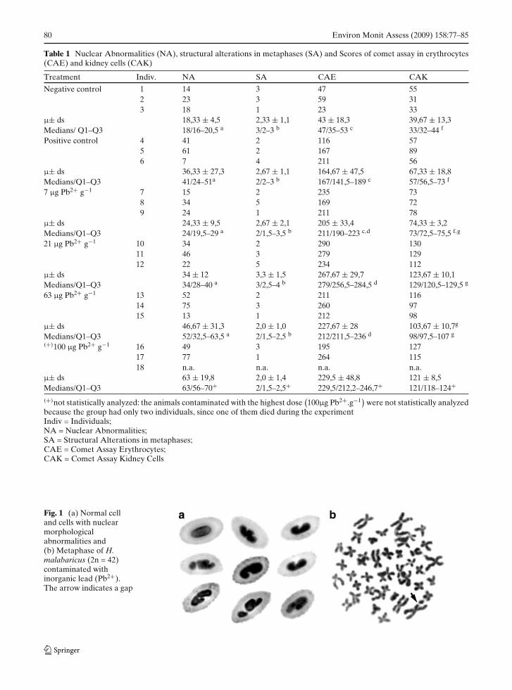
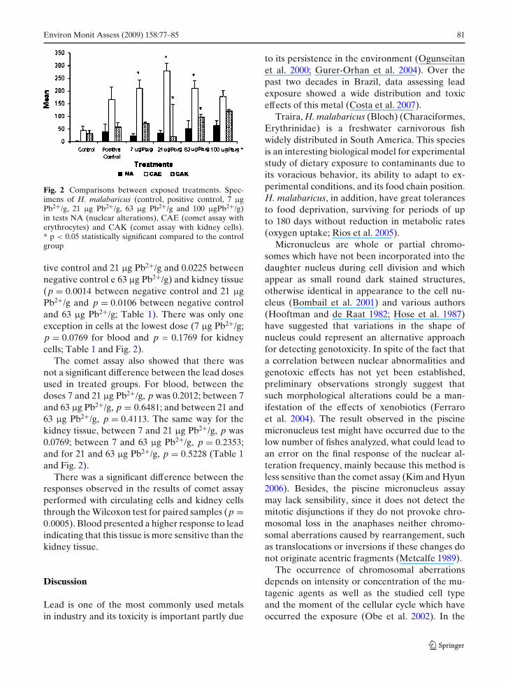
In the piscine micronucleus assay there wereno micronuclei, only nuclear morphological al-terations (Fig. 1a) and after the analysis of 50metaphases of each specimen of H. malabaricus,some structural alterations were found, such asgaps (Fig. 1b). However, the results, after statis-tical analysis, were not significant for both tests(p = 0.6755 in micronucleus test e p = 0.8294 instructural alterations in metaphases; Table 1 andFig. 2).
The comet assays of erythrocytes and kidneycells performed with 30% hydrogen peroxide ap-plication 1 h before sacrifice, initially proposedas positive controls, showed higher damages thanthe negative control groups for both cell types.However, the differences were not statisticallysignificant for neither of the analyzed tissues (p =0.3153 for blood and p = 0.2733 for kidney cells;Table 1).
We also verified that inorganic lead damagesboth cell types, because the results of controlfishes were statistically different than treatedfishes, either for blood (p = 0.019 between nega-
80 Environ Monit Assess (2009) 158:77–85
Table 1 Nuclear Abnormalities (NA), structural alterations in metaphases (SA) and Scores of comet assay in erythrocytes(CAE) and kidney cells (CAK)
Treatment Indiv. NA SA CAE CAK
Negative control 1 14 3 47 552 23 3 59 313 18 1 23 33
μ± ds 18,33 ± 4,5 2,33 ± 1,1 43 ± 18,3 39,67 ± 13,3Medians/ Q1–Q3 18/16–20,5 a 3/2–3 b 47/35–53 c 33/32–44 f
Positive control 4 41 2 116 575 61 2 167 896 7 4 211 56
μ± ds 36,33 ± 27,3 2,67 ± 1,1 164,67 ± 47,5 67,33 ± 18,8Medians/Q1–Q3 41/24–51a 2/2–3 b 167/141,5–189 c 57/56,5–73 f
7 μg Pb2+ g−1 7 15 2 235 738 34 5 169 729 24 1 211 78
μ± ds 24,33 ± 9,5 2,67 ± 2,1 205 ± 33,4 74,33 ± 3,2Medians/Q1–Q3 24/19,5–29 a 2/1,5–3,5 b 211/190–223 c,d 73/72,5–75,5 f,g
21 μg Pb2+ g−1 10 34 2 290 13011 46 3 279 12912 22 5 234 112
μ± ds 34 ± 12 3,3 ± 1,5 267,67 ± 29,7 123,67 ± 10,1Medians/Q1–Q3 34/28–40 a 3/2,5–4 b 279/256,5–284,5 d 129/120,5–129,5 g
63 μg Pb2+ g−1 13 52 2 211 11614 75 3 260 9715 13 1 212 98
μ± ds 46,67 ± 31,3 2,0 ± 1,0 227,67 ± 28 103,67 ± 10,7g
Medians/Q1–Q3 52/32,5–63,5 a 2/1,5–2,5 b 212/211,5–236 d 98/97,5–107 g
(+)100 μg Pb2+ g−1 16 49 3 195 12717 77 1 264 11518 n.a. n.a. n.a. n.a.
μ± ds 63 ± 19,8 2,0 ± 1,4 229,5 ± 48,8 121 ± 8,5Medians/Q1–Q3 63/56–70+ 2/1,5–2,5+ 229,5/212,2–246,7+ 121/118–124+
(+)not statistically analyzed: the animals contaminated with the highest dose(100μg Pb2+.g−1
)were not statistically analyzed
because the group had only two individuals, since one of them died during the experimentIndiv = Individuals;NA = Nuclear Abnormalities;SA = Structural Alterations in metaphases;CAE = Comet Assay Erythrocytes;CAK = Comet Assay Kidney Cells
Fig. 1 (a) Normal celland cells with nuclearmorphologicalabnormalities and(b) Metaphase of H.malabaricus (2n = 42)contaminated withinorganic lead (Pb2+).The arrow indicates a gap
Environ Monit Assess (2009) 158:77–85 81
Fig. 2 Comparisons between exposed treatments. Spec-imens of H. malabaricus (control, positive control, 7 μgPb2+/g, 21 μg Pb2+/g, 63 μg Pb2+/g and 100 μgPb2+/g)in tests NA (nuclear alterations), CAE (comet assay witherythrocytes) and CAK (comet assay with kidney cells).* p < 0.05 statistically significant compared to the controlgroup
tive control and 21 μg Pb2+/g and 0.0225 betweennegative control e 63 μg Pb2+/g) and kidney tissue(p = 0.0014 between negative control and 21 μgPb2+/g and p = 0.0106 between negative controland 63 μg Pb2+/g; Table 1). There was only oneexception in cells at the lowest dose (7 μg Pb2+/g;p = 0.0769 for blood and p = 0.1769 for kidneycells; Table 1 and Fig. 2).
The comet assay also showed that there wasnot a significant difference between the lead dosesused in treated groups. For blood, between thedoses 7 and 21 μg Pb2+/g, p was 0.2012; between 7and 63 μg Pb2+/g, p = 0.6481; and between 21 and63 μg Pb2+/g, p = 0.4113. The same way for thekidney tissue, between 7 and 21 μg Pb2+/g, p was0.0769; between 7 and 63 μg Pb2+/g, p = 0.2353;and for 21 and 63 μg Pb2+/g, p = 0.5228 (Table 1and Fig. 2).
There was a significant difference between theresponses observed in the results of comet assayperformed with circulating cells and kidney cellsthrough the Wilcoxon test for paired samples (p =0.0005). Blood presented a higher response to leadindicating that this tissue is more sensitive than thekidney tissue.
Discussion
Lead is one of the most commonly used metalsin industry and its toxicity is important partly due
to its persistence in the environment (Ogunseitanet al. 2000; Gurer-Orhan et al. 2004). Over thepast two decades in Brazil, data assessing leadexposure showed a wide distribution and toxiceffects of this metal (Costa et al. 2007).
Traira, H. malabaricus (Bloch) (Characiformes,Erythrinidae) is a freshwater carnivorous fishwidely distributed in South America. This speciesis an interesting biological model for experimentalstudy of dietary exposure to contaminants due toits voracious behavior, its ability to adapt to ex-perimental conditions, and its food chain position.H. malabaricus, in addition, have great tolerancesto food deprivation, surviving for periods of upto 180 days without reduction in metabolic rates(oxygen uptake; Rios et al. 2005).
Micronucleus are whole or partial chromo-somes which have not been incorporated into thedaughter nucleus during cell division and whichappear as small round dark stained structures,otherwise identical in appearance to the cell nu-cleus (Bombail et al. 2001) and various authors(Hooftman and de Raat 1982; Hose et al. 1987)have suggested that variations in the shape ofnucleus could represent an alternative approachfor detecting genotoxicity. In spite of the fact thata correlation between nuclear abnormalities andgenotoxic effects has not yet been established,preliminary observations strongly suggest thatsuch morphological alterations could be a man-ifestation of the effects of xenobiotics (Ferraroet al. 2004). The result observed in the piscinemicronucleus test might have occurred due to thelow number of fishes analyzed, what could lead toan error on the final response of the nuclear al-teration frequency, mainly because this method isless sensitive than the comet assay (Kim and Hyun2006). Besides, the piscine micronucleus assaymay lack sensibility, since it does not detect themitotic disjunctions if they do not provoke chro-mosomal loss in the anaphases neither chromo-somal aberrations caused by rearrangement, suchas translocations or inversions if these changes donot originate acentric fragments (Metcalfe 1989).
The occurrence of chromosomal aberrationsdepends on intensity or concentration of the mu-tagenic agents as well as the studied cell typeand the moment of the cellular cycle which haveoccurred the exposure (Obe et al. 2002). In the
82 Environ Monit Assess (2009) 158:77–85
present report, after the analysis of 50 metaphasesof each specimen of H. malabaricus, some littlestructural alterations were found, such as gaps.However, the results, after statistical analysis,were not significant. This way, the absence ofchromosomal aberrations may be related to theshort exposure time (96 h), not enough to causechromosomal damages in the proliferating cells.
Ferraro et al. (2004), working with sub-chronictrophic contamination (13 trophic doses in a pe-riod of 60 days) at the concentration of 21 μgPb2+/g in H. malabaricus, verified an increase inthe frequency of nuclear alterations, chromosomalaberrations and also a significant increase of tailednucleoids in the erythrocytes of fishes treated withPb2+, showing again that extended exposures tocontaminants made of lead are capable of origi-nating damages in the genetic material of fishes.Besides, Cipriano et al. (2004), studying the fre-quency of chromosomal aberrations in Astyanaxsp (lambaris) exposed to TBT (tributyltin) atthe doses of 0.3 mg/kg for 19 and 37 days, ob-served different chromosomal anomalies such asbreakage of one or two chromatids and acentricfragments.
Cytogenetic markers such as chromosomalaberrations, micronuclei and sister chromatid ex-changes are among the most extensively usedmarkers of early biological effects of DNA dam-aging agents. During the last few years, the singlecell gel electrophoresis (SCGE) or comet assaywas introduced as a useful technique for bio-monitoring studies. While biomonitoring studiesemploying cytogenetic techniques are limited tocirculating lymphocytes and involve proliferatingcell populations, the comet assay can be appliedto proliferating and non-proliferating cells andcells of those tissues which are the first sites ofcontact with mutagenic/carcinogenic substances.The difference between effects in comet assay andcytogenetic tests is basically due to variations inthe type of DNA alterations that the test systemsdetect: the comet assay detects repairable DNAlesions or alkali-labile sites while cytogenetic tests(MN and CA) detect fixed mutations which persistat least one mitotic cycle (Kassie et al. 2000).
Several studies show that the comet assay isreally capable of detecting DNA damages caused
by different classes of mutagenic contaminants infishes, attributing high sensibility to the test. Wemay indicate Pandrangi et al. (1995), who showedan increase of the DNA damage in erythrocytes ofbullheads (Ameiurus nebulosus) and carp (Cypri-nus carpio) after exposure to cyclophosphamideand Devaux et al. (1997), who also verified anincrease on the length of the nucleoid tail after theexposure of Onchorynchus mykiss hepatocytes tobenzopyrene and hydrogen peroxide. Thus, thecomet assay is an important tool for monitoringstudies because it shows the genotoxicity of the ex-posure. The answer may, of course, depend on theexperimental conditions of the specie, cell type,mutagenic agent and exposure time (Belpaemeet al. 1998).
The comet assays of erythrocytes and kidneycells performed with 30% hydrogen peroxide ap-plication 1 hour before sacrifice were not sta-tistically significant for neither of the analyzedtissues (p = 0.1936 for blood and p = 0.2211 forkidney cells). This result was partially expectedbecause, despite the high concentration of theagent, the exposure time was very low, considerednot enough to cause damages on the DNA of theanalyzed cells.
We also verified that inorganic lead damagesboth cell types, because the results of controlfishes were statistically different than treatedfishes, either blood (p = 0.01) and kidney tissue(p = 0.01; Table 1). There was one exception inkidney cells at the lowest dose (7 μg Pb2+/g)(p = 0.14) probably due to the short time ex-posure (96 h) and also the low quantity of thecontaminant.
While micronucleated erythrocytes from thehemopoietical organs reflect a genotoxic damagewhich occurred during a time equivalent to thecell cycle, those from the peripheral circulationreflect events that occurred in a time equal to thelifespan of the circulating erythrocytes. Therefore,the application of the micronucleus test on pe-ripheral blood samples is particularly indicated forconditions of chronic exposure. The assay needsthat the cell population undergoes mitosis, sothe duration of the cell cycle should be known(Udroiu 2006). Processes of erythrocyte formationin the fish hemopoietic tissue are submitted to
Environ Monit Assess (2009) 158:77–85 83
pronounced seasonal fluctuations. No general reg-ularities have been revealed. The obtained dataare rather species-specific (Soldatov 2005).
Although the literature shows that this metalis capable of causing damages in the proteins ofDNA structural maintenance or in those involvedwith repair mechanisms (Merian 1991; Pain 1995;Hartwig et al. 2002), our results in the piscine mi-cronucleus assay and evaluation of chromosomalaberration frequency did not indicate contamina-tion, different than the results in the comet assays,which showed differences between control andcontaminated groups for blood and kidney cells.
According to Boelsterli (2002), lead inducesDNA synthesis in a number of cells. Evidence in-dicates that inorganic lead interferes with cellularsignal transduction pathways, in particular withmembers of the calcium-dependent protein kinaseC (PKC) family. These kinases play an importantrole in cell proliferation. The mechanism is not en-tirely clear, but evidence suggests that Pb2+ causesactivation of PKC-mediated pathways by facili-tating the translocation of PKC from the cytosolto the plasma membrane (perhaps by mimickingCa2+). This kinase activation greatly stimulatescell cycle progression.
The whole S phase (DNA replication) of thecell cycle in fresh water fishes lasts near 14 h(Almeida Toledo et al. 1988). Thus, we believethat after 96 h of Pb2+ exposure the decreaseof damage in blood cells in the highest doses,even not statistically significant (the dose 21 μgPb2+/g caused a little bit more damage to theblood cells than the dose 63 μg Pb2+/g), was due tothe induction of the cell cycle by Pb2. The highestPb2 doses could have renewed blood cells morerapidly causing, thus, lower damages.
There was a significant difference between theresponses observed in the results of comet assayperformed with circulating cells and kidney cells.Blood presented a higher response to lead indi-cating that this tissue is more sensitive than thekidney tissue, probably due to the acute contami-nation performed which had immediate responsesfirst detected in the erythrocytes.
Biomonitoring studies with a combination ofcytogenetic tests and comet assay enable com-parison of the relative sensitivity of the two test
systems and may also give a clue about the frac-tion of DNA damage detected in the comet assaythat will lead to fixed mutations (Kassie et al.2000). However, it is important to remember thatthe comet assay performed with peripheral bloodis the most indicated method to analyze the acutecontamination by inorganic lead because it is ableto detect significant differences between controland treated groups even in very low doses (7 μgPb2+/g). This results show that the comet assay, incirculating cells, detects the mutagenic activity ofPb2+ even in low doses and for a short contami-nation period. Despite the low size of our samplesfor the experiments, the results may already showthat low doses of Pb2+ cause alterations in thefish DNA as well as point the best method todetect this genotoxicity (comet assay with blood).However, more studies are needed to verify thegenotoxic damages of Pb2+ for extended timeperiods.
Acknowledgements The authors thank CNPq (ConselhoNacional de Desenvolvimento Científico e Tecnológico)and Aquatóxi group. This research was supported by(CAPES), Coordenação de Aperfeiçoamento de Pessoalde Nível Superior (Brazil).
References
Akaishi, F. M., Silva de Assis, H. C., Jakobi, S. C. G., Eiras-Stofella, D. R., ST-Jean, S. D., Courtenay, S. C., et al.(2004). Morphological and neurotoxicological findingsin tropical freshwater fish (Astyanax sp.) after water-borne and acute exposure to water soluble fraction(WSF) of crude oil. Archives of Environmental Con-tamination and Toxicology, 46, 244–253.
Almeida Toledo, L. F., Viegas-Péquignot, E., Foresti, F.,Toledo Filho, S. A., & Dutrilaux, B. (1988). BrdUreplication patterns demonstrating chromosome ho-moeologies in two fish species, genus Eigenman-nia. Cytogenetics and Cell Genetics, 48, 117–120.doi:10.1159/000132603.
Al-Sabti, K. (1986). Clastogenic effects of live carcinogenic-mutagenic chemicals on the cells of the common carp(Cyprinus carpio L.). Comparative Biochemistry andPhysiology, 85(Part C), 5–9.
Belpaeme, K., Cooreman, K., & Kirsch-Volders, M. (1998).Development and validation of the in vivo alkalinecomet assay for detecting genomic damage in marineflatfish. Mutation Research, 415(3), 167–184.
Boelsterli, U. A. (2002). Mechanistic toxicology. The mole-cular basis of how chemicals disrupt biological targets.London: Taylor and Francis.
84 Environ Monit Assess (2009) 158:77–85
Bombail, V., Aw, D., Gordon, E., & Batty, J. (2001).Application of the comet and micronucleus assaysto butterfish (Pholis gunnellus) erythrocytes from theFirth of Forth, Scotland. Chemosphere, 44, 383–392.doi:10.1016/S0045-6535(00)00300-3.
BRASIL (1986). Resolução do CONAMA No. 20, de 18 dejunho de 1986. Diário Oficial da República Federativado Brasil. Brasília, pp. 72–89.
Burdena, V. M., Sandheinrich, M. B., & Caldwell, C. A.(1998). Effects of lead on the growth and 6-aminolevulinic acid dehydratase activity of juvenilerainbow trout, Oncorhynchus mykiss. EnvironmentalPollution, 101, 285–289. doi:10.1016/S0269-7491(98)00029-3 .
Carrasco, K. R., Tilbury, K. L., & Myers, M. S. (1990).Assessment of the piscine micronucleus test as in situbiological indicator of chemical contaminant effects.Canadian Journal of Fisheries and Aquatic Sciences,47, 2123–2136. doi:10.1139/f90-237.
Cestari, M. M., Lemos, P. M. M., Oliveira - Ribeiro,C. A., Costa, J. R. M. A., Pelletier, E., Ferraro,M. V. M., et al. (2004). Genetic damage inducedby trophic doses of lead in the neotropical fish Ho-plias malabaricus (Characiformes, Erythrinidae) asreveled by the comet assay and chromosomal aberra-tions. Genetics and Molecular Biology, 27(2), 270–274.doi:10.1590/S1415-47572004000200023.
Cipriano, R. R., Oliveira-Ribeiro, C. A., Cestari, M. M., &Fenocchio, A. S. (2004). Evaluation of the effects oftributiltin (TBT) on chromosomes of the neotropicalfish Astyanax sp. (Pisces, Tetragonopterinae). Cytolo-gia, 69(2), 187–190. doi:10.1508/cytologia.69.187
Costa, J. R. M., Mela, M., Assis, H. C. S., Pelletier, É.,Randi, M. A. F., & Oliveira-Ribeiro, C. A. (2007).Enzymatic inhibition and morphological changes inHoplias malabaricus from dietary exposure to lead (II)or methylmercury. Ecotoxicology and EnvironmentalSafety, 67, 82–88. doi:10.1016/j.ecoenv.2006.03.013.
Devaux, A., Pesonen, M., & Monod, G. (1997). Al-kaline comet assay in rainbow trout hepatocytes.Toxicology In Vitro, 11, 71–79. doi:10.1016/S0887-2333(97)00004-0 .
Fenocchio, A. S., & Bertollo, L. A. C. (1988). A sim-ple method for fresh-water fish lymphocyte culture.Brazilian Journal of Genetics, 11(4), 847–852.
Fenocchio, A. S., Venere, P. C., Cesar, A. C. G., Dias, A.L., & Bertollo, L. A. C. (1991). Short term culturefrom solid tissues of fishes. Caryologia, 44, 161–166.
Ferraro, M. V., Fenocchio, A. S., Mantovani, M. S.,Oliveira-Ribeiro, C. A., & Cestari, M. M. (2004).Mutagenic effects of tributyltin (TBT) and inor-ganic lead (PbII) on the fish H. malabaricus asevaluated using the comet assay, piscine micronu-cleus and chromosome aberrations tests. Genetics andMolecular Biology, 27(1), 103–107. doi:10.1590/S1415-47572004000100017.
Gurer-Orhan, H., Sabýr, H. U., & Özgünes, H. (2004).Correlation between clinical indicators of lead poi-soning and oxidative stress parameters in controlsand lead-exposed workers. Toxicology, 195, 147–154.doi:10.1016/j.tox.2003.09.009.
Hartwig, A., Asmuss, M., Blessing, H., Hoffmann, S.,Jahnke, G., Khandelwal, S., et al. (2002). Interfer-ence by toxic metal ions with zinc-dependent pro-teins involved in maintaining genomic stability. Foodand Chemical Toxicology, 40, 1179–1184. doi:10.1016/S0278-6915(02)00043-1 .
Heddle, J. A. (1973). A rapid in vivo test for chro-mosome damage. Mutation Research, 18, 187–192.doi:10.1016/0027-5107(73)90035-3.
Hooftman, R. N., & de Raat, W. K. (1982). Induction of nu-clear anomalies (micronuclei) in the peripheral blooderythrocytes of the eastern mudminnow Umbra pyg-mea by ethyl methanesulphonate. Mutation Research,104, 147–152. doi:10.1016/0165-7992(82)90136-1.
Hose, J. E., Cross, J. N., Smith, S. G., & Diehl, D.(1987). Elevated circulating erythrocyte micronucleiin fishes from contaminated of southern California.Marine Environmental Research, 22, 167–176. doi:10.1016/0141-1136(87)90034-1 .
Johnson, F. M. (1998). The genetic effects of environ-mental lead. Mutation Research/Reviews in MutationResearch, 410, 123–140. doi:10.1016/S1383-5742(97)00032-X .
Kassie, F., Parzefall, W., & Knasmüller, S. (2000) Singlecell gel electrophoresis assay: A new technique forhuman biomonitoring studies. Mutation Research, 463,13–31.
Kim, I. Y., & Hyun, C. K. (2006). Comparative evaluationof the alkaline comet assay with the micronucleus testfor genotoxicity monitoring using aquatic organisms.Ecotoxicology and Environmental Safety, 64, 288–297.doi:10.1016/j.ecoenv.2005.05.019.
Kobayashi, H., Sugiyama, C., Morikawa, Y., Hayashi, M.,& Sofuni, T. (1995) A comparison between manualmicroscopic analysis and computerized image anal-ysis in the single cell gel electrophoresis. Mam-malian Mutagenicity Study Group Communications, 3,103–115.
McCarthy, J. F., & Shugart, L. R. (1990). Biomarkers ofenvironmental contamination. Boca Raton: Lewis.
Merian, E. (Ed.) (1991). Metals and their compounds inthe environment: Occurrence, analysis and biologicalrelevance. New York: VCH.
Metcalfe, C. D. (1989). Testes for predicting carcinogenic-ity in fish. CRC Critical Reviews in Aquatic Sciences, 1,111–129.
Mitchelmore, C. L., Chipman, J. K. (1998). DNA strandbreakage in aquatic organisms and the potential valueof the comet assay in environmental monitoring.Mutation Research—Fundamental and MolecularMechanisms of Mutagenesis, 399, 135–147.
Obe, G., Pfeiffer, P., Savage, J. R. K., Johannes, C.,Goedecke, W., Jeppesen, P., et al. (2002). Chromoso-mal aberrations: formation, identification and distribu-tion. Mutation Research—Fundamental and MolecularMechanisms of Mutagenesis, 504, 17–36.
Ogunseitan, O. A., Yang, S., & Ericson, J. (2000).Microbial δ-aminolevulinate dehydratase as a biosen-sor of lead bioavailability in contaminated environ-ments. Soil Biology & Biochemistry, 32, 1899–1906.doi:10.1016/S0038-0717(00)00164-4.
Environ Monit Assess (2009) 158:77–85 85
Pain, D. J. (1995). Lead in the environment. In D. J.Hoffman, B. A. Rattner, G. A. Burton & J. Cairns(Eds.), Handbook of ecotoxicology (pp. 356–391).Boca Raton: Lewis.
Pandrangi, R., Petras, M., Ralph, S., & Vrzoc, M. (1995).Alkaline single cell gel (comet): Assay and geno-toxicity monitoring using bullheads and carp. Envi-ronmental and Molecular Mutagenesis, 26, 345–356.doi:10.1002/em.2850260411.
Rabitto, I. S., Alves Costa, J. R. M., Silva de Assis, H.C., Pelletier, E., Akaishi, F. M., Anjos, A., et al.(2005). Effects of dietary Pb(II) and tributyltin onneotropical fish, Hoplias malabaricus: histopathologi-cal and biochemical findings. Ecotoxicology and Envi-ronmental Safety, 60(2), 147–156. doi:10.1016/j.ecoenv.2004.03.002.
Rios, F. S., Oba, E. T., Fernandes, M. N., Kalinin, A. L.,& Rantin, F. T. (2005). Erythrocyte senescence andhaematological changes induced by starvation in theneotropical fish traíra, Hoplias malabaricus (Characi-formes, Erythrinidae). Comparative Biochemistry andPhysiology Part A, 140, 281–287. doi:10.1016/j.cbpb.2004.12.006.
Schmid, W. (1975). The micronucleus test. MutationResearch, 31, 9–15.
Soldatov, A. A. (2005). Peculiarities of organization andfunctioning of the fish red blood system. Journalof Evolutionary Biochemistry and Physiology, 41(3),272–281. doi:10.1007/s10893-005-0060-0.
Speit, G., & Hartmann, A. (1999) The comet assay (single-cell gel test), a sensitive test for the detection of DNAdamage and repair. In D. S. Henderson (Ed.), Me-thods in molecular biology: DNA repair protocols—eukaryotic systems (pp. 203–211). Totowa: Human.
Tao, S., Liu, C., Dawson, R., Cao, J., & Li, B. (1999).Uptake of particulate lead via the gills of fish (Caras-sius auratus). Archives of Environmental Contam-ination and Toxicology, 37, 352–357. doi:10.1007/s002449900524 .
Udroiu, I. (2006). The micronucleus test in piscine erythro-cytes. Aquatic Toxicology (Amsterdam, Netherlands),79, 201–204. doi:10.1016/j.aquatox.2006.06.013.
Van Der Oost, R., Beyer, J., & Vermeulen, N. P. E. (2003).Fish bioaccumulation and biomarkers in environmen-tal risk assessment: a review. Environmental Toxi-cology and Pharmacology, 13, 57–149. doi:10.1016/S1382-6689(02)00126-6 .
Vighi, M. (1981). Lead uptake and release in a experimen-tal trophic chain. Ecotoxicology and EnvironmentalSafety, 5, 177–193. doi:10.1016/0147-6513(81)90033-6.


![Mercury heavy-metal-induced physiochemical changes and genotoxic alterations in water hyacinths [Eichhornia crassipes (Mart.)]](https://img.pdfslide.net/doc/110x75/63561fa186104ea1570c08be/mercury-heavy-metal-induced-physiochemical-changes-and-genotoxic-alterations-in.jpg)


