Embed Size (px)
Citation preview
RESEARCH ARTICLE
Gestational Age and Neonatal BrainMicrostructure in Term Born Infants: ABirth Cohort StudyBirit F. P. Broekman1,3, Changqing Wang2, Yue Li2, Anne Rifkin-Graboi3,Seang Mei Saw4, Yap-Seng Chong5, Kenneth Kwek6, Peter D. Gluckman3,7,Marielle V. Fortier8, Michael J. Meaney3,9, Anqi Qiu2,3,10*,for the GUSTO Study Group"
1. Department of Psychological Medicine, Yong Loo Lin School of Medicine, National University of Singapore,National University Health System, Singapore, Singapore, 2. Department of Biomedical Engineering, NationalUniversity of Singapore, Singapore, Singapore, 3. Singapore Institute for Clinical Sciences, the Agency forScience, Technology and Research, Singapore, Singapore, 4. Saw Swee Hock School of Public Health,National University of Singapore, Singapore, Singapore, 5. Department of Obstetrics & Gynaecology, YongLoo Lin School of Medicine, National University of Singapore, National University Health System, Singapore,Singapore, 6. Department of Maternal Fetal Medicine, KK Women’s and Children’s Hospital, Singapore,Singapore, 7. Liggins Institute, University of Auckland, Auckland, New Zealand, 8. Department of Diagnosticand Interventional Imaging, KK Women’s and Children’s Hospital, Singapore, Singapore, 9. Departments ofPsychiatry and Neurology & Neurosurgery, McGill University, Montreal, Canada, 10. Clinical ImagingResearch Centre, National University of Singapore, Singapore, Singapore
" Membership of the GUSTO Study Group is provided in the Acknowledgments.
Abstract
Objective: Understanding healthy brain development in utero is crucial in order to
detect abnormal developmental trajectories due to developmental disorders.
However, in most studies neuroimaging was done after a significant postnatal
period, and in those studies that performed neuroimaging on fetuses, the quality of
data has been affected due to complications of scanning during pregnancy. To
understand healthy brain development between 37–41 weeks of gestational age,
our study assessed the in utero growth of the brain in healthy term born babies with
DTI scanning soon after birth.
Methods: A cohort of 93 infants recruited from maternity hospitals in Singapore
underwent diffusion tensor imaging between 5 to 17 days after birth. We did a
cross-sectional examination of white matter microstructure of the brain among
healthy term infants as a function of gestational age via voxel-based analysis on
fractional anisotropy.
Results: Greater gestational age at birth in term infants was associated with larger
fractional anisotropy values in early developing brain regions, when corrected for
age at scan. Specifically, it was associated with a cluster located at the corpus
OPEN ACCESS
Citation: Broekman BFP, Wang C, Li Y, Rifkin-Graboi A, Saw SM, et al. (2014) Gestational Ageand Neonatal Brain Microstructure in Term BornInfants: A Birth Cohort Study. PLoS ONE 9(12):e115229. doi:10.1371/journal.pone.0115229
Editor: Nouchine Hadjikhani, Harvard MedicalSchool, United States of America
Received: May 4, 2014
Accepted: November 20, 2014
Published: December 23, 2014
Copyright: � 2014 Broekman et al. This is anopen-access article distributed under the terms ofthe Creative Commons Attribution License, whichpermits unrestricted use, distribution, and repro-duction in any medium, provided the original authorand source are credited.
Data Availability: The authors confirm that all dataunderlying the findings are fully available withoutrestriction. All relevant data are provided within thepaper. All additional data not yet covered in thepaper are available from the GUSTO executivecommittee at the Singapore Institute for ClinicalSciences, and can be requested through Anqi Qiu,Department of Biomedical Engineering NationalUniversity of Singapore, email: [email protected],who is also a member of the GUSTO executivecommittee.
Funding: This study is supported by NationalMedical Research Council (NMRC; NMRC/TCR/004-NUS/2008, NMRC/CBRG/0039/2013), theYoung Investigator Award at the National Universityof Singapore (NUSYIA FY10 P07), the NationalUniversity of Singapore MOE AcRF Tier 1, andSingapore Ministry of Education AcademicResearch Fund Tier 2 (MOE2012-T2-2-130). Thefunding agencies did not play any role in studydesign, data collection and analysis, decision topublish, or preparation of the manuscript.
Competing Interests: The authors have declaredthat no competing interests exist.
PLOS ONE | DOI:10.1371/journal.pone.0115229 December 23, 2014 1 / 17
callosum (corrected p,0.001), as well as another cluster spanning areas of the
anterior corona radiata, anterior limb of internal capsule, and external capsule
(corrected p,0.001).
Conclusions: Our findings show variation in brain maturation associated with
gestational age amongst ‘term’ infants, with increased brain maturation when born
with a relatively higher gestational age in comparison to those infants born with a
relatively younger gestational age. Future studies should explore if these
differences in brain maturation between 37 and 41 weeks of gestational age will
persist over time due to development outside the womb.
Introduction
Understanding healthy fetal brain development is essential to be able to detect
abnormal developmental trajectories due to developmental disorders [1]. Previous
studies showed that younger gestational age at birth has been associated with
reduced maturation of the brain. However, current knowledge derives largely
from studies with premature infants [2–8], which do not inform us about healthy
brain development in term born infants. Second, different methods of scanning
have been used. Most of the existing literature relies upon ultrasound or
traditional structural magnetic resonance imaging (MRI). However, Ment et al.
(2009) pointed out in a review article that abnormal neurodevelopmental
trajectories associate with microstructural abnormalities in the brains, especially
white matter abnormalities such as diffuse injury of white matter with neuronal
and axonal disruption [9]. Nevertheless, microstructure development is best
measured by diffusion tensor imaging (DTI) [10], with white matter maturation
characterized by increasing fractional anisotropy (FA) and decreasing mean
diffusivity (MD) [11]. Third, neuroimaging is often done after a significant
postnatal period. The majority of existing studies with term infants used case-
control designs and compared term infants to late preterm infants (34–37 weeks),
preterm (32–34 weeks) and/or very preterm infants (gestational age at birth ,32
weeks) [2–6], scanned at term-equivalent age, after a significant postnatal period.
However, the days from birth to imaging can account for potential postnatal brain
growth with many ex-uterine environmental factors will influence neuronal
connectivity [12]. Indeed, a recent study showed the extraordinary rates of
structural growth in the very early postnatal period [13]. As such imaging after a
significant postnatal period will not inform us much about the in utero brain
development and it remains unclear if altered neuroconnectivitity emerges
prenatally [14]. Hence, performing DTIs during the third trimester directly on
fetuses would be most ideal. Recently some studies used neuroimaging during
pregnancy to understand fetal brain development [1, 12–15]. However, the
understanding of maturation of fetal neural networks remains limited due to
many complications of fetal scanning during pregnancy such as difficulties to
Gestational Age and Brain Development
PLOS ONE | DOI:10.1371/journal.pone.0115229 December 23, 2014 2 / 17
recruit healthy pregnant women for DTI scans, the size of mothers during third
trimester, artifacts due to movements of the fetus and breathing of the mother,
and subsequently the long scanning duration (which is especially challenging for
women in the third trimester) [14, 16]. Hence, although during the third trimester
growth and neurogenesis of the fetal brain increase significantly, the direct effects
of a relatively younger gestational age at birth in healthy term infants on these
processes are not yet well understood [14, 17].
To understand the establishment of brain connections within healthy term born
infants, imaging soon after birth is essential to reduce the effects of postnatal brain
maturation. To the best of our knowledge there have been no DTI studies to date
that have examined differences brain maturation shortly after being born in
healthy term infants. The aim of this study is to investigate the differences in
in utero brain maturation between 37 and 41 weeks of gestational age in a large
cohort of healthy term infants in Singapore. We hypothesize that among healthy
term born infants, a younger gestational age at birth is associated with reduced
white matter maturity in the brain in comparison to healthy term born infants
born with an older gestational age at birth.
Methods
Subjects
One hundred eighty nine infants who participated in a birth cohort study,
Growing Up in Singapore Towards Healthy Outcomes (GUSTO), also
participated in the neuroimaging study. The GUSTO methodology has been
published in detail [18]. The GUSTO cohort consisted of pregnant Asian women
attending the first trimester antenatal ultrasound scan clinic at the National
University Hospital (NUH) and KK Women’s and Children’s Hospital (KKH) in
Singapore, which are the two major maternity hospitals in Singapore. Birth
outcome and pregnancy measures were obtained from hospital records. The
pregnant women and their partners were Singapore citizens or Permanent
Residents of Chinese, Malay or Indian ethnic background. Socioeconomic status
(household income) and prenatal exposure to alcohol (regular alcohol drinking)
and tobacco (regular smoking, daily exposure to smoking at home and job) were
extracted from survey questionnaires conducted as a part of a scheduled
appointment during pregnancy. The GUSTO cohort study, including imaging
procedures, was approved by the National Healthcare Group Domain Specific
Review Board and the Sing Health Centralized Institutional Review Board. All
clinical investigation has been conducted according to the principles expressed in
the Declaration of Helsinki. Written consent was obtained from all guardians on
behalf of the children enrolled in the study.
The current study only included neonates with diffusion tensor imaging (DTI),
gestational age at birth greater or equal to 37 weeks, birth weight larger than
2500 g, and the last recorded APGAR$9, to exclude effects of intra-uterine
growth retardation or co-morbidities. Neonates of mothers with gestational
Gestational Age and Brain Development
PLOS ONE | DOI:10.1371/journal.pone.0115229 December 23, 2014 3 / 17
diabetes, hypertension, and hypoglycemia were excluded from this study, as were
neonates of mothers who reported consuming any alcohol during pregnancy.
MRI Acquisition
Neonates underwent fast spin-echo T2-weighted MRI and single-shot echo-planar
DTI scans using a 1.5-Tesla GE scanner at KKH’s Department of Diagnostic and
Interventional Imaging between 5 and 17 days postpartum. The scans were
acquired when subjects were sleeping in the scanner. No sedation was used and
precautions were taken to reduce exposure to MRI scanner noise. A neonatologist
was present during each scan. A pulse oximeter was used to monitor heart rate
and oxygen saturation through out the entire scans.
The DTI imaging protocol is based on single-shot echo-planar DTI sequence
(TR57000 ms; TE556 ms; flip angle590 , FOV5200 mm6200 mm; matrix
size564664). 40 to 50 axial slices with 3.0 mm thickness were acquired parallel
to the anterior–posterior commissure line. Nineteen diffusion weighted images
(DWIs) with b5600 sec/mm2 and 1 baseline with b50 sec/mm2 were obtained.
All brain scans were reviewed by a neuroradiologist (M.V.F).
DTI Analysis
Diffusion weighted images (DWIs) were first corrected within individual subjects
for motion and eddy current distortions using affine transformation to the image
without diffusion weighting. Six elements of the diffusion tensor were then
determined, from which fractional anisotropy (FA) was calculated using
multivariate least-square fitting.
We constructed a DTI atlas based on the unbiased diffeomorphic atlas
generation algorithm [19] and detailed the processing procedure below. The FA
image and the image without diffusion weighting of each subject were first aligned
to those of the JHU neonate brain single-subject DTI atlas (resolution:
0.660.660.6 mm3, http://lbam.med.jhmi.edu/) [20] via affine and nonlinear
large deformation diffeomorphic metric mapping (LDDMM) transformations
[21]. These affine and nonlinear transformations were then applied to individual
DWIs. The mean DWIs were obtained by averaging the DWIs with the
corresponding gradient direction across all the subjects. The mean FA image was
computed using the mean DWIs. These mean DWIs and FA were considered as a
new atlas. This procedure was repeated three times until the intensity of DWIs was
no longer changed. The mean DWIs and FA at the last iteration were defined as
the final atlas and used as a common anatomical space.
DWIs of individual subjects were then aligned to the atlas based the same
registration procedure as one used for the atlas generation. FA was obtained from
the tensor calculation of DWIs in the atlas and used for the following voxel-based
analysis.
Gestational Age and Brain Development
PLOS ONE | DOI:10.1371/journal.pone.0115229 December 23, 2014 4 / 17
Statistical Analysis
Voxel-based analysis was performed to investigate the association between
gestational age at birth and neonatal brain microstructure (FA) using SPM8. The
FA images were smoothed with a Gaussian kernel with full width half maximum
of 4 mm. Linear regression was performed conducted at every voxel in the white
matter region. The regression model included gestational age at birth as the main
factor and controlled for age at MRI. Most infants were scanned at the second
week after birth. Gestational age was highly correlated with the postmenstrual age-
at-scan (r50.966). As the aim of this study is to investigate the effects of
gestational age at birth on the neonatal brain to assess effects of fetal maturity, we
controlled for age at MRI (postmenstrual age-at-scan - gestational age at birth) in
the regression analysis.
We also adjusted for birth weight [22], maternal age [22], monthly household
income [22, 23], prenatal smoking exposure [22], [23], and ethnicity [24–26], on
the basis of previous knowledge that these variables are potential confounders of
younger gestational age. Family-wise error was computed for correcting multiple
comparisons at a significance level of 0.001.
Birth weight was adjusted based on gestational age using linear regression with
the mean-centered gestational age as a main factor on the GUSTO cohort. The
residual was defined as adjusted birth weight that is statistically independent of
gestational age. As small-for-gestational age has been identified as a risk factor for
prematurity, we included birth weight as a covariate [22]. The brain grows rapidly
in early life even in the first few weeks of life [27, 28] and hence it is crucial to
justify the days from birth to the MRI visit. Several studies have suggested
differences in brain morphology in different ethnic groups [24–26] and we thus
also included ethnicity in the regression model. Also other reproductive risk
factors have been identified, such as advanced maternal age and smoking, for
which we adjusted in our analyses [22]. Furthermore, previous studies showed
significant socioeconomic differences in preterm births, hence we have included
household income [22].
Results
Demographics
Among 189 neonates who underwent MRI scans, 93 were included in the current
study. We excluded 13 neonates with gestational age at birth less than 37 weeks, 11
with birth weight less than 2500 g, 2 with a 5-min Apgar score less than 9, and 52
with no DTI data. Moreover, our study also excluded infants whose mothers had
gestational diabetes (n522), hypertension (n55), and hypoglycemia (n51)
during pregnancy. Mothers who reported consuming any alcohol during
pregnancy (n53) were also excluded from this study. Hence, the total sample
involved in this study was 93. Table 1 lists the demographic information of this
study.
Gestational Age and Brain Development
PLOS ONE | DOI:10.1371/journal.pone.0115229 December 23, 2014 5 / 17
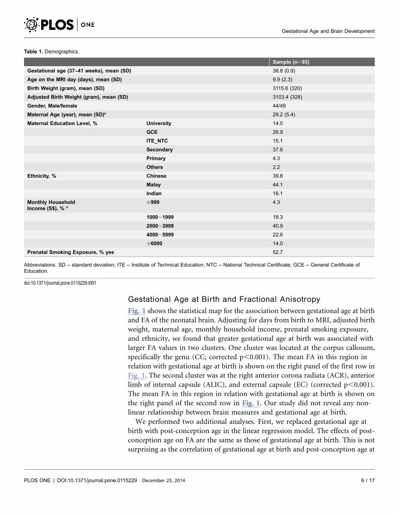
Gestational Age at Birth and Fractional Anisotropy
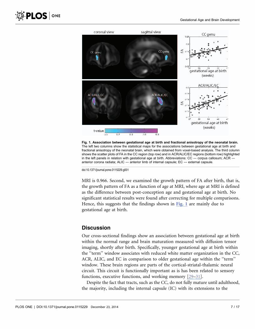
Fig. 1 shows the statistical map for the association between gestational age at birth
and FA of the neonatal brain. Adjusting for days from birth to MRI, adjusted birth
weight, maternal age, monthly household income, prenatal smoking exposure,
and ethnicity, we found that greater gestational age at birth was associated with
larger FA values in two clusters. One cluster was located at the corpus callosum,
specifically the genu (CC; corrected p,0.001). The mean FA in this region in
relation with gestational age at birth is shown on the right panel of the first row in
Fig. 1. The second cluster was at the right anterior corona radiata (ACR), anterior
limb of internal capsule (ALIC), and external capsule (EC) (corrected p,0.001).
The mean FA in this region in relation with gestational age at birth is shown on
the right panel of the second row in Fig. 1. Our study did not reveal any non-
linear relationship between brain measures and gestational age at birth.
We performed two additional analyses. First, we replaced gestational age at
birth with post-conception age in the linear regression model. The effects of post-
conception age on FA are the same as those of gestational age at birth. This is not
surprising as the correlation of gestational age at birth and post-conception age at
Table 1. Demographics.
Sample (n593)
Gestational age (37–41 weeks), mean (SD) 38.8 (0.9)
Age on the MRI day (days), mean (SD) 9.9 (2.3)
Birth Weight (gram), mean (SD) 3115.6 (320)
Adjusted Birth Weight (gram), mean (SD) 3103.4 (328)
Gender, Male/female 44/49
Maternal Age (year), mean (SD)* 29.2 (5.4)
Maternal Education Level, % University 14.0
GCE 26.9
ITE_NTC 15.1
Secondary 37.6
Primary 4.3
Others 2.2
Ethnicity, % Chinese 39.8
Malay 44.1
Indian 16.1
Monthly HouseholdIncome (S$), % *
#999 4.3
1000,1999 18.3
2000,3999 40.9
4000,5999 22.6
$6000 14.0
Prenatal Smoking Exposure, % yes 52.7
Abbreviations. SD – standard deviation; ITE – Institute of Technical Education; NTC – National Technical Certificate; GCE – General Certificate ofEducation.
doi:10.1371/journal.pone.0115229.t001
Gestational Age and Brain Development
PLOS ONE | DOI:10.1371/journal.pone.0115229 December 23, 2014 6 / 17
MRI is 0.966. Second, we examined the growth pattern of FA after birth, that is,
the growth pattern of FA as a function of age at MRI, where age at MRI is defined
as the difference between post-conception age and gestational age at birth. No
significant statistical results were found after correcting for multiple comparisons.
Hence, this suggests that the findings shown in Fig. 1 are mainly due to
gestational age at birth.
Discussion
Our cross-sectional findings show an association between gestational age at birth
within the normal range and brain maturation measured with diffusion tensor
imaging, shortly after birth. Specifically, younger gestational age at birth within
the ‘‘term’’ window associates with reduced white matter organization in the CC,
ACR, ALIC, and EC in comparison to older gestational age within the ‘‘term’’
window. These brain regions are parts of the cortical-striatal-thalamic neural
circuit. This circuit is functionally important as is has been related to sensory
functions, executive functions, and working memory [29–31].
Despite the fact that tracts, such as the CC, do not fully mature until adulthood,
the majority, including the internal capsule (IC) with its extensions to the
Fig. 1. Association between gestational age at birth and fractional anisotropy of the neonatal brain.The left two columns show the statistical maps for the associations between gestational age at birth andfractional anisotropy of the neonatal brain, which were obtained from voxel-based analysis. The third columnshows the scatter plots of FA in the CC region (top row) and in ACR/ALIC/EC regions (bottom row) highlightedin the left panels in relation with gestational age at birth. Abbreviations: CC --- corpus callosum; ACR ---anterior corona radiata; ALIC --- anterior limb of internal capsule; EC --- external capsule.
doi:10.1371/journal.pone.0115229.g001
Gestational Age and Brain Development
PLOS ONE | DOI:10.1371/journal.pone.0115229 December 23, 2014 7 / 17
thalamus and basal ganglia, are in the limited myelinated brain regions at birth
[32]. These tracts may be vulnerable to brain damage in the perinatal and early
postnatal period [33], [34], and are involved in several neural circuits supporting
basic brain functions at birth. For example, the ALIC is a major cortico-
subcortical white matter bundle that contains fibers running from the thalamus to
the basal ganglia as well as connecting the thalamus to the frontal lobe.
Coordinated activity of the thalamus, basal ganglia and frontal lobes is essential
for regulating sensori-motor functioning. Likewise, the corona radiata is the
continuation of the IC as it makes its way to sensorimotor cortex in and near the
central sulcus [35].
During the second trimester of gestation, the commissural fibers [36] and
projection fibers (ALIC and CR) are visible using tractography in ex-vivo DTI
[1, 37]. It has been recently found that structures like ACR, ALIC and EC are likely
to develop rapidly at near-term age during the third trimester, when growth and
neurogenesis of the fetal brain increase [38]. Indeed, our findings show variation
of in utero brain maturation in these early developing structures in healthy term
infants. This is consistent with previously reported associations in studies with
premature infants and as such implies a consistent pattern of white matter
development in the fetal brain. Those studies indeed found an association between
gestational age at birth and maturation of the posterior limb of the internal
capsule (PLIC) [39, 40, 41, 42]. Also, associations between gestational age and
ALIC have been found before [38, 40].
And one previous study, although performed in very-low-birth-weight and
premature children, also reported higher FA in the posterior regions within the
CR. The same study found a positive association between gestational age at birth
and the EC for Mean Diffusivity but not FA [38]. The authors suggested a slower
growth of this region in infants with higher gestational age, possibly caused by
accelerated growth in these regions upon birth in preterm neonates [38].
However, our study represents the observance of associations between gestational
age at birth and higher FA in CR and EC in term born infants. The formation of
the CC is relatively more advanced in the frontal lobe than other brain regions
during the fetal stage, which may suggest the importance of inter-hemispherical
communication in the frontal region beginning early in life [43].
Previous work examining very preterm and near term infants has also
documented an association between CC development and gestational age [38],
[44–46].
Indeed, most of this research demonstrates that FA and diffusion variations in
the CC correlate significantly with gestational age [36, 38, 47], although some
found an association with birth weight instead of gestational age [48].
Nevertheless, extreme prematurity and extremely low birth weights imply multiple
complications that could influence brain development and may not be applicable
to healthy term infants. Indeed, Bonfacio (2010) did a first MRI scan (of a serially
DTI scans) as soon as the premature infants born between 24 and 33 weeks of
gestation were deemed clinically stable by the attending neonatologist, in between
30 weeks and 33 weeks of postmenstrual age. They found that brain
Gestational Age and Brain Development
PLOS ONE | DOI:10.1371/journal.pone.0115229 December 23, 2014 8 / 17
microstructure was independent of (extremely) premature birth, and that only
brain injury and co-morbid conditions were important determinant of
microstructure maturation [49]. This suggests that white matter abnormalities
found with brain DTI in clinical samples may be due to the overwhelming
influences of brain injury and does not inform us about the influence of
gestational age on brain maturation in the ‘‘term window’’ of healthy infants.
Other studies about CC compared preterm and term children with
neuroimaging, but the timings of scanning were after a significant postnatal
period [3, 39, 40, 50–52]. These studies consistently report reduced CC in
premature children [3, 39, 50, 51] in comparison to nearly preterm or term born
infants [3, 40]. However, the multiple confounders that influence postnatal brain
maturation in the time between birth and imaging comprise the ability to directly
associate gestational age at birth to in utero brain development.
Although other studies also found associations between gestational age and the
thalamus in premature infants, we did not find this in our study. This can be
explained by previous findings of myelination of the thalamus around the second
trimester, and makes this structure more vulnerable for effects of prematurity
instead of effects of gestational age within the ‘‘term window’’ [38, 40].
Although some studies in preterm infants suggest that differences in brain
trajectories at birth can be persistent over time [50, 53], other studies in preterm
infants suggest that early life brain development is ongoing and independent of
the degree of gestational age [54–56]. These latter studies suggest that many post
birth environmental factors can potentially influence brain functioning, which
suggests plasticity of the brain. It is still unclear if variation of in utero brain
maturation in healthy term infants will also have implications for functional
outcomes. It is known that there is rapid rate of growth of the brain, inclusive
subcortical areas, during the first 3 months after birth [13]. On the other hand a
previous study in 6 years old Singaporean boys demonstrated an association
between gestational age at birth between 37 and 41 weeks and reaction time on a
stop-signal test that assesses executive functioning dependent upon fronto-striatal
functioning [57], which suggest that gestational age within the ‘‘term’’ window
may have longer lasting effects on functioning. Future studies are required to
investigate the functional significance of the variability in brain maturation found
with DTI in healthy infants at birth and its predictive validity.
A major strength of this study is the use of DTI techniques to image newborn
infants soon after birth. Our study focused on a healthy sample, excluding infants
born preterm as well as infants with very low birth weights, low Apgar scores and
complications during pregnancy. These strict rules for subject inclusion allowed
us to examine the effects of gestational age without the associated influences of
pathologies common in pre-term and high-risk pregnancies. Although inclusion
of infants with APGAR score $9 after maximum of 10 minutes may be over-
conservative, we only excluded two infants based on an APGAR,9, which will not
have changed our results.
Still, our study has limitations. First, FA indices should be interpreted with
caution without knowledge of the possible effects of water concentration in the
Gestational Age and Brain Development
PLOS ONE | DOI:10.1371/journal.pone.0115229 December 23, 2014 9 / 17
newborn infant brain [39]. We employed FA to indicate the integrity of the white
matter in early life. Relatively greater FA in regions such as ALIC and CC at birth
may indicate greater coherence in fibers organization and greater proliferation of
oligodendrocytes prior to myelin ensheathment [58]. Second, as we did scan the
infants as soon as possible after birth, we were not able to compare them at
equivalent gestational ages. Hence, we do not know how the brain regions of the
infants that were born with a relatively younger gestational age will evolve, and if
these infants will eventually ‘‘catch up’’ in brain maturation over time. There was
still a postnatal period of brain maturation of maximum 2.5 weeks. As this is a
significant period given the fast brain development in the early postnatal period
[13] we did not only adjust for days after birth but also have run additional
analyses using post-conception age instead of age at birth to explore the general
maturation due to age since conception.
Third, because DTI is still a relatively new technique, the functional significance
of these findings is unclear. Our study did not explore the functional outcomes of
the differences in brain maturation in term born infants. However, the GUSTO
cohort will be followed over the coming years, which will help us to explore not
only if some variation in brain maturation will persist at later ages but also the
functional significance of our findings of early variation of in utero brain
development, such as effects on executive functioning.
Conclusions
We report that a relatively higher gestational age at birth in healthy term infants is
associated with more white matter maturity in the CC, as well as in ALIC, ACR
and EC, in comparison to healthy term infants born with a relatively lower
gestational age. Future studies in term infants are needed to explore the effects of
variations in brain development at birth and later brain development and
functional outcomes.
Acknowledgments
The GUSTO study group includes:
Allan Sheppard, Developmental Epigenetics Group, Liggins Institute, University
of Auckland, Auckland, New Zealand;
Amutha Chinnadurai, Department of Neonatology, Yong Loo Lin School of
Medicine, National University of Singapore and National University Health
System, Singapore;
Singapore; Anne Ferguson-Smith, Department of Genetics, University of
Cambridge, Cambridge CB2 3EH, UK;
Anne Eng Neo Goh, Allergy Service, Department of Paediatrics, KK Women’s and
Children’s Hospital, Singapore;
Anne Rifkin-Graboi, Singapore Institute for Clinical Sciences, the Agency for
Science, Technology and Research, Singapore;
Gestational Age and Brain Development
PLOS ONE | DOI:10.1371/journal.pone.0115229 December 23, 2014 10 / 17

Arijit Biswas, Obstetrics & Gynaecology, Yong Loo Lin School of Medicine, Yong
Loo Lin School of Medicine, National University Health System, Singapore;
Audrey Chia, Singapore National Eye Center, Singapore and KK Women’s and
Children’s Hospital, Singapore;
Birit Broekman, Singapore Institute for Clinical Sciences, the Agency for Science,
Technology and Research, Singapore and Department of Psychological Medicine,
Yong Loo Lin School of Medicine, National University of Singapore and National
University Health System, Singapore;
Borys Shuter, Department of Diagnostic Radiology, National University of
Singapore, Singapore;
Shirong Cai, Obstetrics & Gynaecology, Yong Loo Lin School of Medicine,
National University of Singapore and National University Health System,
Singapore;
Chan Yiong Huak, Medicine Dean’s Office, Yong Loo Lin School of Medicine,
National University of Singapore;
Cheryl Ngo, Department of Ophthalmology, National University Hospital,
Singapore, Singapore; Chew Fook Tim, Department of Biological Sciences,
National University of Singapore, Singapore;
Chiang Wen Chin, Allergy Service, Department of Paediatrics, KK Woman’s and
Children’s Hospital, Singapore;
Chai Kiat Chng, Dental Service, KK Women’s and Children’s Hospital, Singapore;
Shang Chee Chong, Division of Pediatric Neurology, Developmental and
Behavioural Paediatrics, University Children’s Medical Institute, National
University of Singapore and National University Health System, Singapore;
Christiani Jeyakumar Henry, Clinical Nutrition Research Centre, Singapore
Institute for Clinical Sciences, Singapore;
Mei Chien Chua, Department of Neonatology, KK Women’s and Children’s
Hospital Singapore and Duke-NUS Graduate Medical School;
Cornelia Yin Ing Chee, Department of Psychological Medicine, Yong Loo Lin
School of Medicine, Yong Loo Lin School of Medicine, National University of
Singapore and National University Health System, Singapore;
Yam Thiam Daniel Goh, Department of Paediatrics, Yong Loo Lin School of
Medicine, National University of Singapore and National University Health
System, Singapore;
Dennis Bier, Children’s Nutrition Research Center, Baylor College of Medicine,
Houston;
Chun Ming Ding, Singapore Insitute for Clinical Sciences, the Agency for Science,
Technology and Research, Singapore;
Doris Fok, Obstetrics & Gynaecology, Yong Loo Lin School of Medicine, National
University of Singapore and National University Health System, Singapore;
Eric Andrew Finkelstein, Duke-NUS Graduate Medical School, Singapore and
Duke Global Health Institute, Durham, North Carolina;
Fabian Kok Peng Yap, KK Women’s and Children’s Hospital, Singapore;
Gestational Age and Brain Development
PLOS ONE | DOI:10.1371/journal.pone.0115229 December 23, 2014 11 / 17
George Seow Heong Yeo, Department of Maternal Fetal Medicine, KK Women’s
and Children’s Hospital and Duke-NUS Graduate Medical School and Yong Loo
Lin School of Medicine, National University of Singapore;
Wee Meng Han, Department of Nutrition and Dietetrics, KK Women’s and
Children’s Hospital, Singapore;
Helen Chen, Mental Wellness Service, Department of Psychological Medicine, KK
Women’s and Children’s Hospital;
Helena Marieke Verkooijen, Saw Swee Hock School of Public Health, National
University of Singapore;
Hugo P S Van Bever, Department of Paediatrics, Children’s Medical Institute,
National University Hospital, National University Health System, Singapore;
Hazel Inskip, MRC Lifecourse Epidemiology Unit, University of Southampton,
Southampton General Hospital, Southampton, UK;
Iliana Magiati, Department of Psychology, National University of Singapore,
Singapore;
Inez Bik Yun Wong, Paediatric Ophthalmology and Strabismus Service,
Department of Ophthalmology, National University Hospital, Singapore;
Jeevesh Kapur, Department of Diagnostic Imaging, National University Hospital;
Jenny L Richmond, School of Psychology, University of New South Wales,
Sydney, NSW, Australia;
Jerry Kok Yen Chan, KK Women’s and Children’s Hospital, Singapore;
Johanna Holbrook, Singapore Institute for Clinical Sciences, Agency for Science
Technology and Research (A*STAR), Singapore;
Joshua J Gooley, Program in Neuroscience and Behavioral Disorders, Duke-NUS
Graduate Medical School, Singapore and Division of Sleep and Circadian
Disorders, Departments of Medicine and Neurology, Brigham and Women’s
Hospital, Boston USA and Division of Sleep Medicine, Harvard Medical School,
Boston, USA;
Keith M Godfrey, MRC Lifecourse Epidemiology Unit and NIHR Southampton
Biomedical Research Centre, University of Southampton, United Kingdom;
Kenneth Kwek, KK Women’s and Children’s Hospital, Singapore;
Krishnamoorthy Niduvaje, Department of Neonatology, Yong Loo Lin School of
Medicine, National University of Singapore and National University Health
System, Singapore;
Bee Wah Lee, Department of Paediatrics, University Children’s Medical Institute,
Yong Loo Lin School of Medicine, National University of Singapore and National
University Health System, Singapore;
Yung Seng Lee, Singapore Institute for Clinical Sciences, Agency for Science
Technology and Research (A*STAR), Singapore and Department of Paediatrics,
University Children’s Medical Institute, Yong Loo Lin School of Medicine,
National University of Singapore and National University Health System,
Singapore;
Leher Singh, Department of Psychology, National University of Singapore,
Singapore; Sok Bee Lim, Department of Child Development, KK Women’s &
Children’s Hospital, Singapore;
Gestational Age and Brain Development
PLOS ONE | DOI:10.1371/journal.pone.0115229 December 23, 2014 12 / 17
Lourdes Mary Daniel, Department of Neonatology, KK Women’s and Children’s
Hospital, 100 Bukit Timah Road, Singapore;
Seong Feei Loh, The O&G Specialist Clinic, Thomson Medical Centre, Singapore;
Yen- Ling Low, Saw Swee Hock School of Public Health, National University of
Singapore, Singapore;
Pei-Chi Lynette Shek, Department of Paediatrics, Department of Paediatrics,
University Children’s Medical Institute, Yong Loo Lin School of Medicine,
National University of Singapore and National University Health System,
Singapore;
Marielle Fortier, Department of Diagnostic Imaging, KK Women’s and Children’s
Hospital, Singapore;
Mark Hanson, Institute of Developmental Sciences, Faculty of Medicine,
University of Southampton and NIHR Nutrition Biomedical Research Centre,
University Hospital Southampton, United Kingdom;
Mary Foong-Fong Chong, Clinical Nutrition Research Centre, Singapore Institute
for Clinical Sciences, Agency for Science Technology and Research, Singapore;
Michael H. Heymann, Liggins Institute, University of Auckland;
Michael Meaney, Singapore Institute for Clinical Sciences, Agency for Science
Technology and Research (A*STAR), Singapore and Departments of Psychiatry
and Neurology & Neurosurgery, McGill University, Montreal, Canada;
Mikael Hartman, Department of Surgery, National University of Singapore,
Singapore;
Mya Thway Tint, Obstetrics & Gynaecology, Yong Loo Lin School of Medicine,
National University of Singapore, National University of Singapore and National
University Health System, Singapore;
Susan Morton, National Research Centre for Growth & Development (NRCGD),
University of Auckland;
Wei Wei Pang, Obstetrics & Gynaecology, Yong Loo Lin School of Medicine,
National University of Singapore and National University Health System,
Singapore;
Peter Gluckman, Singapore Institute for Clinical Sciences, Agency for Science
Technology and Research, Singapore and Singapore Liggins Institute, University
of Auckland, Auckland, New Zealand;
Pratibha Agarwal, Department of Neonatology, KK Women’s and Children’s
Hospital, Singapore;
Anqi Qiu, Department of Biomedical Engineering, National University of
Singapore, Singapore and Singapore Institute for Clinical Sciences, Agency for
Science, Technology and Research, Singapore and Clinical Imaging Research
Centre, National University of Singapore, Singapore;
Boon Long Quah, Singapore National Eye Centre, Department of Ophthalmology
and KK Women’s and Children’s Hospital, Singapore;
Rob M van Dam, Saw Swee Hock School of Public Health and Department of
Medicine, Yong Loo Lin School of Medicine, National University of Singapore
and National University Health System, Singapore;
Gestational Age and Brain Development
PLOS ONE | DOI:10.1371/journal.pone.0115229 December 23, 2014 13 / 17

David Stringer, Department of Diagnostic Imaging, KK Women’s and Children’s
Hospital (KKH);
Salome Antonette Rebello, Life Sciences Institute, Centre for Life Sciences,
National University of Singapore, Singapore;
Wing Chee So, Department of Psychology, National University of Singapore,
Singapore;
Seang Mei Saw, Saw Swee Hock School of Public Health, National University of
Singapore and Singapore Eye Research Institute;
So Wing Chee, Department of Psychology, National University of Singapore,
Singapore; Soh Shu E, Saw Swee Hock School of Public Health, National
University of Singapore, Singapore;
Chin-Ying Hsu, Department of Preventive Dentistry, Faculty of Dentistry,
National University of Singapore, Singapore;
Lin Lin Su, Department of Obstetrics and Gynaecology, Yong Loo Lin School of
Medicine, National University of Singapore and National University Health
System, Singapore;
Jenny Tang, Department of Respiratory Medicine, KK Women’s and Children’s
Hospital;
Kok Hian Tan, KK Women’s and Children’s Hospital, Singapore;
Soek Hui Tan, Department of Psychology, National University of Singapore,
Singapore;
Oon Hoe Teoh, Respiratory Medicine Service, Department of Paediatric
Medicine, KK Women’s and Children’s Hospital;
Terry Tong Yoke Yin, Department of Obstetrics & Gynaecology, Yong Loo Lin
School of Medicine, National University of Singapore and National University
Health System;
Thomas Walczyk, Department of Chemistry, National University of Singapore
(NUS); Victor Samuel Rajadurai, Department of Neonatology, KK Women’s and
Children’s Hospital;
Walter Stunkel, Singapore Institute for Clinical Sciences, Agency for Science
Technology and Research, Singapore;
Wayne Cutfield, Clinical Endocrinology Group, Liggins Institute, University of
Auckland and Paediatric Endocrinology, Liggins Institute, University of
Auckland; Wong Peng Cheang, Department of Obstetrics & Gynaecology, Yong
Loo Lin School of Medicine, National University of Singapore and National
University Health System, Singapore;
Yap-Seng Chong, Singapore Institute for Clinical Sciences, Agency for Science
Technology and Research (A*STAR), Singapore and Department of Obstetrics &
Gynaecology, Yong Loo Lin School of Medicine, National University of
Singapore, National University Health System, Singapore;
PC Wong, Department of Obstetrics & Gynaecology, Yong Loo Lin School of
Medicine, National University of Singapore and National University Health
System, Singapore;
Sudhakar K. Venkatesh, Department of Diagnostic Imaging, National University
Hospital, Singapore.
Gestational Age and Brain Development
PLOS ONE | DOI:10.1371/journal.pone.0115229 December 23, 2014 14 / 17
Lead author GUSTO group: A/Prof Yap Seng Chong, email:
Author ContributionsConceived and designed the experiments: BFPB MJM AQ ARG YSC SMS KK
PDG MVF. Performed the experiments: ARG YSC KK PDG MVF. Analyzed the
data: BFPB CW YL AQ. Contributed reagents/materials/analysis tools: CW YL
AQ. Contributed to the writing of the manuscript: BFPB MJM AQ CW YL ARG
SMS YSC KK PDG MVF.
References
1. Huang H, Xue R, Zhang J, Ren T, Richards LJ, et al. (2009) Anatomical Characterization of HumanFetal Brain Development with Diffusion Tensor Magnetic Resonance Imaging. J Neurosc 29: 4263–4273.
2. Eikenes L, Lohaugen GC, Brubakk AM, Skranes J, Haberg AK (2011) Young adults born preterm withvery low birth weight demonstrate widespread white matter alterations on brain DTI. Neuroimage 54:1774–1785.
3. Narberhaus A, Segarra D, Caldu X, Gimenez M, Junque C, et al. (2007) Gestational age at pretermbirth in relation to corpus callosum and general cognitive outcome in adolescents. J Child Neurol 22:761–765.
4. Miao X, Qi M, Cui S, Guan Y, Jia Z, et al. (2014) Assessing sequence and relationship of regionalmaturation in corpus callosum and internal capsule in preterm and preterm and term newborns bydiffusion-tensor imaging. Int J Dev Neurosci 34: 42–47.
5. Rose O, Blanco E, Martinez SM, Sim EK, Castillo M, et al. (2013) Developmental scores at 1 year withincreasing gestational age, 37–41 weeks. Pediatrics 131: e1475–1481.
6. Matthew APG, Kontis D, Walshe M, Wyatt J, Barker GJ, et al. (2011) White matter and cognition inadults who were born preterm. PLoS One 6: e24525.
7. Perenyi A, Amodio J, Katz JS, Stefanov DG (2013) Clinical utility of corpus callosum measurements inhead sonograms of preterm infants: a cohort study. BMJ open 3: e002499.
8. Partridge SC, Mukherjee P, Henry RG, Miller SP, Berman JI, et al. (2004) Diffusion tensor imaging:serial quantitation of white matter tract maturity in premature newborns. Neuroimage 22: 1302–1314.
9. Ment LR, Hirtz D, Huppi PS (2009) Imaging biomarkers of outcome in the developing preterm brain.Lancet 8: 1042–1055.
10. Kaur S, Powell S, He L, Pierson CR, Parikh NA (2014) Reliability and repeatability of quantitativetractography methods for mapping structural white matter connectivity in preterm and term infants atterm-equivalent age. PLoS One 9: e85807.
11. Berman JI, Mukherjee P, Partridge SC, Miller SP, Ferriero DM, et al. (2005) Quantitative diffusiontensor MRI fiber tractography of sensorimotor white matter development in premature infants.Neuroimage 27: 862–871.
12. Dubois J, Dehaene-Lambertz G, Kulikova S, Poupon C, Huppi PS, et al. (2014) The earlydevelopment of brain white matter: a review of imaging studies in fetuses, newborns and infants.Neuroscience 276: 48–71.
13. Holland D, Chang L, Ernst TM, Curran M, Buchthal SD, et al. (2014) Structural growth trajectories andrates of change in the first 3 months of brain development. JAMA Neurol. [Epub ehead of print].
14. Thompson ME, Brown JA, Dassanayake MT, Shastri R, Murasak HA, et al. (2014) Intrinsic functionalbrain architecture derived from graph theoretical analysis in the human fetus. PLoS One 9: e94423.
15. Griffiths PD, Jarvis D, McQuillan H Williams F, Paley M, et al. (2013) MRI of the foetal brain using arapid 3D steady-state sequence. Br J Radiol 86: 20130168.
Gestational Age and Brain Development
PLOS ONE | DOI:10.1371/journal.pone.0115229 December 23, 2014 15 / 17
16. Bulas D, Egloff A (2013) Benefits and risks of MRI in pregnancy. Semin Perinatol 37: 301–304.
17. Nossin-Manor R, Card D, Morris D, Noormohamed S, Shroff MM, et al. (2013) Quantative MRI in thevery preterm brain: assessing tissue organization and myelination using magnetization transfer, diffusiontensor and T1 imaging. Neuroimage 64: 505–516.
18. Soh SE, Tint MT, Gluckman PD, Godfrey KM, Rifkin-Graboi A, et al. (2013) Cohort Profile: GrowingUp in Singapore Towards healthy Outcomes (GUSTO) birth cohort study. Int Journal Epidemiol. E-pubahead of print.
19. Joshi S, Davis B, Jomier M, Gerig G (2004) Unbiased diffeomorphic atlas construction forcomputational anatomy. Neuroimage 23: S151–160.
20. Oishi K, Mori S, Donohue PK, Ernst T, Anderson L, et al. (2011) Multi-contrast human neonatal brainatlas: Application to normal neonate development analysis. Neuroimage 56: 8–20.
21. Du J, Younes L, Qiu A (2011) Whole brain diffeomorphic metric mapping via integration of sulcal andgyral curves, cortical surfaces, and images. Neuroimage 56: 162–173.
22. Raisanen S, Gissler M, Saari J, Kramer M, Heinonen S (2013) Contribution of risk factors to extremely,very and moderately preterm births – register-based analysis of 1,390,742 singleton births. PLoS One 8:e60660.
23. Van den Berg G, van Eijsden M, Vrijkotte TG, Gemke RJ (2012) Educational inequalities in perinataloutcomes: the mediating effect of smoking and environmental tobacco exposure. PLoS One 7: e37002.
24. Kierans WJ, Joseph KS, Luo ZC, Platt R, Wilkins R, et al. (2008) Does one size fit all? The case forethnic-specific standards of fetal growth. BMC Pregnancy Childbirth 8: 1.
25. Tang Y (2010) The construction of a Chinese MRI brain atlas: A morphometric comparison studybetween Chinese and Caucasian cohorts. Neuroimage 51: 33–41.
26. Bai J, Abdul-Rahman MF, Rifkin-Graboi A, Chong YS, Kwek K, et al. (2012) Population Differences inBrain Morphology and Microstructure in Chinese, Malay, and Indian Neonates. PLoS One 7: e47816.
27. Gilmore JH, Lin W, Prastawa MW, Looney CB, Vetsa YS, et al. (2007) Regional gray matter growth,sexual dimorphism, and cerebral asymmetry in the neonatal brain. J Neurosci 27: 1255–1260.
28. Qiu A, Fortier MV, Bai J, Zhang X, Chong YS, et al. (2013) Morphology and Microstructure ofSubcortical Structures at Birth: a Large-Scale Asian Neonatal NeuroImaging Study. Neuroimage 65:315–323.
29. Kato S, Kuramochi M, Kobayashi K, Fukabori R, Okada K, et al. (2011) Selective neural pathwaytargeting reveals key roles of thalamostriatal projection in the control of visual discrimination. J Neurosci31: 17169–17179.
30. Floresco SB, Braaksma DN, Phillips AG (1999) Thalamic-cortical-striatal circuitry subserves workingmemory during delayed responding on a radial arm maze. J Neurosci 19: 11061–11071.
31. Krebs RM, Boehler CN, Roberts KC, Song AW, Woldorff MG (2012) The involvement of thedopaminergic midbrain and cortico-striatal-thalamic circuits in the integration of reward prospect andattentional task demands. Cereb Cortex 22: 607–615.
32. Knaap MS, Valk J (1990) MR imaging of the various stages of normal myelination during the first year oflife. Neuroradiology 31: 459–470.
33. Shinohara T, Sasaki H, Morimatsu Y, Ishihara K (1976) Thalamic lesions during the developmentperiod and their clinical correlation. Appl Neurophysiol 39: 251–256.
34. Hoon AH Jr, Stashinko EE, Nagae LM, Lin DD, Keller J, et al. (2009) Sensory and motor deficits inchildren with cerebral palsy born preterm correlate with diffusion tensor imaging abnormalities inthalamocortical pathways. Dev Med Child Neurol 51: 697–704.
35. Kostovic I, Judas M (2010) The development of the subplate and thalamocortical connections in thehuman foetal brain. Acta Paediatr 99: 1119–1127.
36. Fan J, McCandliss BD, Fossella J, Flombaum JI, Posner MI (2005) The activation of attentionalnetworks. Neuroimage 26: 471–479.
37. Huang H, Zhang J, Wakana S, Zhang W, Ren T, et al. (2006) White and gray matter development inhuman fetal, newborn and pediatric brains. Neuroimage 33: 27–38.
Gestational Age and Brain Development
PLOS ONE | DOI:10.1371/journal.pone.0115229 December 23, 2014 16 / 17
38. Rose J, Vassar R, Cahill-Rowley K, Stecher-Guzman X, Stevenson DK, et al. (2014) Microstructuralbrain development at near-term age in very-low-birth-weight preterm infants: an atlas-based diffusionimaging study. Neuroimage 86: 244–256.
39. Rose J, Butler EE, Lamont LE, Barnes PD, Atlas SW, et al. (2009) Neonatal brain structure on MRIand diffusion tensor imaging, sex, and neurodevelopment in very-low-birthweight preterm children. DevMed Child Neurol 51: 526–535.
40. Aeby A1, Liu Y, De Tiege X, Denolin V, David P, et al. (2009) Maturation of thalamic radiations between34 and 41 weeks’ gestation: a combined voxel-based study and probabilistic tractography with diffusiontensor imaging. AJNR Am J Neuroradiol 30: 1780–1786.
41. Dudink J, Lequin M, van Pul C, Buijs J, Conneman N, et al. (2007) Fractional anisotropy in whitematter tracts of very-low-birth-weight infants. Pediatr Radiol 37: 1216–1223.
42. Kasprian G, Brugger PC, Weber M, Krampl E, Herold C, et al. (2008) In utero tractography of fetalwhite matter development. Neuroimage 43: 213–224.
43. Bai J, Abdul-Rahman MF, Rifkin-Graboi A, Chong YS, Kwek K, et al. (2012) Population differences inbrain morphology and microstructure among Chinese, Malay, and Indian neonates. PloS One 7: e47816.
44. Shim SY, Jeong HJ, Son DW, Jeong JS, Oh SH, et al. (2012) Altered microstructure of white matterexcept the corpus callosum is independent of prematurity. Neonatology102: 309–315.
45. Skiold B, Horsch S, Hallberg B, Engstrom M, Nagy Z, et al. (2010) White matter changes in extremelypreterm infants, a population-based diffusion tensor imaging study. Acta Paediatr 99: 842–849.
46. Hasegawa T, Yamada K, Morimoto M, Morioka S, Tozawa T, et al. (2011) Development of corpuscallosum in preterm infants is affected by the prematurity: in vivo assessment of diffusion tensor imagingat term-equivalent age. Pediatr Res 69: 249–254.
47. Alderson RM, Rapport MD, Kofler MJ (2007) Attention-deficit/hyperactivity disorder and behavioralinhibition: a meta-analytic review of the stop-signal paradigm. J Abnorm Child Psychol 35: 745–758.
48. Lepomaki V, Paavilainen T, Matomaki J, Hurme S, Haataja L, et al. (2012) Effect of antenatal growthand prematurity on brain white matter: diffusion tensor study. Pediatr Radiol. 42: 692–698.
49. Bonifacio SL, Glass HC, Chau V, Berman JI, Xu D, et al. (2010) Extreme premature birth is notassociated with impaired development of brain microstructure. J Pediatr 157: 726–732.
50. Caldu X, Narberhaus A, Junque C, Gimenez M, Vendrell P, et al. (2006) Corpus callosum size andneuropsychologic impairment in adolescents who were born preterm. J Child Neurol 21: 406–410.
51. Jo HM, Cho HK, Jang SH, Yeo SS, Lee E, et al. (2012) A comparison of microstructural maturationalchanges of the corpus callosum in preterm and full-term children: a diffusion tensor imaging study.Neuroradiology 54: 997–1005.
52. van Pul C, van Kooij BJ, de Vries LS, Benders MJ, Vilanova A, et al. (2012) Quantitative fibertracking in the corpus callosum and internal capsule reveals microstructural abnormalities in preterminfants at term-equivalent age. AJNR Am J Neuroradiol 33: 678–684.
53. Soria-Pastor S, Gimenez M, Narberhaus A, Falcon C, Botet F, et al. (2008) Patterns of cerebral whitematter damage and cognitive impairment in adolescents born very preterm. Int J Dev Neurosc 26: 647–654.
54. de Bruine FT, van Wezel-Meijler G, Leijser LM, van den Berg-Huysmans AA, van Steenis A, et al.(2011) Tractography of developing white matter of the internal capsule and corpus callosum in verypreterm infants. Eur Radiol 21: 538–547.
55. Nagy Z, Ashburner J, Andersson J, Jbabdi S, Draganski B, et al. (2009) Structural correlates ofpreterm birth in the adolescent brain. Pediatrics 124: e964–972.
56. Horsch S, Hallberg B, Leifsdottir K, Skiold B, Nagy Z, et al. (2007) Brain abnormalities in extremelylow gestational age infants: a Swedish population based MRI study. Acta Paediatr 96: 979–984.
57. Phua DY, Rifkin-Graboi A, Saw SM, Meaney MJ, Qiu A (2012). Executive functions of six-year-oldboys with normal birth weight and gestational age. PloS One 7: e36502.
58. Dubois J, Dehaene-Lambertz G, Perrin M, Mangin J-F, Cointepas Y, et al. (2008) Asynchrony of theearly maturation of white matter bundles in healthy infants: Quantitative landmarks revealednoninvasively by diffusion tensor imaging. Human Brain Mapp 29: 14–27.
Gestational Age and Brain Development
PLOS ONE | DOI:10.1371/journal.pone.0115229 December 23, 2014 17 / 17


