Embed Size (px)
Citation preview

Pikker et al. Nanoscale Research Letters 2014, 9:143http://www.nanoscalereslett.com/content/9/1/143
NANO EXPRESS Open Access
Gilded nanoparticles for plasmonically enhancedfluorescence in TiO2:Sm
3+ sol-gel filmsSiim Pikker1, Leonid Dolgov1, Siim Heinsalu1, Sergii Mamykin2*, Valter Kiisk1, Sergei Kopanchuk3,Rünno Lõhmus1 and Ilmo Sildos1
Abstract
Silica-gold core-shell nanoparticles were used for plasmonic enhancement of rare earth fluorescence in sol-gel-derivedTiO2:Sm
3+ films. Local enhancement of Sm3+ fluorescence in the vicinity of separate gilded nanoparticles was revealedby a combination of dark field microscopy and fluorescence spectroscopy techniques. An intensity enhancementof Sm3+ fluorescence varies from 2.5 to 10 times depending on the used direct (visible) or indirect (ultraviolet)excitations. Analysis of fluorescence lifetimes suggests that the locally stronger fluorescence occurs becauseof higher plasmon-coupled direct absorption of exciting light by the Sm3+ ions or due to plasmon-assistednon-radiative energy transfer from the excitons of TiO2 host to the rare earth ions.
Keywords: Silica-gold core-shell nanoparticles; Plasmonics; Rare-earth fluorescence; Metal-enhanced fluorescence
PACS: 78; 78.67.-n; 78.67.Bf
BackgroundNoble metal nanoparticles are under intense scientificand applied attention because of their unique opticalproperties [1]. Incident light which is in resonance withthe collective electronic oscillations near the surface ofmetal nanoparticles causes the so-called localized surfaceplasmon resonance. It results in strong concentration oflight energy and electric field in the subwavelengthnanoscale region near the particle. The strong local fieldcauses an increase in the efficiency of light absorption,scattering, and fluorescence [2].Metal-enhanced fluorescence as a branch of nano-
optics was formed on the one hand from the needs offluorescent sensing of minute amounts of matter [2,3]and on the other hand from fundamental interest to thecontrol of light energy on the nanoscale and inducing ofcoherent plasmons with low damping [4]. Effectivecoupling of plasmons with fluorescent light is actual alsofor the fluorescent glasses [5,6] and active optical wave-guides [7]. Trivalent rare earth (RE) ions, which arepopular due to their efficient narrow-band photostablefluorescence, are of special interest as subjects for
* Correspondence: [email protected]. Lashkaryov Institute of Semiconductor Physics of National Academy ofSciences of Ukraine, Kyiv 03680, UkraineFull list of author information is available at the end of the article
© 2014 Pikker et al.; licensee Springer. This is aAttribution License (http://creativecommons.orin any medium, provided the original work is p
plasmonic enhancement. It is because their absorptioncross sections as well as radiative decay rate are bothvery low compared to other emitters, such as dye mole-cules. There are a few studies suggesting local plasmonicenhancement of RE fluorescence induced by noble metalnanodopant in sol-gel-derived optical materials, such assilica glasses and active fibers in the visible [5,6] and in-frared [7] spectral ranges. Yet, the preparation of suchsamples requires specific methods for dispersion ofmetal particles in the host media, avoiding their aggrega-tion and oxidation, especially for the silver nanoparticles[6,8]. As far as we know, detected local enhancement offluorescence intensity in the RE-doped sol-gel materialsdoes not exceed two to three times [5-7]. Plasmonic res-onance in small metal particles (approximately 5 to20 nm) mainly causes a waste of the incident light en-ergy as heat and do not contribute significantly to fluor-escence enhancement. In contrast, plasmonic resonancein bigger nanoparticles (>50 nm) results in a strongerlight scattering, which could support fluorescence moreessentially in the resonance spectral range [3]. However,the synthesis of such bigger nanoparticles with uniformsize is not an easy task.Hereby, we propose to utilize silica-gold core-shell nano-
particles described earlier by Pham et al. [9] for the en-hancement of RE3+ fluorescence. Relatively big uniform size
n Open Access article distributed under the terms of the Creative Commonsg/licenses/by/2.0), which permits unrestricted use, distribution, and reproductionroperly credited.

Pikker et al. Nanoscale Research Letters 2014, 9:143 Page 2 of 6http://www.nanoscalereslett.com/content/9/1/143
of silica core approximately 140 nm together with the plas-monic properties of the gold shell provide the necessaryconditions for the plasmonically enhanced fluorescencewhich will be demonstrated in the case of sol-gel-derivedTiO2:Sm
3+. Used delicate combination of microscopic andspectroscopic techniques allowed investigation of Sm3+
fluorescence in the vicinity of separate gilded nanoparticlesand detection of up to 10 times higher local intensity ofemitted light.
MethodsSilica core nanoparticles were prepared by Stöber method[10] and functionalized by amino groups providing goodcovering of the silica core by the gold seeds. Then, joiningof the gold seeds and formation of a continuous gold shellaround the silica core were realized [9]. Gilded nanoparti-cles dispersed in water were obtained. Plasmonic light ex-tinction by this dispersion was confirmed by using JascoV-570 spectrophotometer (Easton, MD, USA). The gildednanoparticles were redispersed (approximately 0.6 wt.%)in butanol and added into the titanium butoxide precursorcontaining 2 mol% of samarium salt. This mixture wasspin-coated on the glass substrates and annealed at 500°C.Thus, TiO2:Sm
3+ films doped with gilded nanoparticleswere obtained.Optical imaging and microluminescence measurements
were carried out on a home-assembled setup based onOlympus BX41M microscope (Olympus Corporation,Shinjuku-ku, Japan) combined with Andor iXon electronmultiplying charge coupled device (EMCCD) camera(Springvale Business Park, Belfast, UK ) for highly sensitiveoptical imaging and fiber-coupled Andor SR303i spectrom-eter with Andor Newton camera for spectral measure-ments. Colored image of light scattering from biggersample area was made by digital photocamera attached toan ocular of the microscope because the EMCCD cameraused for fluorescence imaging has only black and whitemode. Both dark field and fluorescence measurementswere carried out by using a side illumination. In the case ofdark field imaging, the beam of a bright white light-emitting diode (LED) was used so that the field of viewremains dark if no scattering entities were present in thesample. The fluorescence was excited with a 355 nmdiode-pumped solid-state (DPSS) laser while the signal wasobserved though a long-pass filter. In the latter case, thesmall aperture of the single-mode fiber allowed highly con-focal spectral measurements in spite of the wide-field illu-mination. Alternatively, spectral measurements with pointexcitation were possible by using 532 nm DPSS laser fo-cused onto the sample through the microscope objective.Fluorescent lifetimes were measured by multichannel
analyzer P7882 (FAST ComTec, München, Germany)connected to the photomultiplier. Also, we have deter-mined fluorescence lifetimes in the time-gating lumines-
cence mode (TGL) using an imaging attachment (LIFA-X, Lambert Instruments, Roden, The Netherlands) con-sisting of a signal generator, multi-LED excitation sourcewith a 3-W LED (532 nm) and an intensified chargecoupled device (CCD) Li2CAM-X with GEN-III GaAsphotocathode. The CCD was mounted on the side portof an iMIC inverted digital fluorescence microscope(Till Photonics GmbH, Gräfelfing, Germany) through aTuCam adapter with × 2 magnification (Andor Technol-ogy). Multi-LED was fiber-coupled to the epicondenserof iMIC. The filter cube comprised of a BrightLine HC520/35 nm (Semrock, Rochester, NY, USA) exciter, a Zt532 rdcxt dichroic (Chroma, Bellows Falls, VT, USA)and ET 605/70 M nm (Chroma) emitter. Photons werecollected with × 4 UPLSAPO objective (Olympus, Shinjuku-ku, Japan). Camera binning of 4 × 4 was used. In TGLmode, the delay time between excitation pulses (for 10 μs)trigger off and camera gain trigger on (for 10 μs) was var-ied in the interval between 0.6 and 275 μs at cycle fre-quency of 3 kHz. Full camera exposure time per imagewas 300 ms. Obtained image data analysis was performedusing Lambert instrument fluorescence lifetime imagingmicroscope (Li-FLIM v1.2.22) software.
Results and discussionSilica-gold core-shell nanoparticles were initially pre-pared as dispersion in water. For scanning electron mi-croscopy (SEM) characterization, the droplets of thisdispersion were deposited on a silicon substrate anddried. SEM images indicate globules with a narrow sizedistribution (Figure 1a). The size of silica core approxi-mately 140 nm and thickness of the gold shell approxi-mately 15 to 20 nm were estimated on the basis ofseveral SEM images. Plasmonic properties of thesenanoparticles become apparent already during the syn-thesis process because the spectrally selective plasmoniclight absorption lead to a bluish color of the prepareddispersion. Light extinction spectra measured for the1-cm layer of this dispersion consists of two bands withmaxima at 525 and 675 nm (Figure 1b, curve 1). Theshapes of these bands are related respectively to thequadrupole and dipole plasmonic resonances calculatedaccording to the Mie theory (Figure 1b, curve 2).In the dark field, optical images the single gilded nano-
particles look like colored spots on the dark backgroundbecause of plasmonic light scattering (inset of Figure 2a).The corresponding fluorescence image under UV excita-tion shows bright red spots due to fluorescent Sm3+ ionson the uniform fluorescent background. Generally, thereis an excellent correspondence between the spots de-tected in dark-field scattering (Figure 2a) and those ob-served in fluorescence (Figure 2b). In contrast, in thesimilarly prepared samples without gold co-doping, nobright spots were observed in fluorescence. This is a
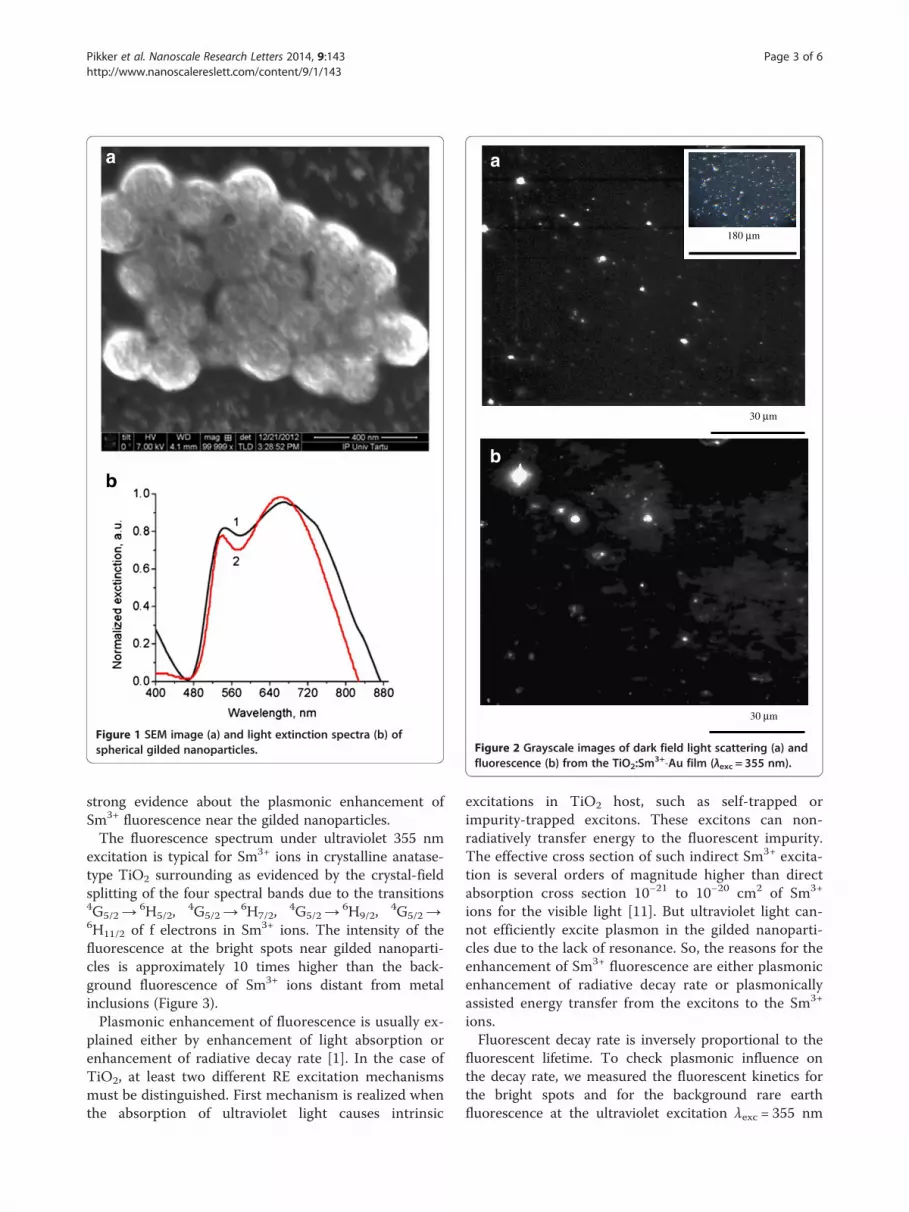
Figure 1 SEM image (a) and light extinction spectra (b) ofspherical gilded nanoparticles.
a
b
30 µm
180 µm
30 µm
Figure 2 Grayscale images of dark field light scattering (a) andfluorescence (b) from the TiO2:Sm
3+-Au film (λexc = 355 nm).
Pikker et al. Nanoscale Research Letters 2014, 9:143 Page 3 of 6http://www.nanoscalereslett.com/content/9/1/143
strong evidence about the plasmonic enhancement ofSm3+ fluorescence near the gilded nanoparticles.The fluorescence spectrum under ultraviolet 355 nm
excitation is typical for Sm3+ ions in crystalline anatase-type TiO2 surrounding as evidenced by the crystal-fieldsplitting of the four spectral bands due to the transitions4G5/2→
6H5/2,4G5/2→
6H7/2,4G5/2→
6H9/2,4G5/2→
6H11/2 of f electrons in Sm3+ ions. The intensity of thefluorescence at the bright spots near gilded nanoparti-cles is approximately 10 times higher than the back-ground fluorescence of Sm3+ ions distant from metalinclusions (Figure 3).Plasmonic enhancement of fluorescence is usually ex-
plained either by enhancement of light absorption orenhancement of radiative decay rate [1]. In the case ofTiO2, at least two different RE excitation mechanismsmust be distinguished. First mechanism is realized whenthe absorption of ultraviolet light causes intrinsic
excitations in TiO2 host, such as self-trapped orimpurity-trapped excitons. These excitons can non-radiatively transfer energy to the fluorescent impurity.The effective cross section of such indirect Sm3+ excita-tion is several orders of magnitude higher than directabsorption cross section 10−21 to 10−20 cm2 of Sm3+
ions for the visible light [11]. But ultraviolet light can-not efficiently excite plasmon in the gilded nanoparti-cles due to the lack of resonance. So, the reasons for theenhancement of Sm3+ fluorescence are either plasmonicenhancement of radiative decay rate or plasmonicallyassisted energy transfer from the excitons to the Sm3+
ions.Fluorescent decay rate is inversely proportional to the
fluorescent lifetime. To check plasmonic influence onthe decay rate, we measured the fluorescent kinetics forthe bright spots and for the background rare earthfluorescence at the ultraviolet excitation λexc = 355 nm

550 600 650 700 7500
1000
2000
3000
4000
5000
6000
2
4G5/2
6H11/2
6H9/2
6H7/2
6H5/2
Background Bright spot
Flu
ores
cenc
e, a
.u.
Wavelength, nm
4G5/2
4G5/2
4G5/2
1
Figure 3 Micro-luminescence spectra of TiO2:Sm3+ films doped with gilded nanoparticles: (1) bright spot, (2)
background (λexc = 355 nm).
Pikker et al. Nanoscale Research Letters 2014, 9:143 Page 4 of 6http://www.nanoscalereslett.com/content/9/1/143
(Figure 4). It was necessary to use up to three exponentialdecay components to satisfactorily model the kinetics:
I tð Þ ¼ A1 exp −tτ1
� �þ A2 exp −
tτ2
� �
þ A3 exp −tτ3
� �; ð1Þ
where A1, A2, and A3 are the coefficients of light inten-sity, τ1, τ2, τ3 are the lifetimes of fluorescence. In such
0 500 100.0
0.2
0.4
0.6
0.8
1.0
2
Ln(I
)
T
1
Figure 4 Normalized experimental fluorescence decay kinetics: from
situation, the overall rate of decay is frequently charac-terized by the average lifetime defined as
τh i ¼
Z∞
0
tI tð Þdt
Z∞
0
I tð Þdt¼
Xi
Aiτi2
Xi
Aiτið2Þ
Obtained lifetimes of fluorescence are in the range oftens and hundreds of microseconds (Table 1). Fluorescence
00 1500 2000
ime, µs
background (1), from bright spot (2) of TiO2:Sm3+-Au films.

Table 1 Lifetimes of fluorescence for the TiO2:Sm3+ film
doped with gilded nanoparticles, λexc = 355 nm
Place on the sample τ1, μs τ2, μs τ3, μs τ, μs
Bright spot 1 2.4 25 156 103
Bright spot 2 6.5 48 299 147
Bright spot 3 10.5 78 294 202
Spot 1 on the background 4.1 35.3 225 138
Spot 2 on the background 7.4 50 220 137
Pikker et al. Nanoscale Research Letters 2014, 9:143 Page 5 of 6http://www.nanoscalereslett.com/content/9/1/143
lifetimes of the order of hundreds of microseconds are typ-ical for the rare earth ions situating in a good crystallineTiO2 anatase host [11]. Lifetimes in the range of tens ofmicroseconds can be caused by Sm3+ fluorescent centerssituating in the areas of TiO2 host having locally differentcrystallinity or local lattice defects. Corresponding lifetimecomponents for the bright spots and for the backgroundSm3+ fluorescence are not very different. Based on this, wecan suppose that the radiative rate of rare earth fluoro-phore is not very strongly influenced by localized plas-mons. Detected approximately 10 times enhancement inthe intensity of Sm3+ fluorescence at the ultraviolet excita-tion could be caused by plasmonic support of energy trans-fer from exciton to rare earth ions. Possibility of non-radiative plasmonic support for the excitons was recentlydemonstrated in the case of plasmonically improvedphotocatalysis [12]. Plasmonic support of Förster reson-ance energy transfer for quantum dot's fluorescence wasdescribed in [13].Excitation by green light, λexc = 532 nm, results in dir-
ect excitation of Sm3+ and also yields a fluorescencespectrum consisting of the four bands. But in this case,the bands are broader and almost featureless (Figure 5).It means that different ensemble of Sm3+ ions is excited
575 600 6250
2000
4000
6000
8000
10000
Flu
ores
cenc
e, a
.u.
Wave
Figure 5 Micro-luminescence spectra of TiO2:Sm3+ films doped with gil
in this case. The absence of spectral features suggeststhat those Sm ions are situated in less ordered TiO2 en-vironment [14]. In spite of the exclusion of excitonic in-fluence at such excitation, we detected still 2.5 timesenhancement of fluorescence in the vicinity of gildednanoparticles (Figure 5). Under 532 nm excitation, theStokes shift of the fluorescence emission is very small[15]. So, both excitation and emission can be influencedby plasmons.Fluorescence lifetimes at 532 nm excitation were mea-
sured in the time-gated mode on a FLIM in the spectralrange of 580 to 660 nm. Obtained fluorescence decay isalso multiexponential because different Sm3+ centerssituate in TiO2 environment with different local sur-roundings. Numerical values of the lifetimes are similarto those presented in Table 1. Because of the insignifi-cant changes in the lifetimes of Sm3+ fluorescence, wesuppose that the detected 2.5 times enhancement in theintensity of fluorescence could be caused mainly byplasmon-enhanced direct absorption of exciting light bySm3+ ions near the gilded nanoparticles.
ConclusionsSilica-gold core-shell nanoparticles were synthesized andsuccessfully adjusted for the incorporation into TiO2:Sm3+ films. Prospective capabilities of these particles forthe local plasmonic enhancement of rare earth fluores-cence are demonstrated. Detected locally strong Sm3+
fluorescence is connected more with local increase inlight absorption and energy transfer than with changesin radiative decay rates since fluorescent lifetimes arenot changed significantly. Detected enhancement offluorescence can be based both on the plasmonic en-hancement of direct light absorption by Sm3+ ions and
650 675 700 725 750
length, nm
Bright spot Background
ded nanoparticles: (1) bright spot, (2) background (λexc = 532 nm).

Pikker et al. Nanoscale Research Letters 2014, 9:143 Page 6 of 6http://www.nanoscalereslett.com/content/9/1/143
on profitable plasmonic support of energy transfer fromexciton to rare earth ions in the case of the indirect exci-tation. As a next step, variation of dielectric core andnoble metal shell sizes can be used for the spectral tun-ing of the plasmon resonance and estimation of its im-pact on the plasmon-enhanced fluorescence.
Competing interestsThe authors declare that they have no competing interests.
Authors’ contributionsSP, LD, and SH developed the idea of the work and participated in thepreparation of sol-gel TiO2 samples activated by Sm3+ ions and in theirdoping by core-shell nanoparticles. SM synthesized silica-gold core-shellnanoparticles. VK and SK provided necessary fluorescent and microscopicmeasurements of the samples. RL made contribution to the revised versionof the manuscript. SP realized scanning electron microscopy of the samplesand proposed fruitful ideas for explanation of obtained results. IS participatedin joint discussions of co-authors and in explanation of scientific results.All authors read and approved the final manuscript.
AcknowledgementsThis work was supported by ESF project Nr. 2013/0202/1DP/1.1.1.2.0/13/APIA/VIAA/010 and EU through the ERDF (Centre of Excellence‘Mesosystems: Theory and Applications’, TK114). The work was also partlysupported by COST Action MP1303 and ETF grant 9007, EstonianNanotechnology Competence Centre (EU29996), ERDF ‘TRIBOFILM’3.2.1101.12-0028, ‘IRGLASS’ 3.2.1101.12-0027, ‘Nano-Com’ 3.2.1101.12-0010,Estonian Research Council (SF0180032s12 and IUT 20-17), and EuropeanUnion through the European Regional Development Fund (TK114 and30020) and partially by the Nanotwinning project FP7-INCO-2011-6 andMarie Curie ILSES project no. 612620.
Author details1Institute of Physics, University of Tartu, Riia 142, Tartu 51014, Estonia. 2V.Lashkaryov Institute of Semiconductor Physics of National Academy ofSciences of Ukraine, Kyiv 03680, Ukraine. 3Institute of Chemistry, University ofTartu, Ravila 14A, Tartu 50411, Estonia.
Received: 3 December 2013 Accepted: 28 February 2014Published: 25 March 2014
References1. Sau TK, Rogach AL: Complex-Shaped Metal Nanoparticles: Bottom-Up Syntheses
and Applications. Wiley-VCH Verlag GmbH & Co. KGaA: Weinheim; 2012.2. Aizpurua J, Hillenbrand R: Localized surface plasmons: basics and
applications in field-enhanced spectroscopy. In Plasmonics From Basics toAdvanced Topics Series: Springer Series in Optical Sciences. Edited by RhodesWT, Adibi A, Asakura T, Hänsch TW, Kamiya T, Krausz F, Monemar Bo AJ,Venghaus H, Weber H, Weinfurter H. Berlin: Springer; 2012:151–176. SpringerSeries in Optical Sciences, vol. 167].
3. Yguerabide J, Yguerabide E: Resonance light scattering particles asultrasensitive labels for detection of analytes in a wide range ofapplications. J Cell Biochem Suppl 2001, 37:71–81.
4. Stockman M: Spasers explained. Nat Photonics 2008, 2:327–329.5. Malashkevich G, Semchenko A, Sukhodola A, Stupak A, Sukhodolov A,
Plyushch B, Sidskii V, Denisenko G: Influence of silver on the Sm3+
luminescence in ‘Aerosil’ silica glasses. Phys Solid State 2008,50(8):1464–1472.
6. Hayakawa T, Selvan S, Nogami M: Field enhancement effect of small Agparticles on the fluorescence from Eu3+- doped SiO2 glass. Appl Phys Lett1999, 74(11):1513–1515.
7. Marques A, Almeida R: Er photoluminescence enhancement in Ag-dopedsol–gel planar waveguides. J Non-Cryst Solids 2007, 353(27):2613–2618.
8. Dolgov L, Kiisk V, Reedo V, Maaroos A, Sildos I, Kikas J: Sol–gel derivedmetal oxides doped with silver nanoparticles as tunable plasmonicmaterials. Phys Stat Solidi A 2010, 207(5):1166–1169.
9. Pham T, Jackson J, Halas N, Lee T: Preparation and characterization ofgold nanoshells coated with self-assembled monolayers. Langmuir 2002,18:4915–4920.
10. Stöber W, Fink A, Bohn E: Controlled growth of monodisperse silicaspheres in the micron size range. J Colloid Interface Sci 1968, 26(1):62–69.
11. Liu G, Jacquier B: Spectroscopic properties of rare earths in opticalmaterials. In Springer Series in Materials Science, vol. 83. Edited by Hull R,Osgood RM, Paris JJ, Warlimont H. Berlin: Springer; 2005.
12. Liu E, Kang L, Wu F, Sun T, Hu X, Yang Y, Liu H, Fan J: Photocatalyticreduction of CO2 into methanol over Ag/TiO2 nanocomposites enhancedby surface plasmon resonance. Plasmonics 2014, 9:61–70.
13. Ozel T, Hernandez-Martinez P, Mutlugun E, Akin O, Nizamoglu S, Ozel I,Zhang Q, Xiong Q, Demir H: Observation of selective plasmon-excitoncoupling in nonradiative energy transfer: donor-selective versusacceptor-selective plexcitons. Nano Lett 2013, 13:3065–3072.
14. Elisa M, Vasiliu I, Grigorescu C, Grigoras B, Niciu H, Niciu D, Meghea A,Iftimie N, Giurginca M, Trodahl H, Dalley M: Optical and structuralinvestigation on rare-earth-doped aluminophosphate glasses.Opt Mater 2006, 28(6–7):621–625.
15. Henderson B, Imbush G: Optical Spectroscopy of Inorganic Solids. Oxford:Clarendon Press; 1989. 2006.
doi:10.1186/1556-276X-9-143Cite this article as: Pikker et al.: Gilded nanoparticles for plasmonicallyenhanced fluorescence in TiO2:Sm
3+ sol-gel films. Nanoscale ResearchLetters 2014 9:143.
Submit your manuscript to a journal and benefi t from:
7 Convenient online submission
7 Rigorous peer review
7 Immediate publication on acceptance
7 Open access: articles freely available online
7 High visibility within the fi eld
7 Retaining the copyright to your article
Submit your next manuscript at 7 springeropen.com


