Embed Size (px)
Citation preview

Abstract
Aims/hypothesis. Viral infections and local productionof IFN-γ might contribute to beta-cell dysfunction/death in Type 1 Diabetes. Double stranded RNA (dsRNA) accumulates in the cytosol of viral-infectedcells, and exposure of purified rat beta cells to dsRNA(tested in the form of polyinosinic-polycytidylic acid,PIC) in combination with IFN-γ results in beta-celldysfunction and apoptosis. To elucidate the molecularmechanisms involved in PIC + IFN-γ-effects, we de-termined the global profile of genes modified by theseagents in primary rat beta cells.Methods. FACS-purified rat beta cells were culturedfor 6 or 24 h in control condition or with IFN-γ, PICor a combination of both agents. The gene expressionprofile was analysed in duplicate by high-density oli-gonucleotide arrays representing 5000 full-lengthgenes and 3000 EST’s. Changes of greater than orequal to 2.5-fold were considered as relevant.Results. Following a 6- or 24-h treatment with IFN-γ,PIC or IFN-γ and PIC, we observed changes in the ex-pression of 51 to 189 genes. IFN-γ modified the ex-pression of MHC-related genes, and also of genes in-
volved in beta-cell metabolism, protein processing,cytokines and signal transduction. PIC affected prefer-entially the expression of genes related to cell adhe-sion, cytokines and dsRNA signal transduction, tran-scription factors and MHC. PIC and/or IFN-γ up-regu-lated the expression of several chemokines and cyto-kines that could contribute to mononuclear cell homingand activation during viral infection, while IFN-γ in-duced a positive feedback on its own signal transduc-tion. PIC + IFN-γ inhibited insulin and GLUT-2 ex-pression without modifying pdx-1 mRNA expression.Conclusion/interpretation. This study provides the firstcomprehensive characterization of the molecular re-sponses of primary beta cells to dsRNA + IFN-γ, twoagents that are probably present in the beta cell milieuduring the course of virally-induced insulitis and Type1 Diabetes. Based on these findings, we propose an in-tegrated model for the molecular mechanisms involvedin dsRNA + IFN-γ induced beta-cell dysfunction anddeath. [Diabetologia (2003) 46:1641–1657]
Keywords Double stranded RNA, microarray analysis,apoptosis, pancreatic beta cells, interferon-γ, nitric ox-ide, NF-κB, diabetes mellitus.
Received: 3 July 2003 / Revised: 11 September 2003Published online: 5 November 2003© Springer-Verlag 2003
Corresponding author: J. Rasschaert, Laboratory of Experimen-tal Medicine, Université Libre de Bruxelles, Route de Lennik,808, CP 618, 1070 Brussels, BelgiumE-mail: [email protected]: AS, argininosuccinate synthetase; ADAR, RNA-specific adenosine deaminase; Bax, bcl-2 associated x protein;BB rats, diabetes-resistant BioBreeding rats; bcl-2, B-cell lym-phoma/leukemia-2; BH3, bcl-2 homology domain-3; Bid, BH3interacting domain death agonist; dsRNA, double stranded RNA;CCK, cholecystokinin; eIF2α, eukaryotic initiation factor 2α;EMC-D, encephalomyocarditis virus; ER, endoplasmic reticu-lum; ESTs, expressed sequence tags; GADD, growth arrest andDNA damage; GAPDH, glyceraldehyde 3-phosphate dehydroge-nase; GH, growth hormone; GIP, gastric inhibitory peptide;HMGCoA, 3-hydroxy 3-methylglutaryl coenzyme A; HO, hemeoxygenase; hsp, heat shock protein; ICE, interleukin convertingenzyme/caspase 1; IFN, interferon; IL, interleukin; iNOS, induc-
Diabetologia (2003) 46:1641–1657DOI 10.1007/s00125-003-1245-y
Global profiling of double stranded RNA- and IFN-γ-induced genesin rat pancreatic beta cellsJ. Rasschaert1, D. Liu1, B. Kutlu1, A. K. Cardozo1, M. Kruhøffer2, T. F. Ørntoft2, D. L. Eizirik1
1 Laboratory of Experimental Medicine, Université Libre de Bruxelles, Brussels, Belgium2 Molecular Diagnostic Laboratory, Department of Clinical Biochemistry, Aarhus University, Denmark
ible nitric oxide synthase; IRF, interferon regulatory factor; JAK-1/2, Janus tyrosine kinase 1 and 2; MAP, mitogen activated pro-tein; MCP-1, macrophage chemoattractant protein-1; MHC, ma-jor histocompatibility complex; MIP, macrophage inflammatoryprotein; MnSOD, manganese superoxide dismutase; MX, myxo-virus; NF, nuclear factor; NO, nitric oxide; NOD mouse, nonobese diabetic mouse; MGMT, O-6-methylguanine-DNA meth-yltransferase; OAS, 2′,5′-oligoadenylate synthetase; PC, prohor-mone convertase; PDX-1, pancreatic duodenal homeobox factor-1; PI, propidium iodide; PIC, polyinosinic-polycytidylic acid;PKR, dsRNA dependent protein kinase; RANTES, regulated up-on activation, normal T-cell expressed, and presumably secreted;SERCA-2, sarco(endo)plasmic reticulum Ca+2 ATPase type 2;STAT, signal transducers and activators of transcription; T1DM,type 1 diabetes mellitus; TLR, toll-like receptor.
J. Rasschaert and D. Liu contributed equally to the article
Electronic Supplementary MaterialSupplementary material is available in the online version ofthis article at http://dx.doi.org/10.1007/s00125-003-1245-y
Type 1 diabetes mellitus is an auto-immune diseaseassociated with a progressive and selective destructionof the insulin-producing pancreatic beta cells [1, 2].Although a genetic susceptibility seems to be a pre-requisite for the development of Type 1 Diabetes [3,4], it is now clear that environmental factors such asviral infections are also important aetiological deter-minants [2, 5].
There is extensive epidemiological evidence for theinvolvement of viral infections in the pathogenesis ofType 1 Diabetes [6]. Up to now, 13 different viruses,most of them belonging to the enterovirus family,have been found to be associated with the onset ofType 1 Diabetes in humans and in various animalmodels [7]. Different mechanisms have been proposedfor the role of viruses in the pathogenesis of Type 1Diabetes. These include: (i) infection and rapid de-struction of beta cells [8]; (ii) triggering of local in-flammation, leading to destruction of the beta cellsthrough the production of NO, cytokines and otherimmune mediators in a mechanism referred to as “in-nocent bystander killing” [9, 10]; (iii) molecular mim-icry, based on a partial sequence homology between aprotein of the infected cells (i.e. GADD65) and a viralantigen, leading to autoimmune destruction of the betacell [11]; (iv) viral infection, coupled with one ormore of the factors described above, acting in con-junction to induce beta cell death [12]. For example,mouse infection with a high titre of the D variant ofthe encephalomyocarditis (EMC-D) virus leads to be-ta-cell destruction and diabetes mainly as a result ofviral replication within beta cells, while mouse infec-tion with a low titre of EMC-D virus leads to diabetesas a chronic process, caused by the destruction of betacells by soluble mediators such as IL-1β, TNF-α/βand NO produced by macrophages or the beta cellsthemselves [7].
The molecular mechanisms involved in beta cellsdamage by viruses, alone or in combination with solu-ble mediators, remain to be elucidated. During viralinfection, accumulation of the viral replicative inter-mediate double stranded RNA (dsRNA) in the cytosolof the infected cell stimulates antiviral activities, suchas dsRNA-dependent protein kinase (PKR) activation,type I interferons and NO production and a general in-hibition of protein translation [13, 14]. These antiviralresponses can be mimicked by treatment of cells withthe synthetic dsRNA polyinosinic-polycytidylic acid(PIC) [15, 16]. PIC triggers the development of hyper-glycaemia in diabetes-resistant BioBreeding (BB) ratsand accelerates the development of the disease in dia-betes-prone BB rats [17, 18]. In vitro, PIC inhibitsglucose-stimulated insulin biosynthesis in mouse is-lets [19] and when used in combination with IFN-γ af-fects rat islet cell function and viability by a mecha-nism involving, at least in part, increased inducible ni-tric oxide synthase (iNOS) expression and NO pro-duction [20, 21].
We have previously shown that exposure of fluo-rescence-activated cell sorting-(FACS)-purified rat be-ta cells to PIC alone does not induce cell death. How-ever, when these cells are exposed to PIC + IL-1β orPIC + IFN-γ they die mostly by apoptosis. The mech-anisms of death induction are either NO-dependent inthe case of PIC + IL-1β, or NO-independent, in thecase of PIC + IFN-γ [21]. Moreover, PIC regulates theexpression of several genes that could participate inthe induction of islet inflammation and beta-celldeath, such as Fas, iNOS, IL-15 and diverse chemo-kines [21, 22]. The complete range of genes inducedby PIC and cytokines in pancreatic beta cells remains,however, to be clarified.
Evaluation of complex patterns of gene expressionis now feasible by the use of microarray analysis [23].We have previously used high-density oligonucleotidearrays to analyse FACS-purified rat beta cells exposedfor 6 or 24 h to IL-1β + IFN-γ [24, 25]. Based on thefindings obtained, we proposed that beta-cell fate fol-lowing cytokines exposure, namely death by apoptosisor survival with or without complete functional recov-ery, depends on the intricate pattern of dozens ofgenes up- or down-regulated in parallel and/or se-quentially. Moreover, these and subsequent data onINS-1 cells [26] allowed us to start an annotated “BetaCell Gene Bank”, including information on nearly3000 genes expressed in beta cells and in INS-1 cells,of which 700 are modified by cytokines. Against thisbackground, we carried out a microarray analysis ofFACS-purified rat pancreatic beta cells exposed for 6or 24 h to PIC, IFN-γ or a combination of both agents.The data obtained provide the first broad picture onhow a primary beta cell responds to dsRNA and theinflammatory cytokine IFN-γ, an experimental condi-tion which bears similarity to the in vivo situation dur-ing the course of a viral infection. The results obtainedallowed us to propose a comprehensive hypothesis forthe mechanisms involved in dsRNA and IFN-γ in-duced beta-cell dysfunction and death.
Materials and methods
Islet cell isolation and culture. Pancreatic islets were isolatedfrom 10–12-week-old male Wistar rats by collagenase diges-tion, and subsequently dissociated into single cells in a calci-um-free medium containing dispase (0.5 mg/ml). Single beta-cells were purified by autofluorescence-activated cell sorting(FACS) [27]. These preparations contain around 90–95% via-ble beta cells [data not shown, 27]. The purified beta cellswere cultured in HAM’s F-10 medium (Invitrogen, Paisley,Scotland) supplemented with 10 mmol/l glucose [28]. For determination of viability, FACS-purified single beta cells (104 cells per well) were cultured for 6 days in Falcon 96-wellmicrotitre plates (Becton Dickinson, New Jersey, N.J., USA)pre-coated with poly-L-lysine and containing 200 µl of medi-um. Culture medium was changed every 3 days and fresh IFN-γ or PIC was added. For RNA extraction for microarray orRT-PCR analysis, single beta cells were re-aggregated for 3 h
1642 J. Rasschaert et al.: Global profiling of double stranded RNA- and IFN-γ-induced genes in rat pancreatic beta cells

in a rotatory shaking incubator [29], cultured for 14–16 h insuspension, and then exposed for 6 or 24 h to IFN-γ(1000 U/ml; 10 U/ng; Invitrogen) or PIC (100 µg/ml; SigmaChemical, St Louis, Mo., USA). In some experiments, recom-binant human IL-1β (50 U/ml, 38 U/ng, a kind gift of Dr. C.W.Reynolds from the National Cancer Institute, Bethesda, Md.,USA) was also utilized as a positive control. The concentra-tions of cytokines and PIC, and the time points for array analy-sis, were selected based on our previous studies in beta cells[21, 22, 24, 25], and aimed to analyse beta cells at time pointswhich precede non-specific changes in mRNA expression in-duced by early apoptosis. Culture media were collected after24 h for nitrite determination (nitrite is a stable product of NOoxidation), which was done spectrophotometrically at 546 nmwavelength after coloured reaction with the Griess reagent [30].
Assessment of beta cell protein synthesis and viability. Totalprotein biosynthesis was determined at 10 mmol/l glucose us-ing L-[4,5-3H] leucine incorporation and trichloroacetic pre-cipitation [31]. The experiments were carried out in duplicate,using 6×10⁴ re-aggregated beta cells per condition. The cellswere exposed for 1, 5 and 23 h to IFN-γ + PIC (same concen-trations as described above) or left untreated (control) beforedetermination of protein biosynthesis for 2 h in the absence(control) or presence of IFN-γ + PIC. These time points wereselected to cover an early time point (3 h), and then two timepoints placed 1 h after the time points used for microarrayanalysis (6 and 24 h), the rationale being that protein synthesisusually lags behind mRNA expression. As a positive controlfor protein biosynthesis inhibition, some control cells were ex-posed to cycloheximide (10 µmol/l) during the final 2 h of theincubation period.
The percentage of viable, apoptotic and necrotic beta cellswas determined after 6 days exposure to IFN-γ and/or PIC [21,22]. For this purpose, beta cells were incubated for 15 minwith propidium iodide (PI, 10 µg/ml) and Hoechst (HO) 342(10 µg/ml) [32]. This fluorescence assay for single beta cells isquantitative and has been validated by systematic comparisonswith electron microscopy observations [32, 33]. The methodhas been successfully used to evaluate apoptosis/necrosis in rat[21, 22, 32], mouse [34, 35] and human [36] beta cells.
Microarray analysis. For microarray analysis, total RNA wasisolated from beta cells (at least 10 µg/sample) and used to pre-pare biotinylated cRNA. The labelled cRNAs were hybridizedin duplicate to the rat U34-A oligonucleotide array (Affyme-trix, Santa Clara, Calif., USA) [24, 25]. Due to difficulties inobtaining a sufficient number of primary beta cells in a singleisolation, and to decrease putative biases caused by biologicalvariation, the cells were pooled from four independent experi-ments, using in each experiment 3.5–3.7×10⁵ cells/group. Wehave shown before that microarray analysis, carried out in du-plicate on pooled beta cell samples, provides a reliable estima-tion of massive changes in mRNA expression, as confirmed bya greater than 90% confirmation by RT-PCR of genes observedas modified in the array [24, 25, 37, present data]. Analysis ofdifferential expression was carried out using the GeneChipSuite software, version 4.0.1 (Affymetrix, Santa Clara, Calif.,USA). Arrays were normalized by global scaling, with the ar-rays scaled to an average intensity of 150. Genes were consid-ered as modified by PIC and/or IFN-γ in case they fulfilled thefollowing criteria [24, 25]: (i) the mRNA was present in eithercontrol or cells exposed to PIC and/or IFN-γ in both dupli-cates; (ii) the mean average fold change (experimental groupvs control) was greater than or equal to 2.5; (iii) the foldchange in each individual duplicate was greater than or equalto 2.0. We have used our own curated “Beta Cell Gene Bank”
to assign the filtered genes into their respective functionalclusters. The expressed sequence tags (ESTs) that had homolo-gy to a known sequence were annotated using the Resourcerer6.0 database [38]. Non-identified ESTs are not shown here.
RT-PCR and real time RT-PCR. RT-PCR was done usingpoly(A)+ RNA as described [24]. The number of cycles was se-lected to allow linear amplification of the cDNA under study.The primer sequences used for amplification of rat cDNAs forGAPDH, GADD153 [24], pdx-1, GLUT-2 and c-Myc [25] andfor sarco(endo)plasmic reticulum Ca2+ ATPase type 2 (alsocalled SERCA-2) [39] were as described in the indicated refer-ences. For the other genes studied, the primer sequences andtheir respective PCR fragment lenghts were as follows: dsRNA-dependent protein kinase (PKR), forward (5′-AATCACGCCA-ACATTGTTCA-3′) and reverse (5′-CACCGGGTCTTGTATC-GACT-3′) (107 bp); RNA-specific adenosine deaminase (ADAR),forward (5′-AAGAAACAGGGCAAG-CAAGA-3′) and reverse(5′-TGTTGGTCAGAGCGTTGAAG-3′) (244 bp); CEBP/β,forward (5′-CAAGCTGAGCGACGAG-TACA-3′) and reverse(5′-CAGCTGCTCCACCTTCTTCT-3′) (147 bp) and Bip/GRP78, forward (5′-CTCAAAGAGCGCATTGACAC-3′) andreverse (5′-GCCACTTGGGCTATAGCATT-3′) (446 bp). Theidentity of the PCR fragments of each gene was confirmed bysize after electrophoretic migration on ethidium bromide-stainedagarose gels photographed under UV-transillumination usingKodak Digital Science DC120 camera (Kodak, Rochester, N.Y.,USA). The data are shown as a representative figure for foursimilar experiments. Expression of the “housekeeping” geneGAPDH is not affected by exposure to cytokines in both wholeislets and FACS-purified beta cells [21, 22, 40].
Real-time RT-PCR was done as described [41] using aLightcycler instrument in a 20 µl reaction containing 3 mmol/lMgCl2, 0.5 µmol/l forward and reverse primers, 2 µl FastStartSYBR Green mix (Roche), and 2 µl template cDNA. The prim-er sequences and their respective PCR fragment lenghts wereas follows: GADD153, forward (5′-CCAGCAGAGGTCACA-AGCA-3′) and reverse (5′-CGCACTGACCACTCTGTTTC-3′) (126 bp); SERCA-2, forward (5′-TTGTGGCCCGAAACT-ACCT-3′) and reverse (5′-TTCATAATGAGCAGCACAAAG-GG-3′) (121 bp). The method used for quantification is thestandard curve approach [42, 43]. To obtain the standard curve,the primer sequences and their respective PCR fragment le-nghts were as follows: GADD153, forward (5′-GTCTCTGCC-TTTCGCCTTTG-3′) and reverse (5′-CTACCCTCAGTCCCC-TCCTC-3′) (605 bp); SERCA-2, forward (5′-TCTAGTCAC-CATAGAGATGTG-3′) and reverse (5′-TACTGACTGAGGT-AGCAGGA-3′) (912 bp).
Promoter studies. A plasmid construct containing the humanpdx-1 gene promoter linked to a luciferase reporter gene waskindly provided by Dr D. Melloul, Department of Endocrinol-ogy, Hadassah University Hospital, Jerusalem, Israel [44].Transfected beta cells were exposed to cytokines and/or PICfor 24 h (same concentrations as above). Luciferase activitywas assayed with the Dual-Luciferase Reporter Assay System(Promega) as previously described [21, 41]. Test values werecorrected for the luciferase activity value of the internal con-trol plasmid, pRL-CMV.
Statistical analysis. Results of microarray analysis are shownas means of two similar determinations. The results for otherexperiments are presented as means±SEM of at least three in-dependent experiments. Statistical differences between thegroups were determined by paired Student’s t-test or ANOVA,as indicated. A p value of less than 0.05 was considered statis-tically significant.
J. Rasschaert et al.: Global profiling of double stranded RNA- and IFN-γ-induced genes in rat pancreatic beta cells 1643

Results
Viability and nitrite production of beta cells exposedto PIC and/or IFN-γ. Exposure of FACS-purified betacells to PIC and IFN-γ for 6 days induced a significant(p<0.001) increase in the percentage of apoptotic cellswhen compared against control cells. Neither thetreatment with PIC nor with IFN-γ alone led to beta-cell death, indicating a synergistic effect between PICand IFN-γ to induce beta-cell apoptosis (Fig. 1). Wedid not detect a significant increase in the percentageof necrotic cells (data not shown).
To identify by microarray analysis early and lateIFN-γ and/or PIC induced or decreased genes in pan-creatic beta cells, the cells were exposed for 6 or 24 hto the following conditions: control condition; IFN-γ(1000 U/ml); PIC (100 µg/ml); or the combination ofboth agents. When the beta cells were treated for 6 hwith PIC and/or IFN-γ, there was no detectable nitriteproduction. However, after a 24-h treatment the combi-nation of PIC and IFN-γ significantly increased nitriteproduction as compared to control, non-treated cells.Thus, the values for PIC + IFN-γ were 5.6±1.1 pmolnitrite·10-3cells·h (means ± SEM; n=6; p<0.05 vs control) while control values were 0.7±0.4 pmol ni-trite·10–3 cells·h (mean ± SEM; n=6). When testedalone, neither PIC nor IFN-γ affected nitrite production(data not shown). These data on viability and nitriteproduction are in good agreement with our previousobservations [21, 22] and confirm that both IFN-γ andPIC were biologically active.
Identification of IFN-γ and/or PIC -modified genes inbeta cells by microarray analysis. Cells from four sep-arate experiments conducted as described above werepooled for RNA extraction, and the resulting biotin-ylated cRNAs were hybridized in duplicate to the Affymetrix rat U34-A oligonucleotide array contain-ing about 8000 probes (77% known genes and 23%ESTs). Approximately 3759 (3300 to 4217) genes orESTs were scored as present in each of the six condi-
tions, in fair agreement with our previous observations[24, 25].
Following a 6-h exposure of beta cells to IFN-γ orPIC we observed changes in the expression of respec-tively 89 and 86 genes, while exposure to PIC + IFN-γnearly doubled the number of modified genes (163genes). A 24-h treatment induced a similar pattern,with 52–57 genes modified by IFN-γ or PIC alone,and a nearly four-fold increase in the number ofchanged genes by the combination of IFN-γ + PIC. Ofnote, except in the case of a 6-h exposure to IFN-γ, themajority (73–95%) of these modifications in gene ex-pression consisted in up-regulation (Table 1). The PICand/or IFN-γ responsive genes were clustered in 15groups according to the putative biological function oftheir encoded proteins. Table 2 shows the percentagesof affected genes observed in each experimental con-dition, expressed relative to the total number of modi-fied genes. After treatment with IFN-γ alone for 6 h,the most frequent changes in gene expression wereobserved in beta-cell metabolism, protein processingand cytokine signal transduction. Of note, after 24-hexposure to IFN-γ, the MHC-related genes represent-ed 31% of all modified genes. Exposure to PIC alonefor 6 or 24 h affected preferentially the expression ofgenes related to cell adhesion, cytokine signal trans-duction, transcription factors and MHC. Beta-celltreatment with both IFN-γ and PIC led to a combinedpattern of the modifications described above, but sev-eral novel genes were detected as changed when cellswere exposed to a combination of both agents.
From the 340 IFN-γ and/or PIC modified genes,161 genes of special relevance are presented in Ta-ble 3. The complete list of modified genes is presentedin S1, deposited as “Supporting online material” atDiabetologia home page: http://dx.doi.org/10.1007/s00125-003-1245 .
Among the metabolism-related genes, a decrease inthe expression of mRNAs encoding for genes in-volved in glucose metabolism, such as the beta-cellspecific glucose transporter GLUT-2 and lactate dehy-
1644 J. Rasschaert et al.: Global profiling of double stranded RNA- and IFN-γ-induced genes in rat pancreatic beta cells
Fig. 1. Percentage of apoptosis observed in FACS purified betacells exposed for 6 days to PIC (100 µg/ml) and/or IFN-γ(1000 U/ml). Cell viability was determined with the DNA-binding dyes Hoechst 342 and PI. Data are means±SEM of sixexperiments. * p<0.001 vs. control, ANOVA
Table 1. Total number of up- and down-regulated genes in ratpancreatic beta cells after 6 or 24 h
6 h 24 h
Increased Decreased Increased Decreased
IFN-γ 25 (28%) 64 (72%) 46 (88%) 6 (12%)PIC 81 (94%) 5 (6%) 54 (95%) 3 (5%)IFN-γ + PIC 130 (80%) 33 (20%) 140 (73%) 51(27%)TOTAL 236 (70%) 102 (30%) 240 (80%) 60 (20%)
IFN-γ- and/or dsRNA-induced differences in gene expressionwere considered as present when the mean fold change of theduplicates was ≥2.5, and both individual fold change valueswere ≥2.0. Data are expressed in absolute numbers, or percent-ages (between parenthesis)

drogenase (isoforms A and B) was observed (Table 3;item 1.1). Interestingly, expression of glucokinase wasnot affected by any of the tested conditions. While ex-pression of glycolytic enzymes was down-regulated,several genes involved or related to lipid metabolism,such as ATP-citrate lyase, 3-hydroxy-3 methylglutar-yl-coenzyme A (HMGCoA) reductase, LDL receptor,mitochondrial cytochrome P450 and stearyl-CoA de-saturase were up-regulated following exposure for 6 or 24 h to both IFN-γ and PIC (Table 3, item 1.4).Of note, exposure to IFN-γ and PIC led to modifica-tions in key genes related to arginine metabolism andnitric oxide formation (Table 3, item 1.2). The modi-fied genes are arginase (decreased), argininosuccinatesynthetase, iNOS and the cationic amino acid trans-porter-1 (all increased) and correlate well with the ob-served increased production of nitrite after 24-h treat-ment by both agents (see above).
After 24-h treatment with IFN-γ and PIC, there wasdown-regulation of mRNAs encoding mitochondrialsubunits of respiratory chain genes involved in ATPproduction, such as NADH dehydrogenase and cyto-chrome b5 reductase, coupled with an up-regulation ofuridine kinase (Table 3, item 1.5). IFN-γ + PIC alsodown-regulated expression of mRNAs encoding re-ceptors for the incretins cholecystokinin-A and gastricinhibitory peptide and of growth hormone receptor(Table 3, item 4.0). This, associated with the observeddecrease in insulin and pro-hormone convertase (PC)1 mRNA levels, and of genes involved in ATP pro-duction, could contribute for the decreased insulin se-
cretion observed after treatment of pancreatic islets orbeta cells with IFN-γ + PIC [20, 45].
Several chemokines, cytokines, as well as genes in-volved in signal transduction, were modified by IFN-γor PIC, alone and specially in combination (Table 3,items 5.0 and 6.0). When tested alone, IFN-γ affectedexpression of the chemokines macrophage inhibitorycytokine-1 (MIC-1) and interferon inducible protein(IP)-10 and of the cytokine IL-15 (Table 3, item 5.0).IL-15, IP-10, MIP-3α, macrophage chemoattractantprotein (MCP)-1 and fractalkine mRNAs were in-duced by PIC and/or the combination of IFN-γ + PIC.Induction of RANTES and TNF-β mRNAs were alsoobserved in pancreatic beta cells exposed to PICand/or PIC + IFN-γ. It has been previously shown thatRANTES and IP-10 are secondarily up-regulated byautocrine production of IFN-β in RAW 264.7 cells, amurine macrophage cell line [46]. In the present mi-croarray analysis however, neither PIC nor IFN-γ, norPIC and IFN-γ together, modified the expression ofIFN-α, IFN-β and IL-1β, at least at our selected timepoints (data not shown).
Interestingly, the concomitant presence of PIC andIFN-γ led to up-regulation of Notch1 receptor and oneof his ligands, Delta-1. Presenilin-2, an enzyme re-quired for intramembraneous proteolysis of Notch wasalso up-regulated (Table 3; item 6.0). PIC and IFN-γ,however, did not modify expression of Jagged, anoth-er Notch ligand (data not shown). There was also up-regulation of JAK-2, Pim-3 serine threonine kinaseand CL100 protein tyrosine phosphatase which, asso-
J. Rasschaert et al.: Global profiling of double stranded RNA- and IFN-γ-induced genes in rat pancreatic beta cells 1645
Table 2. Percentages of differentially expressed genes following a 6 or 24 h exposure to IFN-γ and/or PIC
Gene clusters 6 h 24 h
IFN-γ PIC IFN-γ + PIC IFN-γ PIC IFN-γ + PIC% % % % % %
1. Metabolism 13.5 5.8 12.1 11.8 5 15.32. Protein synthesis, modification and secretion 12.4 8.1 7.9 13.7 5 7.43. Ionic channels, ions transporters and related proteins 7.9 1.2 1.2 2 0 1.64. Hormones, growth factors and related genes 6.7 4.7 3 0 5 8.55. Cytokines, chemokines and related receptors 2.2 7 6.1 3.9 5 2.16. Cytokine processing and signal transduction 14.6 12.8 13.9 9.8 10 11.67. MHC and related genes 9 12.8 11.5 31.4 17 10.18. Cell adhesion and cytoskeleton 9 12.8 9.7 2 15 6.99. Transcription factors and related genes 5.6 11.6 11.5 9.8 15 13.2
10. RNA synthesis and splicing factors 0 0 0 0 0 1.111. Cell cycle 3.4 2.3 2.4 0 0 1.112. Defense/repair 5.6 5.8 6.7 0 3 4.813. Apoptosis 4.5 1.2 1.2 2 3 2.614. Anti-viral response 0 4.7 2.4 0 5 1.615. Miscellaneous 5.6 9.3 10.3 13.7 12 12.2
Total number of genes (absolute values) 89 86 163 52 57 191
IFN-γ- and/or dsRNA-induced differences in gene expressionwere considered as present when the mean fold change of theduplicates was ≥2.5, and both individual fold change valueswere ≥2.0.
Data are expressed as percentage of the total number of modi-fied genes observed in each condition (bold numbers), taken as100%
1646 J. Rasschaert et al.: Global profiling of double stranded RNA- and IFN-γ-induced genes in rat pancreatic beta cells
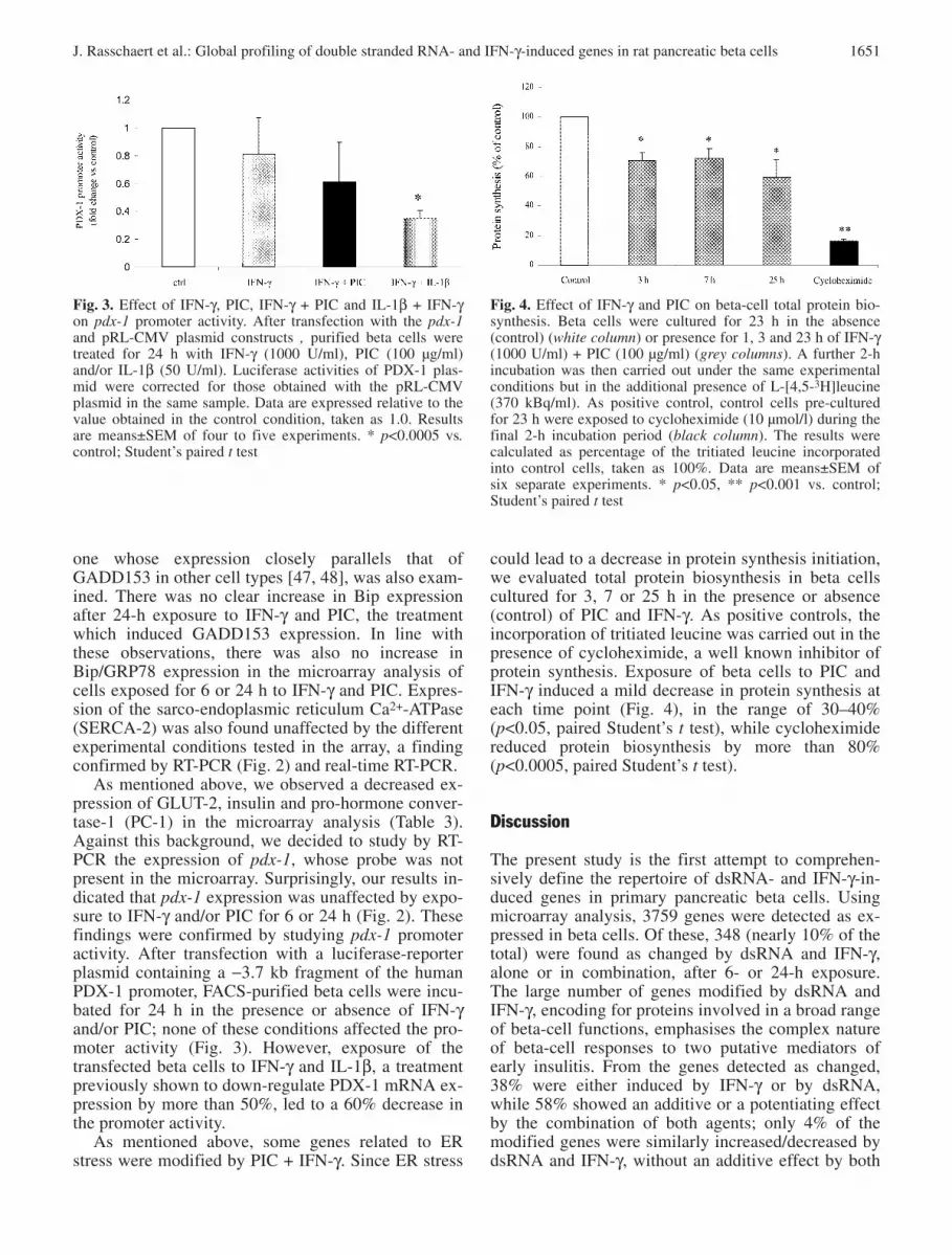
Table 3. List of selected genes modified after a 6- or 24-hour exposure to PIC and/or IFN-γ
Cluster/GAN Gene Name 6h 24h
IFN-γ PIC IFN-γ+PIC IFN-γ PIC IFN-γ+PIC
1.0 Metabolism
1.1 Carbohydrates
X59737 Creatine kinase—ubiquitous * 5.4 11.4 10.9M86240 Fructose-1,6-bisphosphatase 7.9M13979 Glucose transporter type 1 (GLUT 1) 3.1L28126 Glucose transporter type 2 (GLUT 2) * −6.8M54926 Lactate dehydrogenase A −2.7L25387 Phosphofructokinase c (PFK-c) 3.9
1.2 Arginine metabolism and NO formation
J02720 Arginase −4.6X12459 Argininosuccinate synthetase 4.3AA957917 Cationic amino acid transporter-1 3.1D44591 iNOS* 10.5 9.1
1.3 Aminoacids (other than arginine)
AA942685 Cysteine dioxygenase −3.6 −2.8M72422 Glutamic acid decarboxylase 65 (GAD 65) −3.9 −5.2AI176504 Glutaminase −6.5 −8.7 −6.3AA852004 Glutamine synthetase −3.4 2.8 2.8M84648 L-amino acid decarboxylase −11.9
1.4 Lipids
AA799489 Acyl-CoA oxidase −6.0L27075 ATP-cytrate lyase −5.2 2.7 3.2M29249 3-hydroxy-3 methylglutaryl coenzyme A 9.9
(HMGcoA) reductaseX13722 LDL-receptor 4.9AI044900 Long-chain acyl-CoA synthetase 5.6S81497 Lysosomal acid lipase 4.7 6.5 3.1M38566 Mitochondrial cytochrome P450 (P450C27) 30.3 42.1 10.2AF036761 Stearyl-CoA desaturase 2 8.2 9.4S70011 Trycarboxilate carrier (mitochondrial) −2.8
1.5 ATP production and processing
D00636 Cytochrome b5 reductase (NADH)* −11.4AI169265 NADH dehydrogenase (ubiquinone) −2.5
1α subcomplex 4 AA859827 Uridine kinase 8.1
1.6 Miscellaneous
AA799406 Cytochrome P450 monooxygenase −18.4 −2.6 −2.5D87839 GABA transaminase 3.1U27518 UDP-glucuronosyltransferase −3.8 −3.9
2.0 Protein synthesis, modification, and secretion
AA859966 18S rRNA −3.1 −3.2AI008852 Elongation factor 1 α −3.3AA892250 Lysyl-tRNA synthetase 3.2 2.7 3.5M24353 Mannosidase α type II −4.7 −5.0AI169327 Metalloproteinase inhibitor* 3.4 5.0 2.7 3.3 5.9M83676 RAB12 −5.3AA893080 Selenocysteine lyase 3.0 4.3 6.9 5.8 12.9AI007857 SNAP-25 interacting protein hrs-2 2.8AJ006855 Synaptojanin 7.0 9.0 6.4U56261 Synaptosomal associated protein −5.8 −6.4
(SNAP-25a)

J. Rasschaert et al.: Global profiling of double stranded RNA- and IFN-γ-induced genes in rat pancreatic beta cells 1647
3.0 Ionic channels, ions transporters and related proteinsU69884 Calcium-activated potassium channel rSK3 (SK) 4.1M31178 Calbindin −3.8X90642 Multidrug resistance protein (ID:1) 7.0L35771 Potassium channel 3.4AF048828 Voltage dependent anion channel (RVDAC 1)* −6.4
4.0 Hormones, growth factors and related genesX01032 Cholecystokinin (CCK) precursor 58.4 98.9E12746 Cholecystokinin-A receptor* −7.6L20913 Endothelial growth factor form 3 −5.8L19660 Gastric inhibitory peptide receptor −2.9S49003 Growth hormone receptor (short isoform) −25.6Z83757 Growth hormone receptor −5.0M25584 Insulin 1 −2.6Y07534 Mitochondrial vitamin D(3) 25-hydroxylase 19.2 11.5 26.0M83745 Prohormone convertase-1 −2.9S40669 Prohormone convertase 2 2.6M25804 Rev-ErbA-a * 4.1M93273 Somatostatin receptor type 2 −7.2 −7.8X63574 Somatostatin receptor type 3 6.0 6.7M23643 Thyrotropin releasing hormone* −3.1 −11.0M32167 Vascular endothelial growth factor (VEGF)* −9.7AF022952 Vascular endothelial growth factor B 8.3
5.0 Cytokines, chemokines and related receptorsAA799761 CD-40 10.9 88.3 24.3D11445 CINC-1 9.3 8.1AF030358 Fractalkine 7.6U68272 Interferon-γ receptor 5.1U69272 Interleukin 15* 2.7 7.9 3.2 7.7X17053 Macrophage chemoattractant protein-1 (MCP-1)* 7.2 29.3AJ011969 Macrophage inhibitory cytokine-1 (MIC-1) −4.9 16.853312 MIP-3α 3.0 2.7 U17035 Mob-1/ IP-10 26.6 87.0 90.3 17.2 123.0 196.7U94330 Osteoprotegerin 12.8 2.6AI009658 RANTES 155.8 180.4 171.7 284.8L00981 TNF-β 6.5
6.0 Cytokine and dsRNA processing and signal transductionM64780 Agrin* 3.4 2.8 3.1 3.2AI170268 β-2 microgobulin 3.0 2.9 3.2S74351 3CH134/CL100 protein tyrosin phosphatase 4.6 4.9 5.5U78889 Delta1 5.0AA924925 ER transmembrane protein 6.2 5.6M80367 Guanylate nucleotide binding protein 2 15.1 18.5 106.1 27.4 63.4AA891944 INF-γ induced GTPase 37.1 14.3 33.7 17.3 2.9 11.4U13396 Janus protein tyrosine kinase 2 (JAK-2)* 7.7 14.2 8.3AA875327 Lim-kinase 1 4.5 5.9AF013144 MAP-kinase phosphatase (cpg21) −2.7 −3.0D89863 M-Ras* 6.0 4.4X57405 Notch-1* 7.0D84667 Phosphatidylinositol 4-kinase 8.2J03806 Phospholipase C 6.4 7.8AF086624 Pim-3 serine threonine kinase 14.8X99267 Presenilin-2 9.1M18330 Protein kinase C delta subspecies 5.3 5.7 5.1U02553 Protein tyrosine phosphatase 6.2 4.9AF077000 Protein tyrosine phosphatase TD14 (PTP-TD14) 21.0
Table 3. (continued)
Cluster/GAN Gene Name 6h 24h
IFN-γ PIC IFN-γ+PIC IFN-γ PIC IFN-γ+PIC

1648 J. Rasschaert et al.: Global profiling of double stranded RNA- and IFN-γ-induced genes in rat pancreatic beta cells
AA800318 SERPIG 1 7.2 3.5 9.9 4.6 5.0S61868 Syndecan-4 5.0 5.2 2.7
7.0 MHC and related genes
AJ005023 Mature MHC class Ib α chain 6.0 5.6 6.3 7.4M31038 MHC-I non-RT1 α chain 6.5 10.0 5.2 7.0 10.4AF029240 MHC-Ib RT1.S3 * 18.1 29.3 54.6 19.5 12.9 25.9U16025 MHC-Ib RT1* 3.6 6.5 3.4 3.1 7.3M36151 MHC class II A-β RT1.B-b-β 4.7 8.6X13044 MHC-II-assoc. invariant chain γ * 4.9 3.9 38.0 39.6 26.0 178.4X57523 Mtp1* 13.4 4.5 17.6 13.0 19.3X63854 Mtp2 5.7 3.6 10.0 4.7 8.2D10729 Proteasome subunit RC1 16.2 14.9 33.5 20.3 6.9 40.4D10757 Proteasome subunit RING 12 * 15.6 7.4 24.3 16.6 3.0 16.7D45250 Proteasome activator rPA28-b * 4.3 6.6 3.3 7.8
8.0 Cell adhesion, cytoskeleton and related genes
AF017437 Antigen CD-47 * 2.7 4.0 2.5AI070848 β-actin −3.1U72741 36Kd β-galactoside binding lectin * 4.4 4.6 4.0 3.1Y16898 Connexin36 6.7 9.3L16532 2,3-cyclic nucleotide 3-phosphodiesterase (CNPII) 4.9 8.4 7.3 3.7 2.9X67788 Ezrin p81 3.2 2.8 3.3U82612 Fibronectin* −3.1 −10.2D00913 Intercellular adhesion molecule-1 (ICAM-1) 15.4 22.6 4.9 9.7AA875659 Internexin α −4.8 −2.9 −5.0X81449 Keratin 19* 4.6 3.6 10.6 11.4Z12152 Neurofilament protein middle (NF-M) 5.2 8.7U16845 Neurotrimin −7.1 −5.6
9.0 Transcription factors and related genes
S77528 C/EBPβ 5.4 21.3AI045030 C/EBPδ −3.9X17163 c-jun* 8.8 7.6Y00396 c-Myc* 9.6S66024 CREM transcriptional repressor* −4.4U04835 CREMδ C-G gene −2.9J03179 D-binding protein* 8.9X83094 Heat shock transcription factor 1 8.9X62875 High mobility group protein I 3.6Y09507 Hypoxia-inducible factor 1 (HIF1) 4.5X63594 I-kB α-chain* 15.6 17.4 6.0 9.8M34253 Interferon regulatory factor-1 (IRF-1) * 19.1 6.1 15.1 13.1 3.1 19.6AA799861 Interferon regulatory factor-7 (IRF-7)* 9.7 55.2 78.4 7.7 62.5 93.4M63282 Leucine zipper protein 3.0 2.6D32209 Leucine-rich acidic nuclear protein 3.9 6.7 4.6 7.9S71523 Lim-1 5.7U72620 LOT 1 −3.1 −2.9D14448 Max 9.7U58279 Mist1 −3.3AA900476 MRG 1* −3.4L26267 NF-κB-p105 3.4 3.3 2.5AF022081 Small nuclear RING finger protein 4 6.0 5.2 6.5AA892553 STAT-1 56.3 30.5 81.2 33.6 4.4 28.4X91810 STAT-3 3.2AI011498 SWI/SNF related 11.7AA800613 TIS 11* 4.0 7.1 4.1M61725 Transcription factor UBF-2 2.7AF026476 Transcription factor USF-1 4.3 4.9AF001417 Zinc finger protein 9 7.2 9.8 9.1
Table 3. (continued)
Cluster/GAN Gene Name 6h 24h
IFN-γ PIC IFN-γ+PIC IFN-γ PIC IFN-γ+PIC

J. Rasschaert et al.: Global profiling of double stranded RNA- and IFN-γ-induced genes in rat pancreatic beta cells 1649
Table 3. (continued)
Cluster/GAN Gene Name 6h 24h
IFN-γ PIC IFN-γ+PIC IFN-γ PIC IFN-γ+PIC
10.0 RNA synthesis and splicing factors
AF044910 Survival motor neuron* 4.8
11.0 Cell cycleD16308 Cyclin D2 −5.1U24174 p21/WAF1 7.0U75404 SSeCKs 322 17.2 9.9
12.0 Defense/repairAI070295 GADD45 −2.9AF020618 GADD34 (mouse MyD116; rat PEG-3) * 4.2D42148 Gas-6 growth arrest specific −7.1U73174 Glutathione reductase −6.0AI138143 Glutathione S-transferase −5.8Z27118 Hsp 70 gene 1/2 * −17.8 3.9 3.8AA875620 Hsp 70 gene 3 3.0AI176456 Metallothionein 2 2.9Y00497 MnSOD 3.5 4.0X52711 MX1 17.7 49.5 3.6 16.8M76704 O-6-methylguanine-DNA methyltransferase 3.4
(MGMT)J02722 Heme oxygenase 2.7AA848563 Heat shock protein 70-1 −24.3 4.1 3.5AI138143 Glutathione S-transferase −5.8
13.0 Apoptosis and ER stress response and related genesS76511 Bax 2.9M31178 Calbindin d28 K −3.8L18889 Calnexin −2.9U77931 Calreticulin −3.3AF025671 Caspase 2 2.9 2.7 4.0C07012 Cyclophilin c 2.7 3.2 2.7 3.8 4.5U30186 GADD 153 (Growth arrest DNA damage 153) 9.0U41853 150 kDa oxygen regulated protein (ORP150) −4.4 2.6X62950 (pBUS30) with repetitive elements −2.8M96630 Sec 61 homolog −14.8Z14030 TRAP-complex gamma subunit −14.6 −3.7 5.0
14.0 Anti-viral responseL2928 Double-stranded RNA-dependent protein 8.6 9.9
kinase (PKR)U18942 Double-stranded RNA-specific adenosine 5.8 7.2 8.2 9.3
deaminase (ADAR)X52713 Mx3 43.9 36.6 4.7 19.7Z18877 2’,5’-oligoadenylate synthetase (OAS) 11.3 10.7 2.6 2.6
15.0 MiscellaneousY07704 Best-5* 24.4 60.5 60.9 5.2 38.6 99.4D88250 Complement C1 4.7 15.0 7.1 7.9 22.0L21711 Galectin-5 23.8 22.8 7.2 17.7 19.5M62642 Hemopexin 9.1 8.1 13.2 10.9M81855 P glycoprotein 1 11.7 8.6AA875037 Serine protease inhibitor 15 (spi15) 6.4
Data are shown as fold-variation corresponding to the genewith the indicated access number. The genes are ordered alpha-beticaly in each cluster. The data are means of individual du-plicate hybridizations. mRNAs were considered as modifiedby dsRNA and/or IFN-γ when the mean fold change of the du-plicates was ≥2.5, and both individual fold change values were
≥2.0. Decreased expression compared with respective controls(beta cells not exposed to IFN-γ and/or PIC; 6 or 24 h) is indi-cated by “–” preceding the fold-change value; absence of signmeans increased expression. * indicates gene detected by morethan one probe. GAN, GeneBank Accession Number

ciated with a decrease in MAP-kinase phosphatase(cpg21) by exposure to PIC alone or in combinationwith IFN-γ (Table 3; item 6.0), could affect beta-cellsignal transduction by acting on key phosphorylation/dephosphorylation steps.
Genes related to antigen presentation were inducedto a major extent after both 6 and 24 h (Table 3, item7.0). Increased expression of MHC-Ib RT1, MHC-IIand proteasome subunit RC1 and RING12 were ob-served with both IFN-γ and PIC alone, but the twoagents often showed additive effects. MTP-1 andMTP-2, proteins involved in the “machinery” forMHC class I presentation, were also up-regulated byboth agents.
IFN-γ and/or PIC led to up- and down-regulation ofnumerous transcription factors and associated proteins(Table 3, item 9.0). Of note, among the 41 transcrip-tion factors modified, 63% of them were affected onlyin the presence of both agents, the sole condition lead-ing to beta cell apoptosis. There was up-regulation ofc-jun, c-Myc, C/EBPβ, NF-κB, Lim-1 and STAT-1.Up-regulation of these transcription factors was alsoobserved after beta cell exposure to IL-1β and IFN-γ,another treatment leading to apoptosis. The microar-ray results also showed up-regulation of other tran-scription factors not described before in beta cells ex-posed to cytokines, including heat shock transcriptionfactor 1, hypoxia-inducible factor 1, STAT-3, Max andUSF-1 (Table 3; item 9.0). Nuclear Ring Finger pro-tein 4 (RNF4) and SWI/SNF, mRNAs encoding formultiprotein complexes remodelling chromatin, whichare required for positive and negative control of vari-ous cellular pathways, were also up-regulated follow-ing beta-cell exposure to PIC and IFN-γ (Table 3; item9.0).
IFN-γ alone induced an early (6 h) decrease in twomRNAs potentially involved in beta-cell defence/re-pair, namely glutathione reductase and hsp70 (Table 3;item 12.0), and of two endoplasmic reticulum (ER)chaperones that could contribute to defence againstER stress, i.e. calnexin and calreticulin (Table 3; item13.0). In contrast, PIC, especially in combination withIFN-γ, up-regulated several defence/repair genes afterboth 6 and 24 h. Among them, IFN-γ + PIC modifiedexpression of MnSOD, haeme oxygenase, hsp 70,PEG-3/MyD116 (the rat and mouse homologs of hu-man GADD34) and the DNA repair enzyme O-6-methylguanine-DNA methyltransferase (Table 3; item12.0).
Among genes related to apoptosis, we observed in-duction of several putative pro-apoptotic genes by PICalone or combined to IFN-γ. These included Bax,caspase 2, the cyclin-dependent kinase inhibitorp21/WAF1 and GADD153/CHOP, an ER stress-response transcription factor (Table 3, items 11 and13.0). Of interest, Bip/GRP78 and bcl-2 expressionwere unaffected by IFN-γ and/or PIC, at least at ourselected time points (see below).
Treatment of beta cells for 6 or 24 h with PIC aloneinduced expression of genes promoting resistance toviral infection, such as dsRNA-activated protein ki-nase (PKR, also named eIF-2α kinase), 2′,5′-oligoade-nylate synthetase (OAS) and RNA-specific adenosinedeaminase (ADAR) (Table 3; item 14.0); addition ofIFN-γ neither modified the pattern of expression northe magnitude of this response. Moreover, IFN-γ alonedid not induce any of these genes.
Confirmation by RT-PCR and real-time RT-PCR ofgenes identified as modified by IFN-γ and/or PIC.Eight genes of special interest were selected for con-firmation by RT-PCR. Six of the selected genes weredetected as “changed” in at least one of our experi-mental conditions in the array analysis. Thus, ADARand PKR, both involved in anti-viral response; the on-cogene c-Myc; GADD153, an ER stress-induced tran-scription factor; and the transcription factor CEBP/βwere scored as increased; in contrast, expression ofGLUT-2, the beta-cell specific glucose transporter,was detected as decreased by 24 h exposure to thecombination of PIC + IFN-γ. The RT-PCR results con-firmed the data obtained in the microarray for five ofthe six genes under consideration (Fig. 2). The alteredexpression of CEBP/β, however, could not be con-firmed (data not shown). The up-regulation ofGADD153 mRNA observed after 24-h exposure toIFN-γ and PIC (nine-fold increase, as observed by mi-croarray analysis) was also confirmed by real-timeRT-PCR, the ratio of GADD153/GAPDH mRNAsreaching 9.1±1.7-fold increase compared with con-trols (n=3, p<0.05). Bip/GRP78, a resident ER chaper-
1650 J. Rasschaert et al.: Global profiling of double stranded RNA- and IFN-γ-induced genes in rat pancreatic beta cells
Fig. 2. RT-PCR analysis of PKR, dsRNA-specific adenosinedeaminase, c-Myc, GADD153/CHOP, SERCA-2, GLUT-2,PDX-1, Bip/GRP78 and GAPDH mRNA expression by betacells exposed for 6 to 24 h to: IFN-γ (1000 U/ml), PIC(100 µg/ml) or IFN-γ + PIC. The cDNA samples were ampli-fied in parallel with GAPDH-specific primers, confirming sim-ilar loading in all lanes. The figure is representative of foursimilar experiments
J. Rasschaert et al.: Global profiling of double stranded RNA- and IFN-γ-induced genes in rat pancreatic beta cells 1651
one whose expression closely parallels that ofGADD153 in other cell types [47, 48], was also exam-ined. There was no clear increase in Bip expressionafter 24-h exposure to IFN-γ and PIC, the treatmentwhich induced GADD153 expression. In line withthese observations, there was also no increase inBip/GRP78 expression in the microarray analysis ofcells exposed for 6 or 24 h to IFN-γ and PIC. Expres-sion of the sarco-endoplasmic reticulum Ca2+-ATPase(SERCA-2) was also found unaffected by the differentexperimental conditions tested in the array, a findingconfirmed by RT-PCR (Fig. 2) and real-time RT-PCR.
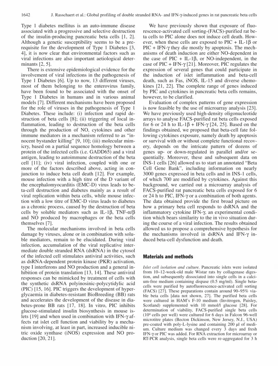
As mentioned above, we observed a decreased ex-pression of GLUT-2, insulin and pro-hormone conver-tase-1 (PC-1) in the microarray analysis (Table 3).Against this background, we decided to study by RT-PCR the expression of pdx-1, whose probe was notpresent in the microarray. Surprisingly, our results in-dicated that pdx-1 expression was unaffected by expo-sure to IFN-γ and/or PIC for 6 or 24 h (Fig. 2). Thesefindings were confirmed by studying pdx-1 promoteractivity. After transfection with a luciferase-reporterplasmid containing a −3.7 kb fragment of the humanPDX-1 promoter, FACS-purified beta cells were incu-bated for 24 h in the presence or absence of IFN-γand/or PIC; none of these conditions affected the pro-moter activity (Fig. 3). However, exposure of thetransfected beta cells to IFN-γ and IL-1β, a treatmentpreviously shown to down-regulate PDX-1 mRNA ex-pression by more than 50%, led to a 60% decrease inthe promoter activity.
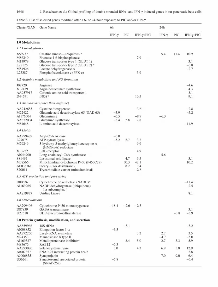
As mentioned above, some genes related to ERstress were modified by PIC + IFN-γ. Since ER stress
could lead to a decrease in protein synthesis initiation,we evaluated total protein biosynthesis in beta cellscultured for 3, 7 or 25 h in the presence or absence(control) of PIC and IFN-γ. As positive controls, theincorporation of tritiated leucine was carried out in thepresence of cycloheximide, a well known inhibitor ofprotein synthesis. Exposure of beta cells to PIC andIFN-γ induced a mild decrease in protein synthesis ateach time point (Fig. 4), in the range of 30–40%(p<0.05, paired Student’s t test), while cycloheximidereduced protein biosynthesis by more than 80%(p<0.0005, paired Student’s t test).
Discussion
The present study is the first attempt to comprehen-sively define the repertoire of dsRNA- and IFN-γ-in-duced genes in primary pancreatic beta cells. Usingmicroarray analysis, 3759 genes were detected as ex-pressed in beta cells. Of these, 348 (nearly 10% of thetotal) were found as changed by dsRNA and IFN-γ,alone or in combination, after 6- or 24-h exposure.The large number of genes modified by dsRNA andIFN-γ, encoding for proteins involved in a broad rangeof beta-cell functions, emphasises the complex natureof beta-cell responses to two putative mediators ofearly insulitis. From the genes detected as changed,38% were either induced by IFN-γ or by dsRNA,while 58% showed an additive or a potentiating effectby the combination of both agents; only 4% of themodified genes were similarly increased/decreased bydsRNA and IFN-γ, without an additive effect by both
Fig. 3. Effect of IFN-γ, PIC, IFN-γ + PIC and IL-1β + IFN-γon pdx-1 promoter activity. After transfection with the pdx-1and pRL-CMV plasmid constructs , purified beta cells weretreated for 24 h with IFN-γ (1000 U/ml), PIC (100 µg/ml)and/or IL-1β (50 U/ml). Luciferase activities of PDX-1 plas-mid were corrected for those obtained with the pRL-CMVplasmid in the same sample. Data are expressed relative to thevalue obtained in the control condition, taken as 1.0. Resultsare means±SEM of four to five experiments. * p<0.0005 vs.control; Student’s paired t test
Fig. 4. Effect of IFN-γ and PIC on beta-cell total protein bio-synthesis. Beta cells were cultured for 23 h in the absence(control) (white column) or presence for 1, 3 and 23 h of IFN-γ(1000 U/ml) + PIC (100 µg/ml) (grey columns). A further 2-hincubation was then carried out under the same experimentalconditions but in the additional presence of L-[4,5-3H]leucine(370 kBq/ml). As positive control, control cells pre-culturedfor 23 h were exposed to cycloheximide (10 µmol/l) during thefinal 2-h incubation period (black column). The results werecalculated as percentage of the tritiated leucine incorporatedinto control cells, taken as 100%. Data are means±SEM of six separate experiments. * p<0.05, ** p<0.001 vs. control;Student’s paired t test

1652 J. Rasschaert et al.: Global profiling of double stranded RNA- and IFN-γ-induced genes in rat pancreatic beta cells
agents. This, and the fact that dsRNA mostly increasesmRNA expression at 6 h (94% of the genes defined asincreased), while IFN-γ has mainly an inhibitory ef-fect at this time point (72% of the genes defined as de-creased), suggests that dsRNA and IFN-γ signal viadifferent and complementary pathways. Comparisonof the present data with our previous microarray anal-ysis of beta cells exposed to IL-1β, alone or in combi-nation with IFN-γ [24, 25], indicates a nearly 50% dif-ference in the pattern of gene expression, suggestingthat dsRNA has also important points of differences insignalling as compared to IL-1β. Of note, and inagreement with our previous observations [22], nei-ther dsRNA nor IFN-γ, nor a combination of bothagents, changed beta-cell expression of IL-1β or IL-1α in the microarray analysis. As previously shown[21, 22], and confirmed in the present study, beta cellsexposed for 3–6 days to dsRNA + IFN-γ, but not to ei-ther agent alone, undergo cell death by apoptosis.These beta cells also present initial adaptive responsesthat are part of the early host reaction to a viral infec-tion and contribute to amplify immune recognitionand immune response against the infective agent [22].How can we integrate these functional responses with
the complex pattern of dsRNA and/or IFN-γ-inducedgene expression observed in our microarray analysis?A general model, based on the present data, is pro-posed in Fig. 5 and discussed below. Due to spacelimitations, not all genes shown in Fig. 5 and Table 3are discussed here; additional information on thesegenes is presented in our previous publications deal-ing with microarray analysis of pancreatic beta cells[24, 25, 26, 37].
Activation of PKR and of the transcription factorNF-κB are important mediators of dsRNA signaltransduction in other cell types [13, 49, 50], and thesepathways have been shown to transduce at least partof the effects of dsRNA, including apoptosis, in betacells [21, 22, 45, 50]. We observed both an inductionof the NF-κB precursor p105 and of several well de-fined beta-cell NF-κB-dependent genes, such as iNOS[51], MCP-1 [41] and MnSOD [52]; there was also anearly 10-fold increase in PKR mRNA expression.Another gene induced to a major extent (more than50-fold) by dsRNA, in an effect potentiated by IFN-γ,is the transcription factor interferon regulatory factor(IRF)-7. IRF-7 is induced by dsRNA and/or interfer-ons (mostly IFN-α and IFN-β) in other cell types, aneffect mediated by the transcription factors NF-κBand STAT-1 [53, 54]. dsRNA increased STAT-1 ex-pression by more than 50-fold, an effect potentiatedby IFN-γ at 6 h (present data). IRF-7 and IRF-3 (notfound as modified in the array; data not shown) playcritical roles in the innate response to a viral infection[53] and IRF-7 also contributes to IFN-γ-mediated ap-optosis [55]. Among the downstream genes regulatedby IRF-7, in cooperation with NF-κB, is the CC che-mokine RANTES [56]. We observed that PIC in-creased RANTES expression by more than 100-fold.
Fig. 5. Proposed model for PIC and/or IFN-γ effects on pan-creatic beta cells. Explanations about the figure are providedwithin the text. Metallothion 2 metallothionein 2; HO haemeoxygenase; MGMT O-6-methylguanine-DNA methyltransfer-ase; ADAR RNA-specific adenosine deaminase; OAS 2′,5′-oli-goadenylate synthetase; glut red glutathione reductase; NOmod. NO module; AS argininosuccinate synthetase; GH recgrowth hormone receptor; CCK rec cholecystokinin receptor;GIP rec gastric inhibitory peptide receptor; IFN-γ rec IFN-γ re-ceptor
The agreement between the effects of dsRNA on theexpression of genes upstream (STAT-1) and down-stream (RANTES) of IRF-7 suggests that this tran-scription factor participates in the signal transductionof dsRNA in beta cells. Another pathway of dsRNAsignalling is via activation of the toll-like receptor 3(TLR3) [57], but it is unknown whether this receptoris expressed and functional in beta cells.
Signal transduction by IFN-γ is mediated via bind-ing of the cytokine to the IFN-γ receptor. This is fol-lowed by autophosphorylation and activation of theJanus tyrosine kinases 1 and 2 (JAK-1/2), which thenphosphorylate and activate STAT-1. Activated STAT-1migrates to the nucleus, where it regulates the expres-sion of several genes, including the transcription fac-tor IRF-1, contributing to both changes in beta-cellfunction and the induction of apoptosis [10]. Beta-cellexposure to dsRNA and IFN-γ up-regulates nearly allthese key steps for IFN-γ signal transduction (Fig. 5).This, and the lack of detectable effects of dsRNAand/or IFN-γ on the expression of IFN-α and IFN-β(probes for both cytokines were present in the array),at least at the time points studied, indicates that IFN-γmight have a major role in the early responses of betacells to a viral challenge.
While the observations described above suggest apositive feedback on IFN-γ signal transduction, thereseems to be a negative feedback operating for dsRNAsignalling. Activation of the transcription factor NF-κB has a pro-apoptotic role in beta cells exposed ei-ther to IL-1β + IFN-γ [58, 59] or to dsRNA + IFN-γ[22]. MnSOD and IκBα are two genes that might par-ticipate in beta-cell defence against apoptosis by de-creasing NF-κB activation [60, 61], and both mRNAswere induced by dsRNA. MnSOD is a NF-κB depen-dent [52] mitochondrial antioxidant enzyme, andoverexpression of MnSOD protects beta cells againstimmune-mediated damage [62]. NF-κB also regulatesIκBα, and increased IκBα concentration both pre-vents NF-κB migration to the nucleus and removesNF-κB already present in the nucleus [63].
As discussed above, another effect of dsRNA is toup-regulate PKR [64]. Once activated, PKR phospho-rylates, among other substrates, the small subunit ofthe eukaryotic initiation factor 2α (eIF2α), reducingtranslation initiation and severely decreasing total cel-lular protein synthesis. This effect hampers viral repli-cation but, if prolonged, could trigger apoptosis [65].We observed, however, that beta cells exposed for dif-ferent time points to dsRNA and IFN-γ have only a30–40% inhibition of total protein biosynthesis. Theseapparently divergent findings can be explained by theobservation that GADD34 (human homolog of mouseMyd116 or rat progression elevated gene-3), a stress-inducible phosphatase, dephosphorylates eIF2α andinduces partial translational recovery after 2–8 h ofcellular stress [66]. We presently observed thatGADD34 mRNA is up-regulated by dsRNA + IFN-γ,
suggesting that this phosphatase provides an addition-al negative feedback on dsRNA effects.
Despite the up-regulation of IκBα, MnSOD,GADD34 and other putative defence/repair genes(Fig. 5), prolonged exposure of beta cells to dsRNAand IFN-γ eventually culminates in apoptosis. Thiscould be at least in part due to inhibition of severalimportant “defence/repair” genes, and induction ofgenes that directly contribute to beta-cell death (Ta-ble 3; Fig. 5). Decreased expression of “defence”genes [24] seems to be mostly an early effect of IFN-γ. Two of these genes, calnexin and calreticulin,are chaperones located in the endoplasmic reticulum(ER) [67], and their early inhibition by IFN-γ couldrender the beta cells more susceptible to ER stress in-duced by the subsequent (after 6 h) production of ni-tric oxide [24, 25, 67, 68]. GADD153, a transcriptionfactor involved in the execution of ER-mediated apop-tosis [67], is up-regulated by dsRNA + IFN-γ at 24 h.IL-1β + IFN-γ also induce GADD153 up-regulation inbeta cells [24, 25], an effect mediated by NF-κB acti-vation and consequent increase in iNOS expressionand nitric oxide formation [25]. Of note, blockingiNOS activity prevents IL-1β + IFN-γ-induced GADD153 expression [25, 26], but does not prevent apopto-sis in human or rodent beta cells [34, 36] or in insulin-producing cells [26]. Similarly, the use of iNOSblockers decreased dsRNA + IFN-γ-induced beta-cellnecrosis, but not apoptosis [21, 22]. Thus, it seemsthat cytokines or dsRNA + IFN-γ lead to ER stress inbeta cells via NO production, but ER stress is not themain mechanism leading to beta cell apoptosis.
Probes for several members of the bcl-2 family ofpro- and anti-apoptotic genes [69], and for differentcaspases [70], were present in the array, including bcl-2, bcl-xL, Bak, Bax, Bid, Bad, caspase 1 (ICE),caspase 2, caspase 6 and caspase 7. Of these, only Baxand caspase 2 were found to be modified, both max-imally induced by dsRNA and IFN-γ at 24 h. The pro-apoptotic Bax homodimerizes through its BH3 do-main, and forms heterodimers with bcl-2 and otherproteins. An increased ratio between Bax and bcl-2contributes to the mitochondrial release of cytochromec, and other pro-apoptotic proteins, triggering the “ex-ecution” phase of apoptosis [71]. dsRNA and IFN-γincrease Bax expression without modifying bcl-2,which could tilt the balance in favour of cell death.Up-regulation of c-Myc was observed under the sameexperimental conditions and at the same time point asBax, and it is conceivable that, as described for othercell types [72], the pathways mediated by both pro-teins synergize to induce cell death. Caspase 2 is anupstream caspase, contributing to apoptosis by acti-vating executioner caspases, such as caspases 3 and 7[70]. Caspase-2 activity is also required for transloca-tion of Bax to the mitochondria and the consequent re-lease of cytochrome c [73]. Finally, dsRNA-inducedFas expression might render the beta cells more sus-
J. Rasschaert et al.: Global profiling of double stranded RNA- and IFN-γ-induced genes in rat pancreatic beta cells 1653

ceptible to death induced by FasL-expressing mono-nuclear cells [22].
Beta cells exposed to dsRNA and IFN-γ have afunctional inhibition that precedes cell death [20, 21,22]. This might be due to excessive production of NOand consequent impairment in glucose oxidation, butother potentially contributory elements were observedin our microarray analysis. Thus, there was a decreasein the glucose transporter GLUT-2, of both insulin andPC-1, an enzyme involved in the conversion of proin-sulin to insulin, and in the receptors for the incretinsGIP and CCK. We have previously observed that IL-1β + IFN-γ decrease the expression of several genesrelated to differentiated beta-cell functions and preser-vation of beta-cell mass [24, 25]; and inhibition ofthese genes was associated to a 50% decrease in theexpression of pdx-1. PDX-1 has a crucial role in main-taining the differentiated phenotype of beta cells [24,25, 74]. In contrast, dsRNA + IFN-γ decrease insulinmRNA expression and release, without affecting nei-ther pdx-1 mRNA expression nor activity of the pdx-1promoter (present data). If pdx-1 is not involved inthis process of loss of beta-cell differentiated func-tions, which other genes could participate in it? c-Mycwas detected as up-regulated in the array, and in-creased expression of this oncogene suppresses insulingene transcription by inhibiting NeuroD/BETA2 [75].Another intriguing finding was the up-regulation ofNotch1, delta-1 (both induced by dsRNA + IFN-γ at6 h), and of presenilin-2 (induced by dsRNA and IFN-γ at 24 h). Delta-1 is a ligand of the Notch recep-tor, while presenilins-1 and 2 are enzymes responsiblefor the intramembraneous proteolysis and activationof Notch [74, 76, 77, 78]. Differentiation is inhibitedin endocrine precursor cells expressing activatedNotch receptors, whereas the signalling cells (express-ing delta-1) are free to differentiate into endocrinecells [74]. It is thus conceivable that dsRNA and IFN-γ-induced re-expression of genes of the Notch signal-ling pathway contributes to both the loss of the differ-entiated beta-cell phenotype, and, together with theobserved inhibition of GH receptor, prevents in aparacrine fashion the growth/differentiation of newlygenerated beta cells.
The first line of cellular defence against a viral in-fection is provided by the local innate immunity,which is followed by the adaptive immune response.Both processes are, at least to some extent, integrated[79, 80]. We have detected induction of several genesthat might impair intracellular viral proliferation, in-cluding PKR, MX3, double-stranded RNA-specificadenosine deaminase (ADAR) and 2′,5′-oligoadenyl-ate synthetase (OAS). These mRNAs were up-regulat-ed by dsRNA at both 6 and 24 h, with little or no addi-tive effect by IFN-γ. MXs are large GTPases whichinterfere with viral replication and spread [81], whileADAR hyperedits dsRNAs by converting adenosineto inosine, targeting the dsRNAs for cleavage and re-
moval from the cytosol [82]. OAS activates RNAseL,which both decreases total protein synthesis and ac-celerates the degradation of RNA, affecting viral rep-lication but also contributing to cell dysfunction andeventually apoptosis [83]. To mount the “second line”of defence, namely the adaptive immune response, theinfected cells must present the viral antigens bound toHLA molecules and attract immune competent cells tothe site of infection. As can be seen from Fig. 5, betacells exposed to dsRNA and/or IFN-γ express severalgenes related to antigen processing and presentation inthe context of MHC class I molecules, and also up-regulate several chemokines, adhesion molecules andcytokines that contribute to homing and activation ofmacrophages, dendritic cells and T-cells. We and oth-ers have described before, by differential display andmicroarray analysis, induction of several of these che-mokines, cytokines and MHC-related molecules byIL-1β and IFN-γ [24, 25, 84], and also confirmed theirexpression at the mRNA and protein level in both ro-dent and human islets, and in islets isolated from pre-diabetic NOD mice [68, 84, 85]. Moreover, by use ofRT-PCR and “gene-by-gene’ analysis we showed thatbeta cells exposed to dsRNA and IFN-γ express IP-10,MCP-1, MIP-3α and fractalkine [22], findings con-firmed in the present microarray analysis. One chemo-kine, however, was not described before in beta cells,namely RANTES. The C-C chemokine RANTES(CCL5) attracts monocytes, activated T-cells and im-mature dendritic cells during inflammation and im-mune responses, suggesting a role for RANTES in vi-rus-related diseases [86]. Of special interest, RANTESexpression in microglia correlates with the initialsymptoms of experimental autoimmune encephalomy-elitis [87, 88] and the chemokine, together with IP-10,MCP-3 and MCP-5, contributes to the distinct Th1 is-let inflammatory infiltrate leading to beta-cell destruc-tion in NOD.scid mice infused with islet-specific TCRtransgenic CD4 cells [89].
Most information available on the broad moleculareffects of dsRNA have been obtained in tumoural celllines [90], and little is known about the effects of dsRNA or actual viral infection on gene regulation bynon- or poorly-dividing cells, such as beta cells. It isconceivable that some of the mechanisms that allowone cell type to eradicate a viral infection in a non-cytopathic and cytokine-dependent way might causedeath in another cell type [79]. This possibility is ofspecial relevance for Type 1 diabetes, where both vi-ruses and their product, dsRNA, together with locallyproduced cytokines, such as IFN-γ and IL-1β, proba-bly play an important role in the initiation and pro-gression of insulitis. Why and how the cellular at-tempts to eradicate/neutralise the invading virus gowrong in some individuals, giving rise to progressiveinflammation and beta-cell death, remain to be deter-mined. Our study, by showing large scale evaluationof mRNAs modified by dsRNA and IFN-γ in beta
1654 J. Rasschaert et al.: Global profiling of double stranded RNA- and IFN-γ-induced genes in rat pancreatic beta cells

cells, provides valuable information to answer thisquestion, and allowed us to propose a comprehensivehypothesis for the molecular regulation of the differ-ent cellular responses involved (Fig. 5). The present“data driven” hypothesis needs now to be tested byboth targeted “hypothesis driven” experiments and bynew microarray and proteomic analysis of rodent andhuman islet cells exposed either to dsRNA, in thepresence of blockers of key signalling pathways, orinfected with viruses with a putative pathogenic rolein human Type 1 diabetes.
Acknowledgements. This work was supported by grants fromthe Juvenile Diabetes Foundation International (JDRF) and theFonds National de la Recherche Scientifique (FNRS), Bel-gium. We thank the personnel from the Laboratory of Experi-mental Medicine, ULB, MA. Neef, J. Schoonheydt, M. Urbainand G. Vandenbroeck for technical assistance and C. Demes-maeker for secretarial help. We thank also R. Leeman and thepersonnel involved in beta-cell purification at the Diabetes Re-search Center, Vrije Universiteit Brussel, for help in the initialpart of the study. This work has been conducted in collabora-tion with and supported by the JDRF Center for Prevention ofBeta-cell Destruction in Europe under grant number 4-2002-457.
References
1. Tisch R, McDevitt H (1996) Insulin-dependent diabetesmellitus. Cell 85:291–297
2. Yoon JW (1997) Pathogenesis of IDDM: environmentalfactors. In: Pickup J, Williams G (eds) Textbook of diabe-tes. Blackwell, London, pp 14.1–14.14
3. Todd JA, Bain SC (1992) A practical approach to identifi-cation of susceptibility genes for IDDM. Diabetes 41:1029–1034
4. Onengut-Gumuscu S, Concannon, P (2002) Mapping genesfor autoimmunity in humans: type 1 diabetes as a model.Immunol Rev 190:182–194
5. Yoon JW, Kim, AM, Jun HS (1999) Role of viruses in in-sulin-dependent diabetes mellitus. In: Turtle JR (ed.) Dia-betes in the New Millenium. Endocrinology and diabetes.Research Foundation of the University of Sydney, Sydney,Australia, pp 105–117
6. Hyöty H, Taylor KW (2002) The role of viruses in humandiabetes. Diabetologia 45:1353–1361
7. Jun HS, Yoon JW (2001) The role of viruses in type 1 dia-betes: two distinct cellular and molecular pathogenic mech-anisms of virus-induced diabetes in animals. Diabetologia44:271–285
8. Jenson AB, Rosenberg HS, Notkins AL (1980) Pancreaticislet-cell damage in children with fatal viral infections.Lancet 2:354–358
9. Horwitz MS, Bradley LM, Harbertson J, Krahl T, Lee J,Sarvetnick N (1998) Diabetes induced by Coxsackie virus:initiation by bystander damage and not molecular mimicry.Nat Med 4:781–785
10. Eizirik DL, Mandrup-Poulsen T (2001) A choice ofdeath—the signal-transduction of immune-mediated betacell apoptosis. Diabetologia 44:2115–2133
11. Vreugdenhil GR, Geluk A, Ottenhoff TH, Melchers WJ,Roep BO, Galama JM (1998) Molecular mimicry in diabe-tes mellitus: the homologous domain in coxsackie B virus
protein 2C and islet autoantigen GAD65 is highly con-served in the coxsackie B-like enteroviruses and binds tothe diabetes associated HLA-DR3 molecule. Diabetologia41:40–46
12. Haverkos HW (1997) Could the aetiology of IDDM bemultifactorial? Diabetologia 40:1235–1240
13. Jacobs BL, Langland JO (1996) When two strands are bet-ter than one: the mediators and modulators of the cellularresponses to double-stranded RNA. Virology 219:339–349
14. Williams BRG (1999) PKR: a sentinel kinase for cellularstress. Oncogene 18:6112–6120
15. Sobel DO, Ewel CH, Zeligs B, Abbassi V, Rossio J, Bellanti JA (1994) Poly I:C induction of alpha-interferonin the diabetes-prone BB and normal Wistar rats. Dose-response relationships. Diabetes 43:518–522
16. Der SD, Yang YL, Weissmann C, Williams BRG (1997) Adouble-stranded RNA-activated protein kinase-dependentpathway mediating stress-induced apoptosis. Proc NatlAcad Sci USA 94:3279–3283
17. Ewel CH, Sobel DO, Zeligs BJ, Bellanti JA (1992) PolyI:C accelerates development of diabetes mellitus in diabe-tes-prone BB rat. Diabetes 41:1016–1021
18. Sobel DO, Newsome J, Ewel CH et al. (1992) Poly I:C in-duces development of diabetes mellitus in BB rat. Diabetes41:515–520
19. Rhodes CJ, Taylor KW (1985) Effect of interferon anddouble-stranded RNA on β-cell function in mouse islets ofLangerhans. Biochem J 228:87–94
20. Heitmeier MR, Scarim AL, Corbett JA (1999) Double-stranded RNA inhibits beta cell function and induces isletdamage by stimulating beta cell production of nitric oxide.J Biol Chem 274:12531–12536
21. Liu D, Darville M, Eizirik DL (2001) Double-stranded ri-bonucleic acid (RNA) induces β-cell Fas messenger RNAexpression and increases cytokine-induced β-cell apopto-sis. Endocrinology 142:2593–2599
22. Liu D, Cardozo AK, Darville MI, Eizirik DL (2002) Dou-ble-stranded RNA cooperates with interferon-γ and IL-1βto induce both chemokine expression and nuclear factor-κB-dependent apoptosis in pancreatic beta cells: potentialmechanisms for viral-induced insulitis and beta cell deathin type 1 diabetes mellitus. Endocrinology 143:1225–1234
23. Stears RL, Martinsky T, Schena M (2003) Trends in micro-array analysis. Nature Med 9:140–145
24. Cardozo AK, Kruhoffer M, Leeman R, Orntoft T, EizirikDL (2001) Identification of novel cytokine-induced genesin pancreatic beta cells by high-density oligonucleotide ar-rays. Diabetes 50:909–920
25. Cardozo AK, Heimberg H, Heremans Y et al. (2001) Acomprehensive analysis of cytokine-induced and nuclearfactor-kappa B-dependent genes in primary rat pancreaticbeta cells. J Biol Chem 276:48879–48886
26. Kutlu B, Cardozo AK, Darville MI, Kruhoffer M, MagnussinN, Orntoft T, Eizirik DL (2003) Discovery of gene net-works regulating cytokine-induced dysfunction and apop-tosis in insulin-producing INS-1 cells. Diabetes 52:2701–2719
27. Pipeleers DG, In’t Veld PA, Van de Winkel M, Maes E,Schuit FC, Gepts W (1985) A new in vitro model for thestudy of pancreatic A and B cells. Endocrinology 117:806–816
28. Ling Z, Hannaert JC, Pipeleers D (1994) Effects of nutri-ents, hormones and serum on survival of rat islet β-cells inculture. Diabetologia 37:15–21
29. Ling Z, Chen M-C, Smismans A et al. (1998) Intercellulardifferences in interleukin-1β-induced suppression of insu-lin synthesis and stimulation of noninsulin protein synthe-
J. Rasschaert et al.: Global profiling of double stranded RNA- and IFN-γ-induced genes in rat pancreatic beta cells 1655

sis by rat pancreatic β-cells. Endocrinology 139:1540–1545
30. Green LC, Wagner DA, Goglowski J, Skipper PL, WishnokJS, Tannenbaum SR (1982) Analysis of nitrate, nitrite, and[15N]nitrate in biological fluids. Anal Biochem 126:131–138
31. Eizirik DL (1991) Interleukin-1β induces an early decreasein insulin release, (pro)insulin biosynthesis and insulinmRNA in mouse pancreatic islets by a mechanism depen-dent on gene transcription and protein synthesis. Autoim-munity 10:107–113
32. Hoorens A, Van de Casteele M, Kloppel G, Pipeleers D(1996) Glucose promotes survival of rat pancreatic β-cellsby activating synthesis of proteins which suppress a consti-tutive apoptotic program. J Clin Invest 98:1568–1574
33. Ling Z, Van de Casteele M, Eizirik DL, Pipeleers DG(2000) Interleukin-1β-induced alteration in a β-cell pheno-type can reduce cellular sensitivity to conditions that causenecrosis but not to cytokine-induced apoptosis. Diabetes49:340–345
34. Liu D, Pavlovic D, Chen MC, Flodström M, Sandler S,Eizirik DL (2000) Cytokines induce apoptosis in β-cellsisolated from mice lacking the inducible isoform of nitricoxide synthase (iNOS–/–). Diabetes 49:1116–1122
35. Pavlovic D, Chen M-C, Gysemans CA, Mathieu C, EizirikDL (1999) The role of interferon regulatory factor-1 in cy-tokine-induced mRNA expression and cell death in murinepancreatic β-cells. Eur Cytokine Netw 10:403–411
36. Delaney CA, Pavlovic D, Hoorens A, Pipeleers DG, EizirikDL (1997) Cytokines induce deoxyribonucleic acid strandbreaks and apoptosis in human pancreatic islet cells. Endo-crinology 138:2610–2614
37. Eizirik DL, Kutlu B, Rasschaert J, Darville M, CardozoAK (2003) Use of microarray analysis to unveil transcrip-tion factor and gene networks contributing to β-cell dys-function and apoptosis. Proc NY Acad Sci (in press)
38. Lee Y, Sultana R, Pertea G et al. (2002) Cross-referencingeukaryotic genomes: TIGR Orthologous Gene Alignments(TOGA). Genome Res 12:493–502
39. Varadi A, Molnar E, Ostenson C-G, Ashcroft SJH (1996)Isoforms of endoplasmic reticulum Ca(2+)-ATPase are dif-ferentially expressed in normal and diabetic islets of Langerhans. Biochem J 319:521–527
40. Eizirik DL, Björklund A, Cagliero E (1993) Genotoxicagents increase expression of growth arrest and DNA dam-age inducible genes GADD 153 and GADD 45 in rat pan-creatic islets. Diabetes 42:738–745
41. Kutlu B, Darville M, Cardozo AK, Eizirik DL (2003) Mo-lecular regulation of monocyte chemoattractant protein-1expression in pancreatic β-cells. Diabetes 52:348–355
42. Overbergh L, Valckx D, Waer M, Mathieu C (1999) Quan-tification of murine cytokine mRNAs using real time quan-titative reverse transcriptase PCR. Cytokine 11:305–312
43. Giulietti A, Overbergh L, Valckx D, Decallonne B, Bouillon R, Mathieu C (2001) An overview of real-timequantitative PCR: applications to quantify cytokine geneexpression. Methods 25:386–401
44. Ben-Shushan E, Marshak S, Shoshkes M, Cerasi E, Melloul DA (2001) Pancreatic beta-cell-specific enhancerin the human PDX-1 gene is regulated by hepatocyte nu-clear factor 3β (HNF-3β), HNF-1α, and SPs transcriptionfactors. J Biol Chem 276:17533–17540
45. Scarim AL, Arnush M, Blair LA et al. (2001) Mechanismsof beta cell death in response to double-stranded (ds)RNAand interferon-γ: dsRNA-dependent protein kinase apopto-sis and nitric oxide-dependent necrosis. Am J Pathol 159:273–283
46. Doyle SE, Vaidya SA, O’Connell R et al. (2002) IRF3 me-diates a TLR3/TLR4-specific antiviral gene program. Im-munity 17:251–263
47. Harding HP, Novoa I, Zhang Y et al. (2000) Regulatedtranslation initiation controls stress-induced gene expres-sion in mammalian cells. Mol Cell 6:1099–1108
48. Zinszner H, Kuroda M, Wang X et al. (1998) CHOP is im-plicated in programmed cell death in response to impairedfunction of the endoplasmic reticulum. Genes Dev 12:982–995
49. Kaufman RJ (1999) Double-stranded RNA-activated pro-tein kinase mediates virus-induced apoptosis: a new rolefor an old actor. Proc Natl Acad Sci USA 96:11693–11695
50. Heitmeier MR, Scarim AL, Corbett JA (1998) Double-stranded RNA-induced inducible nitric-oxide synthase ex-pression and interleukin-1 release by murine macrophagesrequires NF-κB activation. J Biol Chem 273:15301–15307
51. Darville MI, Eizirik DL (1998) Regulation by cytokines ofthe inducible nitric oxide synthase promoter in insulin-pro-ducing cells. Diabetologia 41:1101–1108
52. Darville MI, Ho YS, Eizirik DL (2000) NF-κB is requiredfor cytokine-induced manganese superoxide dismutase ex-pression in insulin-producing cells. Endocrinology 141:153–162
53. Zhang L, Pagano JS (2002) Structure and function of IRF-7. J Interferon Cytokine Res. 22:95–101
54. Barnes B, Lubyova B, Pitha PM (2002) On the role of IRFin host defense. J Interferon Cytokine Res 22:59–71
55. Andrews HN, Mullan PB, McWilliams S et al. (2002)BRCA1 regulates the interferon γ-mediated apoptotic re-sponse. J Biol Chem 277:26225–26232
56. Genin P, Algarte M, Roof P, Lin R, Hiscott J (2000) Regu-lation of RANTES chemokine gene expression requirescooperativity between NF-κB and IFN-regulatory factortranscription factors. J Immunol 164:5352–5361
57. Alexopoulou L, Holt AC, Medzhitov R, Flavell RA (2001)Recognition of double-stranded RNA and activation of NF-κB by Toll-like receptor 3. Nature 413:732–738
58. Giannoukakis N, Rudert WA, Trucco M, Robbins PD(2000) Protection of human islets from the effects of inter-leukin-1β by adenoviral gene transfer of an IκB repressor. J Biol Chem 275:36509–36513
59. Heimberg H, Heremans Y, Jobin C et al. (2001) Inhibitionof cytokine-induced NF-κB activation by adenovirus-medi-ated expression of a NF-κB super-repressor prevents β-cellapoptosis. Diabetes 50:2219–2224
60. Baldwin AS Jr (1996) The NF-κB and IκB proteins: newdiscoveries and insights. Annu Rev Immunol 14:649–683
61. Azevedo-Martins AK, Lortz S, Lenzen S, Curi R, EizirikDL, Tiedge M (2003) Improvement of the mitochondrialantioxidant defense status prevents cytokine-induced nu-clear factor-κB activation in insulin-producing cells. Dia-betes 52:93–101
62. Tiedge M, Lortz S, Munday R, Lenzen S (1999) Protectionagainst the co-operative toxicity of nitric oxide and oxygenfree radicals by overexpression of antioxidant enzymes in bioengineered insulin-producing RINm5F cells. Dia-betologia 42:849–855
63. Karin M (1999) How NF-κB is activated: the role of theIκB kinase (IKK) complex. Oncogene 18:6867–6874
64. Stark GR, Kerr GM, Williams BR, Silvreman RH, Schreiber RD (1998) How cells respond to interferons.Annu Rev Biochem 67:227–264
65. Levin D, London IM (1978) Activation by double-strandedRNA of a protein kinase that phosphorylates eukariotic ini-tiation factor 2. Proc Natl Acad Sci USA 75:1121–1125
1656 J. Rasschaert et al.: Global profiling of double stranded RNA- and IFN-γ-induced genes in rat pancreatic beta cells

66. Novoa I, Zhang Y, Zeng H, Jungreis R, Harding HP, Ron D(2003) Stress-induced gene expression requires pro-grammed recovery from translational repression. EMBO J22:1180–1187
67. Oyadomari S, Araki E, Mori M (2002) Endoplasmic reticu-lum stress-mediated apoptosis in pancreatic β-cells. Apop-tosis 7:335–345
68. Cardozo AK, Proost P, Gysemans C, Chen MC, Mathieu C,Eizirik DL (2003) IL-1β and IFN-γ induce the expressionof diverse chemokines and IL-15 in human and rat pancre-atic islet cells, and in islets from pre-diabetic NOD mice.Diabetologia 46:255–266
69. Borner C (2003) The Bcl-2 protein family: sensors andcheckpoints for life-or-death decisions. Mol Immunol 39:615–647
70. Friedlander RM (2003) Apoptosis and caspases in neurode-generative diseases. N Engl J Med 348:1365–1375
71. Newmeyer DD, Ferguson-Miller S (2003) Mitochondria:releasing power for life and unleashing the machineries ofdeath. Cell 112:481–490
72. Mitchell KO, Ricci MS, Miyashita T et al. (2000) Bax is atranscriptional target and mediator of c-myc-induced apop-tosis. Cancer Res 60:6318–6325
73. Lassus P, Opitz-Araya X, Lazebnik Y (2002) Requirementfor caspase-2 in stress-induced apoptosis before mitochon-drial permeabilization. Science 297:1352–1354
74. Edlund H (2001) Factors controlling pancreatic cell differ-entiation and function. Diabetologia. 44:1071–1079
75. Kaneto H, Sharma A, Suzuma K et al. (2002) Induction ofc-Myc expression suppresses insulin gene transcription byinhibiting NeuroD/BETA2-mediated transcriptional activa-tion. J Biol Chem 277:12998–13006
76. Heremans Y, Van De Casteele M, in’t Veld P et al. (2002)Recapitulation of embryonic neuroendocrine differentiationin adult human pancreatic duct cells expressing neurogenin3. J Cell Biol 159:303–312
77. Artavanis-Tsakonas S, Rand MD, Lake RJ (1999) Notchsignalling: cell fate control and signal integration in devel-opment. Science 284:770–776
78. Kim SK, Hebrok M (2001) Intercellular signals regulatingpancreas development and function. Genes Dev 15:111–127
79. Guidotti LG, Chisari FV (2001) Noncytolytic control of vi-ral infections by the innate and adaptive immune response.Annu Rev Immunol 19:65–91
80. Janeway CA Jr (2001) How the immune system works toprotect the host from infection: a personal view. Proc NatlAcad sci USA 98:7461–7468
81. Arnheiter H, Frese M, Kambadur R, Meier E, Haller O(1996) Mx transgenic mice-animal models of health. CurrTop Microbiol Immunol 206:119–147
82. Bass BL (1997) RNA editing and hypermutation by adeno-sine deamination. Trends Biochem Sci 22:157–162
83. Justesen J, Hartmann R, Kjeldgaard NO (2000) Gene struc-ture and function of the 2′,5′-oligoadenylate synthetasefamily. Cell Mol Life Sci 57:1593–1612
84. Chen MC, Proost P, Gysemans C, Mathieu C, Eizirik DL(2001) Monocyte chemoattractant protein-1 is expressed inpancreatic islets from prediabetic NOD mice and in inter-leukin-1β-exposed human and rat islet cells. Diabetologia44:325–332
85. Frigerio S, Junt T, Lu B et al. (2002) Beta cells are respon-sible for CXCR3-mediated T-cell infiltration in insulitis.Nat Med 8:1414–1420
86. Schall TJ, Bacon K, Toy KJ, Goeddel DV (1990) Selectiveattraction of monocytes and T lymphocytes of the memoryphenotype by the cytokine RANTES. Nature 347:669–671
87. Godiska R, Chantry D, Dietsch GN, Gray PW (1995) Che-mokine expression in murine experimental allergic enceph-alomyelitis. J Neuroimmunol 58:167–176
88. Miyagishi R, Kikuchi S, Takayama C, Inoue Y, Tashiro K(1997) Identification of cell types producing RANTES,MIP-1α and MIP-1β in rat experimental autoimmune en-cephalomyelitis by in situ hybridization. J Neuroimmunol77:17–26
89. Bradley LM, Asensio VC, Schioetz LK et al. (1999) Islet-specific Th1, but not Th2, cells secrete multiple chemo-kines and promote rapid induction of autoimmune diabetes.J Immunol 162:2511–2520
90. Geiss G, Jin G, Guo J, Bumgarner R, Katze MG, Sen GC(2001) A comprehensive view of regulation of gene ex-pression by double-stranded RNA-mediated cell signaling.J Biol Chem. 276:30178–30182
J. Rasschaert et al.: Global profiling of double stranded RNA- and IFN-γ-induced genes in rat pancreatic beta cells 1657


![[Radiotherapy for pancreatic cancer]](https://img.pdfslide.net/doc/110x75/635a6a1ce80f50d1b707a38b/radiotherapy-for-pancreatic-cancer.jpg)


