Embed Size (px)
Citation preview

JOURNAL OF BACTERIOLOGY, Aug. 2008, p. 5412–5430 Vol. 190, No. 150021-9193/08/$08.00�0 doi:10.1128/JB.00259-08Copyright © 2008, American Society for Microbiology. All Rights Reserved.
Glycerol Metabolism and PrfA Activity in Listeria monocytogenes�†Biju Joseph,‡* Sonja Mertins,‡§ Regina Stoll, Jennifer Schar, Kanasinakatte Rudrappa Umesha,¶
Qin Luo,� Stefanie Muller-Altrock, and Werner GoebelLehrstuhl fur Mikrobiologie, Biozentrum, Universitat Wurzburg, D-97074 Wurzburg, Germany
Received 20 February 2008/Accepted 20 May 2008
Listeria monocytogenes is able to efficiently utilize glycerol as a carbon source. In a defined minimal medium, thegrowth rate (during balanced growth) in the presence of glycerol is similar to that in the presence of glucose orcellobiose. Comparative transcriptome analyses of L. monocytogenes showed high-level transcriptional upregulationof the genes known to be involved in glycerol uptake and metabolism (glpFK and glpD) in the presence of glycerol(compared to that in the presence of glucose and/or cellobiose). Levels of expression of the genes encoding a secondputative glycerol uptake facilitator (GlpF2) and a second putative glycerol kinase (GlpK2) were less enhanced underthese conditions. GlpK1 but not GlpK2 was essential for glycerol catabolism in L. monocytogenes under extracellularconditions, while the loss of GlpK1 affected replication in Caco-2 cells less than did the loss of GlpK2 and GlpD.Additional genes whose transcription levels were higher in the presence of glycerol than in the presence of glucoseand cellobiose included those for two dihydroxyacetone (Dha) kinases and many genes that are under carboncatabolite repression control. Transcriptional downregulation in the presence of glycerol (compared to those in thepresence glucose and cellobiose) was observed for several genes and operons that are positively regulated by glucose,including genes involved in glycolysis, N metabolism, and the biosynthesis of branched-chain amino acids. Thehighest level of transcriptional upregulation was observed for all PrfA-dependent genes during early and latelogarithmic growth in glycerol. Under these conditions, a low level of HPr-Ser-P and a high level of HPr-His-P werepresent in the cells, suggesting that all enzyme IIA (EIIA) (or EIIB) components of the phosphotransferase system(PTS) permeases expressed will be phosphorylated. These and other data suggest that the phosphorylation state ofPTS permeases correlates with PrfA activity.
Listeria monocytogenes is known as a facultative intracellularpathogen that can cause severe systemic infections in humans(for recent reviews, see references 15 and 47). This bacterialpathogen has therefore been extensively studied in the lastdecades preferentially with respect to its virulence genes andthe encoded virulence factors. The virulence factors identifiedwere shown to be involved mainly in the intracellular (cytoso-lic) growth cycle, and their genes were highly expressed underintracellular growth conditions (26). Most of the virulencegenes are under the control of the transcription activator PrfA,whose expression is regulated at the transcriptional and theposttranscriptional levels (for recent reviews, see references 21and 27). In addition, the activity of the PrfA protein is modu-lated by an as-yet-unknown factor(s) whose production ap-pears to be linked to the metabolism of L. monocytogenes. Alow level of PrfA activity was observed upon the growth of L.monocytogenes in a defined minimal medium (MM) in the
presence of carbohydrates that are taken up by phosphoenol-pyruvate (PEP):phosphotransferase systems (PTS), such asglucose, mannose, and, particularly, the �-glucosides cellobi-ose and arbutin (19, 34). The inhibitory effect on PrfA activityobserved under these growth conditions is relieved by the ad-dition of activated charcoal (40) or Amberlite Xad-4 (38) tothe growing L. monocytogenes cultures, suggesting that a com-ponent(s) acting directly or indirectly as a negative effector ofPrfA activity and produced during active growth may be ab-sorbed by activated charcoal or Xad (16).
The PTS sugars used in these studies lead to carbon cataboliterepression (CCR) in L. monocytogenes. It was therefore suggestedthat components of global CCR control might be involved in themodulation of the PrfA activity (34). In gram-positive bacteria (towhich L. monocytogenes belongs), CCR control is mediated by theCcpA protein in complex with HPr-Ser-P (for recent reviews, seereferences 10, 13, and 25). In short, the phosphorylation of HPr(encoded by the ptsH gene) occurs in two different ways. Thephosphate group is either transferred from PEP, catalyzed byenzyme I (EI) (encoded by the ptsI gene), to a histidine residue(His-15) of HPr or transferred from ATP catalyzed by the HPrkinase/phosphorylase (encoded by the hprK gene) to a serineresidue (Ser-46). The latter enzyme is activated by metabolites ofthe glycolysis pathway, especially fructose-1,6-bisphosphate andPEP. HPr-His-P transfers the phosphate group further to EIIAcomponents of all PTS and to dihydroxyacetone (Dha) catalyzedby Dha kinase(s). HPr-His-P is thus involved in the transport ofall PTS carbohydrates and of C3 molecules, namely, glycerol andDha. HPr-His-P also activates glycerol kinase (GlpK) by phosphor-ylation (12). HPr-Ser-P, on the other hand, becomes part of theactive catabolite repressor complex (CcpA–HPr-Ser-P), which
* Corresponding author. Mailing address: Institut fur Hygiene undMikrobiologie, Universitat Wurzburg, Josef Schneider Str. 2, GebaudeE1, 97080 Wurzburg, Germany. Phone: 49-931-20146905. Fax: 49-931-20146445. E-mail: [email protected].
† Supplemental material for this article may be found at http://jb.asm.org/.
‡ B.J. and S.M. contributed equally to this work.§ Present address: Vaccine and Infectious Disease Organization,
120 Veterinary Road, Saskatoon, Saskatchewan S7N 5E3, Canada.¶ Present address: Department of Fishery Microbiology, Karnataka
Veterinary, Animal and Fisheries Sciences University, College of Fish-eries, Mangalore 575 002, India.
� Present address: College of Life Science, Central China NormalUniversity, Wuhan 430079, China.
� Published ahead of print on 23 May 2008.
5412
on July 8, 2015 by guesthttp://jb.asm
.org/D
ownloaded from

binds to the specific cre sites located in most cases downstream ofthe promoter sequence of CCR-controlled genes.
Insertion mutations in the ptsH and hprK genes of L. monocy-togenes were shown to lead to a substantial activation of PrfA (33),while an insertion mutation in ccpA did not activate PrfA (4).Thus, CcpA does not seem to affect PrfA activity, but also, thesecond key player in CCR control, HPr-Ser-P, does not seem tobe directly involved in the modulation of PrfA activity (4, 33).
In addition to various PTS sugars, L. monocytogenes can alsoutilize glycerol as a carbon source when cultured in a definedMM (37). The transcription of genes involved in glycerol ca-tabolism was shown to be SigB dependent in L. monocytogenes(1). Our recent studies showed that PrfA activity is highthroughout growth in the presence of this non-PTS carbonsource (33). These data suggested that components of thespecific PTS permeases or those controlling their function mayparticipate in the modulation of PrfA activity.
We therefore decided to study the metabolism of glycerol inL. monocytogenes and its effect on PrfA activity in more detail.For this purpose, we compared the levels of gene expression ofL. monocytogenes cells grown in a glycerol-containing MM tothose in glucose- or cellobiose-containing media.
The results show that L. monocytogenes possesses a rather com-plex set of genes for the metabolism of glycerol and other C3
metabolites. The glycerol metabolism leads to a high level ofactivation of PrfA. The data also show that PrfA activity corre-lates with the phosphorylation state of the PTS permeases.
MATERIALS AND METHODS
Bacterial strains and growth conditions. Strains used in this study are listed inTable 1. Escherichia coli strains were cultivated in Luria-Bertani (LB) medium at37°C. L. monocytogenes EGD-e and mutant strains were grown under aerobicconditions in brain heart infusion (BHI) broth (Difco) or in chemically definedMM (37) supplemented with different sugars at 37°C. When necessary, mediawere supplemented with erythromycin (Sigma, St. Louis, MO) to final concen-trations of 300 �g/ml for E. coli or 5 �g/ml for L. monocytogenes. Fresh stocksolutions of carbohydrates (glucose, cellobiose, and glycerol) were filter sterilizedand added to the culture medium at a final concentration of 50 mM. To deter-mine growth curves, aliquots were removed at regular intervals, and the opticaldensity at 600 nm (OD600) was determined using a spectrophotometer. Allgrowth experiments were performed at least four times independently, and onerepresentative growth curve is shown. For shift experiments, cultures of thestrains grown overnight were diluted in fresh BHI broth, allowed to grow to an
OD600 of 0.5, and washed once in sterile phosphate-buffered saline (PBS); thepellet was resuspended in MM containing the appropriate carbon source; andgrowth was subsequently monitored at 37°C.
General techniques. PCR amplifications, cloning procedures, isolation ofchromosomal DNA, and DNA manipulations were carried out according tostandard procedures (41). Sodium dodecyl sulfate-polyacrylamide gel electro-phoresis was performed according to standard protocols (28). L. monocytogenesHPr was detected by Western blotting using Listeria-specific rabbit polyclonalanti-HPr antibody (1:3,000) (33). The phosphorylation status of HPr (HPr-Ser46/His15-P) in L. monocytogenes was determined as described previously by Mertinset al. (33). Cycle sequencing was conducted using the CEQ Dye TerminatorCycle Sequencing Quick Start kit (Beckman Coulter, Fullerton, CA), and se-quencing reactions were run using a XL2000 Beckman Coulter sequencer. Invitro transcription assays were performed as described previously by Luo et al.(30). Data reported on the Listeria homepage of the Institut Pasteur (http://www.genolist.pasteur.fr/ListiList/) were used for sequence comparisons. All oligonu-cleotides used in this study were synthesized by Sigma Genosys (Steinheim,Germany) and are listed in Table S1 in the supplemental material.
Construction of deletion mutants. In-frame deletions of glpF1 (lmo1539), glpF2
(lmo1167), glpK1 (lmo1538), and glpK2 (lmo1034) were constructed in this studyby using L. monocytogenes EGD-e as the parental strain as described previously(26, 51). glpD (lmo1293) was deleted in L. monocytogenes EGD-e using a dele-tion vector described previously (26).
Construction of complementation mutants. Complementation mutants of�glpD and �1538 (�glpK1) were constructed by homologous recombination usingmutagenesis vector pLSV101 (51). To construct the respective plasmids, thecoding region along with up- and downstream regions of the gene (around 300bp) were amplified using the appropriate oligonucleotide pairs, called c-glpD-1/c-glpD-2 and c-glpK1-1/c-glpK1-2 (sequences of the oligonucleotides are listed inTable S1 in the supplemental material). The purified PCR products were di-gested with the corresponding restriction endonucleases and cloned via therestriction sites into pLSV101 to yield the complementation plasmids. Theseplasmids were transformed into L. monocytogenes EGD-e by electroporation,and erythromycin-resistant bacteria growing at 42°C due to the presence of achromosomally integrated plasmid were selected. The integration mutants weresubcultured at 30°C over several days, and erythromycin-sensitive clones werescreened by PCR to identify a mutant in which the second recombination stephas occurred, resulting in the complementation of the gene. Correct in-framecomplementation mutants were confirmed by sequencing.
Cell culture and infection experiments. Human colon epithelial cells (Caco-2;ACC 169) and mouse monocytes-macrophages (J774A.1; ACC 170) from theDSMZ were cultured at 37°C and 5% CO2 in RPMI 1640 medium supplementedwith 2 mM L-glutamine (Gibco, Eggenstein, Germany) and 10% heat-inactivatedfetal calf serum (Biochrom KG, Berlin, Germany). Cells were seeded into 24-well plates 1 day prior to infection. After a washing step, the cells were infectedat a multiplicity of infection (MOI) of 10 bacteria per cell for 1 h (Caco-2 cells)or an MOI of 1 for 45 min (J774 cells). The cells were washed three times (timezero [t0]) and incubated with medium containing 100 �g/ml gentamicin, whichwas replaced with medium containing 10 �g/ml gentamicin after 1 h (t1). Cells
TABLE 1. Bacterial strains used in this study
Strain Description Source orreference
E. coli DH5� deoR endA1 gyrA96 hsdR17(rK� mK
�) recA1 relA1 supE44 � thi-1�(lacZYA-argF)U169
22
L. monocytogenesEGD-e Wild type, derivative of EGD G. B. MackanessEGD-e::hprK (lmo2483) Inactivation of HPrK by insertion of pLSV101 in lmo2483 33EGD-e::ptsH (lmo1002) Inactivation of HPr by insertion of pLSV101 in lmo1002 33EGD-e�lmo1167 (�glpF2) In-frame deletion of lmo1167 (glycerol uptake facilitator) This studyEGD-e�lmo1539 (�glpF1) In-frame deletion of lmo1539 (glycerol uptake facilitator) This studyEGD-e�lmo1034 (�glpK2) In-frame deletion of lmo1034 (glycerol kinase) This studyEGD-e�lmo1538 (�glpK1) In-frame deletion of lmo1538 (glycerol kinase) This studyEGD-e�lmo1538-39 (�glpFK1) In-frame deletion of lmo1538 (glycerol kinase) and lmo1539
(glycerol uptake facilitator)This study
EGD-e�lmo1293 (�glpD) In-frame deletion of lmo1293 (glycerol-3-P dehydrogenase) This studyEGD-e�lmo1293 (�glpD)-C EGD-e�lmo1293 (�glpD) complemented with glpD This studyEGD-e�lmo1538 (�glpK1)-C EGD-e�lmo1538 (�glpK1) complemented with glpK1 This study
VOL. 190, 2008 GLYCEROL METABOLISM AND PrfA ACTIVITY 5413
on July 8, 2015 by guesthttp://jb.asm
.org/D
ownloaded from

were lysed at various time points (t1, t3, t5, and t7) using cold distilled water, andviable bacterial counts of intracellular bacteria were determined by plating serialdilutions onto BHI agar.
RNA isolation. L. monocytogenes EGD-e was grown in MM with the respectivecarbon sources (glucose, cellobiose, or glycerol), and RNA was isolated from thecells at two different growth phases, namely, early log phase (corresponding to anOD600 of 0.5) and late log phase (corresponding to an OD600 of 1.0), as describedpreviously by Marr et al. (31).
Microarray hybridization and data analysis. Transcriptome analyses wereperformed using whole-genome DNA microarrays as described previously byMarr et al. (31). A total of four independently isolated RNA samples from eachcondition at each growth phase were used for the analysis. RNA from twoisolations were pooled and hybridized onto two microarray slides with dye swap-ping. Another two microarray slides were hybridized using the same principle. Intotal, we used four RNAs and four microarray slides to generate 16 replicateexpression values for each combination except for the comparison betweenglucose and cellobiose, phase B, where data generated from three microarrayslides were used for further analysis. cDNA labeling and hybridization wereperformed as previously described (33). The slides were scanned using Scan-Array HT and analyzed using Scan-Array express software (Perkin-Elmer, Bos-ton, MA). Spots were flagged and eliminated from the analysis when the signal-to-noise ratio was less than 3 or in obvious instances of high background or strayfluorescent signals. The Lowess method of normalization (52) was performed onthe background-corrected median intensity of the spots. The normalized ratioswere analyzed further with Microsoft Excel (Microsoft, Redmond, WA) andSAM (significance analysis of microarrays) software for statistical significance(46). As described previously (33), genes whose expression values were �1.8 or0.55 were considered to be differentially regulated. The data discussed in thiswork are listed in Tables 2 to 4, and the complete list of the differentiallyregulated genes is available in Table S2 in the supplemental material.
Real-time RT-PCR. Real-time reverse transcriptase PCR (RT-PCR) was con-ducted as described previously (26), with total RNA isolated independently fromthat used for transcriptome analysis experiments.
Determination of hemolytic activity. Culture supernatants of wild-type L.monocytogenes and the glycerol metabolism mutants were assayed for hemolyticactivity as described previously (40). The strains, the wild type and the �glpK1
and �glpD mutants, were grown in BHI broth to an OD600 of 0.5 and washedonce in PBS, and the pellets were resuspended in MM with glucose or glycerol.After 2 h of incubation in this medium at 37°C, 50 �l of the culture supernatantwas incubated in 1 ml of a 4% sheep erythrocyte suspension for 30 min at 37°C.After incubation, the tubes were centrifuged at 2,500 rpm for 5 min at roomtemperature. The hemolytic activity was determined by the released hemoglobinmeasured using the OD543.
Microarray data accession number. The data discussed in this publication havebeen deposited in NCBI’s Gene Expression Omnibus (GEO) (http://www.ncbi.nlm.nih.gov/geo/) and are accessible through GEO series accession number GSE11459.
RESULTS
Growth of L. monocytogenes in the presence of glycerol com-pared to that in the presence of glucose and cellobiose. L.monocytogenes EGD-e can grow in a defined MM with glycerol
as a carbon source (33, 37). The growth rate in this mediumwas only slightly lower than that observed in the same mediumwith either of the two PTS sugars glucose and cellobiose (Fig.1A). As shown in Fig. 1B, growth in glycerol-containing me-dium still occurred with an hprK mutant (deficient in HPrkinase/phosphorylase) but not with a ptsH mutant (deficient inHPr production) (33), suggesting that the activity of glycerolkinase initiating glycerol catabolism depends on HPr-His-P-mediated phosphorylation, similar to what has been describedfor the glycerol kinase (GlpK) of Bacillus subtilis. Indeed, thelisterial GlpK1 (encoded by lmo1538) contains a histidyl resi-due (His-231) equivalent to His-230 of GlpK of B. subtilis andother low-G�C gram-positive bacteria, which acts as a phos-phorylation site (11).
Comparison of the L. monocytogenes transcript profiles upongrowth in the presence of glycerol, glucose, and cellobiose ascarbon sources. For a better understanding of the entire me-tabolism of L. monocytogenes during growth in the presence ofglycerol, we carried out comparative transcriptome analysesusing transcripts from L. monocytogenes cultured in MM withglucose, cellobiose, or glycerol. L. monocytogenes cells wereharvested at an early time point (OD600 of 0.5 [5 � 108
bacteria/ml]) (phase A) and a later time point (OD600 of 1.0[109 bacteria/ml]) (phase B) during exponential growth.Equal amounts of RNA from the different combinations,namely, glycerol (phase A)/glucose (phase A), glucose (phaseA)/cellobiose (phase A), glycerol (phase A)/cellobiose (phaseA), glycerol (phase B)/glucose (phase B), glucose (phase B)/cellobiose (phase B), and glycerol (phase B)/cellobiose (phaseB), were hybridized to whole-genome microarrays as describedpreviously (26).
In the following section, we concentrate on the major resultsof these analyses. The complete list of differentially regulatedgenes under the various conditions can be found in Table S2 inthe supplemental material. All PrfA-regulated genes, includingprfA itself, showed high levels of upregulation when phase Atranscripts from L. monocytogenes grown in glycerol-containingmedium were compared to those from L. monocytogenes grownin glucose-containing medium (Table 2). The upregulation ofthese genes was much lower (at most, twofold) in the compar-ative profiles with phase B transcripts.
In contrast, phase A as well as phase B transcripts of PrfA-dependent genes of glycerol-grown L. monocytogenes cultures
FIG. 1. (A) Growth of wild-type L. monocytogenes EGD-e in MM supplemented with 50 mM glucose (filled squares), cellobiose (filled triangles), andglycerol (open triangles). The time points during exponential growth, where L. monocytogenes EGD-e cells were harvested for RNA isolation, areindicated (OD600 of 0.5 [phase A] and OD600 of 1.0 [phase B]). (B) Shift from BHI at an OD600 of 0.5 to glycerol-containing MM. Shown are data forthe growth of wild-type L. monocytogenes EGD-e (WT) (filled squares) and insertion mutants (hprK [filled triangles] and ptsH [open triangles]).
5414 JOSEPH ET AL. J. BACTERIOL.
on July 8, 2015 by guesthttp://jb.asm
.org/D
ownloaded from

TABLE 2. Genes upregulated in glycerol compared to glucose and cellobiose at early log phase (phase A) and latelog phase (phase B) in MMc
Gene FunctionFold induction
YG-A YG-B YC-A YC-B
qoxA AA3-600 quinol oxidase subunit II 2.1 3.9qoxBb AA3-600 quinol oxidase subunit I 3.1qoxC AA3-600 quinol oxidase subunit III 2.6qoxD Highly similar to quinol oxidase AA3-600 chain IV 2.2lmo0021 Similar to PTS; fructose-specific IIA component 2.7 3.5 3.7lmo0022 Similar to PTS; fructose-specific IIB component 4.8lmo0023 Similar to PTS; fructose-specific IIC component 4.6lmo0024 Similar to PTS; mannose-specific IID component 5.4lmo0039 Similar to carbamate kinase 2.5 1.9lmo0043 Similar to arginine deiminase 2.9 3.6 3.2lmo0084 Similar to oxidoreductases 1.8lmo0098b Similar to PTS; mannose specific, factor IID 1.9lmo0105b Highly similar to chitinase B 5.3 6.9lmo0130b Similar to 5-nucleotidase; putative peptidoglycan-bound protein (LPXTG motif) 5.2 9.2 14.7lmo0135 Similar to oligopeptide ABC transport system substrate-binding proteins 1.8lmo0153 Similar to a probable high-affinity zinc ABC transporter �Zn(II)-binding lipoprotein 2 2.8lmo0154 Similar to high-affinity zinc ABC transporter (ATP-binding protein) 2.2lmo0155 Similar to high-affinity zinc ABC transporter (membrane protein) 2.3lmo0169 Similar to a glucose uptake protein 2.2 2.1 3.1 2.8lmo0180 Similar to sugar ABC transporter; permease protein 2.5lmo0181 Similar to sugar ABC transporter; sugar-binding protein 2.5 3.3lmo0182a Similar to alpha-xylosidase and alpha-glucosidase 2.2lmo0183 Similar to alpha-glucosidase 2.3lmo0184 Similar to oligo-1,6-glucosidase 1.9prfAb,d Listeriolysin-positive regulatory protein 3.9 31.1 78.1plcAb,d Phosphatidylinositol-specific phospholipase C 5.1 2 30.5 80.6hlyb,d Listeriolysin O precursor 3.3 37.4 45.5mplb,d Zinc metalloproteinase precursor 2.5 13.9 177.5actAb,d Actin assembly-inducing protein precursor 4.3 40 106.7plcBb,d Phospholipase C 4.3 54 165.5lmo0206 Unknown 4.7 36.9 168.8lmo0207 Hypothetical lipoprotein 4.1 26.3 85.8lmo0231 Similar to arginine kinase 1.8lmo0261 Similar to phospho-beta-glucosidase 2.2 2.2 2.4lmo0265 Similar to succinyldiaminopimelate desuccinylase 4.4 3.8 7.1 4.8lmo0278 Similar to sugar ABC transporter; ATP-binding protein 3lmo0298 Similar to PTS beta-glucoside-specific enzyme IIC component 2.5 3.3lmo0299a Similar to PTS beta-glucoside-specific enzyme IIB component 2.5 2.5 5.8lmo0300 Similar to phospho-beta-glucosidase and phospho-beta-galactosidase 2.5 2.9lmo0342 Similar to transketolase 29.5 2.5 29.8lmo0343 Similar to transaldolase 2.7 118.1 5.1 178.1lmo0344 Similar to dehydrogenase/reductase 69.6 4.7 113.6lmo0345 Similar to sugar-phosphate isomerase 2.8 81.7 3 105.7lmo0346 Similar to triosephosphate isomerase 180.8 6.8lmo0347 Similar to dihydroxyacetone kinase 2.8 40 4.7 85.9lmo0348 Similar to dihydroxyacetone kinase 3.1 39 4.1 120.8lmo0358 Similar to PTS; fructose-specific enzyme IIBC component 2.1lmo0384a,b Similar to B. subtilis IolB protein 4.6 5.4lmo0385 Similar to B. subtilis IolC protein and to fructokinase 2.7 3lmo0386b Similar to B. subtilis IolD protein and to acetolactate synthase 3.4 4.4lmo0400 Similar to fructose-specific phosphotransferase enzyme IIC 2.4 2.5lmo0405 Similar to phosphate transport protein 1.9 2.1 1.9lmo0415 Similar to endo-1,4-beta-xylanase 2.0lmo0426a,b Similar to PTS fructose-specific enzyme IIA component 2.4lmo0427a,b Similar to PTS fructose-specific enzyme IIB component 1.8 3lmo0428a,b Similar to PTS fructose-specific enzyme IIC component 2.8 3.4 3.3 6.4lmo0429a,b Similar to sugar hydrolase 2.7 4.5 6.9lmo0431 Similar to acetyltransferase 3.6inlAb,d Internalin A 4.3 2.2 22.2 16.4inlBb,d Internalin B 4 18.6 17.2lmo0456 Similar to permeases 4.6lmo0458 Similar to hydantoinase 2.0lmo0498 Similar to ribose 5-phosphate isomerase 7.6 8.8lmo0498 Similar to ribose 5-phosphate isomerase 7.6lmo0499 Similar to ribulose-5-phosphate 3 epimerase 9
Continued on following page
VOL. 190, 2008 GLYCEROL METABOLISM AND PrfA ACTIVITY 5415
on July 8, 2015 by guesthttp://jb.asm
.org/D
ownloaded from
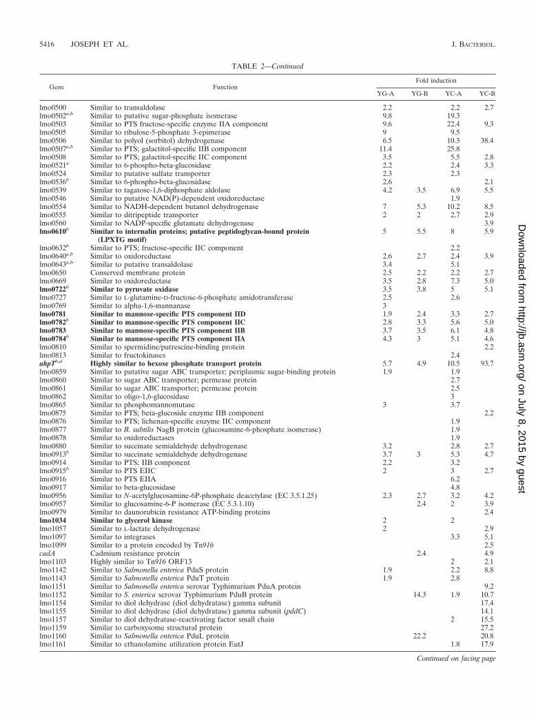
TABLE 2—Continued
Gene FunctionFold induction
YG-A YG-B YC-A YC-B
lmo0500 Similar to transaldolase 2.2 2.2 2.7lmo0502a,b Similar to putative sugar-phosphate isomerase 9.8 19.3lmo0503 Similar to PTS fructose-specific enzyme IIA component 9.6 22.4 9.3lmo0505 Similar to ribulose-5-phosphate 3-epimerase 9 9.5lmo0506 Similar to polyol (sorbitol) dehydrogenase 6.5 10.3 38.4lmo0507a,b Similar to PTS; galactitol-specific IIB component 11.4 25.8lmo0508 Similar to PTS; galactitol-specific IIC component 3.5 5.5 2.8lmo0521a Similar to 6-phospho-beta-glucosidase 2.2 2.4 3.3lmo0524 Similar to putative sulfate transporter 2.3 2.3lmo0536b Similar to 6-phospho-beta-glucosidase 2.6 2.1lmo0539 Similar to tagatose-1,6-diphosphate aldolase 4.2 3.5 6.9 5.5lmo0546 Similar to putative NAD(P)-dependent oxidoreductase 1.9lmo0554 Similar to NADH-dependent butanol dehydrogenase 7 5.3 10.2 8.5lmo0555 Similar to ditripeptide transporter 2 2 2.7 2.9lmo0560 Similar to NADP-specific glutamate dehydrogenase 3.9lmo0610b Similar to internalin proteins; putative peptidoglycan-bound protein
(LPXTG motif)5 5.5 8 5.9
lmo0632b Similar to PTS; fructose-specific IIC component 2.2lmo0640a,b Similar to oxidoreductase 2.6 2.7 2.4 3.9lmo0643a,b Similar to putative transaldolase 3.4 5.1lmo0650 Conserved membrane protein 2.5 2.2 2.2 2.7lmo0669 Similar to oxidoreductase 3.5 2.8 7.3 5.0lmo0722b Similar to pyruvate oxidase 3.5 3.8 5 5.1lmo0727 Similar to L-glutamine-D-fructose-6-phosphate amidotransferase 2.5 2.6lmo0769 Similar to alpha-1,6-mannanase 3lmo0781 Similar to mannose-specific PTS component IID 1.9 2.4 3.3 2.7lmo0782b Similar to mannose-specific PTS component IIC 2.8 3.3 5.6 5.0lmo0783 Similar to mannose-specific PTS component IIB 3.7 3.5 6.1 4.8lmo0784b Similar to mannose-specific PTS component IIA 4.3 3 5.1 4.6lmo0810 Similar to spermidine/putrescine-binding protein 2.2lmo0813 Similar to fructokinases 2.4uhpTb,d Highly similar to hexose phosphate transport protein 5.7 4.9 10.5 93.7lmo0859 Similar to putative sugar ABC transporter; periplasmic sugar-binding protein 1.9 1.9lmo0860 Similar to sugar ABC transporter; permease protein 2.7lmo0861 Similar to sugar ABC transporter; permease protein 2.5lmo0862 Similar to oligo-1,6-glucosidase 3lmo0865 Similar to phosphomannomutase 3 3.7lmo0875 Similar to PTS; beta-glucoside enzyme IIB component 2.2lmo0876 Similar to PTS; lichenan-specific enzyme IIC component 1.9lmo0877 Similar to B. subtilis NagB protein (glucosamine-6-phosphate isomerase) 1.9lmo0878 Similar to oxidoreductases 1.9lmo0880 Similar to succinate semialdehyde dehydrogenase 3.2 2.8 2.7lmo0913b Similar to succinate semialdehyde dehydrogenase 3.7 3 5.3 4.7lmo0914 Similar to PTS; IIB component 2.2 3.2lmo0915b Similar to PTS EIIC 2 3 2.7lmo0916 Similar to PTS EIIA 6.2lmo0917 Similar to beta-glucosidase 4.8lmo0956 Similar to N-acetylglucosamine-6P-phosphate deacetylase (EC 3.5.1.25) 2.3 2.7 3.2 4.2lmo0957 Similar to glucosamine-6-P isomerase (EC 5.3.1.10) 2.4 2 3.9lmo0979 Similar to daunorubicin resistance ATP-binding proteins 2.4lmo1034 Similar to glycerol kinase 2 2lmo1057 Similar to L-lactate dehydrogenase 2 2.9lmo1097 Similar to integrases 3.3 5.1lmo1099 Similar to a protein encoded by Tn916 2.5cadA Cadmium resistance protein 2.4 4.9lmo1103 Highly similar to Tn916 ORF13 2 2.1lmo1142 Similar to Salmonella enterica PduS protein 1.9 2.2 8.8lmo1143 Similar to Salmonella enterica PduT protein 1.9 2.8lmo1151 Similar to Salmonella enterica serovar Typhimurium PduA protein 9.2lmo1152 Similar to S. enterica serovar Typhimurium PduB protein 14.3 1.9 10.7lmo1154 Similar to diol dehydrase (diol dehydratase) gamma subunit 17.4lmo1155 Similar to diol dehydrase (diol dehydratase) gamma subunit (pddC) 14.1lmo1157 Similar to diol dehydratase-reactivating factor small chain 2 15.5lmo1159 Similar to carboxysome structural protein 27.2lmo1160 Similar to Salmonella enterica PduL protein 22.2 20.8lmo1161 Similar to ethanolamine utilization protein EutJ 1.8 17.9
Continued on facing page
5416 JOSEPH ET AL. J. BACTERIOL.
on July 8, 2015 by guesthttp://jb.asm
.org/D
ownloaded from

TABLE 2—Continued
Gene FunctionFold induction
YG-A YG-B YC-A YC-B
lmo1164 Highly similar to Salmonella enterica PduO protein 13.5 12.1lmo1165 Similar to ethanolamine utilization protein EutE 11.3lmo1166b Similar to NADPH-dependent butanol dehydrogenase 6.4glpF Similar to glycerol uptake facilitator protein 13.3 15.3ackA2 Similar to acetate kinase 3.6lmo1180 Similar to putative carboxysome structural protein 2.5lmo1205 Similar to putative cobalt transport protein CbiN 6.1lmo1207 Similar to cobalt transport ATP-binding protein CbiO 8.7glpDa,b Similar to glycerol-3-phosphate dehydrogenase 29.6 24.5 46.5 77.7glnA Highly similar to glutamine synthetases 2.3 1.9lmo1349a,b Similar to glycine dehydrogenase (decarboxylating) subunit 1 2.4 2 3.6lmo1350a,b Similar to glycine dehydrogenase (decarboxylating) subunit 2 2 2 2.6lmo1375 Similar to aminotripeptidase 1.8 2.1lmo1389 Similar to sugar ABC transporter, ATP-binding protein 1.8lmo1390 Similar to ABC transporter (permease proteins) 2lmo1391 Similar to sugar ABC transporter, permease protein 1.9pflBb Pyruvate formate-lyase 2 2.7 3.0pflC Pyruvate formate-lyase-activating enzyme 2 2.2lmo1421 Similar to glycine betaine/carnitine/choline ABC transporter (ATP-binding protein) 1.9opuCD Similar to betaine/carnitine/choline ABC transporter (membrane protein) 1.8 2.6 2.1opuCC Similar to glycine betaine/carnitine/choline ABC transporter (osmoprotectant-
binding protein)2.3 1.9
opuCB Similar to glycine betaine/carnitine/choline ABC transporter (membrane protein) 2.8opuCA Similar to glycine betaine/carnitine/choline ABC transporter (ATP-binding protein) 2 2.3zurA Metal (zinc) transport protein(ABC transporter, ATP-binding protein) 2.3glyQ Similar to glycyl-tRNA synthetase alpha chain 2lmo1538a,b Similar to glycerol kinase 13.5 17.8 26.3 51.1lmo1539 Similar to glycerol uptake facilitator 17.1 21.6 43.8 57.1thrS Threonyl-tRNA synthetase 1.9 3.0lmo1579 Similar to alanine dehydrogenase 1.9argJ Highly similar to ornithine acetyltransferase and amino-acid acetyltransferases 2.1argC Similar to N-acetylglutamate gamma-semialdehyde dehydrogenases 2.7trpA Highly similar to tryptophan synthase (alpha subunit) 2.2trpB Highly similar to tryptophan synthase (beta subunit) 2trpF Phosphoribosyl anthranilate isomerase 2.6trpC Highly similar to indol-3-glycerol phosphate synthases 2.7trpD Highly similar to anthranilate phosphoribosyltransferase 2.8trpG Highly similar to anthranilate synthase beta subunit 2.3trpE Highly similar to anthranilate synthase alpha subunit 1.9lmo1671 Similar to ABC transporter and adhesion proteins 1.8inlCb Internalin C 5.5 3.9 16.3pyrE Highly similar to orotate phosphoribosyltransferases 3.5 2.1pyrF Highly similar to orotidine 5 -phosphate decarboxylases 4.4 2.2pyrD Highly similar to dihydroorotase dehydrogenase 4 2.1pyrDII Highly similar to dihydroorotate dehydrogenase (electron transfer subunit) 2.4pyrAB Highly similar to carbamoyl-phosphate synthetase (catalytic subunit) 2.8 2.2pyrAa Highly similar to carbamoyl-phosphate synthetase (glutaminase subunit) 2 2.8pyrC Highly similar to dihydroorotase 4.5lmo1867 Similar to pyruvate phosphate dikinase 4.2 8.6 7.1 10.0lmo1883a,b Similar to chitinases 2.3 4.1 2.8 6.6pflAa,b Similar to pyruvate formate-lyase 1.8pnpb Similar to purine-nucleoside phosphorylase 2.2 2.1drm Similar to phosphopentomutase 2.5 2.7 3.5 4.1fhuG Similar to ferrichrome ABC transporter (permease) 2.1lmo1972 Similar to pentitol PTS; EIIB component 2 1.9 2.3lmo1992 Similar to alpha-acetolactate decarboxylase 2.5lmo1997 Similar to PTS mannose-specific enzyme IIA component 2.2lmo1998 Similar to opine catabolism protein 3.1 5.8lmo1999b weakly similar to glucosamine-fructose-6-phosphate aminotransferase 2.6 3.4 3.2lmo2000 Similar to PTS mannose-specific EIID component 4.8 7.6lmo2001a,b Similar to PTS mannose-specific EIIC component 3.4 6.2 5.3lmo2002 Similar to PTS mannose-specific EIIB component 3.2 4 2.3alsS Similar to alpha-acetolactate synthase protein (AlsS) 2.3 2.0lmo2007 Weakly similar to putative sugar-binding lipoproteins 1.8lmo2008 Similar to putative ABC transporter; permease protein 2.1 2.6lmo2015 Similar to alpha-mannosidase 1.9
Continued on following page
VOL. 190, 2008 GLYCEROL METABOLISM AND PrfA ACTIVITY 5417
on July 8, 2015 by guesthttp://jb.asm
.org/D
ownloaded from

TABLE 2—Continued
Gene FunctionFold induction
YG-A YG-B YC-A YC-B
ileS Isoleucyl-tRNA synthetase 2.4lmo2067 Similar to conjugated bile acid hydrolase 6.0 4.8 4.8 9.0lmo2085b Putative peptidoglycan-bound protein (LPXTG motif) 3.8 4.7 6.3 9.0lmo2098 Similar to PTS; galactitol-specific EIA component 3.3 3.2lmo2108 Similar to N-acetylglucosamine-6-phosphate deacetylase 2.1lmo2109 Similar to hydrolase 1.8 2.5 2.5lmo2115 Similar to ABC transporter (permease) 3.2lmo2121a,b Similar to maltosephosphorylase 3.6lmo2122 Similar to maltodextrose utilization protein MalA 2.2 4lmo2123 Similar to maltodextrin ABC transport system (permease) 2.7 4.7lmo2124 Similar to maltodextrin ABC transport system (permease) 2 3.2lmo2125b Similar to maltose/maltodextrin ABC transporter (binding protein) 2.7 4.5lmo2134 Similar to fructose-1,6-biphosphate aldolase type II 2.5lmo2135 Similar to PTS; fructose-specific EIIC component 2.9lmo2136 Similar to PTS; fructose-specific EIIB component 3.2lmo2143 Weakly similar to mannose-6-phosphate isomerase 2.3lmo2159b Similar to oxidoreductase 2.2 2.6 3.1lmo2175 Similar to dehydrogenase 3.9 6.3fruA Highly similar to PTS fructose-specific EIIABC component 2.2 2.0lmo2341 Similar to carbohydrate kinases 2.2 3.2 2.6lmo2389 Similar to NADH dehydrogenase 1.8lmo2434 Highly similar to glutamate decarboxylases 3 2.5 4.2 2.7lmo2463 Similar to transport protein 2.1 2.5lmo2469 Similar to amino acid transporter 2.0lmo2569 Similar to dipeptide ABC transporter (dipeptide-binding protein) 2 4.2lmo2573b Similar to zinc-binding dehydrogenase 4.9 6.4 9.2 9.0lmo2580 Similar to ABC transporter; ATP-binding protein 1.8 5.1lmo2584a,b Similar to formate dehydrogenase-associated protein 7.6 8.4lmo2586a,b Similar to formate dehydrogenase alpha chain 13.9 28 39.8 40.5lmo2592 Similar to oxidoreductase; aldo/keto reductase family 2.0lmo2650b Similar to hypothetical PTS enzyme IIB component 2lmo2651a,b Similar to mannitol-specific PTS EIIA component 2.1lmo2659a Similar to ribulose-phosphate 3-epimerase 2.6 2.9lmo2660a Similar to transketolase 2.2lmo2663a Similar to polyol dehydrogenase 2.3 3 7.2lmo2664a Similar to sorbitol dehydrogenase 3.5 7.2 4.9 10.9lmo2665a Similar to PTS; galactitol-specific EIIC component 4.4 6.4 7.4 8.2lmo2666a,b Similar to PTS; galactitol-specific EIIB component 5.2 5 7.6 8.2lmo2667a Similar to PTS; galactitol-specific EIIA component 5.9 4.6 8.8 6.5lmo2674 Similar to ribose 5-phosphate epimerase 2.9 2.6 3.3 4.5kdpB Potassium-transporting ATPase B chain 4.4lmo2683 Similar to cellobiose phosphotransferase EIIB component 2.3 2.2lmo2684 Similar to cellobiose phosphotransferase EIIC component 6.3 3.1lmo2685b Similar to cellobiose phosphotransferase EIIA component 7.4 5.6lmo2689 Highly similar to Mg2� transport ATPase 2.4 3.1lmo2695b Similar to dihydroxyacetone kinase 4.7 4.9 9.3 9.8lmo2696b Similar to hypothetical dihydroxyacetone kinase 3.7 4.3 6.7 8.6lmo2708 Similar to PTS; cellobiose-specific EIIC 18.5 11.1 11.0lmo2733 Similar to PTS; fructose-specific IIABC component 3.3lmo2735b Similar to sucrose phosphorylase 2.1lmo2743 Similar to transaldolase 3.3 3 4.7 4.0lmo2760 Similar to ABC transporter (ATP-binding protein) 3.4 3.1lmo2764a,b Similar to xylose operon regulatory protein and to glucose kinase 2.2 1.9lmo2772a,b Similar to beta-glucoside-specific EIIABC 1.9bvrBa,b Beta-glucoside-specific phosphotransferase EIIABC component 1.8lmo2797b Similar to PTS mannitol-specific EIIA 3.5 5.6lmo2798b Similar to phosphatase 3.1 6.4lmo2799a,b Similar to PTS mannitol-specific EIIBC 3.9 2.7 11.5lmo2800 Similar to dehydrogenase 2.9 4lmo2848 Highly similar to l-rhamnose isomerase 1.9lmo2849 Similar to rhamnulokinase 2.2 2.3lmo2850 Similar to sugar transport proteins 2.1
a Genes upregulated in the ccpA mutant (33).b Genes upregulated in the hprK mutant (33).c Values indicate regulation, and no value indicates no regulation of the gene under the conditions mentioned. The genes discussed in this study are indicated in
boldface type. The complete list of differentially regulated genes is available in Table S2 in the supplemental material. Y, glycerol; G, glucose; C, cellobiose; A, phaseA; B, phase B.
d Genes shown to be directly regulated by PrfA.
5418 JOSEPH ET AL. J. BACTERIOL.
on July 8, 2015 by guesthttp://jb.asm
.org/D
ownloaded from
showed very high levels of upregulation (almost 200-fold) com-pared to those of cellobiose-grown L. monocytogenes cultures(Table 2), indicating that PrfA activity is high throughout thegrowth phase when L. monocytogenes grows in the presence ofglycerol and low in the presence of cellobiose. In the presenceof glucose, PrfA activity is low during early (balanced) growthphases (phase A) but is considerably enhanced in phase B,when bacterial growth may no longer be balanced, probablydue to reduced glucose uptake (33).
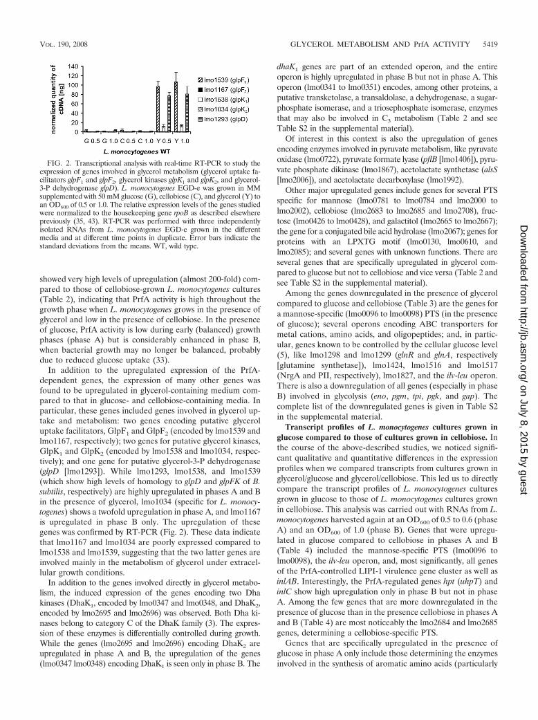
In addition to the upregulated expression of the PrfA-dependent genes, the expression of many other genes wasfound to be upregulated in glycerol-containing medium com-pared to that in glucose- and cellobiose-containing media. Inparticular, these genes included genes involved in glycerol up-take and metabolism: two genes encoding putative glyceroluptake facilitators, GlpF1 and GlpF2 (encoded by lmo1539 andlmo1167, respectively); two genes for putative glycerol kinases,GlpK1 and GlpK2 (encoded by lmo1538 and lmo1034, respec-tively); and one gene for putative glycerol-3-P dehydrogenase(glpD [lmo1293]). While lmo1293, lmo1538, and lmo1539(which show high levels of homology to glpD and glpFK of B.subtilis, respectively) are highly upregulated in phases A and Bin the presence of glycerol, lmo1034 (specific for L. monocy-togenes) shows a twofold upregulation in phase A, and lmo1167is upregulated in phase B only. The upregulation of thesegenes was confirmed by RT-PCR (Fig. 2). These data indicatethat lmo1167 and lmo1034 are poorly expressed compared tolmo1538 and lmo1539, suggesting that the two latter genes areinvolved mainly in the metabolism of glycerol under extracel-lular growth conditions.
In addition to the genes involved directly in glycerol metabo-lism, the induced expression of the genes encoding two Dhakinases (DhaK1, encoded by lmo0347 and lmo0348, and DhaK2,encoded by lmo2695 and lmo2696) was observed. Both Dha ki-nases belong to category C of the DhaK family (3). The expres-sion of these enzymes is differentially controlled during growth.While the genes (lmo2695 and lmo2696) encoding DhaK2 areupregulated in phase A and B, the upregulation of the genes(lmo0347 lmo0348) encoding DhaK1 is seen only in phase B. The
dhaK1 genes are part of an extended operon, and the entireoperon is highly upregulated in phase B but not in phase A. Thisoperon (lmo0341 to lmo0351) encodes, among other proteins, aputative transketolase, a transaldolase, a dehydrogenase, a sugar-phosphate isomerase, and a triosephosphate isomerase, enzymesthat may also be involved in C3 metabolism (Table 2 and seeTable S2 in the supplemental material).
Of interest in this context is also the upregulation of genesencoding enzymes involved in pyruvate metabolism, like pyruvateoxidase (lmo0722), pyruvate formate lyase (pflB [lmo1406]), pyru-vate phosphate dikinase (lmo1867), acetolactate synthetase (alsS[lmo2006]), and acetolactate decarboxylase (lmo1992).
Other major upregulated genes include genes for several PTSspecific for mannose (lmo0781 to lmo0784 and lmo2000 tolmo2002), cellobiose (lmo2683 to lmo2685 and lmo2708), fruc-tose (lmo0426 to lmo0428), and galactitol (lmo2665 to lmo2667);the gene for a conjugated bile acid hydrolase (lmo2067); genes forproteins with an LPXTG motif (lmo0130, lmo0610, andlmo2085); and several genes with unknown functions. There areseveral genes that are specifically upregulated in glycerol com-pared to glucose but not to cellobiose and vice versa (Table 2 andsee Table S2 in the supplemental material).
Among the genes downregulated in the presence of glycerolcompared to glucose and cellobiose (Table 3) are the genes fora mannose-specific (lmo0096 to lmo0098) PTS (in the presenceof glucose); several operons encoding ABC transporters formetal cations, amino acids, and oligopeptides; and, in partic-ular, genes known to be controlled by the cellular glucose level(5), like lmo1298 and lmo1299 (glnR and glnA, respectively[glutamine synthetase]), lmo1424, lmo1516 and lmo1517(NrgA and PII, respectively), lmo1827, and the ilv-leu operon.There is also a downregulation of all genes (especially in phaseB) involved in glycolysis (eno, pgm, tpi, pgk, and gap). Thecomplete list of the downregulated genes is given in Table S2in the supplemental material.
Transcript profiles of L. monocytogenes cultures grown inglucose compared to those of cultures grown in cellobiose. Inthe course of the above-described studies, we noticed signifi-cant qualitative and quantitative differences in the expressionprofiles when we compared transcripts from cultures grown inglycerol/glucose and glycerol/cellobiose. This led us to directlycompare the transcript profiles of L. monocytogenes culturesgrown in glucose to those of L. monocytogenes cultures grownin cellobiose. This analysis was carried out with RNAs from L.monocytogenes harvested again at an OD600 of 0.5 to 0.6 (phaseA) and an OD600 of 1.0 (phase B). Genes that were upregu-lated in glucose compared to cellobiose in phases A and B(Table 4) included the mannose-specific PTS (lmo0096 tolmo0098), the ilv-leu operon, and, most significantly, all genesof the PrfA-controlled LIPI-1 virulence gene cluster as well asinlAB. Interestingly, the PrfA-regulated genes hpt (uhpT) andinlC show high upregulation only in phase B but not in phaseA. Among the few genes that are more downregulated in thepresence of glucose than in the presence cellobiose in phases Aand B (Table 4) are most noticeably the lmo2684 and lmo2685genes, determining a cellobiose-specific PTS.
Genes that are specifically upregulated in the presence ofglucose in phase A only include those determining the enzymesinvolved in the synthesis of aromatic amino acids (particularly
FIG. 2. Transcriptional analysis with real-time RT-PCR to study theexpression of genes involved in glycerol metabolism (glycerol uptake fa-cilitators glpF1 and glpF2, glycerol kinases glpK1 and glpK2, and glycerol-3-P dehydrogenase glpD). L. monocytogenes EGD-e was grown in MMsupplemented with 50 mM glucose (G), cellobiose (C), and glycerol (Y) toan OD600 of 0.5 or 1.0. The relative expression levels of the genes studiedwere normalized to the housekeeping gene rpoB as described elsewherepreviously (35, 43). RT-PCR was performed with three independentlyisolated RNAs from L. monocytogenes EGD-e grown in the differentmedia and at different time points in duplicate. Error bars indicate thestandard deviations from the means. WT, wild type.
VOL. 190, 2008 GLYCEROL METABOLISM AND PrfA ACTIVITY 5419
on July 8, 2015 by guesthttp://jb.asm
.org/D
ownloaded from

TABLE 3. Genes downregulated in glycerol compared to glucose and cellobiose at early log phase (phase A) andlate log phase (phase B) in MMc
Gene FunctionFold induction
YG-A YG-B YC-A YC-B
lmo0018 Beta-glucosidase 0.3 0.3lmo0050 Similar to sensor histidine kinase (AgrC from Staphylococcus) 0.4purA Similar to adenylosuccinate synthetase 0.3 0.3lmo0096 Similar to PTS; mannose-specific, factor IIAB 0.1 0.1lmo0097 Similar to PTS; mannose-specific, factor IIC 0.1 0.1lmo0098 Similar to PTS; mannose-specific, factor IID 0.1 0.1lmo0135 Similar to oligopeptide ABC transport system substrate-binding proteins 0.5lmo0152 Similar to oligopeptide ABC transporter-binding protein 0.4 0.6lmo0176 Similar to glucose uptake protein 0.4 0.4 0.4lmo0218 Polyribonucleotide nucleotidyltransferase domain present 0.5 0.5 0.4lmo0219 Fusion protein; N-terminal part similar to B. subtilis YacA protein; C-terminal part
similar to hypoxanthine-guanine phosphoribosyltransferase0.5 0.4
cysE Similar to serine O-acetyltransferase 0.4 0.5lmo0269 Similar to transporter 0.4lmo0271 Similar to phospho-beta-glucosidase 0.4 0.3lmo0279 Similar to anaerobic ribonucleoside-triphosphate reductase 0.5 0.4lmo0280 Similar to anaerobic ribonucleotide reductase activator protein 0.3 0.5lmo0286 Similar to aminotransferase 0.5 0.4lmo0519b Similar to multidrug resistance protein 0.5lmo0537 Similar to N-carbamyl-l-amino acid amidohydrolase 0.5lmo0560b Similar to NADP-specific glutamate dehydrogenase 0.5 0.5hisD Similar to histidinol dehydrogenases 0.5hisZ Histidyl-tRNA synthetase 0.5 0.5lmo0611 Similar to acyl-carrier protein phosphodiesterase and NAD(P)H dehydrogenase 0.5 0.5lmo0645 Similar to amino acid transporter 0.5 0.5lmo0787 Similar to amino acid transporter 0.5lmo0798 Similar to lysine-specific permease 0.3lmo0802 Weakly similar to GTP-pyrophosphokinase 0.5 0.4lmo0837 Similar to ABC transporter (ATP-binding protein) 0.5lmo0841 Similar to cation (calcium) transporting ATPase 0.5 0.4lmo0847 Similar to glutamine ABC transporter (binding and transport protein) 0.5lmo0897 Similar to transport proteins 0.4lmo0912a Similar to transporters (formate) 0.5lmo0945 Similar to C-terminal part of B. subtilis ComEC protein and to ComEA 0.5lmo0947 Hypothetical transport protein 0.5lmo0960 Similar to proteases 0.4 0.4 0.5lmo0981 Similar to efflux transporter 0.4gbuA Similar to glycine betaine ABC transporter (ATP-binding protein) 0.4 0.5gbuB Similar to glycine betaine ABC transporters (permease) 0.5 0.4 0.4gbuCa,b Similar to glycine betaine ABC transporters (glycine betaine-binding protein) 0.5 0.4lmo1017 Similar to phosphotransferase system glucose-specific enzyme IIA 0.5lmo1073 Similar to metal binding protein (ABC transporter) 0.5pheS Phenylalany-tRNA synthetase alpha subunit 0.2pheT Phenylalanyl-tRNA synthetase beta subunit 0.3proA Gamma-glutamyl phosphate reductase 0.5proB Gamma-glutamyl kinase 0.6glnR Similar to glutamine synthetase repressor 0.1 0.1glnA Similar to glutamine synthetases 0.1lmo1300 Similar to arsenic efflux pump protein 0.4smbA Similar to uridylate kinases 0.4 0.5lmo1424 Similar to manganese transport proteins NRAMP 0.3 0.3 0.3 0.4lmo1431 Similar to ABC transporter (ATP-binding protein) 0.5zurM Metal (zinc) transport protein (ABC transporter, permease protein) 0.5udk Similar to uridine kinase 0.5 0.5lmo1516b Similar to ammonium transporter NrgA 0.02 0.03lmo1517b Similar to nitrogen regulatory PII protein 0.02 0.03relA Similar to (p)ppGpp synthetase 0.5valS Valyl-tRNA synthetase 0.5tyrS Tyrosyl-tRNA synthetase 0.5 0.4aroA 3-Deoxy-D-arabino-heptulosonate 7-phosphate synthase 0.4 0.2 0.1lmo1603 Similar to aminopeptidase 0.5lmo1617 Similar to multidrug-efflux transporter 0.5 0.4daaA D-Amino acid aminotransferase 0.5lmo1624 Similar to putative transporters 0.2lmo1625 Similar to putative transporters 0.3 0.1
Continued on facing page
5420 JOSEPH ET AL. J. BACTERIOL.
on July 8, 2015 by guesthttp://jb.asm
.org/D
ownloaded from

TABLE 3—Continued
Gene FunctionFold induction
YG-A YG-B YC-A YC-B
trpA Similar to tryptophan synthase (alpha subunit) 0.2 0.1trpB Similar to tryptophan synthase (beta subunit) 0.2 0.1trpF Phosphoribosyl anthranilate isomerase 0.3 0.2trpC Similar to indol-3-glycerol phosphate synthases 0.3 0.1trpD Similar to anthranilate phosphoribosyltransferase 0.4 0.2trpG Similar to anthranilate synthase beta subunit 0.4 0.3trpE Similar to anthranilate synthase alpha subunit 0.5 0.4lmo1634 Similar to alcohol-acetaldehyde dehydrogenase 0.2 0.3 0.5 0.1ansB Similar to asparaginyl-tRNA synthetases 0.5 0.4metK Similar to S-methionine adenosyltransferase 0.5lmo1682 Similar to transmembrane transport proteins 0.4lmo1705 Similar to deoxyguanosine kinase/deoxyadenosine kinase(I) subunit 0.5lmo1719 Similar to PTS lichenan-specific EIIA component 0.5lmo1720 Similar to PTS lichenan-specific EIIB component 0.4lmo1730 Similar to sugar ABC transporter-binding protein 0.3lmo1739 Similar to amino acid (glutamine) ABC transporter (ATP-binding protein) 0.6lmo1749 Similar to shikimate kinase 0.3 0.3purD Phosphoribosylglycinamide synthetase 0.5 0.5purH Bifunctional phosphoribosylaminoimidazole carboxy formyl formyltransferase and
inosine-monophosphate cyclohydrolase0.5 0.5
purN Similar to phosphoribosylglycinamide formyltransferases 0.5 0.4purM Phosphoribosylaminoimidazole synthetase 0.5purF Glutamine phosphoribosylpyrophosphate amidotransferase 0.4purQ Phosphoribosylformylglycinamidine synthetase I 0.4 0.4purQ Phosphoribosylformylglycinamidine synthetase I 0.4 0.5 0.4purL Similar to phosphoribosylformylglycinamidine synthetase II 0.4 0.4 0.2 0.4purC Phosphoribosylaminoimidazole succinocarboxamide synthetase 0.5 0.5 0.2 0.4purB Adenylosuccinate lyase 0.3 0.5 0.2purK Phosphoribosylaminoimidazole carboxylase II 0.4 0.3purE Phosphoribosylaminoimidazole carboxylase I 0.2 0.4 0.1lmo1778 Similar to ABC transporter (ATP-binding protein) 0.4rncS Similar to RNase III 0.4lmo1827 Similar to guanylate kinases 0.5 0.5pyrP Similar to uracil permease 0.4lmo1847 Similar to adhesion binding proteins and lipoproteins with multiple specificity for
metal cations (ABC transporter)0.4 0.5
lmo1848 Similar metal cations ABC transporter (permease protein) 0.4 0.4 0.4 0.5lmo1849 Similar to metal cation ABC transporter, ATP-binding proteins 0.3 0.4 0.2 0.5lmo1884 Similar to xanthine permeases 0.3 0.4 0.3 0.4lmo1885 Similar to xanthine phosphoribosyltransferase 0.3 0.4 0.4 0.4aroE Similar to 5-enolpyruvylshikimate-3-phosphate synthase 0.3tyrA Similar to prephenate dehydrogenase 0.4 0.2hisC Similar to histidinol-phosphate aminotransferase and tyrosine/phenylalanine
aminotransferase0.5 0.3
lmo1926 Similar to chorismate mutase 0.4 0.3aroB Similar to 3-dehydroquinate synthase 0.4 0.2aroF Similar to chorismate synthase 0.4 0.2gpsA Similar to NAD(P)H-dependent glycerol-3-phosphate dehydrogenase 0.6lysA Similar to diaminopimelate decarboxylase 0.5fhuC Similar to ferrichrome ABC transporter (ATP-binding protein) 0.5lmo1976 Similar to oxidoreductase 0.5lmo1978 Similar to glucose-6-phosphate 1-dehydrogenase 0.4 0.5 0.4 0.3ilvD Similar to dihydroxy acid dehydratase 0.4 0.4 0.5 0.3ilvB Similar to acetolactate synthase (acetohydroxy acid synthase) (large subunit) 0.5leuA Similar to 2-isopropylmalate synthase 0.5leuBb Similar to 3-isopropylmalate dehydrogenase 0.5lmo2075 Similar to glycoprotein endopeptidase 0.5lmo2110 Similar to mannnose-6 phosphate isomerase 0.3 0.5lmo2114 Similar to ABC transporter (ATP-binding protein) 0.3lmo2152 Similar to thioredoxin 0.5lmo2153 Similar to flavodoxin 0.5lmo2192b Similar to oligopeptide ABC transporter (ATP-binding protein) 0.5lmo2193b Similar to oligopeptide ABC transporter (ATP-binding protein) 0.5lmo2194b Similar to oligopeptide ABC transporter (permease) 0.4 0.5lmo2195b Similar to oligopeptide ABC transporter (permease) 0.5 0.5lmo2196 Similar to pheromone ABC transporter (binding protein) 0.4 0.3
Continued on following page
VOL. 190, 2008 GLYCEROL METABOLISM AND PrfA ACTIVITY 5421
on July 8, 2015 by guesthttp://jb.asm
.org/D
ownloaded from

tryptophan). The trp genes are, however, downregulated inphase B (Table 4).
Growth of mutants defective in glycerol uptake and metab-olism under extra- and intracellular conditions. To study thefunctions of the genes that are most likely involved in glyceroluptake and metabolism, we constructed mutants carrying in-frame deletions of various genes involved in glycerol metabo-lism in L. monocytogenes (Table 1) and tested their growth inMM supplemented with glycerol as a carbon source in com-parison to that of the wild-type strain.
The deletion of genes encoding the two putative glyceroluptake facilitators had little effect on growth (Fig. 3A), sug-gesting that in the presence of 50 mM glycerol, which was usedin these studies, the free diffusion of glycerol provides sufficientsubstrate for glycerol-driven metabolism. In contrast, the de-letion of the glycerol kinase 1 (GlpK1) encoded by lmo1538(part of the glpFK operon) abolished the ability to grow inglycerol-containing MM entirely, indicating that the secondputative glycerol kinase (GlpK2), encoded by lmo1034, cannotreplace the loss of GlpK1, at least not under the applied in vitro
growth conditions. In accord with this assumption is the ob-servation that the deletion of lmo1034 did not affect growth inglycerol-containing medium (Fig. 3A). The deletion oflmo1293 (glpD), encoding glycerol-3-P dehydrogenase, also ledto the complete loss of growth in the presence of glycerol (Fig.3A). To further characterize these mutants (�glpk1 and�glpD), these genes were complemented in the deletion mu-tants, and as can be seen in Fig. 3B, a wild-type phenomenoncould be restored in these complemented strains with respectto growth in MM containing glycerol.
As recently reported (26), mutants with insertions in glpK1
and glpD obtained from a random insertion mutant libraryshowed reduced levels of growth in Caco-2 cells. We thereforetested the intracellular replication of the above-mentioned de-letion mutants in Caco-2 cells and J774 macrophages. In thesegrowth studies, the mammalian host cells were precultured ina glucose-containing cell culture medium. The glpD (lmo1293)deletion mutant as well as the mutant with a deletion in glpK1
(lmo1538) showed a modest but significant reduction in intra-cellular replication in Caco-2 cells (Fig. 3B). Interestingly, the
TABLE 3—Continued
Gene FunctionFold induction
YG-A YG-B YC-A YC-B
lmo2238 Similar to transport system permease protein 0.5 0.3arpJ Similar to amino acid ABC transporter, permease protein 0.5 0.3lmo2346 Similar to amino acid ABC transporter, ATP-binding protein 0.5lmo2348 Similar to amino acid ABC transporter (permease) 0.4 0.5lmo2349 Similar to amino acid ABC transporter (binding protein) 0.5lmo2355 Similar to multidrug resistance protein 0.5 0.5lmo2371 Similar to putative ABC transporter transmembrane subunit 0.5lmo2372 Similar to ABC transporter ATP binding proteins 0.5lmo2374 Similar to aspartate kinase 0.5 0.5lmo2377 Similar to multidrug resistance efflux pump 0.5 0.6lmo2421 Similar to two-component sensor histidine kinase 0.5lmo2430 Similar to B. subtilis ferrichrome ABC transporter (permease) FhuG 0.5 0.5lmo2431 Similar to B. subtilis ferrichrome ABC transporter fhuD precursor
(ferrichrome-binding protein)0.5
eno Similar to enolase 0.4pgm Similar to phosphoglycerate mutase 0.4 0.3tpi Similar to triose phosphate isomerase 0.5 0.3pgk Similar to phosphoglycerate kinase 0.3 0.3gap Similar to glyceraldehyde-3-phosphate dehydrogenase 0.5 0.4 0.3atpH Similar to H�-transporting ATP synthase chain delta 0.4atpF Similar to H�-transporting ATP synthase chain b 0.5atpE Similar to H�-transporting ATP synthase chain c 0.5atpB Similar to H�-transporting ATP synthase chain a 0.5atpI Similar to ATP synthase subunit i 0.5upp Similar to uracil phosphoribosyltransferase 0.5glyA Similar to glycine hydroxymethyltransferase 0.5hom Similar to homoserine dehydrogenase 0.5 0.5 0.4 0.4fbaA Similar to fructose-1,6-bisphosphate aldolase 0.5 0.4lmo2601 Similar to ABC transporter (ATP-binding protein) 0.5lmo2684 Similar to cellobiose phosphotransferase EIIC component 0.6lmo2720 Similar to acetate-coenzyme A ligase 0.5 0.4 0.4serS Seryl-tRNA synthetase 0.5 0.2guaB Similar to inosine-monophosphate dehydrogenase 0.5lmo2769 Similar to ABC transporter, ATP-binding protein 0.5 0.5lmo2824 Similar to D-3-phosphoglycerate dehydrogenase 0.6serC Similar to phosphoserine aminotransferase 0.4
a Gene downregulated in the ccpA mutant (33).b Gene downregulated in the hprK mutant (33).c Values indicate regulation, and no value indicates no regulation of the gene under the conditions mentioned. The genes discussed in this study are indicated in
boldface type. The complete list of differentially regulated genes is available in Table S2 in the supplemental material. Y, glycerol; G, glucose; C, cellobiose; A, phaseA; B, phase B.
5422 JOSEPH ET AL. J. BACTERIOL.
on July 8, 2015 by guesthttp://jb.asm
.org/D
ownloaded from

mutant with the deletion in lmo1034, which encodes GlpK2 (anL. monocytogenes-specific glycerol kinase), also exhibited amodest but significant reduction in levels of intracellular rep-lication in Caco-2 cells, which was more pronounced than thatin the glpK1 (lmo1538) deletion mutant. In J774 macrophages,only the inactivation of glpD led to a significant growth reduc-tion (Fig. 3B), suggesting a cell type-specific dependency onthe glycerol kinase activity.
Growth of L. monocytogenes in the presence of dihydroxyac-etone. The presence of two Dha kinases in L. monocytogenesand the high level of upregulation of the encoding genes in thepresence of glycerol suggest that Dha may also be a carbonsource for L. monocytogenes. When MM was supplementedwith 50 mM Dha instead of glycerol, no growth of L. monocy-togenes was observed (data not shown), but growth in thepresence of Dha was observed when the bacteria where pre-
TABLE 4. Genes differentially regulated in glucose compared to cellobiose at early log phase (phase A) and latelog phase (phase B) in MMa
Gene FunctionFold induction
GC-A GC-B
lmo0018 Beta-glucosidase 0.2lmo0096b Similar to PTS; mannose-specific, factor IIAB 15.8 10.3lmo0097 Similar to PTS; mannose-specific, factor IIC 13.4 9.5lmo0098b Similar to PTS; mannose-specific, factor IID 12.6 8.6prfAc Listeriolysin positive regulatory protein 5.8 46.2plcAb,c Phosphatidylinositol-specific phospholipase c 9.2 48.6hlyb,c Listeriolysin O precursor 10.4 46.1mplc Zinc metalloproteinase precursor 4.5 47.3actAc Actin assembly-inducing protein precursor 15.2 64.8plcBb,c Phospholipase C 12.6 117lmo0271 Highly similar to phospho-beta-glucosidase 0.3inlAb,c Internalin A 4.5 7.7inlBb,c Internalin B 3.2 5.9lmo0560 Similar to NADP-specific glutamate dehydrogenase 2.1uhpTb,c Highly similar to hexose phosphate transport protein 61.8lmo0914 Similar to PTS, IIB component 4.1pheS Phenylalanyl-tRNA synthetase alpha subunit 0.3pheT Phenylalanyl-tRNA synthetase beta subunit 0.4tcsA CD4� T-cell-stimulating antigen; lipoprotein 0.5zurA Metal (zinc) transport protein(ABC transporter, ATP-binding protein) 1.9valS Valyl-tRNA synthetase 3.4aroA 3-Deoxy-D-arabino-heptulosonate 7-phosphate synthase 0.5lmo1625 Similar to putative transporters 0.4trpA Highly similar to tryptophan synthase (alpha subunit) 2.9 0.3trpB Highly similar to tryptophan synthase (beta subunit) 4.7 0.3trpF Phosphoribosyl anthranilate isomerase 7 0.4trpC Highly similar to indol-3-glycerol phosphate synthases 7.8 0.4trpD Highly similar to anthranilate phosphoribosyltransferase 8.1 0.4lmo1719 Similar to PTS lichenan-specific enzyme IIA component 0.4lmo1734 Similar to glutamate synthase (large subunit) 2inlCc Internalin C 55.5ilvB Similar to acetolactate synthase (acetohydroxy acid synthase) (large subunit) 2ilvN Similar to acetolactate synthase (acetohydroxy acid synthase) (small subunit) 2ilvCb Similar to ketol acid reductoisomerase (acetohydroxy acid isomeroreductase) 1.9ilvA Similar to threonine dehydratase 1.9lmo2114 Similar to ABC transporter (ATP-binding protein) 7.2lmo2115 Similar to ABC transporter (permease) 7arpJ Similar to amino acid ABC transporter; permease protein 0.5lmo2390 Similar to hypothetical thioredoxin reductase 1.9lmo2469 Similar to amino acid transporter 2.3glyA Highly similar to glycine hydroxymethyltransferase 0.4lmo2580 Similar to ABC transporter; ATP-binding protein 2.3lmo2650b Similar to hypothetical PTS EIIB component 3.4lmo2651 Similar to mannitol-specific PTS EIIA component 2.7lmo2684 Similar to cellobiose phosphotransferase EIIC component 0.1 0.3lmo2685 Similar to cellobiose phosphotransferase EIIA component 0.1 0.2cydD Highly similar to ABC transporter (ATP-binding protein) required for expression of cytochrome bd 0.5cydC Highly similar to ABC transporter required for expression of cytochrome bd 0.5serS Seryl-tRNA synthetase 0.4
a Values indicate regulation, and no value indicates no regulation of the gene under the conditions mentioned. The genes discussed in this study are indicated inboldface type. The complete list of differentially regulated genes are available in Table S2 in the supplemental material. G, glucose; C, cellobiose; A, phase A; B,phase B.
b Gene regulated in the hprK mutant (33).c Gene shown to be directly regulated by PrfA.
VOL. 190, 2008 GLYCEROL METABOLISM AND PrfA ACTIVITY 5423
on July 8, 2015 by guesthttp://jb.asm
.org/D
ownloaded from

incubated in glycerol-containing medium in order to inducethe two DhaKs (Fig. 4).
The ptsH mutant was unable to grow in Dha-containing me-dium, which suggests that listerial DhaKs (both share the typicalstructure of category C DhaK) (3) are activated by HPr-His-P andtransfer the energy-rich phosphate to Dha, generating Dha-P(17).
PrfA activation is due to glycerol metabolism and not toglycerol itself. A recent study (33) and the data describedabove (Table 2) indicated that PrfA is activated in glycerol-containing MM. A previous structural analysis of PrfA showedthat glycerol can tightly bind to PrfA (Protein Data Bank record41 [http://www.rcsb.org/pdb/explore.do?structureId�10MI]). Totest whether the binding of glycerol may directly activate PrfA,
FIG. 3. (A) Growth of wild-type L. monocytogenes EGD-e (WT), and glycerol metabolism mutants �lmo1539, �lmo1167, and �lmo1034 inglycerol-containing MM at 37°C under aeration. (B) Growth of wild-type L. monocytogenes EGD-e and glycerol metabolism mutants �lmo1538(glpK1), �lmo1293 (glpD), and the complementation mutants of glpK1 and glpD in glycerol-containing MM at 37°C under aeration. (C) Effect ofnonpolar deletions of lmo1293 (glpD), lmo1538 (glpK1), and lmo1034 (glpK2) on the intracellular replication of L. monocytogenes EGD-e. Caco-2epithelial cells or J774 macrophages were infected with either the wild-type strain or the mutants to an MOI of 10 (Caco-2) or an MOI of 1 (J774),and the numbers of bacteria recovered after 1, 3, 5, and 7 h of infection were determined. Three independent infections were performed for eachstrain. Error bars represent the standard deviations from the means.
5424 JOSEPH ET AL. J. BACTERIOL.
on July 8, 2015 by guesthttp://jb.asm
.org/D
ownloaded from

we studied PrfA activity in the glpD and glpK1 mutants, whichare still able to take up but are unable to catabolize glycerol.For this goal, the wild-type strain and the two mutants weregrown in BHI broth to early log phase (OD600 of 0.5). After awash in PBS, one half was shifted into glycerol-containing MM,while the other half was shifted into glucose-containing MM.The hemolytic activity, taken as a measure for the PrfAactivity, was determined 2 h after the shift. As shown in Fig. 5,the wild-type strain was still able to express the PrfA-dependent hly gene after shift into glycerol- or glucose-containing medium, as expected, while the glpK1 or the glpDmutant expressed the hly gene only in the glucose-containingbut not in the glycerol-containing medium, suggesting thatglycerol by itself does not activate PrfA. (The hemolytic activityof the wild-type strain grown in BHI [not shown in Fig. 5] is
very low [0.1 OD543 units], and the hemolytic activityobserved in the glpK1 or the glpD mutant after the shift fromBHI broth into glycerol-containing MM remains at this lowlevel.)
In addition, we purified PrfA using buffers without glycerol anddetermined the specific activities of both PrfA preparations in thepreviously established in vitro runoff transcription assay usingreaction buffers with and without glycerol (6, 29, 30). In vitrotranscription was initiated at the PrfA-dependent hpt (uhpT) pro-moter (Phpt) as previously described (48). As shown in Fig. 6,PrfA activities with glycerol and those without added glycerolwere identical. These results along with those of the above-de-scribed hemolytic activity assays suggest that glycerol by itself doesnot activate PrfA directly but rather that components connectedwith glycerol metabolism may modulate PrfA activity.
CCR control and phosphorylation of HPr in L. monocyto-genes cultures grown in the presence of glycerol. The compar-ative transcript profiles obtained with RNAs from glycerol-grown and glucose- or cellobiose-grown L. monocytogenescultures indicated an induced expression of many genes in theglycerol-grown L. monocytogenes cultures that were recentlyshown to be upregulated in a ccpA mutant, an hprK mutant, orboth mutants (33) (Tables 2 to 4) and, hence, are probablyunder CCR control. These results suggest that CCR control is(at least partially) relieved in the presence of glycerol com-pared to that in the presence glucose and cellobiose as carbonsources. Increased levels of expression of these genes weremore pronounced in growth phase A than in phase B, which isexpected due to the higher carbohydrate concentration inphase A. More CcpA/HPr-Ser-P- and HPrK-controlled geneswere identified as being upregulated in the glycerol/cellobiose
FIG. 4. Growth of wild-type L. monocytogenes EGD-e (WT) (filledsquares) and the L. monocytogenes ptsH mutant (filled triangle) in MMsupplemented with 50 mM Dha. Wild-type L. monocytogenes wasgrown in MM with 50 mM glycerol to an OD600 of 0.9 to induce genesinvolved in Dha metabolism and was then shifted to MM with Dha.The L. monocytogenes ptsH mutant was unable to grow in MM withglycerol and was therefore shifted from BHI broth (OD600 of 0.5) toMM with Dha. The control (open squares) is the shift of WT to MMwithout an additional carbon source to show that the preceding growthin glycerol does not lead to the storage of intermediates of glycerolmetabolism.
FIG. 5. Hemolytic activities of wild-type L. monocytogenes EGD-e(WT) and glycerol metabolism mutants shifted to MM supplementedwith 50 mM glucose (MM�G) or glycerol (MM�Y). The bacteriawere grown in BHI broth to an OD600 of 0.5 and then incubated for 2 hin glucose- or glycerol-containing MM. The hemolytic activity wasdetermined in three independently performed experiments; the errorbars indicate standard deviations of the means for the three experi-ments.
FIG. 6. In vitro transcription starting at the hpt (uhpt) promoter(Phpt). UTP was used as 32P-labeled rNTP present in the lowest con-centration, 0.08 mM, in the assay. The amount of PrfA and the addi-tion of glycerol (1 M) are indicated. Quantification of the transcriptswas performed by phosphorimaging and is shown in the lower graph.The lowest transcription efficiency (transcription from Phpt in the ab-sence of PrfA) is taken as 1, and all other values are normalized to it.Error bars indicate standard deviations of the means for three inde-pendently performed experiments.
VOL. 190, 2008 GLYCEROL METABOLISM AND PrfA ACTIVITY 5425
on July 8, 2015 by guesthttp://jb.asm
.org/D
ownloaded from

transcript pattern than in the glycerol/glucose transcript pat-tern, suggesting that cellobiose may exert a stronger cataboliterepression than glucose.
The level of HPr-Ser-P, the second component of CCRcontrol in gram-positive bacteria (for recent reviews, see ref-erences 7, 45, and 49), was low in L. monocytogenes when cellswere grown in glycerol (Fig. 7C), which may explain the (atleast partial) derepression of CCR-controlled genes in glycerol-grown L. monocytogenes cultures.
PrfA activity and the phosphorylation state of HPr duringgrowth in the presence of glycerol, glucose, and cellobiose.Previous data showed that neither CcpA nor HPr-Ser-P acts asa modulator of PrfA activity. On the other hand, HPr seems tosomehow be involved in the modulation of PrfA activity, sincea ptsH mutant (deficient in the production of HPr) showsgreatly increased levels of PrfA activity (33). The other phos-phorylated HPr derivative, HPr-His-P, is critical for the acti-vation of all PTS permeases but also for the activation of GlpK,DhaK, and different transcription regulators. The data de-scribed above seem to rule out GlpK and DhaK or its sub-strates and products as potential modulators of PrfA activity.
To better understand how PrfA activity is linked to thephosphorylation state of HPr (and hence to that of the PTSpermeases), we determined PrfA activity (by measuring theactivity of PrfA-dependent listeriolysin [Fig. 7A] and the tran-script levels of the PrfA-dependent genes hly and plcA [Fig.7B]) and the amount of HPr-His-P and HPr-Ser-P (Fig. 7C)throughout the growth of L. monocytogenes cells in the pres-ence of glucose, cellobiose, and glycerol.
As shown in Fig. 7A, PrfA activity in the presence of glycerolwas low at the start of growth (lag phase) (Fig. 1); under theseconditions, little phosphorylated HPr (mainly HPr-Ser-P) wasobserved. Levels of PrfA activity then increased quickly andremained high throughout the logarithmic growth and theearly stationary growth phases. During the entire active growthperiod, a rather high level of HPr-His-P and a low level ofHPr-Ser-P were observed (Fig. 7C).
In the presence of glucose, PrfA activity remained lowthroughout the early logarithmic growth phase, where the levelof HPr-His-P was low (consumed by the phosphorylation ofthe transported glucose) and that of HPr-Ser-P was high. Inthe late log phase and the early stationary phase, the PrfAactivity increased and reached levels comparable to those ob-served in the presence of glycerol. In this growth phase, thelevel of HPr-His-P also increased significantly (reduced glu-cose concentration in the medium and hence decreased uptakeof glucose by PTS), while that of HPr-Ser-P slightly decreased.
In the presence of cellobiose, PrfA activity was very lowthroughout the logarithmic growth phase, and this correlatedwith a rather low level of HPr-Ser-P and a rather high level ofHPr-His-P. The latter may be due to the fact that the uptake ofthe disaccharide cellobiose (equivalent of two glucose moi-eties) requires the same amount of HPr-His-P as the uptake ofthe monosaccharide glucose. The unexpected low level of HPr-Ser-P suggests that cellobiose catabolism may not activate theHPr kinase as efficiently as glucose catabolism, possibly by alesser accumulation of glycolytic intermediates (e.g., fructose-1,6-diphosphate), which are known to activate HPr kinase ac-tivity (18, 39). There was a slight increase in levels of PrfAactivity in the stationary phase, and this was accompanied by an
increased level of HPr-Ser-P and the appearance of double-labeled P-Ser-HPr-His-P.
DISCUSSION
L. monocytogenes is a heterotrophic microorganism capableof utilizing a variety of carbohydrates. For the efficient uptakeof these substrates, it carries genes for up to 30 completePEP:PTS specific for mono- and disaccharides and severalgenes encoding single EIIA, EIIB, or EIIC components only(3, 20; R. Stoll, personal communication). The genes for thesePTS appear to be differently regulated. Some of them wereshown to be under global CCR control and, hence, induced ina ccpA mutant and/or an hprK mutant (33). Others are sub-strate induced, more or less constitutively expressed, or evensilent under the applied experimental growth conditions (BHIbroth, LB medium, and MM) (R. Stoll, personal communica-tion). Previous studies indicated that during active PTS-medi-ated sugar transport, the activity of the central regulator ofvirulence gene transcription PrfA is low in general (31, 40).There seems to be a hierarchy among the PTS sugars withrespect to their inhibitory effects on PrfA activity. By far, thestrongest inhibition was observed during PTS-mediated uptakeand subsequent metabolism of the �-glucoside cellobiose,while the uptake of glucose, mannose, or fructose as a carbonsource inhibited PrfA activity to a lesser extent (19, 34; ourunpublished results).
In this study, we used glycerol as a non-PTS carbon source,which allows the growth of L. monocytogenes cultures in de-fined MM (37) with a growth rate similar to that observed withPTS sugars when applied at equimolar concentrations. Underthese growth conditions, the levels of expression of all genesinvolved in the uptake and metabolism of glycerol are highlyupregulated, similar to what has been observed for B. subtilis(11). These genes include (i) the operon of lmo1538 andlmo153939, showing a high level of homology to glpK and glpFof B. subtilis and other gram-positive bacteria (these genesencode the glycerol uptake facilitator GlpF and the glycerolkinase GlpK), and (ii) lmo1293, a gene with a high level ofhomology to glpD of B. subtilis, which encodes glycerol-3-phos-phate dehydrogenase. The organization of the genes involvedin glycerol catabolism is slightly different in L. monocytogenesin comparison to that in B. subtilis. In the latter microorganism,the above-mentioned genes are physically clustered together asglpP (regulator of glpD) in a glpPFKD operon. L. monocyto-genes lacks a homolog of glpP, and glpD is separated from thebicistronic glpFK unit. The levels of expression of the genesencoding a second putative glycerol uptake facilitator(lmo1167) and a second, L. monocytogenes-specific glycerolkinase (lmo1034) are not as high as those of glpFK and glpD.
Together with these genes essential for glycerol metabolism,two sets of genes encoding two Dha kinases (DhaK) are up-regulated. Dha kinases are the key enzymes for the metabolismof Dha, another C3 component that, according to our results,may also act as a carbon source for L. monocytogenes. Dhakinases have been identified in many organisms. Based on theirdifferent structures, DhaKs can be divided into categories A toF. The two DhaKs of L. monocytogenes belong to category C(3).
The common part of the PTS pathway is linked to DhaK and
5426 JOSEPH ET AL. J. BACTERIOL.
on July 8, 2015 by guesthttp://jb.asm
.org/D
ownloaded from

FIG. 7. (A) Hemolytic activity of wild-type L. monocytogenes EGD-e (WT) grown in MM supplemented with 50 mM glucose (G), cellobiose(C), or glycerol (Y). The bacteria were grown to an OD600 of 0.4, 0.6, 1.0, or 1.5, and hemolytic activity was determined in three independentlyperformed experiments; the error bars indicate standard deviations of the means for the three experiments. (B) Transcriptional analysis withreal-time RT-PCR to study the expression of the virulence genes plcA and hly. Wild-type L. monocytogenes EGD-e was grown in MM supplementedwith 50 mM glucose (G), cellobiose (C), or glycerol (Y) to an OD600 of 0.5 or 1.0, and RT-PCR was performed as described in the legend to Fig.2. (C) Western blot analysis of HPr and its phosphorylated forms (HPr-His15-P, HPr-Ser46-P, and double-phosphorylated HPr-Ser46-P-His15-P).Equal amounts of cell extracts untreated (�) or incubated at 70°C for 10 min (�) to hydrolyze the heat-labile HPr-His15-P were separated on a15% nondenaturing polyacrylamide gel and immunoblotted using specific rabbit polyclonal antibodies against HPr. The positions of HPr,HPr-Ser46-P, HPr-His15-P, and HPr-Ser46-P-His15-P are indicated. Equivalent loading of the gels was controlled by Coomassie staining (data notshown). Wild-type L. monocytogenes EGD-e was grown in MM supplemented with 50 mM glucose (G), cellobiose (C), or glycerol (Y) to OD600values of 0.4, 0.6, 1.0, and 1.5.
VOL. 190, 2008 GLYCEROL METABOLISM AND PrfA ACTIVITY 5427
on July 8, 2015 by guesthttp://jb.asm
.org/D
ownloaded from

GlpK in two different ways (2, 39). The DhaK-catalyzed phos-phorylation of its substrate dihydroxyacetone by HPr-His-Poccurs in a way similar to that of the phosphorylation of theEIIA components of PTS permeases and the subsequent trans-fer of the phosphate group to its transported carbohydrate.Indeed, homologous domains essential for phosphorylation arepresent in DhaK and EIIA, respectively (17). The glycerolkinase (GlpK) uses ATP for the phosphorylation of its sub-strate glycerol. However, in order to become active, this en-zyme has to be phosphorylated by HPr-His-P, as shown in B.subtilis and other gram-positive bacteria (11).
The inability of the L. monocytogenes ptsH mutant (deficientin the synthesis of functional HPr) to grow in the presence ofeither glycerol or Dha indicates similar requirements for thelisterial GlpK and DhaK homologues. Indeed, listerial GlpK1
(a gene product of lmo1538) contains the same conservedphosphorylation site (histidyl residue at position 231 sur-rounded by Y and FF) as GlpK of B. subtilis and other low-G�C gram-positive bacteria (9, 50). The second listerial glyc-erol kinase (GlpK2, encoded by lmo1034) lacks this conservedsite but contains a histidyl residue, which may also be phos-phorylated by HPr-His-P, at position 232.
Interestingly, GlpK2, encoded by lmo1034, seems to be moreimportant for intracellular growth than for extracellulargrowth. The opposite is the case for GlpK1; i.e., the deletion oflmo1538 affects intracellular growth little, although this gene isabsolutely required for extracellular growth in the presence ofglycerol. To the contrary, the mutant lacking GlpK2 grows inglycerol-containing MM at a rate similar to that of the wild-type strain. The intracellular replication of the glpK2 mutant inCaco-2 cells is reduced almost to the same extent as that of theglpD deletion mutant, which is unable to oxidize glycerol-3-phosphate to dihydroxyacetone-phosphate. Surprisingly, nei-ther of the two glycerol kinases seems to play a major role inthe macrophage cell line J774, which could mean that thesupply of glycerol is different in the two cell types.
In addition to these genes involved in C3 metabolism (whichseem to also be under CCR control) (33), many other CCR-regulated genes, including genes for several PTS, are likewiseupregulated during growth in the presence of glycerol. How-ever, the transcription of these CCR-controlled genes is not asenhanced as that of the genes involved in glycerol catabolism,and the transcriptional upregulation of these genes is morepronounced in the comparative glycerol/cellobiose than in theglycerol/glucose transcript profiles.
This relief of the CCR in the presence of glycerol can beexplained by the rather low level of HPr-Ser-P produced in L.monocytogenes cells growing in the presence of glycerol (com-pared to glucose), while the amount of CcpA seems to beexpressed rather independently of carbon sources, as alsoshown for Bacillus (23, 24) or Lactobacillus (36). However, theamount of HPr-Ser-P in cellobiose-grown L. monocytogenescultures also seems to be lower than that in the presence ofglucose (at least during logarithmic growth), although CCR-regulated genes are clearly more repressed in the presence ofcellobiose than in the presence of glycerol. A possible expla-nation could be that in addition to its involvement as a cofactorin CcpA-mediated CCR, HPr-Ser-P participates in inducerexclusion of non-PTS carbon compounds by binding to thecorresponding non-PTS transporters (53). Since more of these
transporter genes seem to be expressed in the presence ofglycerol than in the presence of cellobiose, it is likely that asubstantial amount of HPr-Ser-P is titrated out by binding tosuch transporters.
A considerable portion of the energy-rich phosphate of PEPderived from glycerol oxidation is apparently utilized for thegeneration of HPr-His-P by EI-P, as indicated by the highconcentration of cellular HPr-His-P in glycerol-grown cells. Asmentioned above, this component is necessary for the activa-tion of the glycerol kinase (GlpK) and will also lead to phos-phorylation of the EIIA components of the PTS expressed inthe presence of glycerol as a carbon source. The EI-mediatedphosphorylation of HPr to HPr-His-P by PEP will leave pyru-vate behind. The observed upregulation of genes encodingpyruvate-metabolizing enzymes, like pyruvate-formate lyase,pyruvate oxidase, acetolactate synthetase, and pyruvate dehy-drogenase, in the presence of glycerol may therefore be nec-essary to remove excess pyruvate.
The comparative transcript patterns also indicate that in thepresence of glycerol, the genes for glycolysis enzymes (espe-cially those involved in the upper part of glycolysis) are turneddown, while genes involved in gluconeogenesis and the pentosephosphate pathway are upregulated. The level of fructose-1,6-bisphosphate (and possibly even that of ATP) may thereforebe too low for the activation of the HPr kinase, which wouldexplain the small amount of HPr-Ser-P in glycerol-growing L.monocytogenes cells. The downregulation of the genes for ATPsynthase and the upregulation of the qox genes furthermoreindicate that aerobic respiration and hence the production ofATP by the respiratory pathway are also less efficient in thepresence of glycerol.
The most remarkable feature of L. monocytogenes cellsgrowing in the presence of glycerol is, however, the high levelsof upregulation of all PrfA-controlled genes of the LIPI-1cluster as well as of inlAB, inlC, and hpt. The comparativeexpression profiles (L. monocytogenes in glycerol/glucose andglycerol/cellobiose) indeed identify these genes as being thehighest-induced ones during growth in glycerol. The profilesalso show that in contrast to cellobiose, which appears to in-activate PrfA throughout the logarithmic growth phase, glu-cose inhibits PrfA activity less strongly and only during thebalanced growth phase but not in the late logarithmic growthphase. High levels of HPr-His-P and relatively low levels ofHPr-Ser-P are observed throughout logarithmic growth in thepresence of glycerol and cellobiose. But high HPr-Ser-P andlow HPr-His-P levels are found in the presence of glucose. Thelevel of HPr-Ser-P in the presence of cellobiose increases inthe stationary growth phase; at the same time, the HPr-His-Plevel drops (but double-labeled P-His-HPr-Ser-P accumu-lates), and PrfA activity increases. This growth phase reflectsthe situation where the PTS carbon source is consumed, andthe PTS permease(s), which transports cellobiose, probablyremains phosphorylated since the phosphate group is nolonger transferred to the PTS sugar. When cellobiose togetherwith glycerol (which can freely diffuse into the bacterial cell) isadded to MM in equimolar concentrations, the growth kineticsare the same as those in the presence of cellobiose alone.Under these conditions, PrfA activity is also as low as that inthe presence of cellobiose alone (data not shown), suggestingthat glycerol per se is not an activating factor of PrfA.
5428 JOSEPH ET AL. J. BACTERIOL.
on July 8, 2015 by guesthttp://jb.asm
.org/D
ownloaded from

In this context, it is interesting that the bilE (lmo1421 andlmo1422) and vip (lmo0320) genes, which were also recentlyreported as being PrfA regulated (8, 42), are not among theupregulated genes, and bsh (lmo2067), another reported PrfA-regulated gene (14), is only moderately upregulated in thepresence of glycerol. A possible explanation for this unex-pected observation could be that the regulation of the lattergenes requires other transcriptional regulators that are notexpressed or not active in the presence of glycerol in additionto active PrfA.
What are the most significant metabolic differences betweenglucose (or cellobiose)- and glycerol-grown L. monocytogenescells, and what can we learn from these differences with respectto PrfA modulation?
First, clearly, the cellular concentration of glycerol (and glyc-erol-3-phosphate) will be higher in glycerol-grown L. monocy-togenes cells. A direct role of these metabolites in the modu-lation of PrfA activity can, however, be ruled out. First, theglpK and glpD mutants, which are defective in glycerol kinaseand glycerol-3-P dehydrogenase, respectively, no longer acti-vate PrfA when shifted into a glycerol-containing medium. Inboth mutants, glycerol could still be taken up and glycerol-3-Pcould still be produced in the glpD mutant. Second, PrfA ac-tivity is strongly inhibited when L. monocytogenes is grown in aglycerol-containing medium when cellobiose is added.
Second, the energy level of L. monocytogenes cells growing inthe presence of glycerol seems to be lower than that of culturesin the presence of glucose or cellobiose. We have thereforetested the effects of NAD/NADH, ATP/ADP, fructose-1,6-diphosphate, and PEP on PrfA activity in a PrfA-dependent invitro transcription system (6, 30). None of these componentsled to a significant change in PrfA activity (data not shown).
Third, the cellular level of HPr-His-P is high and that ofHPr-Ser-P is low in the presence of glycerol, and the levels arequite similar in the presence of cellobiose but opposite in thepresence of glucose during the logarithmic growth of L. mono-cytogenes. These results again rule out a direct role of either ofthese phosphorylated HPr derivatives, as stated in a recentstudy (33).
Fourth, HPr-His-P in glycerol-grown cells will lead to thephosphorylation of all EIIA components of the many PTSpermeases that are expressed in the presence of glycerol.
This phosphorylation pattern of EIIA components of theexpressed PTS and of GlpK in glycerol-grown L. monocyto-genes cells is clearly different from the situation in L. monocy-togenes cells grown in the presence of PTS sugars. Here, fewerPTS permeases are expressed, and the EIIA components ofthose PTS permeases involved in the transport of the usedcarbohydrate will be unphosphorylated, since the phosphategroup is completely transferred to the imported carbohydrate.It is therefore intriguing to argue that one or more of theunphosphorylated EIIA components of specific PTS may bindPrfA, thereby inhibiting its activity. The phosphorylation ofthese EIIA components may then lead to the release of PrfA,which would be active without any further modification. Puri-fied PrfA protein has indeed been shown to be almost as activeas purified PrfA* in in vitro transcription assays (32). Thishypothesis would be also in line with the recently reportedobservation that the overexpression of PrfA in L. monocyto-genes leads to the inhibition of the PTS-mediated uptake of
glucose and cellobiose (31). In this case, one may assume thatthe binding of excess PrfA to the unphosphorylated EIIA com-ponent(s) of PTS mediating transport of these carbohydratesblocks EIIA phosphorylation and hence their uptake.
The fact that PrfA always has high levels of in vitro transcrip-tion activity even when isolated from L. monocytogenes cultures,where PrfA is highly inactive in vivo (Q. Luo et al., unpublisheddata), suggests that the interaction(s) with cellular components orcovalent bonds leading to the inhibition of PrfA activity is ratherweak, as expected for the two proposed models.
ACKNOWLEDGMENTS
We thank Tobias Muller and Julia Engelmann (Department ofBioinformatics, University of Wurzburg) for helpful discussions withthe transcriptional profiling. We are grateful to M. Frosch, A.Schramm, and G. Gerlach (Institute for Hygiene and Microbiology,University of Wurzburg) for allowing us to use the microarray facilityand for their valuable advice.
This work was supported by the Deutsche Forschungsgemeinschaft(SFB479-B1 and Go-168/27-3), the Network of Excellence/Euro-PathoGenomics, and the Fonds der Chemischen Industrie. Q.L. wassupported by National Natural Science Foundation of China grant30500025.
REFERENCES
1. Abram, F., W. L. Su, M. Wiedmann, K. J. Boor, P. Coote, C. Botting, K. A.Karatzas, and C. P. O’Byrne. 2008. Proteomic analyses of a Listeria mono-cytogenes mutant lacking �B identify new components of the �B regulon andhighlight a role for �B in the utilization of glycerol. Appl. Environ. Microbiol.74:594–604.
2. Bachler, C., K. Flukiger-Bruhwiler, P. Schneider, P. Bahler, and B. Erni.2005. From ATP as substrate to ADP as coenzyme: functional evolution ofthe nucleotide binding subunit of dihydroxyacetone kinases. J. Biol. Chem.280:18321–18325.
3. Barabote, R. D., and M. H. Saier, Jr. 2005. Comparative genomic analyses ofthe bacterial phosphotransferase system. Microbiol. Mol. Biol. Rev. 69:608–634.
4. Behari, J., and P. Youngman. 1998. A homolog of CcpA mediates catabolitecontrol in Listeria monocytogenes but not carbon source regulation of viru-lence genes. J. Bacteriol. 180:6316–6324.
5. Blencke, H. M., G. Homuth, H. Ludwig, U. Mader, M. Hecker, and J. Stulke.2003. Transcriptional profiling of gene expression in response to glucose inBacillus subtilis: regulation of the central metabolic pathways. Metab. Eng.5:133–149.
6. Bockmann, R., C. Dickneite, W. Goebel, and J. Bohne. 2000. PrfA mediatesspecific binding of RNA polymerase of Listeria monocytogenes to PrfA-dependent virulence gene promoters resulting in a transcriptionally activecomplex. Mol. Microbiol. 36:487–497.
7. Bruckner, R., and F. Titgemeyer. 2002. Carbon catabolite repression inbacteria: choice of the carbon source and autoregulatory limitation of sugarutilization. FEMS Microbiol. Lett. 209:141–148.
8. Cabanes, D., S. Sousa, A. Cebria, M. Lecuit, F. Garcia-del Portillo, and P.Cossart. 2005. Gp96 is a receptor for a novel Listeria monocytogenes viru-lence factor, Vip, a surface protein. EMBO J. 24:2827–2838.
9. Charrier, V., E. Buckley, D. Parsonage, A. Galinier, E. Darbon, M. Jaquinod,E. Forest, J. Deutscher, and A. Claiborne. 1997. Cloning and sequencing oftwo enterococcal glpK genes and regulation of the encoded glycerol kinasesby phosphoenolpyruvate-dependent, phosphotransferase system-catalyzedphosphorylation of a single histidyl residue. J. Biol. Chem. 272:14166–14174.
10. Chauvaux, S. 1996. CcpA and HPr(ser-P): mediators of catabolite repressionin Bacillus subtilis. Res. Microbiol. 147:518–522.
11. Darbon, E., P. Servant, S. Poncet, and J. Deutscher. 2002. Antiterminationby GlpP, catabolite repression via CcpA and inducer exclusion triggered byP-GlpK dephosphorylation control Bacillus subtilis glpFK expression. Mol.Microbiol. 43:1039–1052.
12. Deutscher, J., C. Francke, and P. W. Postma. 2006. How phosphotransferasesystem-related protein phosphorylation regulates carbohydrate metabolismin bacteria. Microbiol. Mol. Biol. Rev. 70:939–1031.
13. Deutscher, J., E. Kuster, U. Bergstedt, V. Charrier, and W. Hillen. 1995.Protein kinase-dependent HPr/CcpA interaction links glycolytic activity tocarbon catabolite repression in gram-positive bacteria. Mol. Microbiol. 15:1049–1053.
14. Dussurget, O., D. Cabanes, P. Dehoux, M. Lecuit, C. Buchrieser, P. Glaser,and P. Cossart. 2002. Listeria monocytogenes bile salt hydrolase is a PrfA-regulated virulence factor involved in the intestinal and hepatic phases oflisteriosis. Mol. Microbiol. 45:1095–1106.
VOL. 190, 2008 GLYCEROL METABOLISM AND PrfA ACTIVITY 5429
on July 8, 2015 by guesthttp://jb.asm
.org/D
ownloaded from

15. Dussurget, O., J. Pizarro-Cerda, and P. Cossart. 2004. Molecular determi-nants of Listeria monocytogenes virulence. Annu. Rev. Microbiol. 58:587–610.
16. Ermolaeva, S., S. Novella, Y. Vega, M. T. Ripio, M. Scortti, and J. A.Vazquez-Boland. 2004. Negative control of Listeria monocytogenes virulencegenes by a diffusible autorepressor. Mol. Microbiol. 52:601–611.
17. Erni, B., C. Siebold, S. Christen, A. Srinivas, A. Oberholzer, and U. Baumann.2006. Small substrate, big surprise: fold, function and phylogeny of dihydroxy-acetone kinases. Cell. Mol. Life Sci. 63:890–900.
18. Galinier, A., J. Haiech, M. C. Kilhoffer, M. Jaquinod, J. Stulke, J. Deutscher,and I. Martin-Verstraete. 1997. The Bacillus subtilis crh gene encodes aHPr-like protein involved in carbon catabolite repression. Proc. Natl. Acad.Sci. USA 94:8439–8444.
19. Gilbreth, S. E., A. K. Benson, and R. W. Hutkins. 2004. Catabolite repressionand virulence gene expression in Listeria monocytogenes. Curr. Microbiol.49:95–98.
20. Glaser, P., L. Frangeul, C. Buchrieser, C. Rusniok, A. Amend, F. Baquero,P. Berche, H. Bloecker, P. Brandt, T. Chakraborty, A. Charbit, F. Chetouani,E. Couve, A. de Daruvar, P. Dehoux, E. Domann, G. Domınguez-Bernal, E.Duchaud, L. Durant, O. Dussurget, K. D. Entian, H. Fsihi, F. Garcia-delPortillo, P. Garrido, L. Gautier, W. Goebel, N. Gomez-Lopez, T. Hain, J.Hauf, D. Jackson, L. M. Jones, U. Kaerst, J. Kreft, M. Kuhn, F. Kunst, G.Kurapkat, E. Madueno, A. Maitournam, J. M. Vicente, E. Ng, H. Nedjari, G.Nordsiek, S. Novella, B. de Pablos, J. C. Perez-Diaz, R. Purcell, B. Remmel,M. Rose, T. Schlueter, N. Simoes, A. Tierrez, J. A. Vazquez-Boland, H. Voss,J. Wehland, and P. Cossart. 2001. Comparative genomics of Listeria species.Science 294:849–852.
21. Goebel, W., S. Muller-Altrock, and J. Kreft. 2006. Regulation of virulencegenes in pathogenic Listeria spp., p. 499–506. In V. A. Fischetti, R. P. Novick,J. J. Ferretti, D. A. Portnoy, and J. I. Rood (ed.), Gram-positive pathogens.ASM Press, Washington, DC.
22. Hanahan, D. 1983. Studies on transformation of Escherichia coli with plas-mids. J. Mol. Biol. 166:557–580.
23. Henkin, T. M. 1996. The role of CcpA transcriptional regulator in carbonmetabolism in Bacillus subtilis. FEMS Microbiol. Lett. 135:9–15.
24. Hueck, C. J., A. Kraus, D. Schmiedel, and W. Hillen. 1995. Cloning, expres-sion and functional analyses of the catabolite control protein CcpA fromBacillus megaterium. Mol. Microbiol. 16:855–864.
25. Jones, B. E., V. Dossonnet, E. Kuster, W. Hillen, J. Deutscher, and R. E.Klevit. 1997. Binding of the catabolite repressor protein CcpA to its DNAtarget is regulated by phosphorylation of its corepressor HPr. J. Biol. Chem.272:26530–26535.
26. Joseph, B., K. Przybilla, C. Stuhler, K. Schauer, J. Slaghuis, T. M. Fuchs,and W. Goebel. 2006. Identification of Listeria monocytogenes genes contrib-uting to intracellular replication by expression profiling and mutant screen-ing. J. Bacteriol. 188:556–568.
27. Kreft, J., and J. A. Vazquez-Boland. 2001. Regulation of virulence genes inListeria. Int. J. Med. Microbiol. 291:145–157.
28. Laemmli, U. K. 1970. Cleavage of structural proteins during the assembly ofthe head of bacteriophage T4. Nature 227:680–685.
29. Lalic-Multhaler, M., J. Bohne, and W. Goebel. 2001. In vitro transcription ofPrfA-dependent and -independent genes of Listeria monocytogenes. Mol.Microbiol. 42:111–120.
30. Luo, Q., M. Rauch, A. K. Marr, S. Muller-Altrock, and W. Goebel. 2004. Invitro transcription of the Listeria monocytogenes virulence genes inlC and mplreveals overlapping PrfA-dependent and -independent promoters that aredifferentially activated by GTP. Mol. Microbiol. 52:39–52.
31. Marr, A. K., B. Joseph, S. Mertins, R. Ecke, S. Muller-Altrock, and W.Goebel. 2006. Overexpression of PrfA leads to growth inhibition of Listeriamonocytogenes in glucose-containing culture media by interfering with glu-cose uptake. J. Bacteriol. 188:3887–3901.
32. Mauder, N., R. Ecke, S. Mertins, D. I. Loeffler, G. Seidel, M. Sprehe, W.Hillen, W. Goebel, and S. Muller-Altrock. 2006. Species-specific differencesin the activity of PrfA, the key regulator of listerial virulence genes. J.Bacteriol. 188:7941–7956.
33. Mertins, S., B. Joseph, M. Goetz, R. Ecke, G. Seidel, M. Sprehe, W. Hillen,W. Goebel, and S. Muller-Altrock. 2007. Interference of components of the
phosphoenolpyruvate phosphotransferase system with the central virulencegene regulator PrfA of Listeria monocytogenes. J. Bacteriol. 189:473–490.
34. Milenbachs, A. A., D. P. Brown, M. Moors, and P. Youngman. 1997. Carbon-source regulation of virulence gene expression in Listeria monocytogenes.Mol. Microbiol. 23:1075–1085.
35. Milohanic, E., P. Glaser, J. Y. Coppee, L. Frangeul, Y. Vega, J. A. Vazquez-Boland, F. Kunst, P. Cossart, and C. Buchrieser. 2003. Transcriptome anal-ysis of Listeria monocytogenes identifies three groups of genes differentlyregulated by PrfA. Mol. Microbiol. 47:1613–1625.
36. Monedero, V., M. J. Gosalbes, and G. Perez-Martinez. 1997. Cataboliterepression in Lactobacillus casei ATCC 393 is mediated by CcpA. J. Bacte-riol. 179:6657–6664.
37. Premaratne, R. J., W. J. Lin, and E. A. Johnson. 1991. Development of animproved chemically defined minimal medium for Listeria monocytogenes.Appl. Environ. Microbiol. 57:3046–3048.
38. Rauch, M. 2003. In vitro Transkription von Virulenzgenen aus Listeriamonocytogenes unter der Kontrolle des Transkriptionsregulators PrfA. Bay-erische Julius Maximilians Universitat Wurzburg, Wurzburg, Germany.
39. Reizer, J., C. Hoischen, F. Titgemeyer, C. Rivolta, R. Rabus, J. Stulke, D.Karamata, M. H. Saier, Jr., and W. Hillen. 1998. A novel protein kinase thatcontrols carbon catabolite repression in bacteria. Mol. Microbiol. 27:1157–1169.
40. Ripio, M. T., G. Domınguez-Bernal, M. Suarez, K. Brehm, P. Berche, andJ. A. Vazquez-Boland. 1996. Transcriptional activation of virulence genes inwild-type strains of Listeria monocytogenes in response to a change in theextracellular medium composition. Res. Microbiol. 147:371–384.
41. Sambrook, J., and D. W. Russell. 2001. Molecular cloning: a laboratorymanual, 3rd ed. Cold Spring Harbor Laboratory Press, Cold Spring Harbor,NY.
42. Sleator, R. D., H. H. Wemekamp-Kamphuis, C. G. Gahan, T. Abee, and C.Hill. 2005. A PrfA-regulated bile exclusion system (BilE) is a novel virulencefactor in Listeria monocytogenes. Mol. Microbiol. 55:1183–1195.
43. Sue, D., D. Fink, M. Wiedmann, and K. J. Boor. 2004. SigmaB-dependentgene induction and expression in Listeria monocytogenes during osmotic andacid stress conditions simulating the intestinal environment. Microbiology150:3843–3855.
44. Reference deleted.45. Titgemeyer, F., and W. Hillen. 2002. Global control of sugar metabolism: a
gram-positive solution. Antonie van Leeuwenhoek 82:59–71.46. Tusher, V. G., R. Tibshirani, and G. Chu. 2001. Significance analysis of
microarrays applied to the ionizing radiation response. Proc. Natl. Acad. Sci.USA 98:5116–5121.
47. Vazquez-Boland, J. A., M. Kuhn, P. Berche, T. Chakraborty, G. Domınguez-Bernal, W. Goebel, B. Gonzalez-Zorn, J. Wehland, and J. Kreft. 2001. Lis-teria pathogenesis and molecular virulence determinants. Clin. Microbiol.Rev. 14:584–640.
48. Velge, P., M. Herler, J. Johansson, S. M. Roche, S. Temoin, A. A. Fedorov,P. Gracieux, S. C. Almo, W. Goebel, and P. Cossart. 2007. A naturallyoccurring mutation K220T in the pleiotropic activator PrfA of Listeria mono-cytogenes results in a loss of virulence due to decreasing DNA-binding af-finity. Microbiology 153:995–1005.
49. Warner, J. B., and J. S. Lolkema. 2003. CcpA-dependent carbon cataboliterepression in bacteria. Microbiol. Mol. Biol. Rev. 67:475–490.
50. Wehtje, C., L. Beijer, R. P. Nilsson, and B. Rutberg. 1995. Mutations in theglycerol kinase gene restore the ability of a ptsGHI mutant of Bacillussubtilis to grow on glycerol. Microbiology 141:1193–1198.
51. Wuenscher, M. D., S. Kohler, W. Goebel, and T. Chakraborty. 1991. Genedisruption by plasmid integration in Listeria monocytogenes: insertional in-activation of the listeriolysin determinant lisA. Mol. Gen. Genet. 228:177–182.
52. Yang, Y. H., S. Dudoit, P. Luu, D. M. Lin, V. Peng, J. Ngai, and T. P. Speed.2002. Normalization for cDNA microarray data: a robust composite methodaddressing single and multiple slide systematic variation. Nucleic Acids Res.30:e15.
53. Ye, J. J., and M. H. Saier, Jr. 1995. Allosteric regulation of the glucose:H�
symporter of Lactobacillus brevis: cooperative binding of glucose andHPr(ser-P). J. Bacteriol. 177:1900–1902.
5430 JOSEPH ET AL. J. BACTERIOL.
on July 8, 2015 by guesthttp://jb.asm
.org/D
ownloaded from


