Embed Size (px)
Citation preview

Glycobiology: Toward Understanding the Function of Sugars
Raymond A. Dwek
The Glycobiology Institute, Department of Biochemistry, University of Oxford, South Parks Road, Oxford OX1 3QU, UK
Received July 20, 1995 (Revised Manuscript Received October 28, 1995)
Contents
1. Introduction and Background 6831.1. Biological Macromolecules 6831.2. Glycoproteins 6841.3. Glycobiology 6841.4. Technology Developments 6841.5. There Is No Single Unifying Function for
Oligosaccharides684
2. What Does a Typical Glycoprotein Look like? 6852.1. Implication of the Conformations and
Dynamics of Protein SurfaceOligosaccharides in Protein Function
685
2.2. Molecular Dynamics (MD) Simulation ofMan9GlcNAc2OH
686
2.3. The Core Conformation of theOligosaccharide in N-Linked Glycoproteins IsIndependent of the Protein
686
2.4. Outer Arm Conformation of theOligosaccharide
687
2.5. Dynamic Sugar Model of RNase B 6872.6. Consequences of Protein Side-Chain
Flexibility on the Presentation of theOligosaccharide
687
2.7. Biological Implications 6883. Some Factors Which Control Protein
Glycosylation688
3.1. The Primary Peptide Structure Determinesthe Number and Location of PotentialGlycosylation Sites
688
3.2. Structure and Diversity of N-Linked Glycans 6893.3. Structure of O-Linked Glycans 6893.4. Cell Type Influences Glycosylation 6893.5. The 3D Structure of the Protein Influences
the Extent and Type of Glycosylation690
3.6. Generation of Glycoforms 6914. Oligosaccharide Technology 6924.1. Release of Glycans from Glycoprotein 6924.2. Labeling of the Released Glycans To Enable
Their Detection in Subsequent Procedures693
4.3. Profiling the Pool Glycans To Determine theTypes of Glycans Present and Their RelativeMolar Properties
693
4.4. Structural Analysis 6945. Characteristics of Protein Glycosylation 6955.1. Importance of the Overall Protein
Conformation in Determining Glycosylation695
5.2. Effect of Local ProteinConformationsGlycosylation Shows SiteSpecificity
695
5.3. Glycosylation Is Protein-Specific,Site-Specific, and Tissue/Cell-Specific
697
6. Glycosylation Site Occupancy Can ModulateEnzyme Activities
698
6.1. The Multimolecular Interaction of tPA withPlasminogen and Fibrin Is Modulated byGlycosylation
699
6.2. Variable Glycosylation Site Occupancy onCarbohydrate-Deficient GlycoproteinSyndrome (CDGS)
700
7. Some Structural Roles for Oligosaccharides 7017.1. Glycosyl-Phosphatidylinositol (GPI) Anchors 7017.2. Structure/Function Relationships in IgG 702
8. Oligosaccharide Recognition 7038.1. Specific Interactions with Animal Lectins 7038.2. Neural Glycosylation and Recognition 7058.3. Major Histocompatibility Complex (MHC)
Restricted Recognition of Glycopeptides byT-Cells
706
8.4. Recognition of Oligosaccharides byStimulated T-Cells
707
9. Glycosylation in Disease 7079.1. The IgG Molecule 7079.1.1. Site-Specific Glycosylation of IgG 7079.1.2. IgG Glycoforms Associated with
Rheumatoid Arthritis708
9.1.3. Glycosylation Changes on the IgGMolecule Are “Disease Restricted” andAre an Important Factor in RheumatoidArthritis
708
9.1.4. Structural Changes in IgG Fc on Loss ofGalactose
709
9.1.5. Functional Implications of IgG Glycoforms 7109.1.6. Modeling of the Possible Interaction
between Agalactosyl IgG Fc and MBP711
9.1.7. Ca2+-Dependent Binding of MBP to IgGIs Mediated by the Agalactosyl FcGlycoforms
712
9.1.8. MBP Activation of Complement byAgalactosyl IgG Glycoforms
712
9.1.9. MBP and Agalactosyl IgG Are Present inSynovial Fluid
713
10. Glycosylation Inhibitors as Antiviral Agents 71310.1. Glycosphingolipids 71510.2. Glycosphingolipid Storage Disorders 717
11. Concluding Remarks 71712. Acknowledgments 71813. References 718
1. Introduction and Background
1.1. Biological MacromoleculesFour major classes of macromolecules in biology are
DNA, proteins, carbohydrates, and lipids. Carbohy-drates differ from the other two classes of biologicalpolymers in two important characteristics: they canbe highly branched molecules, and their monomeric
683Chem. Rev. 1996, 96, 683−720
0009-2665/96/0796-0683$25.00/0 © 1996 American Chemical Society
+ +

units may be connected to one another by manydifferent linkage types. Proteins and nucleic acidsare almost exclusively linear and they have only asingle type of linkage between (amide bonds forproteins and 3′-5′ phospho diester bonds for nucleicacids). This complexity allows carbohydrates toprovide almost unlimited variations in their struc-tures. Although carbohydrates can be present with-out being attached to other molecules, the majorityof carbohydrates present in cells are attached toproteins or lipids and the terminology glycoproteinand glycolipids is used to reflect this. Further, theattached carbohydrate is often referred to as anoligosaccharide.
1.2. GlycoproteinsGlycoproteins are fundamental to many important
biological processes including fertilization, immunedefense, viral replication, parasitic infection, cellgrowth, cell-cell adhesion, degradation of blood clots,and inflammation. Glycoproteins and glycolipids aremajor components of the outer surface of mammaliancells. Oligosaccharide structures change dramati-cally during development, and it has been shown thatspecific sets of oligosaccharides are expressed atdistinct stages of differentiation. Further, alterationsin cell surface oligosaccharides are associated withvarious pathological conditions including malignanttransformation.
1.3. GlycobiologyGlycobiology deals with the role of carbohydrates
in biological events. Two main themes haveemerged: (1) How do carbohydrates influence theproperties of the proteins to which they are attached?(2) How are carbohydrates involved in recognitionevents?The use of new technology has shown that at each
glycosylation site on a protein, there is a set (orensemble) of glycosylated structures. This has ledto the concept of a glycoprotein being defined as aset of glycoforms. These all have the same aminoacids but differ in the sequence or position of theattached sugars. It is the populations in this set ofglycoforms that change under a variety of conditionssuch as disease.
1.4. Technology DevelopmentsIn the early 1980s the determination of carbohy-
drate sequences was very difficult and carried out byvery few laboratoriessmainly in Japan by AkiraKobata and colleagues. Some 10 years ago thedetermination of the sequences of the oligosaccha-rides associated with a glycoprotein could take up toone year. As in all newly developing fields, technol-ogy plays a crucial role so that it is now possible toaccomplish this in 1-2 weeks. Some idea of the newtechnologies and approaches for characterizing mi-cromole amounts of glycans is shown in Figure 1. Ingeneral, oligosaccharides are released by hydrazi-nolysis to produce a pool of glycans. These glycansare then fluorescently labeled. A variety of methods,including HPLC, allow profiling of the mixture andseparation of individual glycans. These can then befragmented with arrays of enzymes. The fragmentsare then combined, and the measurement of thesubsequent profile allows the structure to be workedout.
1.5. There Is No Single Unifying Function forOligosaccharidesOne of the aims of this review is to give some
indication of the general principles of glycosylationwhich have emerged as a result of developments incarbohydrate technology. Another, is to describe howindividual sugars or sets of glycoforms affect theproperties of the proteins to which they are attached.What is quite clear, is that there is no single functionfor oligosaccharides. Perhaps their major functionis to serve as recognition markers. Additionally,oligosaccharides can modify the intrinsic propertiesof proteins to which they are attached by altering thestability, protease resistance, or quaternary struc-ture. The large size of oligosaccharides may allowthem to cover functionally important areas of pro-teins, to modulate the interactions of glycoconjugateswith other molecules, and to affect the rate ofprocesses which involve conformational changes.Glycosylation is highly sensitive to alterations incellular function, and abnormal glycosylation isdiagnostic of a number of disease states includingrheumatoid arthritis and cancer. The control ofglycosylation by the cell affords, in principle, a meansof putting the same recognition markers on quitedifferent proteins without having to code the infor-
Raymond A. Dwek b 1941, M.A., M.Sc., D.Phil., D.Sc., C.Chem. F.R.S.C.Research lecturer in Physical Chemistry, Christ Church, Oxford 1966−68, in Biochemistry 1975−76, lecturer in Inorganic Chemistry, ChristChurch, Oxford 1968−1975, in Biochemistry Trinity College, Oxford 1976−84; Fellow Exeter College, Oxford 1976−; Director the Glycobiology Institute1988−; Professor of Glycobiology University of Oxford 1988−; VisitingRoyal Society Research Fellow Weizmann Institute, Rehovot, Israel 1969;Departmental Demonstrator Biochemistry Department, Oxford 1969−74;Royal Society Locke Research Fellow 1974−76; Visiting Professor DukeUniversity, NC 1968; University of Trieste, Italy 1974; University of Lund,Sweden 1977; Institute of Enzymology, Budapest, Hungary 1980; MemberMRC AIDS Antiviral Steering Committee 1987, Oxford Enzyme Group1971−88; founder member Oxford Oligosaccharide Group 1983; Directorand Founding Scientist Oxford GlycoSystems Ltd. 1988, Honorary Life/Founder Member Swedish Biophysical Society 1979, member EuropeanMolecular Biological Organization (EMBO) 1988−; Wellcome Trust Awardfor Research in Biochemistry Related to Medicine; Judge of the MillenniumFund Competition, The Daily Telegraph 1994; Advisory Panel, TheFoundation for Ethnobiology 1994; Scientific Advisory Board HepatitisFoundation, USA 1994, Carbohydrate Bioengineering Meeting 1995;Books: Nuclear Magnetic Resonance (NMR) in Biochemistry. ClarendonPress O.U.P. 1973. Physical Chemistry Principles and Problems forBiochemists. (jtly) O.U.P. 1975 3rd Edition 1983. NMR in Biology. (jtly)Academic Press. London and New York 1977. Biological Spectroscopy.(jtly) Benjamin Cummings 1984.
684 Chemical Reviews, 1996, Vol. 96, No. 2 Dwek
+ +

mation into the DNA of that protein. Site-specificglycosylation of a protein also suggests that the 3Dstructure of the protein plays a role in determiningthe extent and type of its own glycosylation.In the last two years there have been two outstand-
ing experimental contributions to the glycobiologyfield, which although outside the scope of this review,have great significance in understanding the functionof oligosaccharides. The first is the important dem-onstrations of the significance of glycosylation indevelopment by two sets of workers using geneknockout experiments.1,2 In these, deletion of atransferase gene in developing mice resulted inembryonic lethality at about 10 days of gestation. Theembryos had defects in neural tube closure and anoverall stunting of growth. These types of experi-ments open up exciting possibilities for studying therole of glycosylation in development and indeed, in ahost of other situations. The second developmentconcerns the recognition of oligosaccharides by theserum lectin, the mannose-binding protein (MBP).This protein plays a role in immune defense and ingeneral recognizes oligosaccharides on invading patho-gens. What becomes clear from the X-ray data3,4 onthe MBP trimer is that the actual geometry ofpresentation of the oligosaccharides to the receptoris important in order to trigger a biological response.In general, multiple interactions with multivalenttargets are required for physiologically relevantbinding. Thus it is possible to have the same sugaron a whole host of different proteins, none of whichmay present the oligosaccharide with the correctgeometry to the receptor. In this way it is possibleto distinguish oligosaccharides which are “self” fromthose which are “nonself”.
2. What Does a Typical Glycoprotein Look like?
2.1. Implication of the Conformations andDynamics of Protein Surface Oligosaccharides inProtein FunctionThe majority of cell surface and secreted proteins
are glycosylated, with carbohydrates covalently at-tached through either a nitrogen atom (supplied bythe amino acid asparagine) or an oxygen atom(supplied by serine or threonine). The carbohydrate
moiety of a glycoprotein may participate directly inrecognition events,5 but it may also modify theproperties of the protein.6,7 The large size of thecarbohydrates8,9 is probably the most significantfactor in modifying the properties of the proteins towhich they are attached. It should be noted that thedistance across a carbohydrate residue (from O-1 toO-4) is approximately 5.4 Å and that the first threeresidues of the core of an N-linked oligosaccharideextend approximately 16 Å from head to tail. Whenthe dynamic motions of the carbohydrate are takeninto account it becomes apparent that large areas ofthe protein surface may be shielded by a relativelysmall oligosaccharide. As an N-linked oligosaccha-ride will typically have two or three outer arms eachconsisting of three or four sugar residues, the pos-sibility that the oligosaccharide may cover an ex-tremely large area of the protein surface is clear.Furthermore, comparatively small motions of the
protein-carbohydrate linkage, when combined withthe rigidity of the carbohydrate core, will amplify themotion of the terminal arms of the oligosaccharide.This enables the carbohydrate to span an even largerarea of the protein and may have a dramatic effecton the accessibility of the protein in intermolecularinteractions. Accurate quantitation of these proper-ties necessitates a knowledge of both the 3D struc-tures of the carbohydrate and protein and, moreimportantly, of the time evolution of these properties.For many proteins conformational information maybe obtained from crystallographic methods. How-ever, the oligosaccharides present on glycoproteinsappear much less amenable to these techniques.Moreover, for a given time period the carbohydrateexhibits greater dynamic fluctuations than the pro-tein. NMR measurements offers insight into thesedynamics, but NMR data alone are frequently insuf-ficient to determine uniquely the conformations of theoligosaccharide. It is often necessary to combineNMR data with molecular dynamic simulations. Toillustrate this, consider the enzyme ribonuclease(RNase) for which both NMR data and computersimulations have been carried out.RNase is an example of a protein which exists in
vivo in both non-glycosylated and glycosylated forms,A and B, respectively. RNase B has only a single
Figure 1. An overall process for release, labeling, and sequencing of nanomolar amounts of a glycan from a glycoprotein.
Glycobiology Chemical Reviews, 1996, Vol. 96, No. 2 685
+ +

N-linked glycosylation site at Asn-34. In bovinepancreatic RNase B, the glycosylation is character-ized by a set of oligomannose Man5-9GlcNAc2 glyco-forms (see section 4).Despite the existence of a well-resolved X-ray
crystal structure of RNase B the poor definition ofthe electron density associated with the oligosaccha-ride has prohibited any determination of the sugarconformation.10 While NMR spectroscopy has beenwidely applied in the conformational analysis ofproteins, including RNase A,11-14 and RNase B,15unambiguous conformational determinations of oli-gosaccharides are less common. The ambiguity inoligosaccharide conformational analysis arises froma characteristic paucity of nuclear Overhauser effects(NOEs) between sugar residues. Typically two NOEsare observed across a given glycosidic linkage, andin oligomannose structures this has been shown tobe consistent with more than one conformation.16 Acomputer simulation of the dynamic properties of theoligosaccharide offers an additional approach to theconformational analysis.
2.2. Molecular Dynamics (MD) Simulation ofMan9GlcNAc 2OHThe application of MD techniques to proteins is
typically part of the refinement protocol in X-raycrystallography.17 In contrast, MD simulations ofoligosaccharides are often applied in conjunction withNMR refinement. This difference leads to uniquerequirements for the simulations of oligosaccharides.In order to compare MD-generated data with NMR-derived data the duration of the simulation shouldbe sufficient to sample adequately the conformationalspace of the macromolecule. An MD trajectory froman unrestrained simulation that is in agreement withthe NMR-derived data, provides strong support forboth the structure and the computational method.However, the determination of an appropriate dura-tion for an unrestrained simulation requires carefulanalysis of the MD data for each system underexamination.The MD simulation of Man-9 (see Figure 2) used a
novel parameter set for oligosaccharides and glyco-proteins (GLYCAM 93) with the AMBER molecular
mechanical force field.18 The simulation was initi-ated with an energy minimized conformation inwhich the values for the glycosidic torsion angleswere consistent with reported intraresidue NOEdata.16,19-23 In order to emulate as closely as possiblethe conditions of the NMR experiment, the oligosac-charide was immersed in a theoretical box of watermolecules24 and the simulation performed for 750 ps.The temperature was maintained at 300 K through-out the isobaric simulation. During the simulation,each of the glycosidic angles in Man-9 was monitoredto determine whether any significant variationsoccurred.
2.3. The Core Conformation of theOligosaccharide in N-Linked Glycoproteins IsIndependent of the Protein
During the simulation the core residues were foundto maintain a relatively constant conformation, de-spite average fluctuations in the individual glycosidictorsion angles of approximately 15°. An overlaybased on a least-squares fitting of the non-hydrogenatoms of rings 1-3 of 10 randomly selected snapshotsfrom the simulation is presented in Figure 3. Theconstancy of the conformation of these residues isreadily apparent.Although in general the oligosaccharides of glyco-
proteins are not well resolved in X-ray crystal struc-tures, there are a few examples in which the residuesare clearly defined. These are those in the lectinErythrina corallodendron,25 the serine protease hu-man leukocyte elastase (HLE),26 the Fc domain ofhuman IgG1,27 and a variant surface glycoprotein
Figure 2. A schematic representation of Man9GlcNAc2-OH. The hydrogen atoms have been omitted for clarity.
Figure 3. A least-squares overlay of rings 1-3 from 10snapshots from the trajectory of Man-9 each separated intime by 15 ps.
686 Chemical Reviews, 1996, Vol. 96, No. 2 Dwek
+ +

from Trypanosoma brucei.28 Although the oligosac-charides present in the crystal structures varied insequence and by the presence or absence of a fucoseresidue attached to the first GlcNAc residue, the coreconformations were remarkably similar to each other.(A least-squares fitting of the ring atoms of coreresidues 1 and 2 for the average MD conformationwith each of the glycoprotein cores gives atomicdisplacements of only 0.37-0.55 Å.) These similari-ties lead to the conclusion that the conformation ofthe di-N-acetylchitobiose core in N-linked glycopro-teins is independent of the protein and would alsobe that present of the free sugar.
2.4. Outer Arm Conformation of theOligosaccharideThe conformations of the remaining glycosidic
linkages from the MD simulations are in good agree-ment with values of the glycosidic angles derivedfrom previous NMR studies16,19-23,29 and from MDsimulations of related mannobiosides.29,30 Interest-ingly, an analysis of the hydrogen bond and van derWaals interactions in the mannobioside simulationsshow that the Man(R1-2)Man linkage prefers astacked conformation, in which the hydrophobic facesof the two sugars are in close proximity (æ ) -41°,ψ ) 47°). In contrast, the (R1-3)Man linkage prefersan extended conformation that optimizes interresidueand sugar-solvent hydrogen bonding (æ ) -56°, ψ) -32°). The different conformations reflect not onlythe effects of linkage position, but also the attemptto maximize hydrophobic and hydrophilic interac-tions. As observed experimentally, the gauche (gt)orientation of the (R1-6)Man linkage in the disac-charide was the favored orientation during thesimulation.30 This gauche preference was attributedto bulk solvation effects (Figure 4).
2.5. Dynamic Sugar Model of RNase BA structural model for RNase B can be constructed
from the crystal structure of the protein and thesimulation data for Man-9. While the sugar is notresolved in the crystal structure, the side chain ofAsn-34 was well defined. In each of the glycoproteincrystal structures discussed above as well as in thoseof glycopeptides31,32 the Asn-GlcNAc linkage displaysthe same conformation. This conformation has beenreported also to be present in solution.31,33 Given thesimilarity of the C1sNsCdO atomic sequence of anN-linkage to a CRsNsCdO peptide bond, its prefer-ence for planarity and a torsion value of 0° is notsurprising. Provided with this observation it is
possible to link the dynamic model of the free sugar(each snapshot overlaid on GlcNAc-1) to the sidechain of Asn-34 in the crystal structure of RNase Bto generate the structure shown in Figure 5. Thismodel indicates that a considerable area of theprotein surface is shielded by the sugar.
2.6. Consequences of Protein Side-ChainFlexibility on the Presentation of theOligosaccharideMotion of the Asn-GlcNAc linkage can alter the
presentation of the sugar relative to the proteinsurface. For example, consider the case when theside chain angles ø1 and ø2 (see Figure 6) are variedover an arbitrary range of (30° from the crystal-lographic values 30° and -152°, respectively). Forillustrative purposes the average oligosaccharideconformation is used and the variations of (30° (in15° increments) lead to no unfavorable van der Waalscontacts between the oligosaccharide and the protein.Figure 7 shows that even this small movement has
Figure 4. Definition of the angles of rotation of carbohy-drate conformation and flexibility.
Figure 5. The Man-9 glycoform of RNase B based on the2.5 Å X-ray crystal structure with an overlay of 10oligosaccharide conformations from a 750 ps MD trajectoryof Man-9 linked through Asn-34. The side chain of Asn-34was maintained in the crystallographically determinedorientation. In order to ensure a correct position for thereducing terminus, the oligosaccharides were overlaid onthe first GlcNAc residue. All hydrogen atoms have beenomitted for clarity.
Glycobiology Chemical Reviews, 1996, Vol. 96, No. 2 687
+ +

a profound effect on the volume of space potentiallyavailable to be occupied by the oligosaccharide. Inthe absence of specific protein-sugar interactionsthis model must represent a comparatively modestestimate of the extent of the protein surface envel-oped by the oligosaccharide.
2.7. Biological ImplicationsThe functional activity of RNase in its interaction
with double-stranded RNA is decreased by glycosy-
lation (see section 6). One explanation of this is thatthe oligosaccharide sterically hinders the binding ofRNA to the enzyme and is discussed in more detailin section 6.In general, it may be concluded that the sugar
moiety of a glycoprotein may have a significant effecton the properties of the protein. It is apparent thatthe molecular volume occupied by the sugar is largeand therefore able to shield a large section of theprotein surface. When the dynamic nature of theoligosaccharide and the flexibility of the asparagineside chain are also taken into account the portion ofthe protein surface covered by the sugar is even moreextensive. This indicates that the sugar may inter-fere with the normal functioning of the protein,including regions of the protein that are considerablyremoved from the actual linkage site. Since theconformation of the N-glycosidic linkage is both rigidand planar, the actual conformational space availableto an N-linked oligosaccharide in a glycoprotein maydepend to a large extent on the flexibility of theasparagine side chain within the local environmentof amino acids.33
3. Some Factors Which Control ProteinGlycosylationSome rules have emerged with respect to the
factors which control the attachment of oligosaccha-rides to potential glycosylation sites and the subse-quent enzymatic modifications of the glycan chains.While the potential oligosaccharide processing path-ways34 available to a nascent protein are dictated bythe cell in which it is expressed, its final glycosylationpattern is also the result of constraints imposed bythe 3D structure of the individual protein.
3.1. The Primary Peptide Structure Determinesthe Number and Location of PotentialGlycosylation SitesThe two main classes of glycosidic linkages to
proteins (Figure 8) involve either oxygen in the sidechain of serine, threonine, or hydroxylysine (O-linkedglycans) or nitrogen in the side chain of asparagine(N-linked glycans). To be glycosylated, an asparagineresidue must form part of the tripeptide AsnXSerwhere X is any amino acid apart from proline,although the presence of this sequon is not in itselfsufficient to ensure glycosylation. The role of thepeptide sequence in directing O-glycosylation is lessclear, but a Pro residue, at -1 and +3, may make itfavorable. Recently, a consensus sequence (Cys-X-X-Gly-Gly-Ser/Thr-Cys) has been found to correlatewith O-fucosylation in epidermal growth factordomains.35-37 Other O-linked glycans include thoselinked through hydroxylysine are found in collagenand also O-linked GlcNAc residues which are foundin the nucleoplasmic and cytoplasmic compartmentsof cells.38 Physiologically this O-GlcNAc modificationis highly labile and seems to be abundant in alleukaryotes.39
A third type of linkage to proteins has been foundfor an increasing number of cell surface proteins,which are known to be inserted into the lipid bilayervia a glycophosphatidylinositol (GPI) anchor.40 Onlysix amino acids serve as a GPI attachment site; these
Figure 6. Schematic diagram of the Asn-GlcNAc linkageindicating the planarity of the C1sNsCdO glycosidiclinkage and the flexible side chain angles ø1 and ø2.
Figure 7. The effect of flexibility of the Asn-34 side chainon the orientation of the oligosaccharide in the Man-9glycoform of RNase B. The ø1 and ø2 angles of the side chainof Asn-34 were varied by (30° in 15° intervals from thecrystallographically determined orientation. The 25 result-ing orientations are displayed. All hydrogen atoms havebeen omitted for clarity.
688 Chemical Reviews, 1996, Vol. 96, No. 2 Dwek
+ +

are Cys, Asp, Asn, Gly, Ala, and Ser (CDNGAS).41The amino acids Gly, Ala, and Ser predominate atthe +1 positions and are obligatory at +2 positions.
3.2. Structure and Diversity of N-Linked GlycansAll N-linked glycans contain the pentasaccharide
ManR1-6(ManR1-3)Manâ1-4GlcNAcâ1-4GlcNAcas a common core. On the basis of the structure andthe location of glycan residues added to the triman-nosyl core, N-linked oligosaccharides can be classifiedinto four main groups.34,42 These are oligomannose(high mannose), complex, hybrid, and poly-N-acetyl-lactosamine (Figure 9).Oligomannose-type glycans contain only R-man-
nosyl residues attached to the trimannosyl core.Complex-type glycans contain no mannose residuesother than those in the trimannosyl core, but have“antennae” or branches with N-acetylglucosamineresidues (Figure 10a) at their reducing termini at-tached to the core. The number of antennae normallyranges from two (biantennary) to four (tetraanten-nary), but a pentaantennary structure has beenreported in hen ovomuvoid.42 While various monosac-charides can be found in the antennae, the presenceor absence of fucose and a “bisecting” GlcNAc on thecore contributes to the enormous structural variationof complex-type glycans (Figure 10b). Indeed complex-type N-glycans show the largest structural variationin the subgroups resulting mainly from the combina-tions of different numbers of antennae and variationsof monosaccharides in the outer chains. Some of theouter chain structures found in complex-type sugarchains are shown in Figure 10b.The hybrid-type N-glycans have the characteristic
features of both complex-type and high mannose-typeglycans. One or two R-mannosyl residues are linked
to the ManR1-6 arm of the trimannosyl core (as inthe case of oligomannose-type glycans) and usuallyone or two antennae (as found in complex typeglycans) are linked to the ManR1-3 arm of the core.The fourth group is the poly-N-acetyllactosamine
N-glycans containing repeating units of (Galâ1-4GlcNAcâ1-3-) attached to the core. These repeatsare not necessarily uniformly distributed on thedifferent antennae and the lactosamine repeat mayalso be branched. Poly-N-acetyllactosamine exten-sions are most frequently found in tetraantennaryglycans.43
3.3. Structure of O-Linked GlycansIn contrast to N-linked glycans, O-linked glycans
do not share a common core structure. They arebased on a number of different cores.44 So far theycan be categorized into at least six groups accordingto different core structures (Figure 11). These corescan be elongated to form the backbone region byaddition of Gal in â1-3 and â1-4 linkages, andGlcNAc in â1-3 and â1-6 linkages. Although theglycans are often linked to serine or threonineresidues through GalNAc, the linkages may bethrough other residues e.g. fucose. We should alsonote that single glycans such as fucose or GlcNAcmay be O-linked to the peptide backbone. O-GlcNAccan also be â-linked as found in cytoplasmic andnucleoplasmic proteins.38
3.4. Cell Type Influences GlycosylationThe type of cell has a major role in determining
the extent and type of glycosylation, which is bothspecies and tissue specific.9 Oligosaccharides areformed on an “assembly line”. For protein-bound and
Figure 8. A schematic representation of the main forms of attachment of glycans to a polypeptide. Several glycans maybe attached to a single polypeptide and some potential sites may remain unoccupied.
Glycobiology Chemical Reviews, 1996, Vol. 96, No. 2 689
+ +

lipid-bound oligosaccharides this is the endoplasmicreticulum (ER) and the Golgi apparatus.34 A seriesof membrane-bound glycosidases and glycosyltrans-ferases act sequentially on the growing oligosaccha-ride as it moves through the lumen of the ER andGolgi apparatus. Many different enzyme reactions(typically eight for a complex oligosaccharide such asthose on IgG) are involved in the processing path-ways. Each individual enzyme reaction may not goto completion, giving rise to glycoforms or glycosy-lated variants of the polypeptide. The type of en-zymes (glycosidases and transferases), their type,concentrations, kinetic characteristics, and compart-mentalization, reflect both the external and internalenvironment of the individual cell in which theprotein is glycosylated. This explains why the gly-cosylation patterns of natural glycoproteins may beinfluenced by physiological changes such as preg-nancy and also by some diseases which may affectone or more of the enzymes in the cell. For example,in IgG isolated from rheumatoid arthritis patientsthe galactosyl transferase activity may be decreased.This results in an alteration of the glycoform popula-tions in the Fc reflecting an increase in the proportionof agalactosyl N-linked glycans.The glycosylation of recombinant glycoproteins can
be sensitive to changes in manufacturing conditions,
such as the glucose concentration of the culturemedium.45 The glycosylation pattern basically re-flects the type of cell used in the expression systemand the use of different cell lines can result insignificant glycosylation differences. For example,there are differences in the branching structure inthe complex type oligosaccharides. These arise fromthe different expression of the GlcNAc transferasesshown in Figure 12.46,47 The oligosaccharide struc-tures at Asn-289 of recombinant human plasminogenexpressed in both chinese hamster ovary (CHO) andMamestra brassicae cell lines175,176 have been com-pared with those associated with human serumplasminogen.177 While human plasminogen containsonly the variably sialylated complex biantennaryglycan, Gal2, Man3, GlcNAc4, both the CHO and theinsect cell trimer express an additional range ofglycoforms, which includes oligomannose structures,and, in the CHO cell line, tetraantennary complexglycans.
3.5. The 3D Structure of the Protein Influencesthe Extent and Type of Glycosylation
Although the same glycosylation machinery isavailable to all the proteins which are translated ina particular cell and use the secretory pathway, it
Figure 9. Four groups of N-linked glycans: (1) oligomannose (high-mannose); (2) complex; (3) hybrid; (4) poly-N-acetyllactosamine (o > m > n). The structure within the box contains the pentasaccharide core common to all N-linkedglycans.
690 Chemical Reviews, 1996, Vol. 96, No. 2 Dwek
+ +

has been estimated that between 10% and 30% ofpotential glycosylation sites are not occupied.48,49Moreover site analysis has shown that the distribu-tion of different classes of N-linked oligosaccharidestructures is frequently specific for each site on aprotein. In the case of rat brain Thy-1 (Figure 13),for example, site 23 contains only oligomannosestructures, site 74 has only complex and hybrid, whileall three classes of glycans are present at site 98.50,51The 3D structure of the individual protein clearly
has a role in determining the type and extent of itsglycosylation. A number of mechanisms may beinvolved. These include the following:(i) The position of the glycosylation site in the
protein. N-Linked sites at the exposed turns ofâ-pleated sheets, which are sometimes close to pro-line residues, are normally occupied while those nearthe C-terminus are more often vacant.(ii) Access to the glycosylation site on the develop-
ing oligosaccharide. This may be sterically hindered
by the local protein structure or by protein foldingwhich may compete with the initiation of N-glycosy-lation.(iii) Interaction of the developing oligosaccharide
with the protein surface. This may result in a glycanconformation which may alter the accessibility tospecific glycosyltransferases or glycosidases.(iv) Interaction of the glycosyl enzymes with the
protein structure. This can lead to site-specificprocessing.(v) Glycosylation at one site in a multiglycosylated
protein. This may sterically hinder events at asecond site on the same molecule.(vi) The interaction of protein subunits to form
oligomers. This may prevent glycosylation or restrictthe glycoforms at individual sites.
3.6. Generation of GlycoformsThe initial event in N-linked glycosylation is the
cotranslational transfer, to an asparagine residue
Figure 10. Two major elements that create the diversity of structures of complex-type sugar chains: (a) branchingdifferential and (b) variations in chain structures.
Glycobiology Chemical Reviews, 1996, Vol. 96, No. 2 691
+ +

within a glycosylation sequon, of the dolichol-linkedGlc3Man9GlcNAc2 oligosaccharide to the nascentpolypeptide chain (Figure 14a). A series of trimmingevents then occurs. First, glucosidase I hydrolyses,the outermost glucose (R1-2) residue, followed by theremoval of the remaining two R1,3-glucose residuesby glucosidase II. These reactions are reasonablyrapid (on the order of minutes) and the protein isassumed to be fully folded by this stage. Subsequentenzyme reactions may clearly be influenced by the3D structure of the protein in respect of the acces-sibility of the individual enzymes. The routing ofglycoproteins within the cell, the compartmentaliza-tion of trimming enzymes with different specificities,and the competing secretion pathways are alsoimportant factors controlling the biosynthesis ofN-linked oligosaccharides.The synthesis of O-linked oligosaccharides is en-
tirely a post translational event with a series ofenzymes acting sequentially on the fully foldedprotein. Many of the factors discussed above will alsoapply. Initially O-linked oligosaccharides are co-
valently attached through an O-glycosidic monosac-charide, and a serine or threonine. Some of theenzymes that act subsequently may be showed byboth the N- an O-linked biosynthetic pathways.In summary we have seen that both the cell and
the protein influence glycosylation and that compet-ing reactions and pathways give rise to glycoformpopulations (Figure 15).
4. Oligosaccharide Technology
All glycoforms show the same amino acid sequencebut differ with respect to the number, location, orsequence of attached glycan. One of the simplestexamples of glycoforms is found in bovine pancreaticRNase52 which is a globular protein composed of asingle domain. It has one variably occupied glycos-ylation site and occurs naturally as a mixture of theunglycosylated protein (RNase A) and five glycoforms(RNase B Man 5-9) in which from 5-9 mannoseresidues are attached to the di-N-acetylchitobiosecore.Fractionation of the individual glycoforms can be
achieved using capillary electrophoresis (Figure 16).However, the technique is not yet generally ap-plicable for two principal reasons. First, the numberof individual glycoforms is generally too large to allowa practical separation. Second, the fractional differ-ences in physicochemical properties between neutralglycoforms are often relatively small, rendering theirresolution and separation virtually impossible.Analysis of protein glycosylation is most commonly
performed by analyzing the glycans following releasefrom their conjugate peptide. A strategy for proteinglycosylation analysis consists of four distinct steps(Figure 17).
4.1. Release of Glycans from Glycoprotein
In order to release glycans a general method isrequired that is independent of the protein to whichthe glycan is attached. For this reason a chemicalmethod is used, which involves the use of hydrazineto cleave both N- and O-glycosidic bonds (Figure18).178 Hydrazinolysis releases glycans which areintact and with a free reducing terminus and is
Figure 11. Six types of core structures (boxed) amongthose found in O-linked glycans.
Figure 12. The “branching” GlcNAc-transferases. Fiveantennae can be initiated on the ManR1-3Manâ1-4GlcNAcâ1-4GlcNAcâ-Asn core of N-glycans by the actionsof GlcNAc-transferases I, II, IV, V, and VI. A “bisecting”GlcNAc can be added by GlcNAc-transferase III.
Figure 13. A molecular model of rat brain Thy1 demon-strating that glycosylation is site specific. Site 23 (Asn23-AsnThr) contains only oligomannose structures, site 74(Asn74PheThr) has only complex and hybrid, while allthree glycans are present at site 98 (Asn98LysSer).
692 Chemical Reviews, 1996, Vol. 96, No. 2 Dwek
+ +

nonselective (with respect to the glycan), and theprocess is amenable to automation. The GlycoPrep1000 (Figure 19) produced by Oxford GlycoSystemsautomates the hydrazinolysis of glycans and has beenoptimized to release both N- and O-glycans or onlyO-glycans. Released glycans are separated frompeptide fragments, leaving unreduced, intact glycansready for labeling and analysis. Enzymatic methodsare also much used to release glycans from peptidesand denatured glycoproteins. For example, underdenaturing conditions, peptide-N-glycosidase F (F.meningosepticum) will generally cleave the Cγ-Nδbond of the glycosylated asparagine side chain re-leasing the intact N-linked glycans. However, caremust be taken to ensure that the release is nonselec-tive. Enzymatic release of O-glycans is much moredifficult since most O-glycanases require an unsub-stituted disaccharide core as a substrate.
4.2. Labeling of the Released Glycans To EnableTheir Detection in Subsequent ProceduresThis involves a reaction of the reducing terminus
of individual glycans using a method which must beindependent of the glycan sequence. Two methodswhich are commonly used to label glycans are reduc-tive amination with a fluorescent compound, such as2-aminobenzamide,183 and reduction with alkalinesodium borotritide, to give the radiolabeled deriva-tive.182
4.3. Profiling the Pool Glycans To Determine theTypes of Glycans Present and Their RelativeMolar PropertiesThree types of glycan profiling are commonly used:Mass Profile. The molecular weight of each glycan
present can be quickly determined by mass spec-
Figure 14. Representation of some of the steps in oligosaccharide biosynthesis.34
Figure 15. Schematic representation of N- and O-linked glycosylation pathways. The influence of the peptide is initiallyin providing an appropriate glycosylation site, but the 3D structure may influence subsequent processing. The repertoireof enzymes provided by the cell is a major factor in determining the glycosylation patterns of the peptide, as too, are thevarious competing reactions and pathways. Together these events result in the generation of multiple glycoforms, indicatedby the arrows. (Note too the difference between the initial steps in the pathways. O-Linked addition of sugars is entirelya posttranslational event, while the precursor oligosaccharide in N-linked processing is added cotranslationally.)
Glycobiology Chemical Reviews, 1996, Vol. 96, No. 2 693
+ +

trometry. Methods which give only the parent ionfor each glycan include matrix-assisted laser desorp-tion ionization (MALDI) (Figure 20) and producespectra which are fairly easy to interpret on neutralglycans, although laser energy-induced desialylationcan often be observed. Detection limits are in theregion of 1 pmol for a single oligosaccharide. Frag-
mentation, giving information on sequence andbranching, can be observed with time-of-flight instru-ments fitted with a reflectron.53
Size Profile. Gel permeation chromatography (GPC)is frequently used to determine the size profile (inglucose units GU) of a deacidified glycan pool bycoinjection of dextran hydrolysate standard “ladder”with the sample.Charge Profile. Anion exchange chromatography
(AEC) is used to determine the charge profile of aglycan library.
4.4. Structural Analysis
There is no single technique that is able routinelyto provide all the information required for structuralanalysis. This usually involves the combined use ofseveral physical, chemical, and biochemical tech-niques including NMR, mass spectrometric, andenzymatic analysis. These techniques have beencritically reviewed elsewhere.54 The enzymatic isclearly the method of choice in biological systems forwhich often only very small amounts of material(picomoles or less) are available.The basis of enzymatic sequencing for the elucida-
tion of the structure of N- and O-linked glycans is toevaluate the susceptibility of the glycan to a seriesof sequence-grade exoglycosidases of defined specific-ity. There are still many linkages for which specificenzymes are not available. However, a sufficientnumber have been purified to allow the primarysequence analysis of N-linked oligosaccharides usingexoglycosidases in multiple defined mixtures, withanalysis performed in a single chromatographic step.This process called the reagent array analysis method(RAAM) is, in this case, summarized in Figure 21,parts a and b, and involves dividing a purified,labeled N-glycan sample into nine equal aliquots.Each aliquot is incubated with a precisely definedmixture of exoglycosidases called a reagent array.The products of each incubation are combined, anda single analysis is performed on the pool of products.In essence, a mixture of exoglycosidases is used to
digest the sample glycan until a linkage is reachedwhich is resistant to all the exoglycosidases presentin that mix. By omitting one or more differentexoglycosidase(s) from each mixture, different “stoppoint” fragments of the oligosaccharide are generated.Full use is, therefore, made of all positive data (theexoglycosidases hydrolyse linkages up to the “stoppoint”) and negative data (the exoglycosidases do nothydrolyse linkages beyond the “stop point”). Bylabeling the original oligosaccharide at the singlereducing terminus, fragments retaining the originalreducing terminus are readily distinguished fromreleased monosaccharides.Chromatographic separation of the combined “stop
point” fragments generates a pattern that is, in effect,a “signature” of that oligosaccharide treated by theenzyme array used. This signature is characterizedby the size (GU) and relative signal intensity of eachfragment. A RAAM computer program constructsthe carbohydrate structure from the observed signa-ture directly from the output of the RAAM Glycose-quencer (Figure 19).
Figure 16. The glycoforms of bovine ribonuclease pan-creatic B separated at the protein level by capillaryelectrophoresis. The inset shows the analysis of the re-leased glycans by P4 gel permeation chromatography: therelative proportions of the oligomannose sugars also rep-resents the distribution of glycoforms in the intact glyco-protein, since ribonuclease has only one glycosylation site.
Figure 17. A complete strategy for protein glycosylationincludes steps to release, label, profile/fractionate, andsequence glycans.
Figure 18. Method for release of oligosaccharides usinghydrazinolysis.
694 Chemical Reviews, 1996, Vol. 96, No. 2 Dwek
+ +

5. Characteristics of Protein GlycosylationThere are three levels of understanding of protein
glycosylation. These are (i) the influence of thegeneral characteristics of the glycoprotein beingprocessed; (ii) the local conformation at the individualglycosylation sites; and (iii) third, the availablerepertoire of glycosylation processing enzymes for theparticular tissue or cell type.The work on the three members of the immuno-
globulin superfamily, CD4 and CD2,55,56 and Thy-1,50,51 well illustrate the main points of proteinglycosylation. These molecules are shown schemati-cally (Figure 22).
5.1. Importance of the Overall ProteinConformation in Determining GlycosylationThe chromatographic gel filtration profiles of the
sugars released from soluble recombinant forms ofhuman CD4, rat CD4, and rat CD2, expressed inChinese hamster ovary (CHO) cells,55,57 are shownin Figure 23. The glycosylation potential in CHOcells has been well characterized (see references inref 58) and processing to multiantennary and poly-N-acetyllactosamine oligosaccharides. Rat solubleCD2 (sCD2) shows glycosylation typical of the CHOcell line, with bi-, tri-, and tetraantennary complex,glycans, some of which contain poly-N-acetyllac-
tosamine extensions (Figures 23 and 24).55 In con-trast, despite the available repertoire of processingenzymes in this cell line, the N-linked glycans of ratand human soluble CD4 (sCD4) in this cell line hadquite different glycosylation profiles. Most of theoligosaccharides were of the biantennary complex,hybrid, or oligomannose type.56,59-61 These resultsindicate the importance of the protein contributionin determining glycosylation.
5.2. Effect of Local ProteinConformation sGlycosylation Shows SiteSpecificityThe overall degree of processing of CD4 is less than
in CD2. As CD2 is structurally very similar to thefirst two domains of CD462 the general 3D conforma-tion of these members of the immunoglobulin super-family cannot be the only factor influencing theirglycosylation. Clearly the local amino acid sequenceand microenvironment of the glycosylation site mustalso be an important determinant.To illustrate this, the site specificity of glycosyla-
tion in the rat CD4 was determined by isolating theglycopeptides containing the glycosylation sites atAsn-270 and Asn-15956 from the wild type. Theglycosylation patterns at each site were different(Figure 25). In particular, oligomannose and hybridstructures were restricted to Asn-159, the noncon-served site. The conserved site (Figure 22) containedexclusively biantennary complex oligosaccharides.This was identical to that reported for the equivalentsite in human sCD4.60 Therefore, overall differencesin glycosylation between the rat and human glyco-proteins can be accounted for by site-specific glycos-ylation at the nonconserved sites. The detailedanalysis showed that there were three oligosaccha-ride structures associated with Asn-270 and 10 withAsn-159 giving an ensemble for CD4, in this case, of30 glycoforms.There is the further question of whether the
processing at each site is independent of the process-ing at the other. One approach is to produce mutantswith the appropriate glycosylation sites deleted. Inthis case the glycosylation patterns from the variantswith either Asn-270 or Asn-159 mutated show strongsimilarity to that from the glycosylated peptides ofthe wild-type (Figure 25). It can be concluded that
Figure 19. GlycoPrep 1000 and RAAM instruments from Oxford GlycoSystems.
Figure 20. Matrix-assisted mass desorption spectrum ofIgG glycans.
Glycobiology Chemical Reviews, 1996, Vol. 96, No. 2 695
+ +

specific and independent processing occurs at eachglycosylation site.In an extension of this study the glycosylation
profile from a truncated form of rat sCD4, consistingof only domains 1 and 2, were made. This variantcontained the nonconserved glycosylation site at Asn-159.57 There was now more processing at this gly-cosylation site than in either the glycopeptide or the
full length glycosylation variant containing this site(Figure 25).56 It seems therefore that the presenceof domains 3 and 4 affect processing at this site inthe intact molecule.An unexpected finding56 was the presence of ter-
minal R-galactose residues on approximately 20% ofthe oligosaccharides from human sCD4. Althougholigosaccharides terminating in R-galactose residues
Figure 21. (top) The RAAM enzyme array consisting of eight different enzyme mixtures and an enzyme bank and (bottom)a summary of oligosaccharide sequencing by RAAM.
696 Chemical Reviews, 1996, Vol. 96, No. 2 Dwek
+ +

are known to occur normally in glycoproteins fromnonprimate mammals63,64 examination of the glycos-ylation of other recombinant glycoproteins expressedin CHO cells has not revealed the presence of thesemoieties (see references in ref 58). The occurrenceof terminal R-galactose residues in the oligosaccha-rides from human sCD4 is probably not associatedspecifically with this molecule but is more likely to
be caused by the activation by the transfectionprocess of a latent, endogenous R-galactosyltrans-ferase.
5.3. Glycosylation Is Protein-Specific,Site-Specific, and Tissue/Cell-SpecificThe characteristics of the natural expression of
N-glycosylation at individual glycosylation sites havebeen probed within a single immunoglobulin domain,thereby eliminating interdomain effects. Thy-1, aGPI membrane-anchored molecule, has one (V-type)immunoglobulin-like domain and three N-glycosyla-tion sites (Figure 22).65A comparative study of rat Thy-1 from the thy-
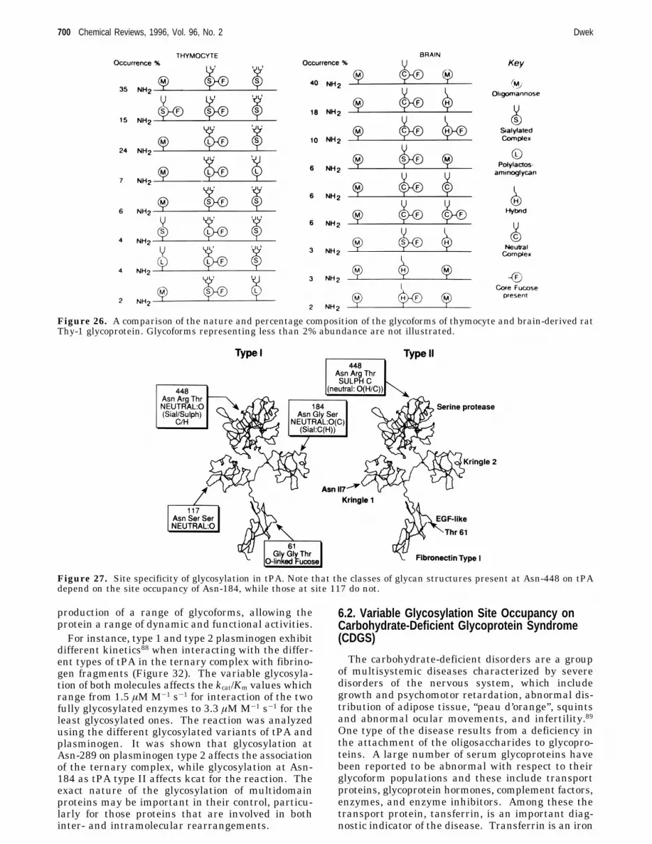
mocyte with that from the brain showed that therewas tissue specificity of N-glycosylation51 althoughthe amino acid sequences are identical. Further-more, the differential effects of tissue glycosylationwere expressed at the level of individual glycosylationsites in the different tissue-derived Thy-1 molecules.51The site distribution of oligosaccharides was suchthat no Thy-1 molecules were found to be in commonbetween the two tissues (Figure 26). Thus eachtissue created unique sets of glycoforms.An important consequence of this is that a glyco-
protein must be viewed as an ensemble or collectionof glycoforms. For instance, tissue plasminogenactivator “tPA” from human colon fibroblast is char-acterized by a different set of glycoforms comparedwith cells from tPA.66,67tPA is a protease glycoprotein consisting of five
domains: a fibronectin “finger” domain, an epidermal-growth factor domain, two kringles, and the catalyticserine protease domain.68,69 Binding sites for fibrinare present within the “finger” and kringle domains.There are two main classes of glycosylated variantsof the tPA molecule, referred to as type I and typeII; type I has three N-linked sugars, at Asn-117, Asn-184, and Asn-448 (Figure 27), whereas type II hasonly two, at Asn-117 and Asn-448. tPA shows site-specific glycosylation and each of the classes ischaracterized by a population of glycoforms. Variableoccupancy of site 184 affects the fine structure of theglycan population at site 448 demonstrating, in thiscase, that glycosylation at one site can influence theprocessing at another. In Bowes melanoma type 1tPA the major species at site 448 were found to beneutral glycans of the complex or hybrid structures.In type II, however 72% of the structures weresulfated complex glycans (Figure 27), and there wererelatively few neutral sugars.9 This is an interestingexample of site-specific glycosylation at one site (448)being controlled or influenced by glycosylation atanother site (184). Further detailed studies70 haveconfirmed that some of the complexed glycans on tPAcontain terminal galactose monosaccharide residuesinstead of, or as well as, terminal GalNAc residues.This suggests the presence of competing â1,4-galac-tosyl andN-acetylgalactosaminyl transferases in theBowes cell line. Interestingly, when the galactoseresidue is present it is always found substituted onthe ManR1-6 arm, suggesting that there may besteric restrictions by the growing glycoconjugatewhich result in arm-specific glycosylation. This isclearly an example of the protein glycoconjugateinfluencing its own glycosylation. Additionally, there
Figure 22. Schematic drawings of rat and human CD4,rat CD2, and Thy-1. The molecules are drawn with thecircles representing immunoglobulin superfamily (IgSF)domains and the “lollipops” N-linked oligosaccharides. Theglycosylphosphatidylinositol membrane anchor of Thy-1 isdepicted as a vertical arrow. The IgSF domains aredesignated as V or C2 on the basis of sequence analysis.166The positions of the mutations introduced in the CD4 andCD2 molecules to produce the recombinant soluble formsare indicated by horizontal arrows. Adapted from ref 166.
Figure 23. Bio-Gel P-4 gel filtration profiles of thedesialylated, tritium-labeled oligosaccharides of recombi-nant soluble CD4 and CD2 expressed in CHO cells: (a)total oligosaccharides of human sCD4; (b) total oligosac-charides of rat sCD4; and (c) total oligosaccharides of ratsCD2. The vertical arrows indicate the elution positions ofisomaltooligosaccharides containing the corresponding num-ber of glucose units. The time axis is marked at 100 minintervals. Data taken from from refs 55 and 56.
Glycobiology Chemical Reviews, 1996, Vol. 96, No. 2 697
+ +

may well be “motifs” in the protein that are recog-nized by particular glycosylating enzymes, such asthe Pro Leu Arg motif, which is recognized by theglycoprotein hormone-specificN-acetylgalactosamine-transferase.
6. Glycosylation Site Occupancy Can ModulateEnzyme ActivitiesThe presence of natural glycosylation variants, as
in tPA, makes it possible to probe the influence ofoligosaccharides on protein interactions directly. Inaddition, to tissue plasminogen activator (tPA typesI and II), ribonuclease (RNase A and B), and plas-minogen (Plg types 1 and 2) (Figure 29) are alsoimportant examples of variable sequon occupancy.While RNase consists of a single domain, both plas-minogen and its activator are multidomain proteins.Ribonuclease (RNase). The size of this molecule
makes it amenable to NMR studies. This has al-lowed the effects of glycosylation on the stability ofthe molecule to be probed15,71 by comparing thehydrogen-deuterium solvent exchange rates for theNH protons of RNase A and RNase B. The presenceof the sugar decreases the exchange rate in 30 of the124 amino acid residues (Figure 28), suggesting thatthe overall dynamic stability of the molecule isenhanced such that it becomes more rigid. Some ofthe affected residues are close to the oligosaccharideattachment site, others are as far away as 30 Å, whileseveral (including His-12, His-119, and Asp-121) areinvolved in the active site, which catalyzes thecleavage of the phospho diester linkages of RNA.The functional variations associated with glycosy-
lation of RNase have been probed in three ways: (1)by determining the relative abilities of RNase A andB to mediate the hydrolysis of double-strandedRNA,52 (2) by examining the resistance of RNase A
and B to proteases,52 and (3) by the abilities ofantibodies to distinguish between each form. Fur-thermore, in the case of RNase B, the enzymeactivities of several glycoforms have been reportedand may be ranked in terms of decreasing activityas: RNase A > RNase Man-0 ) RNase Man-1 >RNase Man-5 ) RNase B.52 The enzyme’s active siteis located in a groove that bisects the protein.72Efficient hydrolysis of RNA necessitates correctalignment of the RNA and the enzyme’s active site.This is achieved in part through an interactionbetween the 5′-terminal phosphate of RNA and acluster of cationic residues on the proteins surface(Lys-31, Lys-37, Arg-10, and Arg-33).73 Since Asn-34 is present on the surface of the protein near thisbinding site, it is tempting to speculate that theattenuated RNase activity of the glycoforms, relativeto that of the nonglycosylated form, arises from sterichindrance between the oligosaccharide and the RNA.tPA. The two variants of tPA differ by occupancy
of site 184 (section 5.3). Various properties of tPAare affected by the occupancy of site 184 with anN-linked oligosaccharide (type I). These include thefollowing:(a) A slower rate for the plasmin-mediated conver-
sion of single chain tPA to two-chain tPA. tPA issynthesized as a single-chain tPA molecule. Plasmincatalyzes the cleavage of the Arg275-Ile bond innative single-chain tPA to form a disulfide bond-linked two-chain species.74 The second-order rateconstant (kcat/Km) for type II tPA was found to beabout twice that for type I tPA,75 indicating thatglycosylation at site 184 hinders the conversion ofsingle- to two-chain tPA. Additionally, single-chaintPA has a lower activity and susceptibility to inhibi-tion compared to the two-chain form76,77 providing
Figure 24. Examples of the structure of the different types of neutral desialylated N-linked oligosaccharides: Fuc, L-fucose;Gal, D-galactose; GlcNAc, D-N-acetylglucsamine; Man, D-mannose.
698 Chemical Reviews, 1996, Vol. 96, No. 2 Dwek
+ +

further evidence that glycosylation of tPA serves tomodulate activity.(b) A decreased affinity of tPA for lysine and a
lower fibrinolytic activity. The decreased affinity oftype I tPA for lysine is well established, and affinitychromatography is commonly used to separate typeI tPA from type II. Fibrin clot assays have shownthat type II tPA binds more efficiently than type ItPA to fibrin,67 probably through lysine sites exposedon degraded fibrin. Moreover, the fibrinolytic activityof type II tPA and plasminogen has consistently been
found to exceed that of type I tPA and plasminogen,regardless of the cell line in which the tPA isproduced. Variable occupancy of site 184 is thereforeone of the factors which controls the rate at whichplasmin is generated, and this may be related to thedifferences in the affinity of type I and type II tPAfor lysine. The differential effects of glycosylation onthe rate, particularly when combined with those forplasmin, may well be a mechanism for ensuringpersistence of fibrinolytic activity.Plasminogen. Plasminogen, the natural substrate
of tPA, is a multidomain protein consisting of fivekringle regions and a serine protease domain (Figure30). It is a mixture of two major glycoforms78,79 whichhave the same amino acid sequence80 and contain oneO-glycosylation site and a potential N-glycosylationsite in kringle 3. Both type 1 and type 2 plasminogencontain an O-linked sugar chain at Thr-345 while theN-glycosylation site at Asn289ArgThr contains abiantennary sugar in type 1, but is unoccupied intype 2.81 Molecular modeling (Figure 30) suggeststhat the presence of the N-linked oligosaccharide(type 1) may result in significant shielding of thekringle 3 domain of the protein. The extent ofglycosylation may have physiological relevance.82Human recombinant plasminogen, expressed inEscherichia coli and consequently not glycosylated,is resistant to activation by tPA.82 The importanceof the O-linked glycoforms (in plasmingoen 2) hasbeen demonstrated,83 who isolated six glycoformsdiffering in sialic acid content. The O-linked sugarchain had previously been assumed to be a tetrasac-charide but the recent work83 which suggests thepresence of polysialic acid should prompt a detailedreexamination. However, they found that the kineticactivity of the different glycoforms was decreased asthe sialylation increased.Some of the properties of plasminogen which are
altered by N-linked glycosylation include(a) A slower rate of the â- to R-conformational
changes which is from an open conformation to acompact form. This change may be important in theformation and rearrangement of the ternary complexof plasminogen with fibrin and tPA.84
(b) A 10-fold weaker binding of plasminogen toU937 cells.85 This suggests that the N-linked glycanmay hinder sterically the recognition event.(c) A lower affinity for lysine sepharose.81 Again
this may be explained sterically by the sugar at site289 limiting access to the lysine binding site inkringle 4.(d) A slower rate of activation of plasminogen by
urokinase. A lower activation rate by tPA of humanneonatal plasminogen.86 This is also true of theactivation of plasminogen by tPA on the surface ofrat hepatocytes.87
6.1. The Multimolecular Interaction of tPA withPlasminogen and Fibrin Is Modulated byGlycosylation
The roles which both the cell and the individualprotein play in determining glycosylation site oc-cupancy and glycan processing may be an importantmeans of control, enabling the cell to respond flexiblyto its internal and external environment through the
Figure 25. Bio-Gel P-4 gel filtration profiles of thedesialylated, tritium-labeled oligosaccharides of the glyco-peptides and glycosylation mutants of rat soluble CD4: (a)total oligosaccharides of rat sCD4-derived glycopeptidefrom the region of the first glycosylation site; (b) totaloligosaccharides of rat sCD4-derived glycopeptide from theregion of the second glycosylation site; (c) total oligosac-charides of rat sCD4 with the second glycosylation siteremoved; (d) total oligosaccharides of rat sCD4 with thefirst glycosylation site removed; (e) total oligosaccharidesof rat sCD4. NH2-terminal-two IgSF domain form. Fordetails of the annotation see legend to Figure 2. (Repro-duced from Figure 8 in ref 56. Copyright 1993 Journal ofBiological Chemistry.)
Glycobiology Chemical Reviews, 1996, Vol. 96, No. 2 699
+ +
production of a range of glycoforms, allowing theprotein a range of dynamic and functional activities.For instance, type 1 and type 2 plasminogen exhibit
different kinetics88 when interacting with the differ-ent types of tPA in the ternary complex with fibrino-gen fragments (Figure 32). The variable glycosyla-tion of both molecules affects the kcat/Km values whichrange from 1.5 µMM-1 s-1 for interaction of the twofully glycosylated enzymes to 3.3 µMM-1 s-1 for theleast glycosylated ones. The reaction was analyzedusing the different glycosylated variants of tPA andplasminogen. It was shown that glycosylation atAsn-289 on plasminogen type 2 affects the associationof the ternary complex, while glycosylation at Asn-184 as tPA type II affects kcat for the reaction. Theexact nature of the glycosylation of multidomainproteins may be important in their control, particu-larly for those proteins that are involved in bothinter- and intramolecular rearrangements.
6.2. Variable Glycosylation Site Occupancy onCarbohydrate-Deficient Glycoprotein Syndrome(CDGS)
The carbohydrate-deficient disorders are a groupof multisystemic diseases characterized by severedisorders of the nervous system, which includegrowth and psychomotor retardation, abnormal dis-tribution of adipose tissue, “peau d’orange”, squintsand abnormal ocular movements, and infertility.89One type of the disease results from a deficiency inthe attachment of the oligosaccharides to glycopro-teins. A large number of serum glycoproteins havebeen reported to be abnormal with respect to theirglycoform populations and these include transportproteins, glycoprotein hormones, complement factors,enzymes, and enzyme inhibitors. Among these thetransport protein, tansferrin, is an important diag-nostic indicator of the disease. Transferrin is an iron
Figure 26. A comparison of the nature and percentage composition of the glycoforms of thymocyte and brain-derived ratThy-1 glycoprotein. Glycoforms representing less than 2% abundance are not illustrated.
Figure 27. Site specificity of glycosylation in tPA. Note that the classes of glycan structures present at Asn-448 on tPAdepend on the site occupancy of Asn-184, while those at site 117 do not.
700 Chemical Reviews, 1996, Vol. 96, No. 2 Dwek
+ +

transport protein containing bi- and triantennarycomplex glycans attached to Asn413LysSer andAsn611ValThr, both of which sites are normallyoccupied. Normal transferrin contains glycoformswhich differ only in the number of sialic acid residuespresent (usually between 2 and 6). Transferrin form
CDGS patients may contain additional glycoformswhere only one of the two glycosylation sites isoccupied, and those in which neither site has beenglycosylated.90 These differences in glycoform popu-lations may be an indication of disease severity, whilealso providing a rapid diagnostic test.91 Furtherstudies of this syndrome may reveal important newfunctional roles for glycosylation, particularly indevelopment.
7. Some Structural Roles for OligosaccharidesWe have seen in the previous section that the major
effects of an oligosaccharide in modulating enzymeactivity arise from the large size of the oligosaccha-rides which affect inter- or intramolecular interac-tions. We have also noted (with RNase) that thepresence of a sugar can affect the dynamic stabilityof the protein.71 More conventional structural rolesfor oligosaccharides are in anchoring a protein to amembrane, and in helping to maintain the quater-nary structure of a protein such as the Fc portion ofthe immunoglobulin G molecule.
7.1. Glycosyl-Phosphatidylinositol (GPI) AnchorsSince 1985 over 100 examples of GPI anchored
proteins have been described.41 The GPI anchor isan alternative anchoring mechanism to the trans-membrane polypeptide domain of type I membraneproteins. The first detailed structural studies on GPIanchors, of T. brucei variant surface glycoprotein(VSG) and of rat brain Thy-1, were carried out inOxford.92,93 The structures were determined by acombination of glycosyl linkage and compositionalanalyses by GC-MS, specific chemical cleavages,
Figure 28. Effects of glycosylation on the rates of amideH/D exchange for selected residues of RNase. A schematicrepresentation of ribonuclease highlighting those residueswhose amide protons (shown by the space-filled balls) showmodified H/D exchange as a result of glycosylation of theenzyme at Asn-34. The structure of Man5GlcNAc (drawnto the same scale as the protein) is shown attached to Asn34 and extending away from the protein into solution.(Reprinted from ref 167. Copyright 1994 American Chemi-cal Society.)
Figure 29. The effect of flexibility of the Asn-34 side chain on the orientation of the oligosaccharides attached to ribonucleaseB. The Man-9 glycoform of RNase B based on the 2.5 Å X-ray crystal structure with an overlay of 10 oligosaccharideconformations from a 500 psec MD trajectory of Man-9. The total Van der Waals surfaces of the oligosaccharides areillustrated by dots.
Glycobiology Chemical Reviews, 1996, Vol. 96, No. 2 701
+ +

sequential enzymatic digestion with exoglycosidasescombined with Bio-Gel P-4 column chromatography,and 1D and 2D 1H NMR spectroscopy. In each case,C-terminus of the protein is linked by ethanolaminephosphate to a glycan with the conserved backbonesequence ManR1-2ManR1-6ManR1-4GlcNH2, whichin turn is linked to the sixth position of the myo-inositol ring of phosphatidylinositol.The conserved sugar sequence of the anchors may
be a consequence of their biosynthesis in which apreassembled core is added to a protein. The generalprinciple is much the same for the biosynthesis ofN-linked oligosaccharides on proteins. In both casesvariations on the respective cores may be protein andcell specific. In VSG this tetrasaccharide backboneis substituted with branched side chains of R-galac-tose. It has been speculated that the arrangementof the GPI anchor of the VSG protein may allow forthe close packing of these molecules on the parasitesurface, with the heterogeneity of the glycan part ofthe GPI anchor allowing close packing in 2D spacein order to create a surface. The solution structure94showed that the glycan exists in an extended config-uration along the plane of the membrane spanningan area of 600 Å2, which is similar to the cross-sectional area of the monomeric N-terminal VSGdomain (Figure 33).By contrast, computer modeling of the structure of
Thy-1 and its GPI anchor, suggests that the lipid partof the GPI may not be the sole membrane anchor.95The model suggests that the glycan part of the GPImay lie within the Thy-1 protein moiety so that theThy-1 protein moiety sits directly on the membranewith most of the GPI actually within the protein(Figure 34a,b). The smaller anchor could thereforeallow access to Thy-1 by other “accessory” moleculeswithin the membrane.
7.2. Structure/Function Relationships in IgGX-ray crystallography has shown that each region
of homology in the IgG molecule corresponds to acompact, independently folded unit and that theseare linked together by short sections of polypeptidechain. Each domain consists of two â-pleated sheetswith antiparallel strands connected by loop regions(the immunoglobulin fold).96 Crystallographic stud-ies of IgG Fc fragments97,98 have shown that, unlikeother immunoglobulin domains, the two CH2 domainsdo not form extensive lateral associations. Theresulting interstitial region accommodates the com-plex oligosaccharides, which are attached to Asn-297on each heavy chain (Figure 35). In the Fc fragmentfor rabbit IgG one of the R-1,3 arms interacts withthe trimannose core of the opposing oligosaccharide,while the other R-1-3 antenna extends toward theinterface with the CH3 domain. In the human Fc onlythe first two monosaccharide residues (Man,GlcNAc)of the 1-3 antenna are defined, but if the nextgalactose residue is built into the structure it wouldappear that this residue may form interactions withThr-335, Ile-336, and Ser-337, on the protein surface.However, in both the human and rabbit Fc theR(1-6) antenna of each oligosaccharide is well de-fined and interacts with the hydrophobic and polarresidues Phe-243 (Man-5 and GlcNAc-6), Pro-244/245(Gal-7), and Thr-260 (GlcNAc-6 and Gal-7) on thedomain surface.The effects of glycosylation may be subtle and
highly specific. Both the C1q and protein A bindingsites on IgG are located in the CH2 domain betweenPhe-319 and Ile-332 (distal or at the carboxyl-terminal side from the N-linked glycosylation site).Neither of these functions is markedly affected by theabsence of carbohydrate.99,100 The binding of mono-cytes, which involves sites 234-237 (proximal to theamino terminus) on the lower hinge region, is elimi-
Figure 30. A schematic molecular model of plasminogen type 1 and type 2. Plasminogen consists of five kringle regionsand a serine protease domain. This model was constructed using the coordinates from the corresponding domains on tPAand is intended to convey the relative size of the oligosaccharides and the protein. Type 1 plasminogen (left) has twooccupied glycosylation sites, at Asn289ArgThr in kringle 3 and at Thr 345 in kringle 4. Type 2 (right) lacks the N-linkedsugar at Asn 289.
702 Chemical Reviews, 1996, Vol. 96, No. 2 Dwek
+ +

nated in aglycosylated monoclonal murine IgG. Thisis consistent with a proposal that the absence ofsugars results in a lateral movement of domains inthe hinge region relative to the normally glycosylatedantibody. Moreover, in aglycosylated IgG a proteinstructural change has been detected by 1H NMR atHis-268,101 which is also in the vicinity of the lowerhinge. These data suggest that the oligosaccharidemay stabilize a particular hinge conformation es-sential for monocyte binding. The spatial relation-ship between the CH2 and CHf domains, on the otherhand, does not seem to depend on presence of the
oligosaccharides, because in their absence protein Aand C1q binding are unaffected.
8. Oligosaccharide Recognition
8.1. Specific Interactions with Animal Lectins
To decode the information present in oligosaccha-ride structures, these must be recognized by othermolecules. Carbohydrate-binding proteins are termedlectins. Following recognition of the oligosaccharideswith the appropriate geometry (see later), these
Figure 31. Schematic model of tissue-type plasminogen activator types I and II. tPA is composed of five domains: afibronectin type 1 finger module, and EGF-like module, two kringles, and a serine protease domain. This model wasconstructed using the coordinates of the finger growth factor pair (Smith, B. O.; Downing, A. K.; Campbell, I. D.) andkringle 2168 from human tPA. Kringle 1 and the serine protease domains were modeled by homology. The high-mannosecarbohydrate at position 117 and the complex sugars at sites 184 and 448 are shown in green. The lysine binding site andthe catalytic triad are highlighted in red and yellow, respectively. Comparison of the glycosylation of type I and type IItPA suggests that glycosylation at site 184 may sterically interfere with lysine binding.
Glycobiology Chemical Reviews, 1996, Vol. 96, No. 2 703
+ +

lectins can mediate many specific biological functions.These include immune defense (e.g. the mannose-binding protein and the macrophage mannose recep-tor), clearance of glycoproteins (e.g. the asialo-glycoprotein receptor), and cell-cell adhesion (e.g. theselectins). This important and major functionalaspect of the glycobiology of animal lectins derivesmainly from the pioneering work of Kurt Dricka-mer.102 No review of glycobiology could be completewithout acknowledgment of the fundamental impor-tance of this field, and the readers attention is drawnto the prime reviews in this area.5,103-105 Approach-ing 100 lectins have been isolated and many of theseclassified on the basis of sequence homology. By farthe largest and most diverse class is that of theC-type lectins. These lectins bind carbohydrates ina Ca2+ -dependent manner and are characterized bythe presence of carbohydrate-recognition domains
(CRDs). These domains contain a common sequencemotif of approximately 120 amino acid residues andare characterized by 31 invariant or highly conservedamino acids.106 Some of the C-type CRDs which havebeen reported are shown in (Figure 36).One C-type lectin which we will refer to in section
9 is the mannose-binding protein (MBP) which medi-ates antibody-independent binding of pathogens whichcontain a high concentration of mannose or N-acetylglucosamine residues on their surface. Therecognition event can lead to complement fixation oropsonization.107 Recent X-ray data of the CRD fromthis protein illustrates the principles involved in therecognition of oligosaccharides. The CRD showsspecificity for terminal residues and involves thesugar chelating to the Ca2+ ion via the 3- and4-hydroxyl groups108 (Figure 37). There are alsointeractions between the CRD and the monosaccha-ride residues in the oligosaccharide that are mediatedby water molecules, effectively increasing the surfacearea of the oligosaccharide in contact with the CRD.C-type lectins and their constituent CRDs displayweak affinity for monosaccharides. In general thetriggering of biological events requires multivalentreceptors interacting with multivated targets (oli-gosaccharides). Multivalency in the receptor can beachieved by clustering CRDs. In the mannose-binding protein for example this occurs by the forma-tion of oligomers of polypeptide chains, each of whichcontains a single CRD. In contrast the macrophagemannose receptor contains multiple CRDs in a singlepolypeptide. Clustering of CRDs could also arisefrom having multiple copies of the lectin in closeproximity. (This may be the case for the selectins.)Similarly, the oligosaccharide may be multivalent byvirtue of its branching if there is the “correct”geometry to bind to the receptor CRDs, or again theremay be multiple copies appropriately presented on asurface.The recent X-ray structures of the MBP trimer3,4
can be used to illustrate the above points. The CRDsin the trimers are separated by some 44-53 Å.Matching multivalency by an oligomannose oligosac-charide to the trimer array can only be obtained ifthe terminal residues have the correct geometry in
Figure 32. The kinetics of the activation of type 1 and 2plasminogen by tPA variants I and II in the presence offibrinogen fragment and their kinetic contents.
Figure 33. Comparison of the structures of the VSG and Thy-1 glycan anchors.
704 Chemical Reviews, 1996, Vol. 96, No. 2 Dwek
+ +

that they must also be separated by similar spacingsof 44-53 Å. For instance, the Man-9 structure hasthree terminal monosaccharides each potentially ableto bind to a CRD in the receptor. NMR studies,together with molecular dynamic calculations onMan-9,109 have shown that in Man-9, the maximumdistances between them is approximately 21 Å. Theconsequence is that the Man-9 structure can bebound via only one terminal residue to MBP, but thismonovalent binding will not result in the biologicaltriggering of complement. (This would only resultfrom multiple presentation of Man-9 structuressason a pathogenswith the appropriate spacings be-tween the individual oligosaccharides to bind to the
different CRDs.) In this way the MBP molecule can“inspect” a range of molecules in the serum. Thosewith a single oligomannose structure will be boundand released, while these multiply presented as ona pathogen will lead to the biological effector func-tions being triggered. This type of mechanism hasall the hallmarks of the discrimination in the immunesystem between self and nonself. Another receptorwhich is believed to form part of a basic defensemechanism against pathogens, is the macrophagemannose receptor.5,110,111 This receptor can alsomediate the clearance of endogenous proteins suchas tissue plasminogen activator (via the oligoman-nose structures at site Asn-117) and lysosomal en-zymes which carry high mannose-type oligosaccha-rides. Such proteins are often released into thecirculation in response to pathological events. Themacrophage mannose receptor internalizes the boundligands and targets these to lysosomes for destruc-tion.
8.2. Neural Glycosylation and Recognition
Glycosylation is a common feature to moleculesimplicated in cell-cell and cell-matrix interactionsin the development and maintenance of the nervoussystem, as is involved, for example, in axon growth,guidance, and targeting. There is now increasingevidence to suggest that oligosaccharides play keyrecognition roles in these processes. A knowledge ofoligosaccharide structures involved is essential for anunderstanding of carbohydrate-mediated interactionsat the molecular level.Technology developments which will help this field
include obtaining milligram quantities of oligosac-charides from the glycoproteins of whole brain byhydrazinolysis. These sugar libraries provide ameans of profiling brain oligosaccharides and deter-mining the relative abundance of different classes.Identification of unusual or novel structures would
Figure 34. Molecular models of VSG (a) and Thy-1 (b)illustrating the potential interactions of the anchors andtheir proteins. In the case of VSG, the anchor is proposedto serve a space-filling role and spans an area along themembrane which is comparable to the cross-sectional areaof the proteins N-terminal domains. In contrast the glycanpart of the GPI anchor of Thy-1 may fit into a pocket inthe protein with only the terminal part of the lipidextending, thus allowing significant direct interactionsbetween the Thy-1 peptide and the membrane surface.Adapted from refs 95 and 169.
Figure 35. The Fc portion data from ref 97 of the IgGmolecule.
Glycobiology Chemical Reviews, 1996, Vol. 96, No. 2 705
+ +

suggest different biosynthetic pathways from glyco-proteins in the systemic side. Further developmentand integration of technologies for the sensitivedetection and chromatographic separation of oli-gosaccharides are being pursued.
One way to probe for functional effects of glycosy-lation is to test glycans from the brain libraries asspecific competitive inhibitors in neurite outgrowthassays in vitro. Preliminary work (Chen, Y. J.;Doherty, P.; Walsh, F.; Wing, D.; Dwek, R. A.,unpublished work) has shown that crude sugarfractions from a rat brain library (at a concentrationof 500 µM) can inhibit neurite outgrowth overastrocytessa process involving recognition betweencell adhesion molecules.
It is well known that carbohydrates are enrichedat the synapse, and as recognition roles for theassociated glycoproteins are becoming established,the exciting possibility exists for a glycoconjugate rolein synaptic efficacy, a field with far-reaching implica-tions in such areas as memory formation.
8.3. Major Histocompatibility Complex (MHC)Restricted Recognition of Glycopeptides byT-CellsThe major histocompatibility complex (MHC) class
1 molecules display peptides from self or foreigncellular proteins, on the antigen presenting cellsurface. A critical event in the immune response isthe recognition of these peptide-MHC complexes by
Figure 36. Summary of the structures of several groups of C-type animal lectins. Representative structures for threegroups of membrane-associated lectins are shown: group II, the chicken hepatic lectin (homologue of the mammalianasialoglycoprotein receptor); group IV, the selectin cell adhesion molecules; and group VI, the macrophage mannose receptor.Group III lectins (collectins), such as mannose-binding protein, are found in extracellular fluids. Group I CRD-containingproteins are proteoglycans of the extracellular matrix. Other domains present in these molecules include EGF, epidermalgrowth factor-like repeats; CR, complement regulatory domains; FN-II, fibronectin type II repeats; COL, collagen-likesequences, GAG, glycosamino-glycan attachment sites; and HA, hyaluronic acid-binding domains. (Reprinted from ref170. Copyright Cold Spring Harbor Laboratory.)
Figure 37. Structure of the C-type CRD from mannose-binding protein.108 The boxed area is enlarged to illustrate thedetails of the interaction with mannose.
706 Chemical Reviews, 1996, Vol. 96, No. 2 Dwek
+ +

cytolytic T-cells. These identify and subsequentlyeliminate cells that present “foreign” peptides.An interesting and exciting finding of carbohydrate-
specific recognition has been the demonstration thatT-cells recognize glycopeptides bound to MHC classI molecules. Harum et al.112 have reported theefficient binding to class I MHC of a syntheticglycopeptide carrying the naturally occurring O-â-linked N-acetylglucosamine (GlcNAc). The peptideused in the investigation was an analogue of theepitope FAPGNYPAL from Sendai virus nuclearprotein, which produces a specific cytotoxic T-cell(CTL) response. The glycosylated analogue (an O-GlcNAc on the serine residue) is highly immunogenicand elicits a specific, MHC-restricted, anti-glycopep-tide CTL response.The relevance of studying T-cell responses to
modifications such as glycosylation finds supportfrom the preliminary data obtained in which thepresence of O-GlcNAc modified peptides has beendetected among the pool of peptides eluted fromhuman spleen class I MHC.112
Interestingly, a study by Michaelsson et al.113 alsosuggested that T-cell recognition of glycosylatedpeptides from type II collagen may be important inthe development of rheumatoid arthritis. Recogni-tion of glycosylated peptides could therefore be ofsignificant importance in immunity toward malig-nant diseases and in viral infections. It is alsopossible that acquisition or loss of glycosylation mightprovide a novel strategy for viruses or malignantlytransformed cells to escape from selection pressureby CTL which does not depend on a mutation at thegene level.
8.4. Recognition of Oligosaccharides byStimulated T-CellsA quite different type of T-cell recognition involving
oligosaccharides may exist in diseases such as rheu-matoid arthritis, Behcet’s syndrome, and IgA neph-ropathy. In these diseases up to 9% of the T-cellsare bound by cytophilic IgA1 compared with none inresting T-cells from healthy subjects. Detailed stud-ies have shown that the binding to this subset ofT-cells can be inhibited by the “O”-linked sugarsassociated with the hinge region of IgA1.114 Whilethe functional significance of the receptor has yet tobe established, the observations suggest that theoligosaccharides indicated recognition may have func-tional implications in these diseases.Although the general area of T-cell recognition of
saccharides is a difficult and challenging one, it isan important one which is well worth pursuing andwhich could provide yet more insights into the rolesof oligosaccharides in the cellular immune response.
9. Glycosylation in DiseaseThe glycosylation of some glycoproteins has been
shown to alter in disease. For example, concanavlinA lectin chromatography suggests that the relativeamounts of complex biantennary glycans on R-1 acidglycoprotein decreases during the acute phase re-sponse.184 Another well-documented example whichis discussed below is the IgG molecule, the glycosy-lation of which is altered in rheumatoid arthritis.
9.1. The IgG Molecule
The IgG molecule contains 2.5 oligosaccharidechains per molecule. Two of these represent theconserved glycosylation sites in the Fc portion of allIgGs at Asn-297. The remainder occur in the hyper-variable regions of the Fab fragment with a frequencyand position dependent on the occurrence of anN-glycosylation site (Asn/Xaa/Ser(Thr). The glycos-ylation is of the complex biantennary class and about30 variants occur in the IgG molecule (Figure 38).This glycosylation of IgG results in many differentglycoforms.
9.1.1. Site-Specific Glycosylation of IgG
There are clear glycosylation site-specific differ-ences between the Fab and Fc, with regard tosialylation and the presence of bisecting GlcNAcresidues (both mainly in the Fab fragment). Char-acteristics of Fc N-glycosylation include a low inci-dence (10%) of monosialylated structures, the absenceof disialylated structures, a low incidence of corescarrying a “bisecting” GlcNAc, and heterogeneity inthe galactose residues. In general, the Fc oligosac-charides are mainly restricted to biantennary oli-gosaccharides terminating in 2, 1, and 0 galactoseresidues (G2, G1, and G0). The unique 3D environ-ment in the Fc may limit the accessibility of sugarprocessing enzymes so resulting in these glycoforms.Fab N-glycosylation is characterized by a high inci-dence of di- and monosialylated structures, and ofcores with the “bisecting” GlcNAc residue.115 Itshould be stressed that the large number of differentstructures associated with IgG is not the result ofstudying a polyclonal population, since a similarheterogeneity is found upon analysis of myeloma andhybridoma-derived IgG.
Figure 38. Primary sequences of the N-linked oligosac-charides associated with IgG. The hydrodynamic volumeof each structure, measured in glucose units, is indicated.
Glycobiology Chemical Reviews, 1996, Vol. 96, No. 2 707
+ +

9.1.2. IgG Glycoforms Associated with RheumatoidArthritisSerum IgG from patients with rheumatoid arthritis
contains the same set of biantennary oligosaccharidesfound in normal individuals, but in very differentproportions (Figure 39). The incidence of structureswith outer arm galactose is dramatically decreased,and the incidence of those structures terminating inouter arm N-acetylglucosamine correspondingly in-creased.8 A comparison of the N-glycosylation of Faband Fc fragments, derived from total serum IgG ofpatients with rheumatoid arthritis or from a controlgroup, shows that the decreased galactosylationfound upon analysis of total serum IgG is largely dueto changes in the N-linked oligosaccharides of the Fc.There are also quantitatively minor, but potentiallysignificant differences in Fab glycosylation (Scragg,I.; Chen Chang, S., unpublished work), in which thereis an increase in the light chain in the proportion offully galactosylated (G2), bisected, and core fucosy-lated oligosaccharides.The cause of the altered glycosylation of IgG in RA
is not clear at present. It may have a biosyntheticorigin116 in which there is decreased levels of thegalactotransferase (GalTase) activity. Indeed, inperipheral blood B and T lymphocytes isolated fromRA patients, there is a decrease in this activity117,118which correlates with the overall decreased galactos-ylation of serum IgG. In general, such a correlationis expected to be insensitive to the Fab glycosylationsince it only contributes a small amount to the overallvalue. However, the detailed site-specific changes ofref 119 in which the Fab and Fc glycosylations arequite differently affected cannot be explained by anoverall decrease in the GalTase activity. Neitherdoes it explain the normal levels of galactose detectedon rheumatoid serum IgA1 when compared withIgA1.120 Analysis of oligosaccharides released fromIgG and its fragments does not give any informationon the combination of the sugars present on themolecule. Thus, it is entirely possible that RA IgGwhich has increased L chain galactosylation is syn-thesized by a different population of B cells thanthose molecules which have decreased Fc fragmentglycosylation. Since B cells have differing levels ofgalactosyltransferase activities121 this provides astraightforward explanation for site-specific galactos-
ylation. Changes in the population of B cells maybe inferred from the finding that there is an increasein the number of B lymphocytes which are CD5+ 122,123
and the discovery that IgG subclass distribution isaltered in rheumatoid arthritis.124 Alternatively,there is the possibility that multiple galactosyltrans-ferases of different protein (and hence potentially IgGfragment) specificity are present in B cells. An IgG-specific galactosyltransferase of reduced activity inRA has been reported118,125 but as yet no otherevidence has confirmed this interesting finding.Destruction of galactose caused by reactive oxygenspecies has also been proposed to contribute to lowerlevels of galactose in rheumatoid arthritis126 but itis difficult to envisage how this mechanism could leadto the site-specific changes between the Fab and Fcfragments. Clearly, more work is necessary to un-derstand the mechanisms which regulate the glycos-ylation of human IgG.There are four human subclasses of IgG (Figure
40). The most striking difference between them liesin the hinge regions. Thus the hinge region of IgG1consists of 23 amino acids, that for IgG2-19, thatfor IgG3-70 and that for IgG4-20 (see Table 1). Inserum the mean concentration (in mg/mL) is -9 forIgG1, 3 for IgG2, 1 for IgG3, and 0.5 for IgG4. Table2 shows that in RA the changes in glycoforms of IgGare seen across all four subclasses.
9.1.3. Glycosylation Changes on the IgG Molecule Are“Disease Restricted” and Are an Important Factor inRheumatoid ArthritisThe change in galactosylation of the serum IgG of
patients with rheumatoid arthritis is not common toall other autoimmune or inflammatory disorders. Onthe other hand, agalactosyl IgG has been consistentlyfound in patients with juvenile rheumatoid arthritis,Crohn’s disease, and tuberculosis.127,128The Fc glycoform distribution changes with age
(Figure 39), with disease severity of rheumatoid
Figure 39. Representative Bio-Gel new P-4 (-400 mesh)gel permeation chromatogram of the asialooligosaccharidesof total serum IgG from a healthy individual and a patientwith rheumatoid arthritis.
Figure 40. Representation of the four human immuno-globulin subclasses. (Reprinted from ref 171. Copyright1990 Pergamon Press.)
708 Chemical Reviews, 1996, Vol. 96, No. 2 Dwek
+ +

arthritis, and with pregnancy. The increase anddecrease may reflect the control of galactosyl trans-ferase activity under different physiological condi-tions. Clearly, rheumatoid arthritis is associatedwith changes in G0 glycoforms so that this parameteris a good biochemical disease marker of diagnosticvalue (Figure 41). In an arthritic woman withpathologically elevated levels of G0 glycoforms,changes in G0 correlated with remission of arthritisduring gestation and post-partum recurrence.129That the G0 glycoforms may be an important factor
in rheumatoid arthritis can be demonstrated in thecollagen-induced arthritis (CIA)174 mouse model.Passive transfer of an acute synovitus in T-cellprimed mice can be achieved using IgG containingautoantibodies to type II collagen only when theseautoantibodies are present in the G0 glycoforms. Thebasic protocol is given in Figure 42. IgG was purifiedfrom pooled sera of mice with CIA at days 17 and 38when peak levels of type II collagen autoantibodieswere produced. The IgG fractions were isolated anddivided. One fraction was treated with â-galactosi-dase from Streptococcus strain 6646K as to generate
the IgG (G0) glycoforms exclusively. Other IgGsamples contain a distribution of glycoforms onlysome of which are the G0 glycoform. Thus byincreasing the level of agalactosyl IgG glycoform ofthe anti-type II collagen antibodies present in thepreparation, the IgG becomes more effective in caus-ing arthritis in passive transfer. This indicates thatthe glycosylation status of an autoantibody is onefactor in determining if an antibody is pathogenic.
9.1.4. Structural Changes in IgG Fc on Loss of GalactoseNMR relaxation data from a control (26% G0) and
RA (55% G0) sample show a very slowly relaxing (andhence a more mobile) subset of proton resonancesbetween 3.5 and 4.25 ppm (Figure 43) which can beassigned to oligosaccharide protons. This subset ischaracterized by T2 relaxation times of∼0.15 s. Thiscompares to the values of 0.005 s for the proteinbackbone and static aromatic side chains, 0.07 s forring flipping aromatic side chains, and ∼0.2-0.5 sfor released oligosaccharides of a similar size. Thereare approximately 18 and 41 protons in this subsetin the control and RA samples respectively. These
Table 1. Amino Acid Sequences of Human IgG HingeRegionsa
upper hinge216 middle hinge
lower hinge231
IgG1 EPKSCDKTHT CPPCP APELLGGPIgG2 ERK CCVECPPCP APPVA GPIgG3 ELKTPLGDTTHT CPRCP APELLGGP
(EPKSCDTPPP-CPRCP)x3
IgG4 ESKYGPP CPSCP APEFLGGPa The upper and middle hinge are encoded by one exon in
the case of IgG1, 2, and 4 and four exons for IgG3. The lowerhinge is encoded by the CH2 exon. The numbering systemrefers to the amino acid sequence position of the beginning ofthe upper and lower hinges.165
Table 2. Galactosylation of Total Human Serum IgGand Its Component Subclasses (%) of G2, G1, and G0Structures
subclassessamplea galactosylationb
serumIgG IgG1 IgG2 IgG3 IgG4
H1 G2 45.0 50.9 38.1 61.7 40.8G1 34.0 33.7 38.6 23.6 34.4G0 21.0 15.4 23.4 14.7 24.8
H2 G2 47.6 52.5 37.8 57.5 46.4G1 38.2 41.1 41.5 30.1 36.7G0 14.2 6.4 20.6 12.4 16.9
H3 G2 39.2 54.2 37.1 61.2 38.3G1 41.7 34.4 38.2 27.6 26.2G0 19.1 11.4 24.7 11.2 25.5
R1 G2 19.4 26.4 18.6 23.6 19.3G1 35.5 35.0 31.3 29.6 32.4G0 45.0 38.6 50.3 46.8 48.3
R2 G2 18.7 19.1 13.8 30.7 16.4G1 30.2 27.4 25.5 23.2 25.5G0 51.1 53.5 60.7 46.1 58.1
R3 G2 14.2 13.9 10.8 20.6 15.3G1 30.8 26.5 20.4 28.4 20.7G0 55.0 59.6 68.8 51.0 64.0
a H1-H3 refer to three nonarthritic individuals while R1-R3 are three patients with rheumatoid arthritis. b Oligoac-charides were assessed to be di- (G2), mono- (G1), or non- (G0)galactosylated. Scragg, I.; Youings, A.; Dwek, R. A. Unpub-lished data.
Figure 41. Variations in the galactosylation of IgG. Thereare changes with age and disease activity as illustratedfor juvenile arthritis. A comparison between the changesin percentage G0 during the course of pregnancy for anormal and rheumatoid arthritis patient, shows that bothpatients tend to increase their galactose levels duringpregnancy. The dashed line indicates the aged matchedexpected value of a normal healthy individual. It is seenthat the pregnant patient with rheumatoid arthritis achievesthis level and this correlates with a remission of arthritisin the patient.
Figure 42. Protocol for passive transfer of IgG in thecollagen-induced arthritis model in mouse. Purified IgG istransferred to a suitably primed mouse or is first treatedwith â-galactosidase to enrich the IgG0 glycoforms.
Glycobiology Chemical Reviews, 1996, Vol. 96, No. 2 709
+ +

are not enough to account for all the oligosaccharideprotons in either sample and the remaining onesmust have relaxation properties very similar to thepeptide backbone. Since the 2-fold increase in thenumber of more mobile protons mirrors the two-foldincrease in G0 values between the two samples, thisallows assignment of these mobile protons to the G0oligosaccharides. By taking into account the per-centage occurrence of G0 in the two samples (andthat there are two oligosaccharides per Fc), each G0oligosaccharide must contribute on average 36 pro-tons to the subset. This suggests that the proteinonly has a very small effect on the mobility of theG0 oligosaccharides. This analysis leads to a modelof G1 and G2 oligosaccharides having restrictedmotion relative to the peptide surface (as seen in thecrystal structure), while the G0 oligosaccharides arefree to move (Figure 44). X-ray data for normal IgGFc97 and calculations of solvent accessibility (Figure45) show that one of the main interactions betweenthe protein surface and the oligosaccharide is medi-ated by interaction of the (1-6) arm terminal galac-tose residue with Thr-260. Loss of this galactosetherefore results in a decreased interaction betweenthe oligosaccharide and the protein surface. On thisbasis, it would be expected that G0 oligosaccharidesand those of G1 with a galactose residue on the (1-3) arm would have extra mobility relative to theprotein. The NMR analysis, however, indicates thatno G1 structures have extra mobility. Furtherinspection of the X-ray data of the Fc shows apotential interaction site between the (1-3) galactoseand residues Thr-335, Ile-336, and Ser-337, on theprotein surface. Thus increased mobility of theoligosaccharide results from loss of both galactoses(Figure 45) and leads to exposure of the nonreducingterminal GlcNAc residues.
9.1.5. Functional Implications of IgG GlycoformsThe studies of the Fc fragment of X-ray crystal-
lography indicate that galactose residues present onthe R (1-6) arms can interact with the protein. An
increase in the level of IgG glycoforms lackingterminal galactose and thereby terminating in N-acetylglucosamine could lead to the exposure ofcertain Fc determinants. This may elicit an immuneresponse, or raise a preexisting subclinical responseto a pathological one which may be relevant torheumatoid arthritis. In addition, the now vacantgalactose sites on the protein may create a lectin-
Figure 43. NMR relaxation weighted spectra of IgG Fc from a rheumatoid arthritic patient. The top insert shows the 500MHz 1D 1H NMR spectrum of IgG Fc isolated from a rheumatoid arthritic patient. Selected regions of the CPMG spectraare shown at total spin echo times of 0.001 s (1) and 0.128 s (2). The first spectrum is essentially the 1D spectrum. Thesecond spectrum shows only the subset of more slowly relaxing proton resonances.
Figure 44. Schematic representation of the oligosaccha-rides of IgG Fc looking along the pseudo-C2 axis. Thesquares show the positions of the GlcNAc 1 residues, thecircles the positions of the other monosaccharide residues,G indicates a nonreducing terminal galactose residue onthe 6-arm of the oligosaccharide. (1) Two G1 oligosaccha-rides (as seen in the crystal structure). Binding of the 6-armto the protein surface holds the rest of the oligosacchariderigid with respect to the protein. (2) A G0 (left) and G1(right) oligosaccharide. Loss of interaction between the G0oligosaccharide 6-arm and the protein surface leaves thewhole of the oligosaccharide free to move.
710 Chemical Reviews, 1996, Vol. 96, No. 2 Dwek
+ +
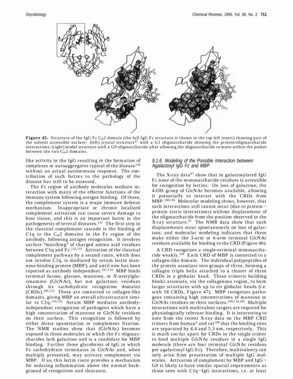
like activity in the IgG resulting in the formation ofcomplexes or autoaggregates typical of the disease130without an actual autoimmune response. The con-tribution of such factors to the pathology of thedisease has still to be assessed.The Fc region of antibody molecules mediate in-
teraction with many of the effector functions of theimmune system following antigen binding. Of these,the complement system is a major immune defensemechanism. Inappropriate or chronic localizedcomplement activation can cause severe damage tohost tissue, and this is an important factor in thepathogenesis of several diseases.131 The first step inthe classical complement cascade is the binding ofC1q to the CH2 domains in the Fc region of theantibody, following antigen recognition. It involvessurface “matching” of charged amino acid residuesbetween C1q and Fc.132,133 Activation of the classicalcomplement pathway by a second route, which doesnot involve C1q, is mediated by serum lectin man-nose-binding protein (MBP), and until now has beenreported as antibody independent.107,134 MBP bindsterminal fucose, glucose, mannose, or N-acetylglu-cosamine (GlcNAc), but not galactose, residuesthrough its carbohydrate recognition domains(CRDs).108,135 These are connected to collagen-likedomains, giving MBP an overall ultrastructure simi-lar to C1q.134,135 Serum MBP mediates antibody-independent recognition of pathogens which have ahigh concentration of mannose or GlcNAc residueson their surface. This recognition is followed byeither direct opsonization or complement fixation.The NMR studies show that (GlcNAc) becomesexposed in those molecules in which the Fc oligosac-charides lack galactose and is a candidate for MBPbinding. Further those glycoforms of IgG in whichFc carbohydrate terminates in GlcNAc and, whenmultiply presented, may activate complement viaMBP. If so, this lectin route provides a mechanismfor inducing inflammation above the normal back-ground of recognition and clearance.
9.1.6. Modeling of the Possible Interaction betweenAgalactosyl IgG Fc and MBP
The X-ray data97 show that in galactosylated IgGFc none of the monosaccharide residues is accessiblefor recognition by lectins. On loss of galactose, the4-OH group of GlcNAc becomes available, allowingit potentially to interact with the CRDs fromMBP.108,136 Molecular modeling shows, however, thatsuch interactions still cannot occur (due to protein-protein steric interactions) without displacement ofthe oligosaccharide from the position observed in theX-ray structure.97 The NMR data show that suchdisplacements occur spontaneously on loss of galac-tose, and molecular modeling indicates that thesemake either the 3-arm or 6-arm terminal GlcNAcresidues available for binding to the CRD (Figure 46).A CRD recognizes a single-terminal monosaccha-
ride weakly.108 Each CRD of MBP is connected to acollagen-like domain. The individual polypeptides ofthe protein associate into groups of three, forming acollagen triple helix attached to a cluster of threeCRDs in a globular head. These trimeric buildingblocks associate, via the collagenous region, to formlarger structures with up to six globular heads (i.e.with 18 CRDs, Figure 47). MBP recognizes patho-gens containing high concentrations of mannose orGlcNAc residues on their surfaces.108,134,185 Multipleinteractions with multivalent targets are required forphysiologically relevant binding. It is interesting tonote from the recent X-ray data on the MBP CRDtrimers from human4 and rat108 that the binding sitesare separated by 4.4 and 5.3 nm, respectively. Thisis much too far apart for CRDs in the single trimerto bind multiple GlcNAc residues in a single IgGmolecule (there are four terminal GlcNAc residuesper agalactosyl IgG Fc). Therefore, multivalency canonly arise from presentation of multiple IgG mol-ecules. Activation of complement by MBP and IgG-G0 is likely to have similar spatial requirements asthose seen with C1q-IgG interactions, i.e. at least
Figure 45. Structure of the IgG Fc CH2 domain (the full IgG Fc structure is shown in the top left insert) showing part ofthe solvent accessible surface: (left) crystal structure97 with a G1 oligosaccharide showing the protein/oligosaccharideinteractions; (right) model structure with a G0 oligosaccharide after allowing the oligosaccharide to move within the pocketbetween the two CH2 domains.
Glycobiology Chemical Reviews, 1996, Vol. 96, No. 2 711
+ +

two globular heads of C1q (or MBP) must bind toimmobilized IgG. In RA patients, autoantibodiescarrying G0 structures may be localized in the jointsbound to collagen and may thus present a multiva-lent surface to the MBP, similar to that presentedby pathogens.
9.1.7. Ca2+-Dependent Binding of MBP to IgG IsMediated by the Agalactosyl Fc GlycoformsThe results in Figure 48 show that there is an
increase in specific MBP binding when normal IgG(G0 ) 20%) is converted enzymatically to 100% IgG-G0. Comparison of the binding data for IgG-G0 andthe corresponding Fab and Fc fragments with thoseof normal IgG and its fragments indicates that theincreased binding arises from interactions betweenMBP and the Fc-associated oligosaccharides. Al-
though normal IgG, its Fab and Fc fragments, andFab-G0 also contain G0 oligosaccharide structures,the data suggest that their density and presentationto the multivalent MBP is insufficient to give rise tostrong binding. In IgG it is, therefore, the alterationof the levels of the Fc glycoforms containing G0structures that could modulate MBP binding to IgG.Figure 48b shows a representative IgG preparationfrom a rheumatoid patient (G0 ) 36%) which exhibitsincreased binding of MBP.It is important to stress that the assays involve
binding IgG or its fragments to microtiter plates. IgGfixed on these plates is multiply presented so thatthe data indicate that multiple presentation of aga-lactosyl IgG leads to interaction with MBP.
9.1.8. MBP Activation of Complement by Agalactosyl IgGGlycoforms
Figure 49 shows the activation of the complementsystem by MBP following binding to IgG or IgG-G0
Figure 46. Theoretical model of the interaction betweenFc97 (light gray) and a single CRD from MBP108 (black).The MBP interacts with the (1-6) arm terminal GlcNAcresidue on a G0 oligosaccharide. [Note: The CRD can alsointeract with the 3-arm nonreducing terminal GlcNAcresidue by an alternative displacement of the Fc oligosac-charide chain.] The C1q binding site on the right-hand ofthe Fc CH2 domain132,133 is also indicated (dark gray).
Figure 47. The relative sizes of C1q, MBP, and IgG basedon refs 135, 172, and 173. C1q has six globular regions eachconsisting of three similar, but non-identical independentlyfolding domains. In contrast the CRDs of MBP are identi-cal. The spacing of carbohydrate binding sites (shown oninsert) on MBP is between 4.5 and 5.3 nm.3,4
Figure 48. Interaction of MBP with IgG, IgG-G0, andtheir fragments. (a) The Ca2+-dependent binding of MBPto IgG-G0 (O) or IgG (b) is inhibited in a concentration-dependent manner by mannose. The binding of MBP in thepresence of EDTA (OD value of 0.3) was subtracted fromthe data to take account of nonspecific binding. The errorbars represent the range of three different determinations.All experiments were done in triplicate. (b) The binding ofMBP in the presence of Ca2+ to normal IgG (20% G0), IgGfrom an RA patient (36% G0), and IgG-G0 (100% G0). Thebackground value of OD ) 0.06 was subtracted from thedata to take account of nonspecific binding (in EDTA). (c)Glycoforms of normal IgG and Fc containing only G0 typesugars show a higher Ca2+-dependent binding of MBP thanthe unmodified normal populations of IgG and Fc. The barsare from left to right Fc, Fab, IgG, Fc-G0, Fab-G0, IgG-G0.
712 Chemical Reviews, 1996, Vol. 96, No. 2 Dwek
+ +

immobilized on microtiter plates. The results inFigure 49a show that the amount of C4 activated anddeposited was dependent on the concentration ofMBP. At the saturation point the amount of C4 fixedwas ca. 5-fold higher when MBP was complexed withIgG-G0 compared with normal pooled IgG. Theresults in Figure 49b show that the MBP-mediatedactivation of complement is induced predominantlyby the interaction between MBP and the Fc/G0 andnot the Fab/G0 fragment. Fixation of C4 was reducedconsiderably when MBP was incubated with IgG,IgG-G0, or their fragments in the presence of man-nose, an inhibitor of binding of MBP to oligosaccha-rides (Figure 49c). This demonstrates that theactivation by MBP is mediated through the IgG-G0sugars. In contrast, the activation by C1q is inde-pendent of the glycosylation state of the IgG (Figure49d), confirming that treatment with glycosidaseshas not altered the conformation of the IgG.
9.1.9. MBP and Agalactosyl IgG Are Present in SynovialFluidThe levels of G0 are elevated in the synovium
compared with serum.137,138 The presence of MBP insynovial fluid has been shown to be very similar tothat in serum.139 The occurrence of MBP in synovialfluid coupled with the presence of high levels of IgG-G0 structures suggests that the activation of comple-ment by MBP could contribute to the chronic inflam-mation of the synovial membrane of affected joints.This would provide a link between the elevated levelsof G0 structures on IgG found in RA and the onsetof tissue damage and a role for G0 structures in thepathogenesis of the disease.
10. Glycosylation Inhibitors as Antiviral AgentsThe biosynthesis of N-linked oligosaccharides in-
volves the cotranslational transfer of a Glc3Man9-GlcNAc2 precursor from a dolichol carrier onto theasparagine residue of an Asn-X-Ser/Thr glycosylation
sequon of the protein. Terminal glucose residues arerapidly cleaved by endoplasmic reticulum R-glucosi-dases of which two distinct activities have beenidentified:140 glucosidase I, which removes the ter-minal R1,2-linked glucose residue, and glucosidaseII hydrolyzing the remaining two R1,3-linked glucoseresidues. Further trimming of the oligosaccharidesby ER- and Golgi-located mannosidases permitssubsequent processing to complex- and hybrid-typestructures through the action of Golgi resident gly-cosyl transferases.34,141
Several compounds that inhibit purified glucosi-dases have been identified.142,143 In the presence ofthese inhibitors complex-type oligosaccharide syn-thesis would be expected to be blocked. However, ithas been shown in a number of systems that whencells are treated with these compounds some complex-type oligosaccharide formation still occurs. There areseveral possible reasons for this. These include notachieving a high enough inhibitor concentrationwithin the endoplasmic reticulum and the presenceof endomannosidase activity which would provide abypass mechanism to circumvent glucosidase inhibi-tion (see ref 144 and reference therein). It has alsobeen found that the effects of glucosidase inhibitionon cellular glycoproteins are selective. Some glyco-proteins require correct oligosaccharide processing forsecretion or cell surface expression, while for otherscomplete processing of their oligosaccharides is lesscritical.When a range of sugar analogues were screened
for anti-HIV activity in vitro,145,146 N-butyldeoxyga-lactonojirimycin (NB-DNJ) was found to be a potentinhibitor of infection and exhibited minimal cytotox-icity. This compound inhibited purified R-glucosidaseI with a Ki of 0.22 µm.The envelope glycoproteins of the HIV virus are
heavily N-glycosylated. HIV-1 gp120 has 20-25potential sites for N-linked glycosylation with thecarbohydrate contributing 50% of its apparent mo-
Figure 49. IgG-G0-induced activation of the complement system. (a) IgG-G0 (O) induces 5 times more deposition ofC4b than IgG (b). Deposition of C4b is dependent on MBP concentration. The nonspecific binding was reflected in an ODof 0.18 which was subtracted from the data points. (b) Increase in C4b deposition occurs when Fc (9) is converted toFc-G0 (0). There is no change in C4b deposition when Fab (2) is converted to Fab-G0 (4). The background value of OD) 0.19 was subtracted from each data point. (c) Binding of MBP is through the oligosaccharide. A complement activationassay was performed as in Figures 6 and 7 except that MBP was incubated in the protein-coated wells in TBS-Ca2+ orTBS-Ca2+ containing mannose. The incubation of MBP with (from left to right) Fc or Fab or IgG or Fc-G0 or Fab-G0 orIgG-G0 in the presence of mannose decreased the deposition of C4b. A background value of OD ) 0.19 was subtractedfrom each data point. (d) Conversion of IgG (b) to IgG-G0 (O) does not markedly alter the C1q-mediated deposition ofC4b. Serial dilutions of C1q (100 µL; 10 µg/mL) were incubated in IgG-G0 or IgG coated wells and C1q-mediated depositionof C4b was measured as above. The background value of OD ) 0.18 was subtracted from each data point in the IgG curve,while a value of 0.32 was subtracted from the IgG-G0 points to account for background due to the residual MBP presentin the depleted serum.
Glycobiology Chemical Reviews, 1996, Vol. 96, No. 2 713
+ +
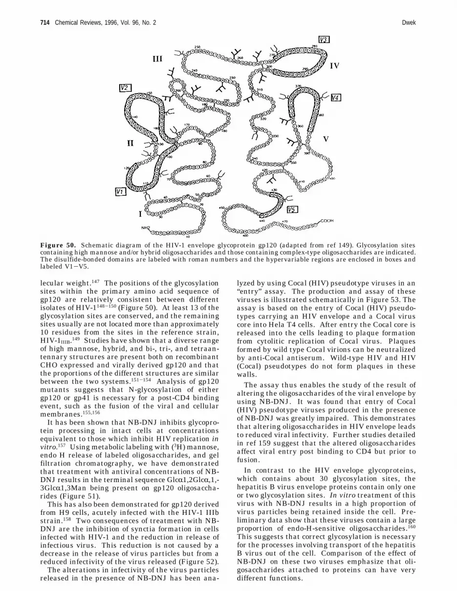
lecular weight.147 The positions of the glycosylationsites within the primary amino acid sequence ofgp120 are relatively consistent between differentisolates of HIV-1148-150 (Figure 50). At least 13 of theglycosylation sites are conserved, and the remainingsites usually are not located more than approximately10 residues from the sites in the reference strain,HIV-1IIIB.149 Studies have shown that a diverse rangeof high mannose, hybrid, and bi-, tri-, and tetraan-tennary structures are present both on recombinantCHO expressed and virally derived gp120 and thatthe proportions of the different structures are similarbetween the two systems.151-154 Analysis of gp120mutants suggests that N-glycosylation of eithergp120 or gp41 is necessary for a post-CD4 bindingevent, such as the fusion of the viral and cellularmembranes.155,156It has been shown that NB-DNJ inhibits glycopro-
tein processing in intact cells at concentrationsequivalent to those which inhibit HIV replication invitro.157 Using metabolic labeling with (3H) mannose,endo H release of labeled oligosaccharides, and gelfiltration chromatography, we have demonstratedthat treatment with antiviral concentrations of NB-DNJ results in the terminal sequence GlcR1,2GlcR,1,-3GlcR1,3Man being present on gp120 oligosaccha-rides (Figure 51).This has also been demonstrated for gp120 derived
from H9 cells, acutely infected with the HIV-1 IIIbstrain.158 Two consequences of treatment with NB-DNJ are the inhibition of synctia formation in cellsinfected with HIV-1 and the reduction in release ofinfectious virus. This reduction is not caused by adecrease in the release of virus particles but from areduced infectivity of the virus released (Figure 52).The alterations in infectivity of the virus particles
released in the presence of NB-DNJ has been ana-
lyzed by using Cocal (HIV) pseudotype viruses in an“entry” assay. The production and assay of theseviruses is illustrated schematically in Figure 53. Theassay is based on the entry of Cocal (HIV) pseudo-types carrying an HIV envelope and a Cocal viruscore into Hela T4 cells. After entry the Cocal core isreleased into the cells leading to plaque formationfrom cytolitic replication of Cocal virus. Plaquesformed by wild type Cocal virions can be neutralizedby anti-Cocal antiserum. Wild-type HIV and HIV(Cocal) pseudotypes do not form plaques in thesewalls.The assay thus enables the study of the result of
altering the oligosaccharides of the viral envelope byusing NB-DNJ. It was found that entry of Cocal(HIV) pseudotype viruses produced in the presenceof NB-DNJ was greatly impaired. This demonstratesthat altering oligosaccharides in HIV envelope leadsto reduced viral infectivity. Further studies detailedin ref 159 suggest that the altered oligosaccharidesaffect viral entry post binding to CD4 but prior tofusion.In contrast to the HIV envelope glycoproteins,
which contains about 30 glycosylation sites, thehepatitis B virus envelope proteins contain only oneor two glycosylation sites. In vitro treatment of thisvirus with NB-DNJ results in a high proportion ofvirus particles being retained inside the cell. Pre-liminary data show that these viruses contain a largeproportion of endo-H-sensitive oligosaccharides.160This suggests that correct glycosylation is necessaryfor the processes involving transport of the hepatitisB virus out of the cell. Comparison of the effect ofNB-DNJ on these two viruses emphasize that oli-gosaccharides attached to proteins can have verydifferent functions.
Figure 50. Schematic diagram of the HIV-1 envelope glycoprotein gp120 (adapted from ref 149). Glycosylation sitescontaining high mannose and/or hybrid oligosaccharides and those containing complex-type oligosaccharides are indicated.The disulfide-bonded domains are labeled with roman numbers and the hypervariable regions are enclosed in boxes andlabeled V1-V5.
714 Chemical Reviews, 1996, Vol. 96, No. 2 Dwek
+ +

10.1. GlycosphingolipidsGlycosphingolipids, as their name implies, are
sugar-containing lipids and are a major class ofglycoconjugates synthesized by eukaryotic cells. Theyconsist of three characteristic building block compo-nents: one of sphingosine (or one of its derivatives)and one molecule of a fatty acid with carbohydrateresidues of varying complexity as their polar headgroups. Sphingosine itself is a non-glycosylated long-chain amino alcohol.181
The amino group of the sphingoid base is attachedto a long-chain fatty acyl group (that can be saturatedor monounsaturated depending on different factors,such as, cell type, diet, or age) to form ceramide.While the ceramide moiety is located in the mem-brane lipid bilayer, the saccharide head group ex-tends out from the plasma membrane surface to theextracellular space. The sugar residue in glycosidiclinkage (always â) to the ceramide is either glucoseor galactose, with glucose the more prevalent. Themost complex sphingolipids are the glycosphingolip-ids, which include neutral lipids containing from one(cerebrosides) to 20 or more glucose units and acidicglycosphingolipids containing one or more sialic acidresidues (gangliosides) or sulfate esters (sulfatides).
Figure 51. Summary of N-glycosylation structures found on untreated and NB-DNJ treated recombinant gp120 expressedin CHO cells. The upper panel shows the structures from untreated gp120151,155 and the lower panel shows the structurespresent following NB-DNJ treatment.158
HOCH2 C
H
C
H3N+
H
OH
C
H
C(CH2)12CH3
H
Glycobiology Chemical Reviews, 1996, Vol. 96, No. 2 715
+ +

Glycosphingolipids are classified according to theircarbohydrate components. These complex lipids canbe divided into two major groups: neutral and acidic.Cerebrosides are the simplest glycosphingolipids andconsist of sphingosine, a fatty acid residue, and asingle monosaccharide polar head group (Figure54).181 More complex neutral glycosphingolipids haveunbranched or branched oligosaccharide head groups
containing up to 20 sugar residues. Acidic glyco-sphingolipids are sulfatides or gangliosides. Gan-gliosides have a similar basic structure to the neutralglycosphingolipids but are differentiated from eachother by the fatty acid residue they contain and bythe sialylated oligosaccharides which comprise thepolar head group. Some examples are shown inFigure 55.181
Over 100 gangliosides have been identified. Gan-gliosides are found in all vertebrate cells but arehighly enriched in nervous tissues. They have con-siderable physiological and medical significance. Theexpression of glycosphingolipids on the cell surfacechanges as the cells divide and differentiate. Thereis considerable evidence that glycosphingolipids arespecific determinants of cell-cell recognition, so thatthey probably have an important role in growth anddifferentiation of tissues. Specific glycosphingolipidsof host cells have been shown to interact with theproteins of viral and bacterial parasites. For in-stance, ganglioside GM1 acts as the cell surfacereceptor for the bacterial toxin that causes thedebilitating diarrhea of cholera.The glycans on glycosphingolipids are in very close
proximity to the plasma membrane, and it is likelythat the spatial proximity of lipid-linked glycans tothe cell surface is biologically important in mediatinginteractions which require intimate cell-cell contact.
Figure 52. Effect of NB-DNJ on virus output and infectiv-ity in H9 cells: (0) reverse transcriptase activity released;(9) infectivity of released virus.
Figure 53. Production and assay of HIV-cocal pseudo-types: (1) no neutralization; (2) anti-Cocal; (3) anti-Cocal+ anti-gp120.
Figure 54. A galactocerebroside. (Reprinted from ref 181.Copyright 1992 International Thompson Publishing Ser-vices.)
Figure 55. Gangliosides GM1, GM2, and GM3. (Reprintedfrom ref 181. Copyright 1992 International ThompsonPublishing Services.)
716 Chemical Reviews, 1996, Vol. 96, No. 2 Dwek
+ +

The common step in the biosynthesis of the vastmajority of glycosphingolipids involves the transferof glucose to ceramide. Partially inhibiting this stepwill result in a reduction of the glycosphingolipidson the cell surface. This approach should allow someof their functional roles to be investigated. Addition-ally, such inhibitors of glycolipid biosynthesis mayalso have therapeutic potential in treating the humanglycosphingolipid storage disorders.179,180
10.2. Glycosphingolipid Storage DisordersThese result from the inheritance of defects in the
genes encoding catabolic enzymes (which are thenimpaired) required for the sequential breakdown ofthe glycolipids within lysosomes. The therapeuticoptions for the treatment of these disorders are verylimited. Currently, only the nonneuronopathic formof Gaucher’s disease (type 1), a condition character-ized by glucocerebrosidase deficiency, which occursat high frequency in Ashkenazi Jews, is beingsuccessfully treated using enzyme-replacementtherapy.161,162 However, the use of specific inhibitorsof glycosphingolipid biosynthesis to reduce theirlevels in cells, is one approach which may be gener-ally applicable to treating storage disorders. Thismight then allow the impaired enzymes to catabolizefully the reduced levels of glycosphingolipids thuspreventing their accumulation.One example of this has recently been shown using
NB-DNJ, which in addition to being an R-glucosidaseI R ΙΙ inhibitor, is also a potent inhibitor of glyco-sphingolipid biosynthesis.163 This compound inhibitsthe initial monosaccharide attachment and so thisapproach may be useful in all glycosphingolipidstorage disorders. Interestingly, the galactose ana-logueN-butyldeoxygalactonojirimycin (NB-DGJ) wasfound to inhibit glycosphingolipid biosynthesis.164This is a more selective compound in that unlike NB-DNJ it does not inhibit the R-galactosidases.Clearly the ability to manipulate glycoprotein and
glycolipid biosynthetic pathways using specific gly-cosyltransferase and glycosidase inhibitor has enor-mous potential. In this way the roles for glycolipidsin such areas as neuronal and embryonic develop-ment can also be investigated, as well as their rolesin adhesion processes and tumor metasis.
11. Concluding RemarksThe chemistry of simple sugars was worked out in
the late nineteenth century by Emil Fischer, and thering structures determined in the interwar years byHaworth and colleagues. Simple polysaccharidessuch as starch, glycogen, and cellulose, as well asmore complex molecules such as chitin and hyalu-ronic acid had also received attention and theircomponent sugars identified by classical means. Bythe 1960s, especially through work on blood-groupdeterminants by Morgan, Watkins, and their associ-ates at the Lister Institute, it had become clear thatbesides simple mono- and polysaccharides, naturallyoccurring carbohydrates were commonly conjugatedto proteins and lipids (as glycoproteins and glycolip-ids).Little progress could be made to determine the
structure or function of these complex molecules until
sensitive and sophisticated techniques became avail-able to analyze the component sugars and the orderand structural details of their attachment to protein.Today automatic techniques are available for analysisof glycoproteins (in picomole amounts) and theprogress in technology has advanced considerably ourunderstanding of carbohydrate structures attachedto proteins.Protein glycosylation is influenced by three main
factors: the overall protein conformation, the effectof local conformation, and the available repertoire ofglycosylation-processing enzymes for the particularcell type. In general, the pattern of glycoforms isprotein specific, site specific, and tissue or cellspecific.Glycobiology deals with the nature and role of
carbohydrates in biological events. Glycoproteins arenow known to be fundamental to many importantbiological processes including fertilization, immunedefense, viral replication, parasitic infection, cellgrowth, cell-cell adhesion, degradation of blood clots,and inflammation. They are major components of theouter surface of mammalian cells. Over half thebiologically important proteins are glycosylated. Oli-gosaccharide structures change dramatically duringdevelopment and it has been shown that specific sets(i.e. specific sequences) of oligosaccharides are ex-pressed at distinct stages of differentiation. Further,alterations in cell surface oligosaccharides are as-sociated with various pathological conditions includ-ing malignant transformation.The finding that glycosylation may vary with
disease also leads to the concept that its manipula-tion might alter the properties of glycoproteins andresult in beneficial therapeutic results. The abilityto manipulate and modify sugar structures alsoprovides an important approach in understanding thedifferent functions of oligosaccharides.The elegant biosynthetic glycan-processing path-
way in the cell allows, in principle, the same oli-gosaccharide to be attached to quite different proteinswithout having to code the information into the DNAof the individual proteins. However, the orientationof the attached oligosaccharide with respect to thepolypeptide may markedly affect the properties of theglycoproteins. Further, different glycoforms of aprotein may display quite different orientations of theoligosaccharides with respect to the protein, thusconferring different properties. A striking exampleis the structure of the Fc fragment of IgG. Theconserved N-linked complex oligosaccharides at Asn-297 on each heavy chain of the CH2 domain occupythe interstitial space between the domains and alsointeract with the domain surface. Loss of the twoterminal galactoses from the oligosaccharide as in theFc fragment from patients with rheumatoid arthritis,results in a loss of interaction between the domainsurface and the oligosaccharide. This permits dis-placement and consequent exposure of the oligosac-charides, giving them the potential to be recognizedby endogenous receptors lectins.The recognition of oligosaccharides (lectins) is
influenced by their accessibility, the number of copiesof the oligosaccharides, and their precise geometryof presentation. These factors introduce a highdegree of specificity and control as to whether the
Glycobiology Chemical Reviews, 1996, Vol. 96, No. 2 717
+ +

recognition is physiologically relevant or not.That one set of structures on different proteins can
result in quite dramatic variations in properties ofglycoproteins or that different glycoproteins mayhave different properties emphasizes that there is nosingle unifying function for oligosaccharides.
12. AcknowledgmentsIt is a privilege and honor to acknowledge my debt
to the late Rodney Porter whose support led me intothe field of immunology and then glycobiology. Heencouraged me to form a relationship with MonsantoCompany to develop and automate the technology ofmicrosequencing of oligosaccharides. The Monsanto/Oxford University relationship was the first majorindustrial partnership for the University. My col-leagues at Monsanto were enlightened and concernedwith developing the science. Their approach also led,with the University of Oxford, to the foundation ofOxford GlycoSystems, the first University spin-offcompany which was to develop technology arisingmainly from within the Glycobiology Institute.I have benefited enormously from the help, advice,
and skills of my colleagues and students in develop-ing the Glycobiology Institute and equipping it so asto be a resource for glycobiology. It is particularlyappropriate to acknowledge fundamental contribu-tions made to the field by Professors T. Rademacher,M. Ferguson, and S. W. Homans. Dr. Parekh, whois now Chief Scientist at Oxford GlycoSystems, madean enormous contribution in the early years.More recently, the work of Drs. F. Platt, T. Butters,
G. B. Karlsson, and P. Fischer helped to establishthe antiviral program on a firm footing and also laidthe foundations for the study of glycolipid storagedisorders using amino sugars. Dr. David Wing hasbeen a resource for the whole Institute. Dr. PaulineRudd has also been a constant source of support,working with me to develop many different projectsand has been of significant help with this manuscript.I would also like to acknowledge contributions
made by Drs. D. Ashford, C. Edge, D. Fernandes, G.Guile, D. Harvey, B. Matthews, N. Karlsson, I.Scragg, J. Thomas, P. Williams, R. Woods, S. Wong,M. Wormald, and S. Zamze much of whose data Ihave used.My thanks are also due to Drs. Maureen Taylor
and Kurt Drickamer for their help in understandingC-type lectins.We thank the Biochemical Society for permission
to reproduce figures from the 7th Wellcome TrustAward for Research in Biochemistry Related toMedicine, 1994 and published in the BiochemicalSociety Transaction, Volume 23, Wellcome TrustAward Lecture.
13. References(1) (1) Metzler, M.; Gertz, A.; Sarkar, M.; Schachter, H.; Schrader,
J. W.; Marth, J. D. EMBO 1994, 13, 2056-2065.(2) Ioffe, E.; Stanley, P. Proc. Natl. Acad. Sci. 1994, 91, 728-732.(3) Weis, W. I.; Drickamer, K. Curr. Biol. 1994, 2, 1227-1240.(4) Sherrif, S.; Chang, C. Y.; Ezekowitz, R. A. B. Nat. Struct. Biol.
1994, 11, 789-794.(5) Drickamer, K.; Taylor, M. E. Annu. Rev. Cell Biol. 1993, 237-
264.(6) Rademacher, T. W.; Parekh, R. B.; Dwek, R. A. Annu. Rev.
Biochem. 1988, 57, 785-838.(7) Varki, A. Glycobiology 1993, 3, 97-130.
(8) Parekh, R. B.; Dwek, R. A.; Sutton, B. J.; Fernandes, D. L.;Leung, A.; Stanworth, D.; Rademacher, T. W.; Mizuochi, T.;Taniguchi, K.; Matsuta, K.; Takeuchi, Y.; Nagano, T.; Miyamoto,T.; Kobata, A. Nature 1985, 316, 452-457.
(9) Parekh, R. B.; Dwek, R. A.; Thomas, J. R.; Rademacher, T. W.;Opdenakker, G.; Wittwer, A. J.; Howard, S. C.; Nelson, R.; Siegel,N. R.; Jennings, M. G.; Harakas, N. K.; Feder, J. Biochemistry1989, 28, 7644-7662.
(10) Williams, R. L.; Greene, S. M.; McPhearson, A. J. Biol. Chem.1987, 262, 16020-16031.
(11) Robertson, A. D.; Purisima, E. O.; Eastman, M. A.; Scheraga,H. A. Biochemistry 1989, 28, 5930-5938.
(12) Rico, M.; et al. J. Biomol. NMR 1991, 1, 283-298.(13) Rico, M.; et al. Annu. Eur. J. Biochem. 1989, 183, 623-638.(14) Santoro, J.; et al. J. Mol. Biol. 1993, 229, 722-734.(15) Joao, H. C.; Scragg, I. G.; Dwek, R. A. FEBS Lett. 1992, 307,
343-346.(16) Wooten, E. W.; Edge, C. J.; Bazzo, R.; Dwek, R. A.; Rademacher,
T. W. Carbohyd. Res. 1990, 203, 13-17.(17) Rao, U.; Teeter, M. M. Protein Eng. 1993, 6, 837-847.(18) Weiner, S. J.; Kollman, P. A.; Nguyen, D. T.; Case, D. A. J.
Comput. Chem. 1986, 7, 230-252.(19) Wooten, E. W.; Bazzo, R.; Edge, C. J.; Zamze, S.; Dwek, R. A.;
Rademacher, T. W. Eur. Biophys. J. 1990, 18, 139-148.(20) Homans, S. W.; Pastore, A.; Dwek, R. A.; Rademacher, T. W.
Biochemistry 1987, 26, 6649-6655.(21) Homans, S. W.; Dwek, R. A.; Boyd, J.; Mahmoudian, M.;
Richards, W. G.; Rademacher, T. W. Biochemistry 1986, 25,6342-6350.
(22) Brisson, J.-R.; Carver, J. P. Biochemistry 1983, 22, 3680-3686.(23) Brisson, J.-R.; Carver, J. P. Biochemistry 1983, 22, 3671-3680.(24) Jorgensen, W. L.; Chandrasekhar, J.; Madura, J. D.; Impey, R.
W.; Klein, M. L. J. Phys. Chem. 1983, 79, 926-935.(25) Shaanan, B.; Lis, H.; Sharon, N. Science 1991, 254, 862-866.(26) Bode, W.; Meyer, E.; Powers, J. C. Biochemistry 1989, 28, 1951-
1963.(27) Marquart, M.; Deisenhofer, J.; Huber, R.; Palm, W. J. Mol. Biol.
1980, 369-391.(28) Freymann, D.; Down, J.; Carrington, M.; Roditit, I.; Turner, M.;
Wilery, D. J. Mol. Biol. 1990, 216, 141-160.(29) Woods, R. J.; Edge, C. J.; Wormald, M. R.; Dwek, R. A. In
Complex Carbohydrates in Drug Research. Alfred Benzon Sym-posium 36.; Clausen, K. B. a. H., Ed.; Munksgaard: Copenhagen,1994.
(30) Woods, R. J.; Fraser-Reid, B.; Dwek, R. A.; Edge, C. J. InModelling the Hydrogen Bond, 1993; Vol. 569.
(31) Bush, C. A.; Blumberg, K.; Brown, J. N. Biopolymers 1982, 21,1971-1977.
(32) Delbaere, L. T. J. Biochem. J. 1974, 143, 197-205.(33) Wormald, M. R.; Wooten, E. W.; Bazzo, R.; Edge, C. J.; Feinstein,
A.; Rademacher, T. W.; Dwek, R. A. Eur. J. Biochem. 1991, 198,131-139.
(34) Kornfeld, R.; Kornfeld, S. Annu. Rev. Biochem. 1985, 54, 631-634.
(35) Harris, R. J. Biochemistry 1993, 32, 6539-6547.(36) Harris, R. J.; Leonard, C. K.; Guzetta, A. W.; Spellman, M. W.
Biochemistry 1991, 30, 2311-2314.(37) Nishimura, H.; Takao, T.; Hase, S.; Shimonishi, Y.; Iwanaga, S.
J. Biol. Chem. 1992, 276, 17520-17525.(38) Haltiwanger, R. S.; Kelly, W. G.; Roquemore, E. P.; Blomberg,
M. A.; Dong, L. D.; Kreppel, L.; Chou, T.; Hart, G. W. Biochem.Soc. Trans. 1992, 20, 264-269.
(39) Hart, G. W. Curr. Opin. Cell Biol. 1992, 4, 1017-1023.(40) Ferguson, M. A. J.; Williams, A. F. Annu. Rev. Biochem. 1988,
1, 522-529.(41) Ferguson, M. A. J. Curr. Opin. Struct. Biol. 1991, 1, 522-529.(42) Yamashita, K.; Kamerling, J. P.; A., K. J. Biol. Chem. 1982, 257,
12809-12814.(43) Fukuda, M. Mol. Glycobiol. 1994, 1-52.(44) Schachter, H.; Brockhausen, I. Glycoconjugates: Composition
Structure, and Function; Dekker: New York, 1992; pp 263-332.(45) Goochee, C. F.; Monica, T. Bio/Technology 1990, 8, 421-427.(46) Schachter, H. Mol. Glycobiol. 1994, 88-162.(47) Schachter, H. Biochem. Cell Biol. 1986, 64, 163-181.(48) Mononen, I.; Karjalainen, E. Biochem. Biophys. Acta 1987, 788,
364-367.(49) Gavel, Y.; Von Heijne, G. Protein Eng. 1990, 3, 433-442.(50) Williams, A. F.; Parekh, R. B.; Wing, D. R.; Willis, A. C.; Barclay,
A. N.; Dalchau, R.; Fabre, J. W.; Dwek, R. A.; Rademacher, T.W. Glycobiology 1993, 3, 339-348.
(51) Parekh, R. B.; Tse, A. G. D.; Dwek, R. A.; Williams, A. F.;Rademacher, T. W. Eur. Mol. Biol. Organ. J. 1987, 6, 1233-1244.
(52) Rudd, P. M.; Joao, H. C.; Coghill, E.; Fiten, P.; Saunders, M. R.;Opdenakker, G.; Dwek, R. A. Biochemistry 1994, 33, 17-22.
(53) Harvey, D. J.; Rudd, P. M.; Bateman, R. H.; Bordoli, R. S.;Howes, K.; Hoyes, J. B.; Vickers, R. G. Org. Mass Spectrom.1994, 29, 753-766.
(54) Dwek, R. A.; Edge, C. J.; Harvey, D. J.; Wormald, M. R.; Parekh,R. B. Annu. Rev. Biochem. 1993, 62, 65-100.
718 Chemical Reviews, 1996, Vol. 96, No. 2 Dwek
+ +

(55) Davis, S. J.; Puklavec, M. J.; Ashfor, D. A.; Harlos, K.; Jones, E.Y.; Stuart, D. I.; Williams, A. F. Protein Eng. 1993, 6, 229-232.
(56) Ashford, D. A.; Alafi, C. D.; Gamble, V. M.; MacKay, D. J. G.;Rademacher, T. W.; Williams, P. J.; Dwek, R. A.; Barclay, A.N.; Davis, S. J.; Somoza, C.; Ward, H. A.; Williams, A. F. J. Biol.Chem. 1993, 268, 3260-3267.
(57) Davis, S. J.; Ward, H. A.; Puklavec, M. J.; Willis, A. C.; Williams,A. F.; Barclay, A. N. J. Biol. Chem. 1990, 265, 10410-10418.
(58) Dwek, R. A.; Ashford, D. A.; Edge, C. J.; Parekh, R. B.;Rademacher, T. W.; Wing, D. R.; Barclay, A. N.; Davis, S. J.;Williams, A. F. Phil. Trans. R. Soc. London Biochem. 1993, 342,43-50.
(59) Carr, S. A.; Hemling, M. E.; Folena-Wasserman, G.; Sweet, R.W.; Anumula, K.; Barr, J. R.; Huddleston, M. J.; Taylor, P. J.Biol. Chem. 1989, 264, 21286-21295.
(60) Spellman, M. W.; Leonard, C. K.; Baa, L. J.; Gelineo, I.; VanHalbeek, H. Biochemistry 1991, 30, 2395-2406.
(61) Harris, R. J.; Chamow, S. M.; Gregory, T. J.; Spellman, M. W.Eur. J. Biochem. 1990, 188, 291-300.
(62) Jones, E. Y.; Davis, S. J.; Williams, A. F.; Harlos, K.; Stuart, D.I. Nature 1992, 360, 232-239.
(63) Spiro, R. G.; Bhoyroo, V. D. J. Biol. Chem. 1984, 259, 9858-9866.
(64) Galili, U.; Shohet, S. B.; Kobrin, E.; Stults, C. L. M.; Macher, B.A. J. Biol. Chem. 1988, 263, 17755-17762.
(65) Williams, A. F.; Gagnon, J. Science 1982, 216, 696-703.(66) Feder, J.; Tolbert, W. R.; Rademacher, T. W.; Parekh, R. B.;
Dwek, R. A. Tissue plasminogen activator from normal humancolon cells, US Patent No. 4,751,084, 1988.
(67) Wittwer, A. J.; Howard, S. C.; Carr, L. S.; Harakas, N. K.; Feder,J.; Parekh, R. B.; Rudd, P. M.; Dwek, R. A.; Rademacher, T. W.Biochemistry 1989, 28, 7662-7669.
(68) Patthy, L. Cell 1985, 41, 657-663.(69) Ny, T.; Elgh, F.; Lund, B. Proc. Natl. Acad. Sci. 1984, 81, 5355-
5359.(70) Zamze, S.; Jacques, A.; Rademacher, T. W.; Opdenakker, G.;
Dwek, R. A. Manuscript in preparation, 1995.(71) Joao, H. C.; Dwek, R. A. Eur. J. Biochem. 1993, 218, 239-244.(72) Shall, S.; Barnard, E. A. J. Mol. Biol. 1969, 41, 237-251.(73) McPherson, A.; Brayer, G.; Cascio, D.; Williams, R. Science 1986,
232, 765-8.(74) Rijken, D. C.; Collen, D. J. Biol. Chem. 1981, 256, 7035-7041.(75) Wittwer, A. J.; Howard, S. C. Biochemistry 1990, 29, 4175-4180.(76) Wallen, P.; Pohl, G.; Bergsdorf, N.; Ranby, M.; Ny, T.; Jornvall,
H. Eur. J. Biochem. 1983, 132, 681-686.(77) Ranby, M.; Bergsdorf, N.; Pohl, G.; Wallen, P. FEBS Lett. 1982,
146, 289-292.(78) Spellman, M. W.; Basa, L. J.; Leonard, C. K.; Chakel, J. A.;
O’Conner, J. V.; Wilson, S.; van Halbeek, H. J. Biochem. 1989,264, 14100-14111.
(79) Brockway, W.; Castellino, F. J. Arch. Biochem. Biophys. 1972,151, 194-199.
(80) Sottrup-Jensen, L.; Claeys, H.; Zajdel, M.; Petersen, T. E.;Magnussen, S. Raven Press: New York, 1978; pp 191-209.
(81) Hayes, M. L.; Castellino, F. J. J. Biol. Chem. 1979, 254, 8768-8780.
(82) Gonzales-Gronow, M.; Grennet, H. E.; Fuller, G. M.; Pizzo, S.V. Biochim. Biophys. Acta 1990, 1039, 269-276.
(83) Pirie-Shepherd, S.; Jett, E. A.; Andon, N. L.; Pizzo, S. V. J. Biol.Chem. 1995, 270, 5877-5881.
(84) Ponting, C. P.; Marshall, J. M.; Cederholm-Williams, S. A.BloodCoagulation Fibrinolysis 1992, 3, 605-614.
(85) Gonzalez-Gronow, M.; Edelberg, J. M.; Pizzo, S. V. Biochemistry1989, 28, 2374-2377.
(86) Edelberg, J. M.; Enghild, J. J.; Pizzo, S. V.; Gonzalez-Gronow,M. J. Clin. Invest. 1990, 86, 107-112.
(87) Hall, S. W.; VandenBerg, S. R.; Gonias, S. L. J. Cell Biochem.1990, 43, 213-227.
(88) Mori, K.; Dwek, R. A.; Downing, A. K.; Opdenakker, G.; Rudd,P. M. J. Biol. Chem. 1995, 270, 3261-3267.
(89) Jaeken, J.; van Eijk, H. G.; van der Heul, C.; Corbeel, L.; Eeckels,R.; Eggermont, E. Clin. Chim. Acta 1984, 144, 245-247.
(90) Yamashita, K.; Ideo, H.; Ohkura, T.; Fukushima, K.; Yusa, I.;Ohno, K.; Takeshita, K. J. Biol. Chem. 1993, 268, 5783-5789.
(91) Iurin, O.; Mattu, T.; Kier, G.; Mian, N.; Rudd, P. M.; Winchester,B.; Dwek, R. A. Submitted for publication, 1995.
(92) Ferguson, M. A. J.; Homans, S. W.; Dwek, R. A.; Rademacher,T. W. Science 1988, 239, 753-759.
(93) Homans, S. W.; Ferguson, M. A. J.; Dwek, R. A.; Rademacher,T. W.; Anand, R.; Williams, A. F. Nature 1988, 333, 269-272.
(94) Homans, S. W.; Edge, C. J.; Ferguson, M. A. J.; Dwek, R. A.;Rademacher, T. W. Biochemistry 1989, 28, 2881-2887.
(95) Rademacher, T. W.; Edge, C. J.; Dwek, R. A. Curr. Biol. 1991,1, 41-42.
(96) Amzel, L. M.; Poljak, R. A. Annu. Rev. Biochem. 1979, 48, 961-997.
(97) Deisenhofer, J. Biochemistry 1981, 20, 2361-2370.(98) Sutton, B. J.; Phillips, D. C. Biochem. Soc. Trans. 1983, 11, 130-
132.
(99) Leatherbarrow, R. J.; Rademacher, T. W.; Dwek, R. A.; Woof, J.M.; Clar, A.; Burton, D. R.; Richardson, N.; Feinstein, A. Mol.Immunol. 1985, 22, 407-415.
(100) Leatherbarrow, R. J.; Dwek, R. A. FEBS Lett. 1983, 164, 227-230.
(101) Lund, J.; Tanaka, T.; Takahashi, N.; Sarmay, G.; Arata, Y.;Jefferis, R. Mol. Immunol. 1990, 27, 1145.
(102) Drickamer, K. J. Biol. Chem. 1988, 263, 9557-9560.(103) Taylor, M. E. Blood Cell Biochem. 1993, 5, 347-370.(104) Drickamer, K. Curr. Opin. Struct. Biol. 1993, 3, 393-400.(105) Drickamer, K. Curr. Opin. Struct. Biol. 1995, in press.(106) Weis, W.; Kahn, R.; Fourme, R.; Drickamer, K.; Hendrickson,
W. A. Science 1991, 254, 1608-1615.(107) Ikeda, K.; Sannoh, T.; Kawasaki, N.; Kawasaki, T.; Yamashina,
I. J. Biol. Chem. 1987, 262, 451-454.(108) Weis, W. I.; Drickamer, K.; Hendrickson, W. A. Nature 1992,
360, 127-134.(109) Woods, R. J.; Edge, C. J.; Dwek, R. A. Nature Struct. Biol. 1994,
1, 499-501.(110) Mullin, N. P.; Hall, K. T.; Taylor, M. E. J. Biol. Chem. 1994,
269, 28405-28413.(111) Taylor, M. E. Biochem. Soc. Trans. 1993, 21, 467-474.(112) Haurum, J. S.; Arsequell, G.; Lellouch, A. C.; Wong, S. Y. C.;
Dwek, R. A.; McMichael, A. J.; Elliot, T. J. Exp. Med. 1994, 180,739-744.
(113) Michaelsson, E.; Malmstrom, V.; Reis, S.; Engstrom, A.;Burkhardt, H.; Holmdahl, R. J. Exp. Med. 1994, 180, 745-749.
(114) Rudd, P. M.; Fortune, F.; Patel, R. B.; Dwek, R. A.; Lehner, T.Immunology 1994, 83, 99-106.
(115) Rademacher, T. W.; Homans, S. W.; Parekh, R. B.; Dwek, R. A.Biochemical Soc. Symp. 1986, 51, 131-148.
(116) Bodman, K. B.; Sumar, N.; Mackenzie, L. E.; Isenberg, D. A.;Hay, F. C.; Roitt, I. M.; Lydyard, P. M. Clin. Exp. Immunol. 1992,88, 420-42335.
(117) Axford, J. S.; Mackenzie, L.; Lydyard, P. M.; Hay, F. C.; Isenberg,D. A.; Roitt, I. M. Lancet 1987, ii, 1486-1488.
(118) Furukawa, K.; Matsuta, K.; Takeuchi, F.; Kosuge, E.; Miyamoto,T.; Kobata, A. Int. Immunol. 1990, 2, 105-112.
(119) Youings, A.; Chang, S.-C.; Dwek, R. A.; Scragg, I. G. J. Biol.Chem. Manuscript in preparation, 1993.
(120) Field, M. C.; Amatayakul-Chantler, S.; Rademacher, T. W.;Rudd, P. M.; Dwek, R. A. Biochem. J. 1994, 299, 261-275.
(121) Jefferis, R.; Lund, J.; Mitzutani, H.; Nakagawa, H.; Kawazoe,Y.; ’Arata, Y.; Takahashi, N. Biochem. J. 1990, 268, 529-537.
(122) Burastero, S. W.; Casali, P.; Wilder, R. L.; Notkins, A. L. J. Exp.Med. 1988, 168, 1979-1992.
(123) Hardy, R. R.; Hayakawa, K.; Shimizu, M.; Yamasaki, K.;Kishimoto, T. Science 1987, 236, 81-83.
(124) Shakib, F.; Stanworth, D. R. Ann. Rheum. Dis. 1976, 35, 263-266.
(125) Kobata, A. Glycobiology 1990, 1, 5-8.(126) Griffiths, H. R.; Dowlin, E. J.; Sahinoglu, T.; Blake, D. R.;
Parnham, M.; Lunec, J. Agents Actions 1992, 36, 107-111.(127) Parekh, R. B.; Isenberg, D. A.; Ansell, B. M.; Roitt, I. M.; Dwek,
R. A.; Rademacher, T. W. Lancet 1988, 1 (8592), 966-969.(128) Parekh, R. B.; Isenberg, D. A.; Rook, G.; Roitt, I. M.; Dwek, R.
A.; Rademacher, T. W. J. Autoimmun. 1989, 2, 101-114.(129) Rook, G. A. W.; Steele, J.; Brealey, R.; Whyte, A.; Isenberg, D.;
Sumar, N.; Nelson, J. L.; Bodman, K. B.; Young, A.; Roitt, I. M.;Williams, P.; Scragg, I.; Edge, C. J.; Arkwright, P. D.; Ashford,D.; Wormald, M.; Rudd, P. M.; Redman, C. W. G.; Dwek, R. A.;Rademacher, T. W. J. Autoimmun. 1991, 4, 779-794.
(130) Roitt, I. M.; Dwek, R. A.; Parekh, R. B.; Rademacher, T. W.;Alavi, A.; Axford, J. S.; Bodman, K. B.; Bond, A.; Cooke, A.; Hay,F. C.; Isenberg, D. A.; Lydyard, P. M.; Mackenzie, L.; Rook, G.;Smith, M.; Sumar, N. J. Autoimmun. 1988, 1, 499-506.
(131) Morgan, B. P. Complement: Clinical aspects and relevance todisease; Academic Press: London, 1990.
(132) Burton, D. R.; Boyd, J.; Brampton, A. D.; Easterbrook-Smith,S. B.; Emanuel, E. J.; Novotny, J.; Rademacher, T. W.; vanSchravendijk, M. R.; Sternburg, M. J. E.; Dwek, R. A. Nature1980, 288, 338-344.
(133) Duncan, A. R.; Winter, G. Nature 1988, 332, 738-740.(134) Malhotra, R.; Lu, J.; Holmskov, U. S.; Sim, R. B. Clin. Exp.
Immunol. 1994, 97 (supp. 2), 4-9.(135) Drickamer, K.; Dordal, M. S.; Reynolds, L. J. Biol. Chem. 1986,
261, 6878-6887.(136) Iobst, S. T.; Wormald, M. R.; Weis, W. I.; Dwek, R. A.; Drickamer,
K. J. Biol. Chem. 1994, 269, 15505-15511.(137) Rademacher, T. W.; Parekh, R. B.; Dwek, R. A.; Isenberg, D.;
Rook, G.; Axford, J.; Roitt, I. Springer Semin. Immunopathol.1988, 10, 231-249.
(138) Tsuchiya, N.; Endo, T.; Matsuta, K.; Yoshinoya, S.; Takenchi,F.; Nagano, Y.; Shiota, M.; Furutawa, K.; Kochibe, N.; Ho, K.;Kobata, A. J. Immunol. 1993, 151, 1137-1146.
(139) Malhotra, R.; Wormald, M. R.; Rudd, P. M.; Fischer, P. B.; Dwek,R. A.; Sim, R. B. Nature Med. 1995, 1, 237-243.
(140) Grinna, L. S.; Robbins, P. W. J. Biol. Chem. 1979, 254, 8814-8818.
(141) Hubbard, S. C.; Ivatt, R. J. Annu. Rev. Biochem. 1981, 50, 555-583.
Glycobiology Chemical Reviews, 1996, Vol. 96, No. 2 719
+ +

(142) Winchester, B.; Fleet, G. W. J. Glycobiology 1992, 2, 199-210.(143) Datema, R.; Olofosson, S.; Romero, P. A. Pharmacol. Ther. 1987,
33, 221-286.(144) Lubas, W. A.; Spiro, R. G. J. Biol. Chem. 1987, 262, 3775-3781.(145) Fleet, G. W. J.; Karpas, A.; Dwek, R. A.; Fellows, L. E.; Tyms,
A. S.; Petrusson, S.; Namgoong, S. K.; Ramsden, N. G.; Smith,P. W.; Son, J. C.; Wilson, F.; Witty, D. R.; Jacob, G. S.;Rademacher, T. W. FEBS Lett. 1988, 237, 128-132.
(146) Karpas, A.; Fleet, G. W. J.; Dwek, R. A.; Petrusson, S.; Nam-goong, S. K.; Ramsden, N. G.; Jacob, G. S.; Rademacher, T. W.Proc. Natl. Acad. Sci. 1988, 85, 9229-9233.
(147) Lasky, L. A.; Groopman, J. E.; Fennie, C. W.; Benz, P. M.; Capon,D. J.; Dowbenko, D. J.; Nakamuira, G. R.; Nunes, W. M.; Renz,M. E. B., P. W. Science 1986, 233, 209-212.
(148) Willey, R. L.; Rutledge, R. A.; Dias, S.; Folks, T.; Theodore, T.;Buckler, C. E.; Martin, M. A. Proc. Natl. Acad. Sci. 1986, 83,5038-5042.
(149) Leonard, C. K.; Spellman, M. W.; Riddle, L.; Harris, R. J.;Thomas, J. N.; Gregory, T. J. J. Biol. Chem. 1990, 265, 10373-10382.
(150) Alizon, M.; Wain-Hobson, S.; Montagnier, L.; Sonigo, P. Cell1986, 46, 63-74.
(151) Mizuochi, T.; Spellman, M. W.; Larkin, M.; Solomon, J.; Basa,L. J.; Feizi, T. Biomed. Chromatogr. 1988, 2, 260-270.
(152) Mizuochi, T.; Spellman, M. W.; Larkin, M.; Solomon, J.; Basa,L. J.; Feizi, T. Biochem. J. 1988, 254, 599-603.
(153) Mizuochi, T.; Matthews, T. J.; Kato, M.; Hamako, J.; Titani, K.;Solomon, J.; Feizi, T. J. Biol. Chem. 1990, 265, 8519-8524.
(154) Geyer, H.; Holschbach, C.; Hunsmann, G.; Schneider, J. J. Biol.Chem. 1988, 263, 11760-11767.
(155) Willey, R. L.; Smith, D. H.; Lasky, L. A.; Theodore, T. S.; Earl,P. L.; Moss B.; Capon, D. J.; Martin, M. A. J. Virol. 1988, 62,139-147.
(156) Lee, W. R.; Yu, X.-F.; Syu, W.-J.; Essex, M.; Lee, T.-H. J. Virol.1992, 66, 1799-1803.
(157) Karlsson, G. B.; Butters, T. D.; Dwek, R. A.; Platt, F. M. J. Biol.Chem. 1993, 268, 570-576.
(158) Karlsson, G. B. University of Oxford: Oxford, 1993.(159) Fischer, P. B.; Collin, M.; Karlsson, G. B.; James, W.; Buters,
T. D.; Gordon, S.; Davis, S. J.; Dwek, R. A.; Platt, F. M. J. Virol.1995, in press.
(160) Block, T. M.; Lu, X. L.; Platt, F. M.; Foster, G. R.; Gerlich, W.H.; Blumberg, B. S.; Dwek, R. A. Proc. Natl. Acad. Sci. 1994,91, 2235-2239.
(161) Barton, N. W.; Brady, R. O.; Dambrosia, J. M.; Di Biscelgie, A.M.; Doppelt, S. H.; Hill, S. C.; Mankin, H. J.; Murray, G. J.;Parker, R. I.; Argoff, C. E.; Grewel, R. P.; Yu, K.-T.; CollaboratorsN. Engl. J. Med. 1991, 324, 1464-1470.
(162) Beutler, R. Proc. Natl. Acad. Sci. 1993, 90, 5384-5390.(163) Platt, F. M.; Neises, G. R.; Dwek, R. A.; Butters, T. D. J. Biol.
Chem. 1994, 269, 8362-8365.
(164) Platt, F. M.; Neises, G. R.; Karlsson, G. B.; Dwek, R. A.; Butters,T. D. J. Biol. Chem. 1994, 269, 27108-27114.
(165) Brekke, O. H.; Michaelsen, T. E.; Sandlie, I. Immunol. Today1995, 16, 85-90.
(166) Williams, A. F.; Davis, S. J.; He, Q.; Barclay, A. N. Cold SpringHarbor Symp. Quant. Biol. 1989, LV1, 637-647.
(167) Rudd, P. M.; Joao, H. C.; Coghill, E.; Fiten, P.; Saunders, M. R.;Opdenakker, G.; Dwek, R. A. Biochemistry 1994, 33, 17-22.
(168) de Vos, A. M.; Ultsch, M. H.; Kelley, R. F.; Padmanabahn, K.;Tulinsky, A.; Westbrook, M. L.; Kossiakoff, A. A. Biochemistry1992, 31, 270-279.
(169) Thomas, J. R.; Dwek, R. A.; Rademacher, T. W. Biochemistry1990, 29, 5413-5422.
(170) Weis, W. I.; Quesenberry, M. S.; Taylor, M. E.; Bezouska, K.;Hendrickson, W. A.; Drickamer, K. Cold Spring Harbor Symp.Quant. Biol. 1992, 57, 281-289.
(171) Pumphrey, R. S. H.Molecular analysis of structure function andregulation; Shakib, F., Ed.; Pergamon Press: Elmsford, NY,1990.
(172) Dwek, R. A.; Sutton, B. J.; Perkins, S. J.; Rademacher, T. W.Biochem. Soc. Symp. 1984, 49, 123-136.
(173) Malhotra, R.; Thiel, S.; Reid, K. B. M.; Sim, R. B. J. Exp. Med.1990, 172, 955-959.
(174) Rademacher, R. W.; Williams, P.; Dwek, R. A. Proc. Natl. Acad.Sci. 1994, 91, 6123-6127.
(175) Davidson, D. J.; Castellino, F. J. Biochemistry 1991, 30, 625-633.
(176) Davidson, D. J.; Castellino, F. J. Biochemistry 1991, 30, 6689-6696.
(177) Hayes, M. L.; Castellino, F. J. J. Biochem. 1979, 254, 8772-8776.
(178) Takasaki, S.; Mizuochi, T.; Kobata, A.Methods Enzymol. 1982,83, 263-268.
(179) Sandhoff, K.; van Echten, G. Adv. Lipid Res. 1993, 26, 119-142.
(180) van Echten, G.; Sandhoff, K. J. Biol. Chem. 1993, 268, 5341-5344.
(181) Olivera, A.; Spiegel, S. Glycoconjugate J. 1992, Number 3, 110-117.
(182) Yoshima, H.; Mizuochi, T.; Ishii, M.; Kobata, A. Cancer Res.1980, 40, 4276-4281.
(183) Bigge, J. C.; Patel, T. P.; Bruce, J. A.; Goulding, P. N.; Charles,S. M.; Parekh, R. B. Anal. Biochem. 1995, 230, 229-238.
(184) Mackiewicz, A.; Dewey, M. J.; Berger, F. G.; Baumann, H.Glycobiology 1991, 1, 265-269.
(185) Ashwell, G.; Harford, J. Annu. Rev. Biochem. 1982, 51, 531-554.
CR940283B
720 Chemical Reviews, 1996, Vol. 96, No. 2 Dwek
+ +


![Quantitative determination of reducing sugars, oligosaccharides, and glycoproteins with [3H]borohydride](https://img.pdfslide.net/doc/110x75/635741203cd558f04e04bd4c/quantitative-determination-of-reducing-sugars-oligosaccharides-and-glycoproteins.jpg)


