Embed Size (px)
Citation preview

Microbiology (2002), 148, 2125–2133 Printed in Great Britain
GPI7 affects cell-wall protein anchorage inSaccharomyces cerevisiae and Candida albicans
Mathias Richard,1† Piet de Groot,2 Olivier Courtin,3 Daniel Poulain,4
Frans Klis2 and Claude Gaillardin1
Author for correspondence: Mathias Richard. Tel : 33 1 30 81 54 53. Fax: 33 1 30 81 54 57.e-mail : richard!grignon.inra.fr
1 Laboratoire de Ge! ne! tiqueMole! culaire et Cellulaire,Institut NationalAgronomique Paris-Grignon, UMR-INRA216,URA-CNRS1925, BP01,78850 Thiverval-Grignon,France
2 Fungal Research Group,Swammerdam Institute forLife Sciences, University ofAmsterdam, NieuweAchtergracht 166, 1018WV Amsterdam, TheNetherlands
3 Aventis Pharma, 102 routede Noisy, 93235Romainville cedex, France
4 Laboratoire de MycologieFondamentale etApplique! e, INSERM EPI9915, Universite! de Lille II,Faculte! de Me! decineH. Warembourg, Po# leRecherche, Place Verdun,59037 Lille Cedex, France
Glycosylphosphatidylinositol (GPI)-anchoring represents a mechanism forattaching proteins to the cell surface of all eukaryotic cells. Two localizationsof GPI proteins have been observed in the yeasts Saccharomyces cerevisiae andCandida albicans : plasma membrane and cell wall. The signals and themechanisms involved in this differential targeting are presently not wellunderstood. Here several cell-wall-related phenotypes of a gpi7/las21 deletionare described, where GPI7/LAS21 encodes a GPI-anchor-modifying activity. Inboth organisms, the structure and composition of the cell wall was modified,with a clear increase in chitin deposition. Cell-wall-targeted proteinsaccumulated in the growth medium, whereas the protein content of the cellwall decreased significantly, suggesting inefficiency of the covalent linkage.The level of plasma-membrane-targeted GPI proteins was not affected.Sequence analyses revealed that gene families involved in the addition ofphosphoethanolamines to the core GPI anchor are highly conserved betweeneukaryotes, with the exception of the Gpi7 family which seems to be fungus-specific. These data are compatible with the notion that thephosphoethanolamine added by Gpi7 protein to the GPI anchor is a key factorin the covalent linkage of cell-wall proteins to fungal cell-wall components.
Keywords : glycosylphosphatidylinositol, Pir proteins, Als proteins
INTRODUCTION
Numerous glycoproteins of lower or higher eukaryotesbecome attached to a glycosylphosphatidylinositol (GPI)anchor, many of which ultimately appear at the plasmamembrane, and in some fungi in the cell wall (Tiede etal., 1999). This anchor has the conserved core structuredescribed in all eukaryotes : lipid-PO
%-myo-inositol-
(6!1)-GlcNH#-(4!1)-αMan-(6!1)-αMan-(2!1)-
αMan-(6)-PO%-(CH
#)#-NH. In yeast, this glycan core is
decorated during its assembly with a fourth mannoseresidue and with additional phosphoethanolamines(EtN-Ps) to form the complete precursor glycolipid(Benghezal et al., 1995; Grimme et al., 2001). Thetransfer of the complete precursor onto proteins occurs
.................................................................................................................................................
†Present address: Department of Microbiology, Columbia University,701 West 168th Street, New York, NY 10032, USA.
Abbreviations: Als, agglutinin-like sequence; ConA, Concanavalin A;CWP, cell-wall protein; EtN, ethanolamine; EtN-P, phosphoethanolamine;GPI, glycosylphosphatidylinositol ; Pir, protein with internal repeat.
in the lumen of the endoplasmic reticulum and iscatalysed by a GPI transamidase complex (Ohishi et al.,2000). GPI-anchoring is not essential at a cellular level inmammalian cells, as many GPI-deficient mutant celllines have been established (Takeda et al., 1993) ;however, in Saccharomyces cerevisiae GPI anchors areessential for growth (Leidich et al., 1995). Thus, all thegenes known for their involvement in the biosynthesis ofGPI anchors in S. cerevisiae are essential, except one,GPI7 (Benachour et al., 1999). Its deletion in S. cerevisiaehampers GPI-anchor addition to newly synthesizedproteins, resulting in cell-wall fragility and matingdefects (Benachour et al., 1999; Toh-e & Oguchi, 1999).In Yarrowia lipolytica, a dimorphic yeast, the mutantcells have a clear defect in morphogenesis and displayreduced invasive growth and hypersensitivity to Calco-fluor White (Richard et al., 2001). Finally, in Cagpi7mutants of Candida albicans, chlamydospore formationis affected, as well as the yeast to hypha transition onsolid, but not in liquid media. The cell-wall structure isalso modified in the mutants, and budding patterns,
0002-5391 # 2002 SGM 2125

M. Richard and others
.................................................................................................................................................
Fig. 1. Representation of the S. cerevisiae GPI core structureand the main proteins involved in the addition of the three EtNgroups on the different mannose groups. Man, Mannose; GlcN,glucosamine; Ins, myo-inositol.
cytokinesis and cell shape are abnormal in both liquidand solid media. In vivo assays revealed that thesemutants have a reduced virulence, a reduced ability tocolonize the gut in mice and are more sensitive to thelytic action of macrophages (Richard et al., 2002).
S. cerevisiae is known to have a GPI core structure witha fourth mannose group; Smp3p, an essential protein, isdescribed as being involved in the addition of this group(Grimme et al., 2001). Various studies have describedthree proteins involved in the transfer of EtN-Ps onto thethree first mannose groups of theGPI anchor (Benachouret al., 1999; Flury et al., 2000; Taron et al., 2000).Mcd4p adds an EtN-P on the first mannose, Gpi7p onthe second and Gpi13p}Yll031c on the third one (Fig. 1)(Flury et al., 2000). Whereas both MCD4 and GPI13 areessential, GPI7 is not. The lethality of the GPI13 deletionis to be expected since its gene product is involved in theaddition of the EtN-P, which links the anchor to theprotein. The function of the other two EtN groups isunknown. Here we present evidence that the side chainadded by Gpi7p participates in the proper anchoring ofGPI-modified proteins targeted to the cell wall.
METHODS
Strains and growth conditions. The C. albicans strains usedwere SC5314 (wild-type), MLR2A42 (ura3 : : imm434}ura3 : : imm434, Cagpi7 : :hisG}Cagpi7 : :hisG-CaURA3-hisG)and MLR4A2 (ura3 : : imm434}ura3 : : imm434, Cagpi7 : :hisG}Cagpi7 : :hisG-CaURA3-hisG) (Richard et al., 2002).The S. cerevisiae strains used were FY23 (MATa ura3-52,trp1D63, leu2D1) and ∆gpi7 (MATa, ura3-52, his3D200,LEU2, LYS2, trp1D63) (de Groot et al., 2001). Candida cellswere grown at 37 °C, whereas Saccharomyces cells weregrown at 30 °C in Yeast Peptone Dextrose (YPD) for pre-culture. During experiments, cells were cultured in SyntheticComplete (SC) medium, at different pH values, containing0±17% yeast nitrogen base without amino acids (Difco), 2%glucose, 0±5% Casamino acids, 100 mM HEPES and 0±01%uracil, histidine, tryptophan or tyrosine, and 0±02% leucinewhen required. Two independent transformants, MLR2A42and MLR4A2, were included in all experiments to ensure thatthe phenotypic traits observed were solely due to the CaGPI7mutation and not to unrelated mutations that may haveoccurred during the construction of ∆Cagpi7 null strains. In a
previous publication, we demonstrated that the phenotypesobserved were due to CaGPI7 disruption (Richard et al., 2002)by reintroducing CaGPI7 in MLR2A42.
Quantazyme and chitinase sensitivity assays. These assayswere carried out to detect possible changes in the organizationof the 1,3-β-glucan and chitin layers of the cell wall.Quantazyme is a recombinant 1,3-β-glucanase (QuantumBiotechnology). The Quantazyme sensitivity assay was con-ducted as described by Ovalle et al. (1998). Briefly, cells inexponential phase (OD
'!!¯1–2) were resuspended in 40 mM
β-mercaptoethanol, 50 mM Tris}HCl, pH¯7±5, to an OD'!!
of 1. After incubation for 1 h, Quantazyme dissolved inglycerol}water (1:1, v}v), was added to a final concentrationof 400 U ml−". Cell lysis was determined by following thedecrease in turbidity at 600 nm over time. Quantazymesensitivity was expressed as a percentage of the initial OD
'!!.
Streptomyces griseus chitinase (Sigma; C1525) was used insome experiments at 0±2 U ml−" for 1 h before Quantazymeaddition.
SDS hypersensitivity tests. Exponential cultures were har-vested and adjusted to an OD
'!!of 1; 5 µl of tenfold serial
dilutions were spotted on YPD containing increasing amountof SDS: 0±01, 0±05 and 0±1%. Plates were read after 24 hculture at 30 °C for S. cerevisiae and 37 °C for C. albicans.
Isolation of cell-wall proteins (CWPs). For these experiments,cultures were grown in SC medium at 30 °C, pH 5±5, for S.cerevisiae strains, and at 37 °C, pH 4±5, for C. albicans strains,since faint differences could only be detected at higher pH inthe CWP content of the wild-type and the gpi7 mutants ofC. albicans. After 16 h culture in SC, cells were harvestedin exponential phase (OD
'!!¯2) and washed with 10 mM
Tris}HCl, pH 7±6, as described previously (Kapteyn et al.,1995; Montijn et al., 1994). Briefly, cell walls were isolated andwashed extensively with 1 M NaCl. Isolated cell walls wereboiled twice in the presence of SDS, EDTA and β-mercapto-ethanol to solubilize the non-covalently linked CWPs and toremove any contaminant derived from the cytosol and}orplasma membrane. SDS-extracted cell walls were freeze-driedafter washing and 3 mg of the pellets were treated either withrecombinant Trichoderma harzianum endo-1,6-β-glucanase(0±8 U g−") to release the GPI-CWPs (Kapteyn et al., 1996) orwith 30 mM ice-cold NaOH for 17 h to release the 1,6-β-glucanase-resistant Pir-like-CWPs. Subsequently, 1,6-β-gluc-anase-treated cell walls were treated with ice-cold 30 mMNaOH for 17 h to release the double linked CWPs. Eachenzymic digestion was stopped by adding SDS at a finalconcentration of 0±4% and heating for 5 min at 100 °C. Priorto heating in SDS, NaOH-treated cell walls were firstneutralized by adding acetic acid.
Analysis of CWPs. CWPs were separated by electrophoresisusing linear 2±6–20% polyacrylamide gels and transferredonto Immobilon-P polyvinylidene difluoride (PVDF, 0±45 µm)membrane (Millipore) using a wet blotting apparatus. CWPswere visualized by probing the membrane with peroxidase-labelled Concanavalin A (ConA; 0±3 µg ml−") in PBS con-taining 3% bovine serum albumin, 2±5 mM CaCl
#and 2±5 mM
MnCl#(Kapteyn et al., 2000). Western immunoblot analyses
were performed with polyclonal antisera directed against 1,6-β-glucan, agglutinin-like sequence (Als) proteins, Cwp1p,Pir2p, Ssr1p and Gas1p. For the antisera directed against Alsproteins, Cwp1p, Pir2p, Ssr1p and Gas1p, the membraneswere treated with 50 mM periodic acid and 100 mM sodiumacetate for 30 min before the blocking step with 5% milk
2126
GPI7}LAS21 function in yeasts
powder in PBS to enhance the specificity of the antisera.Binding of rabbit antibodies was assessed with goat anti-rabbit IgG peroxidase (Pierce) at a dilution of 1:10000 inPBS}5% milk powder. The blots were visualized with ECLWestern blotting detection reagents (Amersham PharmaciaBiotech) and Fuji Medical X-Ray film.
Determination of glucose, mannose and glucosamine con-tent in cell walls. Isolated cell walls (about 1–1±5 mg) werehydrolysed as described previously with minor modifications(Dallies et al., 1998). Briefly, cell walls were hydrolysed with50 µl 72% sulfuric acid for 3 h at 25 °C. Then 650 µl distilledwater was added to obtain a 1 M H
#SO
%solution, followed by
incubation at 100 °C for 3 h. The solution was diluted byadding 3 ml distilled water and precipitation of sulfate wascarried out using 3±3 ml 40 g barium hydroxide l−". Aftercentrifugation, the sugars were quantified using a Dionex Bio-LC system with a CarboPac PA 1 anion-exchange column. Inthis experiment 5 mM galactose was used as internal standard.
RESULTS
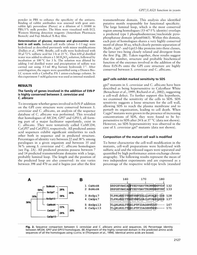
The family of genes involved in the addition of EtN-Pis highly conserved between S. cerevisiae andC. albicans
To investigate whether genes involved in EtN-P additionon the GPI core structure were conserved between S.cerevisiae and C. albicans, an analysis of the sequencedatabase of C. albicans was performed. This revealedthat homologues of MCD4, GPI7 and GPI13, all form-ing part of a major facilitator superfamily, exist inC. albicans. They were tentatively called CaMCD4,CaGPI7 and CaGPI13, respectively. All predicted aminoacid sequences exhibit significant similarities to eachother both in sequence and in predicted structure.Percentages of identity vary between 22 and 38% amongparalogues in a given organism and between 35 and56% among S. cerevisiae and C. albicans homologues(see Fig. 2A). All predicted proteins possess between 7and 14 predicted transmembrane domains with a large,probably luminal loop. The length and the position ofthe predicted loop are also conserved: its size variesbetween 398 and 470 aa and it begins just after the first
.................................................................................................................................................................................................................................................................................................................
Fig. 2. Sequence comparison between S. cerevisiae and C. albicans amino acid sequences. (A) Percentage identitybetween MCD4, GPI7 and GPI13 homologues. (B) Alignment of the highly conserved domain in the predicted amino acidssequences of all the homologues using CLUSTAL W (Thompson et al., 1994). Identical amino acids are boxed.
transmembrane domain. This analysis also identifiedputative motifs responsible for functional specificity.The large luminal loop, which is the most conservedregion among homologues (53–67±5% identity) overlapsa predicted type I phosphodiesterase}nucleotide pyro-phosphatase domain (pfam01663). Within this domain,each pair of homologues shares a very highly conservedmotif of about 30 aa, which clearly permits separation ofMcd4-, Gpi7- and Gpi13-like proteins into three classes,the latter two being closely related and divergent fromthe first (Fig. 2B). Taken together, these results suggestthat the number, structure and probable biochemicalfunction of the enzymes involved in the addition of thethree EtN-Ps onto the GPI core structure are highlyconserved between S. cerevisiae and C. albicans.
gpi7 cells exhibit marked sensitivity to SDS
gpi7 mutants in S. cerevisiae and C. albicans have beendescribed as being hypersensitive to Calcofluor White(Benachour et al., 1999; Richard et al., 2002), suggestinga cell-wall defect. To further support this hypothesis,we examined the sensitivity of the cells to SDS. SDSsensitivity suggests a loose structure for the cell wall,allowing SDS to reach the plasma membrane and toperturb its organization, leading to cell death. WhenCagpi7mutantswere grown in the presence of increasingconcentrations of SDS, they were found to be hy-persensitive to SDS after 24 h at 37 °C (data not shown).However, no SDS hypersensitivity was observed in thecase of S. cerevisiae gpi7 mutants (data not shown).
Composition of the mutant cell wall is modified
To better characterize the cell-wall modification in themutants, cell-wall preparations were hydrolysed withsulfuric acid and the released sugars were separated andquantified by high performance anion-exchange chrom-atography. The following results represent the mean oftwo independent experiments and are expressed as apercentage of the respective wild-type levels (standard
2127

M. Richard and others
.................................................................................................................................................................................................................................................................................................................
Fig. 3. Molecular architecture of the cell wall of S. cerevisiae and C. albicans. The arrows denote the orientation of thepolysaccharides from the non-reducing end to the reducing end. Mature GPI-CWPs, are believed to lose their lipid moietyonce they are covalently incorporated into the cell wall. GPI-CWPs can be linked by either a 1,6-β-glucan or an alkali-sensitive bond, like Pir proteins, or by both. The model is based on data from Kollar et al. (1997), Kapteyn et al. (2000),Smits et al. (1999) and this study.
error on these measurements is less than 5%). The sugarcomposition of the C. albicans Cagpi7 null mutant cellwall was 70, 102 and 412% for mannose, glucose andglucosamine, respectively, and 86, 116 and 261%,respectively, for the S. cerevisiae gpi7 null mutant. TheCagpi7 null mutants showed a fourfold increase in thechitin level, suggesting compensation of a cell-walldefect by chitin deposition, as often observed in cell-wall-defective mutants of S. cerevisiae (Dallies et al.,1998) and C. albicans (Kapteyn et al., 2000). Moreover,the mannose content of the cell wall decreased by 30%,indicating a defect in protein mannosylation or adecrease of mannoproteins in the mutant cell wallcompared to the wild-type. Similar resultswere observedon cell walls of a gpi7 mutant of S. cerevisiae, with agreater than twofold increase in chitin content, althoughthe mannose level was marginally affected. Overall,disruption of GPI7 seems to modify the cell wall of bothorganisms in a qualitatively similar way, resulting in adecrease of mannoproteins, which in turn is compen-sated by increased chitin deposition.
We reasoned that the excess of chitin in mutant cellwalls might protect cells against lytic enzymes. Indeed,we found a higher resistance of the mutant cells of C.albicans and S. cerevisiae toQuantazyme, a recombinant1,3-β-glucanase (data not shown). Furthermore, wechecked whether a chitinase pre-treatment of the mutantmay restore Quantazyme susceptibility. Although a 1 htreatment with chitinase alone had no effect on cell lysisunder our experimental conditions, it induced a signifi-cant increase in mutant cell lysis, comparable to wild-type, during a further Quantazyme treatment in both C.albicans and S. cerevisiae (data not shown).
Fewer proteins are linked to the cell wall in themutant cells
To investigate whether fewer mannoproteins wereindeed present in the cell wall, as suggested by previousdata on cell-wall composition, we analysed differentfractions from hot SDS cell-wall extracts of C. albicans
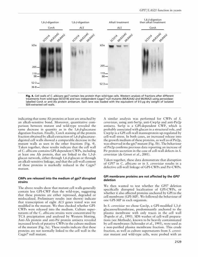
and S. cerevisiae mutant and wild-type cells grown in SCmedium in the yeast form. Indeed, in both S. cerevisiaeand C. albicans two types of CWPs have been identifiedthat are covalently linked to β-glucan, the GPI-de-pendent CWPs (GPI-CWPs) and the Pir-CWPs (Fig. 3).In S. cerevisiae, Pir-CWPs are cross-linked to cell-wall1,3-β-glucan by a covalent link sensitive to mild alkalitreatments (see Methods). GPI-CWPs are cross-linkedby a 1,6-β-glucan to 1,3-β-glucan (and to a lesser extentto chitin), or by an alkali-sensitive bond to 1,3-β-glucan(Pir-CWPs), or by both types of bond (Kapteyn et al.,1999). A recent publication demonstrated that thegeneral organization of the C. albicans cell wall is verysimilar to that of S. cerevisiae (Kapteyn et al., 2000). Tocharacterize the effect of CaGPI7 deletion on CWPs, wecompared the fractions obtained after 1,6-β-glucanasedigestion, mild alkali treatment, or mild alkali treatmentafter 1,6-β-glucanase digestion. Lectin-blotting of wild-type C. albicans 1,6-β-glucanase digests, using peroxi-dase-linked ConA, revealed a large smear and threemannosylated protein bands with apparent molecularmasses of 220, 180 and 100 kDa, respectively (Fig. 4). Asexpected, anti-1,6-β-glucan antiserum did not recognizeany of this material, indicating that the 1,6-β-glucanasedid not leave any detectable 1,6-β-glucan epitope at-tached to the proteins (data not shown). Cagpi7 mutantsexhibited a marked reduction of ConA stained materialin this test, suggesting that fewer proteins were linked tothe cell wall in the mutant (Fig. 4).
Hoyer and colleagues (Hoyer et al., 1995; Hoyer, 2001)have described the ALS gene family, encoding high-molecular-mass cell-surface proteins that are believed tobe covalently linked to cell-wall glucans by a remnantpart of the GPI anchor. Reprobing of the same sampleswith an anti-Als protein antiserum (Hoyer et al., 1995)revealed a band at 400 kDa in the wild-type (Fig. 4),which was greatly reduced when mutant cells wereprobed. Analysis of SDS-extracted, alkali-treated cellwalls with the same anti-Als protein antiserum revealeda protein band with an apparent molecular mass ofC600 kDa for both wild-type and mutant (Fig. 4),
2128
GPI7}LAS21 function in yeasts
205
11998
52·3
36·8
kDa kDa
205
11998
52·3
SC53
14
MLR
2A42
MLR
4A2
SC53
14
MLR
2A42
MLR
4A2
SC53
14
MLR
2A42
MLR
4A2
SC53
14
MLR
2A42
MLR
4A2
ConA ALS ALS ConA
1,6-b-digestionthen alkali treatmentAlkali treatment1,6-b-digestion1,6-b-digestion
.................................................................................................................................................................................................................................................................................................................
Fig. 4. Cell walls of C. albicans gpi7 contain less protein than wild-type cells. Western analysis of fractions after differenttreatments from wild-type (SC5314) and two independent Cagpi7 null mutants (MLR2A42 and MLR4A2) using peroxidase-labelled ConA or anti-Als protein antiserum. Each lane was loaded with the equivalent of 0±3 µg dry weight of isolatedSDS-extracted cell walls.
indicating that some Als proteins at least are attached byan alkali-sensitive bond. Moreover, quantitative com-parison between mutant and wild-type revealed thesame decrease in quantity as in the 1,6-β-glucanasedigestion fraction. Finally, ConA staining of the proteinfraction obtained by alkali extraction of 1,6-β-glucanase-digested cell walls showed a comparable decrease in themutant walls as seen in the other fractions (Fig. 4).Taken together, these results indicate that the cell wallof C. albicans contains GPI-dependent CWPs, includingat least one Als protein, that are linked to the 1,3-β-glucan network, either through 1,6-β-glucan or throughan alkali-sensitive linkage, and that the cell-wall contentof these proteins is markedly reduced in the Cagpi7mutant.
CWPs are released into the medium of gpi7 disruptedstrains
The above results show that mutant cell walls generallycontain less GPI-CWP than the wild-type, suggestingthat these proteins are either repressed, degraded ormislocalized. Preliminary results (not shown) indicatethat transcription of eight ALS genes tested was notmodified in the mutant. We thus checked whether GPI-CWPs were released into the medium. Culture super-natants of the C. albicans strains were concentrated byTCA precipitation and analysed by Western blotting.Anti-Als protein and anti-Pir protein antisera revealedincreased levels of putative CWPs in the culture mediumof the mutant (Fig. 5a). These results indicate that theseproteins are not normally linked to the cell wall in theCagpi7 null mutant.
A similar analysis was performed for CWPs of S.cerevisiae, using anti-Ssr1p, anti-Cwp1p and anti-Pir2pantisera. Ssr1p is a GPI-dependent CWP, which isprobably associated with glucan in a structural role, andCwp1p is a GPI cell-wall mannoprotein up-regulated bycell-wall stress. In both cases, an increased release intothe growth medium of these proteins, as well as of Pir2p,was observed in the gpi7mutant (Fig. 5b). The behaviourof Pir2p confirms previous data reporting an increase ofPir protein secretion in the case of cell-wall defects in S.cerevisiae (de Groot et al., 2001).
Taken together, these data demonstrate that disruptionof GPI7 in C. albicans or in S. cerevisiae results in adefective cell-wall linkage of GPI-CWPs and Pir-CWPs.
GPI membrane proteins are not affected by the GPI7deletion
We then wanted to test whether the GPI7 deletionspecifically disrupted localization of GPI-CWPs, orwhether it also affected proteins anchored by GPI to thecell membrane (GPI-MP). We followed the behaviour ofone GPI-MP in each organism.
In S. cerevisiae we chose Gas1p, a GPI-modified 1,3-β-glucanosyltransferase, predominantly anchored to theplasma membrane with only traces in the cell wall(Popolo et al., 1993). SDS washes of cell-wall prepara-tions (see Methods), known to be heavily contaminatedby cell membranes (Schreuder et al., 1993), were used asa non-purified plasma membrane fraction. This crudefraction, as well as culture supernatants from S. cerevi-siae mutant and wild-type cells, were probed with an
2129

M. Richard and others
205
11998
52·3
36·8
kDa kDa
205
11998
52·3
FY23
Dgpi7
SC53
14
MLR
2A42
MLR
4A2
ALS
30·136·8
SC53
14
MLR
2A42
MLR
4A2
FY23
Dgpi7
FY23
Dgpi7
Pir-like Ssr1 Cwp1 Pir2(b)(a)
.................................................................................................................................................................................................................................................................................................................
Fig. 5. Protein composition of the culture medium of wild-type and gpi7 mutants of S. cerevisiae (a) or C. albicans (b).Western analysis of protein from the culture supernatant from (a) wild-type (SC5314) and two independent Cagpi7 nullmutants (MLR2A42 and MLR4A2) using peroxidase-labelled ConA, anti-Als and anti-Pir2 antisera; (b) wild-type (FY23) andgpi7 null mutant (∆gpi7) using anti-Ssr1p anti-Cwp1 and anti-Pir2 antisera. Each lane was loaded with proteinsprecipitated by TCA from 1 ml culture medium at an OD600 of 2.
205
11998
kDa kDa
205
11998
52·3
FY23
Dgpi7
36·8
SC53
14
MLR
2A42
(b)(a)
FY23
Dgpi7
SC53
14
MLR
2A42
Culturesupernatant SDS extract
Culturesupernatant SDS extract
.....................................................................................................
Fig. 6. GPI-MPs are not affected by the GPI7deletion in S. cerevisiae (a) and C. albicans(b). Western analysis of protein from theculture medium from (a) wild-type (FY23)and gpi7 null mutant (∆gpi7) using anti-Gas1p antiserum; (b) wild-type (SC5314) andCagpi7 null mutant (MLR2A42) using anti-Gas1p antiserum. Each lane was loaded withproteins either precipitated by TCA from1 ml culture medium at an OD600 of 2 orSDS-extracted from 0±2 µg wet weight of cellwalls.
anti-Gas1p antiserum (Fig. 6a). No signal was detectedin the culture supernatants from wild-type and mutantstrains. In contrast, a protein band was present in themembrane fraction of both strains at the expected size of125 kDa and in nearly identical amounts. These datasuggest normal anchorage of at least one GPI-MP to thecell surface in the S. cerevisiae gpi7 mutant.
A previous study (Popolo & Vai, 1998) demonstratedthat anti-Gas1p antiserum cross-reacted with the homo-logues of Gas1p in C. albicans, the Phr1}2}3 proteins.To extend the results obtained in S. cerevisiae to C.albicans, an SDS extract of C. albicans cell walls wasanalysed using the same anti-Gas1p antiserum (Fig. 6b).
In both wild-type and mutant similar patterns wereobtained: no signal was detected in the culture super-natants, whereas in the SDS extract two clear proteinbands and a third, fainter band were detected at theexpected positions for Phr proteins. These resultssuggest that in both organisms membrane anchoring ofat least Gas1p and its homologues, and probably otherGPI-MPs, is not affected by the deletion of GPI7.
DISCUSSION
In this study we investigated the potential role of theEtN-P moiety added by Gpi7p in GPI-anchoring in two
2130

GPI7}LAS21 function in yeasts
(A)
(B)
.................................................................................................................................................
Fig. 7. Alignment of the highly conserved domain in thepredicted amino acid sequences of MCD4 and GPI3 (A) and GPI7(B) with homologues from the non-redundant sequencedatabase.
different organisms, S. cerevisiae and C. albicans. In afirst step, we scanned the C. albicans sequence databaseand found homologues for the three genes, MCD4, GPI7and GPI13, involved in the addition of EtN-P to the GPIcore structure. Interestingly, the C. albicans databasecontains a single homologue for each of these threegenes. Although we confirmed the conservation of thebiochemical function only for CaGpi7p (Richard et al.,2002), we speculate that the function of CaMcd4p andCaGpi13p is also conserved in view of their strongsimilarity to their homologues in S. cerevisiae. Withinthe conserved phosphodiesterase}nucleotide pyrophos-phatase domain shared by all these proteins, a motif ofabout 30 aa is highly conserved between each of thethree pairs of homologues and permits a clear dis-crimination of the three paralogues. An attractivehypothesis is that these motifs may determine thebinding specificity of the proteins for each of the threemannose groups of the GPI core structure: Mcd4p onthe first, Gpi7p on the second and Gpi13p on the third.Several potential homologues of the three genes can befound in lower and higher eukaryotes. As shown in Fig.7(A), the 30 aa motif is well conserved in most of them,permitting identification of four excellent matches withproteins from Schizosaccharomyces pombe, Drosophilamelanogaster, mice and humans for both the Mcd4p andGpi13pmotifs, fromArabidopsis thaliana for theMcd4pmotif and from Caenorhabditis elegans for the Gpi13pmotif. Interestingly, one EtN-P is added on the thirdmannose of GPI anchors in all eukaryotes and in mostcases on the first one, paralleling conservation of Gpi13pand Mcd4p. On the other hand, the modification of thesecond mannose carried out by Gpi7p in yeast is more
variable. We have noticed that Gpi7p tends to be moredivergent. Closely related relatives are found onlyamong proteins of fungal origin, from Yarrowia lipo-lytica, Saccharomyces bayanus and Schizosaccharo-myces pombe (Fig. 7B). More distant ones are detectedamong humans, mice, Drosophila and Caenorhabditisproteins, where the Gpi7p motif is interrupted by twoinsertions of 9 and 5 aa (not shown). This suggests thatGpi7 proteins have a conserved specific function infungi, whereas their homologues in higher eukaryotescarry out different GPI-anchor modifications.
It has been demonstrated that many GPI proteins ofyeast are CWPs or plasma-membrane proteins partici-pating in the building of the cell wall (Caro et al., 1997;Hamada et al., 1998). The phenotype of GPI7-depletedcells strongly suggests that Gpi7 affects cell-wall struc-ture. Increased cell-wall fragility, a common feature ofall gpi mutants, already suggested by previous tests withCalcofluor White (Richard et al., 2002), was confirmedby SDS hypersensitivity (Benghezal et al., 1995; Vossenet al., 1997) and we observed direct evidence of adifference in chitin content in the cell wall of mutant C.albicans and S. cerevisiae cells.
This defect in cell-wall structure correlates with a lowerprotein content of the cell wall in C. albicans and anincreased release of CWPs in the growth medium of themutant. Moreover, mutations in GPI7 do not affectlocalization of the cell membrane-attached GPI proteinGas1p.
The lower levels of GPI-dependent CWPs can beexplained in different ways. First, the lack of EtN-P onthe second mannose of the GPI core structure mightaffect the transport of GPI-dependent CWPs through thesecretory pathway. This does not seem to be the casesince the growth rate and the quantity of Gas1p in theplasma membrane were not affected. Second, the lack ofEtN-P might block the release of the plasma-membrane-bound intermediate form of GPI proteins destined forthe cell wall. This seems unlikely because in the mutantmore soluble forms were found in the culture medium.Third, this side-chain might be essential for efficientlinkage of GPI-CWPs to other cell-wall components.Thus, the lack of this side-chain may trigger mislocali-zation of GPI-CWPs, including some proteins involvedin cell-wall synthesis and in the linkage of Pir proteins.Consequently, a defective cell wall is formed, the cellaggregates, morphogenesis is affected and virulence andthe resistance to macrophages are also affected (Richardet al., 2002).
Finally, although gpi7 mutants of S. cerevisiae and C.albicans share common phenotypes, like secretion ofGPI-CWPs, resistance to Quantazyme and increasedchitin deposition, they also differ. In particular, no SDShypersensitivity was detected in S. cerevisiae mutantsand the amount of proteins in the cell wall was negligiblyaffected. Possibly, these phenotypic differences are dueto the different growth temperatures used for S. cerevi-siae (30 °C) and C. albicans (37 °C).
2131

M. Richard and others
ACKNOWLEDGEMENTS
We thank A. Derniaux for making the cell-wall extraction andthe cell-wall sugars quantification. We also thank A. Le!pinglefor her work on the Candida membranes. We are grateful toL. Hoyer for providing anti-Als antisera. This work wassupported by the French Ministe' re de la Recherche (Re! seauInfections Fongiques, PRFMMIP), the European Commission(QLRT-1999-30795), an EC grant to P.G. and a CNRS-Aventis Pharma grant to M.R.
REFERENCES
Benachour, A., Sipos, G., Flury, I., Reggiori, F., Canivenc-Gansel,E., Vionnet, C., Conzelmann, A. & Benghezal, M. (1999). Deletionof GPI7, a yeast gene required for addition of a side chain to theglycosylphosphatidylinositol (GPI) core structure, affects GPIprotein transport, remodeling, and cell wall integrity. J Biol Chem274, 15251–15261.
Benghezal, M., Lipke, P. N. & Conzelmann, A. (1995). Identifi-cation of six complementation classes involved in the biosynthesisof glycosylphosphatidylinositol anchors in Saccharomyces cerevi-siae. J Cell Biol 130, 1333–1344.
Caro, L. H., Tettelin, H., Vossen, J. H., Ram, A. F., van den Ende, H.& Klis, F. M. (1997). In silico identification of glycosyl-phosphati-dylinositol-anchored plasma-membrane and cell wall proteins ofSaccharomyces cerevisiae. Yeast 13, 1477–1489.
Dallies, N., Francois, J. & Paquet, V. (1998). A new method forquantitative determination of polysaccharides in the yeast cellwall. Application to the cell wall defective mutants of Sac-charomyces cerevisiae. Yeast 14, 1297–1306.
de Groot, P., Ruiz, C., Vazquez de Aldana, C. R. & 13 otherauthors (2001). A genomic approach for identification andclassification of genes involved in the cell wall formation and itsregulation in Saccharomyces cerevisiae. Comp Funct Genomics 2,1–19.
Flury, I., Benachour, A. & Conzelmann, A. (2000). YLL031cbelongs to a novel family of membrane proteins involved in thetransfer of ethanolaminephosphate onto the core structure ofglycosylphosphatidylinositol anchors in yeast. J Biol Chem 275,24458–24465.
Grimme, S. J., Westfall, B. A., Wiedman, J. M., Taron, C. H. &Orlean, P. (2001). The essential smp3 protein is required foraddition of the side-branching fourth mannose during assemblyof yeast glycosylphosphatidylinositols. J Biol Chem 276, 27731–27739.
Hamada, K., Fukuchi, S., Arisawa, M., Baba, M. & Kitada, K.(1998). Screening for glycosylphosphatidylinositol (GPI)-depen-dent cell wall proteins in Saccharomyces cerevisiae. Mol GenGenet 258, 53–59.
Hoyer, L. L. (2001). The ALS gene family of Candida albicans.Trends Microbiol 9, 176–180.
Hoyer, L. L., Scherer, S., Shatzman, A. R. & Livi, G. P. (1995).Candida albicans ALS1: domains related to a Saccharomycescerevisiae sexual agglutinin separated by a repeating motif. MolMicrobiol 15, 39–54.
Kapteyn, J. C., Montijn, R. C., Dijkgraaf, G. J., Van den Ende, H. &Klis, F. M. (1995). Covalent association of beta-1,3-glucan withbeta-1,6-glucosylated mannoproteins in cell walls of Candidaalbicans. J Bacteriol 177, 3788–3792.
Kapteyn, J. C., Montijn, R. C., Vink, E., de la Cruz, J., Llobell, A.,Douwes, J. E., Shimoi, H., Lipke, P. N. & Klis, F. M. (1996).Retention of Saccharomyces cerevisiae cell wall proteins through
a phosphodiester-linked beta-1,3-}beta-1,6-glucan heteropoly-mer. Glycobiology 6, 337–345.
Kapteyn, J. C., van den Ende, H. & Klis, F. M. (1999). Thecontribution of cell wall proteins to the organization of the yeastcell wall. Biochim Biophys Acta 1426, 373–383.
Kapteyn, J. C., Hoyer, L. L., Hecht, J. E., Muller, W. H., Andel, A.,Verkleij, A. J., Makarow, M., Van Den Ende, H. & Klis, F. M.(2000). The cell wall architecture of Candida albicans wild-typecells and cell wall-defective mutants. Mol Microbiol 35, 601–611.
Kollar, R., Reinhold, B. B., Petrakova, E., Yeh, H. J., Ashwell, G.,Drgonova, J., Kapteyn, J. C., Klis, F. M. & Cabib, E. (1997).Architecture of the yeast cell wall. Beta(1!6)-glucan intercon-nects mannoprotein, beta(1!3)-glucan, and chitin. J Biol Chem272, 17762–17775.
Leidich, S. D., Kostova, Z., Latek, R. R., Costello, L. C., Drapp,D. A., Gray, W., Fassler, J. S. & Orlean, P. (1995). Temperature-sensitive yeast Gpi anchoring mutants gpi2 and gpi3 are defectivein the synthesis of N-acetylglucosaminyl phosphatidylinositol.Cloning of the GPI2 gene. J Biol Chem 270, 13029–13035.
Montijn, R. C., van Rinsum, J., van Schagen, F. A. & Klis, F. M.(1994). Glucomannoproteins in the cell wall of Saccharomycescerevisiae contain a novel type of carbohydrate side chain. J BiolChem 269, 19338–19342.
Ohishi, K., Inoue, N., Maeda, Y., Takeda, J., Riezman, H. &Kinoshita, T. (2000). Gaa1p and Gpi8p are components of aglycosylphosphatidylinositol (GPI) transamidase that mediatesattachment of Gpi to proteins. Mol Biol Cell 11, 1523–1533.
Ovalle, R., Lim, S. T., Holder, B., Jue, C. K., Moore, C. W. & Lipke,P. N. (1998). A spheroplast rate assay for determination of cellwall integrity in yeast. Yeast 14, 1159–1166.
Popolo, L. & Vai, M. (1998). Defects in assembly of theextracellular matrix are responsible for altered morphogenesis ofa Candida albicans phr1 mutant. J Bacteriol 180, 163–166.
Popolo, L., Vai, M., Gatti, E., Porello, S., Bonfante, P., Balestrini, R.& Alberghina, L. (1993). Physiological analysis of mutantsindicates involvement of the Saccharomyces cerevisiae GPI-anchored protein gp115 in morphogenesis and cell separation. JBacteriol 175, 1879–1885.
Richard, M., Rosas-Quijano, R., Bezzate, S., Bordon-Pallier, F. &Gaillardin, C. (2001). Tagging morphogenetic genes by insertionalmutagenesis in the yeast Yarrowia lipolytica. J Bacteriol 183,3098–3107.
Richard, M., Ibata Ombetta, S., Dromer, F., Bordon-Pallier, F.,Jouault, T. & Gaillardin, C. (2002). Complete glycosylphosphati-dylinositol anchors are required for full morphogenesis andvirulence in Candida albicans. Mol Microbiol 44, 841–853.
Schreuder, M. P., Brekelmans, S., van den Ende, H. & Klis, F. M.(1993). Targeting of a heterologous protein to the cell wall ofSaccharomyces cerevisiae. Yeast 9, 399–409.
Smits, G. J., Kapteyn, J. C., van den Ende, H. & Klis, F. M. (1999).Cell wall dynamics in yeast. Curr Opin Microbiol 2, 348–352.
Takeda, J., Miyata, T., Kawagoe, K., Iida, Y., Endo, Y., Fujita, T.,Takahashi, M., Kitani, T. & Kinoshita, T. (1993). Deficiency of theGpi anchor caused by a somatic mutation of the PIG-A gene inparoxysmal nocturnal hemoglobinuria. Cell 73, 703–711.
Taron, C. H., Wiedman, J. M., Grimme, S. J. & Orlean, P. (2000).Glycosylphosphatidylinositol biosynthesis defects in Gpi11p- andGpi13p-deficient yeast suggest a branched pathway and implicateGpi13p in phosphoethanolamine transfer to the third mannose.Mol Biol Cell 11, 1611–1630.
Thompson, J. D., Higgins, D. G. & Gibson, T. J. (1994). :improving the sensitivity of progressive multiple sequence align-
2132

GPI7}LAS21 function in yeasts
ment through sequence weighting, position-specific gap penaltiesand weight matrix choice. Nucleic Acids Res 22, 4673–4680.
Tiede, A., Bastisch, I., Schubert, J., Orlean, P. & Schmidt, R. E.(1999). Biosynthesis of glycosylphosphatidylinositols in mammalsand unicellular microbes. Biol Chem 380, 503–523.
Toh-e, A. & Oguchi, T. (1999). Las21 participates in extra-cellular}cell surface phenomena in Saccharomyces cerevisiae.Genes Genet Syst 74, 241–256.
Vossen, J. H., Muller, W. H., Lipke, P. N. & Klis, F. M. (1997).Restrictive glycosylphosphatidylinositol anchor synthesis incwh6}gpi3 yeast cells causes aberrant biogenesis of cell wallproteins. J Bacteriol 179, 2202–2209.
.................................................................................................................................................
Received 4 December 2001; revised 6 February 2002; accepted 4 March2002.
2133


![[Characterization of azole resistance mechanisms in Chilean clinical isolates of Candida albicans]](https://img.pdfslide.net/doc/110x75/634a291cdedec593d70ede27/characterization-of-azole-resistance-mechanisms-in-chilean-clinical-isolates-of.jpg)


