Embed Size (px)
Citation preview

This article appeared in a journal published by Elsevier. The attachedcopy is furnished to the author for internal non-commercial researchand education use, including for instruction at the authors institution
and sharing with colleagues.
Other uses, including reproduction and distribution, or selling orlicensing copies, or posting to personal, institutional or third party
websites are prohibited.
In most cases authors are permitted to post their version of thearticle (e.g. in Word or Tex form) to their personal website orinstitutional repository. Authors requiring further information
regarding Elsevier’s archiving and manuscript policies areencouraged to visit:
http://www.elsevier.com/copyright

Author's personal copy
Journal of Neuroscience Methods 170 (2008) 245–254
Granger causality: Cortico-thalamic interdependencies duringabsence seizures in WAG/Rij rats
Evgenia Sitnikova a,∗, Taras Dikanev b,c,1, Dmitry Smirnov c,2,Boris Bezruchko b,c,3, Gilles van Luijtelaar d,4
a Department of Neuroontogenesis, Institute of Higher Nervous Activity and Neurophysiology, Russian Academy of Sciences,Butlerova Str. 5A, 117485 Moscow, Russia
b Saratov State University, Department of Nano- and Biomedical Technologies, Astrakhanskaya Str. 83, 410012 Saratov, Russiac Laboratory of Nonlinear Dynamical Modeling, Saratov Branch of Institute of RadioEngineering and Electronics,
Russian Academy of Sciences, Zelyonaya Str. 38, 410019 Saratov, Russiad Department of Biological Psychology, Radboud University Nijmegen, Nijmegen Institute of Cognition and Information,
PO 9104, 6500 HE Nijmegen, The Netherlands
Received 12 October 2007; received in revised form 14 December 2007; accepted 18 January 2008
Abstract
Linear Granger causality was used to identify the coupling strength and directionality of information transport between frontal cortex andthalamus during spontaneous absence seizures in a genetic model, the WAG/Rij rats. Electroencephalograms were recorded at the cortical surfaceand from the specific thalamus. Granger coupling strength was measured before, during and after the occurrence of spike-wave discharges (SWD).
Before the onset of SWD, coupling strength was low, but associations from thalamus-to-cortex were stronger than vice versa. The onset of SWDwas associated with a rapid and significant increase of coupling strength in both directions. There were no changes in Granger causalities beforethe onset of SWD. The strength of thalamus-to-cortex coupling remained constantly high during the seizures. The strength of cortex-to-thalamuscoupling gradually diminished shortly after the onset of SWD and returned to the pre-SWD level when SWD stopped. In contrast, the strength ofthalamus-to-cortex coupling remained elevated even after cessation of SWD.
The strong and sustained influence of thalamus-to-cortex may facilitate propagation and maintenance of seizure activity, while rapid reductionof cortex-to-thalamus coupling strength may prompt the cessation of SWD. However, the linear estimation of Granger coupling strength does notseem to be sufficient for predicting episodes with absence epilepsy.© 2008 Elsevier B.V. All rights reserved.
Keywords: Granger causality; Functional coupling; Thalamo-cortical associations; Electroencephalogram; Spike-and-wave seizures; Thalamo-cortical network;Animal model
1. Introduction
Over the years electroencephalography is widely used inclinical practice for the investigation, classification and diag-nosis of epileptic disorders. The electroencephalogram (EEG)
∗ Corresponding author. Tel.: +7 495 334 70 61; fax: +7 495 338 85 00.E-mail addresses: [email protected] (E. Sitnikova), [email protected]
(T. Dikanev), [email protected] (D. Smirnov), [email protected] (B. Bezruchko),[email protected] (G. van Luijtelaar).
1 Tel.: +7 8452 498037; fax: +7 8452 272401.2 Tel.: +7 8452 511180; fax: +7 8452 272401.3 Tel.: +7 8452 511180/498037; fax: +7 8452 272401.4 Tel.: +31 24 3615612; fax: +31 24 3616066.
provides valuable information in patients with typical and atyp-ical epileptic syndromes and offers also important prognosticinformation.
Absence epilepsy, previously known as petit mal, is clas-sically considered as non-convulsive generalized epilepsy(classification of the International League Against Epilepsy,ILAE) of unknown etiology (refs. in Panayiotopoulos, 1997).Clinically, absence seizures occur abrupt, last several secondsup to a minute and are accompanied by a brief decrease ofconsciousness that interrupts normal behavior. Absences mayeither have or have no facial automatisms, e.g. minimal jerks andtwitches of facial muscles, and eye blinks. In humans, EEGs dur-ing typical absence seizures are characterized by the occurrenceof generalized 3–5 Hz spike-wave complexes which have an
0165-0270/$ – see front matter © 2008 Elsevier B.V. All rights reserved.doi:10.1016/j.jneumeth.2008.01.017

Author's personal copy
246 E. Sitnikova et al. / Journal of Neuroscience Methods 170 (2008) 245–254
abrupt on- and offset (Bosnyakova et al., 2007; Panayiotopoulos,1997). Similar EEG paroxysms, spike-and-wave discharges(SWD) appear in rat strains with a genetic predisposition toabsence epilepsy, such as GAERS (Genetic Absence EpilepsyRats from Strasbourg; Vergnes, 1987) and WAG/Rij (Wis-tar Albino Glaxo from Rijswijk, Coenen and van Luijtelaar,2003; see also http://www.socsci.ru.nl/wagrij/info.0.html). TheEEG waveform and duration (1–30 s, mean 5 s) of SWD inrats and in humans are comparable, but the frequency ofSWD in rats is higher, 8–11 Hz (Midzianovskaia et al., 2001;Sitnikova and van Luijtelaar, 2007; van Luijtelaar and Coenen,1986).
Several modern computational techniques and advancedmethods of EEG analysis have been developed to extract “hid-den” information from the EEG in order to localize the regionof onset and to anticipate the onset of ‘absences’ as early aspossible (in rats, Meeren et al., 2002; Refs. for human datain Mormann et al., 2007). Our experiments are carried out inWAG/Rij rats (van Luijtelaar and Coenen, 1986). Every subjectof this strain has typical absence seizures that are accom-panied with spontaneous SWD in the EEG. Previously weused EEG coherence to measure neuronal synchrony betweenpopulations of thalamic and cortical neurons (Sitnikova andvan Luijtelaar, 2006). We found that the onset of SWD wascharacterized by area-specific increase of coherence and sup-ports the idea that the cortico-thalamo-cortical circuitry isprimarily involved in the initiation and propagation of SWD(Meeren et al., 2005; Steriade, 2005). Coherence is a tradi-tional measure of linear correlations between two EEG channelsin the frequency domain, but it does not assume directional-ity of inter-channel interactions and does not provide temporal(time-domain) information of the EEG signals (Challis andKitney, 1991). Granger causality compensates for these limi-tations (Ancona et al., 2004; Feldmann and Bhattacharya, 2004;Granger, 1969; Hlavackova-Schindler et al., 2007; Pereda etal., 2005). Granger causality concept can be used to determinedirectional coupling characteristics between recording sites inintracranial EEG and to reveal active abnormal causal relation-ships in epileptogenic networks (Chavez et al., 2003; Kaminskiet al., 2001). Usually, Granger causality estimations are per-formed in long time intervals that include a dozen or evenmore, basic periods. The pairwise analysis is based on theconstruction of vector autoregressive (AR) models from bivari-ate data that estimates how well the current measure of oneprocess can improve the prediction of the future of anotherprocess (Granger, 1969). Here we apply Granger causalityto measure the strength of bidirectional functional interac-tions between neuronal assembles in thalamus and frontalcortex.
The present work aims to measure bidirectional (feed-forward and feedback) network interdependences betweenlocal field potentials recorded simultaneously from the spe-cific thalamus and the frontal cortex. Granger causalityconcept will be used to characterize the strength of func-tional connectivity between the cortical and thalamic EEGsfor both directions before, during, and after SWD inrats.
2. Materials and methods
2.1. Animals and EEG data acquisition
Experiments were performed in five male 11–12-month oldWAG/Rij rats. The recordings were done at the Departmentof Biological Psychology, Radboud University Nijmegen inaccordance with the European Communities Council Direc-tive (86/609/EEC). Experiments were approved by the EthicalCommittee on Animal Experimentation of Radboud Uni-versity Nijmegen. Distress and suffering of animals wereminimal.
EEGs were recorded from brain areas in which seizure activ-ity is known to be the most robust: in the frontal cortex andin the ventroposteromedial thalamic nucleus, VPM (Vergneset al., 1987). Stainless steel EEG electrodes were implantedduring stereotactic surgery under isofluorane anesthesia. Oneelectrode was placed epidurally over the frontal cortex [AP 2;L 2.5] and the other depth electrode was implanted into theventroposteromedial thalamic nucleus [VPM, AP −3.5; L 2.5;H 7.2]. Two additional electrodes, ground and reference, wereplaced symmetrically over the two hemispheres of the cerebel-lum. All electrodes were identical (diameter 0.25 mm) and hadnon-insulated tips. The coordinates are given in mm relative tobregma according to the rat brain atlas of Paxinos and Watson(1986).
After the surgery, animals were allowed to recover during atleast 10 days. During this recovery period, animals received post-surgery care and their weight was monitored. Upon completionof the EEG recording sessions, rats were deeply anesthetizedwith overdose of sodium pentobarbital (200 mg/kg i.p.) andtheir brains were stained with Nissl. Electrode positioning wasverified using the atlas of the rat brain (Paxinos and Watson,1986).
EEG recordings were made in freely moving rats in a Fara-day cage. Each recording session lasted from 5 to 7 h duringthe dark period of the day–night cycle. EEG signals were fedinto a multi-channel differential amplifier, filtered between 1and 200 Hz, digitized with 1024 samples/second per channel(CODAS software) and stored on hard disk.
SWD appeared in EEG as a train of stereotypic repetitive7–10 Hz spikes-and-waves with high amplitude (that exceededthe background more than three times); SWD lasted longer than1 s (Midzianovskaia et al., 2001; van Luijtelaar and Coenen,1986). SWD were detected automatically in the frontal EEGrecords using the algorithm and original software developedby Dr. Ir. P.L.C. van den Broek (NICI, Radboud UniversityNijmegen, the Netherlands). This method is based on the detec-tion of steep changes in the EEG.
2.2. Granger causality
Let {xn} and {yn} be the two EEG signals (time series)recorded simultaneously in different brain loci. xn and yn areEEG values measured at the n-th time point. In order to studycausal relations between x and y, we use Granger’s approach andanalyze prediction improvement.

Author's personal copy
E. Sitnikova et al. / Journal of Neuroscience Methods 170 (2008) 245–254 247
First, we fit a univariate autoregressive (AR) model to theEEG data. The model takes the form
xn = f (xn−1, xn−2, . . . , xn−d). (1)
It relates current EEG value to the d previous values, f is somefunction, e.g., algebraic polynomial whose order and coefficientsare to be estimated from the observed data. Eq. (2) shows awidely used linear AR model
xn = α0 +d∑
i=1
αixn−i. (2)
Coefficients αi are selected in order to minimize the meansquared error, ε2
x
ε2x = 1
N
∑n
(xn −
(α0 +
d∑i=1
αixn−1
)), (3)
where N is the number of predicted samples in a time series. Ifthe number of model parameters is much less than the numberof data points used for the estimation, a minimal value ε2
x cancharacterize the accuracy of the model: the less the error, thebetter the model (self-predictability of the model).
Causal relations between process y and process x are presentwhen prediction of signal {xn} can be improved by incorporationinto the model the past of signal {yn} (illustrated in Fig. 1A).As a result, we construct a ‘joint’ AR model Eq. (4)
xn = f (xn−1, . . . , xn−d1 ) + g(yn−1, . . . , yn−d2 ), (4)
where f and g are polynomials that we determine from the currentdata. Function f is the same as in the individual model Eq. (1)while function g describes the influence of process y on processx. Number d1 is the same as the dimension d of the individualmodel Eq. (1). The number d2 describes ‘inertial’ propertiesof the influence. If d2 = 1, then the influence is instantaneous,otherwise it is non-local in time. Different values of d1 and d2∈ (1; 25) are tested in order to select those values that providethe most faithful results. In the present study we used linear ARmodels Eq. (5):
xn = α0 +d1∑i=1
αixn−i +d2∑i=1
βiyn−i. (5)
In Eq. (5) coefficients �i are chosen using the least-squaresestimations method. This method examines mean squared pre-diction error, ε2
xy, and when this error appears to be less then the
ε2x, it is assumed that process y influences process x (Fig. 1A). In
order to measure coupling between channels, we use the relativeprediction improvement, the so-called Granger-Sargent statistic(Hlavackova-Schindler et al., 2007):
s2xy = ε2
x − ε2xy
ε2xy
. (6)
Thus, the influence of channel {yn} on channel {xn} is charac-terized by the value of the normalized prediction improvements2xy and the reverse influence of {xn} on {yn}, s2
yx is described
Fig. 1. Estimation of Granger causality between EEG signals as recorded at tha-lamus and frontal cortex. The autoregressive model (AR) is used to characterizeamplitude changes in two EEG signals, {xn} and {yn} over time. (A) In thejoint AR model, the information about the past of EEG signal {yn} improvesthe prediction of EEG signal {xn} (see Eq. (4)) with a reduction of the instan-taneous prediction error, �n. The squared errors, ε2
xy , characterize the quality of
prediction of the chosen AR model. The value of ε2xy depends on the number of
samples in signals {xn} and {yn} used in the AR model, so-called model dimen-sions, d. (B) Dependencies between the prediction errors, ε2
xy , and the modeldimensions in our linear AR that predicted the future of the signal {yn} (d1 andd2 is the number of samples in the signal {xn} and signal {yn} correspondingly).An increase of dimensions d1 and d2 resulted in a decrease of the least meansquared error and the quality of predictions was improved until d1 = d2 = 5. Fur-ther increase of dimensions did not improve the accuracy of prediction, thereforein our computations we used d1 = d2 = 5.
by an equation similar to Eq. (6) in which x and y should beinterchanged.
2.3. Application of Granger causality to EEG data
Nonlinear dynamics techniques consider EEG signals as non-linear process and try to extract some nonlinear features fromit. Typically, they require long epochs of stationary data (e.g.,Arnhold et al., 1999; Le Van Quyen et al., 1999; Schiff et al.,1996). Estimations of Granger causality, either linear or nonlin-ear, are performed under the same conditions. Linear estimatesare usually less sensitive to the epoch length, because they arebased on relatively simple models (with fewer free parameters).However, the EEG is known to be highly non-stationary (Kaplan,1998). This should be taken into account in order to specify the
Author's personal copy
248 E. Sitnikova et al. / Journal of Neuroscience Methods 170 (2008) 245–254
proper time window length for the estimation of autoregressivemodels when estimating Granger causality.
2.3.1. The choice of length of moving windowIn fact, non-stationary is an intrinsic feature of the EEG signal
that accounts for complex dynamics of electrical brain activ-ity (Dikanev et al., 2005). However, the classical estimation ofGranger causality requires stationary data. Therefore we seg-mented the EEG into relatively short epochs in which the EEGsignal reveals quasi-stationary behavior.
In order to get a correct approximation to the non-stationaryEEG data with the AR model, it is important to define theoptimal size of the moving time-window. By shortening thetime-window, it is possible to improve the time resolution, butthis reduces the reliability of the AR models. In non-stationaryEEG data, the time-window should include several repetitive ele-ments. In our case, the time-window lasts 0.5 s; this correspondsto four spike-wave cycles. This size of the time-window wasfound to be optimal. If the estimation window was shorter than0.5 s, Granger causality estimations were unstable and longertime-window (up to 1 s) did not improve the stability of couplingestimates.
2.3.2. Selection of the polynomial order(linearity–nonlinearity in EEG signals)
Originally, Granger causality principles were formulatedwithout any assumption about the linearity or nonlinearity of thesystems. Traditional Granger causality measures were based onlinear models (Granger, 1969; Granger and Newbold, 1977). Inorder to make a choice between linear and nonlinear AR models,we compared prediction accuracy of these two models. It wasfound that the introduction of nonlinearity (such as polynomialsof the second and third order) had no significant influence on theprediction quality of the AR model. It was additionally foundthat a linear AR model was sufficient to describe the dynamicbehavior of the baseline EEG. It suggests a predominance of thelinear causal relations in non-seizure EEG. In contrast, seizureactivity (SWD) contained a nonlinear component, which was notyet modeled. However, the construction of a specific nonlinearAR model that describes seizure-related processes in the EEGis beyond the scope of the present paper.
2.3.3. Adjusting parameters of AR model (the choice of‘dimensions’)
The linear AR models Eqs. (2) and (5) are used to calculatethe coupling characteristics s2
xy and s2yx. Fig. 1B shows the typical
dependence of prediction error ε2xy on the dimensions d1 and d2
of the AR model for a 0.5 s time-interval of SWD. By increasingthe dimensions of the model d1 and d2 from 1 to 5, the error ofprediction decreases and a minimal error was obtained whenboth d-values were equal to 5. Further increase of d1 and d2 didnot improve the accuracy of predictions, therefore, d1 = d2 = 5were selected as optimal dimensions for the chosen model. Sucha dependence is typical for the prediction error ε2 as well.
All this suggests that also in our linear AR model the pastof one signal improves the prediction of the other signal. This
model provided significant and stable predictions with the cho-sen parameters (dimensions and time-window size), therefore,it was an appropriate model for the analysis of pairwise causalrelations in the chosen EEG pairs.
2.3.4. Statistical evaluation of causal interactionsCoefficients of Granger causality (prediction improvements)
were computed using EEG data from the cortical surface (frontalcortex) and from the specific part of the thalamus (ventral basalcomplex). The first and the last spike in spike-wave sequenceswere used to mark the onset and the offset of seizure activity.Statistical analysis of the thalamus-to-cortex (s2
xy) and cortex-
to-thalamus (s2yx) causalities was performed in two 10-s EEG
epochs (Fig. 2A). The first epoch included two 5 s successiveintervals, one before seizure onset (pre-SWD), the second wasthe first 5 s of a seizure (SWD-start). The second 10-s epochincluded the last 5 s of a SWD (SWD-end) and the first 5 s aftera seizure (post-SWD). Coefficients of Granger causality werecomputed with bin = 0.0049 (5 samples/1024) and averaged per0.2 s and per rat. Factorial ANOVA (repeated measures) and posthoc tests (LSD and t-tests for paired observations) were used forthe statistical analysis.
2.3.5. Evaluation of statistical significance with surrogatetest estimation
There are some undesirable factors (EEG noise, nonlinear-ities, etc.) that can influence Granger causality measures andmight mislead analysis. In order to control for these unwantedfactors and evaluate the statistical significance of the Grangercausality parameters, we performed surrogate data tests.Surrogate signals were constructed by taking two apparentlyuncoupled EEG signals recorded in the cortex and thalamus. Forthat purpose, two randomly chosen SWD were selected in thecortex and thalamus. Either the onset or the end of SWD werematched. Surrogate tests were performed using EEG data fromall animals. In total, 1000 random pairs of SWD were made toconstruct an ensemble of surrogate time series which modelleduncoupled process in the thalamo-cortical system. Grangercausalities in surrogate and in original data were computedusing the same algorithm. The surrogate Granger measures inthe uncoupled pairs were analyzed statistically by computing95%-percentiles of their distributions, s2
xy,0.95 and s2yx,0.95,
respectively. The results of surrogate tests, s2xy,0.95 and s2
yx,0.95,were plotted in Fig. 2B and C against the true data values,s2xy and s2
yx. Interdependence in EEG pair can be regarded assignificant (at the significance level p = 0.05) whenever thetrue values s2
xy and s2yx appeared above 95%-percentiles of
the corresponding surrogate values s2xy,0.95 and s2
yx,0.95. Thissurrogate test confirmed that interdependencies between EEGsignals were significant in both directions.
3. Results
All SWD were detected automatically in the full length EEGrecordings (6–7 h). In total, 53, 111, 34, 33 and 63 epilepticdischarges in five rats were detected and analyzed.

Author's personal copy
E. Sitnikova et al. / Journal of Neuroscience Methods 170 (2008) 245–254 249
Fig. 2. Application of Granger causality in WAG/Rij rat model of absence epilepsy. (A) Electroencephalographic records of spike-wave discharges (SWD) in thefrontal cortex and in the specific ventroposteromedial thalamic nucleus. Coefficients of Granger causality were computed in two 10-s epochs of continuous data(5 + 5 s) including pre-SWD/SWD and SWD/post-SWD (indicated by horizontal arrows). (B, C) The presence of SWD was associated with significant (and reversible)changes in Granger causality in both directions. Surrogate data tests (dotted lines) were performed in each animal in order to validate the results of Granger causalityestimations. Surrogate values were very small and did not reveal any dynamic changes, suggesting that seizures affected the coupling strength in both directions.Note the large individual variations and two-hold difference in absolute values of cortex-to-thalamus and thalamus-to-cortex causalities.
Author's personal copy
250 E. Sitnikova et al. / Journal of Neuroscience Methods 170 (2008) 245–254
3.1. Dynamics of cortico-thalamo-cortical casualinteractions at the on- and offset of epileptic discharges
Fig. 2B shows the dynamics of Granger causality duringabsence seizures. Before the onset of SWD, the Granger causal-ity was weak and remained constant until SWD began. Thefirst SWD-related disturbances of Granger’s casual relationshipswere observed about half a second before SWD-onset. Thiseffect was provoked by the seizure itself because the 0.5 s time-window started to include or capture seizure activity. No changesin Granger causalities were found earlier than 0.5 s before SWDonset, suggesting that a linear approximation does not seem tobe suitable for prediction of absence seizures. It can be con-cluded that the linear cortico-thalamo-cortical associations arereinforced during SWD.
It is important that in all rats surrogate values s2xy,0.95 and
s2yx,0.95 were almost constant in time, equal in both directions
and were much smaller (around 0.02–0.04) than the true dataestimates, s2
xy and s2yx (Fig. 2B and C). The difference between
the true (s2xy and s2
yx) and surrogate (s2xy,0.95 and s2
yx,0.95) valuesstrengthened our outcomes and confirmed that mutual inter-dependencies between cortex and thalamus during SWD wasstatistically significant.
The immediate onset of SWD was associated with a rapidgrowth of causal relations (s2
xy and s2yx, Fig. 2B, C and Fig. 3A).
Granger causality reached its maximum within half a secondafter seizure onset (at that moment the time-window completelyshifted from pre-SWD to SWD) and remained high during thefirst 5 s of a seizure.
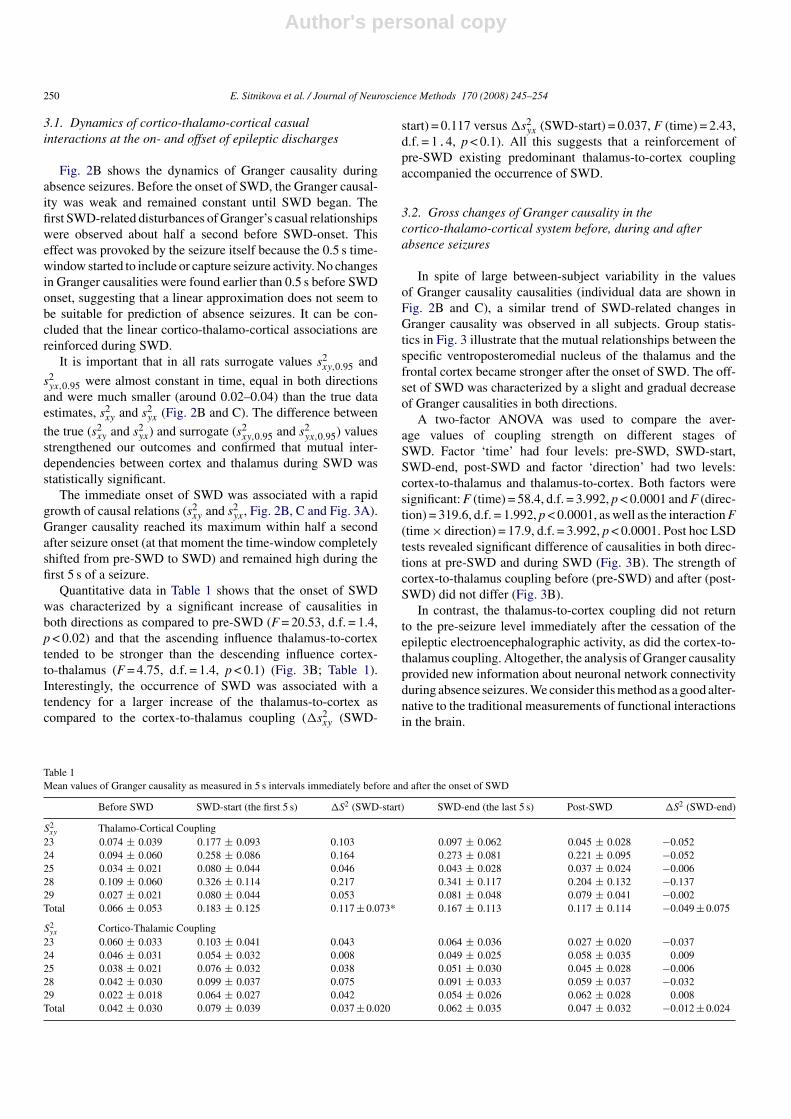
Quantitative data in Table 1 shows that the onset of SWDwas characterized by a significant increase of causalities inboth directions as compared to pre-SWD (F = 20.53, d.f. = 1.4,p < 0.02) and that the ascending influence thalamus-to-cortextended to be stronger than the descending influence cortex-to-thalamus (F = 4.75, d.f. = 1.4, p < 0.1) (Fig. 3B; Table 1).Interestingly, the occurrence of SWD was associated with atendency for a larger increase of the thalamus-to-cortex ascompared to the cortex-to-thalamus coupling (�s2
xy (SWD-
start) = 0.117 versus �s2yx (SWD-start) = 0.037, F (time) = 2.43,
d.f. = 1.4, p < 0.1). All this suggests that a reinforcement ofpre-SWD existing predominant thalamus-to-cortex couplingaccompanied the occurrence of SWD.
3.2. Gross changes of Granger causality in thecortico-thalamo-cortical system before, during and afterabsence seizures
In spite of large between-subject variability in the valuesof Granger causality causalities (individual data are shown inFig. 2B and C), a similar trend of SWD-related changes inGranger causality was observed in all subjects. Group statis-tics in Fig. 3 illustrate that the mutual relationships between thespecific ventroposteromedial nucleus of the thalamus and thefrontal cortex became stronger after the onset of SWD. The off-set of SWD was characterized by a slight and gradual decreaseof Granger causalities in both directions.
A two-factor ANOVA was used to compare the aver-age values of coupling strength on different stages ofSWD. Factor ‘time’ had four levels: pre-SWD, SWD-start,SWD-end, post-SWD and factor ‘direction’ had two levels:cortex-to-thalamus and thalamus-to-cortex. Both factors weresignificant: F (time) = 58.4, d.f. = 3.992, p < 0.0001 and F (direc-tion) = 319.6, d.f. = 1.992, p < 0.0001, as well as the interaction F(time × direction) = 17.9, d.f. = 3.992, p < 0.0001. Post hoc LSDtests revealed significant difference of causalities in both direc-tions at pre-SWD and during SWD (Fig. 3B). The strength ofcortex-to-thalamus coupling before (pre-SWD) and after (post-SWD) did not differ (Fig. 3B).
In contrast, the thalamus-to-cortex coupling did not returnto the pre-seizure level immediately after the cessation of theepileptic electroencephalographic activity, as did the cortex-to-thalamus coupling. Altogether, the analysis of Granger causalityprovided new information about neuronal network connectivityduring absence seizures. We consider this method as a good alter-native to the traditional measurements of functional interactionsin the brain.
Table 1Mean values of Granger causality as measured in 5 s intervals immediately before and after the onset of SWD
Before SWD SWD-start (the first 5 s) �S2 (SWD-start) SWD-end (the last 5 s) Post-SWD �S2 (SWD-end)
S2xy Thalamo-Cortical Coupling
23 0.074 ± 0.039 0.177 ± 0.093 0.103 0.097 ± 0.062 0.045 ± 0.028 −0.05224 0.094 ± 0.060 0.258 ± 0.086 0.164 0.273 ± 0.081 0.221 ± 0.095 −0.05225 0.034 ± 0.021 0.080 ± 0.044 0.046 0.043 ± 0.028 0.037 ± 0.024 −0.00628 0.109 ± 0.060 0.326 ± 0.114 0.217 0.341 ± 0.117 0.204 ± 0.132 −0.13729 0.027 ± 0.021 0.080 ± 0.044 0.053 0.081 ± 0.048 0.079 ± 0.041 −0.002Total 0.066 ± 0.053 0.183 ± 0.125 0.117 ± 0.073* 0.167 ± 0.113 0.117 ± 0.114 −0.049 ± 0.075
S2yx Cortico-Thalamic Coupling
23 0.060 ± 0.033 0.103 ± 0.041 0.043 0.064 ± 0.036 0.027 ± 0.020 −0.03724 0.046 ± 0.031 0.054 ± 0.032 0.008 0.049 ± 0.025 0.058 ± 0.035 0.00925 0.038 ± 0.021 0.076 ± 0.032 0.038 0.051 ± 0.030 0.045 ± 0.028 −0.00628 0.042 ± 0.030 0.099 ± 0.037 0.075 0.091 ± 0.033 0.059 ± 0.037 −0.03229 0.022 ± 0.018 0.064 ± 0.027 0.042 0.054 ± 0.026 0.062 ± 0.028 0.008Total 0.042 ± 0.030 0.079 ± 0.039 0.037 ± 0.020 0.062 ± 0.035 0.047 ± 0.032 −0.012 ± 0.024

Author's personal copy
E. Sitnikova et al. / Journal of Neuroscience Methods 170 (2008) 245–254 251
Fig. 3. Statistical assessment of changes in Granger causalities associated with the onset and end of spike-wave discharges (SWD). (A) Bidirectional causalrelationship between the frontal cortex and the thalamus at the onset and the end of SWD. Coefficients of Granger causality were averaged per 0.2 s intervals and perrat (mean ± S.D.). The increase in Granger causalities at the onset of SWD was abrupt and significant (ANOVA, p < .001 post hoc LCD-test), but seizure offset wascharacterized by smooth and prolonged changes in Granger causalities. (B) Group statistics of Granger causality coefficients averaged in 5 s intervals (five rats, 30SWD per rat). Asterisks show significant differences (the post hoc LSD test).
4. Discussion
This study tackles a challenging problem of predictabilityof absence seizures in EEG. Using the concept of Grangercausality, we measured bidirectional (straightforward and back-ward) linear interdependences between the thalamus and thecortex during absence seizures and obtained new yet comprehen-sive information about functional thalamo-cortical interactionsduring absence epilepsy. Traditional methods, such as cross-correlation analysis of unit activity in a model of generalizedepilepsy in cats (e.g. Steriade and Amzica, 1994) and coher-ence analysis in a genetic absence model (Sitnikova and vanLuijtelaar, 2006) demonstrated that the genesis of generalizedspike-and-wave discharges required mutual interrelationshipbetween thalamus and cortex. The current study aims to evaluatea novel method for assessing directionality in thalamo-corticalnetwork associations during absence epilepsy and this led to
principally new conclusions. (1) Information transfer in thedirection ‘thalamus → frontal cortex’ was more intensive than inthe backward direction. This is the first indication of anisotropyin thalamo-cortical interactions (discussed in Section 4.1). (2)Coupling strength ‘frontal cortex → thalamus’ slightly (but sig-nificantly) increased at the onset of SWD and rapidly restored tothe initial level before cessation of a seizure. A strong and sus-tained increase in ‘thalamus → frontal cortex’ interactions wasfound not only during SWD, but also after the end of the seizure(discussed in Section 4.2).
4.1. Implications of the linear Granger causality in EEGanalysis of absence epilepsy
In patients with absence epilepsy, as well as in WAG/Rij rats,spike-wave seizures appear unpredictably from a normal EEGbackground and associated with sudden behavioral arrest, e.g.

Author's personal copy
252 E. Sitnikova et al. / Journal of Neuroscience Methods 170 (2008) 245–254
‘absences’ (Panayiotopoulos, 1997; van Luijtelaar and Coenen,1986). As known, SWD are produced in the cortico-thalamo-cortical oscillatory network (Avanzini and Franceschetti, 2003;Blumenfeld, 2002; Meeren et al., 2002, 2005; van Luijtelaar andSitnikova, 2006). Traditionally, coherence analysis is used toestimate functional associations between different brain areas(Challis and Kitney, 1991; Pereda et al., 2005). Previously,we have used coherence to measure linear thalamocortical net-work associations in the frequency domain (Sitnikova and vanLuijtelaar, 2006). Granger causality is a time domain measure offunctional interactions, assuming directionality and informationtransfer. Directionality of thalamo-cortical interactions duringSWD in WAG/Rij rats has already been explored by meansof nonlinear association EEG analysis (Meeren et al., 2002). Itwas found that directionally of thalamo-cortical coupling variedthroughout the seizure and it was the most constant during thefirst half a second, when the cortical epileptic focus consistentlyled the thalamus.
Hereby, by measuring Granger causality we also planned toidentify early changes in thalamo-cortical relationships that mayanticipate the onset of absence seizures. We adjusted a linearautoregressive model of Granger causality in order to describecausal relations between cortical and thalamic electrical activ-ity during absence seizures in WAG/Rij rats. Surrogate data testconfirmed the statistical significance of the observed interdepen-dence.
We first found that the linear estimation of Granger causal-ity provided a good approximation to baseline EEG (pre-SWD),e.g. linear autoregressive model was sufficient to obtain stableresults of non-seizure activity. However, with linear estimationsof Granger causality we failed to identify early changes of causalrelationships that may anticipate the onset of absence seizures.It is however possible that early changes of interdependenciescan be described with additional nonlinear autoregressive mod-els or with phase-synchronization methods (Le Van Quyen andBragin, 2007). On the one hand, introduction of nonlinearityinto the model may be necessary to get comprehensive informa-tion about network associations that prerequisite seizure activityor/and take place during a seizure. On the other hand, applicationof nonlinear AR model requires more careful selection of modelparameters (such as dimensions and nonlinear model functions).This piece of work will be done in the future.
Several conclusions can be drawn from the presentresults. First, ‘thalamus → frontal cortex’ coupling characteris-tic, numerically, was greater than that in the opposite direction.This was found before, during and after SWD. The onset ofSWD was associated with an amplification of pre-SWD exist-ing tendencies. However, it is difficult to compare couplingsin both directions to each other since thalamus and cortex sig-nals are essentially different from each other even in respectof their waveforms (Sitnikova and van Luijtelaar, 2007). Moremeaningful is to trace changes in coupling characteristics overtime.
Second, ‘thalamus → frontal cortex’ coupling remained con-stantly high during a seizure and did not return to pre-SWDlevel even after cessation of SWD. It seems intriguing thatalthough SWD were stopped, the thalamo-cortical network did
not rapidly return to the non-epileptic state and causal rela-tionships remained abnormal. Clinically, both start and end ofSWD are regarded as abrupt and unpredictable, but we observethat changes in Granger causalities at the onset of SWD weremore sharp and fast as compared with that at the end of SWD(post-SWD periods were characterized by smooth and prolongedchanges in Granger causalities).
Third, Granger coupling strength increased with seizureonset, although differentially in two directions: reinforcementof ‘thalamus → frontal cortex’ coupling was greater than that inbackward direction. However, this latter comparison should beinterpreted with care, as mentioned above.
4.2. Granger causality and functional interactions inepileptic networks
In the present study we elaborate interactions between thefrontal cortex and the thalamus during absence seizures. In ourrats, EEG records were made in the areas in which seizure activ-ity is known to be the most robust, e.g. in the frontal cortexand specific thalamus (Vergnes et al., 1987). Direct anatomicconnections between these areas are nearly absent, but thesethalamic areas send and receive terminals to the somatosensorycortical (Jones, 1985). This midpoint, the peri-oral region of thesomatosensory cortex in WAG/Rij rats, is known as ‘epilepticfocus’ which initiate SWD (Meeren et al., 2002). In our ani-mals, the frontal EEG electrode was relatively far away fromthe ‘epileptic focus’ and we did not measure electrical activityin focal epileptic zone. Interestingly, a French group has recentlyconfirmed and extended the Meeren et al. (2002) data in GAERS(Polack et al., 2007). They showed that neurons in deep layers ofthe somatosenory cortex started firing much earlier than the firstchanges of local field potential could be visualized at the onsetof SWD. This exaggerated neuronal firing at the early stages ofSWD was only found in neurons localized in the epileptic corti-cal area, but it neither was detected in other areas in epileptic rats,nor in the similar areas in non-epileptic rats. Equally importantis that our previous studies in WAG/Rij rats and others clearlydemonstrated that neuronal activity in cortical regions outsidethe peri-oral area of the somatosensory cortex did not lead tha-lamic activity during SWD (Inoue et al., 1993; Seidenbecher etal., 1998).
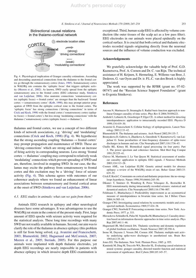
We study interdependencies between two indirectly con-nected structures that communicate via the ‘epileptic focus’:‘thalamus ↔ somatosensory cortex (epileptic focus) ↔ frontalcortex’ (Fig. 4). In order to interpret our results, we put togetherseveral theoretical considerations: the ‘cortical focus’ theory(Meeren et al., 2002, 2005), our concept on global and localsynchronization in oscillatory networks (Sitnikova and vanLuijtelaar, 2006; van Luijtelaar and Sitnikova, 2006) and ideasof ‘driving’ and ‘modulating’ connections in neuronal networks(Crick and Koch, 1998). We propose that the ‘epileptic focus’not merely triggers, but also acts as a distributor of epilepticactivity. In particular, seizure may easily propagate to thoseareas which have dense connections with the ‘epileptic focus’(Sitnikova and van Luijtelaar, 2006). In order to explain thequantitative differences between coupling strength between the
Author's personal copy
E. Sitnikova et al. / Journal of Neuroscience Methods 170 (2008) 245–254 253
Fig. 4. Physiological implication of Granger causality estimations. Ascendingand descending anatomical connections from the thalamus to the frontal cor-tex go through the somatosensory cortex (Jones, 1985). Somatosensory cortexin WAG/Rij rats contains the ‘epileptic focus’ that triggers epileptic activ-ity (Meeren et al., 2002). As known, SWD easily spread from the epilepticsomatosensory area to the frontal cortex (EEG coherence study, Sitnikovaand van Luijtelaar, 2006). Also anatomic connections ‘somatosensory cor-tex (epileptic focus) → frontal cortex’ are stronger than the backward ‘frontalcortex → somatosensory cortex’ (Kolb, 1990), this may prompt anterior prop-agation of SWD from the epileptic cortical zone to the frontal cortex. The‘epileptic focus’ has strong connections (‘driving connections’ in terms ofCrick and Koch, 1998) with the frontal cortex (‘somatosensory cortex (epilep-tic focus) → frontal cortex’), but less strong ‘modulating connections’ with thethalamus (‘somatosensory cortex (epileptic focus) → thalamus’).
thalamus and frontal cortex, we use a concept of two differentkinds of network associations, e.g. ‘driving’ and ‘modulating’connections (Crick and Koch, 1998) (Fig. 4). We hypothesizethat the strong ascending coupling ‘thalamus → frontal cortex’may prompt propagation and maintenance of SWD. These are‘driving connections’ which are strong and induce an increaseof firing activity in corresponding neurons. Less strong ‘frontalcortex → thalamus’ coupling may correspond to relatively weak‘modulating’ connections which prevent spreading of SWD andare, therefore, involved in stopping SWD. In our case, the tha-lamus may excite the pathway from somatosensory to frontalcortex and this excitation may be a ‘driving’ force of seizureactivity (Fig. 4). This schema agrees with outcomes of ourcoherence analysis where we found an enhancement of linearassociations between somatosensory and frontal cortical areasat the onset of SWD (Sitnikova and van Luijtelaar, 2006).
4.3. EEG studies in animals: what can we gain from them?
Animals EEG research in epilepsy and other neurologicaldiseases have some advantages. It was advantageous to use theWAG/Rij rat strain in the context of the present study. First, largeamount of EEG epochs with seizure activity were required forthe statistical analysis. These long time recordings with manySWD are not readily available in humans. Second, our aim was toclarify the role of the thalamus in absence epilepsy (this problemis still far from being solved, e.g. Avanzini and Franceschetti,2003; Blumenfeld, 2002; Huguenard and McCormick, 2007;Meeren et al., 2005; Steriade, 2005). For this purpose ouranimals were implanted with depth thalamic electrodes, yetdepth EEG recordings are nearly impossible in patients withabsence epilepsy in which invasive depth EEG examination is
exceptional. Third, human scalp EEG is affected by volume con-duction (the outer tissues of the scalp act as a low pass filter).EEG electrodes in our animals were placed epidurally on thecortical surface. It is crucial that both cortical and thalamic elec-trodes recorded signals originating directly from the neuronalsources and the influence of volume conduction was excluded.
Acknowledgements
We gratefully acknowledge the valuable help of Prof. G.D.Kuznetsova, Prof. A. Coenen and Dr. C. van Rijn. The technicalassistance of H. Krijnen, S. Hermeling, E. Willems-van Bree, J.Dederen, G. van Oyen and Dr. ir. P.L.C. van den Broek is highlyappreciated.
The work was supported by the RFBR (grant no. 07-02-00747) and the “Russian Science Support Foundation” (grantto Evgenia Sitnikova).
References
Ancona N, Marinazzo D, Stramaglia S. Radial basis function approach to non-linear Granger causality of time series. Phys Rev E 2004;70:056221.
Arnhold J, Lehnertz K, Grassberger P, Elger CE. A robust method for detectinginterdependences: application to intracranially recorded EEG. Physica D1999;134:419–30.
Avanzini G, Franceschetti S. Cellular biology of epileptogenesis. Lancet Neu-rology 2003;2:33–42.
Blumenfeld H. The thalamus and seizures. Arch Neurol 2002;59:135–7.Bosnyakova D, Gabova A, Zharikova A, Gnezditski V, Kuznetsova G, van Lui-
jtelaar G. Some peculiarities of time-frequency dynamics of spike-wavedischarges in humans and rats. Clin Neurophysiol 2007;118:1736–43.
Challis RE, Kitney RI. Biomedical signal processing (in four parts). Part3. The power spectrum and coherence function. Med Biol Eng Comput1991;29:225–41.
Chavez M, Martinerie J, Le Van Quyen M. Statistical assessment of nonlin-ear causality: application to epileptic EEG signals. J Neurosci Methods2003;124:113–28.
Coenen AML, van Luijtelaar ELJM. Genetic animal models for absenceepilepsy: a review of the WAG/Rij strain of rats. Behav Genet 2003;33:635–55.
Crick F, Koch C. Constraints on cortical and thalamic projections: the no-strong-loops hypothesis. Nature 1998;391(6664):245–50.
Dikanev T, Smirnov D, Wennberg R, Perez Velazquez JL, Bezruchko B.EEG nonstationarity during intracranially recorded seizures: statistical anddynamical analysis. Clin Neurophysiol 2005;116:1796–807.
Feldmann U, Bhattacharya J. Predictability improvement as an asymmetricalmeasure of interdependence in bivariate time series. Int J Bifurc Chaos2004;14:505–14.
Granger CWJ. Investigating causal relations by econometric models and cross-spectral methods. Econometrica 1969;37:424–38.
Granger CWJ, Newbold P. Forecasting economic time series. New York: Aca-demic Press; 1977. p. 333.
Hlavackova-Schindler K, Palus M, Vejmelka M, Bhattacharya J. Causality detec-tion based on information-theoretic approaches in time series analysis. PhysRep 2007;441:1–46.
Huguenard JR, McCormick DA. Thalamic synchrony and dynamic regulationof global forebrain oscillations. Trends Neurosci 2007;30:350–6.
Inoue M, Duysens J, Vossen JM, Coenen AM. Thalamic multiple-unit activ-ity underlying spike-wave discharges in anesthetized rats. Brain Res1993;612:35–40.
Jones EG. The thalamus. New York: Plenium Press; 1985. p. 955.Kaminski M, Ding M, Truccolo WA, Bressler SL. Evaluating causal relations in
neural systems: granger causality, directed transfer function and statisticalassessment of significance. Biol Cybern 2001;85:145–57.

Author's personal copy
254 E. Sitnikova et al. / Journal of Neuroscience Methods 170 (2008) 245–254
Kaplan AYA. Nonstationarity of EEG: methodological and experimental anal-ysis. Usp Fiziol Nauk 1998;29(3):35–55.
Kolb B. Organization of the neocortex in rat. In: Kolb B, Tees R, editors. Thecerebral cortex of the rat. Cambridge, Massachusetts: The MIT Press; 1990.p. 21–33.
Le Van Quyen M, Martinerie J, Adam C, Varela F. Nonlinear analyses of inter-ictal EEG map the brain interdependences in human focal epilepsy. PhysicaD 1999;127:250–66.
Le Van Quyen M, Bragin A. Analysis of dynamic brain oscillations: method-ological advances. Trends Neurosci 2007;30:365–73.
Meeren HK, Pijn JP, van Luijtelaar EL, Coenen AM, Lopes da Silva FH. Cor-tical focus drives widespread corticothalamic networks during spontaneousabsence seizures in rats. J Neurosci 2002;22:1480–95.
Meeren H, van Luijtelaar G, Lopes da Silva F, Coenen A. Evolving conceptson the pathophysiology of absence seizures: the cortical focus theory. ArchNeurol 2005;62:371–6.
Midzianovskaia IS, Kuznetsova GD, Coenen AM, Spiridonov AM, van Lui-jtelaar EL. Electrophysiological and pharmacological characteristics of twotypes of spike-wave discharges in WAG/Rij rats. Brain Res 2001;911:62–70.
Mormann F, Andrzejak RG, Elger CE, Lehnertz K. Seizure prediction: the longand winding road. Brain 2007;130(Pt 2):314–33.
Panayiotopoulos CP. Absence epilepsies. In: Engel JJ, Pedley TA, editors.Epilepsy: a comprehensive textbook. Philadelphia: Lippincott-Raven Pub-lishers; 1997. p. 2327–46.
Paxinos G, Watson C. The rat brain in stereotaxic coordinates. 2nd ed. NewYork: Academic Press; 1986.
Pereda E, Quian Quiroga R, Bhattacharya J. Nonlinear multivariate analysis ofneurophysiological signals. Prog Neurobiol 2005;77:1–37.
Polack PO, Guillemain I, Hu E, Deransart C, Depaulis A, Charpier S.Deep layer somatosensory cortical neurons initiate spike-and-wave dis-charges in a genetic model of absence seizures. J Neurosci 2007;27:6590–9.
Schiff SJ, So P, Chang T, Burke RE, Sauer T. Detecting dynamical interde-pendence and generalized synchrony through mutual prediction in a neuralensemble. Phys Rev E 1996;54:6708–24.
Seidenbecher T, Staak R, Pape HC. Relations between cortical and thala-mic cellular activities during absence seizures in rats. Eur J Neurosci1998;10:1103–12.
Sitnikova E, van Luijtelaar G. Cortical and thalamic coherence during spike-wave seizures in WAG/Rij rats. Epilepsy Res 2006;71:159–80.
Sitnikova E, van Luijtelaar G. Electroencephalographic characterization ofspike-wave discharges in cortex and thalamus in WAG/Rij rats. Epilepsia2007;48:2296–311, doi:10.1111/j.1528-1167.2007.01250.x.
Steriade M, Amzica F. Dynamic coupling among neocortical neurons dur-ing evoked and spontaneous spike-wave seizure activity. J Neurophysiol1994;72:2051–69.
Steriade M. Sleep, epilepsy and thalamic reticular inhibitory neurons. TrendsNeurosci 2005;28:317–24.
van Luijtelaar ELJM, Coenen AML. Two types of electrocortical paroxysms inan inbred strain of rats. Neurosci Lett 1986;70:393–7.
van Luijtelaar G, Sitnikova E. Global and focal aspects of absence epilepsy:the contribution of genetic models. Neurosci Biobehav Rev 2006;30:983–1003.
Vergnes M, Marescaux C, Depaulis A, Micheletti G, Warter JM. Spontaneousspike and wave discharges in thalamus and cortex in a rat model of geneticpetit mal-like seizures. Exp Neurol 1987;96:127–36.


