Embed Size (px)
Citation preview

Granuloma annulare in patients with malignantlymphoma: Clinicopathologic study of thirteennew casesSarah Kay Barksdale, MD,* Charles Perniciaro, MD,a Kevin C. Halling, MD,band John G. Strickler, MDb Rochester, Minnesota
Background: Reports of necrobiotic granulomas or granulomaannularein patients withmalignant lymphoma are rare.Objective: Our intentwasto determineany uniqueclinical or histopathologic features in patients with granuloma annulare and lymphoma.Methods: Wereviewed the medical recordsand biopsy material from 13patientswith granuloma annulareand lymphoma.Results:Threepatients had Hodgkin's disease and 10 had non-Hodgkin's lymphoma. Thegranulomaannulare lesions showed typicalhistopathologic features. However, the clinicalpattern wasfrequently atypical, withpainfullesions in unusuallocations includingthe palmsandsoles. Threepatients displayed granulomatous inflammation in noncutaneous sites, eitherwithin the malignant lymphoma or in uninvolved tissues, and all three had atypicalclinicalpresentations of granuloma annulare.Conclusion: Granuloma annularewithatypicalclinical presentations may beassociated withan underlying hematopoietic malignancy and may be part of a generalized granulomatousreaction to malignant lymphoma.(J AM ACAD DERMATOL 1994;31:42-8.)
Granulomatous inflammation associated withmalignant lymphoma is a well-characterized phenomenon. This association is best described forHodgkin's disease, in which epithelioid granulomasfrequently occur in the lymph nodes, spleen, bonemarrow, and liver.1-4 Granulomatous responseshavealso been described in association with nonHodgkin's lymphoma (NHL).5-7 The biologic relation between granulomatous inflammation and malignancy is a matter of speculation. Granulomasmay represent a host response to tumor, as issuggested by reports that their presence confers amore favorable prognosiscompared with that of patients with tumors of the same stage and type but
From the Departmentof Dermatology' and Division ofAnatomicPathology," Mayo ClinicandMayo Foundation.
Acceptedfor publication 000. 18, 1993.
Reprint requests: C. Perniciaro, MD, Mayo Clinicjacksonville, 4500San Pablo Rd., Jacksonville, FL 32224.
*Visiting clinician at the Department of Dermatology. Now with theLaboratoryofPathology, NationalCancerInstitute,National Institutes of Health, Bethesda, Md.
Copyright® 1994by the American Academy of Dermatology, Inc.
0190-9622/94 $3.00+ 0 16/1/53753
42
without granulomas.v 4, 8 Alternatively, granulomatous inflammation in association with malignantlymphoma may be an incidental observation or anaberrant immunologic response in patients with adisordered immune system either resulting from orperhaps predisposing to lymphoma. Studies in whichgranulomatous inflammation did not confer an improved prognosis for malignant lymphoma supportthese two alternative views.9-11 In some cases theremay be a histologiccoexistence of two processes suchas malignant lymphoma and sarcoidosis, althoughthe assessment of such reports is difficultbecause thepresence of sarcoidosis is often not easy to verify.12, 13 From a practical standpoint the recognitionof granulomatous inflammation in malignant lymphoma is important because it can obscure the histologic interpretation of specimens for either diagnosis or staging purposes.
Infrequently, cutaneous granulomatous lesionshave been reported in association with malignantlymphomas, both primary cutaneous lymphomasand types that almost never appear in the skin. Manyreported cases show sarcoid-type granulomatous inflammation14-17; some of these present as granulo-

Journal of the American Academy of DermatologyVolume 31, Number 1
matous rosacea.l'' The unusual skin disorder granulomatous slack skin disease has been associatedwith Hodgkin'sdiseaseand withT-celllymphoma. II
A granulomatous variant of mycosis fungoides alsoexists.B-1O Granulomatous panniculitis has been described in subcutaneous T-cell lymphomas andHodgkin's disease. 19-23 Extravascular necrotizinggranulomas-" and nonspecific necrobiotic granulomas25in patients with lymphoproliferative disordershave also been reported.
The lesions of granuloma annulare (GA) aremostoften locatedon the dorsalsurfacesof the hands andfeet,26 Histologically, GA is characterizedby focaldegeneration of dermal collagensurroundedby aninfiltrate of mainly histiocytes with some lymphocytes.The deposition of acidmucopolysaccharides isprominent in the majority of cases. In a series of 207cases-? several histologic subtypes of GA weredescribed, including infiltrative (71%), palisading(26%),and epithelioid patterns (3%).Unusualclinicopathologic forms exist,includingperforating, generalized, and subcutaneous varieties.P The pathogenesis of GA is unknown, but immunologic studiesimplicatea cell-mediatedimmune response, and delayed hypersensitivity skin tests can produce histopathologic lesions identical to those of GA,28
Only a fewcase reportshave described GA in patients with malignant lymphoma.29-3 I In this reportwe describe 13 additional patients with GA andmalignant lymphoma. Our intent was to determineany unique features of these patients. In each casethe clinicalhistorywasobtained,the pathologic features were reviewed, and the treatment and follow-up information, when available, were determined.
MATERIAL AND METHODS
All patients were seen and received diagnoses at theMayoClinicfrom 1976to 1992. Byusinga computerizeddatabase, 12,415 patients with a diagnosis of malignantlymphoma (Hodgkin'sdisease, NHL, mycosis fungoides,or Sezary syndrome) were cross-referenced with 1355patientswithGA. A subsetof 16 patientswithbothlymphoma and GA was established. The medical records ofthese 16patientswerereviewed for pertinent clinical andpathologic data.
Histologic sections from the original skin biopsy specimensofGA lesions wereevaluated in all but onepatient(case 9). The diagnosis of GA was made on the basisofparaffin-embedded sections stainedwithhematoxylin andeosin. Specialstains for organisms (Fite and periodic acid-Schiff) and acid mucopolysaccharides (alcian blue)
Barksdale et al. 43
wereperformed inallcases. Thehistopathologic featuresweresubclassified according tocriteriadescribed by Urnbert and Winkelmann.e? Three cases were eliminatedfrom the studywhen our review of the skin biopsy specimens disclosed histopathologic features inconsistent withGA. One biopsy specimen from an excluded patientshowed a palisading granuloma most consistent with acutaneous extravascular necrotizing granuloma." Theother two excluded patients had unusual epithelioidgranulomas that lacked mucin deposition, necrobiosis, orother features of GA. A final group of 13 patients wasdefined.
The diagnoses ofNHL were made with the standardcriteriaoftheworking formulation. Earlier cases that hadbeenclassified according to the Rappaport scheme werereclassified according to working formulation criteria.32
Thediagnosis ofHodgkin's disease wasmadebystandardhistologic criteria.P Diagnoses were madefrom formalinfixed, paraffin-embedded sections stained withhematoxylinandeosin. Weobtained andreviewed lymph nodebiopsy specimens in 11 cases, and bone marrow biopsyspecimens were also reviewed in three of thesecases. Incase5 an absolute lymphocyte countof5400 cells/J.LI established the diagnosis of chronic lymphocytic leukemia(CLL) inconjunction witha lymph nodebiopsy specimenthat showed smalllymphocytic lymphoma. In case6 thediagnosis of CLL was based on a bonemarrow biopsyspecimen that displayed focal aggregates of smalllymphoidcells and peripheral blood flow cytometry immunophenotyping, which confirmed a clonal B-cell population. In case 13 the diagnosis of Sezary syndrome wasbasedon a peripheral blood smearthat showed a leukocyte countof7300cellsI J.Ll, with33% circulating Sezarycells. An electron micrograph ofthebuffy coatconfirmedthe presence of Sezarycells.
In selected cases immunoperoxidase studies wereperformed on paraffin-embedded sections by means of astandard streptavidin-biotin technique. The followingantibodies were used: L26 (CD20, Dako, Carpenteria,Calif.), UCHL-l (CD45RO, Dako), Ber-H2 (CD30,Dako), Leu-Ml (CDIS, Becton Dickinson, San Jose,Calif.), andleukocytecommon antigen (C045RB,Dako).Polyclonal CD3 (Dako) staining was performed on paraffin-embedded sections bya modification ofthestandardprocedure.l" A B-cell phenotype was inferred if the neoplastic cells expressed CD20; a T-cell phenotype wasinferred if the cells expressed CD45RO or CD3. TheReed-Sternberg cells in Hodgkin's disease expressedCD15 and CD30.
RESULTS
Age and sex
Eight of the patientswerewomen and five weremen.Theyrangedin agefrom20to 75years(mean,60.9 years) at diagnosis of GA.

44 Barksdale et al.Journal of the American Academy of Dermatology
July 1994
Fig. 1. Granulomatous infiltrates in noncutaneous sites from patients with malignant lymphomas. A, Case 2. Noncaseating granulomas in bone marrow. B, Case 4. Microgranulomasin lymph node involved with small lymphocytic lymphoma with plasmacytoid features. C,Case 11. Epithelioid granuloma in liver. (Hematoxylin-eosin stain; X205.)
Malignant lymphoma
The histopathologic features of the malignantlymphomas were diverse. Three patients had nodular sclerosing Hodgkin's disease (cases 1,2, and 3).The majority of the patients had B-celllymphomasof various types. Three patients had small lymphocytic lymphomas (or CLL) (cases 4, 5, and 6). Threepatients had follicular lymphomas (cases 7, 8, and9), and one of these (case 9) showed progression todiffuse disease. One patient (case 10) had a lymphoma that is best classified according to the working formulation as a diffuse, small cleaved cell lymphoma, but it can also be considered as a lymphocytic lymphoma of intermediate differentiation or
mantle cell lymphoma.P Patient 11 had a diffuselarge-cell lymphoma of B-cell phenotype (CD20positive, CD45RO- and CD3-negative). The lymphoma in case 12was a diffuse large-cell lymphomaby working formulation criteria, but it had morphologic and immunohistochemical features (CD30positive; CD20-, CD45RO-, and CD3-negative) ofan anaplastic large-cell lymphoma, null cell type.One patient had Sezary syndrome (case 13).
Three patients had granulomatous inflammationin sites other than the skin (Fig. 1). In patient 2, a75-year-old man with Hodgkin's disease, noncaseating granulomas were found in the bone marrow thatwas otherwise uninvolved with lymphoma (Fig. 1,
Journal of the American Academy of DermatologyVolume 31, Number I Barksdale et al. 45
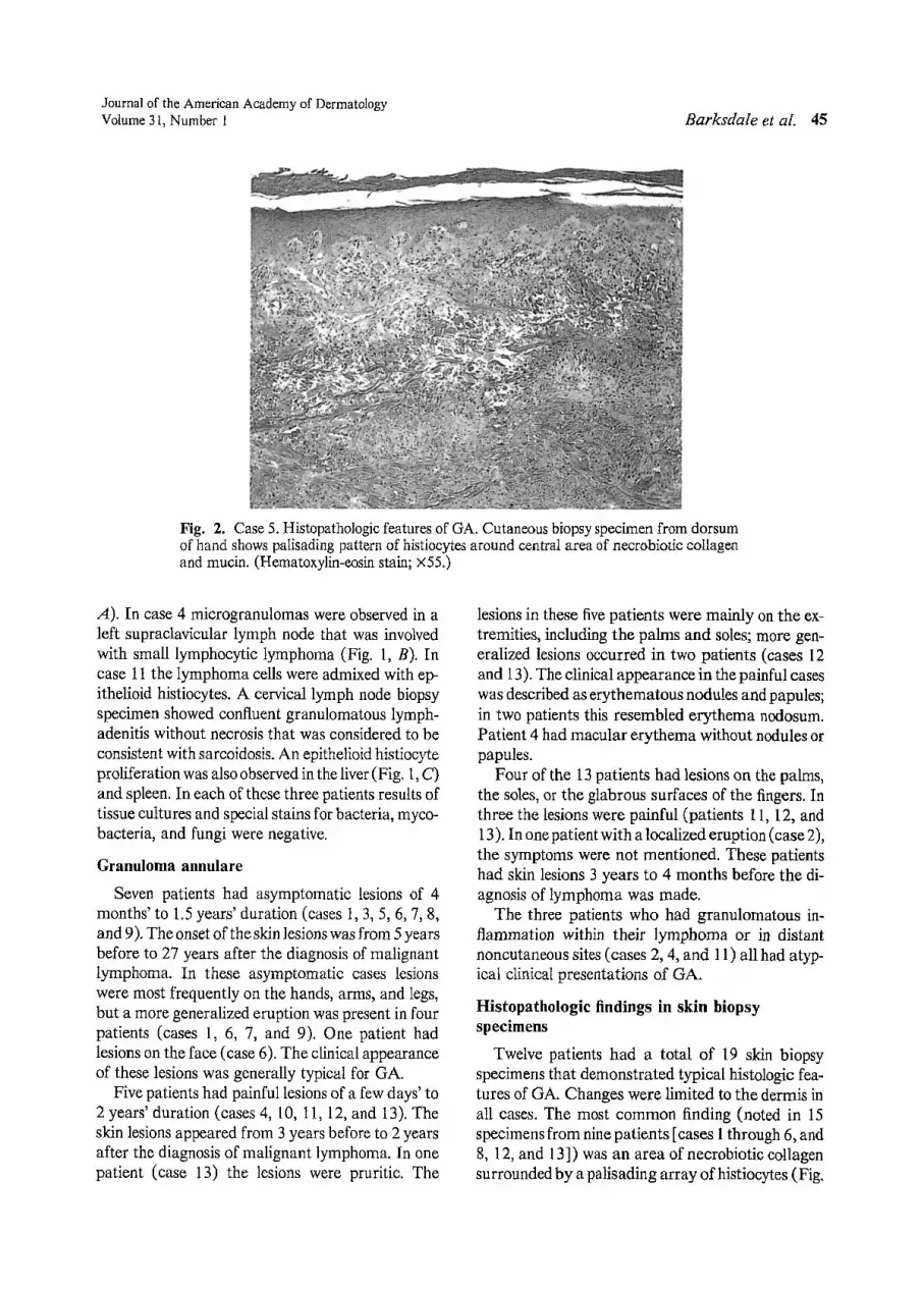
Fig. 2. Case 5. Histopathologic features of GA. Cutaneous biopsy specimen from dorsumof hand shows palisading pattern of histiocytes around central area of necrobiotic collagenand mucin . (Hematoxylin-eosin stain ; X55.)
A ). In case 4 microgranulomas were observed in aleft supraclavicular lymph node that was involvedwith small lymphocytic lymphoma (Fig. 1, B). Incase 11 the lymphoma cells were admixed with epithelioid histiocytes. A cervical lymph node biopsyspecimen showed confluent granulomatous lymphadenitis without necrosis that was considered to beconsistent with sarcoidosis. An epithelioid histiocyteproliferation was also observed in the liver(Fig. 1,C)and spleen. In each of these three patients results oftissue cultures and special stains for bacteria, mycobacteria, and fungi were negat ive.
Granuloma annulare
Seven patients had asymptomatic lesions of 4months'to 1.5 years' duration (cases 1,3,5,6,7,8,and 9). The onset of the skin lesions was from 5 yearsbefore to 27 years after the diagnosis of malignantlymphoma. In these asymptomatic cases lesionswere most frequently on the hands, arms, and legs,but a more generalized eruption was present in fourpatients (cases 1, 6, 7, and 9). ane patient hadlesions on the face (case 6). The clinical appearanceof these lesions was generally typical for GA.
Five patients had painful lesions of a few days' to2 years' duration (cases 4, 10, 11, 12, and 13). Theskin lesions appeared from 3 years before to 2 yearsafter the diagnosis of malignant lymphoma. In onepatient (case 13) the lesions were pruritic. The
lesions in these five patients were mainly on the extremities, including the palms and sales; more generalized lesions occurred in two patients (cases 12and 13). The clinical appearance in the painful caseswas described as erythematous nodules and papules;in two patients this resembled erythema nodosum.Patient 4 had macular erythema without nodules orpapules.
Four of the 13 patients had lesions on the palms,the soles, or the glabrous surfaces of the fingers. Inthree the lesions were painful (patients 11, 12, and13). In one patient with a localized eruption (case 2),the symptoms were not mentioned. These patientshad skin lesions 3 years to 4 months before the diagnosis of lymphoma was made.
The three patients who had granulomatous inflammation within thei r lymphoma or in distantnoncutaneous sites (cases 2, 4, and 11) all had atypical clinical presentations of GA.
Histopathologic findings in skin biopsyspecimens
Twelve patients had a total of 19 skin biopsyspecimens that demonstrated typical histologic features of GA. Changes were limited to the dermis inall cases. The most common finding (noted in 15specimens from nine patients [cases 1through 6, and8, 12, and 13]) was an area of necrobiotic collagensurrounded by a palisading array ofhistiocytes (Fig.

46 Barksdale et al.Journal of the American Academy of Dermatology
July 1994
Table I. Clinical and histologic features of patients with GA and lymphoma
Temporal relationCase Age Symptoms to malignant lymphoma Malignant Follow-up/AgeNo. Sex (yr)* Site ofGA olGA diagnosis lymphoma subtype (yr)
1 F 60 Arms and legs Asymptomatic 27 yr after NSHD Alive/622t M 75 Palms, fingers, Not stated 4 mo before NSHD Dead/76
soles offeet3 M 20 Dorsum of Asymptomatic 3Y2 yr after NSHD Alive/20
hand4t M 75 Fingers Painful erythema 1 yr before SL/CLL Alive/775 M 68 Dorsum of Asymptomatic 5 yr before SLjCLL Alivej75
hand6t F 68 Knees, face, Asymptomatic 3 mo after SL/CLL Alive/70
arms, legs,trunk
7 F 64 Chest, trunk, Asymptomatic 11 rna before Follicular SC Dead/65arms,thighs
8 M 66 Forearm, Asymptomatic 8 yr after Follicular mixed Deadj69helix
9 F 59 Arms, legs, Asymptomatic 11 yr after Follicular LC Alive/66back
lOt F 52 Arms and legs Painful, warm 2 yr after Diffuse SC Alive/56llt F 66 Fingertips Painful 3 yr before Diffuse LC Dead/71l2t F 73 Fingers, Painful 1 yr before Diffuse LC Dead/75
palms,sales, arms,legs, backof head
l3t F 46 Back, flank, Painful, pruritic 2 yr before Sezarysyndrome Dead/50legs, arms,palms
14* F 60 Dorsa of Painful 3 yr after NSHDhands
15* M 25 Neck, hands, Asymptomatic 6 yr after NSHDarms
l6§ M 40 Trunk, limbs Not stated 7 yr after HD (seminoma)1711 M 59 Face, scalp, Not stated 5 yr after Mycosis fungoides
trunk
HD, Hodgkin'sdiseasenot further subclassified; LC, largecell;NSHD, nodularsclerosing Hodgkin'sdisease; SC, smallcleaved; SL, smalilympho-cytic lymphoma.*Age at diagnosis of GA.tAtypical clinical presentation of GA.:j:Datafrom Schwartz et aU'§Data from Harman.P'IlData from Garde et al.29
2). Within the areas of necrobiosis was increased with abundant mucin. Special stains for organismsdeposition of acid mucopolysaccharides, as demon- werenegative in all cases.The clinical and histologicstrated by positivestainingwith alcian blue. An in- features are summarized in Table 1.filtrating pattern with histiocytes and focal necrobi-
DISCUSSIONosis was seen in patients 7 and 12. A second biopsyspecimen from patient 6 showed an epithelioidpat- Previous reports have suggested that cutaneoustern with prominent acid mucopolysaccharide dep- epithelioid granulomas may be the first manifesta-osition. The final biopsy specimen (case 11) dis- tion of an underlying malignant lymphoma IS, 18 andplayed a mixed infiltrating and epithelioid pattern that granulomatous inflammation may be admixed

Journal of the AmericanAcademyof DermatologyVolume 31, Number 1
with lymphoid malignancies of the skin, obscuringtheir true nature.v 22, 23 However, the occurrence inthe same patient of malignant lymphoma and GAhas been reported infrequently.
The clinicopathologic features of the previouslyreported cases of GA in patients with malignantlymphoma are summarized in Table I (cases 14 to17).29-31 Three occurred in patients withHodgkin'sdisease without cutaneous involvement. In thesethree patients GA developed 3 to 7 years after thediagnosis ofand therapyfor Hodgkin's disease. Thefourth caseoccurredin a patient withmycosis fungoides after a 2-year course of chemotherapy andradiation.j? In this last patient,a GA pattern developed within the patient's mycosis fungoides lesion.
Wedescribed 13additional patients withGAandmalignant lymphoma. Our data cannot be used toextrapolate more than a coincidental relation betweenGA and lymphoma because only a verysmallnumberof cases withboth diagnoses were retrievedfrom the database of a referral center with manypatients with these diseases. However, wedid noteseveral interesting features within this series. As inthe previously reported cases, the histopathologicfeatures of the GA lesions were typical. However,the clinical featuresofGA wereatypical inmanyofthese patients. Painful lesions or lesions in unusuallocations (or both), including the palms, soles, andface,occurred in seven of the 13patients. Thisfinding isremarkable when compared witha studyof26patientswithgeneralized GA byDicken et al.36 whohad no patients with involvement of the palms orsoles, onepatientwith a lesion on the face, onewithtenderness, and four with pruritus. Noneof the patients in their study had malignant lymphoma. Infive of our seven patients with atypical clinical features skin lesions developed before the diagnosis oflymphoma. The interval between diagnosis of GAand lymphoma ranged from 4 months to 3 years.Overall, the time between the diagnosis of GA andlymphoma was shorter in the group with atypicalclinical lesions than in the patients withmore typicalclinical features ofGA.Twoofthecases reportedpreviously in the literaturealso had atypical clinicalfeatures (Table I); onepatient with Hodgkin's diseasehad painfullesions.'! and the patientwithmycosis fungoides had lesions on the face. 29
We found that three patients who had granulomatousinflammation elsewhere, whether within thelymphoma or in a remotesite, all had atypical clinicallesions of GA. Each of thesepatients had a dif-
Barksdale et al. 47
ferenttypeofmalignant lymphoma: Hodgkin's disease, diffuse large-cell lymphoma of B-cell phenotype, and small lymphocytic lymphoma withplasmacytoid features. Hodgkin's disease and smalllymphocytic lymphoma have been associated withproliferations of epithelioid histiocytes.l- 7Suchpatients maybe demonstrating a generalized granulomatous response related to a lymphoid malignancy,and atypical GA may manifest as part of thatresponse. Lymphoma was notpresent in any ofthecutaneous biopsy specimens fromthese patients, soit seems unlikely that an immunologic reactiondirected toward tumor cells located in the dennisunderlies the GA.
Our patients demonstrated a wide variety of histologic subtypes oflymphoma, andthereseems tobeno relation between a particular type of malignantlymphoma and GA. Only one of the patients hadSezarysyndrome, andonly this patient hadpruritusas a symptom ofGA.Thepreviously reported casesdemonstrated an excess number ofHodgkin's disease, but this islikely a reporting bias resulting fromthe long-standing interest ingranulomatous inflammation in Hodgkin's disease.
REFERENCES
1. Brincker H. Epithelioid-cell granulomas in Hodgkin's disease. ActaPath Microbio1 Scand 1970;78A:19-32.
2. KadinME, Donaldson SS, Dorfman RF. Isolated granulomas inHodgkin's disease. N Engl J Med 1970;283:85961.
3. O'Connell MJ, SchimpffSC, Kirschner RH, etal. Epithelioid granulomas in Hodgkin's disease: a favorable prognostic sign? JAMA 1975;233:886-9.
4. Sacks EL, Donaldson SS, Gordon J, et a1. Epithelioidgranulomas associated with Hodgkin's disease: clinicalcorrelations in 55 previously untreated patients. Cancer1978;41:562-7.
5. Braylan RC, Long JC, JaffeeES, et a1. Malignant lymphomaobscured byconcomitantextensive epithelioid granulomas: reportofthreecases with similar clinicopathologicfeatures. Cancer 1977;39:1146-55.
6. Hollingsworth HC, Longo DL, Jaffee ES. Small noncleaved cell lymphoma associated with florid epithelioidgranulomatous response: a clinicopathologic studyofsevenpatients. Am J Surg PatholI993;17:51-9.
7. Patsouris E, Noel H, Lennert K. Lymphoplasmacytic/lymphoplasmacytoid immunocytoma with a high contentof epithelioid cells: histologic and immunohistochemicalfindings. Am J Surg Pathol1990;14:660·70.
8. Flaxman BA, Koumans JAD, Ackerman AB. Granulomatous mycosis fungoides: a 14-year follow-up of a case.Am J DermatopathoI1983;5:145-51.
9. Argenyi ZB,Goeken JA,PietteWW,etal.Granulomatousmycosis fungoides: clinicopathologicstudyoftwo cases. AmJ Dermatopathol 1992;14:200-10.
10. Dabski K,StollHL Jr. Granulomatous reactions in mycosisfungoides. J Surg Oncol 1987;34:217-29.

48 Barksdale et al.
11. LeBoitPE, ZackheimHS, White CR Jr. Granulomatousvariantsofcutaneous T-cell lymphoma: the histopathologyof granulomatous mycosis fungoides and granulomatousslackskin. Am J Surg PathoI1988;12:83-95.
12. Brincker H. The sarcoidosis-lymphoma syndrome. Br JCancer 1986;54:467-73.
13. Goldfarb BL, CohenSS. Coexistent disseminated sarcoidosis and Hodgkin's disease. JAMA 1970;2Il:1525-8.
14. Diette KM, Caro WA, Roenigk HH Jr. Malignant lymphomapresenting withcutaneous granulomas. JAM ACADDERMATOL 1984;10:896-902.
15. KahnLB,GordonW, Camp R. Floridsarcoid reaction associated withlymphoma of theskin. Cancer1974;33:111722.
16. Oliwiecki S, Kotecha B, Kingston T, et a1. Sarcoidosislymphoma syndrome. J R Soc Moo 1992;85:176-7.
17. Randle HW, Banks PM, Winkelmann RK. Cutaneousgranulomas in malignant lymphoma. Arch Dermatol1980;116:441-3.
18. SherertzEF, WestwickTJ, Flowers FP.Sarcoidal reactionto lymphoma presenting as granulomatous rosacea. ArchDermatolI986 ;122:1303-5.
19. Ashworth J, CoadyAT, Guy R, et al. Brawny cutaneousinduration and granulomatous panniculitis in large cellnon-Hodgkin's (T suppressor/cytotoxic cell) lymphoma.Br J DermatoI1989;120:563-9.
20. Gonzalez CL, Medeiros U, BrazielRM, et al.T-celllymphoma involving subcutaneous tissue: a clinicopathologicentity commonly associated with hemophagocytic syndrome. Am J Surg PathoI1991;15:17-27.
21. Perniciaro C, Zalla MJ, WhiteJW Jr, et al.SubcutaneousT-celllymphoma: reportoftwoadditional cases andfurtherobservations. Arch DermatoI1993;129:117l-6.
22. PrescottRJ, BanerjeeSS, CrossPA. Subcutaneous T-celllymphoma with florid granulomatous panniculitis. Histopathology 1992;20:535-7.
23. Sina B,Goldner R, Burnett JW. Granulomatous panniculitisand Hodgkin's disease. Cutis 1984;33:403-4.
24. Finan MC, Winkelmann RK. Cutaneous extravascular
Journal of the American Academy of DermatologyJuly 1994
necrotizing granuloma and lymphocytic lymphoma. ArchDermatoI1983;119:419-22.
25. Peltier FA,Pursley TV, Apisarnthanarax P, et a1. Necrobiotic granulomas of the skin associated with Hodgkin'sdisease. ArchDermatoI1981;117:123-4.
26. Muhlbauer JE. Granuloma annulare. J AM ACAD DER.MATOL 1980;3:217-30.
27. Umbert P, Winkelmann RK. Histologic, ultrastructural,and histochemical studies of granuloma annulare. ArchDermatoI1977;113:1681-6.
28. Buechner SA,Winkelmann RK,Banks PM.Identificationof T-cell subpopulations in granuloma annulare. ArchDermatoI1983;119:125-8.
29. Game SA,HirschP, Levan N. Granuloma annulare-likepattern in mycosis fungoides, Arch Dermatol 1972;105:717-9.
30. Harman RRM. Hodgkin's disease, seminoma of testicleandwidespread granuloma annulare. Br J Dermatol1977;97(suppl 15):5G-1,
31. Schwartz RA, Hansen RC, Lynch PJ. Hodgkin's diseaseandgranuloma annulare. ArchDermatoI1981;117:185-6.
32. The Non-Hodgkin's Lymphoma Pathologic ClassificationProject. National Cancer Institute-sponsored study ofclassifications ofnon-Hodgkin's lymphomas: summary anddescription of a working formulation for clinical usage.Cancer 1982;49:2112-35.
33. Lukes RJ, ButlerJJ. The pathology and nomenclature ofHodgkin's disease. CancerRes 1966;26:1063.
34. KurtinPJ, RochePC. Immunoperoxidase staining of nonHodgkin's lymphomas for T-cell lineage associated antigens in paraffin sections: comparison of the performancecharacteristics of four commercially available antibodypreparations. Am J Surg Patholl993;17:898-904.
35. Banks PM, Chan J, Cleary ML, et al. Mantle cell lymphoma: a proposal for unification of morphologic, immunologic, and molecular data. Am J Surg Pathol 1992;16:637-40.
36. Dicken CH, Carrington SG, Winkelmann RK. Generalizedgranuloma annulare. ArchDermatoI1969;99:556-63.


