Embed Size (px)
Citation preview

Mol Gen Genet (1996) 252:503-509 (c) Springer-Verlag 1996
Ti lman Spellig • Arnaud Bottin • Regine Kahmann
Green fluorescent protein (GFP) as a new vital marker in the phytopathogenic fungus Ustilago maydis
Received: 1 March 1996/Accepted: 25 June 1996
Abstract Pathogenic development of Ustilago maydis, the causative agent of corn smut disease, is a multistep process. Compatible yeast-like cells fuse and this gener- ates the infectious dikaryon which grows filamentously. Having entered the plant the dikaryon induces tumors in its host in which massive proliferation of fungal material, karyogamy and spore formation occur. In order to follow fungal development from the initial steps to the final stage we have expressed the green fluorescent protein (GFP) from Aequorea victoria as a vital marker in U. maydis and demonstrate that GFP-tagged strains can be used to study host-patho- gen interactions in vivo.
Key words Green fluorescent protein (GFP) • Ustilaqo maydis • Dimorphism • Reporter gene • Pathogenicity
IMrodu~ion
The basidiomycete fungus Ustila,qo maydis is the causa- tive agent of corn smut disease. Mating and pathogenic development are controlled by two mating-type loci, a and b. Haploid cells that differ in a and b can fuse and form the pathogenic dikaryon, which requires the plant for sustained growth and completion of its life cycle (Christensen 1963; Banuett 1992).
Fusion partner discrimination is based on a phero- mone/receptor-mediated cell recognition system that is encoded by the a mating-type locus (B61ker et al. 1992; Spellig et al. 1994). After fusion the products of the multiallelic b locus initiate the pathogenic program and control, together with the a locus, the morphological
Communicated by H. Saedler
T. Spellig ' A. Bottin • R. Kahmann ( ~ ) Institut ffir Genetik und Mikrobiologie der Universitiit Mfinchen, Maria-Ward-Str. la, 80638 Mfinchen, Germany
switch from yeast-like to filamentous growth (Gillissen et al. 1992; K~imper et al. 1995; Hartmann et al. 1996).
After the dikaryon has entered the plant tissue through wounds (Christensen 1963) or directly (Snet- selaar and Mims 1994), a series of events takes place that are poorly understood. During the initial stages of infection, hyphal growth has been shown to be pri- marily intracellular, with host cell plasma membranes remaining intact (Snetselaar and Mims 1994). At later stages intercellular growth predominates (Snetselaar and Mims 1994), followed by an accumulation of fungal material, nuclear fusion and spore formation. The plant responds to the infection by forming large tumors in which the fungus proliferates and produces massive amounts of diploid teliospores (Christensen 1963). Des- pite this massive reaction of the plant, classical plant defense responses are not observed at the microscopical level (Snetselaar and Mires 1994). Reporter genes like fi-glucuronidase (GUS) have been established for U. maydis (Richard et al. 1992), but their usefulness in detecting single fungal cells in plants appears limited (A. Bottin, unpublished). In addition staining for GUS activity usually involves dissection of the tissue of inter- est, fixation, addition of substrate and bleaching of the sample, and is thus not suitable for the analysis of living tissue.
Recently, a new vital marker, green fluorescent pro- tein (GFP) from the jellyfish Aequorea victoria, has been developed (Chalfie et al. 1994). GFP is a small protein of 238 amino acids responsible for green bio- luminescence in Cnidaria. Since formation of the fluor- escent chromophore is not species dependent, GFP has been successfully expressed in a variety of heterologous systems, e.g. Escherichia coti, Caenorhabditis elegans, Drosophila melanogaster, Saccharomyces cerevisiae, mammals and plants (Chalfie et al. 1994; Wang and Hazelrigg 1994; Haseloff and Amos 1995; Cubitt et al. 1995; Baulcombe et al. 1995; Sheen et al. 1995). When GFP is expressed, blue or UV light and oxygen are the only requirements to induce green fluorescence;

504
exogenous substrates are not needed (for review see Cubitt et al. 1995). These properties make GFP a valu- able tool for the specific labeling of cells and the study of developmental fates.
In this communication we describe the expression of GFP in the phytopathogenic fungus U. maydis and demonstrate that a GFP-expressing strain can be used for the study of host-pathogen interactions.
Materials and methods
Strains
E. coli K12 strain DH5c~ (Bethesda Research Laboratories) was used as host for plasmid amplification. U. maydis strains FB1 (al bl), FB2 (a2 b2) and FBD11 (al a2 bl b2) have been described by Banuett and Herskowitz (1989). FBDll is a solopathogenic diploid strain.
Plasmids and plasmid constructions
Plasmids pSP72 (Promega), pCBX122 (Keon et aL 1991), pBI101 (Clontech), pTZhyg (Schauwecker et al. 1995), pSMUT (Btilker et al. 1995) and pHLN (Schulz et al. 1990) were used for cloning. The plasmid blue-SGFP-TYG-nos SK contains the SGFP-TYG gene (see Sheen et al. 1995). pTEF (M. Dahl, unpublished) contains the tef promoter without the translational start codon, cloned as a 241 bp EcoRI-BamHI fragment into the respective sites of pTZ19R (Phar- macia).
In pTEF-SG the SGFP-TYG gene from blue-SGFP-TYG-nos SK was cloned as a SmaI-ClaI fragment into pSP72. The tef pro- moter was excised as an EcoRI-BamHI fragment and cloned into the PvuII and BamHI sites upstream of SGFP-TYG after appropriate modification of the protruding ends. Subsequently the gene confer- ring carboxin resistance was excised as a SmaI-EcoRV fragment fi'om pCBX122 and inserted into the EcoRV site of the plasmid.
For the construction of pMFAI-SG the mfal promoter was first cloned as a XhoI-Sau3AI fragment (Urban et al. 1996a) into pBI101 linearized with SaII and BamHI. The promoter was sub- sequently excised as a Ps~I-SmaI fragment, and inserted into the respective sites of pSP72. The SGFP-TYG gene was excised as a SmaI-ClaI fragment from blue-SGFP-TYG-nos SK and cloned downstream of the nfal promoter. Subsequently the hygromycin resistance cassette was excised as a BamHI fi'agment from pTZhyg and inserted into the Bg/II site.
pHSP70-SG was generated by a three-fragment ligation of the hsp70 promoter, excised as an XbaI-SmaI fragment from pHLN, SGFP-TYG, excised as a SmaI-XhoI fragment from blue-SGFP- TYG-nos SK, and pSMUT linearized with XhoI and XbaI.
In pOTEF-SG two direct repeats of a fragment from pTop 10 (Wein- mann et al. 1994) containing seven tetracyline-responsive elements are present upstream of the tefpromoter and the SGFP-TYG gene. After refilling the protruding ends a 290 bp Asp718-XhoI fragment from pTopl0 was cloned in tandem into the Sinai site of pSP72. The tef promoter was excised as an EcoRI-SphI fragment from pTEF and cloned into the BamHI and SphI sites downstream of the 290 bp fragments, after appropriate modifications. Subsequently this modi- fied promoter otef was excised as an EcoRI-XbaI fragment and cloned into pSP72 linearized with SalI and Xba] after refilling the protruding ends. The SGFP-TYG gene was excised as a BamHI- ClaI fragment fi'om blue-SGFP-TYG-nos SK and cloned down- stream of this promoter. Subsequently the hygromycin resistance cassette was excised as a Ban*HI fragment from pTZhyg and inserted into the BglII site.
Table I Relative fluorescence of SGFP transformants
Transformant Percentage of Mean fluorescence intensity" (_ SD)
FBD11/pOTEF-SG # 1 84 50 (_+ 28) #2 67 #3 74 #4 23 # 5 29 #6 25
FBD11/pHSP70-SG #1 20 23 (_+4) #2 21 #3 19 #4 30 # 5 24 # 6 24
FBDll/pMFA1-SG # 1 11 8.0 (+ 2.1) #2 6 #3 7 #4 8 #5 6 #6 10
FBDll/pTEF-SG # 1 0 10 (__+ 14) #2 4 #3 39 #4 5 #5 9 #6 5
Fluorescence intensity of a FBD11/pOTEF-SG # 1 culture was set to 100% as a reference. All other values are expressed as relative values using FBD11/pOTEF-SG # 1 as reference. Relative fluores- cence of a FBD11 control culture (16 ___ 4%) was subtracted from all values determined
Growth conditions and pathogenicity test
Strains were grown in YEPS (Tsukuda et al. 1988) or in CM medium (Holliday 1974). To observe filamentous growth water droplet as- says were performed as described in Spellig et al. (1994).
The pathogenicity test has been described (Gillissen et al. 1992). For individual strains at least 20 plants were infected and an- thocyanin biosynthesis and tumor development were scored for a period of 7-21 days after inoculation.
DNA procedures
Standard molecular techniques were employed as described (Sam- brook et al. 1989). Transformation of U. maydis followed published procedures (Schulz et al. 1990).
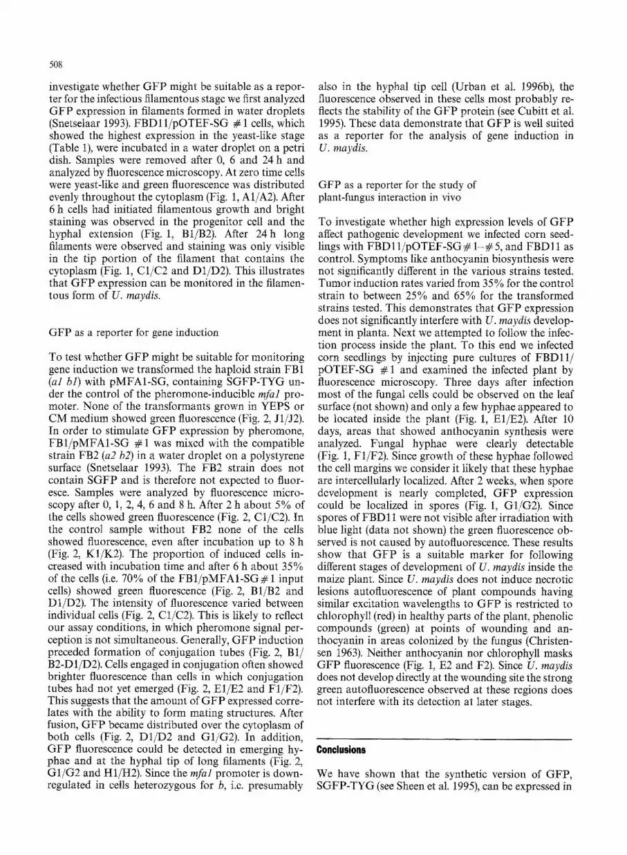
Fig. 1A-G Expression of GFP during different stages of U. maydis development. The U. maydis strain FBDll/pOTEF-SG#1 was incubated in a water droplet (A-D) or injected into corn seedlings (E-G). Samples A-D were assayed for filament formation and green fluorescence after 0 h (A), 6 h (B) and 24 h (C, D). Samples E-G were examined for fungal structures and green fluorescence 3 days (E), 10 days (F) and 14 days (G) after inoculation into maize plants. Samples were analyzed by DIC optics (panels 1) or epifluorescence (panels 2). In E the filament appears in the same focus plane as plant chloroplasts. The bar in G1 represents 30 gm

505
D2

506
Fluorescence and light microscopy
Microscopic analyses were performed using a Zeiss Axiophot without further manipulations of the object. Phase contrast or differential interference contrast (DIC) optics were used for light microscopy. For fluorescence microscopy the Zeiss filter set for fluorescein isothiocyanate fluorescence was employed (BP450-490 excitation filter, 510 nm dichroic and LP 520 emission filter). We have no indication that blue light irradiation leads to substantial damage of U. maydis cells.
Fluorometric determination of fluorescence intensity
Each transformant analyzed was grown to an OD600 of 0.5 in CM medium (Holliday 1974) and the intensity of fluorescence of the cell suspension was determined using a SFM 25 fluorometer (Kontron Instruments). The fluorescence value of FBD 11/pOTEF-SG # 1 was set to 100% as reference. All other values were determined as relative values using FBD11/pOTEF-SG # 1 as reference. Relative fluore- scence of a F B D l l control culture (16 _+ 4%) was subtracted from all values determined.
Results and discussion
Expression of GFP
In order to express GFP in U. maydis we used four different promoters that differ in strength and in their mode of regulation. The tef promoter is constitutively active and controls transcription of the gene for the translation elongation factor 2 of U. maydis (M. Dahl, unpublished). The tufa1 promoter regulates the expres- sion of the mfal pheromone gene. It has a low basal activity, is strongly induced after pheromone stimula- tion and is downregulated in cells heterozygous for b (Urban et al. 1996b). The hsp70 promoter exhibits a strong basal activity that can be further stimulated under stress conditions (Holden et al. 1989; Urban et al. 1996b). The otef promoter is a modified tef promoter in which two direct repeats of a synthetic fragment containing seven tetracycline-responsive elements (Weinmann et al. 1994) were introduced such that they precede the tefpromoter sequence.
Initially we placed the wild-type sequence of the GFP gene (Prasher et al. 1992; Chalfie et al. 1994) under the control of the selected promoters, and iso- lated transformants in which the DNA was integrated at different sites in the U. maydis genome. However, none of these transformants showed the expected green fluorescence when irradiated with blue light. Possible reasons for the failure to detect activity are transcript and/or protein instability and inadequate sensitivity of the detection system. In an attempt to overcome these problems we replaced the wild-type GFP by a recently described improved version of GFP, SGFP-TYG (see Sheen et al. 1995). In SGFP-TYG a Ser65 ~ Thr point mutation has been introduced in the chromophore domain, resulting in a GFP with a single excitation and emission peak, brighter fluorescence and more rapid
chromophore generation (Heim et al. 1995). In addition the codon usage was optimized for higher plants (see Sheen et al. 1995).
SGFP-TYG was transcriptionally fused to the tel mfal, hsp70 and otef promoters, respectively, and the resulting integrative plasmids pTEF-SG, pMFA1-SG, pHSP70-SG and pOTEF-SG were transformed into the solopathogenic strain FBD11 (al a2 bl b2). Six randomly chosen transformants for each promoter construct were analyzed by fluorescence microscopy. Transformants containing pTEF-SG or pMFA1-SG showed either no or barely detectable fluorescence when irradiated with blue light, with one exception, transformant pTEF-SG # 3. All transformants carry- ing pHSP70-SG or pOTEF-SG exhibited a strong green fluorescence after excitation (not shown).
In order to quantify fluorescence and to allow a com- parison of the four different promoter constructs we determined by fluorometry the fluorescence intensity of transformed cells grown in liquid culture. The fluores- cence of individual transformants harboring the same plasmid showed considerable variation, which can be explained by differences in sequence context caused by integration of the plasmid into different chromosomal locations in individual transformants, or by differences in plasmid copy number. Integration events in the U. maydis genome often involve tandem duplications of the integrating DNA (Fotheringham and Holloman 1990). On average, pOTEF-SG transformants showed the highest level of GFP activity (50 + 28% rel. flu- orescence), followed by pHSP70-SG transformants (23 ± 8% rel. fluorescence). Transformants containing pMFA1-SG or pTEF-SG showed GFP activity but the average values were approximately 6 to 8-fold lower compared to pOTEF-SG transformants (see Table 1 and Materials and methods).
Our results clearly demonstrate that SGFP-TYG is functional in U. maydis. However, to allow detection of GFP in single cells high levels of expression appear to be necessary. Interestingly, the artificial promoter showed the highest activity, demonstrating that the fragment containing the tetracycline-responsive elements enhances tef promoter activity by approxi- mately 8-fold.
Detection of GFP in filamentous structures
In the studies described so far, GFP expression was analyzed in cells that grow in a yeast-like manner. To
Fig. 2A-K Induction of pheromone-inducible GF P expression dur- ing mating. FB1 (al bl)/pMFA1-SG# I was mixed with FB2 (a2 b2) and incubated in a water droplet (A-H). As control FB1/pMFA1- SG # 1 was incubated under the same conditions without FB2 (J, K). Samples were assayed for green fluorescence after 0 h (A, J), 2 h (C), 6 h (B-G) and 8 h (H, K). Samples were analyzed by DIC light microscopy (panels 1) or epifluorescence (panels 2). The bar in J1 represents 30 gm (A, B, J and K) and 60 gm (C-H)

A2
507
\
j!
(
/
....... ~ i ¸
D 2 :i
J2 K2
508
investigate whether GFP might be suitable as a repor- ter for the infectious filamentous stage we first analyzed GFP expression in filaments formed in water droplets (Snetselaar 1993). FBD11/pOTEF-SG # 1 cells, which showed the highest expression in the yeast-like stage (Table 1), were incubated in a water droplet on a petri dish. Samples were removed after 0, 6 and 24 h and analyzed by fluorescence microscopy. At zero time cells were yeast-like and green fluorescence was distributed evenly throughout the cytoplasm (Fig. 1, A1/A2). After 6 h cells had initiated filamentous growth and bright staining was observed in the progenitor cell and the hyphal extension (Fig. 1, Bt/B2). After 24h long filaments were observed and staining was only visible in the tip portion of the filament that contains the cytoplasm (Fig. 1, C1/C2 and D1/D2). This illustrates that GFP expression can be monitored in the filamen- tous form of U. maydis.
GFP as a reporter for gene induction
To test whether GFP might be suitable for monitoring gene induction we transformed the haploid strain FB1 (al bl) with pMFA1-SG, containing SGFP-TYG un- der the control of the pheromone-inducible mfal pro- moter. None of the transformants grown in YEPS or CM medium showed green fluorescence (Fig. 2, J1/J2). In order to stimulate GFP expression by pheromone, FB1/pMFA1-SG # 1 was mixed with the compatible strain FB2 (a2 b2) in a water droplet on a polystyrene surface (Snetselaar 1993). The FB2 strain does not contain SGFP and is therefore not expected to fluor- esce. Samples were analyzed by fluorescence micro- scopy after 0, 1, 2, 4, 6 and 8 h. After 2 h about 5 % of the cells showed green fluorescence (Fig. 2, C1/C2). In the control sample without FB2 none of the cells showed fluorescence, even after incubation up to 8 h (Fig. 2, K1/K2). The proportion of induced cells in- creased with incubation time and after 6 h about 35% of the cells (i.e. 70% of the FB1/pMFAI-SG# 1 input cells) showed green fluorescence (Fig. 2, B1/B2 and D1/D2). The intensity of fluorescence varied between individual cells (Fig. 2, C1/C2). This is likely to reflect our assay conditions, in which pheromone signal per- ception is not simultaneous. Generally, GFP induction preceded formation of conjugation tubes (Fig. 2, B1/ B2-D1/D2). Cells engaged in conjugation often showed brighter fluorescence than cells in which conjugation tubes had not yet emerged (Fig. 2, El/E2 and F1/F2). This suggests that the amount of GFP expressed corre- lates with the ability to form mating structures. After fusion, GFP became distributed over the cytoplasm of both cells (Fig. 2, D1/D2 and G1/G2). In addition, GFP fluorescence could be detected in emerging hy- phae and at the hyphal tip of long filaments (Fig. 2, G1/G2 and H1/H2). Since the mJ'hl promoter is down- regulated in cells heterozygous for b, i.e. presumably
also in the hyphal tip cell (Urban et al. 1996b), the fluorescence observed in these cells most probably re- flects the stability of the GFP protein (see Cubitt et at. 1995). These data demonstrate that GFP is well suited as a reporter for the analysis of gene induction in U. maydis.
GFP as a reporter for the study of plant-fungus interaction in vivo
To investigate whether high expression levels of GFP affect pathogenic development we infected corn seed- lings with FBD 11/pOTEF-SG # 1- # 5, and FBD 11 as control. Symptoms like anthocyanin biosynthesis were not significantly different in the various strains tested. Tumor induction rates varied from 35% for the control strain to between 25% and 65% for the transformed strains tested. This demonstrates that GFP expression does not significantly interfere with U. maydis develop- ment in planta. Next we attempted to follow the infec- tion process inside the plant. To this end we infected corn seedlings by injecting pure cultures of FBDl l / pOTEF-SG # 1 and examined the infected plant by fluorescence microscopy. Three days after infection most of the fungal cells could be observed on the leaf surface (not shown) and only a few hyphae appeared to be located inside the plant (Fig. 1, El/E2). After 10 days, areas that showed anthocyanin synthesis were analyzed. Fungal hyphae were clearly detectable (Fig. 1, F1/F2). Since growth of these hyphae followed the cell margins we consider it likely that these hyphae are intercellularly localized. After 2 weeks, when spore development is nearly completed, GFP expression could be localized in spores (Fig. 1, G1/G2). Since spores of FBD11 were not visible after irradiation with blue light (data not shown) the green fluorescence ob- served is not caused by autofluorescence. These results show that GFP is a suitable marker for following different stages of development of U. maydis inside the maize plant. Since U. maydis does not induce necrotic lesions autofluorescence of plant compounds having similar excitation wavelengths to GFP is restricted to chlorophyll (red) in healthy parts of the plant, phenolic compounds (green) at points of wounding and an- thocyanin in areas colonized by the fungus (Christen- sen 1963). Neither anthocyanin nor chlorophyll masks GFP fluorescence (Fig. 1, E2 and F2). Since U. maydis does not develop directly at the wounding site the strong green autofluorescence observed at these regions does not interfere with its detection at later stages.
Conclusions
We have shown that the synthetic version of GFP, SGFP-TYG (see Sheen et al. 1995), can be expressed in

509
Ustilago maydis and its intrinsic green fluorescence is easily detected by fluorescence microscopy in the mor- phologically distinct forms. To observe green light emission at the level of single cells the SGFP-TYG gene must be coupled to a strong promoter. Since detection of SGFP-TYG is not restricted to the yeast-like form but is feasible in filaments and in spores this reporter gene provides an opportunity to observe fungal devel- opment inside the plant. This latter possibility is parti- cularly relevant for the examination of pathogenicity mutants (BNker et al. 1995) which do not show growth defects when cultivated on synthetic media but may be blocked at various stages during the infection process. In addition, SGFP-TYG appears to be an ideal repor- ter gene for screening libraries for genes that are specifi- cally induced when the fungus grows in planta.
Acknowledgements We thank our students Oliver Ladendorf and Christian Gruber for constructing plasmids. Special thanks go to Jen Sheen for providing the synthetic SGFP-TYG clone via Anton Schaeffner. We thank Michael B61ker and Karen Snetselaar for their comments on the manuscript. This work was supported by the Deutsche Forschungsgemeinschaft through SFB 369 and the Leibniz program.
References
Banuett F (1992) Ustilago maydis, the delightful blight. Trends Genet 8 : 174-179
Banuett F, Herskowitz I (1989) Different a alleles of UstiIago rnaydis are necessary for maintenance of filamentous growth but not for meiosis. Proc Natl Acad Sci USA 86 : 5878-5882
Baulcombe DC, Chapman S, Santa Cruz S (1995) Jellyfish green fluorescent protein as a reporter for virus infections. Plant J 7: 1045-1053
B61ker M, Urban M, Kahmann R (1992) The a mating type locus of Ustilago maydis specifies cell signaling components. Cell 68 : 441-450
B/51ker M, B6hnert HU, Braun KH, G6rl J, Kahmann R (1995) Tagging pathogenicity genes in Ustilago maydis by restriction enzyme mediated integration (REMI). Mol Gen Genet 248: 547-552
Chalfie M, Tu Y, Euskirchen G, Ward WW, Prasher DC (1994) Green fluorescent protein as marker for gene expression. Science 263 : 802-805
Christensen JJ (1963) Corn smut caused by UstiIago maydis. Amer Phytopathol Soc Monogr No. 2
Cubitt AB, Helm R, Adams SR, Boyd AE, Gross LA, Tsien RY (1995) Understanding, improving and using green fluorescent proteins. Trends Biochem Sci 20:448-455
Fotheringham S, Holloman WK (1990) Pathways of transformation in UstiIago maydis determined by DNA conformation. Genetics 124: 833-843
Gillissen B, Bergemann J, Sandmann C, Schroeer B, B61ker M, Kahmann R (1992) A two-component regulatory system for self/non-self recognition in Ustilago maydis. Cell 68:647-657
Hartmann HA, Kahmann R, BNker M (1996) The pheromone response factor coordinates flamentous growth and pathogenic development in Ustilago maydis. EMBO J 15:1632-1641
Haseloff J, Amos B (1995) GFP in plants. Trends Genet 11 : 328-329 Helm R, Cubitt AB, Tsien RY (1995) Improved green fluorescence.
Nature 373 : 663-664 Holden DW, Kronstad JW, Leong SA (1989) Mutation in a
heat-regulated hsp70 gene of Ustilago maydis. EMBO J 8 : 1927-1934
Holliday R (1974) Ustilago raaydis. In King RC (ed) Handbook of genetics, vol 1. Plenum Press, New York, pp 575-595
KS.roper J, Reichmann M, Romeis T, B61ker M, Kahmann R (1995) MultialMic recognition: nonself-dependent dimerization of the bE and bW homeodomain proteins in UstiIago maydis. Cell 81:7383
Keon JPR, White GA, Hargreaves JA (1991) Isolation, characteriza- tion and sequence of a gene conferring resistence to the systemic fungicide carboxin from the maize smut pathogen Ustilago maydis. Curr Genet 19:475-481
Prasher DC, Eckenrode VK, Ward WW, Prendergast FG, Cormier MJ (1992) Primary structure of the Aequorea victoria green- fluorescent protein. Gene 111 : 229--233
Richard G, Bailey JA, Keon JPR, Hargreaves JR (1992) Develop- ment of a GUS reporter gene system for the maize pathogen Ustilago maydis. Physiol Mol Plant Pathol 40: 383-.393
Sambrook J, Fritsch EF, Maniatis T (1989) Molecular cloning: a laboratory manual, Cold Spring Harbor Laboratory, Cold Spring Harbor, New York
Schauwecker F, Wanner G, Kahmann R (1995) Filament-specific expression of a cellulase gene in the dimorphic fungus Ustilago maydis. Biol Chem Hoppe-Seyler 376:617-625
Schulz B, Banuett F, Dahl M, Schlesinger R, Schiller W, Martin T, Herskowitz I, Kahmann R (1990) The b alleles of U. maydis, whose combinations program pathogenic development, code for polypeptides containing a homeodomain-related motif. Cell 60 : 295-306
Sheen J, Hwang S, Niwa Y, Kobayashi H, Galbraith DW (1995) Green-fluorescent protein as a new vital marker in plant cells. Plant J 8 : 777-784
Snetselaar KM (1993) Microscopic observation of Ustilago maydis mating interactions. Exp Mycol 17 : 345-355
Snetselaar KM, Mims CW (1994) Light and electron microscopy of UstiIago maydis hyphae in maize. Mycol Res 98 : 34~355
Spellig T, B/51ker M, Lottspeich F, Frank RW, Kahmann R (1994) Pheromones trigger filamentous growth in Ustilago maydis. EMBO J 13 : 1620-1627
Tsukuda T, Carleton S, Fotheringham S, Holloman WK (1988) Isolation and characterization of an autonomously replicating sequence from Ustilago maydis. Mol Cell Biol 8 : 3703-3709
Urban M, Kahmann R, BNker M (1996a) The bialMic a mating type locus of UstiIago maydis: remnants of an additional pheromone gene indicate evolution from a multialMic ancestor. Mol Gen Genet 250:414-420
Urban M, Kahmann R, B61ker M (1996b) Identification of the pheromone response element in Ustilago maydis. Mol Gen Genet 251 : 31-37
Wang S, Hazelrigg T (1994) Implications for bcd mRNA localization from spatial distribution of the exu protein in Drosophila oogen- esis. Nature 369 : 400-403
Weinmann P, Gossen M, Hillen W, Bujard H, Gatz C (1994) A chimeric transactivator allows tetracycline-responsive gene expression in whole plants. The Plant Journal 5 : 559-569


