Embed Size (px)
Citation preview

Mechanistic Studies of Arsenic and Selenium Detoxification
by
Gurnit Kaur
A thesis submitted in partial fulfillment of the requirements for the degree of
Doctor of Philosophy
Medical Sciences - LABORATORY MEDICINE AND PATHOLOGY
University of Alberta
© Gurnit Kaur, 2020

ii
Abstract
Over 200 million people worldwide are exposed to the proven human carcinogen arsenic, due to
contaminated drinking water. Animal studies have shown that arsenic and the essential trace element
selenium can undergo mutual detoxification through the formation of the seleno-bis(S-glutathionyl)
arsinium ion [(GS)2AsSe]- which undergoes biliary excretion, resulting in fecal elimination of both
compounds. The ATP-binding cassette (ABC) transporter multidrug resistance protein 2
(MRP2/ABCC2), localized to the canalicular surface of hepatocytes, is a transporter of this conjugate.
[(GS)2AsSe]- is also formed in animal red blood cells (RBCs), resulting in the sequestration of arsenic
and selenium. In human cells, the influence of arsenic on selenium accumulation, and vice versa, is
largely unknown. The objectives of this thesis were (a) to characterize arsenite and selenite uptake and
metabolism in human RBCs (hRBCs), (b) to characterize the effects of selenite on the hepatobiliary
efflux of arsenic in sandwich-cultured human hepatocytes (SCHH) and (c) to characterize the effects
of different selenium species on arsenic cytotoxicity and accumulation in HepG2 cells. The
overarching hypothesis was that selenium would alter arsenic metabolism and transport through the
formation of [(GS)2AsSe]-. 75Se-selenite uptake inhibition assays and experiments with heterologous
overexpression systems showed selenite uptake in hRBCs to be mediated by the Cl−/bicarbonate
(HCO3−) anion exchanger 1 (AE1). 73As-arsenite uptake inhibition assays showed arsenite uptake in
hRBCs to be partially AQP3-mediated, while no evidence of GLUT1 involvement was found.
Speciation analysis using X-ray absorption spectroscopy showed that 73% of the arsenic and selenium
present in RBCs co-treated with arsenite and selenite was in the form of [(GS)2AsSe]−. The remaining
26% of As was present as As(GS)3, providing the first evidence of the formation of both As-
glutathione conjugates in human cells.

iii
Consistent with published literature, all 73As biliary efflux in SCHH was glutathione-dependent.
Selenite either reduced or did not alter 73As efflux across the sinusoidal and canalicular surfaces of
SCHH. Preliminary data showed that selenide increases or does not change 73As biliary efflux, 75Se-
selenide accumulation in hepatocytes was higher than 75Se-selenite; and selenide was able to protect
SCHH against arsenite-induced toxicity while, selenite only conferred protection when the SCHH
preparation displayed a potential for arsenic biliary efflux. Further characterization of selenite and
selenide uptake and cytotoxicity differences in HepG2 cells also showed a higher IC50 value for cells
treated with arsenite + selenide (138 ± 76 µM) than arsenite + selenite (50 ± 6 µM). Cytotoxicity
assays of arsenite + selenite and arsenite + selenide at different treatment concentration ratios revealed
higher overall mutual antagonism of arsenite + selenide toxicity than arsenite + selenite. In
comparison to 75Se-selenite, HepG2 cells in suspension were at least 3-fold more efficient at
accumulating selenium from reduced 75Se-selenide, and its accumulation was further increased by
arsenite. These results were corroborated by X-ray fluorescence imaging of HepG2 cells which also
showed increased arsenic accumulation in the presence of selenide. These results from studies in
SCHH and HepG2 cells are consistent with a greater intracellular availability of selenide relative to
selenite for protection against arsenite, and the formation and retention of a less toxic product, likely
[(GS)2AsSe]−.
Lastly, although arsenic-induced toxicities are known to display inter-individual variability, selenium
supplementation trials are underway in arsenic-endemic regions in efforts to find cost-effective
therapeutic solutions for arsenic-induced toxicities. As such, the focus of this thesis work was also to
characterize single nucleotide polymorphic variants of MRP2 in order to gain a comprehensive
understanding of the protective role of [(GS)2AsSe]− not only through its sequestration in RBCs, but
also its efflux from hepatocytes. This work provides support in understanding the the mechanistic
aspects of arsenic and selenium cellular handling in humans.

iv
Preface
Chapter 2 of this thesis chapter has been submitted for publication as Gurnit Kaur, Warda Javed,
Olena Ponomarenko, Diane P. Swanlund, Kamran Shekh, Kelly L. Summers, Angela Casini, Margot
Wenzel, Joseph R. Casey, Emmanuelle Cordat, Ingrid J. Pickering, Graham. N. George, and Elaine M.
Leslie., Human red blood cell uptake and sequestration of arsenite and selenite: evidence of seleno-
bis(S-glutathionyl) arsinium ion formation in human cells. Biochemical Pharmacology (2020). Gurnit
Kaur and Warda Javed are co-first authors on the manuscript. This research project was a
collaboration led by Dr. Elaine Leslie, from the University of Alberta and the research groups of Dr.
Graham George and Dr. Ingrid Pickering, from the University of Saskatchewan. Gurnit Kaur initiated
all 73As experiments (with technical replicates completed by Diane Swanlund), and was responsible for
x-ray absorption spectroscopy (XAS) sample preparation and manuscript writing. Warda Javed
completed all 75Se experiments, with support from Diane Swanlund and Kamran Shekh. XAS analysis
was performed by Olena Ponomarenko and Kelly Summers. Angela Casini (with assistance from
Margot Wenzel) provided AQP3 inhibitors. Joseph Casey and Emmanuelle Cordat provided scientific
insight, critical to experiment design. This research project received ethics approval from the
University of Alberta Research Ethics Board, Project Name “Metabolism and Transport of Arsenic
and Selenium by Human Red Blood Cells”, protocol number 5614, January 27, 2010.
In Chapter 3 of this thesis, this research project received ethics approval from the University of Alberta
Research Ethics Board, Project Name “The role of transport proteins in toxicology”, protocol number
00001646, November 28, 2009.
Chapter 4 of this thesis chapter has been accepted for publication as Gurnit Kaur, Olena
Ponomarenko, Janet R. Zhou, Diane P. Swanlund, Kelly L. Summers, Nataliya V. Dolgova, Olga
Antipova, Ingrid J. Pickering, Graham N. George, and Elaine M. Leslie. Studies of selenium and

v
arsenic mutual detoxification in human HepG2 cells, Chemico-Biological Interactions. (2020). This
research project was a collaboration led by Dr. Elaine Leslie, from the University of Alberta and the
research groups of Dr. Graham George and Dr. Ingrid Pickering, from the University of
Saskatchewan. Gurnit Kaur was responsible for manuscript writing, and performed all experiments,
except the experiments in Figure 4-3C, which were performed by Janet Zhou. Diane Swanlund
completed technical replicates for Figure 4-2B, and Janet Zhou completed technical replicates for
Figure 4-4B. Olga Antipova and Olena Ponomarenko (with assistance from Nataliya Dolgova and
Kelly Summers) analyzed x-ray fluorescence imaging (XFI) samples shown in Figure 4-4A.

vi
Dedication
For my wonderful family.
And for those many families living in arsenic-endemic regions,
awaiting solutions.

vii
Acknowledgements
I would like to express my sincere thanks to my PhD supervisor, Dr. Elaine Leslie. Thank you for
your guidance and your patience. I feel fortunate to have a mentor that is supportive, whose door is
always open and with whom I can share ideas and ask questions. Thank you, Elaine, for not only being
an exceptional academic mentor, but also rooting for my personal and professional success.
I would also like to express my gratitude to members of my supervisory committee and candidacy
examination; Drs. X. Chris Le, Amit Bhavsar, Jonathan Martin, Sambasivarao Damaraju and Dawei
Zhang. Thank you for your time, insightful discussions and encouragement. Thank you also to my
external examiners; Drs. Koren Mann and Robin Clugston.
I must also thank my graduate coordinator and teaching mentor, Dr. Monika Keelan for always taking
time out of her busy schedule to check in. Thank you also to Cheryl Titus for being the most amazing
administrator.
Thank you to our collaborators, the research groups of Dr. X. Chris Le at the University of Alberta,
and Dr. Graham George and Dr. Ingrid Pickering at the University of Saskatchewan. My heartfelt
thank you especially to Dr. Olena Ponomarenko for your friendship and for being so generous in
sharing your time and knowledge. My time spent at the beamline is much cherished.
I have had the good fortune of being in the company of exceptional colleagues in the lab, colleagues
all on different career trajectories but brought together by their commitment to science and scholarly
thought. I thank Dr. Michael Carew, Dr. Barbara Roggenbeck, Dr. Mayukh Banerjee, Dr. Vanessa
Marensi, Brayden Whitlock and Janet Zhou for their generosity in sharing their ideas and inspiration.
Thank you to every undergraduate student, past and present, that I have had the opportunity of
working alongside. Ambreen, Teresa, Kate, Michelle, Danielle, Yejun, Han, Bronwen, Lucy, Brooke

viii
and Warda, I wish you all the best. Thank you, Diane Swanlund for your kind encouragement and
technical assistance.
My friends, Ensaf, Daphne, Alka, Ray, Preethi, Ashok, Lacey, Michelle, Anil, Vladimir and Denis;
thank you for the happy memories and unwavering support. Vanessa, you have inspired me incredibly
and taught me so much of what I know. Although it is nice to have friends all over the world, as I
approach the end of my program, you are all missed.
To my parents and siblings, thank you for stepping up infinitely to help us care for Gurjai and Araadh.
Mom and dad, I hope I have made you as proud of me, as I am of you.
Thank you Gurjinder, for your patience and sweet understanding. You are the calm to my storm; I
could not have asked for a better life partner.
My darling Gurjai - your excitement about poisons is all the validation I need for my work; and my
little Araadh - you have made thesis-writing so much sweeter.
Thank you all, for doing this degree with me.
Lastly, thank you to the donors that have provided liver and blood samples, for your trust in research.
Albeit a drop in a vast ocean, I hope that this work adds another piece to the giant puzzle of the
arsenic-modulated interactome.

ix
Table of Contents
1. General Introduction ................................................................................................................................. 1
1.1. Arsenic Background/History .................................................................................................................. 2
1.1.1 Uses of Arsenic ................................................................................................................................. 2
1.1.2. Exposure Scenarios ......................................................................................................................... 2
1.1.2.1. Therapeutic .............................................................................................................................. 2
1.1.2.2. Diet and Water ......................................................................................................................... 3
1.1.2.3. Occupational ............................................................................................................................ 4
1.1.3. Symptoms of Arsenic Exposure....................................................................................................... 4
1.1.4. Regulations on Arsenic Use ............................................................................................................. 5
1.2. Arsenic Mechanism of Toxicity .............................................................................................................. 6
1.2.1. Mechanism of Carcinogenesis ........................................................................................................ 6
1.2.1.1. Arsenic-Induced Oxidative Stress ............................................................................................ 6
1.2.1.2. Genotoxic Potential of Arsenic ................................................................................................ 7
1.2.1.3. Inhibition of DNA Repair .......................................................................................................... 8
1.2.1.4. Inhibition of Apoptosis and Effect on Cell Signalling Pathways ............................................... 8
1.2.1.5. Epigenetic Modulation ............................................................................................................. 9
1.2.2. Mechanism of Other Toxicities ..................................................................................................... 11
1.2.3. Differences in Susceptibility to Arsenic-Induced Toxicities .......................................................... 12
1.2.3.1. Polymorphisms in Arsenic Metabolism ................................................................................. 12
1.2.3.2. Polymorphisms in Arsenic Efflux ............................................................................................ 13
1.3. Arsenic Toxicokinetics .......................................................................................................................... 14
1.3.1. Arsenic Absorption ........................................................................................................................ 14
1.3.1.1. Bioavailability of Arsenic ........................................................................................................ 14
1.3.1.2. Effect of the Gut Microbiome ................................................................................................ 14
1.3.1.3. Arsenic Uptake Transporters ................................................................................................. 15
1.3.2. Cellular Metabolism of Arsenic ..................................................................................................... 15
1.3.3. Cellular Efflux of Arsenic by the ATP-Binding Cassette Transporter Family ................................. 16
1.3.3.1. The Multidrug Resistance Protein 2 ....................................................................................... 18
1.3.4. Arsenic Excretion .......................................................................................................................... 20
1.4. Arsenic-Selenium Interactions ............................................................................................................. 20
1.4.1. Selenium Background ................................................................................................................... 20

x
1.4.2. Selenium Toxicokinetics ................................................................................................................ 21
1.4.2.1. Selenium Absorption .............................................................................................................. 21
1.4.2.2. Selenium Metabolism ............................................................................................................ 22
1.4.2.3. Selenium Excretion ................................................................................................................ 24
1.4.3. Selenium Mechanism of Protection Against Arsenic-Induced Toxicity ........................................ 24
1.4.3.1. Protection Against Oxidative DNA Damage ........................................................................... 24
1.4.3.2. Inhibition of Methylation Pathways ...................................................................................... 25
1.4.3.3. Formation of the Seleno-bis(S-glutathionyl) arsinium Ion in Liver ........................................ 25
1.4.3.4. Selenium and Arsenic Sequestration in Red Blood Cells ....................................................... 26
1.4.4. Selenium Supplementation as a Therapeutic for As-Induced Toxicities ...................................... 26
1.5. Models of Hepatic Transport ............................................................................................................... 27
1.5.1. Primary Hepatocytes ..................................................................................................................... 27
1.5.2. Immortalized Liver Cell Lines ........................................................................................................ 28
1.5.3. Vesicular Transport Assays ........................................................................................................... 29
1.6. Objectives, Hypothesis and Rationale. ................................................................................................ 29
Objective 1: Characterization of the uptake and metabolism of arsenic and selenium in human red
blood cells ............................................................................................................................................... 30
Objective 2: Characterization of the effects of selenium on the hepatobiliary transport of arsenic in
SCHH........................................................................................................................................................ 30
Objective 3: Characterization of arsenic and selenium uptake and cytotoxicity in HepG2 cells ............ 30
1.7. Bibliography ......................................................................................................................................... 32
2. Human Red Blood Cell Uptake and Sequestration of Arsenite and Selenite: Evidence of Seleno-bis(S-
glutathionyl) Arsinium Ion Formation in Human Cells ................................................................................ 57
2.1. Abstract ................................................................................................................................................ 58
2.2. Introduction ......................................................................................................................................... 59
2.3. Materials and Methods ........................................................................................................................ 61
2.3.1. Chemicals and Reagents ............................................................................................................... 61
2.3.2. Blood Collection ............................................................................................................................ 62
2.3.3. 75SeIV Accumulation Assay in RBCs ................................................................................................ 62
2.3.4. 75SeIV Accumulation by AE1 Transfected HEK293 Cells ................................................................. 63
2.3.5. 73As Accumulation Assay in Human RBCs ..................................................................................... 64
2.3.6. 75Se Efflux Assay ............................................................................................................................ 64
2.3.7. Arsenic and Selenium Near-Edge X-ray Absorption Spectroscopy (XAS) ..................................... 65
2.4. Results .................................................................................................................................................. 66

xi
2.4.1. Uptake of SeIV by Human RBCs. .................................................................................................... 66
2.4.2. Uptake of AsIII by Human RBCs ..................................................................................................... 69
2.4.3. The Influence of AsIII on the Accumulation of 75SeIV by Human RBCs ........................................... 70
2.4.4. The Influence of SeIV on the Accumulation of 73AsIII by Human RBCs ........................................... 73
2.4.5. H2DIDS Inhibits SeIV-dependent AsIII Accumulation ...................................................................... 74
2.4.6. Arsenic and Selenium Near-edge XAS of Human RBCs Treated with AsIII and/or SeIV .................. 76
2.5. Discussion ............................................................................................................................................. 77
2.6. Bibliography ......................................................................................................................................... 82
3. Selenium Influence on the Hepatobiliary Efflux of Arsenic in Sandwich Cultured Human Hepatocytes 89
3.1. Abstract ................................................................................................................................................ 90
3.2. Introduction ......................................................................................................................................... 91
3.3. Materials and Methods ........................................................................................................................ 93
3.3.1. Chemicals and Reagents ............................................................................................................... 93
3.3.2. Human Hepatocyte Culture .......................................................................................................... 94
3.3.3. Fluorescence Microscopy ............................................................................................................. 95
3.3.4. SCHH efflux studies ....................................................................................................................... 95
3.3.5. Selenium Accumulation Studies.................................................................................................... 97
3.3.6. Cytotoxicity Studies ....................................................................................................................... 97
3.4. Results .................................................................................................................................................. 98
3.4.1. Canalicular Networks are Formed and Functional in SCHH .......................................................... 98
3.4.2. Arsenic Efflux is Unchanged or Reduced in the Presence of Selenite ........................................ 102
3.4.3. Arsenic Biliary Efflux is GSH-dependent...................................................................................... 104
3.4.4. Arsenic Biliary Efflux is Increased or not Modified in the Presence of Selenide ........................ 106
3.4.5. Preliminary Data Suggest Selenide Accumulation is Higher than Selenite by SCHH .................. 107
3.4.6. Preliminary Data Suggest Selenium Efflux is Increased in the Presence of Arsenic ................... 108
3.4.7. Preliminary Data Suggests Selenide Protects Against Arsenite Cytotoxicity Better than Selenite
.............................................................................................................................................................. 110
3.5. Discussion ........................................................................................................................................... 113
3.6. Bibliography ....................................................................................................................................... 118
4. Studies of Selenium and Arsenic Mutual Detoxification in Human HepG2 Cells.................................. 127
4.1. Abstract .............................................................................................................................................. 128
4.2. Introduction ....................................................................................................................................... 129
4.3. Material and Methods ....................................................................................................................... 131

xii
4.3.1. Chemicals and Reagents ............................................................................................................. 131
4.3.2. Cell Culture .................................................................................................................................. 132
4.3.3. Cytotoxicity Testing ..................................................................................................................... 132
4.3.4. 73As Accumulation Assays with HepG2 Cells in Suspension ........................................................ 132
4.3.5. 75Se Accumulation Assays with HepG2 Cells in Suspension ........................................................ 133
4.3.6. Assessment of Toxicity of Different As and Se Concentration Combinations on HepG2 Cells ... 134
4.3.7. 75Se Accumulation Assays with Adherent HepG2 Cells ............................................................... 135
4.3.8. X-ray Fluorescence Imaging ........................................................................................................ 135
4.3.8.1 Sample Preparation .............................................................................................................. 135
4.3.8.2. X-ray Fluorescence Imaging ................................................................................................. 136
4.3.9. Selection of Arsenic and Selenium Concentrations for Experiments ........................................ 137
4.3.10. Data Analysis and Statistics ...................................................................................................... 137
4.4. Results ................................................................................................................................................ 138
4.4.1. Selenide but Not Selenite Protects HepG2 Cells Against Arsenite Toxicity ............................... 138
4.4.2. Se is Accumulated from Selenide to a Greater Extent than from Selenite, and to a Higher Level
than As is Accumulated from Arsenite, by HepG2 Cells ....................................................................... 139
4.4.3. Arsenite + Selenide Display a Higher Level of Toxicity Antagonism than Arsenite + Selenite in
HepG2 Cells ........................................................................................................................................... 142
4.4.4. Accumulation of 75Se from 75Se-selenite and 75Se-selenide in the Presence and Absence of
Arsenite at Antagonistic Concentrations .............................................................................................. 145
4.4.5. X-ray Fluorescence Imaging shows Greater As and Se Accumulation in Arsenite + Selenide than
Arsenite + Selenite Treated Cells .......................................................................................................... 145
4.5. Discussion ........................................................................................................................................... 150
4.6. Bibliography ....................................................................................................................................... 155
5. General Discussion ................................................................................................................................ 164
5.1. Chapter 2. Human Red Blood Cell Uptake and Sequestration of Arsenite and Selenite: Evidence of
Seleno-bis(S-glutathionyl) Arsinium Ion Formation in Human Cells ..................................................... 164
5.2. Chapter 3. Selenium Influence on the Hepatobiliary Efflux of Arsenic in Sandwich Cultured Human
Hepatocytes .......................................................................................................................................... 165
5.3. Chapter 4. Studies of Selenium and Arsenic Mutual Detoxification in Human HepG2 Cells ......... 166
5.4. Conclusion ...................................................................................................................................... 167
Bibliography .............................................................................................................................................. 169
Appendix 1 ................................................................................................................................................ 218
A.1. Introduction ....................................................................................................................................... 219
A.2. Methods ............................................................................................................................................. 220

xiii
A.2.1. Materials ..................................................................................................................................... 220
A.2.2. Cell lines and Expression Constructs .......................................................................................... 221
A.2.3. Generation of MRP2 Variants ..................................................................................................... 221
A.2.4. MRP2 expression in HEK293T Cells ............................................................................................ 223
A.2.5. Membrane Vesicle Preparation .................................................................................................. 223
A.2.6. Immunoblotting .......................................................................................................................... 224
A.2.7. 3H-E217G Vesicular Transport Assay ......................................................................................... 224
A.2.8. Chemical Synthesis of 73As(GS)3, [(GS)273AsSe]−and [(GS)2As75Se]− ............................................. 225
A.2.9. 73As(GS)3, [(GS)273AsSe]−, [(GS)2As75Se]− Vesicular Transport Assays .......................................... 225
A.3. Results and Discussion ....................................................................................................................... 226
A.4. Bibliography ....................................................................................................................................... 241

xiv
List of Tables
Table 3-1. Summary of the BEI of taurocholate and arsenic in SCHH and viability of freshly isolated hepatocytes…………………………………………………………………………………………………………………………………………100
Table 3-2. Summary of the BEI of taurocholate and arsenic in SCHH and viability of freshly isolated hepatocytes…………………………………………………………………………………………………………………………………………113
Table 4-1. IC50 values for arsenite, selenite, selenide, and a combination of arsenite and selenium species in HepG2 cells after 72 h of exposure……………………………………………………………………………………………………140
Table 4-2. The mean and median (med) areal densities of As and Se in HepG2 cells treated with arsenite in combination with selenite or selenide…………………………………………………………………………………………..…147
Table 4-3. The mean and median (med) areal densities of As and Se in untreated HepG2 cells or treated with arsenite, selenite, or selenide alone……………………………………………………………………….……………………150
Table 4-4. IC50 values for arsenite, selenite, selenide, and a combination of arsenite and selenium species in HepG2 cells after 6 h of exposure………………………………………………………………………………………………………159
Table A-1. Mutagenic primer sequences for synthesis of MRP2 SNP variants…………………………………………223
Table A-2. Single nucleotide polymorphic variants of MRP2 (ABCC2) and their published effects in literature………………………………………………………………………………………………………………………………………..……239

xv
List of Figures
Figure 1-1. Chemical structures of arsenic compounds used in therapeutics……………………………………………3
Figure 1-2. Challenger pathway for arsenic metabolism…………………………………………………………………………..7
Figure 1-3. General predicted topology of the ABCC family “long” (A) and “short” (B) members…………..17
Figure 1-4. Chemical structures of arsenic conjugates found in bile……………………………………………………….19
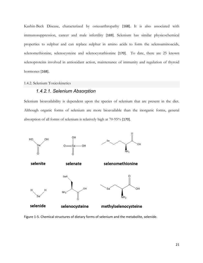
Figure 1-5. Chemical structures of dietary forms of selenium and the metabolite, selenide……………………21
Figure 1-6. Generally accepted selenium uptake and metabolism pathway scheme………………………………23
Figure 2-1. SeIV accumulation by human RBCs and transfected HEK293 cells…………………………………………68
Figure 2-2. AsIII accumulation by human RBCs……………………………………………………………………………………….70
Figure 2-3. The influence of AsIII on 75SeIV accumulation by human RBCs……………………………………………….72
Figure 2-4. The influence of SeIV on 73AsIII accumulation by human RBCs……………………………………………….73
Figure 2-5. Near edge XAS of human RBCs exposed to AsIII and SeIV……………………………………………………….75
Figure 2-6. Summary of human RBC (A) AsIII and SeIV uptake pathways and (B) mechanism of As-
dependent 75SeIV accumulation in the presence and absence of bovine serum albumin (BSA)………………77
Figure 3-1. Light and fluorescent images of SCHH treated with CDF-diacetate………………………………………98
Figure 3-2. Taurocholate efflux from eight different SCHH preparations……………………………………………..101
Figure 3-3. Arsenic efflux from eight different SCHH preparations……………………………………………………….103
Figure 3-4. The effect of GSH-depletion on arsenic efflux…………………………………………………………………….105
Figure 3-5. Influence of selenium species on sinusoidal and canalicular arsenic efflux from SCHH………107
Figure 3-6. Accumulation differences between 75Se-selenite and 75Se-selenide…………………………………..108
Figure 3-7. Influence of arsenite on sinusoidal and canalicular 75Se-selenide efflux from SCHH……………109
Figure 3-8. Effect of arsenite, selenite and arsenite + selenite on the viability of SCHH……………………….110
Figure 3-9. Effect of arsenite, selenite, selenide, arsenite + selenite and arsenite + selenide on the
viability of SCHH………………………………………………………………………………………………………………………………….111
Figure 4-1. Effect of selenite and selenide on arsenite cytotoxicity in HepG2 cells………………………………138
Figure 4-2. Accumulation of arsenite, selenite, and selenide by HepG2 cells in suspension…………………141
Figure 4-3. Toxicity and accumulation evaluation of arsenite + selenide and arsenite + selenite
combination concentrations in HepG2 cells…………………………………………………………………………………………144
Figure 4-4. (A) XFI elemental maps for S, Zn, As and Se collected from HepG2 cell cultures and, B) 75Se
accumulation under XFI sample preparation conditions………………………………………………………………………147

xvi
Figure 4-5. XFI elemental maps for S, Zn, As and Se collected from HepG2 cultures either untreated or
treated with arsenite, selenite, or selenide…………………………………………………………………………………………148
Figure 4-6. Overlay of arsenic, selenium, and sulphur XFI elemental maps collected from HepG2 cell
cultures treated with arsenite + selenite or arsenite + selenide…………………………………………………………..153
Figure 5-1. Overview of arsenic and selenium uptake and cellular handling in red blood cells and hepatocytes…………………………………………………………………………………………………………………………………………168
Figure A-1. Localization of the human MRP2 single nucleotide polymorphic variants in a general
predicted topology of MRP2………………………………………………………………………………………………………………..227
Figure A-2. WT-MRP2 and variant-MRP2 protein levels in transiently transfected HEK cells………………..228
Figure A-3. Effect of MRP2 variants on 3H- E217G, 73As(GS)3 and [(GS)273AsSe]− transport using
membrane vesicles prepared from WT- and variant-MRP2 transfected HEK293T cells………………..………231
Figure A-4. Optimization of transfection methods and 73As(GS)3 transport………………………………………….233
Figure A-5. Optimization of [(GS)273AsSe]− transport…………………………………………………………………………….236

xvii
List of Abbreviations
AE1 anion-exchanger 1
AMP adenosine monophosphate
AQP3 aquaglyceroporin 3
AQP9 aquaglyceroporin 9
As arsenic
AsV arsenate
AsIII arsenite
As(GS)3 arsenic triglutathione
ATP adenosine triphosphate
ATCC American Type Culture Collection
Auphen [Au (1,10-phenanthroline)Cl2]Cl
BCA bicinchoninic acid
BEI biliary excretion index
BSA bovine serum albumin
BSO buthionine sulfoximine
CDF 5(6)-carboxy-2′,7′-dichlorofluorescein
DJS Dubin Johnson syndrome
DMAV dimethylarsinic acid
DMAIII dimethylarsinous acid
DNDS 4,4’-dinitrosostilbene-2,2’-disulfonate
GLUT1 glucose transporter 1
xviii
[(GS)2AsSe]− seleno-bis(S-glutathionyl) arsinium ion
GSH glutathione
HA hemagglutinin
HBSS Hanks’ balanced salt solution
H2DIDS 4,4’-diisothiocyanatodihydrostilbene-2,2’-disulfonic acid
HEPES 4-(2-hydroxyethyl)-1-piperazineethanesulfonic acid
HSA Highest Single Agent
HSe- hydrogen selenide
LDH lactate dehydrogenase
MSD membrane spanning domain
MeSeCys methylselenocysteine
mAb monoclonal antibody
MMAV monomethylarsonic acid
MMAIII monomethylarsonous acid
MMA(GS)2 monomethylarsenic diglutathione
MMMTAV monomethylmonothioarsonic acid
MOPS 3-(N-morpholino) propanesulfonic acid
MRPs multidrug resistance proteins
MRP2 multidrug resistance protein 2
NaB sodium butyrate
NBD nucleotide binding domains
PBS phosphate buffered saline
pAb polyclonal antibody
PVDF polyvinylidene difluoride

xix
RIPA radioimmunoprecipitation assay
RBCs red blood cells
ROI regions of interest
ROS reactive oxygen species
SCHH sandwich cultured human hepatocytes
Se selenium
SeIV selenite
SeMet selenomethionine
Si3N4 silicon nitride
SNVs single nucleotide polymorphic variants
TM transmembrane
XAS X-ray absorption spectroscopy
XFI X-ray fluorescence imaging

1
1. General Introduction

2
1.1. Arsenic Background/History
1.1.1 Uses of Arsenic
Throughout history, arsenic has an infamous reputation for being the poison of kings and the king of
poisons [1,2]. However, arsenic is naturally present in the environment and has become ubiquitous to
the human experience also. We are exposed to arsenic in our soil, in our water and in our medicines
[3].
1.1.2. Exposure Scenarios
1.1.2.1. Therapeutic
As early as the year 2000 BC, arsenic has been used therapeutically [4]. Hippocrates, Aristotle and
Paracelsus have all been known to use arsenic in medicinal treatments. Fowler’s solution, a solution
of 1% potassium arsenite was used in the treatment of syphilis, psoriasis, asthma, and malaria (Figure
1-1) [5]. The first arsenic-containing commercial drug to be licensed was by the German company
called Hoescht, under the brand name of Arsphenamine/Salvarsan to treat syphilis (Figure 1-1) [6,7].
It was the most widely prescribed drug at one point and very effective, but was taken off the market
when penicillin came out in the 1940s [7].
Since the early 2000s, arsenic trioxide (ATO) has been used to treat acute promyelocytic leukemia
(APL) (Figure 1-1) [8]. The pathogenesis of APL is characterized by the fusion of the promyelocytic
leukemia/retinoic acid receptor alpha (PML/RARα) genes, leading to inhibition of myeloid cell
development and thus, increased levels of abnormal promyelocytes [9,10]. Due to the high affinity of
arsenite for thiol groups of proteins, ATO targets the ring finger of the PML protein, resulting in the
degradation of the PML/RARα fusion protein [11]. When used in combination with all-trans retinoic
acid (ATRA), the remission rate is greater than 90% [12]. Common side effects with ATO use include

3
hyperleukocytosis and cardiac arrythmia, with interindividual variability due to differences in arsenic
accumulation, metabolism and efflux [13].
Figure 1-1. Chemical structures of arsenic compounds used in therapeutics.
1.1.2.2. Diet and Water
Millions of people worldwide are exposed to unacceptable levels of arsenic in their drinking water
[14]. Arsenic is naturally found in the environment and can often leach into the groundwater supply
through the weathering of rocks or when the arsenic deposits are disturbed. The presence of high
levels of arsenic in aquifers is a public health problem in many countries [15]. Contaminated drinking
water has been reported in North America, Latin America, Taiwan, China (Inner Mongolia and
Xinjiang), India (West Bengal), Bangladesh, affecting the health of more than 200 million people
worldwide [3,16–20].
Rice and cereal grown using contaminated groundwater can also be a source of arsenic contamination
[21]. From all food types, rice contains the highest levels of inorganic arsenic contamination [21,22].
This is likely due to the paddy farming of rice, which requires rice to be grown in flooded water bodies.
Arsenic is highly soluble in water, when rice is grown in contaminated irrigation water, arsenic is easily
4
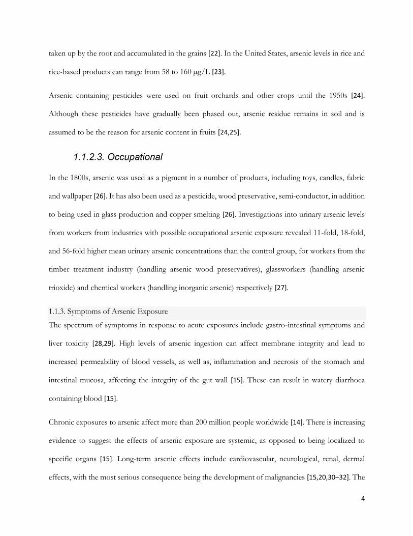
taken up by the root and accumulated in the grains [22]. In the United States, arsenic levels in rice and
rice-based products can range from 58 to 160 µg/L [23].
Arsenic containing pesticides were used on fruit orchards and other crops until the 1950s [24].
Although these pesticides have gradually been phased out, arsenic residue remains in soil and is
assumed to be the reason for arsenic content in fruits [24,25].
1.1.2.3. Occupational
In the 1800s, arsenic was used as a pigment in a number of products, including toys, candles, fabric
and wallpaper [26]. It has also been used as a pesticide, wood preservative, semi-conductor, in addition
to being used in glass production and copper smelting [26]. Investigations into urinary arsenic levels
from workers from industries with possible occupational arsenic exposure revealed 11-fold, 18-fold,
and 56-fold higher mean urinary arsenic concentrations than the control group, for workers from the
timber treatment industry (handling arsenic wood preservatives), glassworkers (handling arsenic
trioxide) and chemical workers (handling inorganic arsenic) respectively [27].
1.1.3. Symptoms of Arsenic Exposure
The spectrum of symptoms in response to acute exposures include gastro-intestinal symptoms and
liver toxicity [28,29]. High levels of arsenic ingestion can affect membrane integrity and lead to
increased permeability of blood vessels, as well as, inflammation and necrosis of the stomach and
intestinal mucosa, affecting the integrity of the gut wall [15]. These can result in watery diarrhoea
containing blood [15].
Chronic exposures to arsenic affect more than 200 million people worldwide [14]. There is increasing
evidence to suggest the effects of arsenic exposure are systemic, as opposed to being localized to
specific organs [15]. Long-term arsenic effects include cardiovascular, neurological, renal, dermal
effects, with the most serious consequence being the development of malignancies [15,20,30–32]. The

5
clinical presentation of arsenic-induced symptoms varies among populations and individuals, with
studies showing that a small percentage of the exposed population develop arsenic-induced skin
lesions [33–35].
1.1.4. Regulations on Arsenic Use
The U.S. Agency for Toxic Substances and Disease Registry (ATSDR) classified arsenic as No. 1 on
their Substance Priority List in 2017, based on its toxicity and potential for human exposure [36].
The International Agency for Research on Cancer (IARC) classifies arsenic in Group 1, meaning it is
a proven human carcinogen [37]. The U.S. Environmental Protection Agency (EPA) also classifies
arsenic as a confirmed human carcinogen [38].
The World Health Organization (WHO) has set a maximum acceptable limit of 10 µg/L on arsenic
in drinking water [39]. The same standard is followed in Canada, where municipal water is well below
this regulated value [40]. However, approximately 30% of Canadians rely on ground water for their
daily use [41]. If ground water is from a private well there is no requirement for arsenic contamination
testing, and more importantly, even when they are, residents may not have the knowledge or the means
to implement the correct interventions [42].
Regulation standards on arsenic consumption have been based primarily on arsenic contamination of
drinking water. While this is the predominant route of exposure, exposures from food products such
as rice, rice products and fruit juices can also amount to significant values [22]. As such, the current
regulations do not factor in the complete scope of arsenic exposure.
In addition, the current guideline of maximum arsenic levels in drinking water being at 10 µg/L is
based on a cost-benefit analysis. Based on arsenic exposure and hazard, it has been suggested that the
regulatory standards ought to be as close to zero as possible, or at least at 3 µg/L for a 1 in 10,000
estimate of cancer risk [25].

6
1.2. Arsenic Mechanism of Toxicity
1.2.1. Mechanism of Carcinogenesis
Despite the fact that arsenic has been established by IARC as a Group I, proven human carcinogen
[37], the mechanism of arsenic-induced carcinogenicity remains inadequately understood. This is
partly due to the lack of animal models of arsenic-induced carcinogenesis [43]. Moreover, the
biotransformation process of arsenic, which yields multiple arsenic metabolites, with varying degrees
of toxicities. Arsenic metabolism and speciation play a central role in arsenic-induced carcinogenesis.
This multi-faceted pathway includes a network of genotoxic and epigenetic mechanisms that are
discussed below, each of which are not independent of the other.
1.2.1.1. Arsenic-Induced Oxidative Stress
Arsenic has high affinity for cellular thiols; it can bind to free sulfhydryl groups of proteins and
enzymes leading to macromolecule oxidation and the inactivation of many enzymes [44]. In this
manner, arsenic can also compromise the mitochondrial membrane integrity, leading to the release of
ROS from the mitochondria to the cytoplasm [45]. In addition, arsenic biotransformation consumes
the cell’s antioxidant reserves by depleting glutathione in phase II metabolism through the formation
of arsenic-glutathione conjugates [46,47].
Excessive ROS formation, and depletion of the cell’s antioxidant capacity, lead to oxidative damage
[48]. The effects of arsenic-induced ROS formation are seen in the form of DNA damage, lipid and
protein damage and effects on cell signalling pathways; all of which are discussed below [48].

7
Figure 1-2. Challenger pathway for arsenic metabolism [49,50]. Taken from [49], with permission.
1.2.1.2. Genotoxic Potential of Arsenic
Although arsenic is a well-established carcinogen, it does not act as a direct mutagen. Its genotoxic
potential is largely a product of increased oxidative stress and inhibition of DNA repair pathways [51].
As such, arsenic has the capacity to induce micronuclei, DNA strand breaks, sister chromatid
exchanges, chromosomal aberrations and aneuploidy [51] which can then lead to genetic mutations .
Arsenic biotransformation involves a series of reduction followed by oxidative methylation steps,
leading to trivalent and pentavalent methylated species of arsenic [49]. Trivalent arsenic species are
orders of magnitude more toxic than the pentavalent forms. Methylated trivalents, in turn, are even
more toxic than inorganic arsenite (AsIII) [52]. When testing arsenate (AsV), AsIII, monomethylarsonic
acid (MMAV), monomethylarsonous acid (MMAIII), dimethylarsinic acid (DMAV), and
dimethylarsinous acid (DMAIII) for the effect on electrophoretic migration of the plasmid, øX174 RF
I DNA, Mass et al. found that MMAIII and DMAIII were the only species that had altered the

8
conformation of øX174 RF I DNA [53]. DNA nicking was seen at concentrations of 0.15 mM for
DMAIII and 30 mM for MMAIII, while complete DNA degradation was seen at higher concentrations
(10 mM for DMAIII, 60 mM for MMAIII) [53]. This provides evidence for the direct genotoxic potential
of DMAIII and MMAIII at high concentrations, however AsIII and AsV had no effect even at higher
concentrations.
Epidemiological data also provides insight into genotoxic mechanisms of arsenic, as well as, possible
biomarkers that can be used to evaluate genotoxicity in biomonitoring studies. In a West Bengal study,
with 163 exposed individuals and 154 unexposed individuals, increased micronuclei prevalence was
found in lymphocytes, oral mucosa cells and urothelial cells of the exposed group [54]. Similar results
have been observed in individuals from Chilean populations [55]. Other studies coming from the West
Bengal population, have also shown an increase in sister chromatid exchanges and chromosomal
aberrations [56].
1.2.1.3. Inhibition of DNA Repair
Arsenic down regulates the expression of the DNA repair enzyme, DNA polymerase β [51]. In
addition, the expression and activity of genes involved in the nucleotide excision repair and base
excision repair pathways are also altered [57]. These can lead to disruption of protein coding and non-
coding RNA genes, thus affecting apoptosis [57].
1.2.1.4. Inhibition of Apoptosis and Effect on Cell Signalling Pathways
Apoptosis is an active and gene directed mechanism of programmed cell death [58]. The generation
of ROS, reduces the mitochondrial membrane potential, leading to cytochrome C release and the
activation of caspases which result in apoptosis [59].

9
Increased levels of the transcription factors, activation protein 1 (AP-1) and nuclear factor кB (NF-
кB) lead to upregulation of stress proteins and mitogen-activated protein kinase (MAPK) pathways,
which regulate heat shock protein synthesis, cell transformation and apoptosis [31,57,60]. It has been
shown that DMAIII and MMAIII are more potent than AsIII in the activation of AP-1 [60].
1.2.1.5. Epigenetic Modulation
Epigenetic modulation refers to changes in transcription that can alter gene expression without any
effect on the DNA sequence [61]. These changes can be due to diet and environmental factors.
Examples of epigenetic modulation include DNA methylation, histone modifications and small
noncoding microRNAs [62–65].
1.2.1.5.1. Changes in DNA Methylation
Arsenic metabolism occurs through a series of reduction followed by oxidative methylation steps (Fig
1-1) [49,50]. During methylation, S-adenosyl methionine (SAM) acts as the methyl group donor, and
the reaction is catalysed by arsenite methyltransferase (AS3MT) [51,66]. SAM is required for arsenic
biotransformation, but it is also a methyl donor during DNA methylation [67]. The rapid depletion of
SAM results in global hypomethylation, which is often seen in cancer [28,61,68]. SAM synthesis
requires homo-cysteine, folate, vitamin B12 and other vitamins and cofactors [51]. Dietary deficiencies
in these can compromise SAM synthesis, offering an explanation for why arsenic induced toxicities
are correlated with nutritional deficiencies [69]. However, arsenic induced global hypomethylation
occurs irrespective of nutritional status. Hypomethylation can result in promoter activation, leading
to aberrant gene expression including increased expression of oncogenes [68,70]. Arsenic-induced
changes in DNA hypermethylation can also result in carcinogenesis. It has been shown that exposure
to inorganic arsenic results in increased levels of cytosine methyltransferase in cells, while upregulation

10
of DNA methyltransferase 3a was observed in mice [71]. Arsenic-induced hypermethylation of
promoter regions which affect conserved CpG islands of tumour suppressor genes can lead to their
inhibition [68]. It is likely that early exposure to arsenic leads to DNA hypermethylation, however,
with increasing arsenic exposures leading to increased methylation and depletion of SAM, arsenic-
induced hypomethylation results [71]. The specific genes targeted by the altered methylation profiles
likely hold the key to understanding arsenic-induced carcinogenesis.
1.2.1.5.2. Histone Modifications
Histone modifications in the form of methylation, acetylation, phosphorylation, ubiquitination,
sumoylation, proline isomerization or ADP-ribosylation, can restructure the packaging of chromatin,
leading to increased or decreased transcriptional activity [61,66,68]. An ex vivo study of 63 steel
workers examined the effect of metal and metalloid-rich particulate matter, including arsenic, on
histone modifications [72]. The results showed a positive association between histone 3 lysine
demethylation and histone 3 lysine deacetylation in blood leukocytes and arsenic exposure [72]. In
vitro studies using T-lymphocytes showed modifications in the histone deacetylase 2 (HDAC2) and
histone 3 (acetyl K9) [73].
1.2.1.5.3. Small Noncoding MicroRNAs
Until recently, the regulatory function of non-coding RNAs had been overlooked. MicroRNAs
(miRNAs) are a class of short non-coding RNAs, ranging from 19-25 nucleotides in length, that are
involved in gene silencing [48]. They achieve this through the inhibition of mRNA translation or by
inducing mRNA degradation [48]. In vitro studies have shown that arsenic can upregulate the
expression of the miRNAs, miR-190 and miR-21 [74]. In turn, miR-190 can suppress the translation

11
of the pH domain and leucine rich repeat phosphatases (PHLPP) protein and miR-21 can suppress
the translation of phosphatase and tensin homolog (PTEN), both of which are negative regulators of
protein kinase B (PKB) [74]. The activation of PKB mediates cell survival, growth, proliferation, cell
migration and angiogenesis [75]. Overactivation of PKB is common in cancer cells and likely plays a
role in arsenic-induced carcinogenesis [75–77].
1.2.2. Mechanism of Other Toxicities
The immunological response to arsenic exposure depends on the dose and length of exposures.
Arsenic can lead to both suppression and activation of the immune system [78]. Immunosuppression
leads to increased susceptibility to infectious diseases, while immunological activation can lead to
hypersensitivity disorder [78]. In infants, arsenic can impair thymic development, in addition to
increased morbidity which may be due to immunosuppression upon arsenic exposure [78].
In addition to immunological effects, arsenic can also interfere with glucose homeostasis.
Epidemiological data shows that chronic arsenic exposure is correlated with the incidence of diabetes
in Bangladesh, Mexico and Taiwan [79–81]. This is likely linked to effects of arsenite on insulin
signalling cascades [82,83]. It has been shown in murine pancreatic islet cells that glucose-stimulated
insulin secretion is inhibited by methylated trivalent arsenic species [84]. Arsenite has also been shown
to lead to the downregulation of glucose transporters and impaired functioning of enzymes involved
in glucose metabolism, such as pyruvate dehydrogenase [85–87]. Occupational exposures to arsenic in
copper smelters and glass industries are also correlated with arsenic-induced diabetes [88–90].
Epidemiological data has also established a correlation between arsenic exposure and the risk of
atherosclerosis [91–93]. The apolipoprotein E (ApoE) protein removes plasma lipoproteins from
blood [94]. In ApoE deficient mice, arsenic exposure in drinking water for 13 weeks led to the
increases in plaque size in the aortic sinus, from as low as 10 µg/L arsenic [95]. Possible molecular

12
mechanism for arsenic-induced atherosclerosis is arsenic-induced oxidative stress and its binding to
sulfhydryl groups of proteins, which in turn can lead to endothelial dysfunction causing inflammation,
platelet aggregation and loss of vasodilation, leading to the development of arsenic-induced
atherosclerosis [91,96]. Consistent with methylated trivalent arsenicals being more toxic than
pentavalent species, it has also been shown in ApoE deficient and AS3MT double knockout mice,
that AS3MT is required for arsenic-enhanced atherosclerosis [32].
1.2.3. Differences in Susceptibility to Arsenic-Induced Toxicities
Differences in diet and nutrition can lead to differences in arsenic induced toxicities. As discussed
above, folate is necessary for SAM which is required for arsenic methylation. Lifestyle factors such as
smoking can also lead to increased risk of arsenic induced carcinogenesis [31,97]. Genetic components
also likely play a significant role. Polymorphisms in various proteins involved in arsenic metabolism
and transport could potentially lead to differences in the toxicity and body burden of arsenic:
1.2.3.1. Polymorphisms in Arsenic Metabolism
The major genetic players of arsenic metabolism and detoxification that have been associated with
interindividual variability are AS3MT, proteins from the glutathione S-transferase (GST) family, and
DNA repair proteins [98]. GSTs are important phase II metabolism enzymes, catalyzing the
conjugation of substrates with glutathione. Purine nucleoside phosphorylase (PNP) has been shown
to reduce arsenate in cells. Polymorphisms in GSTO1, GSTM1, GSTP1, GSTT1 and PNP have been
associated with altered arsenic urinary excretion profiles [99–103].
AS3MT was first isolated from rat liver cytosol and determined to be the enzyme responsible for the
majority of arsenic methylation [104]. Arsenic biotransformation can lead to the presence of more
toxic intermediate metabolites, as such, AS3MT plays an integral role in arsenic metabolism and
toxicity. Studies of the influence of AS3MT polymorphisms on arsenic metabolism have relied on

13
urinary arsenic excretion profiles as indicators of methylation status. Engstrom et al., also investigated
the urinary arsenic profiles in indigenous women in Argentina exposed to high levels of arsenic in
drinking water; polymorphisms in AS3MT, GSTT1 and GSTM1 were shown to alter the urinary
elimination profile of arsenic [105]. Antonelli et al., screened 360 publications with a focus on AS3MT,
GSTO1 and PNP polymorphisms in arsenic metabolism [100]. While they concluded that there was
insufficient data on the role of PNP in arsenic metabolism, and GSTO polymorphisms did not exhibit
statistically significant associations with arsenic methylation profiles, three polymorphisms in AS3MT
were identified that altered the urinary MMA percentage (rs3740390, rs11191439, and rs11191453)
[100]. Ratio of DMA:MMA in urine reflects As metabolism efficiency; a high MMA:DMA ratio is
associated with increased toxicity [106].
1.2.3.2. Polymorphisms in Arsenic Efflux
In addition to metabolism, efflux transporters also play a role in the modulation of arsenic toxicity.
Efflux of arsenic metabolites has been shown to be mediated by the ATP-binding cassette (ABC)
transporters multidrug resistance protein 1 (MRP1, gene ABCC1), MRP2 (ABCC2) and MRP4
(ABCC4) [46,107,108]. MRP4 is localized to the basolateral surface of hepatocytes in the liver and to
the apical surface of the proximal tubule cells in the kidney, ideally positioned for urinary elimination
of hepatic metabolites. Banerjee et al., characterized the localization and transport function of 8 single
nucleotide polymorphisms (SNPs) of ABCC4 [109]. Two SNP variants (MRP4-V776I and MRP4-
C956S) did not correctly localize to the plasma membrane, while one variant (MRP4-K304N) reduced
transport activity for both arsenic substrates (monomethylarsenic diglutathione, MMA(GS)2 and
dimethylarsinic acid, DMAV) of MRP4 [109]. Individuals carrying these SNPs could have reduced
efflux ability of arsenic species, leading to their increased body burden and toxicity.

14
1.3. Arsenic Toxicokinetics
1.3.1. Arsenic Absorption
As discussed, the primary route of exposure to arsenic is through diet and drinking water. The
predominant species of arsenic that the intestinal epithelium first encounters are AsIII and AsV. At
physiological pH, AsIII exists in solution as the neutral molecule, H3AsO3 with a pKa of 9.2 [110].
Arsenate exists predominantly as H2AsO4- with pKa values of 2.3, 6.8 and 11.3 [110].
1.3.1.1. Bioavailability of Arsenic
Bioavailability of soluble forms of arsenic is rather high, reaching 90% when it is ingested in drinking
water [111]. Arsenic bioavailability is lower in a more complex matrix, such as soil [112], and in the
presence of insoluble forms of arsenicals, such as arsenic triselenide [111]. Other factors that can
influence arsenic bioavailability include pH, diet composition and gut microbiota.
1.3.1.2. Effect of the Gut Microbiome
The gut microbiome can be thought of as the “gate keeper” to the traditional absorption, distribution,
metabolism and excretion (ADME) model [113,114]. The interplay between xenobiotics and the gut
microbiome can result in pre-systemic metabolism of xenobiotics, and potential toxicity of the
microbiota; leading to changes in absorption dynamics [69]. A 2010 study from Van de Wiele et al.,
2010, showed that the human colon microbes, supplemented with a methyl donor, could metabolize
AsIII and AsV into monomethylated and thiolated forms [115]. Thiolated forms of arsenic are those
where the oxygen atom bound to arsenic is replaced by a sulfur [116]. The influence of the gut
microbiome on arsenic absorption kinetics is likely through transformation of arsenic by microbes, as
well as, the likelihood of bacteria to sequester arsenic [117].

15
1.3.1.3. Arsenic Uptake Transporters
Since AsIII exists as a small, neutral molecule at physiological pH, it can substitute for other small,
neutral molecules such as glycerol and water, during uptake. Aquaglyceroporins (AQP3, AQP7 and
possibly, AQP10) are involved in AsIII uptake in enterocytes [118–121]. Proteins from the solute carrier
group (SLC family) of membrane proteins have been implicated in arsenic uptake. Rat and human
glucose transporters 1 (GLUT1), when expressed in Xenopus laevis oocytes have been shown to increase
AsIII and MMAIII uptake [122]. However, GLUT1 is not localized to enterocytes, while GLUT5 is.
There is some evidence for GLUT5 involvement in AsIII uptake; in Caco-2 cells, knockdown of
GLUT5 decreased AsIII accumulation, however, corroboration with heterologous expression studies
is necessary [120]. AsV is very similar chemically and structurally to phosphate, explaining its cellular
uptake through the sodium/phosphate co-transporter type IIb protein (SLC34A2) [123]. The organic
anion transporting polypeptides family also has members that are potentially involved in the uptake
of AsIII (OATP1B1 & OATP2B1) [120,121]. As discussed above, pre-systemic metabolism by the gut
microbiota can result in methylated species of As. In addition to transcellular uptake of arsenicals,
arsenite, arsenate and methylated arsenic species can cross the intestinal epithelium through
paracellular transport reviewed in [121].
1.3.2. Cellular Metabolism of Arsenic
Arsenic biotransformation occurs predominantly in the liver through a series of oxidative methylation,
followed by, reduction steps, resulting in monomethylated and dimethylated trivalent and pentavalent
species (Figure 1-2) [50]. Electrons for the reduction of arsenic are likely provided by thioredoxin or
glutaredoxin, where two thiol groups are oxidized to a disulfide [49]. Arsenic methylation is catalyzed
by AS3MT, with SAM as the methyl donor in the reaction [124]. Methylation of arsenic can lead to
the formation of more toxic intermediates; methylated trivalent arsenicals are more reactive and toxic
than methylated pentavalent and inorganic arsenicals[116].

16
High affinity of trivalent arsenicals for sulphur leads to thiolation (protein bound arsenic through a
thiol group) [47,125]. The presence of thiolated arsenicals have been identified in vivo; wild-type and
AS3MT knockout mice were administered a single oral dose of 0.5 mg/kg body weight arsenic and
urine was analyzed for arsenic speciation. Urinary analysis of wild-type mice indicated the presence of
methylated arsenicals and the thiolated arsenic species, monomethylmonothioarsonic acid
(MMMTAV) and dimethylmonothioarsinic acid (DMMTAV), while the AS3MT knockout mice only
showed the presence of inorganic arsenic species in urine [126]. Urinary analysis of populations of
Bangladeshi women chronically exposed to arsenic, and Japanese men (on a diet high in arsenosugars),
also revealed the presence of DMMTAV [127,128].
Trivalent arsenic species can also undergo glutathionylation to form arsenic-glutathione conjugates,
which are transported out of cells by members of the ATP-binding cassette transporter (ABC)
subfamily C [46,107,108,129,130].
1.3.3. Cellular Efflux of Arsenic by the ATP-Binding Cassette Transporter Family
The ABC transporter family, subfamily C; are transmembrane proteins that play a role in the transport
of physiological, pharmacological, and toxicological compounds, as well as drug resistance.
MRP1/ABCC1, MRP2/ABCC2 and MRP4/ABCC4 are established transporters of arsenic
metabolites [46,107,108,131,132].

17
Figure 1-3. General predicted topology of the ABCC family “long” (A) and “short” (B) members. CL, cytoplasmic loop; ECL, extracellular loop; MSD, membrane spanning domain; NBD, nucleotide binding domain. Taken from [133], with permission.
ABCC transport proteins are localized to the plasma membrane and are involved in the primary active
transport of their substrates. Transport can occur against a chemical gradient and requires ATP
hydrolysis as the driving force [134,135]. ABCC transport proteins are unidirectional and carry several
structural and sequence similarities [136]. Typical ABCC proteins have a minimum of four functional
domains; two cytosolic nucleotide binding domains (NBDs) and two polytopic membrane spanning
domains (MSDs) (Figure 1-3) [133,136–138]. Several ABCC family members, such as MRP1 and
MRP2 have an additional amino-terminal MSD (Figure 1-3) (MSD0) [133,139]. There are highly
conserved regions in the NBDs that are common among all ATP-binding proteins; these are referred
to as the Walker A and Walker B sequence motifs. The Walker A motif, which is characterized by the
sequence: GXXGXGK(S/T), where X can be any amino acid [140,141]. The Walker B motif is

18
characterized by the sequence: φφφφD, where φ can be any hydrophobic amino acid [141]. The
conserved aspartic acid residue in this sequence chelates the magnesium ion that is bound to the
nucleotide [142]. The aspartate residue is often followed by a glutamate residue, which facilitates the
nucleophilic attack of ATP by water [142]. All ABC proteins have a highly conserved signature motif
(LSGGQ), located between Walker A and Walker B, known as the C-motif [143]. The glycine residues
in this motif have been shown to be involved in nucleotide binding [143,144]. Crystal structures show
that the NBDs of the ABC transport proteins dimerize to allow for the binding of two ATP molecules
at the interface of the Walker A motif of one NBD and the C-motif of the other NBD [145]. Two
transmembrane domains can then alternate between an inward facing conformation and an outward
facing conformation. This allows for substrate binding from the cytosol and release into extracellular
space [134,144–146].
1.3.3.1. The Multidrug Resistance Protein 2
MRP2 is localized to the apical surface of polarized cells. Apical localization in enterocytes allows
MRP2 to transport substrates into the intestinal lumen, limiting the bioavailability of xenobiotics
and/or their metabolites [147]. The liver and the kidney are the major organs responsible for the
elimination of xenobiotics and their metabolites. In hepatocytes and in proximal tubule cells, MRP2
is strategically positioned to play a crucial role in the biliary excretion and urinary elimination of both
endogenous and exogenous compounds [148,149]. It is the only ABC subfamily C member to be
found on the apical hepatocyte surface, transporting its substrates from the hepatocyte into bile.
Arsenite has been shown to upregulate MRP2 and MRP4 levels in human hepatocytes [150]. In vivo
studies have shown majority of the arsenic in bile to be present as As(GS)3 and MMA(GS)2 (Figure 1-
4) [129]. Studies with Mrp2-deficient Wistar rats have shown all arsenic transport into bile to be Mrp2-

19
dependent [129]. Direct transport studies have shown As(GS)3 and [(GS)2AsSe]− transport to be
MRP2-mediated (Figure 1-4) [108].
Figure 1-4. Chemical structures of arsenic conjugates found in bile. As(GS)3, arsenic triglutathione; MMA(GS)2, monomethylarsenic diglutathione; [(GS)2AsSe]-, seleno-bis(S-glutathionyl) arsinium ion.
1.3.3.1.2. SNPs (& Dubin Johnson Syndrome)
There are more than 200 known naturally occurring MRP2 variants [149]. A large number of these are
silent mutations that do not result in amino acid changes and are hence likely to have no functional
consequences. Variants that result in the loss of function or absence of MRP2 carry deletions,
nonsense mutations, splice variants, premature stop codons or frameshift mutations [149,151,152].
These MRP2 variants that alter MRP2 function cause the Dubin-Johnson syndrome (DJS), which is
an autosomal, recessively inherited disorder which is characterized by conjugated hyperbilirubinemia,

20
caused by the impaired efflux of bilirubin glucuronide [153–158]. DJS is rare, with prevalence in the
Japanese population of 1 in 300,000 and in the Iranian Jewish population of 1 in 1,300. [149,157].
1.3.4. Arsenic Excretion
As mentioned above, MRP2, the only MRP located on the biliary surface of the hepatocyte, can
transport arsenic-glutathione complexes into bile [108,129]. However, these complexes are not stable
at the alkaline pH of bile and dissociate, thereby undergo enterohepatic circulation through the
intestine and back to the liver, where they can be transported across the basolateral surface of the
hepatocyte into sinusoidal blood for elimination via the urine [147]. In concordance with this, the
majority of arsenic (60-80%) is eliminated via the urine [159]. The speciation profile of arsenic in
human urine is 10-30% as inorganic As, 10-20% as monomethylated species of arsenic and the
remaining 60-80% as dimethylated species [159–161]. A high MMA:iAs and high MMA:DMA ratio
has been associated with increased prevalence of arsenic-induced cancers in human populations [162].
The presence of thiolated arsenicals has also been detected in human urine [163]. In a Mrp2-deficient
rat model, Bu et al., found very low levels of thiolated arsenicals in urine, whereas, wild-type rats
showed high levels of thiolated arsenical in urine, likely due to transformations in the gut during
entero-hepatic recirculation [164].
1.4. Arsenic-Selenium Interactions
1.4.1. Selenium Background
Selenium was first identified as an environmental toxin from observation studies in animals grazing
on selenium rich soil. It was later found to be an essential nutrient with a narrow therapeutic window
[165]. Selenium imbalance in the environment is a problem in many parts of the world [166]. Selenium
toxicity is known as selenosis, characterized by brittle nails, garlic breath and hair loss [167]. Selenium
deficiency is known as the Keshan Disease, characterized by juvenile cardiomyopathy and as the
21
Kashin-Beck Disease, characterized by osteoarthropathy [168]. It is also associated with
immunosuppression, cancer and male infertility [169]. Selenium has similar physicochemical
properties to sulphur and can replace sulphur in amino acids to form the selenoaminoacids,
selenomethionine, selenocysteine and selenocystathionine [170]. To date, there are 25 known
selenoproteins involved in antioxidant action, maintenance of immunity and regulation of thyroid
hormones [168].
1.4.2. Selenium Toxicokinetics
1.4.2.1. Selenium Absorption
Selenium bioavailability is dependent upon the species of selenium that are present in the diet.
Although organic forms of selenium are more bioavailable than the inorganic forms, general
absorption of all forms of selenium is relatively high at 70-95% [170].
Figure 1-5. Chemical structures of dietary forms of selenium and the metabolite, selenide.

22
Dietary forms of selenium are predominantly selenomethionine, selenocysteine, selenite and selenate,
all of which are readily absorbed (Figure 1-5) [171]. Selenocysteine and selenomethionine uptake
occurs through the amino acid transport system [172,173]. Selenate uptake occurs paracellularly and
it is reduced to selenite within the cell (Figure 1-6) [165]. Most in vitro studies involving inorganic
selenium have been performed with selenite. At physiological pH, selenite exists predominantly as
HSeO3- , H2SeO3 has pKa values of 2.6 and 8.3 [174]. Selenite uptake is likely mediated by the
Cl−/bicarbonate (HCO3−) anion exchanger 1 (AE1 or Band 3, gene SLC4A1) in red blood cells [175].
Other transport proteins that have been implicated in selenite uptake include the zinc import family
transport protein, ZIP8, which transports substrates in a zinc and bicarbonate dependent manner.
ZIP8 protein levels are associated with selenite uptake in human prostate cancer DU145 cells and
Xenopus laevis oocytes [176]. Aquaglyceroporin 9 (AQP9) transports the selenium metabolite,
monomethylselilinic acid at physiological pH and can also transport selenite at acidic pH [177]. This
is consistent with the plant aquaglyceroporin protein, NIP III, being involved in selenite uptake [178].
1.4.2.2. Selenium Metabolism
Total selenium in the body exists essentially in two different pools. The first is the selenomethionine
pool; this pool of selenium-containing proteins is not regulated as the translation machinery in the
body cannot differentiate between methionine and selenomethionine. Selenoproteins, characterized
by the presence of selenocysteine, and selenium metabolites form the second pool of selenium in the
body [171].
Upon absorption, selenium is carried to the liver in the portal circulation. Here, through the
transsulfuration pathway, selenium from selenomethionine can join the selenoproteins pool (pathway
(e) in Figure 1-6) [179,180]. Methylselenocysteine and other selenium-cysteine conjugates form
methylselenol upon cleavage of the cysteine group (pathway (j) in Figure 1-6) [180]. Methylation of
23
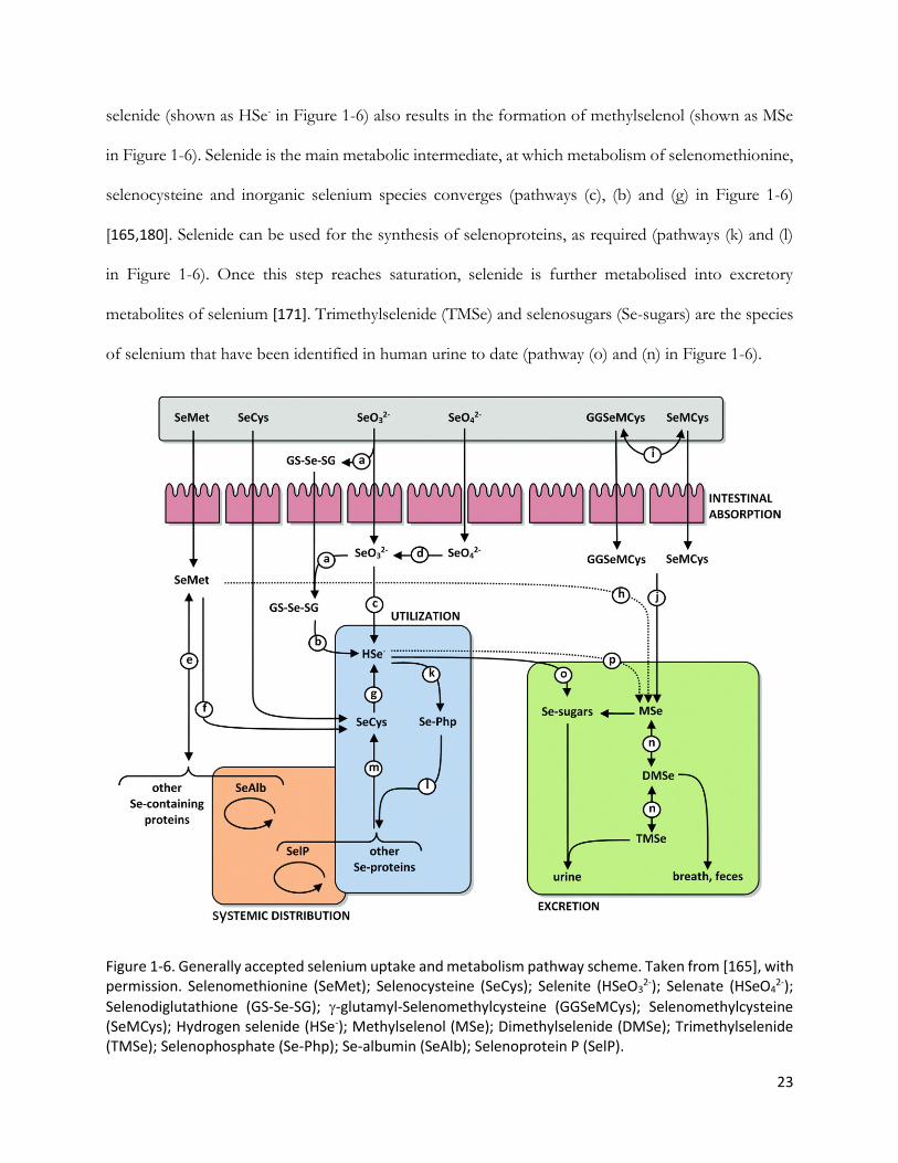
selenide (shown as HSe- in Figure 1-6) also results in the formation of methylselenol (shown as MSe
in Figure 1-6). Selenide is the main metabolic intermediate, at which metabolism of selenomethionine,
selenocysteine and inorganic selenium species converges (pathways (c), (b) and (g) in Figure 1-6)
[165,180]. Selenide can be used for the synthesis of selenoproteins, as required (pathways (k) and (l)
in Figure 1-6). Once this step reaches saturation, selenide is further metabolised into excretory
metabolites of selenium [171]. Trimethylselenide (TMSe) and selenosugars (Se-sugars) are the species
of selenium that have been identified in human urine to date (pathway (o) and (n) in Figure 1-6).
Figure 1-6. Generally accepted selenium uptake and metabolism pathway scheme. Taken from [165], with permission. Selenomethionine (SeMet); Selenocysteine (SeCys); Selenite (HSeO3
2-); Selenate (HSeO42-);
Selenodiglutathione (GS-Se-SG); -glutamyl-Selenomethylcysteine (GGSeMCys); Selenomethylcysteine (SeMCys); Hydrogen selenide (HSe-); Methylselenol (MSe); Dimethylselenide (DMSe); Trimethylselenide (TMSe); Selenophosphate (Se-Php); Se-albumin (SeAlb); Selenoprotein P (SelP).
24
1.4.2.3. Selenium Excretion
50-60% of selenium excretion occurs through the urine. Urinary elimination of selenium maintains
selenium homeostasis in the body [170]. Selenium excretion does not go up when selenium intake is
increased during selenium-deficient conditions [181]. It is hypothesized that once selenoprotein
synthesis is saturated, the remaining selenium is then metabolized for excretion via the urine. The ratio
of urinary:fecal excretion of selenium is increased, when selenium levels in the diet are high [182].
Plasma and erythrocyte selenium levels are good biomarkers for selenium intake. As with arsenic,
short-term selenium exposure can also be measured through the urine, and long-term selenium
exposure can be measured from hair and nails [183,184].
1.4.3. Selenium Mechanism of Protection Against Arsenic-Induced Toxicity
The antagonism between arsenic and selenium was first observed in the 1930s when selenium toxicity
in rats, from being fed seleniferous grains, was reduced with the addition of arsenic to drinking water
[185]. Arsenic and selenium sit as neighbours on the periodic table with atomic numbers 33 and 34,
respectively. As such, arsenite and selenite have a similar electronic structure. Since their chemical
and physical properties can be similar, arsenic and selenium can act in an antagonistic manner in
biological systems [60].
1.4.3.1. Protection Against Oxidative DNA Damage
One of the main mechanisms of arsenic toxicity is through the generation of reactive oxygen species.
It has been shown in vitro and in animal models that exposure to arsenite reduces the activity of
glutathione peroxidase and thioredoxin reductase [186–188]. Since both GPx and TrxR are
selenoproteins, co-exposure of selenite and arsenite has been shown to restore the activity of these
proteins and neutralize arsenite-induced oxidative damage [187]. Selenium can thus reduce the
mutagenic effects of arsenic by inhibiting oxidative damage caused to DNA, lipids and proteins [189].

25
1.4.3.2. Inhibition of Methylation Pathways
Selenium metabolism, as for arsenic, involves methylation [171,180]. Its methylated metabolites,
trimethylselenide and selenosugars, are also excreted in urine. As such, selenium inhibits arsenic
metabolism by competing for the methyl donor, SAM [168,186,190].
Glutathione is also required in both arsenic and selenium metabolism. Glutathione is involved in the
reduction of pentavalent arsenic species to trivalent [125,191]. In turn, selenate and selenite are
reduced by glutathione to the metabolite, selenodiglutathione, which is further metabolized to
hydrogen selenide by glutathione reductase [168,192]. The parallels in arsenic and selenium
metabolism mean that the reductive methylation of both species, results in competition for glutathione
and SAM [60].
1.4.3.3. Formation of the Seleno-bis(S-glutathionyl) arsinium Ion in
Liver
In the 1970s, Levander showed that arsenic and selenium lead to the decreased retention of the other
in rat liver and biliary excretion of both species was increased [193]. The molecular basis for this
interaction was later identified in rabbit bile to be the seleno-bis(S-glutathionyl) arsinium ion,
[(GS)2AsSe]- [194,195]. The Leslie lab determined that the human multidrug resistance protein 2
(MRP2) can transport both the glutathione-conjugated arsenic metabolites, arsenic triglutathione
(As(GS)3) and [(GS)2AsSe]- (at least in vitro) providing potential mechanistic insight into the increased
biliary efflux of both arsenic and selenium [108].
As(GS)3 is not stable at alkaline biliary pH and thereby dissociates in bile and undergoes enterohepatic
recirculation, preventing fecal arsenic elimination. [(GS)2AsSe]-, on the other hand, is relatively stable

26
at biliary pH; high levels of glutathione protect it from dissociation, thereby allowing excretion via
feces [131,194].
1.4.3.4. Selenium and Arsenic Sequestration in Red Blood Cells
The presence of arsenite in rat blood has been shown to delay selenite clearance from blood and vice
versa [196,197]. Glutathione levels in erythrocytes are approximately 3 mM, allowing for the in vivo
formation of [(GS)2AsSe]-. The formation of this conjugate has been shown in rabbit erythrocyte
lysate; 95% of RBC selenium was in the form of [(GS)2AsSe]-, 30 minutes after treatment [198]. Efflux
studies with human RBC membrane ghosts did not exhibit any ATP-dependent efflux of [(GS)2AsSe]-
, providing further evidence for the formation and sequestration of this conjugate [108]. Not only
does selenium influence arsenic metabolism and efflux but also plays a significant role in arsenic
sequestration in blood, influencing the organ-level distribution and toxicity of arsenic.
1.4.4. Selenium Supplementation as a Therapeutic for As-Induced Toxicities
The socio-economic status of arsenic-endemic regions is often poor, and arsenic removal from water
is costly, and not often feasible [199]. Alternatively, selenium supplementation is being investigated as
an inexpensive dietary palliative for the efficient elimination of arsenic. Selenium intake varies globally
and is largely dependent upon selenium content in agricultural soil [180]. In most arsenic-endemic
regions, low levels of selenium in soil may contribute to the high incidence of arsenic-induced toxicities
[184,200,201]. Chen et al., have shown that the risk of arsenic-induced skin lesions is greater among
individuals with lower blood selenium levels [202].
Epidemiological studies have investigated the effects of selenium on arsenic induced toxicities. A
supplementation trial using selenium-enriched yeast with a rural community in Inner Mongolia that
had been exposed to elevated levels of arsenic in their drinking water showed reduced arsenic
concentrations in blood and hair [203]. Evaluation of skin lesions also showed marked improvement

27
in the selenium supplementation group vs the placebo control group but interestingly, selenium
supplementation was most effective in ameliorating symptoms of adults that were most severely
affected by arsenic toxicity [203]. Kibriya et al., investigated changes in gene expression profiles in
individuals with pre-malignant arsenic-induced skin lesion supplemented with selenomethionine for 6
months daily [204]. Genes found to be up-regulated by selenium supplementation were previously
found to be down-regulated in the presence of arsenic by the same group, in particular the oxidative
stress related gene, superoxide dismutase 2 (SOD2) [204]. A 6-month selenomethionine
supplementation trial in Bangladesh also found slight improvement in arsenic-induced skin lesions
[205]. A case-control study in preschool children in Taiwan has shown that plasma selenium levels are
correlated with increased dimethylated arsenic species in urine [206]. Smits et al., also reported
increased urinary elimination of dimethylarsinic acid in individuals on a diet of selenium-enriched
lentils, in comparison to the control group [106].
Despite all the advances in understanding made with epidemiological studies and with in vivo animal
studies, little is known about the cellular handling and interactions between arsenic and selenium in
humans or human cell lines. The focus of this thesis is the influence of selenium on arsenic transport
and accumulation in human RBCs and hepatocytes.
1.5. Models of Hepatic Transport
Models of transport utilized in this thesis research are described below, providing a perspective on
the limitations and advantages of each model.
1.5.1. Primary Hepatocytes
Since the liver is the largest organ in the body involved in detoxification, primary human hepatocytes
are the gold standard for in vitro xenobiotic toxicity studies. However, in conventional culture,
hepatocytes lose their morphology and other characteristics after 24-48 hours in culture, making them

28
of limited use in longer-term metabolism studies [207]. Further to this, correct localization of
membrane proteins, cell polarization, cell-cell interactions and cell membrane integrity, especially the
bile canaliculi, results in disparate results from what may be occurring in vivo [207]. In contrast, when
hepatocytes are cultured between two layers of collagen, aptly termed sandwich-cultured hepatocytes
(SCH), they retain their in vivo-like features [208]. Sandwich-cultured hepatocytes retain the correct
liver functions, cell morphology and display the formation of intact biliary networks and excretory
function [209–211]. Most notably, transport function across the basolateral and apical surfaces of
hepatocytes can be elucidated using the sandwich-cultured model. As demonstrated by Liu et al.,
calcium can be used to modulate the integrity of tight junctions [212]. Tights junctions act as barriers
between the canalicular lumen and extracellular space. Tight junctions are disrupted when calcium is
depleted, allowing for the release of canalicular network contents and the design of experimental
studies to investigate biliary efflux. The biliary excretion index (BEI) is a measure of the percent of
total efflux that is going into bile. It is determined using the following equation:
Efflux(sinusoidal + canalicular) - Efflux(sinusoidal) BEI = x 100%
Efflux(sinusoidal + canalicular)
1.5.2. Immortalized Liver Cell Lines
The shortage of primary human hepatocytes leads to the use of immortalized cell lines in investigations
of liver function. The human hepatoma cell line, HepG2 is the most commonly used in research [213].
However, genetic expression of proteins involved in metabolism can vary over cell passages [214].
Not only do HepG2 cells lack the formation of canalicular networks, but protein profiles of membrane
transporters and metabolic enzymes are different in HepG2 cells than human hepatocytes [215].
Despite that, liver-specific cell function can be maintained in these cell lines, providing a readily
available model for high throughput optimization work [207].

29
More recently, HepaRG cells have been developed as a surrogate for human hepatocytes. Like HepG2
cells, the HepaRG cell line is a human hepatoma cell line, however, it can differentiate into both
cholangiocyte-like and hepatocyte-like cells, retaining features of human hepatocytes [216,217]. Since
the HepaRG cells represent the phenotype from one donor, human variability cannot be represented
using this cell line [207]. However, having a high proliferation capacity, allows for data replication and
reproducibility, making this cell line useful for xenobiotic metabolism and transport studies [216,217].
HepaRG cells also maintain cell polarity and expression of hepatocyte transport proteins [218]. The
correct localization and efflux function of MRP2 has been shown in HepaRG cells [218].
1.5.3. Vesicular Transport Assays
Characterization of ABC-transporter function is commonly done using vesicular transport assays.
Membrane vesicles can be isolated upon heterologous overexpression of a transporter of interest in
immortalized cell lines [209]. MRP2 is unidirectional transport protein, effluxing substrates out of the
cell [219]. Functional characterization of ABC proteins can be carried out using inside-out membrane
vesicles [220,221]. Inside-out vesicles have the ATP-binding site and substrate-binding sites of
proteins facing out. Their benefits are two-fold; (i) the need for prior accumulation of substrate within
cells prior to the commencement of efflux studies is eliminated, and (ii) substrates transported into
the vesicles will be ‘trapped’ for quantification.
1.6. Objectives, Hypothesis and Rationale.
Despite advances in selenium supplementation trials in efforts to alleviate arsenic-induced toxicities,
the cellular handling and interactions of arsenic and selenium in humans is poorly understood. Animal
studies have shown the protective mechanism of selenium to potentially be two-fold; through the
increased sequestration of arsenic in RBCs and secondly, through the increased biliary efflux of
arsenic. The objective of this thesis work was to characterize the effects of selenium on the uptake,

30
metabolism and efflux of arsenic using human RBCs and relevant human hepatic models. The main
hypothesis was that selenium would alter arsenic metabolism and transport through the formation of
[(GS)2AsSe]-. To address this hypothesis, specific objectives were as follows:
Objective 1: Characterization of the uptake and metabolism of arsenic and selenium in human red
blood cells
The first objective was to elucidate the mechanism of uptake of arsenic and selenium in human RBCs
upon crossing the intestinal epithelia following the most common route of exposure, ingestion.
Presented in Chapter 2 uptake transporters of arsenite and selenite in human RBCs were characterized
using inhibition studies, as well as, heterologous overexpression systems. In addition, the effect of
selenite on arsenite uptake, and vice versa was investigated. Lastly, in order to elucidate the effect of
selenite on arsenite metabolism, x-ray absorption spectroscopy was used to determine the speciation
of arsenic and selenium in red blood cells treated with both compounds.
Objective 2: Characterization of the effects of selenium on the hepatobiliary transport of arsenic in
SCHH
Interactions between arsenic and selenium likely begin in the portal circulation. First-pass metabolism
of arsenic and selenium occurs in the liver. As such, the second objective of my work was to use the
SCHH model to investigate the influence of selenium species on the biliary and sinusoidal efflux of
arsenic. SCHH preparations from 8 different donors were used to characterize arsenic efflux.
Preliminary experiments investigating the effect of arsenic on selenium accumulation and efflux were
also performed. In addition to efflux and accumulation studies, the effect of selenium on arsenic-
induced cytotoxicity in primary human hepatocytes was also investigated.
Objective 3: Characterization of arsenic and selenium uptake and cytotoxicity in HepG2 cells
To further extend our observations from the SCHH model and to optimize treatment condition
combinations for arsenic and selenium, studies were completed using HepG2 cells, as presented in

31
Chapter 4. The focus of this chapter was on uptake differences in HepG2 cells between the selenium
species, selenite and selenide. Cytotoxicity experiments were carried out for both selenium species and
in combination with arsenic to determine treatment combinations that resulted in antagonism of
toxicity between arsenic and selenium. Lastly, synchrotron-based techniques were used to investigate
accumulation differences of arsenic in the presence and absence of both selenite and selenide. The
results of this chapter provide important information for the better design of in vitro arsenic-selenium
interaction studies.

32
1.7. Bibliography
[1] T.M. Cox, N. Jack, S. Lofthouse, J. Watling, J. Haines, M.J. Warren, King George III and
porphyria: an elemental hypothesis and investigation, Lancet. 366 (2005) 332–335.
https://doi.org/10.1016/S0140-6736(05)66991-7.
[2] J. Parascandola, King of Poisons : a History of Arsenic., Potomac Books Inc, 2012.
https://books.google.ca/books?id=L6Ahq0FuVK8C&source=gbs_navlinks_s (accessed May
29, 2020).
[3] I. Palma-Lara, M. Martínez-Castillo, J.C. Quintana-Pérez, M.G. Arellano-Mendoza, F. Tamay-
Cach, O.L. Valenzuela-Limón, E.A. García-Montalvo, A. Hernández-Zavala, Arsenic
exposure: A public health problem leading to several cancers, Regul. Toxicol. Pharmacol. 110
(2020) 104539. https://doi.org/10.1016/j.yrtph.2019.104539.
[4] J.N. Pirl, G.F. Townsend, A.K. Valaitis, D. Grohlich, J.J. Spikes, Death by arsenic: A
comparative evaluation of exhumed body tissues in the presence of external contamination, J.
Anal. Toxicol. 7 (1983) 216–219. https://doi.org/10.1093/jat/7.5.216.
[5] S. Gibaud, G. Jaouen, Arsenic-based drugs: from Fowler’s solution to modern anticancer
chemotherapy, in: Springer, Berlin, Heidelberg, 2010: pp. 1–20.
https://doi.org/10.1007/978-3-642-13185-1_1.
[6] D.M. Jolliffe, A history of the use of arsenicals in man., J. R. Soc. Med. 86 (1993) 287–9.
http://www.ncbi.nlm.nih.gov/pubmed/8505753 (accessed March 2, 2020).
[7] J. Parascandola, History of Salvarsan (Arsphenamine), in: Encycl. Life Sci., John Wiley &
Sons, Ltd, Chichester, UK, 2001. https://doi.org/10.1038/npg.els.0003622.
[8] C. V. Rao, S. Pal, A. Mohammed, M. Farooqui, M.P. Doescher, A.S. Asch, H.Y. Yamada,
Biological effects and epidemiological consequences of arsenic exposure, and reagents that
can ameliorate arsenic damage <i>in vivo</i>, Oncotarget. 8 (2017) 57605–57621.
https://doi.org/10.18632/oncotarget.17745.
[9] A.R. Mistry, E.W. Pedersen, E. Solomon, D. Grimwade, The molecular pathogenesis of acute
promyelocytic leukaemia: implications for the clinical management of the disease., Blood Rev.
17 (2003) 71–97. https://doi.org/10.1016/s0268-960x(02)00075-9.

33
[10] K. Davison, K.K. Mann, W.H. Miller, Arsenic trioxide: Mechanisms of action, Semin.
Hematol. 39 (2002) 3–7. https://doi.org/10.1053/shem.2002.33610.
[11] C. Kaiming, Y. Sheng, S. Zheng, S. Yuan, G. Huang, Y. Liu, Arsenic trioxide preferentially
binds to the ring finger rotein PML: understanding target selection of the drug, Metallomics.
10 (2018) 1564–1569.
[12] A. Testi, M. D’Angiò, F. Locatelli, A. Pession, F. Lo Coco, Acute promyelocytic leukemia
(APL): Comparison between children and adults, Mediterr. J. Hematol. Infect. Dis. 6 (2014)
e2014032. https://doi.org/10.4084/MJHID.2014.032.
[13] P. Zhang, On arsenic trioxide in the clinical treatment of acute promyelocytic leukemia.,
Leuk. Res. Reports. 7 (2017) 29–32. https://doi.org/10.1016/j.lrr.2017.03.001.
[14] M.F. Naujokas, B. Anderson, H. Ahsan, H. Vasken Aposhian, J.H. Graziano, C. Thompson,
W.A. Suk, The broad scope of health effects from chronic arsenic exposure: Update on a
worldwide public health problem, Environ. Health Perspect. 121 (2013) 295–302.
https://doi.org/10.1289/ehp.1205875.
[15] R.N. Ratnaike, Acute and chronic arsenic toxicity., Postgrad. Med. J. 79 (2003) 391–6.
https://doi.org/10.1136/pmj.79.933.391.
[16] W.P. Tseng, H.M. Chu, S.W. How, J.M. Fong, C.S. Lin, S. Yeh, Prevalence of skin cancer in
an endemic area of chronic arsenicism in Taiwan, JNCI J. Natl. Cancer Inst. 40 (1968) 453–
463. https://doi.org/10.1093/jnci/40.3.453.
[17] M.E. Cebrian, A. Albores, M. Aguilar, E. Blakely, Chronic arsenic poisoning in the north of
Mexico, Hum. Toxicol. 2 (1983) 121–133. https://doi.org/10.1177/096032718300200110.
[18] C. Hopenhayn-Rich, M. Lou Biggs, A.H. Smith, Lung and kidney cancer mortality associated
with arsenic in drinking water in Cordoba, Argentina, Int. J. Epidemiol. 27 (1998) 561–569.
https://doi.org/10.1093/ije/27.4.561.
[19] A.H. Smith, M. Goycolea, R. Haque, M.L. Biggs, Marked increase in bladder and lung cancer
mortality in a region of Northern Chile due to arsenic in drinking water, Am. J. Epidemiol.
147 (1998) 660–669. https://doi.org/10.1093/oxfordjournals.aje.a009507.
[20] D.N. Guha Mazumder, Chronic arsenic toxicity: clinical features, epidemiology, and

34
treatment: experience in West Bengal, J. Environ. Sci. Heal. Part A. 38 (2003) 141–163.
https://doi.org/10.1081/ESE-120016886.
[21] M.A. Davis, A.J. Signes-Pastor, M. Argos, F. Slaughter, C. Pendergrast, T. Punshon, A.
Gossai, H. Ahsan, M.R. Karagas, Assessment of human dietary exposure to arsenic through
rice, Sci. Total Environ. 586 (2017). https://doi.org/10.1016/j.scitotenv.2017.02.119.
[22] U. Gundert-Remy, G. Damm, H. Foth, A. Freyberger, T. Gebel, K. Golka, C. Röhl, T.
Schupp, K.M. Wollin, J.G. Hengstler, High exposure to inorganic arsenic by food: the need
for risk reduction, Arch. Toxicol. 89 (2015). https://doi.org/10.1007/s00204-015-1627-1.
[23] U.S. Food and Drug Administration, Arsenic in rice and rice products: Risk assessment
report, 2016.
http://www.fda.gov/Food/FoodScienceResearch/RiskSafetyAssessment/default.htm.
[24] P.L.M. Veneman, J.R. Murray, J.H. Baker, Spatial distribution of pesticide residues in a
former apple orchard, J. Environ. Qual. 12 (1983) 101–104.
https://doi.org/10.2134/jeq1983.00472425001200010017x.
[25] S. Sauvé, Time to revisit arsenic regulations: comparing drinking water and rice, BMC Public
Health. 14 (2014) 465. https://doi.org/10.1186/1471-2458-14-465.
[26] W.T. Frankenberger, Environmental chemistry of arsenic, illustrate, CRC Press, 2001.
[27] J.G. Farmer, L.R. Johnson, Assessment of occupational exposure to inorganic arsenic based
on urinary concentrations and speciation of arsenic., Br. J. Ind. Med. 47 (1990) 342–8.
https://doi.org/10.1136/oem.47.5.342.
[28] M.F. Hughes, Arsenic toxicity and potential mechanisms of action, Toxicol. Lett. 133 (2002)
1–16. https://doi.org/10.1016/S0378-4274(02)00084-X.
[29] A. Franzblau, R. Lilis, Acute arsenic intoxication from environmental arsenic exposure, Arch.
Environ. Heal. An Int. J. 44 (1989) 385–390.
https://doi.org/10.1080/00039896.1989.9935912.
[30] A. Vahidnia, G.B. van der Voet, F.A. de Wolff, Arsenic neurotoxicity — A review:,
Http://Dx.Doi.Org/10.1177/0960327107084539. (2016).
https://doi.org/10.1177/0960327107084539.

35
[31] P. Bhattacharjee, D. Chatterjee, K.K. Singh, A.K. Giri, Systems biology approaches to
evaluate arsenic toxicity and carcinogenicity: An overview, Int. J. Hyg. Environ. Health. 216
(2013) 574–586. https://doi.org/10.1016/j.ijheh.2012.12.008.
[32] L.F. Negro Silva, M. Lemaire, C.A. Lemarié, D. Plourde, A.M. Bolt, C. Chiavatti, D.S. Bohle,
V. Slavkovich, J.H. Graziano, S. Lehoux, K.K. Mann, Effects of inorganic arsenic, methylated
arsenicals, and arsenobetaine on atherosclerosis in the apoE−/− mouse model and the role
of As3mt-mediated methylation, Environ. Health Perspect. 125 (2017) 077001.
https://doi.org/10.1289/EHP806.
[33] M. Rahman, M. Vahter, M.A. Wahed, N. Sohel, M. Yunus, P.K. Streatfield, S. El Arifeen, A.
Bhuiya, K. Zaman, A.M.R. Chowdhury, E.-C. Ekström, L.A. Persson, Prevalence of arsenic
exposure and skin lesions. A population based survey in Matlab, Bangladesh., J. Epidemiol.
Community Health. 60 (2006) 242–8. https://doi.org/10.1136/jech.2005.040212.
[34] A. Hernández, R. Marcos, Genetic variations associated with interindividual sensitivity in the
response to arsenic exposure., Pharmacogenomics. 9 (2008) 1113–32.
https://doi.org/10.2217/14622416.9.8.1113.
[35] N. Das, A. Giri, S. Chakraborty, P. Bhattacharjee, Association of single nucleotide
polymorphism with arsenic-induced skin lesions and genetic damage in exposed population
of West Bengal, India, Mutat. Res. Toxicol. Environ. Mutagen. 809 (2016) 50–56.
https://doi.org/10.1016/J.MRGENTOX.2016.09.006.
[36] Agency for Toxic Substances and Disease Registry (ASTDR), Toxicological profile for
arsenic, 2007.
[37] International Agency for Research on Cancer, Arsenic, metals, fibres, and dusts., 2012.
http://www.ncbi.nlm.nih.gov/pubmed/23189751 (accessed July 23, 2019).
[38] U.E.O. of Water, Technical fact sheet: Proposed rule for arsenic in drinking water and
clarifications to compliance and new source contaminants monitoring, (2014).
https://archive.epa.gov/water/archive/web/html/regulations_pro-techfactsheet.html
(accessed March 2, 2020).
[39] World Health Organization, Arsenic in Drinking-water: Background document for
development of WHO guidelines for drinking-water quality, 2003.
36
https://apps.who.int/iris/bitstream/handle/10665/75375/WHO_SDE_WSH_03.04_75_en
g.pdf (accessed March 2, 2020).
[40] Health Canada, Drinking water quality in Canada, (2019). https://www.canada.ca/en/health-
canada/services/environmental-workplace-health/water-quality/drinking-water.html
(accessed March 2, 2020).
[41] C.F. McGuigan, C.L.A. Hamula, S. Huang, S. Gabos, X.C. Le, A review on arsenic
concentrations in Canadian drinking water, Environ. Rev. 18 (2010) 291–307.
https://doi.org/10.1139/A10-012.
[42] T.J.B. Dummer, Z.M. Yu, L. Nauta, J.D. Murimboh, L. Parker, Geostatistical modelling of
arsenic in drinking water wells and related toenail arsenic concentrations across Nova Scotia,
Canada, Sci. Total Environ. 505 (2015) 1248–1258.
https://doi.org/10.1016/J.SCITOTENV.2014.02.055.
[43] P. Goering, H. V Aposhian, M.J. Mass, M. Cebrián, B.D. Beck, M.P. Waalkes, The enigma of
arsenic carcinogenesis: role of metabolism, Toxicol. Sci. 49 (1999) 5–14.
https://doi.org/10.1093/toxsci/49.1.5.
[44] S. Shen, X.-F. Li, W.R. Cullen, M. Weinfeld, X.C. Le, Arsenic binding to proteins, Chem.
Rev. 113 (2013) 7769–7792. https://doi.org/10.1021/cr300015c.
[45] Y. Hu, J. Li, B. Lou, R. Wu, G. Wang, C. Lu, H. Wang, J. Pi, Y. Xu, The role of reactive
oxygen species in arsenic toxicity, Biomolecules. 10 (2020) 240.
https://doi.org/10.3390/biom10020240.
[46] E.M. Leslie, A. Haimeur, M.P. Waalkes, Arsenic transport by the human multidrug resistance
protein 1 (MRP1/ABCC1). Evidence that a tri-glutathione conjugate is required., J. Biol.
Chem. 279 (2004) 32700–8. https://doi.org/10.1074/jbc.M404912200.
[47] K. Rehman, H. Naranmandura, Arsenic metabolism and thioarsenicals, Metallomics. 4 (2012)
881. https://doi.org/10.1039/c2mt00181k.
[48] L. Li, F. Chen, Oxidative stress, epigenetics, and cancer stem cells in arsenic carcinogenesis
and prevention, Curr. Pharmacol. Reports. 2 (2016) 57–63. https://doi.org/10.1007/s40495-
016-0049-y.

37
[49] W.R. Cullen, Chemical mechanism of arsenic biomethylation, Chem. Res. Toxicol. 27 (2014)
457–461. https://doi.org/10.1021/tx400441h.
[50] F. Challenger, Biological methylation., Sci. Prog. 35 (1947) 396–416.
https://doi.org/10.1039/qr9550900255.
[51] E. Bustaffa, A. Stoccoro, F. Bianchi, L. Migliore, Genotoxic and epigenetic mechanisms in
arsenic carcinogenicity, Arch. Toxicol. 88 (2014) 1043–1067.
https://doi.org/10.1007/s00204-014-1233-7.
[52] M. Vahter, G. Concha, Role of metabolism in arsenic toxicity., Pharmacol. Toxicol. 89 (2001)
1–5. https://doi.org/10.1111/j.1600-0773.2001.890101.x.
[53] M.J. Mass, A. Tennant, B.C. Roop, W.R. Cullen, M. Styblo, D.J. Thomas, A.D. Kligerman,
Methylated trivalent arsenic species are genotoxic, Chem. Res. Toxicol. 14 (2001) 355–361.
https://doi.org/10.1021/tx000251l.
[54] A. Basu, P. Ghosh, J.K. Das, A. Banerjee, K. Ray, A.K. Giri, Micronuclei as biomarkers of
carcinogen exposure in populations exposed to arsenic through drinking water in West
Bengal , India : A comparative study in three cell types micronuclei as biomarkers of
carcinogen exposure in populations exposed to arsenic, 13 (2004) 820–827.
[55] V. Martínez, A. Creus, W. Venegas, A. Arroyo, J.P. Beck, T.W. Gebel, J. Surrallés, R. Marcos,
Micronuclei assessment in buccal cells of people environmentally exposed to arsenic in
northern Chile, Toxicol. Lett. 155 (2005) 319–327.
https://doi.org/10.1016/J.TOXLET.2004.10.007.
[56] J. Mahata, A. Basu, S. Ghoshal, J.N. Sarkar, a K. Roy, G. Poddar, a K. Nandy, A. Banerjee,
K. Ray, a T. Natarajan, R. Nilsson, a K. Giri, Chromosomal aberrations and sister chromatid
exchanges in individuals exposed to arsenic through drinking water in West Bengal, India.,
Mutat. Res. 534 (2003) 133–43. https://doi.org/http://dx.doi.org/10.1016/S1383-
5718(02)00255-3.
[57] P. Mandal, Molecular insight of arsenic-induced carcinogenesis and its prevention, Naunyn.
Schmiedebergs. Arch. Pharmacol. 390 (2017) 443–455. https://doi.org/10.1007/s00210-017-
1351-x.

38
[58] R. Nakano, Apoptosis: Gene-Directed Cell Death, Horm. Res. 48 (1997) 2–4.
https://doi.org/10.1159/000191292.
[59] V. Gogvadze, S. Orrenius, B. Zhivotovsky, Multiple pathways of cytochrome c release from
mitochondria in apoptosis, Biochim. Biophys. Acta - Bioenerg. 1757 (2006) 639–647.
https://doi.org/10.1016/J.BBABIO.2006.03.016.
[60] H. Zeng, E.O. Uthus, G.F. Combs, Mechanistic aspects of the interaction between selenium
and arsenic, J. Inorg. Biochem. (2005) 1269–1274.
https://doi.org/10.1016/j.jinorgbio.2005.03.006.
[61] F. Khan, S. Momtaz, K. Niaz, F.I. Hassan, M. Abdollahi, Epigenetic mechanisms underlying
the toxic effects associated with arsenic exposure and the development of diabetes, Food
Chem. Toxicol. 107 (2017) 406–417. https://doi.org/10.1016/j.fct.2017.07.021.
[62] M.J. Barter, C. Bui, D.A. Young, Epigenetic mechanisms in cartilage and osteoarthritis: DNA
methylation, histone modifications and microRNAs, Osteoarthr. Cartil. 20 (2012) 339–349.
https://doi.org/10.1016/J.JOCA.2011.12.012.
[63] N. Valeri, I. Vannini, F. Fanini, F. Calore, B. Adair, M. Fabbri, Epigenetics, miRNAs, and
human cancer: a new chapter in human gene regulation, Mamm. Genome. 20 (2009) 573–
580. https://doi.org/10.1007/s00335-009-9206-5.
[64] S. Udali, P. Guarini, S. Moruzzi, S.-W. Choi, Cardiovascular epigenetics: From DNA
methylation to microRNAs, Mol. Aspects Med. 34 (2013) 883–901.
https://doi.org/10.1016/J.MAM.2012.08.001.
[65] C. Moutinho, M. Esteller, MicroRNAs and Epigenetics, Adv. Cancer Res. 135 (2017) 189–
220. https://doi.org/10.1016/BS.ACR.2017.06.003.
[66] R.V. Roy, Y.-O. Son, P. Pratheeshkumar, L. Wang, J.A. Hitron, S.P. Divya, R. D, D. Kim, Y.
Yin, Z. Zhang, X. Shi, Epigenetic targets of arsenic: Emphasis on epigenetic modifications
during carcinogenesis, J. Environ. Pathol. Toxicol. Oncol. 34 (2015) 63–84.
https://doi.org/10.1615/JEnvironPatholToxicolOncol.2014012066.
[67] D.S. Dheeman, C. Packianathan, J.K. Pillai, B.P. Rosen, Pathway of human AS3MT arsenic
methylation, Chem. Res. Toxicol. 27 (2014). https://doi.org/10.1021/tx500313k.

39
[68] B.C. Minatel, A.P. Sage, C. Anderson, R. Hubaux, E.A. Marshall, W.L. Lam, V.D. Martinez,
Environmental arsenic exposure: From genetic susceptibility to pathogenesis, Environ. Int.
112 (2018) 183–197. https://doi.org/10.1016/j.envint.2017.12.017.
[69] L. Chi, B. Gao, P. Tu, C.-W. Liu, J. Xue, Y. Lai, H. Ru, K. Lu, Individual susceptibility to
arsenic-induced diseases: the role of host genetics, nutritional status, and the gut microbiome,
Mamm. Genome. 29 (2018) 63–79. https://doi.org/10.1007/s00335-018-9736-9.
[70] K.E. Pelch, E.J. Tokar, B.A. Merrick, M.P. Waalkes, Differential DNA methylation profile of
key genes in malignant prostate epithelial cells transformed by inorganic arsenic or cadmium,
Toxicol. Appl. Pharmacol. 286 (2015) 159–167. https://doi.org/10.1016/j.taap.2015.04.011.
[71] S. Majumdar, S. Chanda, B. Ganguli, D.N.G. Mazumder, S. Lahiri, U.B. Dasgupta, Arsenic
exposure induces genomic hypermethylation, Environ. Toxicol. 25 (2009) 315–318.
https://doi.org/10.1002/tox.20497.
[72] L. Cantone, F. Nordio, L. Hou, P. Apostoli, M. Bonzini, L. Tarantini, L. Angelici, V. Bollati,
A. Zanobetti, J. Schwartz, P.A. Bertazzi, A. Baccarelli, Inhalable metal-rich air particles and
histone H3K4 dimethylation and H3K9 acetylation in a cross-sectional study of steel workers,
Environ. Health Perspect. 119 (2011) 964–969. https://doi.org/10.1289/ehp.1002955.
[73] A. Pournara, M. Kippler, T. Holmlund, R. Ceder, R. Grafström, M. Vahter, K. Broberg, A.E.
Wallberg, Arsenic alters global histone modifications in lymphocytes in vitro and in vivo, Cell
Biol. Toxicol. 32 (2016) 275–284. https://doi.org/10.1007/s10565-016-9334-0.
[74] B. Chen, J. Liu, Q. Chang, K. Beezhold, Y. Lu, F. Chen, JNK and STAT3 signaling pathways
converge on Akt-mediated phosphorylation of EZH2 in bronchial epithelial cells induced by
arsenic, Cell Cycle. 12 (2013) 112–121. https://doi.org/10.4161/cc.23030.
[75] J.Q. Cheng, C.W. Lindsley, G.Z. Cheng, H. Yang, S. V Nicosia, The Akt/PKB pathway:
molecular target for cancer drug discovery, Oncogene. 24 (2005) 7482–7492.
https://doi.org/10.1038/sj.onc.1209088.
[76] C.R. Webster, M.S. Anwer, Role of the PI3K/PKB signaling pathway in cAMP-mediated
translocation of rat liver Ntcp, Am. J. Physiol. 277 (1999) G1165–G1172.
[77] A. Carnero, The PKB/AKT Pathway in Cancer, Curr. Pharm. Des. 16 (2010) 34–44.

40
https://doi.org/10.2174/138161210789941865.
[78] R. Gera, V. Singh, S. Mitra, A.K. Sharma, A. Singh, A. Dasgupta, D. Singh, M. Kumar, P.
Jagdale, S. Patnaik, D. Ghosh, Arsenic exposure impels CD4 commitment in thymus and
suppress T cell cytokine secretion by increasing regulatory T cells, Sci. Rep. 7 (2017) 1–13.
https://doi.org/10.1038/s41598-017-07271-z.
[79] L.M. Del Razo, G.G. García-Vargas, O.L. Valenzuela, E.H. Castellanos, L.C. Sánchez-Peña,
J.M. Currier, Z. Drobná, D. Loomis, M. Stýblo, Exposure to arsenic in drinking water is
associated with increased prevalence of diabetes: a cross-sectional study in the Zimapán and
Lagunera regions in Mexico, Environ. Heal. 10 (2011) 73. https://doi.org/10.1186/1476-
069X-10-73.
[80] C.H. Tseng, T.Y. Tai, C.K. Chong, C.P. Tseng, M.S. Lai, B.J. Lin, H.Y. Chiou, Y.M. Hsueh,
K.H. Hsu, C.J. Chen, Long-term arsenic exposure and incidence of non-insulin-dependent
diabetes mellitus: a cohort study in arseniasis-hyperendemic villages in Taiwan., Environ.
Health Perspect. 108 (2000) 847–851. https://doi.org/10.1289/ehp.00108847.
[81] M. Rahman, M. Tondel, S.A. Ahmad, O. Axelson, Diabetes mellitus associated with arsenic
exposure in Bangladesh, Am. J. Epidemiol. 148 (1998) 198–203.
https://doi.org/10.1093/oxfordjournals.aje.a009624.
[82] Y. Kumagai, D. Sumi, Arsenic: Signal transduction, transcription factor, and
biotransformation involved in cellular response and toxicity, Annu. Rev. Pharmacol. Toxicol.
47 (2007) 243–262. https://doi.org/10.1146/annurev.pharmtox.47.120505.105144.
[83] C. Huang, Q. Ke, M. Costa, X. Shi, Molecular mechanisms of arsenic carcinogenesis, Mol.
Cell. Biochem. 255 (2004) 57–66. https://doi.org/10.1023/B:MCBI.0000007261.04684.78.
[84] C. Douillet, J. Currier, J. Saunders, W.M. Bodnar, Matoušek, T, M. Styblo, Methylated
trivalent arsenicals are potent inhibitors of glucose stimulated insulin secretion by murine
pancreatic islets, Toxicol. Appl. Pharmacol. 267 (2013) 11–15.
https://www.sciencedirect.com/science/article/pii/S0041008X12005340 (accessed March 1,
2020).
[85] T. Samikkannu, C.-H. Chen, L.-H. Yih, A.S.S. Wang, S.-Y. Lin, T.-C. Chen, K.-Y. Jan,
Reactive oxygen species are involved in arsenic trioxide inhibition of pyruvate dehydrogenase

41
activity, Chem. Res. Toxicol. 16 (2003) 409–414. https://doi.org/10.1021/TX025615J.
[86] M. Jochem, L. Ende, M. Isasa, J. Ang, H. Schnell, A. Guerra-Moreno, Y. Micoogullari, M.
Bhanu, S.P. Gygi, J. Hanna, Targeted degradation of glucose transporters protects against
arsenic toxicity., Mol. Cell. Biol. 39 (2019). https://doi.org/10.1128/MCB.00559-18.
[87] E.M. Martin, M. Stýblo, R.C. Fry, Genetic and epigenetic mechanisms underlying arsenic-
associated diabetes mellitus: a perspective of the current evidence., Epigenomics. 9 (2017)
701–710. https://doi.org/10.2217/epi-2016-0097.
[88] M.-S. Lai, Y.-M. Hsueh, C.-J. Chen, M.-P. Shyu, S.-Y. Chen, T.-L. Kuo, M.-M. Wu, T.-Y. Tai,
Ingested inorganic arsenic and prevalence of diabetes mellitus, Am. J. Epidemiol. 139 (1994)
484–492. https://doi.org/10.1093/oxfordjournals.aje.a117031.
[89] C.-H. Tseng, The potential biological mechanisms of arsenic-induced diabetes mellitus,
Toxicol. Appl. Pharmacol. 197 (2004) 67–83. https://doi.org/10.1016/J.TAAP.2004.02.009.
[90] M. Rahman, O. Axelson, Diabetes mellitus and arsenic exposure: a second look at case-
control data from a Swedish copper smelter., Occup. Environ. Med. 52 (1995) 773–4.
https://doi.org/10.1136/oem.52.11.773.
[91] P.P. Simeonova, M. Luster, Arsenic and atherosclerosis, Toxicol. Appl. Pharmacol. 198
(2004) 444–449.
[92] C.-H. Wang, J.-S. Jeng, P.-K. Yip, C.-L. Chen, L.-I. Hsu, Y.-M. Hsueh, H.-Y. Chiou, M.-M.
Wu, C.-J. Chen, Biological gradient between long-term arsenic exposure and carotid
atherosclerosis., Circulation. 105 (2002) 1804–9.
https://doi.org/10.1161/01.cir.0000015862.64816.b2.
[93] M.-M. Wu, H.-Y. Chiou, Y.-M. Hsueh, C.-T. Hong, C.-L. Su, S.-F. Chang, W.-L. Huang, H.-
T. Wang, Y.-H. Wang, Y.-C. Hsieh, C.-J. Chen, Effect of plasma homocysteine level and
urinary monomethylarsonic acid on the risk of arsenic-associated carotid atherosclerosis,
Toxicol. Appl. Pharmacol. 216 (2006) 168–175.
https://doi.org/10.1016/J.TAAP.2006.05.005.
[94] P.P. Simeonova, T. Hulderman, D. Harki, M.I. Luster, Arsenic exposure accelerates
atherogenesis in apolipoprotein E(-/-) mice., Environ. Health Perspect. 111 (2003) 1744–
42
1748. https://doi.org/10.1289/ehp.6332.
[95] K. Makhani, C. Chiavatti, D. Plourde, L.F. Negro Silva, M. Lemaire, C.A. Lemarié, S. Lehoux,
K.K. Mann, Using the apolipoprotein E knock-out mouse model to define atherosclerotic
plaque changes induced by low dose arsenic, Toxicol. Sci. 166 (2018) 213–218.
https://doi.org/10.1093/toxsci/kfy201.
[96] A. Pulliero, R. Godschalk, M.G. Andreassi, D. Curfs, F.J. Van Schooten, A. Izzotti,
Environmental carcinogens and mutational pathways in atherosclerosis, Int. J. Hyg. Environ.
Health. 218 (2015) 293–312. https://doi.org/10.1016/j.ijheh.2015.01.007.
[97] H. Shen, Q. Niu, M. Xu, D. Rui, S. Xu, G. Feng, Y. Ding, S. Li, M. Jing, Factors affecting
arsenic methylation in arsenic-exposed humans: A systematic review and meta-analysis, Int. J.
Environ. Res. Public Health. 13 (2016). https://doi.org/10.3390/ijerph13020205.
[98] F. Faita, L. Cori, F. Bianchi, M.G. Andreassi, Arsenic-induced genotoxicity and genetic
susceptibility to arsenic-related pathologies, Int. J. Environ. Res. Public Health. 10 (2013).
https://doi.org/10.3390/ijerph10041527.
[99] T. Agusa, J. Fujihara, H. Takeshita, H. Iwata, Individual variations in inorganic arsenic
metabolism associated with AS3MT genetic polymorphisms, 2011.
https://doi.org/10.3390/ijms12042351.
[100] R. Antonelli, K. Shao, D.J. Thomas, R. Sams, J. Cowden, AS3MT, GSTO, and PNP
polymorphisms: Impact on arsenic methylation and implications for disease susceptibility,
Environ. Res. 132 (2014). https://doi.org/10.1016/j.envres.2014.03.012.
[101] K.M. McCarty, Y.C. Chen, Q. Quamruzzaman, M. Rahman, G. Mahiuddin, Y.M. Hsueh, L.
Su, T. Smith, L. Ryan, D.C. Christiani, Arsenic methylation, GSTT1, GSTM1, GSTP1
polymorphisms, and skin lesions, Environ. Health Perspect. 115 (2007) 341–345.
https://doi.org/10.1289/ehp.9152.
[102] B. Janasik, E. Reszka, M. Stanislawska, E. Wieczorek, W. Fendler, W. Wasowicz, Biological
monitoring and the influence of genetic polymorphism of As3MT and GSTs on distribution
of urinary arsenic species in occupational exposure workers, Int. Arch. Occup. Environ.
Health. 88 (2015) 807–818. https://doi.org/10.1007/s00420-014-1009-7.

43
[103] A. Hollman, P. Tchounwou, H.-C. Huang, The association between gene-environment
interactions and diseases involving the human GST superfamily with SNP variants, Int. J.
Environ. Res. Public Health. 13 (2016) 379. https://doi.org/10.3390/ijerph13040379.
[104] S. Lin, Q. Shi, F.B. Nix, M. Styblo, M.A. Beck, K.M. Herbin-Davis, L.L. Hall, J.B.
Simeonsson, D.J. Thomas, A novel S-adenosyl-L-methionine:arsenic(III) methyltransferase
from rat liver cytosol., J. Biol. Chem. 277 (2002) 10795–803.
https://doi.org/10.1074/jbc.M110246200.
[105] K.S. Engström, K. Broberg, G. Concha, B. Nermell, M. Warholm, M. Vahter, Genetic
polymorphisms influencing arsenic metabolism: Evidence from Argentina, Environ. Health
Perspect. 115 (2007) 599–605. https://doi.org/10.1289/ehp.9734.
[106] J.E. Smits, R.M. Krohn, E. Akhtar, S.K. Hore, M. Yunus, A. Vandenberg, R. Raqib, Food as
medicine: Selenium enriched lentils offer relief against chronic arsenic poisoning in
Bangladesh, Environ. Res. 176 (2019) 108561.
https://doi.org/10.1016/j.envres.2019.108561.
[107] M. Banerjee, M.W. Carew, B.A. Roggenbeck, B.D. Whitlock, H. Naranmandura, X.C. Le,
E.M. Leslie, A novel pathway for arsenic elimination : Human multidrug resistance protein 4 (
MRP4 / ABCC4 ) mediates cellular export of dimethylarsinic acid ( DMA V ) and the
diglutathione conjugate of monomethylarsonous acid ( MMA III ), Mol. Pharmacol. 86
(2014) 168–179.
[108] M.W. Carew, E.M. Leslie, Selenium-dependent and -independent transport of arsenic by the
human multidrug resistance protein 2 (MRP2/ABCC2): implications for the mutual
detoxification of arsenic and selenium, Carcinogenesis. 31 (2010) 1450–1455.
https://doi.org/10.1093/carcin/bgq125.
[109] M. Banerjee, V. Marensi, G. Conseil, X.C. Le, S.P.C. Cole, E.M. Leslie, Polymorphic variants
of MRP4/ABCC4 differentially modulate the transport of methylated arsenic metabolites and
physiological organic anions, Biochem. Pharmacol. 120 (2016) 72–82.
https://doi.org/10.1016/J.BCP.2016.09.016.
[110] M.R.I. Chowdhury, C.N. Mulligan, Biosorption of arsenic from contaminated water by
anaerobic biomass, J. Hazard. Mater. 190 (2011) 486–492.

44
https://doi.org/10.1016/j.jhazmat.2011.03.070.
[111] D. Caussy, Case studies of the impact of understanding bioavailability: arsenic, Ecotoxicol.
Environ. Saf. 56 (2003) 164–173. https://doi.org/10.1016/S0147-6513(03)00059-9.
[112] G.B. Freeman, R.A. Schoof, M.V. Ruby, A.O. Davis, J.A. Dill, S.C. Liao, C.A. Lapin, P.D.
Bergstrom, Bioavailability of arsenic in soil and house dust impacted by smelter activities
following oral administration in Cynomolgus monkeys, Fundam. Appl. Toxicol. 28 (1995)
215–222. https://doi.org/10.1006/FAAT.1995.1162.
[113] R.R. Dietert, E.K. Silbergeld, Biomarkers for the 21st century: Listening to the microbiome,
Toxicol. Sci. 144 (2015) 208–216. https://doi.org/10.1093/toxsci/kfv013.
[114] B.A. Roggenbeck, E.M. Leslie, S.T. Walk, E.E. Schmidt, Redox metabolism of ingested
arsenic: Integrated activities of microbiome and host on toxicological outcomes, Curr. Opin.
Toxicol. 13 (2019) 90–98. https://doi.org/10.1016/J.COTOX.2018.09.003.
[115] T. Van de Wiele, C.M. Gallawa, K.M. Kubachk, J.T. Creed, N. Basta, E.A. Dayton, S.
Whitacre, G. Du Laing, K. Bradham, Arsenic metabolism by human gut microbiota upon in
vitro digestion of contaminated soils, Environ. Health Perspect. 118 (2010) 1004–1009.
https://doi.org/10.1289/ehp.0901794.
[116] Q.Q. Wang, D.J. Thomas, H. Naranmandura, Importance of being thiomethylated:
Formation, fate, and effects of methylated thioarsenicals, Chem. Res. Toxicol. 28 (2015) 281–
289. https://doi.org/10.1021/tx500464t.
[117] M. Coryell, B.A. Roggenbeck, S.T. Walk, The human gut microbiome’s influence on arsenic
toxicity, Curr. Pharmacol. Reports. 5 (2019) 491–504. https://doi.org/10.1007/s40495-019-
00206-4.
[118] Z. Liu, J.M. Carbrey, P. Agre, B.P. Rosen, Arsenic trioxide uptake by human and rat
aquaglyceroporins, Biochem. Biophys. Res. Commun. 316 (2004) 1178–1185.
https://doi.org/10.1016/J.BBRC.2004.03.003.
[119] B.P. Rosen, Z. Liu, Transport pathways for arsenic and selenium: a minireview., Environ. Int.
35 (2009) 512–5. https://doi.org/10.1016/j.envint.2008.07.023.
[120] M. Calatayud, J.A. Barrios, V. Devesa, In vitro study of transporters involved in intestinal

45
absorption of inorganic arsenic, Chem. Res. Toxicol. 25 (2012) 446–453.
[121] B.A. Roggenbeck, M. Banerjee, E.M. Leslie, Cellular arsenic transport pathways in mammals,
J. Environ. Sci. (China). 49 (2016) 38–58. https://doi.org/10.1016/j.jes.2016.10.001.
[122] Z. Liu, M.A. Sanchez, X. Jiang, E. Boles, S.M. Landfear, B.P. Rosen, Mammalian glucose
permease GLUT1 facilitates transport of arsenic trioxide and methylarsonous acid., Biochem.
Biophys. Res. Commun. 351 (2006) 424–30. https://doi.org/10.1016/j.bbrc.2006.10.054.
[123] R. Villa-Bellosta, V. Sorribas, Arsenate transport by sodium/phosphate cotransporter type
IIb, Toxicol. Appl. Pharmacol. 247 (2010) 36–40.
https://doi.org/10.1016/j.taap.2010.05.012.
[124] T. Hayakawa, Y. Kobayashi, X. Cui, S. Hirano, A new metabolic pathway of arsenite:
arsenic?glutathione complexes are substrates for human arsenic methyltransferase Cyt19,
Arch. Toxicol. 79 (2005) 183–191. https://doi.org/10.1007/s00204-004-0620-x.
[125] D.J. Thomas, Unraveling arsenic - glutathione connections, Toxicol. Sci. 107 (2009) 309–311.
https://doi.org/10.1093/toxsci/kfn257.
[126] H. Naranmandura, K. Rehman, X.C. Le, D.J. Thomas, Formation of methylated
oxyarsenicals and thioarsenicals in wild-type and arsenic (+3 oxidation state)
methyltransferase knockout mice exposed to arsenate, Anal. Bioanal. Chem. 405 (2013)
1885–1891. https://doi.org/10.1007/s00216-012-6207-0.
[127] R. Raml, W. Goessler, K.A. Francesconi, Improved chromatographic separation of thio-
arsenic compounds by reversed-phase high performance liquid chromatography-inductively
coupled plasma mass spectrometry (HPLC–ICPMS), J. Chromatogr. A. 1128 (2006) 164–170.
[128] R. Raml, A. Rumpler, W. Goessler, M. Vahter, L. Li, T. Ochi, K.A. Francesconi, Thio-
dimethylarsinate is a common metabolite in urine samples from arsenic-exposed women in
Bangladesh, Toxicol. Appl. Pharmacol. 222 (2007) 374–380.
[129] S. V Kala, M.W. Neely, G. Kala, C.I. Prater, D.W. Atwood, J.S. Rice, M.W. Lieberman, The
MRP2/cMOAT transporter and arsenic-glutathione complex formation are required for
biliary excretion of arsenic., J. Biol. Chem. 275 (2000) 33404–8.
https://doi.org/10.1074/jbc.M007030200.

46
[130] S. V. Kala, G. Kala, C.I. Prater, A.C. Sartorelli, M.W. Lieberman, Formation and urinary
excretion of arsenic triglutathione and methylarsenic diglutathione, Chem. Res. Toxicol. 17
(2004) 243–249. https://doi.org/10.1021/tx0342060.
[131] E.M. Leslie, Arsenic-glutathione conjugate transport by the human multidrug resistance
proteins (MRPs/ABCCs), J. Inorg. Biochem. 108 (2012) 141–149.
https://doi.org/10.1016/j.jinorgbio.2011.11.009.
[132] M.W. Carew, H. Naranmandura, C.B. Shukalek, X.C. Le, E.M. Leslie, Monomethylarsenic
diglutathione transport by the human multidrug resistance protein 1 ( MRP1 / ABCC1 ),
Drug Metab. Dispos. 39 (2011) 2298–2304. https://doi.org/10.1124/dmd.111.041673.ing.
[133] B.D. Whitlock, E.M. Leslie, Efflux transporters in anti-cancer drug resistance: Molecular and
functional identification and characterization of multidrug resistance proteins
(MRPs/ABCCs), Academic Press, 2020. https://doi.org/10.1016/B978-0-12-816434-
1.00002-4.
[134] P.M. Jones, A.M. George, A reciprocating twin-channel model for ABC transporters, Q. Rev.
Biophys. 3 (2014) 1–32. https://doi.org/10.1017/S0033583514000031.
[135] Z. Chen, T. Shi, L. Zhang, P. Zhu, M. Deng, C. Huang, T. Hu, L. Jiang, J. Li, Mammalian
drug efflux transporters of the ATP binding cassette (ABC) family in multidrug resistance: A
review of the past decade, Cancer Lett. 370 (2016) 153–164.
https://doi.org/10.1016/j.canlet.2015.10.010.
[136] E.M. Leslie, R.G. Deeley, S.P.C. Cole, Multidrug resistance proteins: role of P-glycoprotein,
MRP1, MRP2, and BCRP (ABCG2) in tissue defense., Toxicol. Appl. Pharmacol. 204 (2005)
216–37. https://doi.org/10.1016/j.taap.2004.10.012.
[137] S. Wilkens, Structure and mechanism of ABC transporters., F1000Prime Rep. 7 (2015) 14.
https://doi.org/10.12703/P7-14.
[138] L. Schmitt, R. Tampé, Structure and mechanism of ABC transporters, Curr. Opin. Struct.
Biol. 12 (2002) 754–760.
https://www.sciencedirect.com/science/article/pii/S0959440X02003998 (accessed March
20, 2020).

47
[139] P.E. Bandler, C.J. Westlake, C.E. Grant, S.P.C. Cole, R.G. Deeley, Identification of regions
required for apical membrane localization of human multidrug resistance protein 2., Mol.
Pharmacol. 74 (2008) 9–19. https://doi.org/10.1124/mol.108.045674.
[140] J.E. Walker, M. Saraste, M.J. Runswick, N.J. Gay, Distantly related sequences in the alpha-
and beta-subunits of ATP synthase, myosin, kinases and other ATP-requiring enzymes and a
common nucleotide binding fold., EMBO J. 1 (1982) 945–951.
https://doi.org/10.1002/j.1460-2075.1982.tb01276.x.
[141] C. Oswald, I.B. Holland, L. Schmitt, The motor domains of ABC-transporters, Naunyn.
Schmiedebergs. Arch. Pharmacol. 372 (2006) 385–399. https://doi.org/10.1007/s00210-005-
0031-4.
[142] C.R. Zito, E. Antony, J.F. Hunt, D.B. Oliver, M.M. Hingorani, Role of a conserved glutamate
residue in the Escherichia coli SecA ATPase mechanism., J. Biol. Chem. 280 (2005) 14611–9.
https://doi.org/10.1074/jbc.M414224200.
[143] Z. Szentpétery, A. Kern, K. Liliom, B. Sarkadi, A. Váradi, É. Bakos, The role of the
conserved glycines of ATP-binding cassette signature motifs of MRP1 in the communication
between the substrate-binding site and the catalytic centers, J. Biol. Chem. 279 (2004) 41670–
41678. https://doi.org/10.1074/jbc.M406484200.
[144] P.C. Smith, N. Karpowich, L. Millen, J.E. Moody, J. Rosen, P.J. Thomas, J.F. Hunt, ATP
binding to the motor domain from an ABC transporter drives formation of a nucleotide
sandwich dimer., Mol. Cell. 10 (2002) 139–49. https://doi.org/10.1016/S1097-
2765(02)00576-2.
[145] Z.L. Johnson, J. Chen, ATP binding enables substrate release from multidrug resistance
protein 1, Cell. 172 (2018) 81-89.e10. https://doi.org/10.1016/J.CELL.2017.12.005.
[146] V. Materna, H. Lage, Homozygous mutation Arg768Trp in the ABC-transporter encoding
gene MRP2/cMOAT/ABCC2 causes Dubin-Johnson syndrome in a Caucasian patient., J.
Hum. Genet. 48 (2003) 484–6. https://doi.org/10.1007/s10038-003-0057-8.
[147] C.G. Dietrich, R. Ottenhoff, D.R. de Waart, R.P.. Oude Elferink, Role of MRP2 and GSH in
intrahepatic cycling of toxins, Toxicology. 167 (2001) 73–81. https://doi.org/10.1016/S0300-
483X(01)00459-0.

48
[148] A.T. Nies, Y. Cui, I. Leier, D. Keppler, Conjugate export pumps of the multidrug resistance
protein ( MRP ) family : localization , substrate speci ¢ city , and MRP2-mediated drug
resistance, 1461 (1999).
[149] A.T. Nies, D. Keppler, The apical conjugate efflux pump ABCC2 (MRP2), Pflügers Arch. -
Eur. J. Physiol. 453 (2007) 643–659. https://doi.org/10.1007/s00424-006-0109-y.
[150] B.A. Roggenbeck, M.W. Carew, G.J. Charrois, D.N. Douglas, N.M. Kneteman, X. Lu, X.C.
Le, E.M. Leslie, Characterization of arsenic hepatobiliary transport using sandwich-cultured
human hepatocytes, Toxicol. Sci. 145 (2015) 307–320.
https://doi.org/10.1093/toxsci/kfv051.
[151] O. Bruhn, I. Cascorbi, Polymorphisms of the drug transporters ABCB1, ABCG2, ABCC2
and ABCC3 and their impact on drug bioavailability and clinical relevance, Expert Opin.
Drug Metab. Toxicol. 10 (2014) 1337–1354. https://doi.org/10.1517/17425255.2014.952630.
[152] O. Fardel, E. Jigorel, M. Le Vee, L. Payen, Physiological, pharmacological and clinical
features of the multidrug resistance protein 2., Biomed. Pharmacother. 59 (2005) 104–14.
https://doi.org/10.1016/j.biopha.2005.01.005.
[153] V. Keitel, A.T. Nies, M. Brom, J. Hummel-Eisenbeiss, H. Spring, D. Keppler, A common
Dubin-Johnson syndrome mutation impairs protein maturation and transport activity of
MRP2 (ABCC2)., Am. J. Physiol. Gastrointest. Liver Physiol. 284 (2003) G165-74.
https://doi.org/10.1152/ajpgi.00362.2002.
[154] J. Kartenbeck, U. Leuschner, R. Mayer, D. Keppler, Absence of the canalicular isoform of
theMRP gene-encoded conjugate export pump from the hepatocytes in Dubin-Johnson
syndrome, Hepatology. 23 (1996) 1061–1066. https://doi.org/10.1002/hep.510230519.
[155] R. Mor-Cohen, A. Zivelin, N. Rosenberg, M. Shani, S. Muallem, U. Seligsohn, Identification
and functional analysis of two novel mutations in the multidrug resistance protein 2 gene in
Israeli patients with Dubin-Johnson syndrome., J. Biol. Chem. 276 (2001) 36923–30.
https://doi.org/10.1074/jbc.M105047200.
[156] M. Itoda, Y. Saito, A. Soyama, M. Saeki, N. Murayama, S. Ishida, K. Sai, M. Nagano, H.
Suzuki, Y. Sugiyama, S. Ozawa, J. Sawada Ji, Polymorphisms in the ABCC2 (cMOAT/MRP2)
gene found in 72 established cell lines derived from Japanese individuals: an association

49
between single nucleotide polymorphisms in the 5’-untranslated region and exon 28., Drug
Metab. Dispos. 30 (2002) 363–4. http://www.ncbi.nlm.nih.gov/pubmed/11901087 (accessed
April 15, 2014).
[157] H. Suzuki, Y. Sugiyama, Single nucleotide polymorphisms in multidrug resistance associated
protein 2 ( MRP2 / ABCC2 ): its impact on drug disposition, 54 (2002) 1311–1331.
[158] I.N. Dubin, F.B. Johnson, Chronic idiopathic jaundice with unidentified pigment in liver
cells, Medicine (Baltimore). 33 (1954) 155–198.
[159] M. Vahter, Variation in human metabolism of arsenic, Arsen. Expo. Heal. Eff. III. (1999)
267–279. https://doi.org/10.1016/B978-008043648-7/50031-5.
[160] B.K. Mandal, Y. Ogra, K.T. Suzuki, Identification of dimethylarsinous and
monomethylarsonous acids in human urine of the arsenic-affected areas in West Bengal,
India, (2001). https://doi.org/10.1021/TX000246H.
[161] X.C. Le, X. Lu, M. Ma, W.R. Cullen, H. V. Aposhian, B. Zheng, Speciation of key arsenic
metabolic intermediates in human urine, Anal. Chem. 72 (2000) 5172–5177.
https://doi.org/10.1021/AC000527U.
[162] C.-H. Tseng, Arsenic methylation, urinary arsenic metabolites and human diseases: Current
perspective, J. Environ. Sci. Heal. Part C. 25 (2007) 1–22.
https://doi.org/10.1080/10590500701201695.
[163] V. Taylor, B. Goodale, A. Raab, T. Schwerdtle, K. Reimer, S. Conklin, M.R. Karagas, K.A.
Francesconi, Human exposure to organic arsenic species from seafood, Sci. Total Environ.
580 (2017). https://doi.org/10.1016/j.scitotenv.2016.12.113.
[164] N. Bu, H.Y. Wang, W.H. Hao, X. Liu, S. Xu, B. Wu, Y. Anan, Y. Ogra, Y.J. Lou, H.
Naranmandura, Generation of thioarsenicals is dependent on the enterohepatic circulation in
rats, Metallomics. 3 (2011) 1064. https://doi.org/10.1039/c1mt00036e.
[165] M. Roman, C. Barbante, Selenium biochemistry and its role for human health, Metallomics. 6
(2014) 25–54. https://doi.org/10.1039/c3mt00185g.
[166] J.A. Plant, J. Bone, N. Voulvoulis, D.G. Kinniburgh, P.L. Smedley, F.M. Fordyce, B. Klinck,
Arsenic and selenium, 2nd ed., Elsevier Ltd., 2013. https://doi.org/10.1016/B978-0-08-
50
095975-7.00902-5.
[167] J.K. Wrobel, R. Power, M. Toborek, Critical review biological activity of selenium : Revisited,
Int. Union Biochem. Mol. Biol. 68 (2016) 97–105. https://doi.org/10.1002/iub.1466.
[168] I. Zwolak, H. Zaporowska, Selenium interactions and toxicity: a review, Cell Biol. Toxicol. 28
(2012) 31–46. https://doi.org/10.1007/s10565-011-9203-9.
[169] S.E. Jackson-Rosario, W.T. Self, Targeting selenium metabolism and selenoproteins: novel
avenues for drug discovery., Metallomics. 2 (2010) 112–6. https://doi.org/10.1039/b917141j.
[170] M. Navarro-Alarcon, C. Cabrera-Vique, Selenium in food and the human body: A review, Sci.
Total Environ. 400 (2008) 115–141. https://doi.org/10.1016/J.SCITOTENV.2008.06.024.
[171] R.F. Burk, K.E. Hill, Regulation of selenium metabolism and transport, Annu. Rev. Nutr. 35
(2015) 109–134. https://doi.org/10.1146/annurev-nutr-071714-034250.
[172] T. Imai, H. Mihara, T. Kurihara, N. Esaki, Possible role of red blood cells in selenocysteine
metabolism, Trace Nutr. Res. 26 (2009) 22–25.
[173] Y. Wang, L. Fu, Forms of selenium affect its transport, uptake and glutathione peroxidase
activity in the caco-2 cell model, Biol. Trace Elem. Res. 149 (2012) 110–116.
https://doi.org/10.1007/s12011-012-9395-8.
[174] V.R. Preedy, ed., Selenium: chemistry, analysis, function and effects, Royal Society of
Chemistry, Cambridge, 2015. https://doi.org/DOI:10.1039/9781782622215.
[175] K.T. Suzuki, Y. Shiobara, M. Itoh, M. Ohmichi, Selective uptake of selenite by red blood
cells, Analyst. 123 (1998) 63–67. https://doi.org/10.1039/a706230c.
[176] J.R. McDermott, X. Geng, L. Jiang, M. Gálvez-Peralta, F. Chen, D.W. Nebert, Z. Liu, Zinc-
and bicarbonate-dependent ZIP8 transporter mediates selenite uptake, Oncotarget. 7 (2016)
35327–40. https://doi.org/10.18632/oncotarget.9205.
[177] X. Geng, J. McDermott, J. Lundgren, L. Liu, K.J. Tsai, J. Shen, Z. Liu, Role of AQP9 in
transport of monomethyselenic acid and selenite, BioMetals. 30 (2017) 747–755.
https://doi.org/10.1007/s10534-017-0042-x.
[178] N. Mitani-Ueno, N. Yamaji, F.J. Zhao, J.F. Ma, The aromatic/arginine selectivity filter of

51
NIP aquaporins plays a critical role in substrate selectivity for silicon, boron, and arsenic, J.
Exp. Bot. 62 (2011) 4391–4398. https://doi.org/10.1093/jxb/err158.
[179] N. Esaki, T. Nakamura, H. Tanaka, T. Suzuki, Y. Morino, K. Soda, Enzymic synthesis of
selenocysteine in rat liver, Biochemistry. 20 (1981) 4492–4496.
https://doi.org/10.1021/bi00518a039.
[180] C.M. Weekley, H.H. Harris, Which form is that? the importance of selenium speciation and
metabolism in the prevention and treatment of disease, Chem. Soc. Rev. 42 (2013) 8870–
8894. https://doi.org/10.1039/c3cs60272a.
[181] R.F. Burk, R.J. Seely, K.W. Kiker, Selenium: Dietary threshold for urinary excretion in the rat,
Exp. Biol. Med. 142 (1973) 214–216. https://doi.org/10.3181/00379727-142-36991.
[182] L.F.C. Pedrosa, A.K. Motley, T.D. Stevenson, K.E. Hill, R.F. Burk, Fecal selenium excretion
is regulated by dietary selenium intake, Biol. Trace Elem. Res. 149 (2012) 377–381.
https://doi.org/10.1007/s12011-012-9430-9.
[183] J.E. Spallholz, L.M. Boylan, V. Palace, J. Chen, L. Smith, M.M. Rahman, J.D. Robertson,
Arsenic and selenium in human hair: A comparison of five countries with and without
arsenicosis, Biol. Trace Elem. Res. 106 (2005) 133–144.
https://doi.org/10.1385/BTER:106:2:133.
[184] H. Skröder, M. Kippler, J. De Loma, R. Raqib, M. Vahter, Predictors of selenium biomarker
kinetics in 4–9-year-old Bangladeshi children, Environ. Int. 121 (2018) 842–851.
https://doi.org/10.1016/J.ENVINT.2018.10.018.
[185] A.L. Moxon, The effect of arsenic on the toxicity of seleniferous grains, Science. 88 (1938)
81. https://doi.org/10.1126/science.88.2273.81.
[186] T.G. Rossman, A.N. Uddin, Selenium prevents spontaneous and arsenite-induced
mutagenesis, Int. Congr. Ser. 1275 (2004) 173–179.
https://doi.org/10.1016/J.ICS.2004.09.038.
[187] Z. Xu, Z. Wang, J. Li, C. Chen, P. Zhang, L. Dong, J. Chen, Q. Chen, X. Zhang, Z. Wang,
Protective effects of selenium on oxidative damage and oxidative stress related gene
expression in rat liver under chronic poisoning of arsenic, Food Chem. Toxicol. 58 (2013) 1–
52
7. https://doi.org/10.1016/J.FCT.2013.03.048.
[188] M. Messarah, F. Klibet, A. Boumendjel, C. Abdennour, N. Bouzerna, M.S. Boulakoud, A. El
Feki, Hepatoprotective role and antioxidant capacity of selenium on arsenic-induced liver
injury in rats, Exp. Toxicol. Pathol. 64 (2012) 167–174.
https://doi.org/10.1016/J.ETP.2010.08.002.
[189] M.H. Lewin, J.R. Arthur, R.A. Riemersma, F. Nicol, S.W. Walker, E.M. Millar, A.F. Howie,
G.J. Beckett, Selenium supplementation acting through the induction of thioredoxin
reductase and glutathione peroxidase protects the human endothelial cell line EAhy926 from
damage by lipid hydroperoxides, Biochim. Biophys. Acta - Mol. Cell Res. 1593 (2002) 85–92.
https://doi.org/10.1016/S0167-4889(02)00333-6.
[190] I. Zwolak, The role of selenium in arsenic and cadmium toxicity: An updated review of
scientific literature, Biol. Trace Elem. Res. (2019). https://doi.org/10.1007/s12011-019-
01691-w.
[191] B. Németi, M.E. Anderson, Z. Gregus, Glutathione synthetase promotes the reduction of
arsenate via arsenolysis of glutathione, Biochimie. 94 (2012) 1327–1333.
https://doi.org/10.1016/j.biochi.2012.02.033.
[192] H.E. Ganther, Reduction of the selenotrisulfide derivative of glutathione to a persulfide
analog by glutathione reductase, Biochemistry. 10 (1971) 4089–4098.
https://doi.org/10.1021/bi00798a013.
[193] O.A. Levander, Metabolic interrelationships between arsenic and selenium., Environ. Health
Perspect. 19 (1977) 159–64.
http://www.pubmedcentral.nih.gov/articlerender.fcgi?artid=1637401&tool=pmcentrez&ren
dertype=abstract (accessed September 8, 2015).
[194] J. Gailer, S. Madden, M.F. Burke, M.B. Denton, H. V. Aposhian, Simultaneous multielement-
specific detection of a novel glutathione-arsenic-selenium ion [(GS)2AsSe]- by ICP AES after
micellar size-exclusion chromatography, Appl. Organomet. Chem. 14 (2000) 355–363.
https://doi.org/10.1002/1099-0739(200007)14:7<355::AID-AOC8>3.0.CO;2-0.
[195] J. Gailer, G.N. George, I.J. Pickering, R.C. Prince, H.S. Younis, J.J. Winzerling, Biliary
excretion of [(GS) 2 AsSe] - after intravenous injection of rabbits with arsenite and selenate,

53
Chem. Res. Toxicol. 15 (2002) 1466–1471. https://doi.org/10.1021/tx025538s.
[196] I.S. Palmer, C.W. Bonhorst, Modification of selenite metabolism by arsenite, J. Agric. Food
Chem. 5 (1957) 928–930. https://doi.org/10.1017/CBO9781107415324.004.
[197] Z. Gregus, Á. Gyurasics, L. Koszorús, Interactions between selenium and group Va-
metalloids (arsenic, antimony and bismuth) in the biliary excretion, Environ. Toxicol.
Pharmacol. 5 (1998) 89–99. https://doi.org/10.1016/S1382-6689(97)10008-4.
[198] S. a Manley, G.N. George, I.J. Pickering, R.S. Glass, E.J. Prenner, R. Yamdagni, Q. Wu, J.
Gailer, The seleno bis(S-glutathionyl) arsinium ion is assembled in erythrocyte lysate., Chem.
Res. Toxicol. 19 (2006) 601–7. https://doi.org/10.1021/tx0503505.
[199] S.A. Joya, G. Mostofa, J. Yousuf, A. Islam, A. Elahi, G. Mahiuddin, M. Rahman, Q.
Quamruzzaman, R. Wilson, One solution to the arsenic problem: A return to surface
(improved dug) wells, J. Heal. Popul. Nutr. 24 (2006) 363–375.
http://ovidsp.ovid.com/ovidweb.cgi?T=JS&PAGE=reference&D=emed7&NEWS=N&A
N=17366778.
[200] D.A. Ahsan, T.A. DelValls, J. Blasco, Distribution of arsenic and trace metals in the
floodplain agricultural soil of Bangladesh, Bull. Environ. Contam. Toxicol. 82 (2009) 11–15.
https://doi.org/10.1007/s00128-008-9502-x.
[201] J.E. Spallholz, L.M. Boylan, M.M. Rhaman, Environmental hypothesis: Is poor dietary
selenium intake an underlying factor for arsenicosis and cancer in Bangladesh and West
Bengal, India?, Sci. Total Environ. 323 (2004) 21–32.
https://doi.org/10.1016/j.scitotenv.2003.09.034.
[202] Y. Chen, M. Hall, J.H. Graziano, V. Slavkovich, A. Van Geen, F. Parvez, H. Ahsan, A
prospective study of blood selenium levels and the risk of arsenic-related premalignant skin
lesions, Cancer Epidemiol. Biomarkers Prev. 16 (2007) 207–213.
[203] L. Yang, W. Wang, S. Hou, P.J. Peterson, W.P. Williams, Effects of selenium
supplementation on arsenism : an intervention trial in Inner Mongolia, Environ. Geochem.
Health. 24 (2002) 359–374.
[204] M.G. Kibriya, F. Jasmine, M. Argos, W.J. Verret, M. Rakibuz-Zaman, A. Ahmed, F. Parvez,

54
H. Ahsan, Changes in gene expression profiles in response to selenium supplementation
among individuals with arsenic-induced pre-malignant skin lesions, Toxicol. Lett. 169 (2007)
162–176. https://doi.org/10.1016/j.toxlet.2007.01.006.
[205] W.J. Verret, Y. Chen, A. Ahmed, T. Islam, F. Parvez, M.G. Kibriya, J.H. Graziano, H. Ahsan,
A randomized, double-blind placebo-controlled trial evaluating the effects of vitamin E and
selenium on arsenic-induced skin lesions in Bangladesh, J. Occup. Environ. Med. 47 (2005)
1026–1035. https://doi.org/10.1097/01.jom.0000183095.45050.97.
[206] C.T. Su, R.L. Hsieh, C.J. Chung, P.T. Huang, Y.C. Lin, P.L. Ao, H.S. Shiue, W.J. Chen, S.R.
Huang, M.I. Lin, S.C. Mu, Y.M. Hsueh, Plasma selenium influences arsenic methylation
capacity and developmental delays in preschool children in Taiwan, Environ. Res. 171 (2019)
52–59. https://doi.org/10.1016/j.envres.2019.01.003.
[207] V.Y. Soldatow, E.L. LeCluyse, L.G. Griffith, I. Rusyn, In vitro models for liver toxicity
testing, Toxicol. Res. 2 (2013) 23–39. https://doi.org/10.1039/C2TX20051A.
[208] B. Swift, N.D. Pfeifer, K.L.R. Brouwer, Sandwich-cultured hepatocytes: an in vitro model to
evaluate hepatobiliary transporter-based drug interactions and hepatotoxicity., Drug Metab.
Rev. 42 (2010) 446–71. https://doi.org/10.3109/03602530903491881.
[209] K.L.R. Brouwer, D. Keppler, K.A. Hoffmaster, D.A.J. Bow, Y. Cheng, Y. Lai, J.E. Palm, B.
Stieger, R. Evers, In vitro methods to support transporter evaluation in drug discovery and
development, Clin. Pharmacol. Ther. 94 (2013) 95–112.
https://doi.org/10.1038/clpt.2013.81.
[210] P. Zhang, X. Tian, P. Chandra, K.L.R. Brouwer, Role of glycosylation in trafficking of Mrp2
in sandwich-cultured rat hepatocytes., Mol. Pharmacol. 67 (2005) 1334–1341.
https://doi.org/10.1124/mol.104.004481.
[211] G. Ghibellini, L.S. Vasist, E.M. Leslie, W.D. Heizer, R.J. Kowalsky, B.F. Calvo, K.L.R.
Brouwer, In vitro–in vivo correlation of hepatobiliary drug clearance in humans, Clin.
Pharmacol. Ther. 81 (2007) 406–413. https://doi.org/10.1038/sj.clpt.6100059.
[212] X. Liu, E.L. LeCluyse, K.R. Brouwer, R.M. Lightfoot, J.I. Lee, K.L. Brouwer, Use of Ca2+
modulation to evaluate biliary excretion in sandwich-cultured rat hepatocytes., J. Pharmacol.
Exp. Ther. 289 (1999) 1592–1599.

55
[213] A. Baquerizo, R. Bañares, F. Saliba, Current clinical status of the extracorporeal liver support
devices, in: Transplant. Liver, Elsevier, 2015: pp. 1463–1487. https://doi.org/10.1016/B978-
1-4557-0268-8.00107-X.
[214] C. Guguen-Guillouzo, A. Guillouzo, General review on in vitro hepatocyte models and their
applications, in: Methods Mol. Biol., Clifton, N.J., 2010: pp. 1–40.
https://doi.org/10.1007/978-1-60761-688-7_1.
[215] J.R. Wiśniewski, A. Vildhede, A. Norén, P. Artursson, In-depth quantitative analysis and
comparison of the human hepatocyte and hepatoma cell line HepG2 proteomes, J.
Proteomics. 136 (2016) 234–247. https://doi.org/10.1016/J.JPROT.2016.01.016.
[216] M.-J. Marion, O. Hantz, D. Durantel, The HepaRG cell line: Biological properties and
relevance as a tool for cell biology, drug metabolism, and virology studies, in: Methods Mol.
Biol., 2010: pp. 261–272. https://doi.org/10.1007/978-1-60761-688-7_13.
[217] A. Guillouzo, A. Corlu, C. Aninat, D. Glaise, F. Morel, C. Guguen-Guillouzo, The human
hepatoma HepaRG cells: A highly differentiated model for studies of liver metabolism and
toxicity of xenobiotics, Chem. Biol. Interact. 168 (2007) 66–73.
https://doi.org/10.1016/J.CBI.2006.12.003.
[218] M. Le Vee, G. Noel, E. Jouan, B. Stieger, O. Fardel, Polarized expression of drug transporters
in differentiated human hepatoma HepaRG cells, Toxicol. Vitr. 27 (2013) 1979–1986.
https://doi.org/10.1016/J.TIV.2013.07.003.
[219] V. Vasiliou, K. Vasiliou, D.W. Nebert, Human ATP-binding cassette (ABC) transporter
family, Hum. Genomics. 3 (2009) 281–290.
[220] D.W. Loe, K.C. Almquist, S.P.C. Cole, R.G. Deeley, ATP-dependent 17B-Estradiol 17-(B-D-
Glucuronide) transport by multidrug resistance protein, J. Biol. Chem. 271 (1996) 9683–9689.
https://www.jbc.org/content/271/16/9683.full.pdf (accessed February 24, 2020).
[221] D.W. Loe, K.C. Almquist, R.G. Deeley, S.P. Cole, Multidrug resistance protein (MRP)-
mediated transport of leukotriene C4 and chemotherapeutic agents in membrane vesicles.
Demonstration of glutathione-dependent vincristine transport., J. Biol. Chem. 271 (1996)
9675–82. https://doi.org/10.1074/jbc.271.16.9675.

56

57
2. Human Red Blood Cell Uptake and Sequestration of
Arsenite and Selenite: Evidence of Seleno-bis(S-glutathionyl)
Arsinium Ion Formation in Human Cells
This thesis chapter has been submitted for publication as: Gurnit Kaur*, Warda Javed*, Olena
Ponomarenko, Diane P. Swanlund, Kamran Shekh, Kelly L. Summers, Angela Casini, Margot Wenzel,
Joseph R. Casey, Emmanuelle Cordat, Ingrid J. Pickering, Graham. N. George, and Elaine M. Leslie.,
Human red blood cell uptake and sequestration of arsenite and selenite: evidence of seleno-bis(S-
glutathionyl) arsinium ion formation in human cells. Biochemical Pharmacology. (2020).
*Gurnit Kaur and Warda Javed are co-first authors on the manuscript.

58
2.1. Abstract
Over 200 million people worldwide are exposed to the Group 1 human carcinogen, arsenic, in
contaminated drinking water. In laboratory animals, arsenic and the essential trace element, selenium,
can undergo mutual detoxification through the formation of the seleno-bis(S-glutathionyl) arsinium
ion [(GS)2AsSe]−, which undergoes biliary and fecal elimination. [(GS)2AsSe]−, formed in animal red
blood cells (RBCs), sequesters arsenic and selenium, and slows the distribution of both compounds
to peripheral tissues susceptible to toxic effects. In human RBCs, the influence of arsenic on selenium
accumulation, and vice versa, is largely unknown. The aims of this study were to characterize arsenite
(AsIII) and selenite (SeIV) uptake by human RBCs, to determine if SeIV and AsIII increase the respective
accumulation of the other in human RBCs, and ultimately to determine if this occurs through the
formation and sequestration of [(GS)2AsSe]−. 75SeIV accumulation was inhibited by 4,4’-
diisothiocyanatodihydrostilbene-2,2’-disulfonic acid (H2DIDS) (IC50 1 ± 0.2 µM), suggesting uptake is
mediated by the erythrocyte anion-exchanger 1 (AE1 or Band 3, gene SLC4A1). HEK293 cells
overexpressing AE1 showed concentration-dependent 75SeIV uptake. 73AsIII uptake by human RBCs
was temperature-dependent and partly reduced by aquaglyceroporin 3 inhibitors. AsIII increased 75SeIV
accumulation (in the presence of albumin) and SeIV increased 73AsIII accumulation in human RBCs.
Near-edge X-ray absorption spectroscopy revealed the formation of [(GS)2AsSe]− in human RBCs
exposed to both AsIII and SeIV. The sequestration of [(GS)2AsSe]− in human RBCs potentially slows
arsenic distribution to susceptible tissues and could reduce arsenic-induced disease.

59
2.2. Introduction
An estimated 200 million people world-wide are exposed to inorganic arsenic (As) in their drinking
water at levels above the World Health Organization acceptable limit of 10 µg/L [1]. This is a massive
global public health issue due to the fact that As is a Group 1 (proven) human carcinogen [2], and
chronic exposure is also associated with numerous other adverse health outcomes [3,4]. Decreasing
exposure to As should be a major priority, however, evidence suggests that humans are susceptible to
As-induced disease for decades following cessation of chronic exposure [5,6]. Furthermore, in many
As-endemic regions, removing exposure is not feasible for socioeconomic and geographical reasons.
Thus, strategies for the treatment and prevention of As-induced disease, in addition to exposure
reduction, are necessary.
Selenium (Se) is an essential trace element with a recommended dietary allowance of 55 μg/day [7,8].
In excess, Se is toxic with a tolerable upper intake level of 400 μg/day [8]. A surprising mutually
protective relationship was discovered between As and Se when As (in the form of arsenite, which is
As(OH)3 at physiological pH, and abbreviated in the present chapter as AsIII) protected rats against
Se-induced liver damage [9]. This mutual detoxification has also been observed in other animal species
[10]. In laboratory animals, Se and As enhanced the fecal elimination of each other through increased
biliary excretion, resulting in a decreased retention of both compounds in the liver [11,12]. More
recently, the seleno-bis(S-glutathionyl) arsinium ion [(GS)2AsSe]− was identified as the major As
metabolite in the bile of rabbits and rats co-injected with selenite and AsIII, providing a molecular basis
for the antagonistic interaction between these two compounds [13–15]. Selenite is ~90% [Se(OH)O2]-
at physiological pH, and selenite is abbreviated in the present chapter as SeIV.
Further to the biliary excretion of [(GS)2AsSe]−, sequestration of this conjugate in RBCs may be the
first line of defence against As and Se toxicity [16]. Co-treatment of rats with AsIII and SeIV resulted in
a dramatically increased accumulation of both species in RBCs [17–19]. The loss of As and Se from

60
RBCs was closely followed by the accumulation of Se in liver [19], suggesting that RBCs slow or even
prevent the distribution of these toxicants to other tissues. In rabbit RBC lysate, ~70% of AsIII and
SeIV were converted to [(GS)2AsSe]− in two min [16]. Further, membrane ghosts prepared from human
RBCs had no ATP-dependent export of [(GS)2AsSe]− [20]. Taken together, these data suggest that
[(GS)2AsSe]− is the sequestered form of As and Se in RBCs. Evidence supports that both rapid
formation of [(GS)2AsSe]− in RBCs and [(GS)2AsSe]− biliary excretion prevent the extensive
methylation of As in the liver (and the resulting production of toxic methylated trivalent species) and
distribution of As and Se to other organs [15,21–23].
Consistent with animal studies, Se-deficiency in people living in As-endemic regions is associated with
increased risk of As-induced skin lesions [24,25]. Together with the laboratory animal data, these
observations have led to human Se-supplementation trials in As-endemic regions where human
exposure to As is high. Preliminary reports and small population studies show promising results,
suggesting that supplementation with L-selenomethionine or selenized yeast (at ≤ 200 μg/day) reduces
the symptoms of chronic As exposure [26,27]. Recently, consumption of high-Se lentils (55 μg Se/day)
in a Bangladeshi population increased urinary elimination of total As [28]. Other Se-supplementation
trials are underway or have yet to report results [29–31]. Despite the use of Se-supplementation in
human trials, the influence of Se on As metabolism and elimination in humans remains largely
unknown. In particular, it is not known if [(GS)2AsSe]− is formed in human tissues.
The first objective of this study was to characterize the human RBC uptake pathways for SeIV and AsIII
individually. Understanding these pathways lays the foundation for recognizing genetic variation that
may dictate which As-exposed individuals benefit from Se-supplementation. For SeIV, previous studies
have concluded that uptake by human and rat RBCs is through the Cl−/bicarbonate (HCO3−) anion
exchanger 1 (AE1 or Band 3, gene SLC4A1). The evidence for AE1 involvement is inhibition of SeIV
uptake into RBCs by the AE1-inhibitors, 4,4’-diisothiocyano-2,2’-stilbene sisulfonic acid (DIDS) or

61
4,4’-dinitrosostilbene-2,2’-disulfonate (DNDS) [32,33]. The HCO3−-dependent Zn2+ transporter,
ZIP8 (gene SLC39A8), takes up SeIV in different cell models (including mouse RBCs) in a Zn2+- and
HCO3−-dependent manner [34]. One report suggests that ZIP8 is present in the plasma membrane of
human RBCs, therefore ZIP8 must also be considered a candidate SeIV transporter in human RBCs
[35].
For As uptake, multiple pathways have been identified using over-expression systems and knock-
down experiments in different cell lines (reviewed in [36]), however, very little has been done directly
with human RBCs to characterize As uptake. That said, aquaglyceroporin 3 (AQP3) and glucose
transporter 1 (GLUT1, gene SLC2A1) are candidates because they have been shown to promote the
cellular uptake of AsIII using heterologous expression systems, and are also expressed in human RBCs
[37–39].
The second objective of this study was to characterize the influence of AsIII on SeIV accumulation, and
vice versa, in human RBCs. The As and Se species present in human RBCs after AsIII and SeIV
treatment (individually and in combination) were also investigated.
2.3. Materials and Methods
2.3.1. Chemicals and Reagents
73AsV was purchased from Los Alamos Meson Production Facility (Los Alamos, NM). 75SeIV was
obtained from the University of Missouri Research Reactor (MURR, Columbia, MO). Sodium selenite,
sodium (meta)arsenite, bovine serum albumin (BSA), BAY-876, glucose, sodium chloride, potassium
chloride, potassium gluconate, calcium gluconate, magnesium sulfate, 4-(2-hydroxyethyl)-1-
piperazineethanesulfonic acid (HEPES), NaH2PO4, and NaHCO3 were purchased from
MilliporeSigma (Oakville, Ontario). 3-(N-morpholino) propanesulfonic acid (MOPS) was purchased
from BioShop Canada (Burlington, ON). 4,4’-Diisothiocyanatodihydrostilbene-2,2’-disulfonic acid

62
(H2DIDS) was purchased from Invitrogen. Auphen ([Au (1,10-phenanthroline)Cl2]Cl) and
[Au(PbImMe)Cl2]PF6 (PbImMe = 1-methyl-2-(pyridine-2-yl)-benzimidazole) were synthesized as
previously described [40]. Bicinchoninic acid assay reagents were purchased from Thermo Scientific
(Rockford, IL).
2.3.2. Blood Collection
Human blood was obtained with the approval of the University of Alberta Human Ethics Review
Board (protocol number 5614) with donor consent. Blood (5-10 mL) was drawn from healthy human
volunteers into K2 EDTA Vacutainer® blood collection tubes (BD, Franklin Lanes, NJ). Blood was
centrifuged at 950 g for 10 min, plasma and buffy coat were removed and RBCs were washed three
times with isotonic saline solution (140 mM sodium chloride, 5 mM potassium chloride, 10 mM
MOPS buffer, 5 mM glucose, pH 7.4). RBCs were stored at 4ºC in isotonic saline for a maximum of
8 days and diluted to 10% hematocrit before use in accumulation experiments, in the indicated buffer.
2.3.3. 75SeIV Accumulation Assay in RBCs†
The H2DIDS-dependence of 75SeIV (1 µM, 1.85 kBq) accumulation was tested by incubating human
RBCs at a 10% hematocrit in 100 or 200 µl of isotonic saline at 37˚C in the presence and absence of
H2DIDS (50 µM) for the indicated time points. H2DIDS was freshly prepared prior to each experiment
in DMSO. Concentration dependence of H2DIDS (0.1 to 30 µM) inhibition of 75SeIV (1 µM, 1.85 kBq)
uptake was determined as described above at a 1 min time point. IC50 values were determined by fitting
the data with the log[inhibitor] vs response curve using GraphPad Prism 6 software. All other 75SeIV
accumulation by RBCs was done in standard Ringer’s solution (5 mM potassium gluconate, 1 mM
calcium gluconate, 1 mM magnesium sulfate, 10 mM HEPES, 2.5 mM NaH2PO4, 5 mM glucose, 140
mM NaCl, 24 mM NaHCO3 and pH of 7.4). To test the Cl− -dependence of SeIV accumulation,
Ringer’s solution was prepared with 140 mM sodium gluconate in place of NaCl. To test the
† Warda Javed completed all 75Se experiments, with technical assistance from Diane Swanlund and Kamran Shekh Angela Casini (with assistance from Margot Wenzel) provided AQP3 inhibitors.

63
dependence on HCO3-, the standard Ringer’s buffer was prepared without the addition of NaHCO3.
The influence of ZnCl2 (10, 20, and 100 μM) was tested in standard Ringer’s solution. The influence
of AsIII (1 μM) on 75SeIV (1 μM, 1.85 kBq) uptake was investigated in isotonic saline or Ringer’s solution
in the presence and absence of physiological levels of BSA (35 mg/ml). Prior to use in accumulation
assays RBCs were washed twice in the appropriate ice-cold buffer.
At each time point, uptake was stopped by transferring the reaction to ice then centrifuging at 6800 g
for 1 min at 4˚C. Supernatant was removed and RBC pellets were washed twice with 1 ml of the
appropriate 4˚C buffer. RBC pellets were resuspended in 140 µl Ringer’s buffer and transferred to 4.5
ml polypropylene gamma counter tubes (Sarstedt, Numbrecht, Germany). Radioactivity was
quantified using a Packard Cobra II gamma counter. Untreated human RBCs were run as background
controls and a known volume of the 75SeIV solution used to treat RBCs was quantified.
2.3.4. 75SeIV Accumulation by AE1 Transfected HEK293 Cells
HEK293 cells were obtained from the American Type Culture Collection (ATCC), and were
maintained in Dulbecco’s modified Eagle’s medium containing 7.5% fetal bovine serum, for less than
three months. HEK293 cells were negative for Mycoplasma contamination (ATCC Universal
Mycoplasma Testing Kit). pcDNA3.1-AE1 with a hemagglutinin (HA)-tag at position 557 in
extracellular loop 3 was constructed as previously described [41]. For transient transfections, HEK293
cells were seeded at 4 to 5 x 10⁶ cells in 150 mm dishes and 24 h later transfected with pcDNA3.1-
HA-AE1 or empty pcDNA3.1 using the calcium phosphate method as described previously [20]. Cells
were harvested for transport assays at 48 h post-transfection.
Transport assays were completed as described for the human RBCs with the following modifications.
Empty pcDNA3.1 and pcDNA3.1-HA-AE1 transfected cells were washed and resuspended at 1 x 10⁶
cells/200 μl in 4°C Cl−-free Ringer’s solution without the addition of HCO3−. 75SeIV (0.3, 1, and 3 μM,
1.85 kBq) was incubated with the cells for 10 min at 37˚C and then uptake stopped by transfer to ice,

64
cell suspensions were then centrifuged at 1000 g for 2 min, supernatant was discarded, and cell pellets
washed twice with 4°C Cl−- and HCO3−- free Ringer’s solution. Transport activity was normalized to
total protein of cell lysates, determined using a bicinchoninic acid protein assay.
2.3.5. 73As Accumulation Assay in Human RBCs†
Reduction of 73AsV into 73AsIII was performed as described previously [42]. Human RBCs at a 10%
hematocrit in 200 µl of isotonic saline were incubated at 37ºC with 73AsIII (1 µM, 1.85 kBq) for the
indicated time points.
To test the influence of transport inhibitors on the accumulation of 73AsIII (1 µM, 1.85 kBq) human
RBCs were pre-incubated with the GLUT1 inhibitor BAY-876 (1 to100 µM), or AQP3 inhibitors
Auphen and [Au(PbImMe)Cl2]PF6 (1 to100 µM) or vehicle (DMSO) control for 30 min at room
temperature. Inhibitor stocks were prepared in DMSO, aliquoted and stored at -20ºC. To test the
influence of SeIV on 73AsIII (1 µM, 1.85 kBq) accumulation, SeIV (1 µM) was added to human RBCs at
the same time as 73AsIII. The dose dependence of SeIV (0.1 to 10 µM) on 73AsIII accumulation was
determined with 73AsIII at 0.3 µM (1.85 kBq/data point). H2DIDS (1 to 10 µM) was added to human
RBCs 1 minute before the simultaneous addition of SeIV (1 µM) and 73AsIII (1 µM, 1.85 kBq).
After the appropriate time period of accumulation, RBC suspensions were placed on ice then
centrifuged at 6800 g for 1 min at 4ºC. Pelleted RBCs were then washed twice with 1 ml of ice cold
isotonic saline. 73AsIII accumulated was then quantified by counting samples on a Packard Cobra II
gamma counter, as described for 75SeIV accumulation.
2.3.6. 75Se Efflux Assay
Efflux assays were modelled after a previous study [43]. Human RBCs at a 10% hematocrit were
preloaded with 75SeIV (1 μM, 1.85 kBq/100 μl RBCs) for 20 min at 37ºC. Cells were then centrifuged
at 6800 g for 3 minutes at 4ºC and washed twice with 4ºC Ringer’s solution (1 ml of wash buffer/ml
† Gurnit Kaur initiated all 73As experiments, with technical replicates completed by Diane Swanlund.

65
of RBC suspension). Cells were then resuspended at a 10% hematocrit in 37ºC Ringer’s ± BSA,
incubated at 37ºC, and 100 µl removed at the indicated time points. RBCs were then processed and
75SeIV quantified as described for accumulation assays.
2.3.7. Arsenic and Selenium Near-Edge X-ray Absorption Spectroscopy (XAS)†
Human RBCs at a 5% haematocrit in isotonic saline were incubated at 37˚C with AsIII (30 µM) or SeIV
(30 µM) or the combination of AsIII (30 µM) plus SeIV (30 µM) for 45 minutes. Accumulation was
stopped by diluting the reaction in 800 µl of ice-cold isotonic saline solution. Cells were then
centrifuged at 2000 g for 10 min at 4˚C and washed twice with isotonic saline solution. The RBCs were
then transferred to 2 mm-thick Lucite cuvettes, sealed with metal- and bromine-free Mylar adhesive
tape, frozen in an isopropanol bath in liquid nitrogen and then stored in liquid nitrogen until analysis.
Bulk As and Se K near-edge XAS was measured on the structural molecular biology beamline 7-3 at
the Stanford Synchrotron Radiation Lightsource (SSRL) using the data acquisition program XAS
Collect. An incident cutoff energy of 15 keV was obtained using a Si(220) double-crystal
monochromator with harmonic rejection from an upstream Rh-coated mirror.
Incident X-ray intensities were monitored using nitrogen-filled gas ionization chambers. Near-edge
XAS data were measured as the primary fluorescence excitation spectrum using a 30-element
germanium array detector (Canberra Ltd, Meriden, CT, USA). Soller slits and (Z-1) X-ray filters were
employed to reject spurious scattered radiation contributing to background and to limit detector count
rates to linear regime. The samples were kept at ~ 10 K temperature in a liquid helium flow cryostat
CF1204 (Oxford Instruments, Concord, MA) in order to minimize photodamage, and resultant
change of speciation, in particular ensuring maximum stability of redox-active As and Se thiol-bound
compounds
† Gurnit Kaur was responsible for x-ray absorption spectroscopy (XAS) sample preparation. XAS analysis was performed by Olena Ponomarenko and Kelly Summers.

66
X-ray energy calibration used the lowest energy K-edge inflection point of the downstream standard
foils of elemental As and hexagonal elemental Se which were assumed to be 11,867.0 and 12,658.0 eV
for As and Se, respectively. Primary data reduction and the least-square fitting of the samples’ near-
edge XAS spectra were conducted using the EXAFSPAK suite of programs [44]. The library of
standard spectra was collected previously on the 7-3 beamline using reference Se and As compounds.
The details of least square fitting and rejection criteria in the refinements are discussed in [15] and the
references therein.
2.4. Results
2.4.1. Uptake of SeIV by Human RBCs.
Previous studies have concluded that SeIV is taken up by human and rat RBCs through AE1 [32,33].
Consistent with this, SeIV accumulation by human RBCs was inhibited by the AE1 antagonist H2DIDS
(Figure 2-1A and B). In Figure 1A, RBCs were treated with either 50 µM of H2DIDS or the vehicle
control (DMSO) and accumulation of 1 µM 75SeIV in the RBCs was followed for 3 min. In the absence
of H2DIDS, 75SeIV accumulation was eight-fold higher than in the presence of H2DIDS at the 3 min
time point. Investigation of this inhibition was continued by treating RBCs with increasing
concentrations of H2DIDS (0.1 to 30 µM), and 1 µM 75SeIV accumulation was measured at a 1 min
time point (Figure 2-1B). The IC50 of H2DIDS on 75SeIV accumulation was 1.0 ± 0.2 µM, consistent
with previous H2DIDS IC50 values reported for AE1 for non-SeIV permeants (0.8 µM in [45]).
To further characterize SeIV uptake by human RBCs, Cl−, temperature, and HCO3− dependence were
evaluated. AE1 exchanges HCO3− for Cl−. SeIV is predominantly HSeO3
− at physiological pH and is
likely exchanged for Cl− in place of HCO3− [32]. SeIV uptake was measured in the presence and absence
of Cl−. Cl−-depleted conditions (-Cl−) resulted in a stimulation of SeIV uptake (Figure 2-1C), as would
be expected if SeIV uptake is mediated through the exchange of Cl− for SeIV by AE1. The -Cl− condition

67
results in a large downhill Cl−-concentration gradient from inside to outside of the RBC combined
with the large inward SeIV concentration gradient. SeIV uptake was temperature dependent with an
average uptake of 1.3 pmol/min at 4ºC and 14 pmol/min at 37ºC (Figure 2-1C), providing further
evidence that SeIV enters RBCs through a transporter-mediated process. SeIV uptake was measured in
buffer with and without the addition of HCO3−. An increased SeIV uptake trend was observed without
HCO3− (Figure 2-1C), however, this difference did not reach statistical significance.
To further test the involvement of AE1 in SeIV uptake (without the use of a pharmacological inhibitor),
the ability of AE1 to transport SeIV in a heterologous expression system was determined. HEK293
cells transiently transfected with AE1 displayed a dose-dependent increase in SeIV accumulation
relative to cells transfected with empty pcDNA3.1 (Figure 2-1D). AE1 expressing cells accumulated
3-, 4-, and 6-fold more SeIV at 0.3, 1, and 3 µM, respectively, than cells transfected with empty
pcDNA3.1.
In mouse RBCs the transporter, ZIP8, was reported to take up SeIV through a HCO3− and Zn2+
stimulated pathway [34]. This led us to determine the influence of ZnCl2 (10, 20, and 100 µM) on SeIV
(1 µM) uptake by human RBCs in the presence of HCO3− (24 mM). No significant difference was
observed between SeIV uptake alone or in the presence of ZnCl2 (Figure 2-1E), suggesting that ZIP8
is not involved in human RBC uptake of SeIV.

68
Figure 2-1. SeIV accumulation by human RBCs and transfected HEK293 cells. (A-C, E) Human RBCs at a 10% hematocrit in 200 µl of the indicated buffer were incubated at 37ºC (or the temperature indicated) with 75SeIV (1 µM, 1.85 kBq) for the indicated time point. RBC suspensions were then centrifuged at 6800 g for 1 min at 4ºC, pelleted RBCs were then washed twice with 1 ml of ice-cold isotonic saline. 75SeIV accumulation was quantified by counting samples on a gamma counter. (A) Time course of SeIV uptake in the presence (○) and absence (●) of the AE1 inhibitor H2DIDS (50 µM) in isotonic saline. Symbols represent means of triplicate determinations (± S.D.) in a single experiment. Similar results were obtained in three additional experiments. (B) Dose dependence of H2DIDS inhibition of 75SeIV accumulation by human RBCs in isotonic saline at a 1 min time point. Symbols represent means of three independent experiments (± S.D.). (C) Temperature-, HCO3
−-, and Cl−-dependence of 75SeIV accumulation were tested at 1 min time points in Ringer’s solution. Bars represent mean of at least four independent experiments (± S.D.), *** P < 0.001, **** P < 0.0001, (ANOVA with Dunnett’s multiple comparisons test). (D) HEK293 cells were transfected with either empty pcDNA3.1 (open bars) or pcDNA3.1-HA-AE1 (black bars). Cells in suspension were washed twice with Ringer’s solution without Cl− or HCO3
− and suspended at 5 x 10⁶ cells/ml. 75SeIV (0.3, 1, or 3 μM, 1.85 kBq) was incubated with the cells for 10 min at 37˚C and then uptake stopped by transfer to ice, cell suspensions were then centrifuged at 1000 g for 2 min, supernatant was discarded, and cell pellets washed twice with Cl−- and HCO3
−- free Ringer’s solution. 75SeIV accumulation was then quantified by counting samples on a gamma counter. Bars represent means of three independent experiments (± S.D.), AE1 expressing HEK293 cells were significantly different from empty vector at each concentration indicated with ** P < 0.01, *** P < 0.001 (t-test with Holm-Sidak correction for multiple comparisons). (E) Influence of increasing concentrations of ZnCl2 (10, 20, and 100 µM) on 75SeIV (1 µM, 1.85 kBq) uptake at a 1 min time point in Ringer’s solution. Bars represent means of three independent experiments (± S.D.). There was no significant difference between conditions. (F) Chemical structure of AE1-inhibitor, H2DIDS used in panel A and B.

69
2.4.2. Uptake of AsIII by Human RBCs
To begin characterization of AsIII transport into human RBCs, 73AsIII (1 µM, 1.85 kBq) uptake time
courses were performed and transport was linear up to 15 min and then plateaued (Figure 2-2A).
Transport was also temperature dependent with an activity of 0.62 pmol/min at 4ºC and 2.6 pmol/min
at 37ºC (Figure 2-2B), suggesting that AsIII does not enter RBCs purely through simple diffusion.
The transporter and channel proteins, GLUT1 and AQP3, respectively, are candidates for human
RBC AsIII uptake pathways [37,38]. Thus, the influences of GLUT1 and AQP3 inhibitors on 73AsIII
uptake by human RBCs were investigated. BAY-876 has been reported as a highly selective and potent
GLUT1 inhibitor [46] (structure in Figure 2-2C). The IC50 value of BAY-876 for GLUT1, determined
using an indirect measure of glucose uptake, was 2 nM [46]. At concentrations of BAY-876 exceeding
the glucose IC50 by at least two orders of magnitude, no inhibition of AsIII (1 µM) uptake by human
RBCs was observed (Figure 2-2D).
Auphen and [Au(PbImMe)Cl2]PF6, AuIII complexes developed as AQP3 inhibitors, inhibit the uptake
of glycerol by human RBCs with IC50 values of 0.8 µM and 0.6 µM, respectively [40,47] (structures in
Figure 2-2C). Auphen inhibited AsIII (1 µM) uptake by human RBCs by 24, 33, and 37% at 10, 30, and
100 µM, respectively (Figure 2-2E). Similarly, [Au(PbImMe)Cl2]PF6 inhibited AsIII (1 µM) uptake by
20, 36, and 43% at 10, 30, and 100 µM, respectively (Figure 2-2F). Thus, AsIII uptake by human RBCs
was reduced by these AQP3 inhibitors, suggesting that AQP3 may be important for some RBC uptake
of AsIII.

70
Figure 2-2. AsIII accumulation by human RBCs. Human RBCs at a 10% hematocrit in 200 µl of isotonic saline were incubated at 37ºC (or the temperature indicated) with 73AsIII (1 µM, 1.85 kBq) for the indicated time point. RBC suspensions were then centrifuged at 1000 g for 10 min at 4ºC, pelleted RBCs were then washed twice with 1 ml of ice cold isotonic saline. 73AsIII accumulation was then quantified by counting samples on a gamma counter. (A) Time course of AsIII uptake. Symbols represent means of triplicate determinations (± S.D.) in a single experiment. Similar results were obtained in two additional experiments. (B) Temperature dependence of 73AsIII accumulation was tested at a 15 min time point. Bars represent means of three independent experiments (± S.D.). A significant difference between the two temperatures was indicated with *** P < 0.001 (Student’s t-test). (C) Structures of inhibitors used in panels (D-F). (D-F) Human RBCs were pre-incubated with the indicated concentrations of the (D) GLUT1 inhibitor BAY-876, or AQP3 inhibitors (E) Auphen and (F) [Au(PbImMe)Cl2]PF6 or vehicle control for 30 min at room temperature. Accumulation of 73AsIII was then measured for 15 min at 37ºC. Bars represent means of 3-5 independent experiments (± S.D.). Significant differences between ± inhibitor are indicated with ** P < 0.01, *** P < 0.001, and **** P < 0.0001 (ANOVA with Dunnett’s multiple comparisons test).
2.4.3. The Influence of AsIII on the Accumulation of 75SeIV by Human RBCs
One mechanism for the mutual protection of As and Se observed in laboratory animals, is
sequestration of both compounds in RBCs as [(GS)2AsSe]− [16]. To our knowledge this sequestration
has never been reported in human RBCs. To begin to investigate this, 75SeIV accumulation was
measured under different conditions in the presence and absence of AsIII (Figure 2-3). The influence
of AsIII (1 µM) on the accumulation of 75SeIV (1.85 kBq, 1 µM) over time was measured (Figure 2-3A).

71
Unexpectedly, the presence of AsIII resulted in no difference in 75SeIV accumulation, when compared
to 75SeIV alone.
In blood, SeIV is effluxed from RBCs and bound to plasma proteins (initially albumin and then globular
proteins) [48,49]. Plasma proteins likely prevent the rapid re-uptake of Se by the RBCs. To determine
the influence of physiological concentrations of albumin on the sequestration of SeIV and AsIII, 75SeIV
(1.85 kBq, 1 µM) accumulation in the presence and absence of AsIII (1 µM) with BSA (35 mg/ml) was
measured (Figure 2-3B). At 10, 20, and 40 minutes, there was 1.5-, 2.3-, and 2.9-fold higher 75SeIV
accumulation, respectively, in the presence of AsIII compared to AsIII-free conditions. These data
suggest that to detect a difference between ± AsIII conditions, the reuptake of 75Se by RBCs must be
prevented by the inclusion of albumin in the assay. This suggestion is supported by the fact that efflux
of 75SeIV from preloaded human RBCs was not detected in the absence of BSA (Figure 2-3C).
The concentration dependence of AsIII on 75SeIV accumulation was next evaluated using a single
concentration of 75SeIV (1.85 kBq, 0.3 µM) and increasing concentrations of AsIII (0.1-10 µM) for a 20
min time point in the presence of BSA (35 mg/ml) (Figure 2-3D). A significant increase in 75SeIV
accumulation was observed with increasing concentrations of AsIII starting at 1 µM (Figure 2-3D).
This suggests that the presence of AsIII can result in the sequestration of Se in RBCs even at SeIV levels
as low as 0.3 µM.

72
Figure 2-3. The influence of AsIII on 75SeIV accumulation by human RBCs. Unless otherwise indicated, human RBCs at a 10% hematocrit in 200 µl of the indicated buffer were incubated at 37ºC with 75SeIV (1 µM, 1.85 kBq) for the indicated time point. RBC suspensions were then centrifuged at 6800 g for 1 min at 4ºC, pelleted RBCs were then washed twice with 1 ml of 4ºC isotonic saline. 75SeIV accumulated was then quantified by counting samples on a gamma counter. (A) Time course of 75SeIV (1 µM, 1.85 kBq) uptake in the presence (●) and absence (○) of AsIII (1 µM) in isotonic saline. Symbols represent mean of three independent experiments (± S.D.). (B) Time course of 75SeIV (1 µM, 1.85 kBq) uptake in the presence (●) and absence (○) of AsIII (1 µM) in Ringer’s solution plus BSA (35 mg/ml). Symbols represent means of five independent experiments (± S.D.). Significant differences between ± AsIII conditions are indicated with * P < 0.05, ** P < 0.01, *** P < 0.001 (ANOVA with Sidak’s multiple comparisons test). (C) Human RBCs were incubated with 75SeIV (1 μM, 1.85 kBq/data point) for 20 min at 37ºC, then washed and resuspended at a 10% haematocrit in ice cold Ringer’s buffer with (▲) and without (Δ) BSA (35 mg/ml). RBCs were then incubated at 37ºC for the indicated time points and the 75SeIV remaining quantified. Symbols represent means of three independent experiments (± S.D.). Significant differences between ± BSA conditions are indicated with ** P < 0.01, **** P < 0.0001, (ANOVA with Sidak’s multiple comparisons test). (D) Dose dependence of AsIII (0.1-10 µM) on 75SeIV (0.3 μM, 1.85 kBq) accumulation in human RBCs was measured in Ringer’s buffer containing BSA (35 mg/ml) at a 20 min time point. Data are expressed as a % of the 0 AsIII control and symbols represent means of four independent experiments (± S.D.). Significant differences between the ± AsIII conditions are indicated with * P < 0.05, **** P < 0.0001, (ANOVA with Dunnett’s multiple comparisons test).

73
2.4.4. The Influence of SeIV on the Accumulation of 73AsIII by Human RBCs
After finding that AsIII increases the accumulation of SeIV, the ability of SeIV to increase the
accumulation of 73AsIII (1.85 kBq, 1 µM) was measured (Figure 2-4). At 15, 30 and 45 minutes, there
was 1.8-, 2.3-, and 2.0-fold higher 73AsIII accumulation, respectively, in the presence of SeIV compared
to SeIV-free conditions (Figure 2-4A). Unlike AsIII-stimulated 75SeIV accumulation, there was no
requirement for BSA for SeIV-stimulated 73AsIII accumulation.
The concentration dependence of SeIV on 73AsIII accumulation was next evaluated using a single
concentration of 73AsIII (1.85 kBq, 0.3 µM) and increasing concentrations of SeIV (0.1-10 µM) over 15
min (Figure 2-4B). Consistent with 75SeIV accumulation shown in Figure 2-3D, a significant increase
in 73AsIII accumulation was observed with increasing concentrations of SeIV starting at 1 µM (Figure
4B). This suggests that SeIV can result in the sequestration of AsIII in RBC even at AsIII levels as low
as 0.3 µM.
Figure 2-4. The influence of SeIV on 73AsIII accumulation by human RBCs. Human RBCs at 10% hematocrit in 200 µl of the indicated buffer were incubated at 37ºC with 73AsIII (1 µM, 1.85 kBq) for the indicated time point. RBC suspensions were then cenrifuged at 950 g for 10 min at 4ºC, pelleted RBCs were then washed twice with 1 ml of 4ºC isotonic saline. 73AsIII accumulated was then quantified by counting samples on a gamma counter. (A) Time course of 73AsIII (1 µM, 1.85 kBq) uptake in the presence (■) and absence (□) of SeIV (1 µM) in isotonic saline. Symbols represent means of three independent experiments (± S.D.). Significant differences between the ± SeIV conditions are indicated with ** P < 0.01, and **** P < 0.0001 (ANOVA with Sidak’s multiple comparisons test). (B) Dose dependence of SeIV (0.1-10 µM) on 73AsIII (0.3 μM, 1.85 kBq) accumulation in human RBCs was measured in isotonic saline at a 15 min time point. Symbols represent means of three independent experiments (± S.D.). Significant differences between the concentrations, compared with 0.1 µM SeIV, are indicated with * P < 0.05, and **** P < 0.0001 (ANOVA with Dunnett’s multiple comparisons test). (C) Human RBCs at 10% hematocrit in 200 µl of isotonic saline

74
were incubated at 37ºC with 73AsIII (1 µM, 1.85 kBq) for 15 min either untreated (open bar), treated with H2DIDS only (1, 3 , or 10 µM, gray bars), treated with SeIV only (1 µM, black bar), or SeIV (1 µM) plus H2DIDS (1, 3 , or 10 µM, hatched gray bars). Bars represent means of four independent experiments (± S.D.). Treatment with SeIV significantly increased 73AsIII accumulation (****P < 0.0001) compared to untreated control and SeIV-dependent 73AsIII accumulation was significantly inhibited by H2DIDS at 3 µM (***P < 0.001) and 10 µM (****P < 0.0001) (ANOVA with Sidak’s multiple comparisons test).
2.4.5. H2DIDS Inhibits SeIV-dependent AsIII Accumulation
The influence of H2DIDS on SeIV-dependent 73AsIII accumulation was next investigated. H2DIDS (1,
3, and 10 µM) had no inhibitory effect on 73AsIII (1 µM) accumulation by human RBCs (Figure 2-4C,
open bar versus light gray bars). Consistent with data in Figure 2-4A and B, SeIV increased the
accumulation of 73AsIII by 1.7-fold (Figure 2-4C open bar versus black bar). An H2DIDS dose-
dependent reduction in SeIV-dependent 73AsIII accumulation was observed and at 10 µM H2DIDS there
was no significant difference between the –SeIV condition and the +SeIV, suggesting that inhibition of
SeIV uptake completely prevented the Se-dependent accumulation of 73As (Figure 2-4C).

75
Figure 2-5. Near edge XAS of human RBCs exposed to AsIII and SeIV. Human RBCs were exposed to either 30 µM AsIII and 30 µM SeIV or to their combination for 45 min. Se K near-edge XAS of human RBCs exposed to (A) combination of AsIII and SeIV and, (B) SeIV alone. Arsenic K near edge XAS of human RBCs exposed to (C) combination of AsIII and SeIV and (D) AsIII alone. The data are represented by the open circles, the linear combination fit is denoted by a green solid line and the components of the fit are scaled according to their percentage contribution to the fit as identified in the labels. α-Se, alpha phase of elemental selenium; [Cys-Se]−, selenocysteinate; As(GS)3, tris-glutathione AsIII; [(GS)2AsSe]−, seleno‐bis(S‐glutathionyl) arsinium ion. [Cys-Se]- standard fit can be interpreted as selenium in the form of a selenide [R-Se]− or a selenopersulfide [R-S-Se]−.
76
2.4.6. Arsenic and Selenium Near-edge XAS of Human RBCs Treated with AsIII and/or SeIV
Bulk near-edge XAS was used to determine Se and As speciation in human RBC samples exposed
either to AsIII and SeIV in combination (30 µM each) or individually AsIII (30 µM) or SeIV (30 µM),
followed by flash freezing in an isopropanol bath in liquid nitrogen.
Analysis of the Se K near-edge XAS spectrum collected from the dual treatment scenario using least
square fitting by linear combination of standard spectra suggested that ~73% of all Se present in the
human RBCs treated with AsIII and SeIV, was in the form of [(GS)2AsSe]− (Figure 2-5A). The remaining
27% was fit by the combination of 16% alpha phase of elemental selenium (α-Se) and 11%
selenocysteinate [Cys-Se]− standards. Contrastingly, the Se K near-edge spectrum collected from the
human RBC exposed to SeIV as a single treatment, was best fit by the combination of 75% α-Se and
25% [Cys-Se]− (Figure 2-5B). XAS is sensitive to the type of bonding environment of the absorbing
atom, therefore the data generated from the [Cys-Se]- standard fit can be interpreted as selenium in
the form of a selenide [R-Se]- or a selenopersulfide [R-S-Se]−.
Analysis of the As K near-edge XAS linear combination fit of the spectrum collected from the sample
treated with both AsIII and SeIV showed that ~73% of all As present in the human RBCs was in the
form of [(GS)2AsSe] −, with remaining 27% in form of tris-S-glutathionyl AsIII (As(GS)3) (Figure 2-5C).
The sample of human RBC treated with AsIII alone was well represented by 100% As(GS)3 standard
spectrum (Figure 2-5D). Consistent with previous reports that human RBCs do not methylate As [50],
trivalent and pentavalent methylated As species were not detected in XAS measurements.

77
Figure 2-6. Summary of human RBC (A) AsIII and SeIV uptake pathways and (B) mechanism of As-dependent 75SeIV accumulation in the presence and absence of bovine serum albumin (BSA). (A) Data from this and other studies strongly support that the anion exchanger 1 (AE1/SLC4A1) is the major SeIV uptake transport pathway in human RBCs [32,33]. AQP3 inhibitors Auphen and [Au(PbImMe)Cl2]PF6
partially reduced the accumulation of AsIII while the GLUT1 inhibitor BAY-876 had no effect. These data suggest that AQP3 is likely involved in AsIII accumulation by human RBCs, but additional transport pathways (or simple diffusion) are likely involved. Once inside the human RBC, SeIV is rapidly converted to hydrogen selenide (HSe−) and in the presence of AsIII and reduced GSH (present at mM levels in RBCs) the seleno‐bis(S‐glutathionyl) arsinium ion [(GS)2AsSe]− is formed. For simplicity, the chemical reactions involved in the formation of HSe− and [(GS)2AsSe]− have been omitted, and the reader is referred to [16]. (B) (top panel) SeIV is accumulated by human RBCs and rapidly converted to HSe− which is subsequently exported from RBCs where it is bound to plasma albumin [33,48,49,51]. In the absence of BSA the net export of Se is low due to rapid re-uptake of Se by the human RBCs. When physiological concentrations of BSA were included, a net loss of Se from human RBCs was observed, likely due to a reduced Se re-uptake after binding BSA (for simplicity, transport pathways for AsIII and SeIV have been left off). (bottom panel) In the presence of As, the accumulation of Se was high due to the formation of [(GS)2AsSe]−, this occurred in the presence and absence of physiological concentrations of BSA.
2.5. Discussion
Chronic exposure to As is a global health concern of massive proportions, affecting millions of people
world-wide [1]. In humans, As has been proven to cause skin, lung, and bladder cancers and is
associated with increased incidences of kidney, liver and prostate tumours [2]. Animal and human
studies suggest that Se supplementation is a promising therapy to prevent and treat adverse health

78
effects of chronic As exposure [9,10,22,24–27,52]. In animal models, the formation of [(GS)2AsSe]−
is thought to be the molecular basis for As and Se mutual detoxification. [(GS)2AsSe]− formation is
protective through two major mechanisms; first through its biliary excretion, and second, through its
sequestration in RBCs [16]. To begin to address the paucity of knowledge with regard to the influence
of Se on As (and vice versa) handling in humans, the present study focused on As and Se transport
and metabolism in human RBCs. The human RBC accumulation of SeIV and AsIII individually and in
combination was characterized. Near-edge XAS provided the first evidence of [(GS)2AsSe]− and
As(GS)3 formation in human cells. No detectable methylated arsenicals were observed using the XAS
method. The major findings in this chapter are summarized in Figure 2-6A.
SeIV uptake by human RBCs was H2DIDS sensitive and stimulated by an outward Cl- gradient
providing evidence that AE1 is important for human RBC SeIV uptake. Compared with empty vector
controls, AE1-overexpressing HEK293 cells showed increased concentration dependent SeIV
accumulation, at physiologically relevant concentrations. In combination, these results provide strong
support that AE1 is the major pathway for SeIV uptake by human RBCs (Figure 2-6A).
Recently it has been reported that ZIP8 plays a role in SeIV uptake, experiments in mammalian cell
lines, X. laevis oocytes and mouse models showed SeIV uptake to be associated with ZIP8, HCO3− and
Zn2+ levels [34]. Human RBCs showed a lack of Zn2+-dependent SeIV uptake (Figure 2-1E), and the
exclusion of HCO3- from the Ringer’s solution had no effect on SeIV uptake (Figure 2-1C), indicating
ZIP8 is not involved in human RBC SeIV uptake.
The GLUT1 inhibitor, BAY-876, did not inhibit AsIII uptake by human RBCs even at concentrations
500 to 50,000-times higher than the IC50 reported for inhibition of glucose uptake [46]. This result was
somewhat surprising because rat GLUT1, overexpressed in both yeast and X. laevis oocytes, increases
the efficiency of AsIII accumulation, and GLUT1 is found at high levels in human RBCs [53,54]. AsIII
may use a different GLUT1 translocation pathway than glucose, and therefore GLUT1 could allow

79
permeation of AsIII even in the presence of BAY-876 [55]. Thus, our data cannot rule out the
involvement of GLUT1. Firm conclusions about GLUT1 involvement in AsIII uptake by human RBCs
await the development of different tools to study GLUT1 AsIII transport in situ. AsIII uptake by human
RBCs was partially inhibited by the AQP3 inhibitors Auphen and [Au(PbImMe)Cl2]PF6, suggesting
that AQP3 may be important for uptake of AsIII. The high concentrations required, and incomplete
inhibition observed suggest that additional uptake pathways are likely involved. The low temperature
sensitivity combined with incomplete AQP3 inhibition might suggest a simple diffusion component
or a different transport pathway (Figure 2-6A).
In earlier studies, AsIII delayed the clearance of SeIV from rat blood by ~20 hours [19]. Subsequent
animal and in vitro studies have shown the presence of [(GS)2AsSe]− in RBCs to be the molecular basis
of As and Se sequestration [16,20]. To investigate the influence of AsIII on SeIV accumulation in human
RBCs, 75SeIV uptake assays were conducted in the presence and absence of AsIII. Surprisingly, there
was no effect on SeIV uptake in the presence of AsIII (Figure 2-3A). In blood, SeIV is rapidly taken up
by RBCs and reduced to hydrogen selenide. Hydrogen selenide is unstable at physiological pH but
stabilized by excess GSH levels in cells. Selenide is then effluxed from RBCs and bound to plasma
albumin, likely preventing the immediate reuptake of Se by the RBCs [33,48,49,51] (Figure 2-6B). The
increased accumulation of 75SeIV observed in the presence of AsIII with a physiologically relevant
concentration of albumin present in the extracellular media supports the theory that albumin present
in the extracellular matrix would halt the recycling process of Se, resulting in the increased
accumulation of Se in the presence of As to be evident (Figures 2-3B and 2-6B). Consistent with
published literature [43], further characterization of the influence of albumin on 75SeIV efflux showed
that 75SeIV efflux is albumin-dependent (Figure 2-3C). This is likely due to the concentration gradient
of free Se being maintained as most effluxed Se is rapidly albumin-bound (Figure 2-6B).

80
Consistent with the AsIII influence on 75SeIV accumulation, SeIV also increased 73AsIII accumulation in
human RBCs. This supports the theory that sequestration of As and Se in RBCs occurs through the
formation of the As-Se complex, [(GS)2AsSe]-. Moreover, 73AsIII accumulation, at an environmentally
relevant dose (0.3 µM), increased as SeIV concentration increased; consistent with previously discussed
AsIII-dependent 75SeIV accumulation. As expected, the AE1-inhibitor, H2DIDS did not affect 73AsIII
accumulation. However, H2DIDS diminished SeIV-dependent 73AsIII accumulation in a concentration-
dependent manner. This provides further evidence for the role of AE1 in SeIV uptake and
sequestration with AsIII in human RBCs.
While the mutually increased accumulation of AsIII and SeIV provide support for the reciprocal
detoxification by reducing the levels of free AsIII present in blood and tissues, the molecular basis for
this sequestration in human RBCs had not been investigated. Near-edge XAS data (Figure 2-5) showed
that the majority of As and Se present in human RBCs in the dual treatment scenario were in the form
of the single ion complex, [(GS)2AsSe]−. This provides the first evidence for the formation of the
[(GS)2AsSe]− complex in human tissue.
Numerous animal studies have investigated As and Se interactions. However, due to their toxicokinetic
differences, humans are more sensitive to arsenic toxicity than rodents [56,57], resulting in a lack of
good animal models for arsenic-induced carcinogenesis [58,59]. This work provides valuable
information on the uptake pathways and mutual sequestration of As and Se in human RBCs. Selenite
and AsIII increased the accumulation of each other from doses as low as 0.3 µM, mimicking
environmental exposures. Uptake of AsIII was found to be partially AQP3 mediated, while uptake of
SeIV was found to be AE1-mediated. While inorganic As is the predominant form of As in drinking
water, humans chronically exposed to As in drinking water can have multiple forms of inorganic and
methylated As in their blood. Our future work will consider the influence of methylated As species
on human RBC sequestration of these species. Understanding how human RBCs sequester

81
[(GS)2AsSe]− and the different pathways involved, ultimately provides valuable information for
influencing these pathways to prevent and treat As-induced disease.

82
2.6. Bibliography
[1] M.F. Naujokas, B. Anderson, H. Ahsan, H. Vasken Aposhian, J.H. Graziano, C. Thompson,
W.A. Suk, The broad scope of health effects from chronic arsenic exposure: Update on a
worldwide public health problem, Environ. Health Perspect. 121 (2013) 295–302.
https://doi.org/10.1289/ehp.1205875.
[2] International Agency for Research on Cancer, Arsenic, metals, fibres, and dusts., 2012.
http://www.ncbi.nlm.nih.gov/pubmed/23189751 (accessed July 23, 2019).
[3] B.C. Minatel, A.P. Sage, C. Anderson, R. Hubaux, E.A. Marshall, W.L. Lam, V.D. Martinez,
Environmental arsenic exposure: From genetic susceptibility to pathogenesis, Environ. Int.
112 (2018) 183–197. https://doi.org/10.1016/j.envint.2017.12.017.
[4] P. Bhattacharjee, D. Chatterjee, K.K. Singh, A.K. Giri, Systems biology approaches to
evaluate arsenic toxicity and carcinogenicity: An overview, Int. J. Hyg. Environ. Health. 216
(2013) 574–586. https://doi.org/10.1016/j.ijheh.2012.12.008.
[5] E.M. Hall, J. Acevedo, F.G. López, S. Cortés, C. Ferreccio, A.H. Smith, C.M. Steinmaus,
Hypertension among adults exposed to drinking water arsenic in Northern Chile, Environ.
Res. 153 (2017) 99–105. https://doi.org/10.1016/j.envres.2016.11.016.
[6] C.M. Steinmaus, C. Ferreccio, J.A. Romo, Y. Yuan, S. Cortes, G. Marshall, L.E. Moore, J.R.
Balmes, J. Liaw, T. Golden, A.H. Smith, Drinking water arsenic in Northern Chile: high
cancer risks 40 years after exposure cessation, Cancer Epidemiol. Biomarkers Prev. 22 (2013)
623–630. https://doi.org/10.1158/1055-9965.EPI-12-1190.
[7] S. Kurokawa, M.J. Berry, Selenium. Role of the Essential Metalloid in Health, in: Met. Ions
Life Sci., 2013: pp. 499–534. https://doi.org/10.1007/978-94-007-7500-8_16.
[8] Institute of Medicine (US) Panel on Dietary Antioxidants and Related Compounds, Dietary
Reference Intakes for Vitamin C, Vitamin E, Selenium, and Carotenoids, National Academies
Press (US), Washington DC, 2000. https://doi.org/10.17226/9810.
[9] A.L. Moxon, The effect of arsenic on the toxicity of seleniferous grains, Science. 88 (1938)
81. https://doi.org/10.1126/science.88.2273.81.

83
[10] O.A. Levander, Metabolic interrelationships between arsenic and selenium., Environ. Health
Perspect. 19 (1977) 159–64.
http://www.pubmedcentral.nih.gov/articlerender.fcgi?artid=1637401&tool=pmcentrez&ren
dertype=abstract (accessed September 8, 2015).
[11] M. Ishizaki, S. Ueno, T. Okazaki, T. Suzuki, N. Oyamada, Interaction of arsenic and selenium
on the metabolism of these elements in hamsters, Appl. Organomet. Chem. 2 (1988) 323–
331. https://doi.org/10.1002/aoc.590020408.
[12] O.A. Levander, C.A. Baumann, Selenium metabolism VI. Effect of arsenic on the excretion
of selenium in the bile, Toxicol. Appl. Pharmacol. 9 (1966) 106–115.
https://doi.org/https://doi-org.login.ezproxy.library.ualberta.ca/10.1016/0041-
008X(66)90035-4.
[13] J. Gailer, G.N. George, I.J. Pickering, R.C. Prince, H.S. Younis, J.J. Winzerling, Biliary
excretion of [(GS) 2 AsSe] - after intravenous injection of rabbits with arsenite and selenate,
Chem. Res. Toxicol. 15 (2002) 1466–1471. https://doi.org/10.1021/tx025538s.
[14] J. Gailer, S. Madden, G.A. Buttigieg, M.B. Denton, H.S. Younis, Identification of
[(GS)2AsSe]- in rabbit bile by size-exclusion chromatography and simultaneous multielement-
specific detection by inductively coupled plasma atomic emission spectroscopy, Appl.
Organomet. Chem. 16 (2002) 72–75. https://doi.org/10.1002/aoc.260.
[15] G.N. George, J. Gailer, O. Ponomarenko, P.F. La Porte, K. Strait, M. Alauddin, H. Ahsan, S.
Ahmed, J. Spallholz, I.J. Pickering, Observation of the seleno bis-(S-glutathionyl) arsinium
anion in rat bile, J. Inorg. Biochem. 2 (2016) 1–6.
https://doi.org/10.1016/j.jinorgbio.2016.01.022.
[16] S. a Manley, G.N. George, I.J. Pickering, R.S. Glass, E.J. Prenner, R. Yamdagni, Q. Wu, J.
Gailer, The seleno bis(S-glutathionyl) arsinium ion is assembled in erythrocyte lysate., Chem.
Res. Toxicol. 19 (2006) 601–7. https://doi.org/10.1021/tx0503505.
[17] I. Csanaky, Z. Gregus, Effect of selenite on the disposition of arsenate and arsenite in rats,
Toxicology. 186 (2003) 33–50. www.elsevier.com/locate/toxicol (accessed August 1, 2019).
[18] Z. Gregus, Á. Gyurasics, L. Koszorús, Interactions between selenium and group Va-
metalloids (arsenic, antimony and bismuth) in the biliary excretion, Environ. Toxicol.

84
Pharmacol. 5 (1998) 89–99. https://doi.org/10.1016/S1382-6689(97)10008-4.
[19] I.S. Palmer, C.W. Bonhorst, Modification of selenite metabolism by arsenite, J. Agric. Food
Chem. 5 (1957) 928–930. https://doi.org/10.1017/CBO9781107415324.004.
[20] M.W. Carew, E.M. Leslie, Selenium-dependent and -independent transport of arsenic by the
human multidrug resistance protein 2 (MRP2/ABCC2): implications for the mutual
detoxification of arsenic and selenium, Carcinogenesis. 31 (2010) 1450–1455.
https://doi.org/10.1093/carcin/bgq125.
[21] O. Ponomarenko, F. La Porte, S.P. Singh, G. Langan, D.E.B. Fleming, J.E. Spallholz, M.
Alauddin, H. Ahsan, G.N. George, I.J. Pickering, Selenium-mediated arsenic excretion in
mammals:a synchrotron-based study of whole-bodydistribution and tissue-specific chemistry,
Metallomics. 9 (2017) 1585–1595. https://doi.org/10.1039/c7mt00201g.
[22] F.J. Burns, T. Rossman, K. Vega, A. Uddin, S. Vogt, B. Lai, R.J. Reeder, Mechanism of
selenium-induced inhibition of arsenic-enhanced UVR carcinogenesis in mice., Environ.
Health Perspect. 116 (2008) 703–8. https://doi.org/10.1289/ehp.10978.
[23] J. Gailer, Reactive selenium metabolites as targets of toxic metals/metalloids in mammals: A
molecular toxicological perspective, Appl. Organomet. Chem. 16 (2002) 701–707.
https://doi.org/10.1002/aoc.376.
[24] Y. Chen, M. Hall, J.H. Graziano, V. Slavkovich, A. Van Geen, F. Parvez, H. Ahsan, A
prospective study of blood selenium levels and the risk of arsenic-related premalignant skin
lesions, Cancer Epidemiol. Biomarkers Prev. 16 (2007) 207–213.
[25] Z. Huang, Q. Pei, G. Sun, S. Zhang, J. Liang, Y. Gao, X. Zhang, Low selenium status affects
arsenic metabolites in an arsenic exposed population with skin lesions, Clin. Chim. Acta. 387
(2008) 139–144. https://doi.org/10.1016/j.cca.2007.09.027.
[26] W.J. Verret, Y. Chen, A. Ahmed, T. Islam, F. Parvez, M.G. Kibriya, J.H. Graziano, H. Ahsan,
A randomized, double-blind placebo-controlled trial evaluating the effects of vitamin E and
selenium on arsenic-induced skin lesions in Bangladesh, J. Occup. Environ. Med. 47 (2005)
1026–1035. https://doi.org/10.1097/01.jom.0000183095.45050.97.
[27] L. Yang, W. Wang, S. Hou, P.J. Peterson, W.P. Williams, Effects of selenium

85
supplementation on arsenism : an intervention trial in Inner Mongolia, Environ. Geochem.
Health. 24 (2002) 359–374.
[28] J.E. Smits, R.M. Krohn, E. Akhtar, S.K. Hore, M. Yunus, A. Vandenberg, R. Raqib, Food as
medicine: Selenium enriched lentils offer relief against chronic arsenic poisoning in
Bangladesh, Environ. Res. 176 (2019) 108561.
https://doi.org/10.1016/j.envres.2019.108561.
[29] Selenium in the Treatment of Arsenic Toxicity and Cancers (SETAC), (n.d.).
https://clinicaltrials.gov/ct2/show/NCT01442727.
[30] Bangladesh Vitamin E and Selenium Trial (BEST), (n.d.).
https://clinicaltrials.gov/ct2/show/NCT00392561.
[31] Selenium and Arsenic Pharmacodynamics (SEASP), (n.d.).
https://clinicaltrials.gov/ct2/show/NCT02377635.
[32] W.L. Galanter, M. Hakimian, R.J. Labotka, Structural determinants of substrate specificity of
the erythrocyte anion transporter., Am. J. Physiol. 265 (1993) C918-26.
https://doi.org/10.1152/ajpcell.1993.265.4.C918.
[33] K.T. Suzuki, Y. Shiobara, M. Itoh, M. Ohmichi, Selective uptake of selenite by red blood
cells, Analyst. 123 (1998) 63–67. https://doi.org/10.1039/a706230c.
[34] J.R. McDermott, X. Geng, L. Jiang, M. Gálvez-Peralta, F. Chen, D.W. Nebert, Z. Liu, Zinc-
and bicarbonate-dependent ZIP8 transporter mediates selenite uptake, Oncotarget. 7 (2016)
35327–40. https://doi.org/10.18632/oncotarget.9205.
[35] M.-S. Ryu, G.J. Guthrie, A.B. Maki, T.B. Aydemir, R.J. Cousins, Proteomic analysis shows the
upregulation of erythrocyte dematin in zinc-restricted human subjects 1-3, Am J Clin Nutr. 95
(2012) 1096–102. https://doi.org/10.3945/ajcn.111.032862.
[36] B.A. Roggenbeck, M. Banerjee, E.M. Leslie, Cellular arsenic transport pathways in mammals,
J. Environ. Sci. (China). 49 (2016) 38–58. https://doi.org/10.1016/j.jes.2016.10.001.
[37] T.-C. Lee, I.-C. Ho, W.-J. Lu, J. Huang, Enhanced expression of multidrug resistance-
associated protein 2 and reduced expression of aquaglyceroporin 3 in an arsenic-resistant
human cell line., J. Biol. Chem. 281 (2006) 18401–7.

86
https://doi.org/10.1074/jbc.M601266200.
[38] Z. Liu, M.A. Sanchez, X. Jiang, E. Boles, S.M. Landfear, B.P. Rosen, Mammalian glucose
permease GLUT1 facilitates transport of arsenic trioxide and methylarsonous acid., Biochem.
Biophys. Res. Commun. 351 (2006) 424–30. https://doi.org/10.1016/j.bbrc.2006.10.054.
[39] N. Roudier, J.M. Verbavatz, C. Maurel, P. Ripoche, F. Tacnet, Evidence for the presence of
aquaporin-3 in human red blood cells., J. Biol. Chem. 273 (1998) 8407–12.
https://doi.org/10.1074/jbc.273.14.8407.
[40] A. de Almeida, A.F. Mósca, D. Wragg, M. Wenzel, P. Kavanagh, G. Barone, S. Leoni, G.
Soveral, A. Casini, The mechanism of aquaporin inhibition by gold compounds elucidated by
biophysical and computational methods, Chem. Commun. 53 (2017) 3830–3833.
https://doi.org/10.1039/C7CC00318H.
[41] E. Cordat, J. Li, R.A.F. Reithmeier, Carboxyl-Terminal Truncations of Human Anion
Exchanger Impair its Trafficking to the Plasma Membrane, Traffic. 4 (2003) 642–651.
https://doi.org/10.1034/j.1600-0854.2003.00123.x.
[42] P.F. Reay, C.J. Asher, Preparation and purification of 74As-labeled arsenate and arsenite for
use in biological experiments, Anal. Biochem. 78 (1977) 557–560.
https://doi.org/10.1016/0003-2697(77)90117-8.
[43] M. Haratake, M. Hongoh, M. Miyauchi, R. Hirakawa, M. Ono, M. Nakayama, Albumin-
mediated selenium transfer by a selenotrisulfide relay mechanism, Inorg. Chem. 47 (2008)
6273–6280. https://doi.org/10.1021/ic800310j.
[44] G. George, I. Pickering, EXAFSPAK: A suite of computer programs for analysis of X-ray
absorption spectra, (1995).
[45] Z.I. Cabantchik, R. Greger, Chemical probes for anion transporters of mammalian cell
membranes., Am. J. Physiol. 262 (1992) C803-27.
https://doi.org/10.1152/ajpcell.1992.262.4.C803.
[46] H. Siebeneicher, A. Cleve, H. Rehwinkel, R. Neuhaus, I. Heisler, T. Müller, M. Bauser, B.
Buchmann, Identification and Optimization of the First Highly Selective GLUT1 Inhibitor
BAY-876, ChemMedChem. 11 (2016) 2261–2271.
87
https://doi.org/10.1002/cmdc.201600276.
[47] A.P. Martins, A. Marrone, A. Ciancetta, A. Galán Cobo, M. Echevarría, T.F. Moura, N. Re,
A. Casini, G. Soveral, Targeting aquaporin function: potent inhibition of aquaglyceroporin-3
by a gold-based compound., PLoS One. 7 (2012) e37435.
https://doi.org/10.1371/journal.pone.0037435.
[48] M. Sandholm, Function of erythrocytes in attaching selenite-Se onto specific plasma proteins,
Acta Pharmacol. Toxicol. (Copenh). 36 (1975) 321–327. https://doi.org/10.1111/j.1600-
0773.1975.tb00799.x.
[49] M. Sandholm, The initial fate of a trace amount of intravenously administered selenite, Acta
Pharmacol. Toxicol. (Copenh). 33 (1973) 1–5. https://doi.org/10.1111/j.1600-
0773.1973.tb01500.x.
[50] G.K.H. Tam, S.M. Charbonneau, G. Lacroix, F. Bryce, In vitro methylation of74As in urine,
plasma and red blood cells of human and dog, Bull. Environ. Contam. Toxicol. 22 (1979) 69–
71. https://doi.org/10.1007/BF02026910.
[51] Y. Shiobara, K.T. Suzuki, Binding of selenium (administered as selenite) to albumin after
efflux from red blood cells, J. Chromatogr. B Biomed. Appl. 710 (1998) 49–56.
https://doi.org/10.1016/S0378-4347(98)00142-X.
[52] J. Gailer, G.N. George, I.J. Pickering, R.C. Prince, S.C. Ringwald, J.E. Pemberton, R.S. Glass,
H.S. Younis, D.W. DeYoung, H. Vasken Aposhian, A metabolic link between arsenite and
selenite: The seleno-bis(S-glutathionyl) arsinium Ion, J. Am. Chem. Soc. 122 (2000) 4637–
4639. https://doi.org/10.1021/ja993064m.
[53] Z. Liu, J.M. Carbrey, P. Agre, B.P. Rosen, Arsenic trioxide uptake by human and rat
aquaglyceroporins, Biochem. Biophys. Res. Commun. 316 (2004) 1178–1185.
https://doi.org/10.1016/J.BBRC.2004.03.003.
[54] M. Mueckler, B. Thorens, The SLC2 (GLUT) family of membrane transporters, Mol. Aspects
Med. 34 (2013) 121–138. https://doi.org/10.1016/J.MAM.2012.07.001.
[55] X. Jiang, J.R. McDermott, A.A. Ajees, B.P. Rosen, Z. Liu, Trivalent arsenicals and glucose use
different translocation pathways in mammalian GLUT1, Metallomics. 2 (2010) 211–219.

88
https://doi.org/10.1039/B920471G.
[56] M. Vahter, Species differences in the metabolism of arsenic compounds, Appl. Organomet.
Chem. 8 (1994) 175–182. https://doi.org/10.1002/aoc.590080304.
[57] M. Vahter, Variation in human metabolism of arsenic, Arsen. Expo. Heal. Eff. III. (1999)
267–279. https://doi.org/10.1016/B978-008043648-7/50031-5.
[58] E.J. Tokar, L. Benbrahim-Tallaa, J.M. Ward, R. Lunn, R.L. Sams, M.P. Waalkes, Cancer in
experimental animals exposed to arsenic and arsenic compounds, Crit. Rev. Toxicol. 40
(2010) 912–927. https://doi.org/10.3109/10408444.2010.506641.
[59] S.M. Cohen, L.L. Arnold, M. Eldan, A.S. Lewis, B.D. Beck, Methylated Arsenicals: The
Implications of Metabolism and Carcinogenicity Studies in Rodents to Human Risk
Assessment, Crit. Rev. Toxicol. 36 (2006) 99–133.
https://doi.org/10.1080/10408440500534230.

89
3. Selenium Influence on the Hepatobiliary Efflux of Arsenic
in Sandwich Cultured Human Hepatocytes

90
3.1. Abstract
Arsenic is a proven human carcinogen, causing cancers of the skin, lung and bladder. More than two
hundred million people worldwide are exposed to levels of arsenic above the World Health
Organization guideline of 10 µg/L. Yet, the cellular handling of arsenic remains inadequately
understood. Selenium is an essential trace element that is known to interact with As, to form the the
seleno-bis(S-glutathionyl) arsinium ion [(GS)2AsSe]−. In contrast to other arsenic-glutathione
metabolites, [(GS)2AsSe]− is stable at biliary pH and has the potential to break the enterohepatic
recirculation loop of As and promote its fecal elimination. Sandwich cultured human hepatocytes
(SCHH) were used as a physiological model to characterize the influence of selenium on human
arsenic hepatobiliary transport. Six out of eight SCHH preparations showed both canalicular and
sinusoidal efflux of 73As-arsenic at 10 min, while two out of six SCHH preparations showed only
sinusoidal efflux. In the presence of selenite, sinusoidal efflux of 73As was decreased or unchanged,
while biliary efflux was decreased. Selenide, the reduced form of selenite, also decreased or did not
change the sinusoidal efflux of 73As but biliary efflux was unchanged or increased. Preliminary data
from two independent SCHH preparations showed that selenide accumulation in hepatocytes was at
least 59% higher than selenite after 24 hrs; and when treated with 75Se-selenide ± arsenite, efflux of
75Se was increased in the presence of As. Selenite did not protect hepatocytes against arsenite
cytotoxicity except when biliary efflux of 73As-arsenic was observed from as early as 3 min. In one
SCHH preparation tested, selenide protected cells against arsenite toxicity, while selenite did not.
These results suggest that selenide is likely more efficiently taken up by SCHH than selenite, and
selenide is the species of selenium responsible for the mutual antagonism with arsenic.

91
3.2. Introduction
Contamination of drinking water sources with inorganic arsenic (As) is a global health concern [1].
More than 200 million people worldwide are exposed to levels of this Group 1 carcinogen, greater
than the World Health Organization limit of 10 µg/L [2]. The predominant forms of As in drinking
water are arsenite and arsenate [3]. Upon uptake into cells, these arsenicals undergo a series of
reduction followed by oxidative methylation reactions to yield monomethylated and dimethylated
pentavalent and trivalent As species, namely: monomethylarsonic acid (MMAV), monomethylarsonous
acid (MMAIII), dimethylarsinic acid (DMAV), and dimethylarsinous acid (DMAIII) [4,5]. The trivalent
metabolites are toxic intermediaries which can react with glutathione (GSH) to form As-GSH
complexes which undergo cellular efflux through the multidrug resistance proteins (MRPs) [6].
Approximately 60-80% of As consumed undergoes urinary elimination, of which 60-70% is
dimethylated forms of As, 10-20% monomethylated and 10-30% inorganic arsenic [7–9].
The majority of As metabolism takes place in the liver [10], where MRP4 located on the sinusoidal
surface of hepatocytes, and MRP2, located on the apical surface, efflux As-GSH conjugates out of
hepatocytes into the sinusoid and the canalicular lumen, respectively [11–13]. Arsenic-GSH conjugates
are not stable at biliary pH, and dissociation is thought to occur [14,15]. This leads to the reuptake of
As in the gut and enterohepatic recirculation until it is eliminated via urine [16,17].
Selenium (Se) is an essential trace element that is known to interact with As, to form the the seleno-
bis(S-glutathionyl) arsinium ion [(GS)2AsSe]− [18]. [(GS)2AsSe]− is more stable at biliary pH than As-
GSH conjugates, and has the potential to break the enterohepatic recirculation loop of As and
promote its fecal elimination [6,15]. Animals treated with arsenite and selenite show the presence of
[(GS)2AsSe]− in bile and the Leslie lab has identified [(GS)2AsSe]− efflux to be human MRP2-mediated,
at least in vitro [13,15,19]. In animal models, it is also seen that co-treatment with arsenite and selenite

92
resulted in increased levels of As and Se in bile and lower amounts of As and Se being retained in the
liver [20,21]. However, little work has been done in human cell lines. Despite this, Se supplementation
trials are in place in As-endemic regions to investigate the effects of Se supplementation [22–24].
Organic forms of Se, such as selenomethionine (SeMet) and methylselenocysteine (MeSeCys) are
found in the diet in grains, vegetables and legumes [25]. Selenomethionine is a major component of
selenized yeast and enriched lentils, which are commonly used in Se-supplementation trials [24,26,27].
Methylselenocysteine is dominant in cruciferous vegetables and is also used in dietary selenium
supplement tablets [28,29]. Clinical trials using purified L-SeMet have shown that it improves arsenic-
induced skin lesions, and may alter genetic expression profiles [30,31]. Studies with selenized yeast
have shown decreased As content in hair, nails and urine in As-exposed individuals in comparison to
controls [22]. Studies with selenium enriched lentils have shown an increased urinary elimination of
DMAV [32].
Since human trials are in place to study the effects of Se on As-induced toxicities, this makes it crucial
to develop physiologically relevant in vitro human models for As and Se studies to better understand
the cellular handling of these compounds. Since hepatocytes are the major site of As methylation and
subsequent export to the systemic circulation, the sandwich-cultured hepatocyte (SCH) model was
used in this study to evaluate As and Se interactions [11,33,34]. Hepatocytes cultured between two
layers of collagen retain cell polarity, and establish extensive canalicular networks [34]. Biliary efflux
in the sandwich culture model is strongly correlated with in vivo biliary clearance in humans and rats
[35,36].
The first aim of this study was to use sandwich cultured human hepatocytes (SCHH) to investigate
the effects of selenite on As efflux. The second aim was to determine the effects of different Se
species on As cytotoxicity and efflux. Our hypothesis was that Se would protect against As cytotoxicity

93
through increasing its biliary efflux. The results presented in this study suggest that different chemical
species of Se influence the cytotoxicity and efflux of As differently, possibly due to accumulation
differences. Our results add to the increasing body of evidence suggesting that selenide, in comparison
with selenite, is more efficiently taken up by hepatocytes, as it is likely the form presented to the liver
after metabolism in enterocytes and RBCs (36-38) (Chapter 4). This has important implications for
future in vitro studies involving As and suggests that the form of Se used in both in vitro experimental
studies and supplemental form needs to be carefully considered.
3.3. Materials and Methods
3.3.1. Chemicals and Reagents
[3H(G)]-Taurocholic acid was purchased from PerkinElmer Life and Analytical Sciences (Woodbridge,
Ontario, Canada). 73As-arsenate was purchased from Los Alamos Meson Production Facility (Los
Alamos, New Mexico). 75Se-selenite (25.7 mCi/ml) was obtained from the University of Missouri
Research Reactor (MURR, Columbia, MO).
ITS+ (insulin, transferrin and selenium), BioCoat culture plates and Matrigel were purchased from
Fisher/BD Biosciences (Franklin Lakes, New Jersey). Phenol red-free Dulbecco’s modified Eagle’s
medium (DMEM), penicillin, streptomycin, human recombinant insulin, modified essential media
nonessential amino acids, L-glutamine were purchased from Life Technologies (Carlsbad, California).
Sodium selenite, sodium (meta)arsenite, fetal bovine serum, taurocholate, Ethylene glycol-bis(2-
aminoethylether)-N,N,N′,N′-tetraacetic acid (EGTA), Hanks’ balanced salt solution (HBSS),
dexamethasone, bovine serum albumin, glutathione (GSH), buthionine sulfoximine (BSO), sodium
bicarbonate (HCO3-), 5(6)-Carboxy-2′,7′-dichlorofluorescein (CDF) diacetate, and methyl
selenocysteine (MeSeCys) were purchased from Sigma-Aldrich (Oakville, Ontario, Canada). Sodium

94
selenide was purchased from Alfa Aesar (Ward Hill, MA). Triton X-100 was purchased from Fisher
Scientific (Fair Lawn, NJ). Selenomethionine (SeMet) was purchased from BioVision (Miltipas, CA).
The bicinchoninic acid (BCA) protein assay kit was purchased from Pierce Chemicals (Rockford,
Illinois). The lactate dehydrogenase (LDH) cytotoxicity detection kit was purchased from Roche,
Applied Sciences (Indianapolis, Indiana).
3.3.2. Human Hepatocyte Culture†
With the approval of the University of Alberta human ethics review board (protocol number
00001646), human hepatocytes were isolated from the liver of patients undergoing hepatic resection
at the University of Alberta Hospital. These patients did not have any significant liver nor infectious
disease (HepB/HepC/HIV). Hepatocytes were counted and cell viability was determined using trypan
blue exclusion. Hepatocytes with viability greater than 85% were seeded in 12-well Biocoat culture
plates at 6.25 x 10⁵ cells/well in seeding media (phenol red-free DMEM supplemented with 5% FBS,
0.1 mM modified essential medium nonessential amino acids, 2 mM L-glutamine, 100 U/ml penicillin,
100 µg/ml streptomycin, 4 µg/ml human recombinant insulin, 1 µM dexamethasone). Cells were
maintained at 37ºC in a humidified incubator with 95% air/5% CO2. Cells were allowed to attach for
1- 3 hrs, after which, seeding media was replaced with growth media (phenol red-free DMEM
supplemented as above with dexamethasone at 0.1 µM and the addition of ITS+ (6.25 µg/ml insulin,
6.25 µg/ml transferrin, 6.25 ng/ml selenium) and no FBS for remaining time in culture. On day 2 of
culture (12-16 hrs later), cells were overlaid with 0.25 mg/ml Matrigel in ice-cold medium. Growth
media was changed daily for the remaining time in culture. SCHH were cultured for 6 or 7 days prior
† Gurnit Kaur completed all experiments with sandwich-cultured human hepatocyte (SCHH) (preparations 26-30 and 34-36). Drs. Norman M. Kneteman and Donna D. Douglas provided hepatocytes for all preparations.

95
to experiments, to allow for the development of canalicular networks and to limit variability due to
donor health and lifestyle.
Cryopreserved human hepatocytes were used for SCHH preparations 35 and 36. Hepatocytes stored
in liquid nitrogen were quickly transferred to a 37ºC water bath for 30 sec with loosened cap, followed
by gentle shaking of tube with tightened cap in water bath for 1 min. Cells were then transferred to a
50 ml falcon tube. Seeding media (40 ml) was then added drop-wise. Cells were centrifuged at 100g
for 4 min at room temperature and resuspended in 3 ml of seeding media for viability assessment,
counting and seeding was completed as described above.
3.3.3. Fluorescence Microscopy
On day 7 of culture, 5(and 6)-carboxy 2’,7’dichlorofluorescein (CDF) accumulation was measured as
previously described [11]. Briefly, cells were washed three times with 1 ml HBSS buffer (37ºC) and
incubated with 2 µM of CDF-diacetate for 10 min at 37ºC. Non-accumulated CDF was removed by
washing cells three times with 1 ml HBSS at 37ºC and viewed under a fluorescence microscope
(Olympus, 1X81, Olympus Canada, Richmond Hill, Ontario).
3.3.4. SCHH efflux studies
73As-arsenite was prepared from 73As-arsenate using metabisulfite-thiosulfate reduction, as previously
described [40]. 75Se-selenide was prepared by reducing 75Se-selenite in the presence of GSH, as
previously described [41]. Briefly, 75Se-selenite was combined with GSH at a molar ratio of at least
1:550 and incubated at room temperature for 20 min, this was supplemented with the indicated
concentration of non-radioactive sodium selenide. Excess GSH reduces selenite to selenide in a
multistep reaction process resulting in the formation of Se-GSH conjugates as intermediaries;
however, the final product has been shown to be predominantly selenide [41–43]. Going forward, this
solution with reduced 75Se-selenite and non-radioactive sodium selenide, will be referred to as 75Se-
selenide throughout the chapter.
96
On day 5, SCHH were incubated in media two, with 1 µM 73As-arsenite ± 1 µM
selenite/selenide/SeMet/MeSeCys or 1 µM 75Se-selenite/75Se-selenide ± 1 µM arsenite for 24 hours
to allow for accumulation and metabolism, prior to initiating efflux. In order to distinguish between
sinusoidal and canalicular efflux, efflux was measured either in the presence or absence of calcium
using B-CLEAR® technology, as described [11]. In brief, SCHH were washed twice with ice cold
HBSS buffer. To initiate efflux, HBSS buffer with 4 mM HCO3- (Ca2+ free HBSS buffer with 1 mM
EGTA and 4.2 mM HCO3-) was added to wells in triplicate. Calcium depletion disrupts the canalicular
tight junctions, allowing the As or Se contained in the canalicular networks to be released into the
media for measurement [33]. Efflux ± Ca was measured at 1, 3, 5, 10 and 15 min by removing 100 µl
of the efflux media for scintillation counting. The biliary excretion index (BEI) is a measure of the
percent of total efflux that is going into bile. It is determined using the following equation:
Efflux(sinusoidal + canalicular) - Efflux(sinusoidal) BEI = x 100%
Efflux(sinusoidal + canalicular)
As a positive control for hepatocyte canalicular network formation and function, the bile salt [3H(G)]-
taurocholate (1 µM, 100 nCi/well) was used in efflux studies as described above, with the exception
that accumulation was allowed for 15 min, instead of 24 hr. As an additional control, cytotoxicity
assays were performed on each transport well to ensure that observed efflux was not due to cell
leakiness and to ensure that there was no toxicity from treatment conditions, 5 µl of media was
collected at each efflux time point to measure LDH release. Media from untreated cells was used as a
low release control and media from cells treated with 0.1% Triton X-100 was used as a high release
control. Cytotoxicity (percentage LDH release) was calculated using the following equation:
LDHsample - LDHblank Cytotoxicity = x 100%
LDHTriton X-100 - LDHblank

97
Efflux data from cells with LDH release greater than 5% were not included, with the exception of
SCHH preparation 29, efflux data from cells with LDH release greater than 10% were not included.
Upon completion of efflux experiments, cells were washed three times with ice-cold HBSS and lysed
in 500 µl of 0.1% Triton X-100 and cell lysate collected for protein determination using a BCA assay.
3.3.5. Selenium Accumulation Studies
The accumulation of 75Se-selenite or 75Se-selenide in the presence and absence of arsenite was
measured using SCHH. SCHH were cultured in 12-well plates as described above. Cells were then
treated with 75Se-selenite (1 µM, 100 nCi), 75Se-selenide or in combination with arsenite (1 µM) in
supplemented culture media for 24 hours. Cells were then washed three times with 1 ml of ice-cold
standard HBSS, lysed in 500 µl of 0.5% Triton X-100 and the lysate (250 µl) collected for
quantification. Radioactivity was measured using the Packard Cobra II gamma counter. Accumulation
was normalized for total protein, determined using a BCA assay.
3.3.6. Cytotoxicity Studies
The viability of SCHH in the presence of arsenite with and without Se species was measured using
the tetrazolium based CellTiter96® Aqueous Non-Radioactive Cell Proliferation Assay from Promega
(Madison, MI), as previously described [44]. Briefly, cells were seeded in 96-well plates in quadruplicate
at the same cell density as used in efflux studies, of 40,000 cells/well and grown in culture as described
above. On day 6 of culture, cells were treated with arsenite (0.01-300 µM), selenite (0.01-300 µM),
selenide (0.01-300 µM), arsenite + selenite (0.01-300 µM), arsenite + selenide (0.01-300 µM) for 24
hrs. All treatment solutions were prepared in supplemented media. Data analyses were completed
using GraphPad Prism version 6 (GraphPad Software, San Diego, CA) and IC50 values were
determined using the sigmoidal dose response fit.

98
3.4. Results
3.4.1. Canalicular Networks are Formed and Functional in SCHH
When grown using conventional cell culture techniques, hepatocytes do not retain their morphological
properties and lack polarity [45]. The sandwich configuration, where hepatocytes are grown between
two layers of collagen, helps these cells maintain their cuboidal shape, correct polarization, form
canalicular networks and allows for the correct expression and localization of transport proteins [34].
In order to test canalicular network formation, accumulation of CDF in canalicular networks was
investigated. CDF is a multidrug resistance protein 2 (MRP2/ABCC2) substrate that is effluxed into
canalicular networks [45,46]. Figure 3-1 shows a light image of SCHH and a fluorescent image of CDF
accumulation in networks, suggesting (a) the formation of networks in SCHH and (b) the presence of
functional MRP2.
Figure 3-1. Light and fluorescent images of SCHH treated with CDF-diacetate. (Left) Light and (right) fluorescence microscopy images of SCHH#28 on day 7 of culture, incubated with CDF-diacetate (2 µM) in HBSS for 10 min at 37ºC. CDF accumulation (right) in canalicular networks is indicated with arrows. SCHH were viewed with a fluorescence microscope (Olympus 1X81, Olympus Canada). CDF accumulation was also imaged for SCHH#29 and SCHH#30 with similar results.
As an additional measure of canalicular network formation and SCHH transport function,
hepatobiliary transport of the bile acid taurocholate was measured for each SCHH preparation. Tight
junctions between the canalicular and sinusoidal membranes seal the canalicular networks. Integrity

99
of these tight junctions depends on the availability of extracellular calcium. Depletion of extracellular
calcium disrupts tight junctions, allowing for taurocholate efflux to be measured across both sinusoidal
and canalicular surfaces [33]. Taking taurocholate efflux from both surfaces and subtracting sinusoidal
only efflux from it, allows measurement of biliary efflux. Figure 3-2 shows taurocholate efflux profiles
for each SCHH preparations used in arsenic efflux experiments. Sinusoidal efflux of taurocholate
ranged from 3 to 16 pmol mg-1 10 min-1. Taurocholate BEI values were calculated for all preparations
and are listed in Table 3-1. The mean taurocholate BEI was 56 ± 14% (range 40 - 74 %) at 10 min,
consistent with published literature [11,35].
Table 3-1. Summary of the BEI of taurocholate and arsenic in SCHH and viability of freshly
isolated hepatocytes
10 min BEI (%) % Viability* Type

100
SCHH
preparation
no.
Taurocholate Arsenic Arsenic +
Selenite
26 70 17 15 89 Freshly isolated
27 74 17 NS >90 Freshly isolated
28 52 37 28 >90 Freshly isolated
29 40 NS NS >90 Freshly isolated
30 49 11 NS 100 Freshly isolated
34 69 17 NS 90 Freshly isolated
35 45 25 21 85 Cryopreserved,
purchased from
Triangle Research
Labs
36 68 NS NS 92 Cryopreserved,
purchased from
Cellz Direct
NS, not significant.
*Determined using trypan blue exclusion
BEI values were only calculated when the -Ca2+ condition was significantly higher (P<0.05,
student’s t-test) than the +Ca2+ condition for triplicate determinations from a single experiment.

101
Figure 3-2. Taurocholate efflux from eight different SCHH preparations. SCHH were incubated at 37ºC with [3H(G)]-taurocholate at 1 µM, 100 nCi for 15 min, incubation media was then replaced with HBSS containing Ca2+ (sinusoidal efflux ●) or without Ca2+ (sinusoidal + canalicular efflux ○), as described in methods. The difference between the closed and open circles represents taurocholate efflux into canalicular networks. Points represent means ± S.D. for triplicate determinations within a single experiment.

102
3.4.2. Arsenic Efflux is Unchanged or Reduced in the Presence of Selenite
To investigate the hepatobiliary efflux of As, SCHH were treated with 1 µM 73As-arsenite for 24 hrs
to allow for accumulation within hepatocytes and canalicular networks. Sinusoidal efflux of As ranged
from 5 to 42 pmol mg-1 10 min-1 (Figure 3-3). At 10 min, 6 out of 8 SCHH preparations showed
differences between As efflux conditions in the presence and absence of Ca2+, indicating there was
biliary efflux (Figure 3A, B, C, E, F, G). The BEI values ranged from 11-25% in SCHH preparations
26, 27, 30, 34, 35 (Table 3-1). SCHH preparation 28 showed significant biliary excretion from as early
as 3 min, with a BEI value of 35%. Consistently, Roggenbeck et al., reported predominant sinusoidal
efflux of As in hepatocyte preparations with only 8 out of 14 preparations displaying biliary excretion
at 10 min [11].
Based on the mutually protective effects of arsenite and selenite in animal models, we expected to see
increased efflux of As in the presence of Se. However, our investigation on the influence of selenite
on As efflux showed at 10 min, SCHH biliary efflux of As in the presence of selenite was reduced by
10-100% across 6 preparations (Figure 3-3A, B, C, E, F, G), whereas sinusoidal efflux was reduced by
20 to 47% across all preparations, with the exception of SCHH preparation 34, where there was no
change in efflux when Se was present (Figure 3-3A to E, G and H versus Fig 3-3F). SCHH preparation
28 showed significant biliary excretion of As in the presence of selenite (BEI 28%) at 10 min, however,
this BEI was reduced relative to As alone treatment (BEI 37%) (Figure 3-3C and Table 3-1).
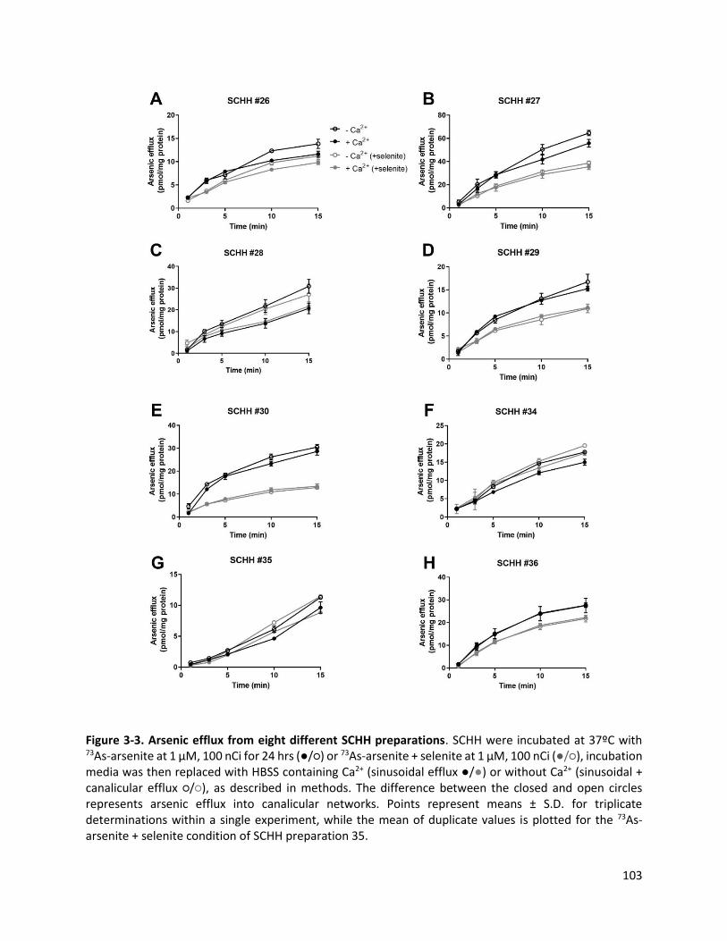
103
Figure 3-3. Arsenic efflux from eight different SCHH preparations. SCHH were incubated at 37ºC with 73As-arsenite at 1 µM, 100 nCi for 24 hrs (●/○) or 73As-arsenite + selenite at 1 µM, 100 nCi (●/○), incubation media was then replaced with HBSS containing Ca2+ (sinusoidal efflux ●/●) or without Ca2+ (sinusoidal + canalicular efflux ○/○), as described in methods. The difference between the closed and open circles represents arsenic efflux into canalicular networks. Points represent means ± S.D. for triplicate determinations within a single experiment, while the mean of duplicate values is plotted for the 73As-arsenite + selenite condition of SCHH preparation 35.

104
3.4.3. Arsenic Biliary Efflux is GSH-dependent
Since inorganic As and methylated metabolites can form GSH conjugates and be effluxed from cells
by the MRPs; GSH-dependence of As efflux in the presence of selenite was examined. Consistent
with Figure 3-3, where As efflux is inhibited in the presence of selenite, and this inhibition is further
pronounced in the presence of BSO, an inhibitor of the rate-limiting step of GSH synthesis. In SCHH
preparation 28, which shows biliary efflux of As in the presence and absence of selenite, GSH
depletion eliminates all efflux into bile. Sinusoidal efflux of As was also reduced in the presence of
BSO with a mean 61 ± 6 % (mean ± SD, n=4) reduction in efflux at 10 min in the presence of Ca2+
in the 73As-arsenite only condition, and mean 64 ± 7 % (mean ± SD, n=4) reduction in efflux in the
73As-arsenite + selenite condition, across 4 preparations (SCHH preparations 27-30) (Figure 3-4).
These results suggest that As efflux, in the presence and absence of selenite, across the sinusoidal and
canalicular membranes is GSH-dependent.

105
Figure 3-4. The effect of GSH-depletion on arsenic efflux was assessed from four different SCHH preparations pretreated with 0.5 mM BSO for 48 hrs. SCHH were incubated at 37ºC with (A,C,E,G) 73As-arsenite or (B,D,F,H) 73As-arsenite + selenite at 1 µM, 100 nCi for 24 hrs (●/○) or in the presence of BSO pretreatment (■/□), incubation media was then replaced with HBSS containing Ca2+ (sinusoidal efflux ●/■) or without Ca2+ (sinusoidal + canalicular efflux ○/□). The difference between the closed and open circles represents arsenic efflux into canalicular networks. Points represent means ± S.D. for triplicate determinations within a single experiment.

106
3.4.4. Arsenic Biliary Efflux is Increased or not Modified in the Presence of Selenide
Given the large body of evidence surrounding the mutual detoxification of arsenite and selenite, the
surprising lack of increase in As efflux in the presence of selenite led to the investigation of different
Se species and their effect on As efflux, especially the more reduced form, selenide, which may be the
predominant form of Se presented to the liver [38,47].
Sinusoidal and canalicular efflux of As in the presence of selenite alongside selenide was investigated
in 3 SCHH preparations (SCHH 34, 35 and 36). Consistent with SCHH preparations presented in
Figure 3-3, selenite inhibited As efflux or did not alter it. In the presence of selenide, As efflux in the
presence of Ca2+ (sinusoidal efflux), showed a similar trend to selenite, where sinusoidal As efflux was
either unchanged (SCHH 34 and 35) or inhibited by 31% (SCHH 36) (Figure 3-5A to 3-5C). Unlike
selenite (where biliary efflux of As was reduced), selenide treatment did not the change As biliary
efflux in preparations which were able to efflux As in 73As-arsenite alone treatment conditions (Figure
3-5A and 3-5C). Interestingly, upon treatment with 73As-arsenite alone, SCHH preparation 36 showed
no biliary efflux, whereas when treated with selenide and 73As-arsenite, biliary efflux of As was
observed with a BEI value of 11% (Figure 3-5B).
SeMet and MeSeCys are components of selenized yeast which has been used in As-Se studies and has
shown to have beneficial effects. The influence of these Se species on As sinusoidal efflux was
investigated. Preliminary data shows no difference between As and SeMet sinusoidal efflux, while
MeSeCys increased As sinusoidal efflux by 12% (Figure 3-5D).

107
Figure 3-5. Influence of selenium species on sinusoidal and canalicular arsenic efflux from SCHH. SCHH were incubated at 37ºC with 73As-arsenite ± selenium (1 µM, 100 nCi) for 24 hrs. (A-C) The effects of selenite and selenide on arsenic efflux from SCHH preparations 34, 35 and 36 was measured in the presence (sinusoidal efflux, closed bars) and absence (sinusoidal + canalicular efflux, open bars) of calcium. (D) The effects of selenomethionine (SeMet) and methyl selenocysteine (MeSeCys) on only arsenic sinusoidal efflux from SCHH preparation 34 was measured (i.e., in the presence of calcium Ca2+). Bars represent means ± SD of triplicate determinations at 10 min in a single experiment. (while the mean of duplicate values are plotted for the 73As-arsenite + selenite condition of SCHH preparation 35). BEI values were only calculated when the -Ca2+ condition was significantly higher (P<0.05, Student’s t-test) than the +Ca2+ condition for triplicate determinations from a single experiment.
3.4.5. Preliminary Data Suggest Selenide Accumulation is Higher than Selenite by SCHH
In order to investigate whether poor selenite uptake resulted in the lack of stimulation of As efflux,
SCHH accumulation studies were carried out to investigate uptake differences between selenite and

108
selenide. It has been shown that Se metabolism takes place in RBCs, resulting in selenite reduction to
selenide [48]. It is likely then that selenide is predominantly the species presented to the liver for uptake
into hepatocytes. Consistent with this, over a 24 hr period, our experiments showed that the amount
of 75Se-selenide accumulation in SCHH is at least 59% higher than 75Se-selenite (Figure 3-6A and 3-
6B). These results are consistent with previous reports in yeast, human keratinocytes, lung cancer cell
lines and trout hepatocytes, as well as in HepG2 cells (Chapter 4), where it has been shown that
selenide is more efficiently taken up into cells, in comparison with selenite [37,39,41,43,49].
Investigation into the effect of As uptake on Se accumulation showed a trend towards higher levels
of Se present in cells when exposed to As in SCHH 35 (Figure 3-6A), with differences in Se
accumulation between the selenite + arsenite and selenide + arsenite treatment conditions in SCHH
36 (Figure 3-6B), likely due to differences in selenite vs selenide uptake.
Figure 3-6. Accumulation differences between 75Se-selenite and 75Se-selenide, in the presence of arsenite. SCHH were incubated at 37ºC with 75Se-selenite or 75Se-selenide ± arsenite (1 µM, 100 nCi) for 24 hrs. Accumulation was measured from SCHH preparations 35 (A) and 36 (B). Bars represent means ± SD of triplicate determinations at 10 min in a single experiment. (while the mean of duplicate values is plotted for SCHH preparation 35).
3.4.6. Preliminary Data Suggest Selenium Efflux is Increased in the Presence of Arsenic
Based on the increased accumulation of selenium when SCHH are treated with selenide compared to
selenite, and the potential for increased biliary efflux of As in the presence of selenide, Se efflux in the
presence and absence of arsenite in two SCHH preparations was investigated. Total efflux of Se (±
Ca2+, across both the sinusoidal and canalicular surfaces) was increased in the presence of arsenite

109
across both preparations (28% increase in SCHH 35 and 17% increase in SCHH 36) (open bars, Figure
3-7A and 3-7B). 75Se efflux experiments in SCHH preparation 35 showed a 10% increase in sinusoidal
efflux of Se in the presence of arsenite (compare closed bars Figure 3-7A). Co-treatment with arsenite
also resulted in the biliary efflux of 75Se with a BEI value changing from 0% to 18% (Figure 3-7A).
Figure 3-7. Influence of arsenite on sinusoidal and canalicular 75Se-selenide efflux from SCHH. SCHH were incubated at 37ºC with 75Se-selenide ± arsenite (1 µM, 100 nCi) for 24 hrs. The effects of arsenite on 75Se-selenide efflux from SCHH preparations 35 (A) and 36 (B) was measured in the presence (sinusoidal efflux, closed bars) and absence (sinusoidal + canalicular efflux, open bars) of calcium. Bars represent means ± SD of triplicate determinations at 10 min in a single experiment. BEI values were only calculated when the -Ca2+ condition was significantly higher (P <0.05, Student’s t-test) than the +Ca2+ condition for triplicate determinations from a single experiment.

110
3.4.7. Preliminary Data Suggests Selenide Protects Against Arsenite Cytotoxicity Better than Selenite
Figure 3-8. Effect of arsenite, selenite and arsenite + selenite on the viability of SCHH. SCHH from four preparations, (A) SCHH 26 (B) SCHH 28 (C) SCHH 29 (D) SCHH 30 were incubated with arsenite, selenite and arsenite + selenite for 72 hrs. Cell viability was determined using a tetrazolium-based cytotoxicity assay and IC50 values were determined using the sigmoidal dose response fit (GraphPad Prism 6). Data points are means (± S.D.) of quadruplicate determinations within a single experiment.
In addition to uptake and efflux, the effect of Se on As cytotoxicity was also investigated for 5 SCHH
preparations (Figure 3-8 and 3-9). In almost all preparations (SCHH 26, 29, 30, 34), selenite (arsenite
+ selenite mean IC50 22 ± 5 µM) did not protect against arsenite (mean IC50 29 ± 14 µM) toxicity
(Table 3-1, Figure 3-8A, C, D). Our results show that when there is biliary efflux of arsenic, from as
early as 3 min, selenite protects against arsenite cytotoxicity (arsenite IC50 10 µM; arsenite + selenite
IC50 26 µM) (SCHH 28, Figure 3-8B). This supports our hypothesis that increase in biliary efflux of
As in the presence of Se is a protective mechanism. In addition, selenide protected against arsenite-

111
induced cytotoxicity better than selenite (arsenite + selenite IC50 25 µM; arsenite + selenide IC50 109
µM) (Figure 3-9, Table 3-2).
Figure 3-9. Effect of arsenite, selenite, selenide, arsenite + selenite and arsenite + selenide on the viability of SCHH. SCHH from preparation 34 were incubated with (A) arsenite, selenite and arsenite + selenite or (B) arsenite, selenide and arsenite + selenide for 72 hrs. (C) Combinations of selenite + arsenite and selenide + arsenite shown in A and B, respectively are plotted together. Cell viability was determined using a tetrazolium-based cytotoxicity assay and IC50 values were determined using the sigmoidal dose response fit (GraphPad Prism 6). Data points are means (± S.D.) of quadruplicate determinations within a single experiment.

112
Table 3-2. IC50 values for arsenite, selenite, selenide, and a combination of arsenite and selenium species in SCHH cells after 24 hrs of exposure.
SCHH preparation no. IC50 (µM)
Arsenite selenite arsenite +
selenite
selenide arsenite + selenide
26 31 66 15 - -
28 10 100 26 - -
29 45 38 23 - -
30 28 63 22 - -
Mean IC50 ± S.D. 29 ± 14 67 ± 25* 22 ± 5
34 25 > 300 25 > 300 109
*P < 0.05 compared with arsenite + selenite (analysis of variance followed by a Tukey’s multiple comparison test).

113
3.5. Discussion
The molecular basis for the mutual antagonism of toxicity between As and Se is through the formation
of [(GS)2AsSe]−, which is thought to increase As fecal elimination [15,18,19,50]. In the current study,
it has been shown that As efflux from SCHH is predominantly sinusoidal which is consistent with
previously published data [11]. Arsenic biliary efflux, in the presence and absence of selenite was found
to be GSH-dependent. Unexpectedly, we found that selenite either decreased As biliary efflux or had
no effect on it. In contrast, selenide, expected to be the predominant form of inorganic Se presented
to the liver either increased or did not alter As biliary efflux. Further characterization of differences in
selenite and selenide accumulation revealed selenide accumulation in SCHH was higher. Se efflux was
increased in the presence of arsenite, when SCHH were treated with 75Se-selenide and arsenite. In
corroboration of selenide being the form of Se that likely interacts with As, cytotoxicity assays revealed
that selenide decreased arsenite-induced toxicity, while selenite did not.
Consistent with urinary excretion being the major route for As elimination, As efflux from hepatocytes
takes place predominantly across the sinusoidal surface to reach the kidneys via the systemic
circulation. Methylated arsenicals comprise the majority of As species present in urine, with more than
60% being present as dimethylated arsenic [7,9]. Hepatocytes methylate inorganic As to produce
DMAs and MMAs in the same proportions as found in urinary analysis [51,52].
Selenite has been shown to be an inhibitor of As methylation, in both rat and human hepatocytes
[53,54]. Furthermore, previous studies have shown that rat hepatocyte exposure to selenite leads to
increased accumulation of arsenite [55]. Consistent with these studies, our results show selenite leads
to inhibition of sinusoidal efflux of As. This could be due in part to the inhibition of As methylation
by Se. MRP4/ABCC4 is present on the sinusoidal surface of the hepatocyte and in vitro transport
assays have shown MRP4 to mediate the transport of the di-methylated and mono-methylated arsenic

114
metabolites, DMAV and monomethylarsenic diglutathione (MMA(GS)2) [12]. Roggenbeck et al., have
shown through arsenic speciation studies that both methylated and inorganic As forms are effluxed
across the sinusoidal surface of the hepatocyte, with the majority of As present as inorganic As [11].
Thus, inhibition of methylation only provides a potential partial explanation for the decrease of As
sinusoidal efflux in the presence of selenite.
Selenium has been investigated as an important modifier of As metabolism and toxicity; protecting
against As-induced toxicity by forming a glutathione complex with As, [(GS)2AsSe]− [18,56,57]. In vitro
efflux of [(GS)2AsSe]− has been shown to be mediated by the human multidrug resistance protein 2
(MRP2), which is located on the canalicular surface of the hepatocyte, allowing efflux of this conjugate
into bile [13]. Surprisingly, our SCHH efflux data shows that biliary efflux of As was inhibited in the
presence of selenite, across all preparations that displayed the capacity for As biliary efflux. In vivo
studies with WT and Mrp2-deficient Wistar rats showed that Mrp2 is responsible for almost all of
arsenic efflux into bile in the forms of arsenic triglutathione (As(GS)3) and MMA(GS)2 [14].
[(GS)2AsSe]−, has been identified in rat and rabbit bile [15,19,50].
Consistent with published data in Roggenbeck et al., GSH depletion results in a complete loss of As
biliary efflux from SCHH [11]. This is also the case in arsenite + selenite treatment conditions,
suggesting As efflux into bile is GSH-dependent. MRP2 is highly expressed on the canalicular surface
of the hepatocytes and in this location is the only known protein which transports As GSH-conjugates
[14]. This supports the observed loss of biliary efflux of As in the absence of GSH, both in the
presence and absence of selenite.
Depletion of glutathione does not completely inhibit As sinusoidal efflux due to the potential for
efflux of non-glutathionylated metabolites of As (such as DMAV) by transport proteins like MRP4
and aquaglyceroporin 9 (AQP9) which is a channel shown to allow permeation of neutral species of

115
As, such as arsenite and MMAIII, down their concentration gradient [12,58,59]. Rat studies have also
shown a 50% decrease in urinary elimination of As when administered BSO to deplete GSH synthesis
[60].
After oral ingestion of selenite, it is thought to cross the intestinal epithelium paracellularly and
transcellularly [61]. During transcellular passage some reduction of selenite to selenide might occur in
the enterocyte, while selenite transported paracellularly will reach the portal blood, be taken up by
RBCs and rapidly reduced to selenide [48] . As such, the form of Se that reached the liver is likely
selenide. Indeed, we see either an increased biliary efflux of As in the presence of selenide or at
minimum, no change in the biliary efflux (Figure 3-5A to C). Investigation of accumulation differences
between these two species, showed selenide uptake to be significantly higher than selenite (Figure 3-
6). This is consistent with other studies demonstrating increased selenide uptake in trout hepatocytes
and HepG2 cells (Chapter 4) [37,39].
Previous studies have shown that organic forms of selenium have higher bioavailability and are thus
preferred for use in therapeutic applications [62–64]. Among commercially available selenium
supplements, selenized yeast (major component, SeMet) and MeSeCys are common components
identified on manufacturer’s labels [28,65,66]. In small population studies, treatment of As-exposed
individuals with selenized yeast led to lower levels of As in hair, urine and blood [22]. In vitro studies
with human osteosarcoma cells showed SeMet and MeSeCys blocked As-induced mutagenesis [67].
All Se sources are metabolized to a selenide pool before being incorporated into selenoproteins or
cellular reactions, while SeMet, as a Se analogue of methionine, is also incorporated into proteins non-
specifically [25,26,68–70]. Suzuki et al., show SeMet and MeSeCys to be transported to target organs
in their intact forms in rats [71]. SeMet undergoes trans-selenation to form selenocysteine, which then
forms selenide in cells, while MeSeCys is first transformed into methyl selenol, followed by

116
demethylation to form selenide [25,42,72,73]. When undergoing transformation to selenide, not only
do these seleno-amino acids not compete for methylation, but MeSeCys conversion to selenide
releases a methyl group [25,26]. Preliminary data shows sinusoidal As efflux is increased in the
presence of MeSeCys (Figure 3-5D), possibly due to increased levels of demethylated and
monomethylated As available for efflux.
Previous studies have also investigated the effects of As on Se efflux. It was shown that Se levels in
bile increased from 4% of total Se dose to 41% of total Se dose in the presence of As [21]. Consistent
with these in vivo observations, SCHH experiments with 75Se-selenide in the presence and absence of
arsenite, revealed an increase in biliary efflux in SCHH preparation 35 (Figure 3-7A). Preliminary data
from a limited study in Bangladesh show increased urinary and fecal Se levels in 5 participants that
were exposed to arsenic in drinking water [74]. These participants were accommodated in a clinic for
10 days and fed a fixed diet with drinking water from their homes and received a dose of 77Se-selenite
or a placebo on the 6th day. This preliminary data is consistent with the observed increase in sinusoidal
and canalicular efflux of 75Se in the presence of arsenite in the present study.
While epidemiological studies and in vivo work with animal models show a protective effect of selenite
on arsenite-induced toxicity, Styblo et al., reported that in vitro selenite (2 µM) increased the cytotoxicity
of arsenite (10 µM) in rat hepatocytes [22,30,31,55,75,76]. Cytotoxicity assays show that in 3 out 5
SCHH preparations, selenite decreases the IC50 of arsenite (Figure 3-8). This is likely due to the
inhibition of As efflux in the presence of selenite, leading to the increased retention of toxic As species.
Preliminary results show that co-treatment of arsenite with selenide (IC50 109 µM) leads to a higher
IC50 than arsenite treatment alone (IC50 25 µM), and arsenite plus selenite treatment (IC50 25 µM)
(Figure 3-9). This led us to explore the differences between selenide and selenite in HepG2 cells
(Chapter 4) using a readily available model system.

117
This work shows that consideration of the physiological relevance of the species of Se for better design
of experimental studies is required. The reduction of selenite to selenide, means that this is likely the
form of Se presented to the liver, resulting in its increased uptake in comparison to selenite. In
addition, using primary human hepatocytes, this work has shown that there are inter-individual
differences in the cytotoxicity and efflux of As and Se. The evidence of direct interaction between As
and Se through the [(GS)2AsSe]− conjugate, has led to clinical trials and small population studies in
arsenic-endemic regions [22,32,77]. The complex nature of interactions between As and Se require
careful extrapolation of in vivo and in vitro studies.
118
3.6. Bibliography
[1] B.C. Minatel, A.P. Sage, C. Anderson, R. Hubaux, E.A. Marshall, W.L. Lam, V.D. Martinez,
Environmental arsenic exposure: From genetic susceptibility to pathogenesis, Environ. Int.
112 (2018) 183–197. https://doi.org/10.1016/j.envint.2017.12.017.
[2] M. Tolins, M. Ruchirawat, P. Landrigan, The developmental neurotoxicity of arsenic:
Cognitive and behavioral consequences of early life exposure, Ann. Glob. Heal. 80 (2014)
303–314. https://doi.org/10.1016/j.aogh.2014.09.005.
[3] T.-H. Lin, Y.-L. Huang, Y. Ming, Arsenic species in drinking water, hair, fingernails and urine
of patients with blackfoot disease, J. Toxicol. Environ. Heal. Part A. 53 (1998) 85–93.
https://doi.org/10.1080/009841098159376.
[4] F. Challenger, Biological methylation., Sci. Prog. 35 (1947) 396–416.
https://doi.org/10.1039/qr9550900255.
[5] K. Rehman, H. Naranmandura, Arsenic metabolism and thioarsenicals, Metallomics. 4 (2012)
881. https://doi.org/10.1039/c2mt00181k.
[6] E.M. Leslie, Arsenic-glutathione conjugate transport by the human multidrug resistance
proteins (MRPs/ABCCs), J. Inorg. Biochem. 108 (2012) 141–149.
https://doi.org/10.1016/j.jinorgbio.2011.11.009.
[7] B.K. Mandal, Y. Ogra, K.T. Suzuki, Identification of dimethylarsinous and
monomethylarsonous acids in human urine of the arsenic-affected areas in West Bengal,
India, (2001). https://doi.org/10.1021/TX000246H.
[8] M. Vahter, Variation in human metabolism of arsenic, Arsen. Expo. Heal. Eff. III. (1999)
267–279. https://doi.org/10.1016/B978-008043648-7/50031-5.
[9] X.C. Le, X. Lu, M. Ma, W.R. Cullen, H. V. Aposhian, B. Zheng, Speciation of key arsenic
metabolic intermediates in human urine, Anal. Chem. 72 (2000) 5172–5177.
https://doi.org/10.1021/AC000527U.
[10] M. Vahter, G. Concha, Role of metabolism in arsenic toxicity., Pharmacol. Toxicol. 89 (2001)
1–5. https://doi.org/10.1111/j.1600-0773.2001.890101.x.

119
[11] B.A. Roggenbeck, M.W. Carew, G.J. Charrois, D.N. Douglas, N.M. Kneteman, X. Lu, X.C.
Le, E.M. Leslie, Characterization of arsenic hepatobiliary transport using sandwich-cultured
human hepatocytes, Toxicol. Sci. 145 (2015) 307–320.
https://doi.org/10.1093/toxsci/kfv051.
[12] M. Banerjee, M.W. Carew, B.A. Roggenbeck, B.D. Whitlock, H. Naranmandura, X.C. Le,
E.M. Leslie, A novel pathway for arsenic elimination : Human multidrug resistance protein 4 (
MRP4 / ABCC4 ) mediates cellular export of dimethylarsinic acid ( DMA V ) and the
diglutathione conjugate of monomethylarsonous acid ( MMA III ), Mol. Pharmacol. 86
(2014) 168–179.
[13] M.W. Carew, E.M. Leslie, Selenium-dependent and -independent transport of arsenic by the
human multidrug resistance protein 2 (MRP2/ABCC2): implications for the mutual
detoxification of arsenic and selenium, Carcinogenesis. 31 (2010) 1450–1455.
https://doi.org/10.1093/carcin/bgq125.
[14] S. V Kala, M.W. Neely, G. Kala, C.I. Prater, D.W. Atwood, J.S. Rice, M.W. Lieberman, The
MRP2/cMOAT transporter and arsenic-glutathione complex formation are required for
biliary excretion of arsenic., J. Biol. Chem. 275 (2000) 33404–8.
https://doi.org/10.1074/jbc.M007030200.
[15] J. Gailer, S. Madden, G.A. Buttigieg, M.B. Denton, H.S. Younis, Identification of
[(GS)2AsSe]- in rabbit bile by size-exclusion chromatography and simultaneous multielement-
specific detection by inductively coupled plasma atomic emission spectroscopy, Appl.
Organomet. Chem. 16 (2002) 72–75. https://doi.org/10.1002/aoc.260.
[16] Y. Shiobara, Y. Ogra, K.T. Suzuki, Animal species difference in the uptake of
dimethylarsinous acid ( DMA III ) by red blood cells, Chem. Res. Toxicol. 14 (2001) 1446–
1452.
[17] C.G. Dietrich, R. Ottenhoff, D.R. de Waart, R.P.. Oude Elferink, Role of MRP2 and GSH in
intrahepatic cycling of toxins, Toxicology. 167 (2001) 73–81. https://doi.org/10.1016/S0300-
483X(01)00459-0.
[18] J. Gailer, G.N. George, I.J. Pickering, R.C. Prince, S.C. Ringwald, J.E. Pemberton, R.S. Glass,
H.S. Younis, D.W. DeYoung, H. Vasken Aposhian, A metabolic link between arsenite and

120
selenite: The seleno-bis(S-glutathionyl) arsinium Ion, J. Am. Chem. Soc. 122 (2000) 4637–
4639. https://doi.org/10.1021/ja993064m.
[19] G.N. George, J. Gailer, O. Ponomarenko, P.F. La Porte, K. Strait, M. Alauddin, H. Ahsan, S.
Ahmed, J. Spallholz, I.J. Pickering, Observation of the seleno bis-(S-glutathionyl) arsinium
anion in rat bile, J. Inorg. Biochem. 2 (2016) 1–6.
https://doi.org/10.1016/j.jinorgbio.2016.01.022.
[20] O.A. Levander, C.A. Baumann, Selenium metabolism VI. Effect of arsenic on the excretion
of selenium in the bile, Toxicol. Appl. Pharmacol. 9 (1966) 106–115.
https://doi.org/https://doi-org.login.ezproxy.library.ualberta.ca/10.1016/0041-
008X(66)90035-4.
[21] O.A. Levander, Metabolic interrelationships between arsenic and selenium., Environ. Health
Perspect. 19 (1977) 159–64.
http://www.pubmedcentral.nih.gov/articlerender.fcgi?artid=1637401&tool=pmcentrez&ren
dertype=abstract (accessed September 8, 2015).
[22] L. Yang, W. Wang, S. Hou, P.J. Peterson, W.P. Williams, Effects of selenium
supplementation on arsenism : an intervention trial in Inner Mongolia, Environ. Geochem.
Health. 24 (2002) 359–374.
[23] C.T. Su, R.L. Hsieh, C.J. Chung, P.T. Huang, Y.C. Lin, P.L. Ao, H.S. Shiue, W.J. Chen, S.R.
Huang, M.I. Lin, S.C. Mu, Y.M. Hsueh, Plasma selenium influences arsenic methylation
capacity and developmental delays in preschool children in Taiwan, Environ. Res. 171 (2019)
52–59. https://doi.org/10.1016/j.envres.2019.01.003.
[24] S. Sah, A. Vandenberg, J. Smits, Treating chronic arsenic toxicity with high selenium lentil
diets, Toxicol. Appl. Pharmacol. 272 (2013) 256–262.
https://doi.org/10.1016/j.taap.2013.06.008.
[25] C.M. Weekley, H.H. Harris, Which form is that? the importance of selenium speciation and
metabolism in the prevention and treatment of disease, Chem. Soc. Rev. 42 (2013) 8870–
8894. https://doi.org/10.1039/c3cs60272a.
[26] B. Gammelgaard, M.I. Jackson, C. Gabel-Jensen, Surveying selenium speciation from soil to
cell-forms and transformations, Anal. Bioanal. Chem. 399 (2011) 1743–1763.
121
https://doi.org/10.1007/s00216-010-4212-8.
[27] D. Thavarajah, A. Vandenberg, G.N. George, I.J. Pickering, Chemical form of selenium in
naturally selenium-rich lentils (Lens culinaris L.) from Saskatchewan, J. Agric. Food Chem. 55
(2007) 7337–7341. https://doi.org/10.1021/JF070681I.
[28] P.O. Amoako, P.C. Uden, J.F. Tyson, Speciation of selenium dietary supplements; formation
of S-(methylseleno)cysteine and other selenium compounds, Anal. Chim. Acta. 652 (2009)
315–323. https://doi.org/10.1016/j.aca.2009.08.013.
[29] M. Rayman, H. Infante, M. Sargent, Food-chain selenium and human health: Spotlight on
speciation., Br. J. Nutr. 100 (2008) 238–253.
https://doi.org/doi:10.1017/S0007114508922522.
[30] W.J. Verret, Y. Chen, A. Ahmed, T. Islam, F. Parvez, M.G. Kibriya, J.H. Graziano, H. Ahsan,
A randomized, double-blind placebo-controlled trial evaluating the effects of vitamin E and
selenium on arsenic-induced skin lesions in Bangladesh, J. Occup. Environ. Med. 47 (2005)
1026–1035. https://doi.org/10.1097/01.jom.0000183095.45050.97.
[31] M.G. Kibriya, F. Jasmine, M. Argos, W.J. Verret, M. Rakibuz-Zaman, A. Ahmed, F. Parvez,
H. Ahsan, Changes in gene expression profiles in response to selenium supplementation
among individuals with arsenic-induced pre-malignant skin lesions, Toxicol. Lett. 169 (2007)
162–176. https://doi.org/10.1016/j.toxlet.2007.01.006.
[32] J.E. Smits, R.M. Krohn, E. Akhtar, S.K. Hore, M. Yunus, A. Vandenberg, R. Raqib, Food as
medicine: Selenium enriched lentils offer relief against chronic arsenic poisoning in
Bangladesh, Environ. Res. 176 (2019) 108561.
https://doi.org/10.1016/j.envres.2019.108561.
[33] X. Liu, E.L. LeCluyse, K.R. Brouwer, R.M. Lightfoot, J.I. Lee, K.L. Brouwer, Use of Ca2+
modulation to evaluate biliary excretion in sandwich-cultured rat hepatocytes., J. Pharmacol.
Exp. Ther. 289 (1999) 1592–1599.
[34] B. Swift, N.D. Pfeifer, K.L.R. Brouwer, Sandwich-cultured hepatocytes: an in vitro model to
evaluate hepatobiliary transporter-based drug interactions and hepatotoxicity., Drug Metab.
Rev. 42 (2010) 446–71. https://doi.org/10.3109/03602530903491881.

122
[35] G. Ghibellini, L.S. Vasist, E.M. Leslie, W.D. Heizer, R.J. Kowalsky, B.F. Calvo, K.L.R.
Brouwer, In vitro–in vivo correlation of hepatobiliary drug clearance in humans, Clin.
Pharmacol. Ther. 81 (2007) 406–413. https://doi.org/10.1038/sj.clpt.6100059.
[36] X. Liu, J.P. Chism, E.L. Lecluyse, K.R. Brouwer, K.I.M.L.R. Brouwer, Correlation of biliary
excretion in sandwich-cultured rat hepatocytes and in vivo in rats, Drug Metab. Dispos. 27
(1999) 637–644.
[37] S. Misra, R.W.M. Kwong, S. Niyogi, Transport of selenium across the plasma membrane of
primary hepatocytes and enterocytes of rainbow trout, J. Exp. Biol. 215 (2012) 1491–1501.
https://doi.org/10.1242/jeb.062307.
[38] M. Haratake, M. Hongoh, M. Miyauchi, R. Hirakawa, M. Ono, M. Nakayama, Albumin-
mediated selenium transfer by a selenotrisulfide relay mechanism, Inorg. Chem. 47 (2008)
6273–6280. https://doi.org/10.1021/ic800310j.
[39] G. Kaur, O. Ponomarenko, J.R. Zhou, D.P. Swanlund, K.L. Summers, N. V. Dolgova, O.
Antipova, I.J. Pickering, G.N. George, E.M. Leslie, Studies of selenium and arsenic mutual
detoxification in human HepG2 cells, (Submitted).
[40] P.F. Reay, C.J. Asher, Preparation and purification of 74As-labeled arsenate and arsenite for
use in biological experiments, Anal. Biochem. 78 (1977) 557–560.
https://doi.org/10.1016/0003-2697(77)90117-8.
[41] D. Ganyc, W.T. Self, High affinity selenium uptake in a keratinocyte model, FEBS Lett. 582
(2008) 299–304. https://doi.org/10.1016/j.febslet.2007.12.022.
[42] H.E. Ganther, Reduction of the selenotrisulfide derivative of glutathione to a persulfide
analog by glutathione reductase, Biochemistry. 10 (1971) 4089–4098.
https://doi.org/10.1021/bi00798a013.
[43] A. Tarze, M. Dauplais, I. Grigoras, M. Lazard, N.-T. Ha-Duong, F. Barbier, S. Blanquet, P.
Plateau, Extracellular production of hydrogen selenide accounts for thiol-assisted toxicity of
selenite against Saccharomyces cerevisiae, J. Biol. Chem. 282 (2007) 8759–8767.
https://doi.org/10.1074/JBC.M610078200.
[44] M.W. Carew, H. Naranmandura, C.B. Shukalek, X.C. Le, E.M. Leslie, Monomethylarsenic
123
diglutathione transport by the human multidrug resistance protein 1 ( MRP1 / ABCC1 ),
Drug Metab. Dispos. 39 (2011) 2298–2304. https://doi.org/10.1124/dmd.111.041673.ing.
[45] D.A.J. Bow, J.L. Perry, D.S. Miller, J.B. Pritchard, K.L.R. Brouwer, Localization of P-gp
(Abcb1) and Mrp2 (Abcc2) in freshly isolated rat hepatocytes., Drug Metab. Dispos. 36
(2008) 198–202. https://doi.org/10.1124/dmd.107.018200.
[46] M.M. Malinen, K. Ito, H.E. Kang, P. Honkakoski, K.L.R. Brouwer, Protein expression and
function of organic anion transporters in short-term and long-term cultures of Huh7 human
hepatoma cells, Eur. J. Pharm. Sci. 130 (2019) 186–195.
https://doi.org/10.1016/j.ejps.2019.01.022.
[47] M. Sandholm, Function of erythrocytes in attaching selenite-Se onto specific plasma proteins,
Acta Pharmacol. Toxicol. (Copenh). 36 (1975) 321–327. https://doi.org/10.1111/j.1600-
0773.1975.tb00799.x.
[48] Y. Shiobara, K.T. Suzuki, Binding of selenium (administered as selenite) to albumin after
efflux from red blood cells, J. Chromatogr. B Biomed. Appl. 710 (1998) 49–56.
https://doi.org/10.1016/S0378-4347(98)00142-X.
[49] E. Olm, A.P. Fernandes, C. Hebert, A.-K. Rundlöf, E.H. Larsen, O. Danielsson, M.
Björnstedt, Extracellular thiol-assisted selenium uptake dependent on the x(c)- cystine
transporter explains the cancer-specific cytotoxicity of selenite., Proc. Natl. Acad. Sci. U. S. A.
106 (2009) 11400–5. https://doi.org/10.1073/pnas.0902204106.
[50] J. Gailer, G.N. George, I.J. Pickering, R.C. Prince, H.S. Younis, J.J. Winzerling, Biliary
excretion of [(GS) 2 AsSe] - after intravenous injection of rabbits with arsenite and selenate,
Chem. Res. Toxicol. 15 (2002) 1466–1471. https://doi.org/10.1021/tx025538s.
[51] M. Styblo, L.M. Del Razo, E.L. LeCluyse, G.A. Hamilton, C. Wang, W.R. Cullen, D.J.
Thomas, Metabolism of arsenic in primary cultures of human and rat hepatocytes, Chem.
Res. Toxicol. 12 (1999) 560–565. https://doi.org/10.1021/TX990050L.
[52] Z. Drobná, S.B. Waters, F.S. Walton, E.L. LeCluyse, D.J. Thomas, M. Stýblo, Interindividual
variation in the metabolism of arsenic in cultured primary human hepatocytes, Toxicol. Appl.
Pharmacol. 201 (2004) 166–177. https://doi.org/10.1016/J.TAAP.2004.05.004.

124
[53] I. Csanaky, Z. Gregus, Effect of selenite on the disposition of arsenate and arsenite in rats,
Toxicology. 186 (2003) 33–50. www.elsevier.com/locate/toxicol (accessed August 1, 2019).
[54] F.S. Walton, S.B. Waters, S.L. Jolley, E.L. LeCluyse, D.J. Thomas, M. Styblo, Selenium
compounds modulate the activity of recombinant rat AsIII-methyltransferase and the
methylation of arsenite by rat and human hepatocytes, Chem. Commun. 16 (2003) 261–265.
https://doi.org/10.1021/TX025649R.
[55] M. Styblo, D.J. Thomas, Selenium modifies the metabolism and toxicity of arsenic in primary
rat hepatocytes, Toxicol. 172 (2001) 52–61. https://doi.org/10.1006/taap.2001.9134.
[56] S. a Manley, G.N. George, I.J. Pickering, R.S. Glass, E.J. Prenner, R. Yamdagni, Q. Wu, J.
Gailer, The seleno bis(S-glutathionyl) arsinium ion is assembled in erythrocyte lysate., Chem.
Res. Toxicol. 19 (2006) 601–7. https://doi.org/10.1021/tx0503505.
[57] J. Gailer, Chronic toxicity of AsIII in mammals: The role of (GS)2AsSe-, Biochimie. 91
(2009) 1268–1272. https://doi.org/10.1016/j.biochi.2009.06.004.
[58] J.R. McDermott, X. Jiang, L.C. Beene, B.P. Rosen, Z. Liu, Pentavalent methylated arsenicals
are substrates of human AQP9, BioMetals. 23 (2010) 119–127.
https://doi.org/10.1007/s10534-009-9273-9.
[59] Z. Liu, J. Shen, J.M. Carbrey, R. Mukhopadhyay, P. Agre, B.P. Rosen, Arsenite transport by
mammalian aquaglyceroporins AQP7 and AQP9, 99 (2002).
[60] S. V. Kala, G. Kala, C.I. Prater, A.C. Sartorelli, M.W. Lieberman, Formation and urinary
excretion of arsenic triglutathione and methylarsenic diglutathione, Chem. Res. Toxicol. 17
(2004) 243–249. https://doi.org/10.1021/tx0342060.
[61] H. Zeng, M.I. Jackson, W.H. Cheng, G.F. Combs, Chemical form of selenium affects its
uptake, transport, and glutathione peroxidase activity in the human intestinal Caco-2 cell
model, Biol. Trace Elem. Res. 143 (2011) 1209–1218. https://doi.org/10.1007/s12011-010-
8935-3.
[62] C. Wang, R.T. Lovell, Organic selenium sources, selenomethionine and selenoyeast, have
higher bioavailability than an inorganic selenium source, sodium selenite, in diets for channel
catfish (Ictalurus punctatus), Aquaculture. 152 (1997) 223–234.

125
https://doi.org/10.1016/S0044-8486(96)01523-2.
[63] Y. Wang, L. Fu, Forms of selenium affect its transport, uptake and glutathione peroxidase
activity in the caco-2 cell model, Biol. Trace Elem. Res. 149 (2012) 110–116.
https://doi.org/10.1007/s12011-012-9395-8.
[64] X. Zhan, M. Wang, R. Zhao, W. Li, Z. Xu, Effects of different selenium source on selenium
distribution, loin quality and antioxidant status in finishing pigs, Anim. Feed Sci. Technol. 132
(2007) 202–211. https://doi.org/10.1016/J.ANIFEEDSCI.2006.03.020.
[65] A.E. Veatch, J.D. Brockman, V.L. Spate, J.D. Robertson, J.S. Morris, Selenium and nutrition:
The accuracy and variability of the selenium content in commercial supplements, J.
Radioanal. Nucl. Chem. 264 (2005) 33–38. https://doi.org/10.1007/s10967-005-0671-3.
[66] A. Połatajko, M. Śliwka-Kaszyńska, M. Dernovics, R. Ruzik, J. Ruiz Encinar, J. Szpunar, A
systematic approach to selenium speciation in selenized yeast, J. Anal. At. Spectrom. 19
(2004) 114–120. https://doi.org/10.1039/B308756P.
[67] T.G. Rossman, A.N. Uddin, Selenium prevents spontaneous and arsenite-induced
mutagenesis, Int. Congr. Ser. 1275 (2004) 173–179.
https://doi.org/10.1016/J.ICS.2004.09.038.
[68] H.E. Ganther, Selenium metabolism, selenoproteins and mechanisms of cancer prevention:
complexities with thioredoxin reductase., Carcinogenesis. 20 (1999) 1657–66.
https://doi.org/10.1093/carcin/20.9.1657.
[69] S.E. Jackson-Rosario, W.T. Self, Targeting selenium metabolism and selenoproteins: novel
avenues for drug discovery., Metallomics. 2 (2010) 112–6. https://doi.org/10.1039/b917141j.
[70] R.F. Burk, K.E. Hill, Regulation of selenium metabolism and transport, Annu. Rev. Nutr. 35
(2015) 109–134. https://doi.org/10.1146/annurev-nutr-071714-034250.
[71] K.T. Suzuki, C. Doi, N. Suzuki, Metabolism of 76Se-methylselenocysteine compared with
that of 77Se-selenomethionine and 82Se-selenite, Toxicol. Appl. Pharmacol. 217 (2006) 185–
195. https://doi.org/10.1016/j.taap.2006.09.006.
[72] N. Esaki, T. Nakamura, H. Tanaka, T. Suzuki, Y. Morino, K. Soda, Enzymic synthesis of
selenocysteine in rat liver, Biochemistry. 20 (1981) 4492–4496.

126
https://doi.org/10.1021/bi00518a039.
[73] J.N.M. Commandeur, I. Andreadou, M. Rooseboom, M. Out, L.J. de Leur, E. Groot, N.P.E.
Vermeulen, Bioactivation of selenocysteine se-Conjugates by a highly purified rat renal
cysteine conjugate β-lyase/glutamine transaminase K, J. Pharmacol. Exp. Ther. 294 (2000)
753–761.
[74] M. Alauddin, R. Cekovic, S. Alauddin, L. Bolevic, S. Saha, J.E. Spallholz, P.F. Laporte,
Pharmacodynamic study of the selenium-mediated arsenic excretion in arsenicosis patients in
Bangladesh, in: Y.-G. Zhu, H. Guo, P. Bhattacharya, J. Bundschuh, A. Ahmad, R. Naidu
(Eds.), Environ. Arsen. a Chang. World, CRC Press, 2019.
https://doi.org/10.1201/9781351046633.
[75] Z. Huang, Q. Pei, G. Sun, S. Zhang, J. Liang, Y. Gao, X. Zhang, Low selenium status affects
arsenic metabolites in an arsenic exposed population with skin lesions, Clin. Chim. Acta. 387
(2008) 139–144. https://doi.org/10.1016/j.cca.2007.09.027.
[76] S. Biswas, T. Geeta, A. Sharma, Prevention of cytotoxic effects of arsenic by short-term
dietary supplementation with selenium in mice in vivo RN - Mutation Research, vol. 441, pp.
155-160, Mutat. Res. 441 (1999) 155–160.
[77] Y. Chen, M. Hall, J.H. Graziano, V. Slavkovich, A. Van Geen, F. Parvez, H. Ahsan, A
prospective study of blood selenium levels and the risk of arsenic-related premalignant skin
lesions, Cancer Epidemiol. Biomarkers Prev. 16 (2007) 207–213.

127
4. Studies of Selenium and Arsenic Mutual Detoxification in
Human HepG2 Cells
This thesis chapter has been accepted for publication in Chemico-Biological Interactions: Gurnit
Kaur, Olena Ponomarenko, Janet R. Zhou, Diane P. Swanlund, Kelly L. Summers, Nataliya V.
Dolgova, Olga Antipova, Ingrid J. Pickering, Graham N. George, and Elaine M. Leslie. Studies of
selenium and arsenic mutual detoxification in human HepG2 cells. Chemico-biological Interactions
(2020).

128
4.1. Abstract
Hundreds of millions of people worldwide are exposed to unacceptable levels of carcinogenic
inorganic arsenic. Animal models have shown that selenium and arsenic are mutually protective
through the formation and elimination of the seleno-bis(S-glutathionyl) arsinium ion [(GS)2AsSe]−.
Consistent with this, human selenium deficiency in arsenic-endemic regions is associated with arsenic-
induced disease, leading to the initiation of human selenium supplementation trials. In contrast to the
protective effect observed in vivo, in vitro studies have suggested that selenite increases arsenite cellular
retention and toxicity. This difference might be explained by the rapid conversion of selenite to
selenide in vivo. In the current study, selenite did not protect the human hepatoma (HepG2) cell line
against the toxicity of arsenite at equimolar concentrations, however selenide increased the IC50 by
2.3-fold. Cytotoxicity assays of arsenite + selenite and arsenite + selenide at different molar ratios
revealed higher overall mutual antagonism of arsenite + selenide toxicity than arsenite + selenite.
Despite this protective effect, in comparison to 75Se-selenite, HepG2 cells in suspension were at least
3-fold more efficient at accumulating selenium from reduced 75Se-selenide, and its accumulation was
further increased by arsenite. X-ray fluorescence imaging of HepG2 cells also showed that arsenic
accumulation, in the presence of selenide, was higher than in the presence of selenite. These results
are consistent with a greater intracellular availability of selenide relative to selenite for protection
against arsenite, and the formation and retention of a less toxic product, possibly [(GS)2AsSe]−.

129
4.2. Introduction
Inorganic arsenic (As) is a Group I (proven) human carcinogen, found throughout the environment
[1]. Arsenic contamination of drinking water is a global problem, with more than 200 million people
exposed to levels higher than the World Health Organization standard of 10 µg/L [2]. In Bangladesh,
a nation-wide program to evade microbial contamination in surface water, resulted in the
contamination of well water through the disturbance of As-containing strata [3]. Chronic As
consumption can have adverse health consequences for decades after exposure cessation [4]. Poor
socioeconomic status of developing countries means cost-effective As exposure solutions are
required.
Selenium (Se) is an essential trace element required in human nutrition [5]. There is an intriguing
toxicity antagonism between some forms of As and Se, first observed decades ago when Se toxicity in
rats (from seleniferous grains), was reduced with the addition of As in the form of arsenite to drinking
water [6]. More recently, the seleno-bis(S-glutathionyl) arsinium ion [(GS)2AsSe]− was identified in the
bile of rabbits and rats [7–10]. In contrast to other As-glutathione (As-GSH) metabolites,
[(GS)2AsSe]− is more stable at alkaline biliary pH and is thought to break the enterohepatic
recirculation of As-GSH complexes (reviewed in [11]). [(GS)2AsSe]− biliary efflux is likely mediated
by the ATP-binding cassette transporter multidrug resistance protein 2 (MRP2/ABCC2) [11,12].
Further to its biliary elimination, [(GS)2AsSe]− is sequestered in red blood cells and, likely delays the
distribution of As and Se to tissues [13–16]
Low blood Se levels have been associated with a higher incidence of As-induced skin lesions [17,18].
Due to this correlation, and the evidence from animal studies, Se supplementation trials have been
initiated [19–22]. Initial research into Se supplementation with L-selenomethionine and selenized yeast
suggest that Se may improve As-induced toxicities [23–25]. Following suggestions from George,

130
Pickering and co-workers [26], a recent study supplementing a Bangladeshi population with lentils
grown in soil with a high Se content, showed higher urinary As elimination [27]. If successful, Se
supplementation has the potential to be a cost-effective solution for populations that are chronically
exposed to As. However, the interaction between As and Se, especially in humans, is not fully
understood.
Within cells, arsenate is reduced to arsenite and undergoes a series of reduction and oxidative
methylation steps. Inorganic Se biotransformation is similar to As and as such, Se competes with As
for methyl donors, decreasing As methylation [28]. In addition to decreasing the formation of the
highly toxic methylated trivalent arsenicals, Se also reacts with arsenite to form [(GS)2AsSe]− [29].
Most of the characterization of As and Se interactions have been done in laboratory animals dosed
with arsenite and selenite, and very little work has been done with human cell lines [30]. It was
established early on in rats that the mutual detoxification of arsenite and selenite was not due to a
reduced bioavailability of either compound [31]. In rats that had been dosed individually with arsenite
or selenite compared with a combination of both, it was found that arsenite and selenite together
resulted in lower retention levels of both compounds in the liver, and increased fecal elimination [32].
More recently, in rats injected with arsenite and selenite, all the As and Se detected in the bile was in
the form of [(GS)2AsSe]− [9].
There are inconsistencies between what happens in vitro and in vivo. For example, rats dosed with
arsenite and selenite are protected through increased biliary elimination of [(GS)2AsSe]−, however, in
vitro work with primary rat hepatocytes showed that selenite increased cellular retention of As,
decreased As methylation and slightly increased As toxicity [9,15,33,34]. Selenite is known to be rapidly
converted to selenide (HSe- at physiological pH) within cells, including red blood cells [35–37].
Selenide can also be formed non-enzymatically from selenite, in the presence of reduced glutathione

131
(GSH), however, extracellular GSH levels might not be sufficient for this reduction to occur [38,39].
Furthermore, it has been shown that selenide uptake is more efficient than selenite in yeast, trout
hepatocytes, lung cancer cells and keratinocytes [39–42]. This information has led us to hypothesize
that extra-hepatic formation of selenide may be necessary for sufficient hepatocyte uptake of Se for
protection against As. Thus, the aim of this study was to investigate the influence of selenite and
selenide on arsenite toxicity and cellular accumulation using the human hepatoma cell line HepG2.
HepG2 cells are a convenient and well characterized model, widely used in toxicology studies, and are
functional for As methylation [43–46]. HepG2 cells also have functional glutathione reductase and
thioredoxin reductases [47,48], enzymes important for the conversion of selenite to selenide [49,50].
4.3. Material and Methods
4.3.1. Chemicals and Reagents
73As-arsenate (1.2 mCi/ml) was purchased from Los Alamos Meson Production Facility (Los Alamos,
NM). 75Se-selenite (25.7 mCi/ml) was obtained from the University of Missouri Research Reactor
(MURR, Columbia, MO). Sodium selenite, sodium (meta)arsenite, bovine serum albumin (BSA),
glucose, sodium chloride, potassium chloride, magnesium sulphate heptahydrate, Tris base, 4-(2-
hydroxyethyl)-1-piperazineethanesulfonic acid (HEPES), reduced glutathione (GSH), and poly-L-
lysine solution were purchased from MilliporeSigma (Oakville, ON). Sodium selenide was purchased
from Alfa Aesar (Ward Hill, MA). Triton X-100, calcium chloride and paraformaldehyde was
purchased from Fisher Scientific (Fair Lawn, NJ). Silicon nitride windows (5x5 mm frame, 2x2 mm
membrane, 200 nm thickness) were purchased from Norcada (Edmonton, AB). Glass fibre type A/E
filters were purchased from Pall Corporation (Mississauga, ON). PierceTM bicinchoninic acid (BCA)
protein assay reagent was purchased from Thermo Scientific (Rockford, IL).

132
4.3.2. Cell Culture
The human liver carcinoma cell line, HepG2, was obtained from the American Type Culture
Collection (ATCC catalog No. HB-8065) (Rockville, MD) and maintained in Dulbecco’s modified
Eagle’s medium (DMEM) supplemented with 1 mM sodium pyruvate and 10% fetal bovine serum in
a humidified incubator with 5% CO2. Routine testing for mycoplasma contamination was performed,
using the ATCC Universal Mycoplasma Testing Kit (Manassas, VA).
4.3.3. Cytotoxicity Testing
The cytotoxicity of arsenite in the presence and absence of Se species was measured using HepG2
cells, as previously described [51]. Briefly, cells were seeded in 96-well plates in quadruplicate at a cell
density of 10,000 cells/well. After 24 hrs, cells were treated with arsenite (0.01-300 µM), selenite (0.01-
300 µM), selenide (0.01-300 µM), arsenite + selenite (0.01-300 µM), or arsenite + selenide (0.01-300
µM) for 72 hrs. All As and Se were prepared in supplemented media, with the exception of selenide,
which was weighed into BSA (33 mg/ml) in phosphate buffered saline (PBS), followed by serial
dilutions in supplemented media. Cell viability was determined using the tetrazolium based
CellTiter96® Aqueous Non-Radioactive Cell Proliferation Assay from Promega (Madison, MI). Data
analyses were completed using GraphPad Prism version 6 (GraphPad Software, San Diego, CA) and
IC50 values were determined using the sigmoidal dose response fit.
4.3.4. 73As Accumulation Assays with HepG2 Cells in Suspension†
73As-arsenite was prepared from 73As-arsenate using metabisulfite-thiosulfate reduction, as previously
described [52]. To measure 73As-arsenite accumulation, HepG2 cells were suspended at 1 x 10⁶
cells/ml in Hank’s balanced salt solution (HBSS) (137 mM sodium chloride, 10 mM Tris, 5 mM
glucose, 5.4 mM potassium chloride, 1.8 mM calcium chloride, 0.9 mM magnesium sulphate
heptahydrate, 10 mM HEPES, pH 7.4) at 37ºC. 73As-arsenite (1 µM, 50 nCi/pt) was added to the cell
† All 73As experiments were performed by Gurnit Kaur.

133
suspension and uptake was stopped at the indicated time points (1, 3, 5, 15, and 30 min) by diluting
the reaction in 800 µl of ice-cold HBSS. Samples were filtered over type A/E glass fibre filters, washed
twice with ice-cold HBSS, and radioactivity on filters was quantified using the PerkinElmer MicroBeta2
liquid scintillation counter.
4.3.5. 75Se Accumulation Assays with HepG2 Cells in Suspension†
Excess GSH reduces selenite to selenide in a multistep reaction process and prevents selenide from
re-oxidation [38–40]. This reaction process results in the formation of Se-GSH conjugates as
intermediaries; with the final product being inorganic selenide [39,40]. 75Se-selenite was reduced in the
presence of excess GSH, as previously described [40]. Briefly, 75Se-selenite (50 nCi/ data point) was
combined with GSH at a molar ratio of at least 1:550 and incubated at room temperature for 20 min;
this was supplemented with the indicated concentration of non-radioactive Na2Se (dissolved in 33
mg/ml BSA in PBS) before the accumulation assay. For simplicity, this solution of 75Se-selenite +
GSH and non-radioactive Na2Se, will be referred to as 75Se-selenide throughout the chapter, although
it is likely that not all Se will be present as selenide (some 75Se-GSH conjugates as well as 75Se-selenite
oxidized to elemental 75Se may be present. These conditions resulted in the presence of 0.35 µM GSH
and 0.075 mg/ml BSA in 75Se-selenide containing transport assays.
For all assays, HepG2 cells were suspended at 1 x 10⁶ cells/ml in HBSS at 37ºC and 75Se-selenite or
75Se-selenide (0.1 µM, 50 nCi/pt) was added to the cell suspension. At the indicated time points (1, 3,
5, 15, 30 min), uptake was stopped by diluting the reaction in 800 µl of ice-cold HBSS. Temperature
dependence was measured by determining uptake of 75Se-selenite or 75Se-selenide (0.1 µM, 50 nCi/pt)
at 4ºC and 37ºC at 1 and 30 min time points. For concentration dependence assays, 75Se-selenite or
75Se-selenide (50 – 75 nCi/pt) was added to the HepG2 cell suspension at the indicated concentrations
† All 75Se accumulation experiments in suspension were initiated by Gurnit Kaur with technical replicates for Figure 4-2B completed by Diane Swanlund and Figure 4-2D completed by Janet Zhou.

134
(0.1 – 2.5 µM). At 1 min, uptake was stopped by diluting the reaction in 800 µl of ice-cold HBSS.
Samples were filtered over type A/E glass fibre filters, washed twice with ice-cold HBSS, and
radioactivity on filters was quantified using the Packard Cobra II gamma counter.
In order to investigate the influence of arsenite on Se uptake, 75Se uptake assays were carried out in
the presence of arsenite, as described above. 75Se-selenite ± arsenite or 75Se-selenide ± arsenite (0.1
µM of each and 50 nCi/pt of 75Se) was added to the cell suspension and uptake measured at a 1 min
time point, as described above.
4.3.6. Assessment of Toxicity of Different As and Se Concentration Combinations on HepG2 Cells†
To assess the toxicity of different arsenite and selenide/selenite concentration combinations on
HepG2 cells, cells were seeded in 96-well plates in triplicate at a density of 10,000 cells/well. After 24
hrs, cells were treated with arsenite (0.01-300 µM) plus selenite (0.01-300 µM) and arsenite (0.01-300
µM) plus selenide (0.01-300 µM) for 72 hrs. This resulted in each concentration of As being paired
with every concentration of Se, resulting in 81 different treatment combinations over 9 different
concentrations. Assessment of cytotoxicity was done using the tetrazolium-based assay as described
above.
Cytotoxicity data were analyzed using the ComBenefit software [53]. The software compares dose-
response experimental data (for arsenite and Se combinations) to a mathematical model derived dose-
response matrix (based on the As and Se single agent dose response curves), resulting in a synergy
distribution output. The Loewe, Bliss and Highest Single Agent (HSA) mathematical models were
used to analyze the cytotoxicity data to capture synergy/antagonism profiles [53–59]. The
SUM_SYN_ANT value captures the summary between the synergistic and antagonistic effect of As
† All experiments were completed by Gurnit Kaur
135
and Se with a negative value indicating toxicity antagonism and a positive value indicating toxicity
synergy.
4.3.7. 75Se Accumulation Assays with Adherent HepG2 Cells†
The accumulation of 75Se-selenide or 75Se-selenite in the presence and absence of arsenite was
measured using adherent cells. HepG2 cells were seeded in 6-well plates at 500,000 cells/well and
allowed to reach 60-70% confluence. Cells were then treated with 75Se-selenite (3 µM, 100 nCi), 75Se-
selenide or in combination with arsenite (30 µM) in supplemented culture media for 24 hrs. Cells were
then washed three times with 1 ml of ice-cold phosphate buffered saline, lysed in 500 µl of 0.5%
Triton X-100 and the lysate collected. Radioactivity was measured using the Packard Cobra II gamma
counter. Accumulation was normalized for total protein determined using a BCA assay.
4.3.8. X-ray Fluorescence Imaging
4.3.8.1 Sample Preparation‡
HepG2 cells were seeded on 200 nm-thick low stress silicon nitride windows (Norcada ®, Edmonton,
AB) with window dimensions of 1.5 × 1.5 mm or 1.75 x 1.75 mm, manually coated with 0.01% poly-
L-lysine and allowed to reach 60-70% confluency. Cells were treated with selenite, selenite + arsenite,
selenide or selenide + arsenite all at 60 µM in culture media for 2 hrs. These concentrations were
necessary for optimal detection by X-ray fluorescence imaging (XFI). After 2 hrs, cells were washed
twice with ice-cold PBS, fixed in 4% paraformaldehyde and rinsed in ammonium acetate, then allowed
to dry at room temperature. Samples were stored at 4˚C in the presence of desiccant until analysis.
† These experiments were performed by Janet Zhou. ‡ All sample preparation for XFI was carried out by Gurnit Kaur.

136
To determine if the high concentrations of As and Se used for XFI were toxic, tetrazolium-based
cytotoxicity assays were conducted as described above, except exposure was for 6 hrs instead of 72
hrs.
The accumulation of 75Se under conditions used for XFI was also investigated using adherent cells as
described above in Section 4.3.7. Cells were treated with 75Se-selenite, 75Se-selenite + arsenite, 75Se-
selenide or 75Se-selenide + arsenite (at 60 µM each, with 100 nCi of 75Se/well) in supplemented culture
media for 2 hrs.
4.3.8.2. X-ray Fluorescence Imaging†
XFI was conducted at the 2-ID-E X-ray fluorescence microprobe beamline of the Advanced Photon
Source (APS), a 7 GeV synchrotron light source, at Argonne National Laboratory (Lemont, IL) [60].
The 2-ID-E station is equipped with Fresnel zone plates which allow focusing of the incident beam
to a sub-micron (400 nm) spot size. The HepG2 cultures prepared on Si3N4 windows, first were
imaged using the Leica optical microscope with encoded stages in order to align the samples and find
areas of interest. The Si3N4 window then was attached to a motorized stage and raster-scanned, with
the incident beam energy set to 13.5 KeV. Initially, “fly” scans (in which the sample is continuously
moved) with pixel size 2.5 μm × 2.5 μm and 100 ms effective dwell time/pixel were performed on a
large area of the sample, after which higher resolution 500 nm × 500 nm or 300 nm x 300 nm “step”
scans with 0.4 s or 1 s dwell time/pixel were conducted on smaller groups of cells.
The fluorescence radiation excited at each scan point was collected by an energy dispersive silicon
drift detector (Vortex-ME4, SII Nanotechnology, Northridge, California, USA). Spectra for each pixel
† Data collection was carried out by Gurnit Kaur and Olena Ponomarenko (with assistance from Nataliya Dolgova and Kelly Summers). Olga Antipova and Olena Ponomarenko analyzed x-ray fluorescence imaging (XFI) samples shown in Figure 4-4A.

137
were fitted and quantified with reference to an AXO 1X thin-film standard (AXO Dresden GmbH,
Germany). The fitting of two‐dimensional elemental maps and analysis of the regions of interest (ROI)
were performed using MAPS software [61].
4.3.9. Selection of Arsenic and Selenium Concentrations for Experiments
In humans, chronic As exposure has been reported to result in blood total As levels of 0.01-0.8 µM,
generally with means in 10-100 nM range in populations exposed to 10-100 µg/L of As in water
[17,19,62,63]. Mean blood total Se levels range from ~1.6 µM (typical North American diet) to 4 µM
(population with high Se intake, without adverse effects) [62,64,65]. For suspended HepG2 transport
experiments, As and Se were used at sub to low µM concentrations, and therefore are relevant to As
exposed and Se supplemented populations. Higher concentrations (60 µM) were used for As and Se
XFI due to detection limits.
4.3.10. Data Analysis and Statistics
Cell viability assays were analyzed using the GraphPad Prism 6 software and IC50 values were
determined using the sigmoidal dose response fit. Significant differences between IC50 values were
determined using one-way ANOVA followed by Tukey’s post hoc test. Cell viability assays to assess
antagonism were analyzed using the Combenefit software and synergy distribution plots were
determined using the Loewe additivity synergy model [53,55]. Significant differences between uptake
time courses were determined using the mixed-effects analysis followed by the Sidak post hoc test.
Uptake differences between 75Se-selenite and 75Se-selenide at multiple concentrations and
temperatures were compared with two-way ANOVA followed by the Sidak post hoc. Uptake
differences between all other groups were compared using one-way ANOVA followed by Tukey’s
post hoc test. Differences were considered significant with P < 0.05.

138
4.4. Results
4.4.1. Selenide but Not Selenite Protects HepG2 Cells Against Arsenite Toxicity
To determine if selenite protects HepG2 cells against the toxicity of arsenite and vice versa HepG2
cells were treated with increasing concentrations of arsenite and selenite alone, and in combination
(arsenite + selenite) at equimolar concentrations. IC50 values were determined (Table 4-1 and Figure
4-1A), and were not significantly different for any of these conditions (P > 0.05, ANOVA with Tukey’s
post-hoc test).
Figure 4-1. Effect of selenite and selenide on arsenite cytotoxicity in HepG2 cells. Cells were seeded in a 96-well plate at 1 x 10⁴ cells/well and treated for 72 h with (A) selenite, arsenite and selenite + arsenite, and (B) selenide, arsenite, and selenide + arsenite. (C) Combinations of selenite + arsenite and selenide + arsenite shown in A and B, respectively are plotted together. Cell viability was determined using a tetrazolium-based cytotoxicity assay and IC50 values were determined using the sigmoidal dose response fit (GraphPad Prism 6). Data points are means (± S.D.) of quadruplicate determinations in a representative experiment; similar results were obtained in at least three additional experiments (see means of independent experiments in Table 4-1).
The lack of effect of selenite on arsenite toxicity was somewhat surprising due to the reported mutual
antagonism of these two compounds in vivo [32,66]. Interestingly, upon transport across the intestinal
epithelium, and subsequent uptake into RBCs, selenite is metabolized to selenide, exported, then
bound to albumin for distribution to other tissues [35–37]. Thus, hepatocytes in vivo might be exposed
to Se in the form of selenide (stabilized by albumin) rather than selenite, and be more efficient at
taking up selenide than selenite. To determine if selenide was capable of protecting HepG2 cells

139
against the toxicity of arsenite and vice versa, cells were treated with increasing concentrations of
arsenite and selenide individually and in combination (arsenite + selenide) at equimolar
concentrations. IC50 values were then determined (Table 4-1 and Figure 4-1B). The IC50 of the
arsenite + selenide condition was significantly increased compared to HepG2 cells treated with
arsenite and selenide individually (the IC50 for arsenite + selenide was 2.3- and 3.5-fold higher than
arsenite and selenide, respectively). Furthermore, the IC50 value for arsenite + selenide was 2.8- and
6-fold higher (P < 0.05, ANOVA with Tukey’s post hoc test) than the combination of selenite +
arsenite and selenite alone (Table 4-1 and Figure 4-1C).
Table 4-1: IC50 values for arsenite, selenite, selenide, and a combination of arsenite and selenium species (0.01 – 300 µM) in HepG2 cells after 72 h of exposure.
Arsenic/Selenium Species IC50 ± S.D. (μM)
Arsenite (n=4) 61 ± 6*
Selenite (n=4) 23 ± 3**
Selenide (n=4) 40 ± 15**
Arsenite + Selenite (n=4) 50 ± 6*
Arsenite + Selenide (n=4) 138 ± 76
*P < 0.05 and **P < 0.01 compared with arsenite + selenide (analysis of variance followed by a Tukey’s post hoc test).
4.4.2. Se is Accumulated from Selenide to a Greater Extent than from Selenite, and to a Higher
Level than As is Accumulated from Arsenite, by HepG2 Cells
To determine differences in cellular accumulation, time course experiments with 73As-arsenite at 1 µM,
and both 75Se-selenite, and 75Se-selenide at 0.1 µM were performed with HepG2 cells in suspension
(Figure 4-2A and B). Despite the fact that 73As-arsenite accumulation studies were done at a 10-fold
higher concentration than 75Se experiments, it was observed that accumulation of 73As from 73As-

140
arsenite was at least 3 to 5-fold lower than accumulation of Se from 75Se-selenite, and 22 to 46-fold
lower than the accumulation of Se from 75Se-selenide at all time points. For example, at 5 min, 75Se
accumulation from 75Se-selenite was 4-fold higher than 73As accumulation from 73As-arsenite, while
75Se accumulation from 75Se-selenide was 46-fold higher (Figure 4-2A and B). Between the two Se
species, 75Se accumulation over time from 75Se-selenide was higher than from 75Se-selenite (P < 0.01)
(Figure 4-2B).
Temperature sensitivity of 73As-arsenite (1 µM), 75Se-selenite (0.1 µM), and 75Se-selenide (0.1 µM)
accumulation by HepG2 cells was determined at 1 and 30 min time points (Figure 4-2C). At a 1 min
time point, accumulation of 73As-arsenite was not temperature dependent, however at 30 min it was,
with a 1.6-fold increase in accumulation at 37°C compared with 4°C. 75Se-selenite and 75Se-selenide
accumulation was temperature-dependent at both time points. For 75Se-selenite there were 2.6-and
6.7-fold increases in accumulation at 37°C compared with 4°C, at 1 and 30 min, respectively (P < 0.05
and 0.0001, respectively). For 75Se-selenide there were 2.2-and 4.3-fold increases in accumulation at
37°C compared with 4°C, at 1 and 30 min, respectively (P < 0.05 and 0.0001, respectively). This
temperature dependence provides support for a carrier mediated process, at least for selenite and
selenide uptake by HepG2 cells.
To further investigate the differences in accumulation of Se from selenide and selenite, concentration
dependent accumulation experiments were conducted at 0.1 to 2.5 µM for 1 min (Figure 4-2D). Over
the different concentrations, Se accumulation from 75Se-selenide was significantly higher (by 3 to 8-
fold) than Se accumulation from 75Se-selenite (P < 0.0001).
In order to investigate the influence of As on Se accumulation, 75Se accumulation studies at 0.1 µM
with equimolar arsenite concentrations were completed (Figure 4-2E). Consistent with the time course
studies (Figure 4-2B), 75Se accumulation from 75Se-selenide was significantly higher than from 75Se-

141
selenite. Accumulation of 75Se from 75Se-selenide in the presence of arsenite was 23% higher than
accumulation from 75Se-selenide alone, while 75Se-selenite accumulation from 75Se-selenite remained
unchanged.
Figure 4-2. Accumulation of arsenite, selenite, and selenide by HepG2 cells in suspension. HepG2 cells at 1 x 10⁶ cells/ml in HBSS buffer were incubated at 37ºC (or 4 ºC where indicated) with (A) 73As- arsenite (1 µM, 50 nCi) for the indicated time points, (B) 75Se-selenite or 75Se-selenide (0.1 µM, 50 nCi), for the

142
indicated time points, (C) (i) 73As- arsenite (1 µM, 50 nCi), (ii) 75Se-selenite (0.1 µM, 50 nCi), or (iii) 75Se-selenide (0.1 µM, 50 nCi), for the indicated time points (presented as % of 30 min 37°C control), (D) 75Se-selenite or 75Se-selenide (0.1 - 0.2 µM, 50 nCi and 1 - 2.5 µM, 75 nCi) for 1 min, or (E) 75Se-selenite ± arsenite or 75Se-selenide ± arsenite (0.1 µM, 50 nCi) for 1 min. Uptake was stopped by diluting the reaction in 800 µl of ice-cold HBSS. Samples were filtered over type A/E glass fibre filters and washed twice with ice-cold HBSS; radioactivity on filters was then quantified using a Packard Cobra II gamma counter. Symbols represent means (± S.D.) of at least three independent experiments. Significant differences between conditions are indicated with * P < 0.05, ** P < 0.01, *** P < 0.001, **** P < 0.0001 for (B) using mixed-effects analysis followed by Sidak post hoc test; for (C) using two-way ANOVA, followed by Sidak post hoc test; for (D) using one-way ANOVA, followed by Tukey’s post hoc test.
4.4.3. Arsenite + Selenide Display a Higher Level of Toxicity Antagonism than Arsenite + Selenite
in HepG2 Cells
The influence of arsenite and Se (selenite or selenide) concentration combinations on HepG2
cytotoxicity were assessed. Cells were treated with arsenite + selenite or arsenite + selenide in different
combinations over nine concentrations, resulting in 81 dose-combinations, which were analyzed using
the Combenefit program [53].
Arsenite and selenite matrix plots analyzed using the Loewe, BLISS and HSA models, showed a
mutually antagonistic effect on cytotoxicity when both compounds were present at high
concentrations (Figure 4-3A) [53–58]. Statistically significant antagonism, i.e., reduced cytotoxicity,
was observed at 30 µM arsenite in combination with 0.1, 1, 3, 10, 30 and 100 µM selenite compared
to untreated cells. At 30 µM selenite, statistically significant antagonism was observed with 0.01, 1, 10
and 30 µM arsenite, and at 100 µM selenite in combination with 30, 100 and 300 µM arsenite. The 1:1
combination of 30 µM arsenite and 30 µM selenite had the highest degree of mutual toxicity
antagonism. The summary metric value for arsenite and selenite antagonism and synergy
(SUM_SYN_ANT) was -140, the negative value indicates that these two compounds have overall
mutual toxicity antagonism.
Arsenite and selenide matrix plots show significant antagonism at many more concentrations than
arsenite and selenite, most notably including lower concentrations, of relevance to environmental and

143
dietary exposures. Antagonism was observed at concentration combinations as low as 0.01-10 µM
arsenite paired with 0.1-3 µM selenide, in addition to the 10, 30 and 100 µM block observed with
arsenite plus selenite. The SUM_SYN_ANT score for arsenite and selenide was -590, indicating a
higher level of overall mutual toxicity antagonism between the two species in comparison with arsenite
and selenite. Overall, a greater antagonism effect was observed at higher concentration combinations
of arsenite and selenite, whereas with arsenite and selenide, treatment combinations at both low and
high concentrations yielded antagonistic interactions.

144
Figure 4-3. Toxicity and accumulation evaluation of arsenite + selenide and arsenite + selenite combination concentrations in HepG2 cells. Cells were seeded in a 96-well plate at 1 x 10⁴ cells/well and treated with different concentration combinations of (A) selenite + arsenite, (B) selenide + arsenite for 72 h. Cell viability was determined using a tetrazolium-based cytotoxicity assay. Data used in analyses were

145
means of at least three independent experiments. Synergy distribution contour (i) and matrix (ii) plots were obtained using the Loewe additivity synergy model [54,55]. Statistically significant interactions (P < 0.05) are indicated with yellow/red for an antagonism effect, and blue for a synergistic effect. (C) Accumulation of 75Se-selenite and 75Se-selenide by adherent HepG2 cells in the presence and absence of arsenite at antagonistic concentrations was measured. HepG2 cells were seeded in a 6-well plate at 500,000 cells/well and treated with 75Se-selenite (3 µM, 100 nCi), 75Se-selenide (3 µM, 100 nCi) and in combination with arsenite (30 µM) for 24 h. Cells were then washed with ice-cold phosphate buffered saline, lysed in 0.5% Triton X-100 and the lysate was collected for gamma counting. Data points are means (± S.D.) of at least three independent experiments. Significant differences between the conditions are indicated with ** P < 0.01, *** P < 0.001 and **** P < 0.0001 (One-way ANOVA, followed by Tukey’s multiple comparison post hoc test)
4.4.4. Accumulation of 75Se from 75Se-selenite and 75Se-selenide in the Presence and Absence of
Arsenite at Antagonistic Concentrations
To determine if mutual antagonism of arsenite and Se toxicity is due, at least in part, to influences on
their accumulation, accumulation experiments using adherent HepG2 cells with a concentration
combination that displayed mutual antagonism were completed. Arsenite (30 µM) in combination
with selenite (3 µM) or selenide (3 µM) showed mutual antagonism of toxicity in dose response assays
(Figure 4-3A and 4-3B). HepG2 accumulation of 75Se delivered as 75Se-selenite and 75Se-selenide ±
arsenite at the above-mentioned concentrations under similar conditions was measured. An increased
accumulation of 75Se from both 75Se-selenite and 75Se-selenide (3.8- and 3.5-fold, respectively) was
observed in the presence of arsenite (Figure 4-3C).
4.4.5. X-ray Fluorescence Imaging shows Greater As and Se Accumulation in Arsenite + Selenide
than Arsenite + Selenite Treated Cells
Cells were exposed to arsenite and selenite or selenide at a 1:1 ratio, at 60 µM for 2 hrs. High resolution
XFI elemental maps for S, Zn, As and Se collected from HepG2 cultures are shown in Figure 4-4A.
The S and Zn elemental maps allow for the definition of the whole cell. Mean and median densities
(concentrations) for As and Se were calculated in different regions of interest of the cell images, and
background support, are presented in Table 4-2. The background values were calculated in the areas
of silicon nitride windows that were free from cells. The areal density of Se was 1.5-2 times higher in

146
HepG2 cells treated with arsenite + selenide, compared with that in cells treated with arsenite +
selenite (Table 4-2). The areal density of As, was almost 3 times higher in cells treated with arsenite +
selenide compared to similar areas of the cells treated with arsenite + selenite.
Table 4-2. The mean and median (med) areal densities of As and Se in HepG2 cells treated with arsenite in combination with selenite or selenide.
As [ng/cm2] Se [ ng/cm2]
Treatment mean med S.D. mean med S.D.
arsenite + selenite
ROI#1 – area of high As and Se in the cell 14 13 6 22 21 9
ROI#2 – cell and its external skeleton 9 8 6 15 13 9
ROI#3 – background 2 2 2 3 3 3
arsenite + selenide
ROI#1 – area of high As and Se in the cell 38 37 12 44 43 14
ROI#2 – cell and its external skeleton 29 29 14 32 31 16
ROI#3 – background 5 3 8 7 4 9
The standard deviation (denoted by S.D.) in this case is a measure of non-homogeneity of elemental distributions within the sample.
High resolution XFI elemental maps for S, Zn, As and Se collected from HepG2 cultures either
untreated or treated individually with arsenite, selenite, or selenide are shown in Figure 4-5. There was
no Se or As detected in untreated controls, no Se detected in the arsenite only treated control, and no
As detected in the Se treated controls (Table 4-3). The level of As in the arsenite only treated control
was similar to levels observed in the arsenite + selenite treatment group, but lower than the arsenite
+ selenide treatment group. Selenite-alone treated cells had similar levels of Se, while selenide-alone

147
treated cells had much lower levels of Se, than their respective + arsenite dual treatments (Figure 4-5,
Table 4-3). Discrepancies in Se levels in the selenide-alone treatment sample are likely due to poor
sample quality, as a result of a high number of crystals formed during the fixing and washing steps of
sample preparation.
75Se accumulation studies with 75Se-selenite, 75Se-selenide, 75Se-selenite + arsenite and 75Se-selenite +
arsenite (with all Se and As at 60 µM) corroborated the dual treatment results, with significantly higher
accumulation of 75Se-selenide in the presence of arsenite compared to 75Se-selenide alone (Figure 4-
4B). Consistent with XFI results, accumulation of 75Se-selenide + arsenite was also significantly higher
than accumulation of 75Se-selenite + arsenite.
Figure 4-4. (A) XFI elemental maps for S, Zn, As and Se collected from HepG2 cell cultures treated with arsenite + selenite (left) or arsenite + selenide (right) for 2 h. Lower values correspond to a blue range, and higher values are denoted by the red range, as shown in the color bar. The images were collected using a step size of 300 nm, and 1 s dwell time/pixel. (B) 75Se accumulation under XFI sample preparation conditions. HepG2 cells were seeded in a 6-well plate at 500,000 cells/well and treated with 75Se-selenite, 75Se-selenite + arsenite, 75Se-selenide or 75Se-selenide+ arsenite (60 µM, 100 nCi) for 2 h. Cells were then washed with ice-cold phosphate buffered saline, lysed in 0.5% Triton X-100 and the lysate was collected for scintillation counting to detect radioactivity. Data points are means (± S.D.) of at least three experiments. Significant differences between the conditions are indicated with * P < 0.05 and *** P < 0.001 (One-way ANOVA, followed by Tukey’s multiple comparison post hoc test)

148
Figure 4-5. XFI elemental maps for S, Zn, As and Se collected from HepG2 cultures either untreated or treated with arsenite, selenite, or selenide. HepG2 cultures were exposed to 60 µM arsenite, selenite, or selenide for 2 h. Lower values correspond to a blue range, and higher values are denoted by the red range. The images were collected using a step size of 300 nm, and 1 s dwell time/pixel.

149
Table 4-3. The mean and median (med) areal densities of As and Se in untreated HepG2 cells or treated with arsenite, selenite, or selenide alone.
As [ng/cm2] Se [ ng/cm2]
Treatment mean Med S.D. mean med S.D.
Untreated 1 1 2 3 2 2
arsenite 9 8 6 3 2 2
Selenite 1 0 2 27 24 20
Selenide 1 0 2 6 5 4
The standard deviation (denoted by S.D.) in this case is a measure of non-homogeneity of elemental distributions within the sample.
The ROIs analyzed were from the total cell area. To determine if cells were viable at concentrations used for XFI experiments, cytotoxicity assays were
performed and IC50 values calculated for all treatment conditions at 6 hrs (Table 4-4). At this short
time point, no significant cytotoxicity was detected for arsenite, selenide, arsenite + selenite and
arsenite + selenide up to 300 µM, whereas, selenite had an IC50 of 69 µM at 6 hrs. This was in contrast
with the 72-hr data (Figure 4-1 and Table 4-1), and suggests that toxicity induced by these compounds
(detected with the tetrazolium-based assay), was relatively slow. Overall, the two-hour exposure to 60
µM of arsenite, selenite, or selenide either alone or in combination, for XFI, allowed us to image cells
that were viable prior to fixation.

150
Table 4-4: IC50 values for arsenite, selenite, selenide, and a combination of arsenite and selenium species (0.01 – 300 µM) in HepG2 cells after 6 h of exposure.
Arsenic/Selenium Species IC50 ± S.D. (μM)
Arsenite (n=4) > 300
Selenite (n=4) 69 ± 7
Selenide (n=4) > 300
Arsenite + Selenite (n=4) > 300
Arsenite + Selenide (n=4) > 300
4.5. Discussion
Arsenic and Se display a remarkable mutual antagonism of toxicity despite similarities in their chemical
properties and metabolic pathways. Arsenite leads to the increased production of toxic reactive oxygen
species (ROS) [67]. Selenium is an essential micronutrient and critical for the enzymatic activity of Se-
containing enzymes such as glutathione peroxidase and thioredoxin reductase, important in reducing
cellular ROS levels [68,69]. Depending upon chemical form, Se can be toxic in excess and can increase
ROS production through reaction with thiol groups [70]. One direct mechanism for their mutual
detoxification is through the formation of [(GS)2AsSe]−, which has been identified in the bile of
laboratory animals co-treated with arsenite and selenite [7–10,71].
Small population studies with selenized yeast in Inner Mongolia have shown decreased levels of As
content in blood, hair and urine in comparison to controls [19]. For these reasons, Se supplementation
is being explored as a therapeutic in As-endemic regions. However, little work has been done to
understand the handling of As and Se in human cell lines. The focus of this study was to investigate

151
the effects of exogenous Se (as selenite and selenide) on As toxicity in the human hepatoma cell line,
HepG2. Different Se species (selenite vs selenide) were found to have differing effects on arsenite
toxicity.
Although selenite is the form that has been used in most animal studies observing As and Se mutual
antagonism, it is rapidly reduced in red blood cells and possibly enterocytes prior to efflux into plasma,
where it is bound to albumin [35,37]. Thus, albumin-bound selenide is potentially the predominant
form of Se presented to the liver after ingestion of selenite. Selenium accumulation in HepG2 cells
was higher when treatment was with selenide rather than selenite, and selenide had a greater protective
effect on As-induced cytotoxicity in HepG2 cells than selenite. Cytotoxicity assays at equimolar Se
and As concentrations showed that selenite did not protect cells from arsenite toxicity, but selenide
did (Figure 4-1). The reducing intracellular environment results in the conversion of selenite to
selenide, likely by GSH [39]. The differences in IC50 between arsenite + selenite and arsenite + selenide
led us to hypothesize that differences in cellular accumulation between selenite and selenide could
influence their ability to antagonize arsenite toxicity.
Consistent with published literature, our results with HepG2 cells show that 75Se accumulation was 6-
to 13- fold higher with 75Se-selenide than 75Se-selenite treatment over multiple concentrations (0.1 to
2.5 µM) (Figure 4-2B and 4-2D). In human keratinocytes, selenite uptake was linear and lower than
selenide uptake over a range of concentrations, while selenide uptake followed saturation kinetics with
a Km of 279 nM [40]. Furthermore, Ganyc and Self provided strong evidence that it was selenide being
taken up by cells and not a conjugate [40]. Olm et al., have shown in cancer cells that extracellular
cysteine increases the uptake of selenite, presumably through the formation of selenide [41]. In trout
hepatocytes, uptake differences between selenite and its reduced form were investigated by conducting
experiments in the presence and absence of GSH. The rate of selenite + GSH (presumably, selenide)
152
uptake was 8-fold higher compared to selenite [42]. Thus, consistently in multiple different model
systems, selenide is taken up by cells more efficiently than selenite.
Our results show that statistically significant mutual antagonism between arsenite and selenite is
observed at higher concentrations of both species (Figure 4-3A), whereas mutual antagonism between
arsenite and selenide is observed at both low and high concentrations (Figure 4-3B). This difference
in arsenite toxicity antagonism between selenite and selenide is likely explained by the lower amount
of Se accumulation within the cell with selenite versus selenide treatment for intracellular reaction with
arsenite and GSH.
This is consistent with the observed increase in 75Se-selenide accumulation in the presence of arsenite,
when HepG2 cells are exposed to 0.1 µM of both species (Figure 4-2E), while the presence of arsenite
has no effect on selenite accumulation at this concentration. Additionally, at this same concentration
combination for arsenite:selenide (0.1:0.1 µM), dose response assays show an antagonism of toxicity
between these two species but no significant change in viability for arsenite:selenite (0.1:0.1 µM)
(Figure 4-3A and 4-3B). However, when cells were exposed to a concentration combination (As:Se,
30:3 µM) that showed strong mutual antagonism for both arsenite + selenite and arsenite + selenide
in the cytotoxicity assays (Figure 4-3A and 4-3B), we observed increased Se accumulation with both
selenite and selenide in the presence of arsenite, while noting that Se accumulation for selenide was
still higher (Figure 4-3C).
Both these differences favour the increased accumulation of selenide + arsenite in cells. In addition,
trivalent arsenicals and selenite can bind to sulfhydryl groups of proteins in cells with high affinity
[72,73]. It is interesting that arsenite and selenite binding to sulfhydryl groups of proteins is one of the
reasons for their individual cytotoxicity, yet when together they are bound to GSH to form
[(GS)2AsSe]−, it is also the reason for their mutual antagonism [74]. Co-localization data for S, As, and

153
Se (Figure 4-6) supports that [(GS)2AsSe]− is likely formed in HepG2 cells, higher accumulation of
75Se-selenide could explain the increased protective effect. Arsenic and Se also demonstrated high co-
localization in brain regions of rainbow trout given arsenite and selenomethionine in diet, and in the
liver, gall bladder and small intestine of hamsters injected with arsenite and selenite [75,76].
Additionally, dose response (Figure 4-3A and 4-3B) and radioactive accumulation data (Figure 4-4B)
support the increased accumulation seen through XFI studies for both arsenite:selenite and
arsenite:selenide at 60:60 µM (Figure 4-4A).
Figure 4-6. Overlay of arsenic, selenium, and sulphur XFI elemental maps collected from HepG2 cell cultures treated with arsenite + selenite or arsenite + selenide. HepG2 cell cultures were treated with (left) 60 µM arsenite + selenite or (right) 60 uM arsenite + selenide for 2 h. Sulphur is denoted by green, arsenic by red and selenium by blue color. Higher intensity of the color corresponds to the higher areal density (areal concentration) of the respective elements. The images were collected using a step size of 300 nm, and 1 s dwell time/pixel.
It was unanticipated that increased accumulation of toxicants should be indicative of protection.
Indeed, it has been shown in rat and human hepatocytes that selenite prevents As biotransformation,
10 µm 10 µm

154
resulting in increased levels of As in cells [34,77]. However, in vivo studies have shown arsenite and
selenite to increase biliary efflux of each other, leading to reduced retention in the liver [32]. Biliary
efflux of [(GS)2AsSe]−, is likely mediated by the ATP-binding cassette transporter multidrug resistance
protein 2 (MRP2/ABCC2) [12]. Thus, both the formation and subsequent efflux of [(GS)2AsSe]−
would be important in As and Se mutual antagonism. Future studies should utilize human hepatic
models that are properly polarized. One of the limitations of the HepG2 cells is the absence of
extensive canalicular networks and the polarized phenotype, which may impact the ability of
hepatocyte canalicular proteins to transport substrates [78]. This study provides evidence for the
physiological role of selenide in As detoxification and lays the groundwork for better design of future
in vitro As-Se studies.

155
4.6. Bibliography
[1] International Agency for Research on Cancer, Arsenic, metals, fibres, and dusts., 2012.
http://www.ncbi.nlm.nih.gov/pubmed/23189751 (accessed July 23, 2019).
[2] M.F. Naujokas, B. Anderson, H. Ahsan, H. Vasken Aposhian, J.H. Graziano, C. Thompson,
W.A. Suk, The broad scope of health effects from chronic arsenic exposure: Update on a
worldwide public health problem, Environ. Health Perspect. 121 (2013) 295–302.
https://doi.org/10.1289/ehp.1205875.
[3] S.A. Joya, G. Mostofa, J. Yousuf, A. Islam, A. Elahi, G. Mahiuddin, M. Rahman, Q.
Quamruzzaman, R. Wilson, One solution to the arsenic problem: A return to surface
(improved dug) wells, J. Heal. Popul. Nutr. 24 (2006) 363–375.
http://ovidsp.ovid.com/ovidweb.cgi?T=JS&PAGE=reference&D=emed7&NEWS=N&A
N=17366778.
[4] H.-J. Sun, B. Rathinasabapathi, B. Wu, J. Luo, L.-P. Pu, L.Q. Ma, Arsenic and selenium
toxicity and their interactive effects in humans, Environ. Int. 69 (2014) 148–158.
https://doi.org/10.1016/J.ENVINT.2014.04.019.
[5] C.M. Weekley, H.H. Harris, Which form is that? the importance of selenium speciation and
metabolism in the prevention and treatment of disease, Chem. Soc. Rev. 42 (2013) 8870–
8894. https://doi.org/10.1039/c3cs60272a.
[6] A.L. Moxon, The effect of arsenic on the toxicity of seleniferous grains, Science. 88 (1938)
81. https://doi.org/10.1126/science.88.2273.81.
[7] J. Gailer, S. Madden, G.A. Buttigieg, M.B. Denton, H.S. Younis, Identification of
[(GS)2AsSe]- in rabbit bile by size-exclusion chromatography and simultaneous multielement-
specific detection by inductively coupled plasma atomic emission spectroscopy, Appl.
Organomet. Chem. 16 (2002) 72–75. https://doi.org/10.1002/aoc.260.
[8] J. Gailer, G.N. George, I.J. Pickering, R.C. Prince, H.S. Younis, J.J. Winzerling, Biliary
excretion of [(GS) 2 AsSe] - after intravenous injection of rabbits with arsenite and selenate,
Chem. Res. Toxicol. 15 (2002) 1466–1471. https://doi.org/10.1021/tx025538s.
[9] G.N. George, J. Gailer, O. Ponomarenko, P.F. La Porte, K. Strait, M. Alauddin, H. Ahsan, S.

156
Ahmed, J. Spallholz, I.J. Pickering, Observation of the seleno bis-(S-glutathionyl) arsinium
anion in rat bile, J. Inorg. Biochem. 2 (2016) 1–6.
https://doi.org/10.1016/j.jinorgbio.2016.01.022.
[10] J. Gailer, G.N. George, I.J. Pickering, R.C. Prince, S.C. Ringwald, J.E. Pemberton, R.S. Glass,
H.S. Younis, D.W. DeYoung, H. Vasken Aposhian, A metabolic link between arsenite and
selenite: The seleno-bis(S-glutathionyl) arsinium Ion, J. Am. Chem. Soc. 122 (2000) 4637–
4639. https://doi.org/10.1021/ja993064m.
[11] E.M. Leslie, Arsenic-glutathione conjugate transport by the human multidrug resistance
proteins (MRPs/ABCCs), J. Inorg. Biochem. 108 (2012) 141–149.
https://doi.org/10.1016/j.jinorgbio.2011.11.009.
[12] M.W. Carew, E.M. Leslie, Selenium-dependent and -independent transport of arsenic by the
human multidrug resistance protein 2 (MRP2/ABCC2): implications for the mutual
detoxification of arsenic and selenium, Carcinogenesis. 31 (2010) 1450–1455.
https://doi.org/10.1093/carcin/bgq125.
[13] S. a Manley, G.N. George, I.J. Pickering, R.S. Glass, E.J. Prenner, R. Yamdagni, Q. Wu, J.
Gailer, The seleno bis(S-glutathionyl) arsinium ion is assembled in erythrocyte lysate., Chem.
Res. Toxicol. 19 (2006) 601–7. https://doi.org/10.1021/tx0503505.
[14] I.S. Palmer, C.W. Bonhorst, Modification of selenite metabolism by arsenite, J. Agric. Food
Chem. 5 (1957) 928–930. https://doi.org/10.1017/CBO9781107415324.004.
[15] I. Csanaky, Z. Gregus, Effect of selenite on the disposition of arsenate and arsenite in rats,
Toxicology. 186 (2003) 33–50. www.elsevier.com/locate/toxicol (accessed August 1, 2019).
[16] G. Kaur, W. Javed, O. Ponomarenko, D.P. Swanlund, K. Shekh, K.L. Summers, A. Casini, M.
Wenzel, J.R. Casey, E. Cordat, I.J. Pickering, G.N. George, E.M. Leslie, Human red blood
cell uptake and sequestration of arsenite and selenite: Evidence of seleno-bis(S-glutathionyl)
arsinium ion formation in human cells, (Submitted).
[17] Y. Chen, M. Hall, J.H. Graziano, V. Slavkovich, A. Van Geen, F. Parvez, H. Ahsan, A
prospective study of blood selenium levels and the risk of arsenic-related premalignant skin
lesions, Cancer Epidemiol. Biomarkers Prev. 16 (2007) 207–213.

157
[18] Z. Huang, Q. Pei, G. Sun, S. Zhang, J. Liang, Y. Gao, X. Zhang, Low selenium status affects
arsenic metabolites in an arsenic exposed population with skin lesions, Clin. Chim. Acta. 387
(2008) 139–144. https://doi.org/10.1016/j.cca.2007.09.027.
[19] L. Yang, W. Wang, S. Hou, P.J. Peterson, W.P. Williams, Effects of selenium
supplementation on arsenism : an intervention trial in Inner Mongolia, Environ. Geochem.
Health. 24 (2002) 359–374.
[20] S. Sah, A. Vandenberg, J. Smits, Treating chronic arsenic toxicity with high selenium lentil
diets, Toxicol. Appl. Pharmacol. 272 (2013) 256–262.
https://doi.org/10.1016/j.taap.2013.06.008.
[21] C.T. Su, R.L. Hsieh, C.J. Chung, P.T. Huang, Y.C. Lin, P.L. Ao, H.S. Shiue, W.J. Chen, S.R.
Huang, M.I. Lin, S.C. Mu, Y.M. Hsueh, Plasma selenium influences arsenic methylation
capacity and developmental delays in preschool children in Taiwan, Environ. Res. 171 (2019)
52–59. https://doi.org/10.1016/j.envres.2019.01.003.
[22] T.G. Rossman, A.N. Uddin, Selenium prevents spontaneous and arsenite-induced
mutagenesis, Int. Congr. Ser. 1275 (2004) 173–179.
https://doi.org/10.1016/J.ICS.2004.09.038.
[23] M.G. Kibriya, F. Jasmine, M. Argos, W.J. Verret, M. Rakibuz-Zaman, A. Ahmed, F. Parvez,
H. Ahsan, Changes in gene expression profiles in response to selenium supplementation
among individuals with arsenic-induced pre-malignant skin lesions, Toxicol. Lett. 169 (2007)
162–176. https://doi.org/10.1016/j.toxlet.2007.01.006.
[24] W.J. Verret, Y. Chen, A. Ahmed, T. Islam, F. Parvez, M.G. Kibriya, J.H. Graziano, H. Ahsan,
A randomized, double-blind placebo-controlled trial evaluating the effects of vitamin E and
selenium on arsenic-induced skin lesions in Bangladesh, J. Occup. Environ. Med. 47 (2005)
1026–1035. https://doi.org/10.1097/01.jom.0000183095.45050.97.
[25] J.E. Spallholz, L.M. Boylan, M.M. Rhaman, Environmental hypothesis: Is poor dietary
selenium intake an underlying factor for arsenicosis and cancer in Bangladesh and West
Bengal, India?, Sci. Total Environ. 323 (2004) 21–32.
https://doi.org/10.1016/j.scitotenv.2003.09.034.
[26] D. Thavarajah, A. Vandenberg, G.N. George, I.J. Pickering, Chemical form of selenium in

158
naturally selenium-rich lentils (Lens culinaris L.) from Saskatchewan, J. Agric. Food Chem. 55
(2007) 7337–7341. https://doi.org/10.1021/JF070681I.
[27] J.E. Smits, R.M. Krohn, E. Akhtar, S.K. Hore, M. Yunus, A. Vandenberg, R. Raqib, Food as
medicine: Selenium enriched lentils offer relief against chronic arsenic poisoning in
Bangladesh, Environ. Res. 176 (2019) 108561.
https://doi.org/10.1016/j.envres.2019.108561.
[28] X. Song, Z. Geng, C. Li, X. Hu, Z. Wang, Transition metal ions and selenite modulate the
methylation of arsenite by the recombinant human arsenic (+3 oxidation state)
methyltransferase (hAS3MT), J. Inorg. Biochem. 104 (2010) 541–550.
https://doi.org/10.1016/J.JINORGBIO.2010.01.005.
[29] J. Gailer, S. Madden, M.F. Burke, M.B. Denton, H. V. Aposhian, Simultaneous multielement-
specific detection of a novel glutathione-arsenic-selenium ion [(GS)2AsSe]- by ICP AES after
micellar size-exclusion chromatography, Appl. Organomet. Chem. 14 (2000) 355–363.
https://doi.org/10.1002/1099-0739(200007)14:7<355::AID-AOC8>3.0.CO;2-0.
[30] R.C. Prince, J. Gailer, D.E. Gunson, R.J. Turner, G.N. George, I.J. Pickering, Strong poison
revisited, J. Inorg. Biochem. 101 (2007) 1891–1893.
https://doi.org/10.1016/j.jinorgbio.2007.06.008.
[31] A.L. Moxon, C.R. Paynter, A.W. Halverson, Effect of route of administration on
detoxification of selenium by arsenic, J. Pharmacol. Exp. Ther. 84 (1945).
[32] O.A. Levander, Metabolic interrelationships between arsenic and selenium., Environ. Health
Perspect. 19 (1977) 159–64.
http://www.pubmedcentral.nih.gov/articlerender.fcgi?artid=1637401&tool=pmcentrez&ren
dertype=abstract (accessed September 8, 2015).
[33] O.A. Levander, C.A. Baumann, Selenium metabolism VI. Effect of arsenic on the excretion
of selenium in the bile, Toxicol. Appl. Pharmacol. 9 (1966) 106–115.
https://doi.org/https://doi-org.login.ezproxy.library.ualberta.ca/10.1016/0041-
008X(66)90035-4.
[34] M. Styblo, D.J. Thomas, Selenium modifies the metabolism and toxicity of arsenic in primary
rat hepatocytes, Toxicol. 172 (2001) 52–61. https://doi.org/10.1006/taap.2001.9134.

159
[35] M. Sandholm, The initial fate of a trace amount of intravenously administered selenite, Acta
Pharmacol. Toxicol. (Copenh). 33 (1973) 1–5. https://doi.org/10.1111/j.1600-
0773.1973.tb01500.x.
[36] Y. Shiobara, K.T. Suzuki, Binding of selenium (administered as selenite) to albumin after
efflux from red blood cells, J. Chromatogr. B Biomed. Appl. 710 (1998) 49–56.
https://doi.org/10.1016/S0378-4347(98)00142-X.
[37] M. Haratake, M. Hongoh, M. Miyauchi, R. Hirakawa, M. Ono, M. Nakayama, Albumin-
mediated selenium transfer by a selenotrisulfide relay mechanism, Inorg. Chem. 47 (2008)
6273–6280. https://doi.org/10.1021/ic800310j.
[38] H.E. Ganther, Reduction of the selenotrisulfide derivative of glutathione to a persulfide
analog by glutathione reductase, Biochemistry. 10 (1971) 4089–4098.
https://doi.org/10.1021/bi00798a013.
[39] A. Tarze, M. Dauplais, I. Grigoras, M. Lazard, N.-T. Ha-Duong, F. Barbier, S. Blanquet, P.
Plateau, Extracellular production of hydrogen selenide accounts for thiol-assisted toxicity of
selenite against Saccharomyces cerevisiae, J. Biol. Chem. 282 (2007) 8759–8767.
https://doi.org/10.1074/JBC.M610078200.
[40] D. Ganyc, W.T. Self, High affinity selenium uptake in a keratinocyte model, FEBS Lett. 582
(2008) 299–304. https://doi.org/10.1016/j.febslet.2007.12.022.
[41] E. Olm, A.P. Fernandes, C. Hebert, A.-K. Rundlöf, E.H. Larsen, O. Danielsson, M.
Björnstedt, Extracellular thiol-assisted selenium uptake dependent on the x(c)- cystine
transporter explains the cancer-specific cytotoxicity of selenite., Proc. Natl. Acad. Sci. U. S. A.
106 (2009) 11400–5. https://doi.org/10.1073/pnas.0902204106.
[42] S. Misra, R.W.M. Kwong, S. Niyogi, Transport of selenium across the plasma membrane of
primary hepatocytes and enterocytes of rainbow trout, J. Exp. Biol. 215 (2012) 1491–1501.
https://doi.org/10.1242/jeb.062307.
[43] S. Knasmüller, V. Mersch-Sundermann, S. Kevekordes, F. Darroudi, W. Huber, C. Hoelzl, J.
Bichler, B. Majer, Use of Human-Derived Liver Cell Lines for the Detection of
Environmental and Dietary Genotoxicants; Current State of Knowledge, Toxicology. 198
(2004). https://doi.org/10.1016/J.TOX.2004.02.008.

160
[44] A. V Tyakht, E.N. Ilina, D.G. Alexeev, D.S. Ischenko, A.Y. Gorbachev, T.A. Semashko,
A.K. Larin, O. V Selezneva, E.S. Kostryukova, P.A. Karalkin, I. V Vakhrushev, L.K.
Kurbatov, A.I. Archakov, V.M. Govorun, RNA-Seq gene expression profiling of HepG2
cells: the influence of experimental factors and comparison with liver tissue, BMC Genomics.
15 (2014) 1108. https://doi.org/10.1186/1471-2164-15-1108.
[45] Z. Drobná, W. Xing, D.J. Thomas, M. Stýblo, shRNA Silencing of AS3MT Expression
Minimizes Arsenic Methylation Capacity of HepG2 Cells, Chem. Res. Toxicol. 19 (2006) 894.
https://doi.org/10.1021/TX060076U.
[46] M. Tokumoto, N. Kutsukake, E. Yamanishi, D. Katsuta, Y. Anan, Y. Ogra, Arsenic (+3
oxidation state) methyltransferase is a specific but replaceable factor against arsenic toxicity,
Toxicol. Reports. 1 (2014) 589. https://doi.org/10.1016/J.TOXREP.2014.08.011.
[47] M.S. Yang, H.W. Chan, L.C. Yu, Glutathione peroxidase and glutathione reductase activities
are partially responsible for determining the susceptibility of cells to oxidative stress,
Toxicology. 226 (2006) 126–130. https://doi.org/10.1016/J.TOX.2006.06.008.
[48] J. Zhang, V. Švehlíková, Y. Bao, A.F. Howie, G.J. Beckett, G. Williamson, Synergy between
sulforaphane and selenium in the induction of thioredoxin reductase 1 requires both
transcriptional and translational modulation, Carcinogenesis. 24 (2003) 497–503.
https://doi.org/10.1093/carcin/24.3.497.
[49] T. Tamura, K. Sato, K. Komori, T. Imai, M. Kuwahara, T. Okugochi, H. Mihara, N. Esaki,
K. Inagaki, Selenite Reduction by the Thioredoxin System: Kinetics and Identification of
Protein-Bound Selenide, Biosci. Biotechnol. Biochem. 75 (2011) 1184–1187.
https://doi.org/10.1271/bbb.100847.
[50] M. Roman, C. Barbante, Selenium biochemistry and its role for human health, Metallomics. 6
(2014) 25–54. https://doi.org/10.1039/c3mt00185g.
[51] M.W. Carew, H. Naranmandura, C.B. Shukalek, X.C. Le, E.M. Leslie, Monomethylarsenic
diglutathione transport by the human multidrug resistance protein 1 ( MRP1 / ABCC1 ),
Drug Metab. Dispos. 39 (2011) 2298–2304. https://doi.org/10.1124/dmd.111.041673.ing.
[52] P.F. Reay, C.J. Asher, Preparation and purification of 74As-labeled arsenate and arsenite for
use in biological experiments, Anal. Biochem. 78 (1977) 557–560.
161
https://doi.org/10.1016/0003-2697(77)90117-8.
[53] G.Y. Di Veroli, C. Fornari, D. Wang, J.L. Bramhall, F.M. Richards, D.I. Jodrell, Combenefit :
an interactive platform for the analysis and visualization of drug combinations, Syst. Biol.
(Stevenage). 32 (2016) 2866–2868. https://doi.org/10.1093/bioinformatics/btw230.
[54] S. Loewe, H. Muischnek, Über Kombinationswirkungen, Arch. Für Exp. Pathol. Und
Pharmakologie. 114 (1926) 313–326. https://doi.org/10.1007/BF01952257.
[55] S. Loewe, The problem of synergism and antagonism of combined drugs,
Arzneimittelforschung. 3 (1953) 285–290. https://ci.nii.ac.jp/naid/10016543066/ (accessed
December 18, 2019).
[56] C.I. Bliss, The toxicity of poisons applied jointly, Ann. Appl. Biol. 26 (1939) 585–615.
https://doi.org/10.1111/j.1744-7348.1939.tb06990.x.
[57] X. Tan, L. Hu, L.J. Luquette, G. Gao, Y. Liu, H. Qu, R. Xi, Z.J. Lu, P.J. Park, S.J. Elledge,
Systematic identification of synergistic drug pairs targeting HIV, Nat. Biotechnol. 30 (2012)
1125–1130. https://doi.org/10.1038/nbt.2391.
[58] L.A. Mathews Griner, R. Guha, P. Shinn, R.M. Young, J.M. Keller, D. Liu, I.S. Goldlust, A.
Yasgar, C. McKnight, M.B. Boxer, D.Y. Duveau, J.-K. Jiang, S. Michael, T. Mierzwa, W.
Huang, M.J. Walsh, B.T. Mott, P. Patel, W. Leister, D.J. Maloney, C.A. Leclair, G. Rai, A.
Jadhav, B.D. Peyser, C.P. Austin, S.E. Martin, A. Simeonov, M. Ferrer, L.M. Staudt, C.J.
Thomas, High-throughput combinatorial screening identifies drugs that cooperate with
ibrutinib to kill activated B-cell-like diffuse large B-cell lymphoma cells., Proc. Natl. Acad. Sci.
U. S. A. 111 (2014) 2349–54. https://doi.org/10.1073/pnas.1311846111.
[59] E. Criscuolo, N. Clementi, N. Mancini, R. Burioni, M. Miduri, M. Castelli, M. Clementi,
Synergy evaluation of anti-Herpes Simplex Virus type 1 and 2 compounds acting on di ff
erent steps of virus life cycle, Antiviral Res. 151 (2018) 71–77.
https://doi.org/10.1016/j.antiviral.2018.01.009.
[60] M.J. Pushie, I.J. Pickering, M. Korbas, M.J. Hackett, G.N. George, Elemental and chemically
specific X-ray fluorescence imaging of biological systems, Chem. Rev. 114 (2014) 8499–8541.
https://doi.org/10.1021/cr4007297.
162
[61] T. Nietzold, B.M. West, M. Stuckelberger, B. Lai, S. Vogt, M.I. Bertoni, Quantifying X-Ray
Fluorescence Data Using MAPS, J. Vis. Exp. (2018) e56042. https://doi.org/10.3791/56042.
[62] J.R. Pilsner, M.N. Hall, X. Liu, H. Ahsan, V. Ilievski, V. Slavkovich, D. Levy, P. Factor-
Litvak, J.H. Graziano, M. V. Gamble, Associations of Plasma Selenium with Arsenic and
Genomic Methylation of Leukocyte DNA in Bangladesh, Environ. Health Perspect. 119
(2011) 113. https://doi.org/10.1289/EHP.1001937.
[63] C.M. George, M. Gamble, V. Slavkovich, D. Levy, A. Ahmed, H. Ahsan, J. Graziano, A
Cross-sectional Study of the Impact of Blood Selenium on Blood and Urinary Arsenic
Concentrations in Bangladesh, Environ. Heal. 12 (2013) 52. https://doi.org/10.1186/1476-
069X-12-52.
[64] G.F. Combs, Jr., Biomarkers of Selenium Status, Nutrients. 7 (2015) 2209.
https://doi.org/10.3390/NU7042209.
[65] A.S. Niskar, D.C. Paschal, S.M. Kieszak, K.M. Flegal, B. Bowman, E.W. Gunter, J.L. Pirkle,
C. Rubin, E.J. Sampson, M. Mcgeehin, Serum Selenium Levels in the US Population : Third
National Health and Nutrition Examination Survey, 1988-1994, Biol. Trace Elem. Res. 91
(2003) 1–10. https://doi.org/10.1385/BTER:91:1:1.
[66] K.P. Dubois, A.L. Moxon, O.E. Olson, Further Studies on the Effectiveness of Arsenic in
Preventing Selenium Poisoning, J. Nutr. 19 (1940) 477–482.
https://doi.org/10.1093/jn/19.5.477.
[67] T.K. Hei, M. Filipic, Role of oxidative damage in the genotoxicity of arsenic, Free Radic. Biol.
Med. 37 (2004) 574–581. https://doi.org/10.1016/J.FREERADBIOMED.2004.02.003.
[68] L.V. Papp, J. Lu, A. Holmgren, K.K. Khanna, From selenium to selenoproteins: Synthesis,
identity, and their role in human health, Antioxid. Redox Signal. 9 (2007) 775–806.
https://doi.org/10.1089/ars.2007.1528.
[69] Y. Wang, L. Fu, Forms of selenium affect its transport, uptake and glutathione peroxidase
activity in the caco-2 cell model, Biol. Trace Elem. Res. 149 (2012) 110–116.
https://doi.org/10.1007/s12011-012-9395-8.
[70] L. Letavayová, V. Vlčková, J. Brozmanová, Selenium: From cancer prevention to DNA

163
damage, Toxicology. 227 (2006) 1–14. https://doi.org/10.1016/J.TOX.2006.07.017.
[71] J. Gailer, Reactive selenium metabolites as targets of toxic metals/metalloids in mammals: A
molecular toxicological perspective, Appl. Organomet. Chem. 16 (2002) 701–707.
https://doi.org/10.1002/aoc.376.
[72] J.L. Kice, T.W.S. Lee, S.-T. Pan, Mechanism of the reaction of thiols with selenite, J. Am.
Chem. Soc. 102 (1980) 4448–4455. https://doi.org/10.1021/ja00533a025.
[73] S. Shen, X.-F. Li, W.R. Cullen, M. Weinfeld, X.C. Le, Arsenic binding to proteins, Chem.
Rev. 113 (2013) 7769–7792. https://doi.org/10.1021/cr300015c.
[74] L. Harper, S. Antony, C. Bayse, Thiol Reduction of Arsenite and Selenite: DFT Modeling of
the Pathways to an As–Se Bond, Chem. Res. Toxicol. 27 (2014) 2119–2127.
https://doi.org/10.1021/tx500384h.
[75] A. Jamwal, Y. Saibu, T.C. Macdonald, G.N. George, S. Niyogi, The effects of dietary
selenomethionine on tissue-specific accumulation and toxicity of dietary arsenite in rainbow
trout (: Oncorhynchus mykiss) during chronic exposure, Metallomics. 11 (2019) 643–655.
https://doi.org/10.1039/c8mt00309b.
[76] O. Ponomarenko, F. La Porte, S.P. Singh, G. Langan, D.E.B. Fleming, J.E. Spallholz, M.
Alauddin, H. Ahsan, G.N. George, I.J. Pickering, Selenium-mediated arsenic excretion in
mammals:a synchrotron-based study of whole-bodydistribution and tissue-specific chemistry,
Metallomics. 9 (2017) 1585–1595. https://doi.org/10.1039/c7mt00201g.
[77] F.S. Walton, S.B. Waters, S.L. Jolley, E.L. LeCluyse, D.J. Thomas, M. Styblo, Selenium
compounds modulate the activity of recombinant rat AsIII-methyltransferase and the
methylation of arsenite by rat and human hepatocytes, Chem. Commun. 16 (2003) 261–265.
https://doi.org/10.1021/TX025649R.
[78] C. Decaens, M. Durand, B. Grosse, D. Cassio, Which in vitro models could be best used to
study hepatocyte polarity?, Biol. Cell. 100 (2008) 387–398.
https://doi.org/10.1042/BC20070127.

164
5. General Discussion
In this chapter, the main findings of my thesis will be summarized, and future directions described.
5.1. Chapter 2. Human Red Blood Cell Uptake and Sequestration of Arsenite and Selenite: Evidence
of Seleno-bis(S-glutathionyl) Arsinium Ion Formation in Human Cells
The mutually protective relationship between As and Se has been established since the early 1900s.
Animal studies have shown that the first mechanism of Se protection against As-induced toxicity is
likely through the sequestration of As and Se in RBCs through the formation of [(GS)2AsSe]−. This
sequestration of [(GS)2AsSe]− would likely slow the distribution of As to tissues, reducing As burden.
However, [(GS)2AsSe]− has not been identified in human cell lines, and As and Se cellular interactions
are poorly understood. The objectives were to first characterize As and Se uptake pathways in RBCs,
secondly to investigate the effect of Se on As accumulation in RBCs, and vice versa, and lastly to
determine the species of As and Se present in RBCs when co-treated with both compounds. These
studies showed that SeIV uptake is AE1-mediated, and AsIII uptake is partially AQP3-mediated, while
no evidence of GLUT1-mediated AsIII uptake was found. SeIV resulted in increased accumulation of
AsIII, at doses as low as 0.3 µM. AsIII also resulted in increased accumulation of SeIV, in the presence
of albumin; corroborating evidence in the literature that Se effluxed from RBCs is likely albumin
bound in plasma. Speciation analysis using XAS showed that 73% of the As and Se present in RBCs
co-treated with AsIII and SeIV (30 µM, 45 min) was in the form of [(GS)2AsSe]−. The remaining 26%
of As was present as As(GS)3, providing the first evidence of the formation of both As-GSH
conjugates in human cells.
Future speciation analysis will be carried out at lower treatment concentrations of As and Se, to
determine the extent of [(GS)2AsSe]− formation at physiologically relevant concentrations mimicking
blood As and Se levels of individuals with chronic As exposure. The more sensitive technique, high

165
energy resolution fluorescence detection (HERFD) X-ray absorption near-edge spectroscopy
(XANES) will be employed.
Inter-individual variability is a common phenomenon in As-induced toxicities. This can be attributed
to differences in nutrition and lifestyle habits, as well as, polymorphic variants in metabolism enzymes
and transporters. The next steps of this study are also to characterize SNP variants of AE1, to
investigate their influence on Se uptake.
In addition to inorganic As, methylated species of As can also be present in the blood of people
chronically exposed to arsenic in drinking water. Investigations of the uptake and sequestration
mechanism of methylated arsenical in RBCs may also be of importance.
5.2. Chapter 3. Selenium Influence on the Hepatobiliary Efflux of Arsenic in Sandwich Cultured
Human Hepatocytes
The second mechanism of Se protection against As-induced toxicity in humans is likely through the
formation and biliary efflux of [(GS)2AsSe]−. [(GS)2AsSe]− is stable at biliary pH and can potentially
break the enterohepatic recirculation cycle of As, allowing for increased fecal elimination. In this
chapter, the influence of Se on the hepatobiliary efflux of As in SCHH was investigated. Consistent
with published literature, all human hepatocyte preparations displayed sinusoidal efflux of As, and all
As biliary efflux observed was GSH-dependent. Surprisingly, it was found that selenite resulted in the
reduced or unchanged efflux of As across the sinusoidal and canalicular surfaces of SCHH.
Preliminary data shows that selenide increases or does not change As biliary efflux; selenide
accumulation in hepatocytes is higher than selenite; increased Se biliary efflux is observed in the
presence of As; and selenide is able to protect SCHH against As-induced toxicity while, selenite only
confers protection when the preparation displays a potential for As biliary efflux.

166
Due to the limited availability of primary human hepatocytes, future studies to investigate the effects
of different Se compounds on the hepatobiliary efflux of As can be carried out in HepaRG cells. These
are immortalized cells, often used as a surrogate for hepatocytes, which can differentiate and develop
functional bile canalicular networks. Results from HepaRG cells can then be corroborated in human
hepatocytes, when and as they become available.
MRP2 is the transporter present on the canalicular surface of hepatocytes, that has been shown to
efflux As(GS)3 and [(GS)2AsSe]−. MRP2 is encoded by the ABCC2 gene, which is highly polymorphic.
Inter-individual variation in As-induced toxicity has also been attributed to differences in arsenic
metabolism, likely caused by arsenic methyltransferase (AS3MT). Samples from all hepatocyte
preparations have been saved for future genotyping studies to correlate observed differences in
cytotoxicity and efflux among SCHH preparations with possible ABCC2 and AS3MT SNP variants.
Lastly, as part of optimization efforts to carry out As and Se speciation in bile canalicular network,
SCHH samples treated with As and Se alone and in combination have been saved for XAS analysis.
Speciation of As and Se species in canalicular networks would provide valuable information as to the
interactions of As and Se in human hepatocytes.
5.3. Chapter 4. Studies of Selenium and Arsenic Mutual Detoxification in Human HepG2 Cells
Following up on unanswered questions from Chapter 4, HepG2 cells, being readily available, were
utilized to characterize differences in selenite and selenide uptake and cytotoxicity. In addition, HepG2
cells were used to determine optimal treatment combinations of As and Se, which resulted in mutual
antagonism of toxicity. Our results showed selenide uptake in HepG2 cells to be higher than selenite,
in a concentration dependent manner. In addition, selenide displays a greater overall antagonism of
toxicity with arsenic, compared to selenite which displayed antagonism with arsenic at higher
concentrations only. Functional uptake assays corroborated observed results from the cytotoxicity

167
assays, showing increased accumulation of Se in the presence of As, at antagonistic concentrations. If
[(GS)2AsSe]− is formed within HepG2 cells, the increased accumulation would be explained by the
lack of polarized phenotype and canalicular networks in HepG2 cells, resulting in limited efflux of As
metabolites.
As mentioned above, HepaRG cells are a more suitable in vitro model for investigation of hepatocyte
metabolic and transport functions in the absence of primary human hepatocytes. They would also
serve as an excellent tool for the continued optimization of As and Se interactions for functional
studies in primary human hepatocytes.
Since the cellular handling of both As and Se is inadequately understood, the use of genomics
techniques to identify genes in HepaRG cells that are up-regulated in As and Se treatments, followed
by functional assays to characterize activity would be an excellent next step to better understand As
and Se interactions. In addition, the use of knockdown systems, such as CRISPR-Cas9 would provide
direct evidence for the physiological role of MRP2 in As transport.
5.4. Conclusion
This thesis work provides insight into As and Se interactions in human cell lines. Figure 5-1 provides
a general overview of the physiological interactions between As and Se species in human cells. Upon
oral exposure and absorption across enterocytes, arsenite uptake into human RBCs is partially AQP3-
mediated. Selenite uptake is mediated by AE1, followed by its reduction to selenide. High levels of
GSH in RBCs favour the formation of [(GS)2AsSe]−. Sequestration of As and Se in RBCs in the form
of [(GS)2AsSe]−, leads to the delayed release of As to susceptible tissues. This detoxification
mechanism lowers the burden of As at target organs. Following release from RBCs, arsenite and
selenide are the likely forms presented to hepatocytes. Upon uptake, arsenite and selenide possibly

168
interact to form the non-toxic metabolite, [(GS)2AsSe]−, which may then be effluxed into bile, likely
by MRP2.
Figure 5-1. Overview of arsenic and selenium uptake and cellular handling in red blood cells and hepatocytes.
Information on arsenic and selenium interactions has mainly come from studies using animal models
and human exposure studies. Although arsenic is a carcinogen in humans, it is a poor one in rodents
and because of this, the focus of this thesis has been to use human cell models to understand
mechanisms of arsenic and selenium detoxification. Epidemiological studies of Se-supplementation in
As-endemic regions need to be supported by robust in vitro data in human cell lines. This thesis work
lays the foundation to understand how human RBCs sequester [(GS)2AsSe]−, provides support to the
physiological role of selenide and characterizes the influence of different selenium species on the
hepatobiliary efflux of arsenic. A better understanding of the mechanistic basis of As and Se
interactions would inform better therapeutic strategies to prevent and treat As-induced disease.

169
Bibliography
Chapter 1
[1] T.M. Cox, N. Jack, S. Lofthouse, J. Watling, J. Haines, M.J. Warren, King George III and
porphyria: an elemental hypothesis and investigation, Lancet. 366 (2005) 332–335.
https://doi.org/10.1016/S0140-6736(05)66991-7.
[2] J. Parascandola, King of Poisons : a History of Arsenic., Potomac Books Inc, 2012.
https://books.google.ca/books?id=L6Ahq0FuVK8C&source=gbs_navlinks_s (accessed May
29, 2020).
[3] I. Palma-Lara, M. Martínez-Castillo, J.C. Quintana-Pérez, M.G. Arellano-Mendoza, F. Tamay-
Cach, O.L. Valenzuela-Limón, E.A. García-Montalvo, A. Hernández-Zavala, Arsenic
exposure: A public health problem leading to several cancers, Regul. Toxicol. Pharmacol. 110
(2020) 104539. https://doi.org/10.1016/j.yrtph.2019.104539.
[4] J.N. Pirl, G.F. Townsend, A.K. Valaitis, D. Grohlich, J.J. Spikes, Death by arsenic: A
comparative evaluation of exhumed body tissues in the presence of external contamination, J.
Anal. Toxicol. 7 (1983) 216–219. https://doi.org/10.1093/jat/7.5.216.
[5] S. Gibaud, G. Jaouen, Arsenic-based drugs: from Fowler’s solution to modern anticancer
chemotherapy, in: Springer, Berlin, Heidelberg, 2010: pp. 1–20.
https://doi.org/10.1007/978-3-642-13185-1_1.
[6] D.M. Jolliffe, A history of the use of arsenicals in man., J. R. Soc. Med. 86 (1993) 287–9.
http://www.ncbi.nlm.nih.gov/pubmed/8505753 (accessed March 2, 2020).
[7] J. Parascandola, History of Salvarsan (Arsphenamine), in: Encycl. Life Sci., John Wiley &
Sons, Ltd, Chichester, UK, 2001. https://doi.org/10.1038/npg.els.0003622.
[8] C. V. Rao, S. Pal, A. Mohammed, M. Farooqui, M.P. Doescher, A.S. Asch, H.Y. Yamada,
Biological effects and epidemiological consequences of arsenic exposure, and reagents that
can ameliorate arsenic damage <i>in vivo</i>, Oncotarget. 8 (2017) 57605–57621.
https://doi.org/10.18632/oncotarget.17745.
[9] A.R. Mistry, E.W. Pedersen, E. Solomon, D. Grimwade, The molecular pathogenesis of acute
170
promyelocytic leukaemia: implications for the clinical management of the disease., Blood Rev.
17 (2003) 71–97. https://doi.org/10.1016/s0268-960x(02)00075-9.
[10] K. Davison, K.K. Mann, W.H. Miller, Arsenic trioxide: Mechanisms of action, Semin.
Hematol. 39 (2002) 3–7. https://doi.org/10.1053/shem.2002.33610.
[11] C. Kaiming, Y. Sheng, S. Zheng, S. Yuan, G. Huang, Y. Liu, Arsenic trioxide preferentially
binds to the ring finger rotein PML: understanding target selection of the drug, Metallomics.
10 (2018) 1564–1569.
[12] A. Testi, M. D’Angiò, F. Locatelli, A. Pession, F. Lo Coco, Acute promyelocytic leukemia
(APL): Comparison between children and adults, Mediterr. J. Hematol. Infect. Dis. 6 (2014)
e2014032. https://doi.org/10.4084/MJHID.2014.032.
[13] P. Zhang, On arsenic trioxide in the clinical treatment of acute promyelocytic leukemia.,
Leuk. Res. Reports. 7 (2017) 29–32. https://doi.org/10.1016/j.lrr.2017.03.001.
[14] M.F. Naujokas, B. Anderson, H. Ahsan, H. Vasken Aposhian, J.H. Graziano, C. Thompson,
W.A. Suk, The broad scope of health effects from chronic arsenic exposure: Update on a
worldwide public health problem, Environ. Health Perspect. 121 (2013) 295–302.
https://doi.org/10.1289/ehp.1205875.
[15] R.N. Ratnaike, Acute and chronic arsenic toxicity., Postgrad. Med. J. 79 (2003) 391–6.
https://doi.org/10.1136/pmj.79.933.391.
[16] W.P. Tseng, H.M. Chu, S.W. How, J.M. Fong, C.S. Lin, S. Yeh, Prevalence of skin cancer in
an endemic area of chronic arsenicism in Taiwan, JNCI J. Natl. Cancer Inst. 40 (1968) 453–
463. https://doi.org/10.1093/jnci/40.3.453.
[17] M.E. Cebrian, A. Albores, M. Aguilar, E. Blakely, Chronic arsenic poisoning in the north of
Mexico, Hum. Toxicol. 2 (1983) 121–133. https://doi.org/10.1177/096032718300200110.
[18] C. Hopenhayn-Rich, M. Lou Biggs, A.H. Smith, Lung and kidney cancer mortality associated
with arsenic in drinking water in Cordoba, Argentina, Int. J. Epidemiol. 27 (1998) 561–569.
https://doi.org/10.1093/ije/27.4.561.
[19] A.H. Smith, M. Goycolea, R. Haque, M.L. Biggs, Marked increase in bladder and lung cancer
mortality in a region of Northern Chile due to arsenic in drinking water, Am. J. Epidemiol.

171
147 (1998) 660–669. https://doi.org/10.1093/oxfordjournals.aje.a009507.
[20] D.N. Guha Mazumder, Chronic arsenic toxicity: clinical features, epidemiology, and
treatment: experience in West Bengal, J. Environ. Sci. Heal. Part A. 38 (2003) 141–163.
https://doi.org/10.1081/ESE-120016886.
[21] M.A. Davis, A.J. Signes-Pastor, M. Argos, F. Slaughter, C. Pendergrast, T. Punshon, A.
Gossai, H. Ahsan, M.R. Karagas, Assessment of human dietary exposure to arsenic through
rice, Sci. Total Environ. 586 (2017). https://doi.org/10.1016/j.scitotenv.2017.02.119.
[22] U. Gundert-Remy, G. Damm, H. Foth, A. Freyberger, T. Gebel, K. Golka, C. Röhl, T.
Schupp, K.M. Wollin, J.G. Hengstler, High exposure to inorganic arsenic by food: the need
for risk reduction, Arch. Toxicol. 89 (2015). https://doi.org/10.1007/s00204-015-1627-1.
[23] U.S. Food and Drug Administration, Arsenic in rice and rice products: Risk assessment
report, 2016.
http://www.fda.gov/Food/FoodScienceResearch/RiskSafetyAssessment/default.htm.
[24] P.L.M. Veneman, J.R. Murray, J.H. Baker, Spatial distribution of pesticide residues in a
former apple orchard, J. Environ. Qual. 12 (1983) 101–104.
https://doi.org/10.2134/jeq1983.00472425001200010017x.
[25] S. Sauvé, Time to revisit arsenic regulations: comparing drinking water and rice, BMC Public
Health. 14 (2014) 465. https://doi.org/10.1186/1471-2458-14-465.
[26] W.T. Frankenberger, Environmental chemistry of arsenic, illustrate, CRC Press, 2001.
[27] J.G. Farmer, L.R. Johnson, Assessment of occupational exposure to inorganic arsenic based
on urinary concentrations and speciation of arsenic., Br. J. Ind. Med. 47 (1990) 342–8.
https://doi.org/10.1136/oem.47.5.342.
[28] M.F. Hughes, Arsenic toxicity and potential mechanisms of action, Toxicol. Lett. 133 (2002)
1–16. https://doi.org/10.1016/S0378-4274(02)00084-X.
[29] A. Franzblau, R. Lilis, Acute arsenic intoxication from environmental arsenic exposure, Arch.
Environ. Heal. An Int. J. 44 (1989) 385–390.
https://doi.org/10.1080/00039896.1989.9935912.
172
[30] A. Vahidnia, G.B. van der Voet, F.A. de Wolff, Arsenic neurotoxicity — A review:,
Http://Dx.Doi.Org/10.1177/0960327107084539. (2016).
https://doi.org/10.1177/0960327107084539.
[31] P. Bhattacharjee, D. Chatterjee, K.K. Singh, A.K. Giri, Systems biology approaches to
evaluate arsenic toxicity and carcinogenicity: An overview, Int. J. Hyg. Environ. Health. 216
(2013) 574–586. https://doi.org/10.1016/j.ijheh.2012.12.008.
[32] L.F. Negro Silva, M. Lemaire, C.A. Lemarié, D. Plourde, A.M. Bolt, C. Chiavatti, D.S. Bohle,
V. Slavkovich, J.H. Graziano, S. Lehoux, K.K. Mann, Effects of inorganic arsenic, methylated
arsenicals, and arsenobetaine on atherosclerosis in the apoE−/− mouse model and the role
of As3mt-mediated methylation, Environ. Health Perspect. 125 (2017) 077001.
https://doi.org/10.1289/EHP806.
[33] M. Rahman, M. Vahter, M.A. Wahed, N. Sohel, M. Yunus, P.K. Streatfield, S. El Arifeen, A.
Bhuiya, K. Zaman, A.M.R. Chowdhury, E.-C. Ekström, L.A. Persson, Prevalence of arsenic
exposure and skin lesions. A population based survey in Matlab, Bangladesh., J. Epidemiol.
Community Health. 60 (2006) 242–8. https://doi.org/10.1136/jech.2005.040212.
[34] A. Hernández, R. Marcos, Genetic variations associated with interindividual sensitivity in the
response to arsenic exposure., Pharmacogenomics. 9 (2008) 1113–32.
https://doi.org/10.2217/14622416.9.8.1113.
[35] N. Das, A. Giri, S. Chakraborty, P. Bhattacharjee, Association of single nucleotide
polymorphism with arsenic-induced skin lesions and genetic damage in exposed population
of West Bengal, India, Mutat. Res. Toxicol. Environ. Mutagen. 809 (2016) 50–56.
https://doi.org/10.1016/J.MRGENTOX.2016.09.006.
[36] Agency for Toxic Substances and Disease Registry (ASTDR), Toxicological profile for
arsenic, 2007.
[37] International Agency for Research on Cancer, Arsenic, metals, fibres, and dusts., 2012.
http://www.ncbi.nlm.nih.gov/pubmed/23189751 (accessed July 23, 2019).
[38] U.E.O. of Water, Technical fact sheet: Proposed rule for arsenic in drinking water and
clarifications to compliance and new source contaminants monitoring, (2014).
https://archive.epa.gov/water/archive/web/html/regulations_pro-techfactsheet.html

173
(accessed March 2, 2020).
[39] World Health Organization, Arsenic in Drinking-water: Background document for
development of WHO guidelines for drinking-water quality, 2003.
https://apps.who.int/iris/bitstream/handle/10665/75375/WHO_SDE_WSH_03.04_75_en
g.pdf (accessed March 2, 2020).
[40] Health Canada, Drinking water quality in Canada, (2019). https://www.canada.ca/en/health-
canada/services/environmental-workplace-health/water-quality/drinking-water.html
(accessed March 2, 2020).
[41] C.F. McGuigan, C.L.A. Hamula, S. Huang, S. Gabos, X.C. Le, A review on arsenic
concentrations in Canadian drinking water, Environ. Rev. 18 (2010) 291–307.
https://doi.org/10.1139/A10-012.
[42] T.J.B. Dummer, Z.M. Yu, L. Nauta, J.D. Murimboh, L. Parker, Geostatistical modelling of
arsenic in drinking water wells and related toenail arsenic concentrations across Nova Scotia,
Canada, Sci. Total Environ. 505 (2015) 1248–1258.
https://doi.org/10.1016/J.SCITOTENV.2014.02.055.
[43] P. Goering, H. V Aposhian, M.J. Mass, M. Cebrián, B.D. Beck, M.P. Waalkes, The enigma of
arsenic carcinogenesis: role of metabolism, Toxicol. Sci. 49 (1999) 5–14.
https://doi.org/10.1093/toxsci/49.1.5.
[44] S. Shen, X.-F. Li, W.R. Cullen, M. Weinfeld, X.C. Le, Arsenic binding to proteins, Chem.
Rev. 113 (2013) 7769–7792. https://doi.org/10.1021/cr300015c.
[45] Y. Hu, J. Li, B. Lou, R. Wu, G. Wang, C. Lu, H. Wang, J. Pi, Y. Xu, The role of reactive
oxygen species in arsenic toxicity, Biomolecules. 10 (2020) 240.
https://doi.org/10.3390/biom10020240.
[46] E.M. Leslie, A. Haimeur, M.P. Waalkes, Arsenic transport by the human multidrug resistance
protein 1 (MRP1/ABCC1). Evidence that a tri-glutathione conjugate is required., J. Biol.
Chem. 279 (2004) 32700–8. https://doi.org/10.1074/jbc.M404912200.
[47] K. Rehman, H. Naranmandura, Arsenic metabolism and thioarsenicals, Metallomics. 4 (2012)
881. https://doi.org/10.1039/c2mt00181k.

174
[48] L. Li, F. Chen, Oxidative stress, epigenetics, and cancer stem cells in arsenic carcinogenesis
and prevention, Curr. Pharmacol. Reports. 2 (2016) 57–63. https://doi.org/10.1007/s40495-
016-0049-y.
[49] W.R. Cullen, Chemical mechanism of arsenic biomethylation, Chem. Res. Toxicol. 27 (2014)
457–461. https://doi.org/10.1021/tx400441h.
[50] F. Challenger, Biological methylation., Sci. Prog. 35 (1947) 396–416.
https://doi.org/10.1039/qr9550900255.
[51] E. Bustaffa, A. Stoccoro, F. Bianchi, L. Migliore, Genotoxic and epigenetic mechanisms in
arsenic carcinogenicity, Arch. Toxicol. 88 (2014) 1043–1067.
https://doi.org/10.1007/s00204-014-1233-7.
[52] M. Vahter, G. Concha, Role of metabolism in arsenic toxicity., Pharmacol. Toxicol. 89 (2001)
1–5. https://doi.org/10.1111/j.1600-0773.2001.890101.x.
[53] M.J. Mass, A. Tennant, B.C. Roop, W.R. Cullen, M. Styblo, D.J. Thomas, A.D. Kligerman,
Methylated trivalent arsenic species are genotoxic, Chem. Res. Toxicol. 14 (2001) 355–361.
https://doi.org/10.1021/tx000251l.
[54] A. Basu, P. Ghosh, J.K. Das, A. Banerjee, K. Ray, A.K. Giri, Micronuclei as biomarkers of
carcinogen exposure in populations exposed to arsenic through drinking water in West
Bengal , India : A comparative study in three cell types micronuclei as biomarkers of
carcinogen exposure in populations exposed to arsenic, 13 (2004) 820–827.
[55] V. Martínez, A. Creus, W. Venegas, A. Arroyo, J.P. Beck, T.W. Gebel, J. Surrallés, R. Marcos,
Micronuclei assessment in buccal cells of people environmentally exposed to arsenic in
northern Chile, Toxicol. Lett. 155 (2005) 319–327.
https://doi.org/10.1016/J.TOXLET.2004.10.007.
[56] J. Mahata, A. Basu, S. Ghoshal, J.N. Sarkar, a K. Roy, G. Poddar, a K. Nandy, A. Banerjee,
K. Ray, a T. Natarajan, R. Nilsson, a K. Giri, Chromosomal aberrations and sister chromatid
exchanges in individuals exposed to arsenic through drinking water in West Bengal, India.,
Mutat. Res. 534 (2003) 133–43. https://doi.org/http://dx.doi.org/10.1016/S1383-
5718(02)00255-3.

175
[57] P. Mandal, Molecular insight of arsenic-induced carcinogenesis and its prevention, Naunyn.
Schmiedebergs. Arch. Pharmacol. 390 (2017) 443–455. https://doi.org/10.1007/s00210-017-
1351-x.
[58] R. Nakano, Apoptosis: Gene-Directed Cell Death, Horm. Res. 48 (1997) 2–4.
https://doi.org/10.1159/000191292.
[59] V. Gogvadze, S. Orrenius, B. Zhivotovsky, Multiple pathways of cytochrome c release from
mitochondria in apoptosis, Biochim. Biophys. Acta - Bioenerg. 1757 (2006) 639–647.
https://doi.org/10.1016/J.BBABIO.2006.03.016.
[60] H. Zeng, E.O. Uthus, G.F. Combs, Mechanistic aspects of the interaction between selenium
and arsenic, J. Inorg. Biochem. (2005) 1269–1274.
https://doi.org/10.1016/j.jinorgbio.2005.03.006.
[61] F. Khan, S. Momtaz, K. Niaz, F.I. Hassan, M. Abdollahi, Epigenetic mechanisms underlying
the toxic effects associated with arsenic exposure and the development of diabetes, Food
Chem. Toxicol. 107 (2017) 406–417. https://doi.org/10.1016/j.fct.2017.07.021.
[62] M.J. Barter, C. Bui, D.A. Young, Epigenetic mechanisms in cartilage and osteoarthritis: DNA
methylation, histone modifications and microRNAs, Osteoarthr. Cartil. 20 (2012) 339–349.
https://doi.org/10.1016/J.JOCA.2011.12.012.
[63] N. Valeri, I. Vannini, F. Fanini, F. Calore, B. Adair, M. Fabbri, Epigenetics, miRNAs, and
human cancer: a new chapter in human gene regulation, Mamm. Genome. 20 (2009) 573–
580. https://doi.org/10.1007/s00335-009-9206-5.
[64] S. Udali, P. Guarini, S. Moruzzi, S.-W. Choi, Cardiovascular epigenetics: From DNA
methylation to microRNAs, Mol. Aspects Med. 34 (2013) 883–901.
https://doi.org/10.1016/J.MAM.2012.08.001.
[65] C. Moutinho, M. Esteller, MicroRNAs and Epigenetics, Adv. Cancer Res. 135 (2017) 189–
220. https://doi.org/10.1016/BS.ACR.2017.06.003.
[66] R.V. Roy, Y.-O. Son, P. Pratheeshkumar, L. Wang, J.A. Hitron, S.P. Divya, R. D, D. Kim, Y.
Yin, Z. Zhang, X. Shi, Epigenetic targets of arsenic: Emphasis on epigenetic modifications
during carcinogenesis, J. Environ. Pathol. Toxicol. Oncol. 34 (2015) 63–84.

176
https://doi.org/10.1615/JEnvironPatholToxicolOncol.2014012066.
[67] D.S. Dheeman, C. Packianathan, J.K. Pillai, B.P. Rosen, Pathway of human AS3MT arsenic
methylation, Chem. Res. Toxicol. 27 (2014). https://doi.org/10.1021/tx500313k.
[68] B.C. Minatel, A.P. Sage, C. Anderson, R. Hubaux, E.A. Marshall, W.L. Lam, V.D. Martinez,
Environmental arsenic exposure: From genetic susceptibility to pathogenesis, Environ. Int.
112 (2018) 183–197. https://doi.org/10.1016/j.envint.2017.12.017.
[69] L. Chi, B. Gao, P. Tu, C.-W. Liu, J. Xue, Y. Lai, H. Ru, K. Lu, Individual susceptibility to
arsenic-induced diseases: the role of host genetics, nutritional status, and the gut microbiome,
Mamm. Genome. 29 (2018) 63–79. https://doi.org/10.1007/s00335-018-9736-9.
[70] K.E. Pelch, E.J. Tokar, B.A. Merrick, M.P. Waalkes, Differential DNA methylation profile of
key genes in malignant prostate epithelial cells transformed by inorganic arsenic or cadmium,
Toxicol. Appl. Pharmacol. 286 (2015) 159–167. https://doi.org/10.1016/j.taap.2015.04.011.
[71] S. Majumdar, S. Chanda, B. Ganguli, D.N.G. Mazumder, S. Lahiri, U.B. Dasgupta, Arsenic
exposure induces genomic hypermethylation, Environ. Toxicol. 25 (2009) 315–318.
https://doi.org/10.1002/tox.20497.
[72] L. Cantone, F. Nordio, L. Hou, P. Apostoli, M. Bonzini, L. Tarantini, L. Angelici, V. Bollati,
A. Zanobetti, J. Schwartz, P.A. Bertazzi, A. Baccarelli, Inhalable metal-rich air particles and
histone H3K4 dimethylation and H3K9 acetylation in a cross-sectional study of steel workers,
Environ. Health Perspect. 119 (2011) 964–969. https://doi.org/10.1289/ehp.1002955.
[73] A. Pournara, M. Kippler, T. Holmlund, R. Ceder, R. Grafström, M. Vahter, K. Broberg, A.E.
Wallberg, Arsenic alters global histone modifications in lymphocytes in vitro and in vivo, Cell
Biol. Toxicol. 32 (2016) 275–284. https://doi.org/10.1007/s10565-016-9334-0.
[74] B. Chen, J. Liu, Q. Chang, K. Beezhold, Y. Lu, F. Chen, JNK and STAT3 signaling pathways
converge on Akt-mediated phosphorylation of EZH2 in bronchial epithelial cells induced by
arsenic, Cell Cycle. 12 (2013) 112–121. https://doi.org/10.4161/cc.23030.
[75] J.Q. Cheng, C.W. Lindsley, G.Z. Cheng, H. Yang, S. V Nicosia, The Akt/PKB pathway:
molecular target for cancer drug discovery, Oncogene. 24 (2005) 7482–7492.
https://doi.org/10.1038/sj.onc.1209088.

177
[76] C.R. Webster, M.S. Anwer, Role of the PI3K/PKB signaling pathway in cAMP-mediated
translocation of rat liver Ntcp, Am. J. Physiol. 277 (1999) G1165–G1172.
[77] A. Carnero, The PKB/AKT Pathway in Cancer, Curr. Pharm. Des. 16 (2010) 34–44.
https://doi.org/10.2174/138161210789941865.
[78] R. Gera, V. Singh, S. Mitra, A.K. Sharma, A. Singh, A. Dasgupta, D. Singh, M. Kumar, P.
Jagdale, S. Patnaik, D. Ghosh, Arsenic exposure impels CD4 commitment in thymus and
suppress T cell cytokine secretion by increasing regulatory T cells, Sci. Rep. 7 (2017) 1–13.
https://doi.org/10.1038/s41598-017-07271-z.
[79] L.M. Del Razo, G.G. García-Vargas, O.L. Valenzuela, E.H. Castellanos, L.C. Sánchez-Peña,
J.M. Currier, Z. Drobná, D. Loomis, M. Stýblo, Exposure to arsenic in drinking water is
associated with increased prevalence of diabetes: a cross-sectional study in the Zimapán and
Lagunera regions in Mexico, Environ. Heal. 10 (2011) 73. https://doi.org/10.1186/1476-
069X-10-73.
[80] C.H. Tseng, T.Y. Tai, C.K. Chong, C.P. Tseng, M.S. Lai, B.J. Lin, H.Y. Chiou, Y.M. Hsueh,
K.H. Hsu, C.J. Chen, Long-term arsenic exposure and incidence of non-insulin-dependent
diabetes mellitus: a cohort study in arseniasis-hyperendemic villages in Taiwan., Environ.
Health Perspect. 108 (2000) 847–851. https://doi.org/10.1289/ehp.00108847.
[81] M. Rahman, M. Tondel, S.A. Ahmad, O. Axelson, Diabetes mellitus associated with arsenic
exposure in Bangladesh, Am. J. Epidemiol. 148 (1998) 198–203.
https://doi.org/10.1093/oxfordjournals.aje.a009624.
[82] Y. Kumagai, D. Sumi, Arsenic: Signal transduction, transcription factor, and
biotransformation involved in cellular response and toxicity, Annu. Rev. Pharmacol. Toxicol.
47 (2007) 243–262. https://doi.org/10.1146/annurev.pharmtox.47.120505.105144.
[83] C. Huang, Q. Ke, M. Costa, X. Shi, Molecular mechanisms of arsenic carcinogenesis, Mol.
Cell. Biochem. 255 (2004) 57–66. https://doi.org/10.1023/B:MCBI.0000007261.04684.78.
[84] C. Douillet, J. Currier, J. Saunders, W.M. Bodnar, Matoušek, T, M. Styblo, Methylated
trivalent arsenicals are potent inhibitors of glucose stimulated insulin secretion by murine
pancreatic islets, Toxicol. Appl. Pharmacol. 267 (2013) 11–15.
https://www.sciencedirect.com/science/article/pii/S0041008X12005340 (accessed March 1,
178
2020).
[85] T. Samikkannu, C.-H. Chen, L.-H. Yih, A.S.S. Wang, S.-Y. Lin, T.-C. Chen, K.-Y. Jan,
Reactive oxygen species are involved in arsenic trioxide inhibition of pyruvate dehydrogenase
activity, Chem. Res. Toxicol. 16 (2003) 409–414. https://doi.org/10.1021/TX025615J.
[86] M. Jochem, L. Ende, M. Isasa, J. Ang, H. Schnell, A. Guerra-Moreno, Y. Micoogullari, M.
Bhanu, S.P. Gygi, J. Hanna, Targeted degradation of glucose transporters protects against
arsenic toxicity., Mol. Cell. Biol. 39 (2019). https://doi.org/10.1128/MCB.00559-18.
[87] E.M. Martin, M. Stýblo, R.C. Fry, Genetic and epigenetic mechanisms underlying arsenic-
associated diabetes mellitus: a perspective of the current evidence., Epigenomics. 9 (2017)
701–710. https://doi.org/10.2217/epi-2016-0097.
[88] M.-S. Lai, Y.-M. Hsueh, C.-J. Chen, M.-P. Shyu, S.-Y. Chen, T.-L. Kuo, M.-M. Wu, T.-Y. Tai,
Ingested inorganic arsenic and prevalence of diabetes mellitus, Am. J. Epidemiol. 139 (1994)
484–492. https://doi.org/10.1093/oxfordjournals.aje.a117031.
[89] C.-H. Tseng, The potential biological mechanisms of arsenic-induced diabetes mellitus,
Toxicol. Appl. Pharmacol. 197 (2004) 67–83. https://doi.org/10.1016/J.TAAP.2004.02.009.
[90] M. Rahman, O. Axelson, Diabetes mellitus and arsenic exposure: a second look at case-
control data from a Swedish copper smelter., Occup. Environ. Med. 52 (1995) 773–4.
https://doi.org/10.1136/oem.52.11.773.
[91] P.P. Simeonova, M. Luster, Arsenic and atherosclerosis, Toxicol. Appl. Pharmacol. 198
(2004) 444–449.
[92] C.-H. Wang, J.-S. Jeng, P.-K. Yip, C.-L. Chen, L.-I. Hsu, Y.-M. Hsueh, H.-Y. Chiou, M.-M.
Wu, C.-J. Chen, Biological gradient between long-term arsenic exposure and carotid
atherosclerosis., Circulation. 105 (2002) 1804–9.
https://doi.org/10.1161/01.cir.0000015862.64816.b2.
[93] M.-M. Wu, H.-Y. Chiou, Y.-M. Hsueh, C.-T. Hong, C.-L. Su, S.-F. Chang, W.-L. Huang, H.-
T. Wang, Y.-H. Wang, Y.-C. Hsieh, C.-J. Chen, Effect of plasma homocysteine level and
urinary monomethylarsonic acid on the risk of arsenic-associated carotid atherosclerosis,
Toxicol. Appl. Pharmacol. 216 (2006) 168–175.

179
https://doi.org/10.1016/J.TAAP.2006.05.005.
[94] P.P. Simeonova, T. Hulderman, D. Harki, M.I. Luster, Arsenic exposure accelerates
atherogenesis in apolipoprotein E(-/-) mice., Environ. Health Perspect. 111 (2003) 1744–
1748. https://doi.org/10.1289/ehp.6332.
[95] K. Makhani, C. Chiavatti, D. Plourde, L.F. Negro Silva, M. Lemaire, C.A. Lemarié, S. Lehoux,
K.K. Mann, Using the apolipoprotein E knock-out mouse model to define atherosclerotic
plaque changes induced by low dose arsenic, Toxicol. Sci. 166 (2018) 213–218.
https://doi.org/10.1093/toxsci/kfy201.
[96] A. Pulliero, R. Godschalk, M.G. Andreassi, D. Curfs, F.J. Van Schooten, A. Izzotti,
Environmental carcinogens and mutational pathways in atherosclerosis, Int. J. Hyg. Environ.
Health. 218 (2015) 293–312. https://doi.org/10.1016/j.ijheh.2015.01.007.
[97] H. Shen, Q. Niu, M. Xu, D. Rui, S. Xu, G. Feng, Y. Ding, S. Li, M. Jing, Factors affecting
arsenic methylation in arsenic-exposed humans: A systematic review and meta-analysis, Int. J.
Environ. Res. Public Health. 13 (2016). https://doi.org/10.3390/ijerph13020205.
[98] F. Faita, L. Cori, F. Bianchi, M.G. Andreassi, Arsenic-induced genotoxicity and genetic
susceptibility to arsenic-related pathologies, Int. J. Environ. Res. Public Health. 10 (2013).
https://doi.org/10.3390/ijerph10041527.
[99] T. Agusa, J. Fujihara, H. Takeshita, H. Iwata, Individual variations in inorganic arsenic
metabolism associated with AS3MT genetic polymorphisms, 2011.
https://doi.org/10.3390/ijms12042351.
[100] R. Antonelli, K. Shao, D.J. Thomas, R. Sams, J. Cowden, AS3MT, GSTO, and PNP
polymorphisms: Impact on arsenic methylation and implications for disease susceptibility,
Environ. Res. 132 (2014). https://doi.org/10.1016/j.envres.2014.03.012.
[101] K.M. McCarty, Y.C. Chen, Q. Quamruzzaman, M. Rahman, G. Mahiuddin, Y.M. Hsueh, L.
Su, T. Smith, L. Ryan, D.C. Christiani, Arsenic methylation, GSTT1, GSTM1, GSTP1
polymorphisms, and skin lesions, Environ. Health Perspect. 115 (2007) 341–345.
https://doi.org/10.1289/ehp.9152.
[102] B. Janasik, E. Reszka, M. Stanislawska, E. Wieczorek, W. Fendler, W. Wasowicz, Biological

180
monitoring and the influence of genetic polymorphism of As3MT and GSTs on distribution
of urinary arsenic species in occupational exposure workers, Int. Arch. Occup. Environ.
Health. 88 (2015) 807–818. https://doi.org/10.1007/s00420-014-1009-7.
[103] A. Hollman, P. Tchounwou, H.-C. Huang, The association between gene-environment
interactions and diseases involving the human GST superfamily with SNP variants, Int. J.
Environ. Res. Public Health. 13 (2016) 379. https://doi.org/10.3390/ijerph13040379.
[104] S. Lin, Q. Shi, F.B. Nix, M. Styblo, M.A. Beck, K.M. Herbin-Davis, L.L. Hall, J.B.
Simeonsson, D.J. Thomas, A novel S-adenosyl-L-methionine:arsenic(III) methyltransferase
from rat liver cytosol., J. Biol. Chem. 277 (2002) 10795–803.
https://doi.org/10.1074/jbc.M110246200.
[105] K.S. Engström, K. Broberg, G. Concha, B. Nermell, M. Warholm, M. Vahter, Genetic
polymorphisms influencing arsenic metabolism: Evidence from Argentina, Environ. Health
Perspect. 115 (2007) 599–605. https://doi.org/10.1289/ehp.9734.
[106] J.E. Smits, R.M. Krohn, E. Akhtar, S.K. Hore, M. Yunus, A. Vandenberg, R. Raqib, Food as
medicine: Selenium enriched lentils offer relief against chronic arsenic poisoning in
Bangladesh, Environ. Res. 176 (2019) 108561.
https://doi.org/10.1016/j.envres.2019.108561.
[107] M. Banerjee, M.W. Carew, B.A. Roggenbeck, B.D. Whitlock, H. Naranmandura, X.C. Le,
E.M. Leslie, A novel pathway for arsenic elimination : Human multidrug resistance protein 4 (
MRP4 / ABCC4 ) mediates cellular export of dimethylarsinic acid ( DMA V ) and the
diglutathione conjugate of monomethylarsonous acid ( MMA III ), Mol. Pharmacol. 86
(2014) 168–179.
[108] M.W. Carew, E.M. Leslie, Selenium-dependent and -independent transport of arsenic by the
human multidrug resistance protein 2 (MRP2/ABCC2): implications for the mutual
detoxification of arsenic and selenium, Carcinogenesis. 31 (2010) 1450–1455.
https://doi.org/10.1093/carcin/bgq125.
[109] M. Banerjee, V. Marensi, G. Conseil, X.C. Le, S.P.C. Cole, E.M. Leslie, Polymorphic variants
of MRP4/ABCC4 differentially modulate the transport of methylated arsenic metabolites and
physiological organic anions, Biochem. Pharmacol. 120 (2016) 72–82.

181
https://doi.org/10.1016/J.BCP.2016.09.016.
[110] M.R.I. Chowdhury, C.N. Mulligan, Biosorption of arsenic from contaminated water by
anaerobic biomass, J. Hazard. Mater. 190 (2011) 486–492.
https://doi.org/10.1016/j.jhazmat.2011.03.070.
[111] D. Caussy, Case studies of the impact of understanding bioavailability: arsenic, Ecotoxicol.
Environ. Saf. 56 (2003) 164–173. https://doi.org/10.1016/S0147-6513(03)00059-9.
[112] G.B. Freeman, R.A. Schoof, M.V. Ruby, A.O. Davis, J.A. Dill, S.C. Liao, C.A. Lapin, P.D.
Bergstrom, Bioavailability of arsenic in soil and house dust impacted by smelter activities
following oral administration in Cynomolgus monkeys, Fundam. Appl. Toxicol. 28 (1995)
215–222. https://doi.org/10.1006/FAAT.1995.1162.
[113] R.R. Dietert, E.K. Silbergeld, Biomarkers for the 21st century: Listening to the microbiome,
Toxicol. Sci. 144 (2015) 208–216. https://doi.org/10.1093/toxsci/kfv013.
[114] B.A. Roggenbeck, E.M. Leslie, S.T. Walk, E.E. Schmidt, Redox metabolism of ingested
arsenic: Integrated activities of microbiome and host on toxicological outcomes, Curr. Opin.
Toxicol. 13 (2019) 90–98. https://doi.org/10.1016/J.COTOX.2018.09.003.
[115] T. Van de Wiele, C.M. Gallawa, K.M. Kubachk, J.T. Creed, N. Basta, E.A. Dayton, S.
Whitacre, G. Du Laing, K. Bradham, Arsenic metabolism by human gut microbiota upon in
vitro digestion of contaminated soils, Environ. Health Perspect. 118 (2010) 1004–1009.
https://doi.org/10.1289/ehp.0901794.
[116] Q.Q. Wang, D.J. Thomas, H. Naranmandura, Importance of being thiomethylated:
Formation, fate, and effects of methylated thioarsenicals, Chem. Res. Toxicol. 28 (2015) 281–
289. https://doi.org/10.1021/tx500464t.
[117] M. Coryell, B.A. Roggenbeck, S.T. Walk, The human gut microbiome’s influence on arsenic
toxicity, Curr. Pharmacol. Reports. 5 (2019) 491–504. https://doi.org/10.1007/s40495-019-
00206-4.
[118] Z. Liu, J.M. Carbrey, P. Agre, B.P. Rosen, Arsenic trioxide uptake by human and rat
aquaglyceroporins, Biochem. Biophys. Res. Commun. 316 (2004) 1178–1185.
https://doi.org/10.1016/J.BBRC.2004.03.003.

182
[119] B.P. Rosen, Z. Liu, Transport pathways for arsenic and selenium: a minireview., Environ. Int.
35 (2009) 512–5. https://doi.org/10.1016/j.envint.2008.07.023.
[120] M. Calatayud, J.A. Barrios, V. Devesa, In vitro study of transporters involved in intestinal
absorption of inorganic arsenic, Chem. Res. Toxicol. 25 (2012) 446–453.
[121] B.A. Roggenbeck, M. Banerjee, E.M. Leslie, Cellular arsenic transport pathways in mammals,
J. Environ. Sci. (China). 49 (2016) 38–58. https://doi.org/10.1016/j.jes.2016.10.001.
[122] Z. Liu, M.A. Sanchez, X. Jiang, E. Boles, S.M. Landfear, B.P. Rosen, Mammalian glucose
permease GLUT1 facilitates transport of arsenic trioxide and methylarsonous acid., Biochem.
Biophys. Res. Commun. 351 (2006) 424–30. https://doi.org/10.1016/j.bbrc.2006.10.054.
[123] R. Villa-Bellosta, V. Sorribas, Arsenate transport by sodium/phosphate cotransporter type
IIb, Toxicol. Appl. Pharmacol. 247 (2010) 36–40.
https://doi.org/10.1016/j.taap.2010.05.012.
[124] T. Hayakawa, Y. Kobayashi, X. Cui, S. Hirano, A new metabolic pathway of arsenite:
arsenic?glutathione complexes are substrates for human arsenic methyltransferase Cyt19,
Arch. Toxicol. 79 (2005) 183–191. https://doi.org/10.1007/s00204-004-0620-x.
[125] D.J. Thomas, Unraveling arsenic - glutathione connections, Toxicol. Sci. 107 (2009) 309–311.
https://doi.org/10.1093/toxsci/kfn257.
[126] H. Naranmandura, K. Rehman, X.C. Le, D.J. Thomas, Formation of methylated
oxyarsenicals and thioarsenicals in wild-type and arsenic (+3 oxidation state)
methyltransferase knockout mice exposed to arsenate, Anal. Bioanal. Chem. 405 (2013)
1885–1891. https://doi.org/10.1007/s00216-012-6207-0.
[127] R. Raml, W. Goessler, K.A. Francesconi, Improved chromatographic separation of thio-
arsenic compounds by reversed-phase high performance liquid chromatography-inductively
coupled plasma mass spectrometry (HPLC–ICPMS), J. Chromatogr. A. 1128 (2006) 164–170.
[128] R. Raml, A. Rumpler, W. Goessler, M. Vahter, L. Li, T. Ochi, K.A. Francesconi, Thio-
dimethylarsinate is a common metabolite in urine samples from arsenic-exposed women in
Bangladesh, Toxicol. Appl. Pharmacol. 222 (2007) 374–380.
[129] S. V Kala, M.W. Neely, G. Kala, C.I. Prater, D.W. Atwood, J.S. Rice, M.W. Lieberman, The
183
MRP2/cMOAT transporter and arsenic-glutathione complex formation are required for
biliary excretion of arsenic., J. Biol. Chem. 275 (2000) 33404–8.
https://doi.org/10.1074/jbc.M007030200.
[130] S. V. Kala, G. Kala, C.I. Prater, A.C. Sartorelli, M.W. Lieberman, Formation and urinary
excretion of arsenic triglutathione and methylarsenic diglutathione, Chem. Res. Toxicol. 17
(2004) 243–249. https://doi.org/10.1021/tx0342060.
[131] E.M. Leslie, Arsenic-glutathione conjugate transport by the human multidrug resistance
proteins (MRPs/ABCCs), J. Inorg. Biochem. 108 (2012) 141–149.
https://doi.org/10.1016/j.jinorgbio.2011.11.009.
[132] M.W. Carew, H. Naranmandura, C.B. Shukalek, X.C. Le, E.M. Leslie, Monomethylarsenic
diglutathione transport by the human multidrug resistance protein 1 ( MRP1 / ABCC1 ),
Drug Metab. Dispos. 39 (2011) 2298–2304. https://doi.org/10.1124/dmd.111.041673.ing.
[133] B.D. Whitlock, E.M. Leslie, Efflux transporters in anti-cancer drug resistance: Molecular and
functional identification and characterization of multidrug resistance proteins
(MRPs/ABCCs), Academic Press, 2020. https://doi.org/10.1016/B978-0-12-816434-
1.00002-4.
[134] P.M. Jones, A.M. George, A reciprocating twin-channel model for ABC transporters, Q. Rev.
Biophys. 3 (2014) 1–32. https://doi.org/10.1017/S0033583514000031.
[135] Z. Chen, T. Shi, L. Zhang, P. Zhu, M. Deng, C. Huang, T. Hu, L. Jiang, J. Li, Mammalian
drug efflux transporters of the ATP binding cassette (ABC) family in multidrug resistance: A
review of the past decade, Cancer Lett. 370 (2016) 153–164.
https://doi.org/10.1016/j.canlet.2015.10.010.
[136] E.M. Leslie, R.G. Deeley, S.P.C. Cole, Multidrug resistance proteins: role of P-glycoprotein,
MRP1, MRP2, and BCRP (ABCG2) in tissue defense., Toxicol. Appl. Pharmacol. 204 (2005)
216–37. https://doi.org/10.1016/j.taap.2004.10.012.
[137] S. Wilkens, Structure and mechanism of ABC transporters., F1000Prime Rep. 7 (2015) 14.
https://doi.org/10.12703/P7-14.
[138] L. Schmitt, R. Tampé, Structure and mechanism of ABC transporters, Curr. Opin. Struct.

184
Biol. 12 (2002) 754–760.
https://www.sciencedirect.com/science/article/pii/S0959440X02003998 (accessed March
20, 2020).
[139] P.E. Bandler, C.J. Westlake, C.E. Grant, S.P.C. Cole, R.G. Deeley, Identification of regions
required for apical membrane localization of human multidrug resistance protein 2., Mol.
Pharmacol. 74 (2008) 9–19. https://doi.org/10.1124/mol.108.045674.
[140] J.E. Walker, M. Saraste, M.J. Runswick, N.J. Gay, Distantly related sequences in the alpha-
and beta-subunits of ATP synthase, myosin, kinases and other ATP-requiring enzymes and a
common nucleotide binding fold., EMBO J. 1 (1982) 945–951.
https://doi.org/10.1002/j.1460-2075.1982.tb01276.x.
[141] C. Oswald, I.B. Holland, L. Schmitt, The motor domains of ABC-transporters, Naunyn.
Schmiedebergs. Arch. Pharmacol. 372 (2006) 385–399. https://doi.org/10.1007/s00210-005-
0031-4.
[142] C.R. Zito, E. Antony, J.F. Hunt, D.B. Oliver, M.M. Hingorani, Role of a conserved glutamate
residue in the Escherichia coli SecA ATPase mechanism., J. Biol. Chem. 280 (2005) 14611–9.
https://doi.org/10.1074/jbc.M414224200.
[143] Z. Szentpétery, A. Kern, K. Liliom, B. Sarkadi, A. Váradi, É. Bakos, The role of the
conserved glycines of ATP-binding cassette signature motifs of MRP1 in the communication
between the substrate-binding site and the catalytic centers, J. Biol. Chem. 279 (2004) 41670–
41678. https://doi.org/10.1074/jbc.M406484200.
[144] P.C. Smith, N. Karpowich, L. Millen, J.E. Moody, J. Rosen, P.J. Thomas, J.F. Hunt, ATP
binding to the motor domain from an ABC transporter drives formation of a nucleotide
sandwich dimer., Mol. Cell. 10 (2002) 139–49. https://doi.org/10.1016/S1097-
2765(02)00576-2.
[145] Z.L. Johnson, J. Chen, ATP binding enables substrate release from multidrug resistance
protein 1, Cell. 172 (2018) 81-89.e10. https://doi.org/10.1016/J.CELL.2017.12.005.
[146] V. Materna, H. Lage, Homozygous mutation Arg768Trp in the ABC-transporter encoding
gene MRP2/cMOAT/ABCC2 causes Dubin-Johnson syndrome in a Caucasian patient., J.
Hum. Genet. 48 (2003) 484–6. https://doi.org/10.1007/s10038-003-0057-8.

185
[147] C.G. Dietrich, R. Ottenhoff, D.R. de Waart, R.P.. Oude Elferink, Role of MRP2 and GSH in
intrahepatic cycling of toxins, Toxicology. 167 (2001) 73–81. https://doi.org/10.1016/S0300-
483X(01)00459-0.
[148] A.T. Nies, Y. Cui, I. Leier, D. Keppler, Conjugate export pumps of the multidrug resistance
protein ( MRP ) family : localization , substrate speci ¢ city , and MRP2-mediated drug
resistance, 1461 (1999).
[149] A.T. Nies, D. Keppler, The apical conjugate efflux pump ABCC2 (MRP2), Pflügers Arch. -
Eur. J. Physiol. 453 (2007) 643–659. https://doi.org/10.1007/s00424-006-0109-y.
[150] B.A. Roggenbeck, M.W. Carew, G.J. Charrois, D.N. Douglas, N.M. Kneteman, X. Lu, X.C.
Le, E.M. Leslie, Characterization of arsenic hepatobiliary transport using sandwich-cultured
human hepatocytes, Toxicol. Sci. 145 (2015) 307–320.
https://doi.org/10.1093/toxsci/kfv051.
[151] O. Bruhn, I. Cascorbi, Polymorphisms of the drug transporters ABCB1, ABCG2, ABCC2
and ABCC3 and their impact on drug bioavailability and clinical relevance, Expert Opin.
Drug Metab. Toxicol. 10 (2014) 1337–1354. https://doi.org/10.1517/17425255.2014.952630.
[152] O. Fardel, E. Jigorel, M. Le Vee, L. Payen, Physiological, pharmacological and clinical
features of the multidrug resistance protein 2., Biomed. Pharmacother. 59 (2005) 104–14.
https://doi.org/10.1016/j.biopha.2005.01.005.
[153] V. Keitel, A.T. Nies, M. Brom, J. Hummel-Eisenbeiss, H. Spring, D. Keppler, A common
Dubin-Johnson syndrome mutation impairs protein maturation and transport activity of
MRP2 (ABCC2)., Am. J. Physiol. Gastrointest. Liver Physiol. 284 (2003) G165-74.
https://doi.org/10.1152/ajpgi.00362.2002.
[154] J. Kartenbeck, U. Leuschner, R. Mayer, D. Keppler, Absence of the canalicular isoform of
theMRP gene-encoded conjugate export pump from the hepatocytes in Dubin-Johnson
syndrome, Hepatology. 23 (1996) 1061–1066. https://doi.org/10.1002/hep.510230519.
[155] R. Mor-Cohen, A. Zivelin, N. Rosenberg, M. Shani, S. Muallem, U. Seligsohn, Identification
and functional analysis of two novel mutations in the multidrug resistance protein 2 gene in
Israeli patients with Dubin-Johnson syndrome., J. Biol. Chem. 276 (2001) 36923–30.
https://doi.org/10.1074/jbc.M105047200.

186
[156] M. Itoda, Y. Saito, A. Soyama, M. Saeki, N. Murayama, S. Ishida, K. Sai, M. Nagano, H.
Suzuki, Y. Sugiyama, S. Ozawa, J. Sawada Ji, Polymorphisms in the ABCC2 (cMOAT/MRP2)
gene found in 72 established cell lines derived from Japanese individuals: an association
between single nucleotide polymorphisms in the 5’-untranslated region and exon 28., Drug
Metab. Dispos. 30 (2002) 363–4. http://www.ncbi.nlm.nih.gov/pubmed/11901087 (accessed
April 15, 2014).
[157] H. Suzuki, Y. Sugiyama, Single nucleotide polymorphisms in multidrug resistance associated
protein 2 ( MRP2 / ABCC2 ): its impact on drug disposition, 54 (2002) 1311–1331.
[158] I.N. Dubin, F.B. Johnson, Chronic idiopathic jaundice with unidentified pigment in liver
cells, Medicine (Baltimore). 33 (1954) 155–198.
[159] M. Vahter, Variation in human metabolism of arsenic, Arsen. Expo. Heal. Eff. III. (1999)
267–279. https://doi.org/10.1016/B978-008043648-7/50031-5.
[160] B.K. Mandal, Y. Ogra, K.T. Suzuki, Identification of dimethylarsinous and
monomethylarsonous acids in human urine of the arsenic-affected areas in West Bengal,
India, (2001). https://doi.org/10.1021/TX000246H.
[161] X.C. Le, X. Lu, M. Ma, W.R. Cullen, H. V. Aposhian, B. Zheng, Speciation of key arsenic
metabolic intermediates in human urine, Anal. Chem. 72 (2000) 5172–5177.
https://doi.org/10.1021/AC000527U.
[162] C.-H. Tseng, Arsenic methylation, urinary arsenic metabolites and human diseases: Current
perspective, J. Environ. Sci. Heal. Part C. 25 (2007) 1–22.
https://doi.org/10.1080/10590500701201695.
[163] V. Taylor, B. Goodale, A. Raab, T. Schwerdtle, K. Reimer, S. Conklin, M.R. Karagas, K.A.
Francesconi, Human exposure to organic arsenic species from seafood, Sci. Total Environ.
580 (2017). https://doi.org/10.1016/j.scitotenv.2016.12.113.
[164] N. Bu, H.Y. Wang, W.H. Hao, X. Liu, S. Xu, B. Wu, Y. Anan, Y. Ogra, Y.J. Lou, H.
Naranmandura, Generation of thioarsenicals is dependent on the enterohepatic circulation in
rats, Metallomics. 3 (2011) 1064. https://doi.org/10.1039/c1mt00036e.
[165] M. Roman, C. Barbante, Selenium biochemistry and its role for human health, Metallomics. 6

187
(2014) 25–54. https://doi.org/10.1039/c3mt00185g.
[166] J.A. Plant, J. Bone, N. Voulvoulis, D.G. Kinniburgh, P.L. Smedley, F.M. Fordyce, B. Klinck,
Arsenic and selenium, 2nd ed., Elsevier Ltd., 2013. https://doi.org/10.1016/B978-0-08-
095975-7.00902-5.
[167] J.K. Wrobel, R. Power, M. Toborek, Critical review biological activity of selenium : Revisited,
Int. Union Biochem. Mol. Biol. 68 (2016) 97–105. https://doi.org/10.1002/iub.1466.
[168] I. Zwolak, H. Zaporowska, Selenium interactions and toxicity: a review, Cell Biol. Toxicol. 28
(2012) 31–46. https://doi.org/10.1007/s10565-011-9203-9.
[169] S.E. Jackson-Rosario, W.T. Self, Targeting selenium metabolism and selenoproteins: novel
avenues for drug discovery., Metallomics. 2 (2010) 112–6. https://doi.org/10.1039/b917141j.
[170] M. Navarro-Alarcon, C. Cabrera-Vique, Selenium in food and the human body: A review, Sci.
Total Environ. 400 (2008) 115–141. https://doi.org/10.1016/J.SCITOTENV.2008.06.024.
[171] R.F. Burk, K.E. Hill, Regulation of selenium metabolism and transport, Annu. Rev. Nutr. 35
(2015) 109–134. https://doi.org/10.1146/annurev-nutr-071714-034250.
[172] T. Imai, H. Mihara, T. Kurihara, N. Esaki, Possible role of red blood cells in selenocysteine
metabolism, Trace Nutr. Res. 26 (2009) 22–25.
[173] Y. Wang, L. Fu, Forms of selenium affect its transport, uptake and glutathione peroxidase
activity in the caco-2 cell model, Biol. Trace Elem. Res. 149 (2012) 110–116.
https://doi.org/10.1007/s12011-012-9395-8.
[174] V.R. Preedy, ed., Selenium: chemistry, analysis, function and effects, Royal Society of
Chemistry, Cambridge, 2015. https://doi.org/DOI:10.1039/9781782622215.
[175] K.T. Suzuki, Y. Shiobara, M. Itoh, M. Ohmichi, Selective uptake of selenite by red blood
cells, Analyst. 123 (1998) 63–67. https://doi.org/10.1039/a706230c.
[176] J.R. McDermott, X. Geng, L. Jiang, M. Gálvez-Peralta, F. Chen, D.W. Nebert, Z. Liu, Zinc-
and bicarbonate-dependent ZIP8 transporter mediates selenite uptake, Oncotarget. 7 (2016)
35327–40. https://doi.org/10.18632/oncotarget.9205.
[177] X. Geng, J. McDermott, J. Lundgren, L. Liu, K.J. Tsai, J. Shen, Z. Liu, Role of AQP9 in

188
transport of monomethyselenic acid and selenite, BioMetals. 30 (2017) 747–755.
https://doi.org/10.1007/s10534-017-0042-x.
[178] N. Mitani-Ueno, N. Yamaji, F.J. Zhao, J.F. Ma, The aromatic/arginine selectivity filter of
NIP aquaporins plays a critical role in substrate selectivity for silicon, boron, and arsenic, J.
Exp. Bot. 62 (2011) 4391–4398. https://doi.org/10.1093/jxb/err158.
[179] N. Esaki, T. Nakamura, H. Tanaka, T. Suzuki, Y. Morino, K. Soda, Enzymic synthesis of
selenocysteine in rat liver, Biochemistry. 20 (1981) 4492–4496.
https://doi.org/10.1021/bi00518a039.
[180] C.M. Weekley, H.H. Harris, Which form is that? the importance of selenium speciation and
metabolism in the prevention and treatment of disease, Chem. Soc. Rev. 42 (2013) 8870–
8894. https://doi.org/10.1039/c3cs60272a.
[181] R.F. Burk, R.J. Seely, K.W. Kiker, Selenium: Dietary threshold for urinary excretion in the rat,
Exp. Biol. Med. 142 (1973) 214–216. https://doi.org/10.3181/00379727-142-36991.
[182] L.F.C. Pedrosa, A.K. Motley, T.D. Stevenson, K.E. Hill, R.F. Burk, Fecal selenium excretion
is regulated by dietary selenium intake, Biol. Trace Elem. Res. 149 (2012) 377–381.
https://doi.org/10.1007/s12011-012-9430-9.
[183] J.E. Spallholz, L.M. Boylan, V. Palace, J. Chen, L. Smith, M.M. Rahman, J.D. Robertson,
Arsenic and selenium in human hair: A comparison of five countries with and without
arsenicosis, Biol. Trace Elem. Res. 106 (2005) 133–144.
https://doi.org/10.1385/BTER:106:2:133.
[184] H. Skröder, M. Kippler, J. De Loma, R. Raqib, M. Vahter, Predictors of selenium biomarker
kinetics in 4–9-year-old Bangladeshi children, Environ. Int. 121 (2018) 842–851.
https://doi.org/10.1016/J.ENVINT.2018.10.018.
[185] A.L. Moxon, The effect of arsenic on the toxicity of seleniferous grains, Science. 88 (1938)
81. https://doi.org/10.1126/science.88.2273.81.
[186] T.G. Rossman, A.N. Uddin, Selenium prevents spontaneous and arsenite-induced
mutagenesis, Int. Congr. Ser. 1275 (2004) 173–179.
https://doi.org/10.1016/J.ICS.2004.09.038.

189
[187] Z. Xu, Z. Wang, J. Li, C. Chen, P. Zhang, L. Dong, J. Chen, Q. Chen, X. Zhang, Z. Wang,
Protective effects of selenium on oxidative damage and oxidative stress related gene
expression in rat liver under chronic poisoning of arsenic, Food Chem. Toxicol. 58 (2013) 1–
7. https://doi.org/10.1016/J.FCT.2013.03.048.
[188] M. Messarah, F. Klibet, A. Boumendjel, C. Abdennour, N. Bouzerna, M.S. Boulakoud, A. El
Feki, Hepatoprotective role and antioxidant capacity of selenium on arsenic-induced liver
injury in rats, Exp. Toxicol. Pathol. 64 (2012) 167–174.
https://doi.org/10.1016/J.ETP.2010.08.002.
[189] M.H. Lewin, J.R. Arthur, R.A. Riemersma, F. Nicol, S.W. Walker, E.M. Millar, A.F. Howie,
G.J. Beckett, Selenium supplementation acting through the induction of thioredoxin
reductase and glutathione peroxidase protects the human endothelial cell line EAhy926 from
damage by lipid hydroperoxides, Biochim. Biophys. Acta - Mol. Cell Res. 1593 (2002) 85–92.
https://doi.org/10.1016/S0167-4889(02)00333-6.
[190] I. Zwolak, The role of selenium in arsenic and cadmium toxicity: An updated review of
scientific literature, Biol. Trace Elem. Res. (2019). https://doi.org/10.1007/s12011-019-
01691-w.
[191] B. Németi, M.E. Anderson, Z. Gregus, Glutathione synthetase promotes the reduction of
arsenate via arsenolysis of glutathione, Biochimie. 94 (2012) 1327–1333.
https://doi.org/10.1016/j.biochi.2012.02.033.
[192] H.E. Ganther, Reduction of the selenotrisulfide derivative of glutathione to a persulfide
analog by glutathione reductase, Biochemistry. 10 (1971) 4089–4098.
https://doi.org/10.1021/bi00798a013.
[193] O.A. Levander, Metabolic interrelationships between arsenic and selenium., Environ. Health
Perspect. 19 (1977) 159–64.
http://www.pubmedcentral.nih.gov/articlerender.fcgi?artid=1637401&tool=pmcentrez&ren
dertype=abstract (accessed September 8, 2015).
[194] J. Gailer, S. Madden, M.F. Burke, M.B. Denton, H. V. Aposhian, Simultaneous multielement-
specific detection of a novel glutathione-arsenic-selenium ion [(GS)2AsSe]- by ICP AES after
micellar size-exclusion chromatography, Appl. Organomet. Chem. 14 (2000) 355–363.

190
https://doi.org/10.1002/1099-0739(200007)14:7<355::AID-AOC8>3.0.CO;2-0.
[195] J. Gailer, G.N. George, I.J. Pickering, R.C. Prince, H.S. Younis, J.J. Winzerling, Biliary
excretion of [(GS) 2 AsSe] - after intravenous injection of rabbits with arsenite and selenate,
Chem. Res. Toxicol. 15 (2002) 1466–1471. https://doi.org/10.1021/tx025538s.
[196] I.S. Palmer, C.W. Bonhorst, Modification of selenite metabolism by arsenite, J. Agric. Food
Chem. 5 (1957) 928–930. https://doi.org/10.1017/CBO9781107415324.004.
[197] Z. Gregus, Á. Gyurasics, L. Koszorús, Interactions between selenium and group Va-
metalloids (arsenic, antimony and bismuth) in the biliary excretion, Environ. Toxicol.
Pharmacol. 5 (1998) 89–99. https://doi.org/10.1016/S1382-6689(97)10008-4.
[198] S. a Manley, G.N. George, I.J. Pickering, R.S. Glass, E.J. Prenner, R. Yamdagni, Q. Wu, J.
Gailer, The seleno bis(S-glutathionyl) arsinium ion is assembled in erythrocyte lysate., Chem.
Res. Toxicol. 19 (2006) 601–7. https://doi.org/10.1021/tx0503505.
[199] S.A. Joya, G. Mostofa, J. Yousuf, A. Islam, A. Elahi, G. Mahiuddin, M. Rahman, Q.
Quamruzzaman, R. Wilson, One solution to the arsenic problem: A return to surface
(improved dug) wells, J. Heal. Popul. Nutr. 24 (2006) 363–375.
http://ovidsp.ovid.com/ovidweb.cgi?T=JS&PAGE=reference&D=emed7&NEWS=N&A
N=17366778.
[200] D.A. Ahsan, T.A. DelValls, J. Blasco, Distribution of arsenic and trace metals in the
floodplain agricultural soil of Bangladesh, Bull. Environ. Contam. Toxicol. 82 (2009) 11–15.
https://doi.org/10.1007/s00128-008-9502-x.
[201] J.E. Spallholz, L.M. Boylan, M.M. Rhaman, Environmental hypothesis: Is poor dietary
selenium intake an underlying factor for arsenicosis and cancer in Bangladesh and West
Bengal, India?, Sci. Total Environ. 323 (2004) 21–32.
https://doi.org/10.1016/j.scitotenv.2003.09.034.
[202] Y. Chen, M. Hall, J.H. Graziano, V. Slavkovich, A. Van Geen, F. Parvez, H. Ahsan, A
prospective study of blood selenium levels and the risk of arsenic-related premalignant skin
lesions, Cancer Epidemiol. Biomarkers Prev. 16 (2007) 207–213.
[203] L. Yang, W. Wang, S. Hou, P.J. Peterson, W.P. Williams, Effects of selenium

191
supplementation on arsenism : an intervention trial in Inner Mongolia, Environ. Geochem.
Health. 24 (2002) 359–374.
[204] M.G. Kibriya, F. Jasmine, M. Argos, W.J. Verret, M. Rakibuz-Zaman, A. Ahmed, F. Parvez,
H. Ahsan, Changes in gene expression profiles in response to selenium supplementation
among individuals with arsenic-induced pre-malignant skin lesions, Toxicol. Lett. 169 (2007)
162–176. https://doi.org/10.1016/j.toxlet.2007.01.006.
[205] W.J. Verret, Y. Chen, A. Ahmed, T. Islam, F. Parvez, M.G. Kibriya, J.H. Graziano, H. Ahsan,
A randomized, double-blind placebo-controlled trial evaluating the effects of vitamin E and
selenium on arsenic-induced skin lesions in Bangladesh, J. Occup. Environ. Med. 47 (2005)
1026–1035. https://doi.org/10.1097/01.jom.0000183095.45050.97.
[206] C.T. Su, R.L. Hsieh, C.J. Chung, P.T. Huang, Y.C. Lin, P.L. Ao, H.S. Shiue, W.J. Chen, S.R.
Huang, M.I. Lin, S.C. Mu, Y.M. Hsueh, Plasma selenium influences arsenic methylation
capacity and developmental delays in preschool children in Taiwan, Environ. Res. 171 (2019)
52–59. https://doi.org/10.1016/j.envres.2019.01.003.
[207] V.Y. Soldatow, E.L. LeCluyse, L.G. Griffith, I. Rusyn, In vitro models for liver toxicity
testing, Toxicol. Res. 2 (2013) 23–39. https://doi.org/10.1039/C2TX20051A.
[208] B. Swift, N.D. Pfeifer, K.L.R. Brouwer, Sandwich-cultured hepatocytes: an in vitro model to
evaluate hepatobiliary transporter-based drug interactions and hepatotoxicity., Drug Metab.
Rev. 42 (2010) 446–71. https://doi.org/10.3109/03602530903491881.
[209] K.L.R. Brouwer, D. Keppler, K.A. Hoffmaster, D.A.J. Bow, Y. Cheng, Y. Lai, J.E. Palm, B.
Stieger, R. Evers, In vitro methods to support transporter evaluation in drug discovery and
development, Clin. Pharmacol. Ther. 94 (2013) 95–112.
https://doi.org/10.1038/clpt.2013.81.
[210] P. Zhang, X. Tian, P. Chandra, K.L.R. Brouwer, Role of glycosylation in trafficking of Mrp2
in sandwich-cultured rat hepatocytes., Mol. Pharmacol. 67 (2005) 1334–1341.
https://doi.org/10.1124/mol.104.004481.
[211] G. Ghibellini, L.S. Vasist, E.M. Leslie, W.D. Heizer, R.J. Kowalsky, B.F. Calvo, K.L.R.
Brouwer, In vitro–in vivo correlation of hepatobiliary drug clearance in humans, Clin.
Pharmacol. Ther. 81 (2007) 406–413. https://doi.org/10.1038/sj.clpt.6100059.

192
[212] X. Liu, E.L. LeCluyse, K.R. Brouwer, R.M. Lightfoot, J.I. Lee, K.L. Brouwer, Use of Ca2+
modulation to evaluate biliary excretion in sandwich-cultured rat hepatocytes., J. Pharmacol.
Exp. Ther. 289 (1999) 1592–1599.
[213] A. Baquerizo, R. Bañares, F. Saliba, Current clinical status of the extracorporeal liver support
devices, in: Transplant. Liver, Elsevier, 2015: pp. 1463–1487. https://doi.org/10.1016/B978-
1-4557-0268-8.00107-X.
[214] C. Guguen-Guillouzo, A. Guillouzo, General review on in vitro hepatocyte models and their
applications, in: Methods Mol. Biol., Clifton, N.J., 2010: pp. 1–40.
https://doi.org/10.1007/978-1-60761-688-7_1.
[215] J.R. Wiśniewski, A. Vildhede, A. Norén, P. Artursson, In-depth quantitative analysis and
comparison of the human hepatocyte and hepatoma cell line HepG2 proteomes, J.
Proteomics. 136 (2016) 234–247. https://doi.org/10.1016/J.JPROT.2016.01.016.
[216] M.-J. Marion, O. Hantz, D. Durantel, The HepaRG cell line: Biological properties and
relevance as a tool for cell biology, drug metabolism, and virology studies, in: Methods Mol.
Biol., 2010: pp. 261–272. https://doi.org/10.1007/978-1-60761-688-7_13.
[217] A. Guillouzo, A. Corlu, C. Aninat, D. Glaise, F. Morel, C. Guguen-Guillouzo, The human
hepatoma HepaRG cells: A highly differentiated model for studies of liver metabolism and
toxicity of xenobiotics, Chem. Biol. Interact. 168 (2007) 66–73.
https://doi.org/10.1016/J.CBI.2006.12.003.
[218] M. Le Vee, G. Noel, E. Jouan, B. Stieger, O. Fardel, Polarized expression of drug transporters
in differentiated human hepatoma HepaRG cells, Toxicol. Vitr. 27 (2013) 1979–1986.
https://doi.org/10.1016/J.TIV.2013.07.003.
[219] V. Vasiliou, K. Vasiliou, D.W. Nebert, Human ATP-binding cassette (ABC) transporter
family, Hum. Genomics. 3 (2009) 281–290.
[220] D.W. Loe, K.C. Almquist, S.P.C. Cole, R.G. Deeley, ATP-dependent 17B-Estradiol 17-(B-D-
Glucuronide) transport by multidrug resistance protein, J. Biol. Chem. 271 (1996) 9683–9689.
https://www.jbc.org/content/271/16/9683.full.pdf (accessed February 24, 2020).
[221] D.W. Loe, K.C. Almquist, R.G. Deeley, S.P. Cole, Multidrug resistance protein (MRP)-

193
mediated transport of leukotriene C4 and chemotherapeutic agents in membrane vesicles.
Demonstration of glutathione-dependent vincristine transport., J. Biol. Chem. 271 (1996)
9675–82. https://doi.org/10.1074/jbc.271.16.9675.
Chapter 2
[1] M.F. Naujokas, B. Anderson, H. Ahsan, H. Vasken Aposhian, J.H. Graziano, C. Thompson,
W.A. Suk, The broad scope of health effects from chronic arsenic exposure: Update on a
worldwide public health problem, Environ. Health Perspect. 121 (2013) 295–302.
https://doi.org/10.1289/ehp.1205875.
[2] International Agency for Research on Cancer, Arsenic, metals, fibres, and dusts., 2012.
http://www.ncbi.nlm.nih.gov/pubmed/23189751 (accessed July 23, 2019).
[3] B.C. Minatel, A.P. Sage, C. Anderson, R. Hubaux, E.A. Marshall, W.L. Lam, V.D. Martinez,
Environmental arsenic exposure: From genetic susceptibility to pathogenesis, Environ. Int.
112 (2018) 183–197. https://doi.org/10.1016/j.envint.2017.12.017.
[4] P. Bhattacharjee, D. Chatterjee, K.K. Singh, A.K. Giri, Systems biology approaches to
evaluate arsenic toxicity and carcinogenicity: An overview, Int. J. Hyg. Environ. Health. 216
(2013) 574–586. https://doi.org/10.1016/j.ijheh.2012.12.008.
[5] E.M. Hall, J. Acevedo, F.G. López, S. Cortés, C. Ferreccio, A.H. Smith, C.M. Steinmaus,
Hypertension among adults exposed to drinking water arsenic in Northern Chile, Environ.
Res. 153 (2017) 99–105. https://doi.org/10.1016/j.envres.2016.11.016.
[6] C.M. Steinmaus, C. Ferreccio, J.A. Romo, Y. Yuan, S. Cortes, G. Marshall, L.E. Moore, J.R.
Balmes, J. Liaw, T. Golden, A.H. Smith, Drinking water arsenic in Northern Chile: high
cancer risks 40 years after exposure cessation, Cancer Epidemiol. Biomarkers Prev. 22 (2013)
623–630. https://doi.org/10.1158/1055-9965.EPI-12-1190.
[7] S. Kurokawa, M.J. Berry, Selenium. Role of the Essential Metalloid in Health, in: Met. Ions
Life Sci., 2013: pp. 499–534. https://doi.org/10.1007/978-94-007-7500-8_16.
[8] Institute of Medicine (US) Panel on Dietary Antioxidants and Related Compounds, Dietary

194
Reference Intakes for Vitamin C, Vitamin E, Selenium, and Carotenoids, National Academies
Press (US), Washington DC, 2000. https://doi.org/10.17226/9810.
[9] A.L. Moxon, The effect of arsenic on the toxicity of seleniferous grains, Science. 88 (1938)
81. https://doi.org/10.1126/science.88.2273.81.
[10] O.A. Levander, Metabolic interrelationships between arsenic and selenium., Environ. Health
Perspect. 19 (1977) 159–64.
http://www.pubmedcentral.nih.gov/articlerender.fcgi?artid=1637401&tool=pmcentrez&ren
dertype=abstract (accessed September 8, 2015).
[11] M. Ishizaki, S. Ueno, T. Okazaki, T. Suzuki, N. Oyamada, Interaction of arsenic and selenium
on the metabolism of these elements in hamsters, Appl. Organomet. Chem. 2 (1988) 323–
331. https://doi.org/10.1002/aoc.590020408.
[12] O.A. Levander, C.A. Baumann, Selenium metabolism VI. Effect of arsenic on the excretion
of selenium in the bile, Toxicol. Appl. Pharmacol. 9 (1966) 106–115.
https://doi.org/https://doi-org.login.ezproxy.library.ualberta.ca/10.1016/0041-
008X(66)90035-4.
[13] J. Gailer, G.N. George, I.J. Pickering, R.C. Prince, H.S. Younis, J.J. Winzerling, Biliary
excretion of [(GS) 2 AsSe] - after intravenous injection of rabbits with arsenite and selenate,
Chem. Res. Toxicol. 15 (2002) 1466–1471. https://doi.org/10.1021/tx025538s.
[14] J. Gailer, S. Madden, G.A. Buttigieg, M.B. Denton, H.S. Younis, Identification of
[(GS)2AsSe]- in rabbit bile by size-exclusion chromatography and simultaneous multielement-
specific detection by inductively coupled plasma atomic emission spectroscopy, Appl.
Organomet. Chem. 16 (2002) 72–75. https://doi.org/10.1002/aoc.260.
[15] G.N. George, J. Gailer, O. Ponomarenko, P.F. La Porte, K. Strait, M. Alauddin, H. Ahsan, S.
Ahmed, J. Spallholz, I.J. Pickering, Observation of the seleno bis-(S-glutathionyl) arsinium
anion in rat bile, J. Inorg. Biochem. 2 (2016) 1–6.
https://doi.org/10.1016/j.jinorgbio.2016.01.022.
[16] S. a Manley, G.N. George, I.J. Pickering, R.S. Glass, E.J. Prenner, R. Yamdagni, Q. Wu, J.
Gailer, The seleno bis(S-glutathionyl) arsinium ion is assembled in erythrocyte lysate., Chem.
Res. Toxicol. 19 (2006) 601–7. https://doi.org/10.1021/tx0503505.

195
[17] I. Csanaky, Z. Gregus, Effect of selenite on the disposition of arsenate and arsenite in rats,
Toxicology. 186 (2003) 33–50. www.elsevier.com/locate/toxicol (accessed August 1, 2019).
[18] Z. Gregus, Á. Gyurasics, L. Koszorús, Interactions between selenium and group Va-
metalloids (arsenic, antimony and bismuth) in the biliary excretion, Environ. Toxicol.
Pharmacol. 5 (1998) 89–99. https://doi.org/10.1016/S1382-6689(97)10008-4.
[19] I.S. Palmer, C.W. Bonhorst, Modification of selenite metabolism by arsenite, J. Agric. Food
Chem. 5 (1957) 928–930. https://doi.org/10.1017/CBO9781107415324.004.
[20] M.W. Carew, E.M. Leslie, Selenium-dependent and -independent transport of arsenic by the
human multidrug resistance protein 2 (MRP2/ABCC2): implications for the mutual
detoxification of arsenic and selenium, Carcinogenesis. 31 (2010) 1450–1455.
https://doi.org/10.1093/carcin/bgq125.
[21] O. Ponomarenko, F. La Porte, S.P. Singh, G. Langan, D.E.B. Fleming, J.E. Spallholz, M.
Alauddin, H. Ahsan, G.N. George, I.J. Pickering, Selenium-mediated arsenic excretion in
mammals:a synchrotron-based study of whole-bodydistribution and tissue-specific chemistry,
Metallomics. 9 (2017) 1585–1595. https://doi.org/10.1039/c7mt00201g.
[22] F.J. Burns, T. Rossman, K. Vega, A. Uddin, S. Vogt, B. Lai, R.J. Reeder, Mechanism of
selenium-induced inhibition of arsenic-enhanced UVR carcinogenesis in mice., Environ.
Health Perspect. 116 (2008) 703–8. https://doi.org/10.1289/ehp.10978.
[23] J. Gailer, Reactive selenium metabolites as targets of toxic metals/metalloids in mammals: A
molecular toxicological perspective, Appl. Organomet. Chem. 16 (2002) 701–707.
https://doi.org/10.1002/aoc.376.
[24] Y. Chen, M. Hall, J.H. Graziano, V. Slavkovich, A. Van Geen, F. Parvez, H. Ahsan, A
prospective study of blood selenium levels and the risk of arsenic-related premalignant skin
lesions, Cancer Epidemiol. Biomarkers Prev. 16 (2007) 207–213.
[25] Z. Huang, Q. Pei, G. Sun, S. Zhang, J. Liang, Y. Gao, X. Zhang, Low selenium status affects
arsenic metabolites in an arsenic exposed population with skin lesions, Clin. Chim. Acta. 387
(2008) 139–144. https://doi.org/10.1016/j.cca.2007.09.027.
[26] W.J. Verret, Y. Chen, A. Ahmed, T. Islam, F. Parvez, M.G. Kibriya, J.H. Graziano, H. Ahsan,

196
A randomized, double-blind placebo-controlled trial evaluating the effects of vitamin E and
selenium on arsenic-induced skin lesions in Bangladesh, J. Occup. Environ. Med. 47 (2005)
1026–1035. https://doi.org/10.1097/01.jom.0000183095.45050.97.
[27] L. Yang, W. Wang, S. Hou, P.J. Peterson, W.P. Williams, Effects of selenium
supplementation on arsenism : an intervention trial in Inner Mongolia, Environ. Geochem.
Health. 24 (2002) 359–374.
[28] J.E. Smits, R.M. Krohn, E. Akhtar, S.K. Hore, M. Yunus, A. Vandenberg, R. Raqib, Food as
medicine: Selenium enriched lentils offer relief against chronic arsenic poisoning in
Bangladesh, Environ. Res. 176 (2019) 108561.
https://doi.org/10.1016/j.envres.2019.108561.
[29] Selenium in the Treatment of Arsenic Toxicity and Cancers (SETAC), (n.d.).
https://clinicaltrials.gov/ct2/show/NCT01442727.
[30] Bangladesh Vitamin E and Selenium Trial (BEST), (n.d.).
https://clinicaltrials.gov/ct2/show/NCT00392561.
[31] Selenium and Arsenic Pharmacodynamics (SEASP), (n.d.).
https://clinicaltrials.gov/ct2/show/NCT02377635.
[32] W.L. Galanter, M. Hakimian, R.J. Labotka, Structural determinants of substrate specificity of
the erythrocyte anion transporter., Am. J. Physiol. 265 (1993) C918-26.
https://doi.org/10.1152/ajpcell.1993.265.4.C918.
[33] K.T. Suzuki, Y. Shiobara, M. Itoh, M. Ohmichi, Selective uptake of selenite by red blood
cells, Analyst. 123 (1998) 63–67. https://doi.org/10.1039/a706230c.
[34] J.R. McDermott, X. Geng, L. Jiang, M. Gálvez-Peralta, F. Chen, D.W. Nebert, Z. Liu, Zinc-
and bicarbonate-dependent ZIP8 transporter mediates selenite uptake, Oncotarget. 7 (2016)
35327–40. https://doi.org/10.18632/oncotarget.9205.
[35] M.-S. Ryu, G.J. Guthrie, A.B. Maki, T.B. Aydemir, R.J. Cousins, Proteomic analysis shows the
upregulation of erythrocyte dematin in zinc-restricted human subjects 1-3, Am J Clin Nutr. 95
(2012) 1096–102. https://doi.org/10.3945/ajcn.111.032862.
[36] B.A. Roggenbeck, M. Banerjee, E.M. Leslie, Cellular arsenic transport pathways in mammals,

197
J. Environ. Sci. (China). 49 (2016) 38–58. https://doi.org/10.1016/j.jes.2016.10.001.
[37] T.-C. Lee, I.-C. Ho, W.-J. Lu, J. Huang, Enhanced expression of multidrug resistance-
associated protein 2 and reduced expression of aquaglyceroporin 3 in an arsenic-resistant
human cell line., J. Biol. Chem. 281 (2006) 18401–7.
https://doi.org/10.1074/jbc.M601266200.
[38] Z. Liu, M.A. Sanchez, X. Jiang, E. Boles, S.M. Landfear, B.P. Rosen, Mammalian glucose
permease GLUT1 facilitates transport of arsenic trioxide and methylarsonous acid., Biochem.
Biophys. Res. Commun. 351 (2006) 424–30. https://doi.org/10.1016/j.bbrc.2006.10.054.
[39] N. Roudier, J.M. Verbavatz, C. Maurel, P. Ripoche, F. Tacnet, Evidence for the presence of
aquaporin-3 in human red blood cells., J. Biol. Chem. 273 (1998) 8407–12.
https://doi.org/10.1074/jbc.273.14.8407.
[40] A. de Almeida, A.F. Mósca, D. Wragg, M. Wenzel, P. Kavanagh, G. Barone, S. Leoni, G.
Soveral, A. Casini, The mechanism of aquaporin inhibition by gold compounds elucidated by
biophysical and computational methods, Chem. Commun. 53 (2017) 3830–3833.
https://doi.org/10.1039/C7CC00318H.
[41] E. Cordat, J. Li, R.A.F. Reithmeier, Carboxyl-Terminal Truncations of Human Anion
Exchanger Impair its Trafficking to the Plasma Membrane, Traffic. 4 (2003) 642–651.
https://doi.org/10.1034/j.1600-0854.2003.00123.x.
[42] P.F. Reay, C.J. Asher, Preparation and purification of 74As-labeled arsenate and arsenite for
use in biological experiments, Anal. Biochem. 78 (1977) 557–560.
https://doi.org/10.1016/0003-2697(77)90117-8.
[43] M. Haratake, M. Hongoh, M. Miyauchi, R. Hirakawa, M. Ono, M. Nakayama, Albumin-
mediated selenium transfer by a selenotrisulfide relay mechanism, Inorg. Chem. 47 (2008)
6273–6280. https://doi.org/10.1021/ic800310j.
[44] G. George, I. Pickering, EXAFSPAK: A suite of computer programs for analysis of X-ray
absorption spectra, (1995).
[45] Z.I. Cabantchik, R. Greger, Chemical probes for anion transporters of mammalian cell
membranes., Am. J. Physiol. 262 (1992) C803-27.

198
https://doi.org/10.1152/ajpcell.1992.262.4.C803.
[46] H. Siebeneicher, A. Cleve, H. Rehwinkel, R. Neuhaus, I. Heisler, T. Müller, M. Bauser, B.
Buchmann, Identification and Optimization of the First Highly Selective GLUT1 Inhibitor
BAY-876, ChemMedChem. 11 (2016) 2261–2271.
https://doi.org/10.1002/cmdc.201600276.
[47] A.P. Martins, A. Marrone, A. Ciancetta, A. Galán Cobo, M. Echevarría, T.F. Moura, N. Re,
A. Casini, G. Soveral, Targeting aquaporin function: potent inhibition of aquaglyceroporin-3
by a gold-based compound., PLoS One. 7 (2012) e37435.
https://doi.org/10.1371/journal.pone.0037435.
[48] M. Sandholm, Function of erythrocytes in attaching selenite-Se onto specific plasma proteins,
Acta Pharmacol. Toxicol. (Copenh). 36 (1975) 321–327. https://doi.org/10.1111/j.1600-
0773.1975.tb00799.x.
[49] M. Sandholm, The initial fate of a trace amount of intravenously administered selenite, Acta
Pharmacol. Toxicol. (Copenh). 33 (1973) 1–5. https://doi.org/10.1111/j.1600-
0773.1973.tb01500.x.
[50] G.K.H. Tam, S.M. Charbonneau, G. Lacroix, F. Bryce, In vitro methylation of74As in urine,
plasma and red blood cells of human and dog, Bull. Environ. Contam. Toxicol. 22 (1979) 69–
71. https://doi.org/10.1007/BF02026910.
[51] Y. Shiobara, K.T. Suzuki, Binding of selenium (administered as selenite) to albumin after
efflux from red blood cells, J. Chromatogr. B Biomed. Appl. 710 (1998) 49–56.
https://doi.org/10.1016/S0378-4347(98)00142-X.
[52] J. Gailer, G.N. George, I.J. Pickering, R.C. Prince, S.C. Ringwald, J.E. Pemberton, R.S. Glass,
H.S. Younis, D.W. DeYoung, H. Vasken Aposhian, A metabolic link between arsenite and
selenite: The seleno-bis(S-glutathionyl) arsinium Ion, J. Am. Chem. Soc. 122 (2000) 4637–
4639. https://doi.org/10.1021/ja993064m.
[53] Z. Liu, J.M. Carbrey, P. Agre, B.P. Rosen, Arsenic trioxide uptake by human and rat
aquaglyceroporins, Biochem. Biophys. Res. Commun. 316 (2004) 1178–1185.
https://doi.org/10.1016/J.BBRC.2004.03.003.

199
[54] M. Mueckler, B. Thorens, The SLC2 (GLUT) family of membrane transporters, Mol. Aspects
Med. 34 (2013) 121–138. https://doi.org/10.1016/J.MAM.2012.07.001.
[55] X. Jiang, J.R. McDermott, A.A. Ajees, B.P. Rosen, Z. Liu, Trivalent arsenicals and glucose use
different translocation pathways in mammalian GLUT1, Metallomics. 2 (2010) 211–219.
https://doi.org/10.1039/B920471G.
[56] M. Vahter, Species differences in the metabolism of arsenic compounds, Appl. Organomet.
Chem. 8 (1994) 175–182. https://doi.org/10.1002/aoc.590080304.
[57] M. Vahter, Variation in human metabolism of arsenic, Arsen. Expo. Heal. Eff. III. (1999)
267–279. https://doi.org/10.1016/B978-008043648-7/50031-5.
[58] E.J. Tokar, L. Benbrahim-Tallaa, J.M. Ward, R. Lunn, R.L. Sams, M.P. Waalkes, Cancer in
experimental animals exposed to arsenic and arsenic compounds, Crit. Rev. Toxicol. 40
(2010) 912–927. https://doi.org/10.3109/10408444.2010.506641.
[59] S.M. Cohen, L.L. Arnold, M. Eldan, A.S. Lewis, B.D. Beck, Methylated Arsenicals: The
Implications of Metabolism and Carcinogenicity Studies in Rodents to Human Risk
Assessment, Crit. Rev. Toxicol. 36 (2006) 99–133.
https://doi.org/10.1080/10408440500534230.
Chapter 3
[1] B.C. Minatel, A.P. Sage, C. Anderson, R. Hubaux, E.A. Marshall, W.L. Lam, V.D. Martinez,
Environmental arsenic exposure: From genetic susceptibility to pathogenesis, Environ. Int.
112 (2018) 183–197. https://doi.org/10.1016/j.envint.2017.12.017.
[2] M. Tolins, M. Ruchirawat, P. Landrigan, The developmental neurotoxicity of arsenic:
Cognitive and behavioral consequences of early life exposure, Ann. Glob. Heal. 80 (2014)
303–314. https://doi.org/10.1016/j.aogh.2014.09.005.
[3] T.-H. Lin, Y.-L. Huang, Y. Ming, Arsenic species in drinking water, hair, fingernails and urine
of patients with blackfoot disease, J. Toxicol. Environ. Heal. Part A. 53 (1998) 85–93.
https://doi.org/10.1080/009841098159376.
[4] F. Challenger, Biological methylation., Sci. Prog. 35 (1947) 396–416.

200
https://doi.org/10.1039/qr9550900255.
[5] K. Rehman, H. Naranmandura, Arsenic metabolism and thioarsenicals, Metallomics. 4 (2012)
881. https://doi.org/10.1039/c2mt00181k.
[6] E.M. Leslie, Arsenic-glutathione conjugate transport by the human multidrug resistance
proteins (MRPs/ABCCs), J. Inorg. Biochem. 108 (2012) 141–149.
https://doi.org/10.1016/j.jinorgbio.2011.11.009.
[7] B.K. Mandal, Y. Ogra, K.T. Suzuki, Identification of dimethylarsinous and
monomethylarsonous acids in human urine of the arsenic-affected areas in West Bengal,
India, (2001). https://doi.org/10.1021/TX000246H.
[8] M. Vahter, Variation in human metabolism of arsenic, Arsen. Expo. Heal. Eff. III. (1999)
267–279. https://doi.org/10.1016/B978-008043648-7/50031-5.
[9] X.C. Le, X. Lu, M. Ma, W.R. Cullen, H. V. Aposhian, B. Zheng, Speciation of key arsenic
metabolic intermediates in human urine, Anal. Chem. 72 (2000) 5172–5177.
https://doi.org/10.1021/AC000527U.
[10] M. Vahter, G. Concha, Role of metabolism in arsenic toxicity., Pharmacol. Toxicol. 89 (2001)
1–5. https://doi.org/10.1111/j.1600-0773.2001.890101.x.
[11] B.A. Roggenbeck, M.W. Carew, G.J. Charrois, D.N. Douglas, N.M. Kneteman, X. Lu, X.C.
Le, E.M. Leslie, Characterization of arsenic hepatobiliary transport using sandwich-cultured
human hepatocytes, Toxicol. Sci. 145 (2015) 307–320.
https://doi.org/10.1093/toxsci/kfv051.
[12] M. Banerjee, M.W. Carew, B.A. Roggenbeck, B.D. Whitlock, H. Naranmandura, X.C. Le,
E.M. Leslie, A novel pathway for arsenic elimination : Human multidrug resistance protein 4 (
MRP4 / ABCC4 ) mediates cellular export of dimethylarsinic acid ( DMA V ) and the
diglutathione conjugate of monomethylarsonous acid ( MMA III ), Mol. Pharmacol. 86
(2014) 168–179.
[13] M.W. Carew, E.M. Leslie, Selenium-dependent and -independent transport of arsenic by the
human multidrug resistance protein 2 (MRP2/ABCC2): implications for the mutual
detoxification of arsenic and selenium, Carcinogenesis. 31 (2010) 1450–1455.

201
https://doi.org/10.1093/carcin/bgq125.
[14] S. V Kala, M.W. Neely, G. Kala, C.I. Prater, D.W. Atwood, J.S. Rice, M.W. Lieberman, The
MRP2/cMOAT transporter and arsenic-glutathione complex formation are required for
biliary excretion of arsenic., J. Biol. Chem. 275 (2000) 33404–8.
https://doi.org/10.1074/jbc.M007030200.
[15] J. Gailer, S. Madden, G.A. Buttigieg, M.B. Denton, H.S. Younis, Identification of
[(GS)2AsSe]- in rabbit bile by size-exclusion chromatography and simultaneous multielement-
specific detection by inductively coupled plasma atomic emission spectroscopy, Appl.
Organomet. Chem. 16 (2002) 72–75. https://doi.org/10.1002/aoc.260.
[16] Y. Shiobara, Y. Ogra, K.T. Suzuki, Animal species difference in the uptake of
dimethylarsinous acid ( DMA III ) by red blood cells, Chem. Res. Toxicol. 14 (2001) 1446–
1452.
[17] C.G. Dietrich, R. Ottenhoff, D.R. de Waart, R.P.. Oude Elferink, Role of MRP2 and GSH in
intrahepatic cycling of toxins, Toxicology. 167 (2001) 73–81. https://doi.org/10.1016/S0300-
483X(01)00459-0.
[18] J. Gailer, G.N. George, I.J. Pickering, R.C. Prince, S.C. Ringwald, J.E. Pemberton, R.S. Glass,
H.S. Younis, D.W. DeYoung, H. Vasken Aposhian, A metabolic link between arsenite and
selenite: The seleno-bis(S-glutathionyl) arsinium Ion, J. Am. Chem. Soc. 122 (2000) 4637–
4639. https://doi.org/10.1021/ja993064m.
[19] G.N. George, J. Gailer, O. Ponomarenko, P.F. La Porte, K. Strait, M. Alauddin, H. Ahsan, S.
Ahmed, J. Spallholz, I.J. Pickering, Observation of the seleno bis-(S-glutathionyl) arsinium
anion in rat bile, J. Inorg. Biochem. 2 (2016) 1–6.
https://doi.org/10.1016/j.jinorgbio.2016.01.022.
[20] O.A. Levander, C.A. Baumann, Selenium metabolism VI. Effect of arsenic on the excretion
of selenium in the bile, Toxicol. Appl. Pharmacol. 9 (1966) 106–115.
https://doi.org/https://doi-org.login.ezproxy.library.ualberta.ca/10.1016/0041-
008X(66)90035-4.
[21] O.A. Levander, Metabolic interrelationships between arsenic and selenium., Environ. Health
Perspect. 19 (1977) 159–64.

202
http://www.pubmedcentral.nih.gov/articlerender.fcgi?artid=1637401&tool=pmcentrez&ren
dertype=abstract (accessed September 8, 2015).
[22] L. Yang, W. Wang, S. Hou, P.J. Peterson, W.P. Williams, Effects of selenium
supplementation on arsenism : an intervention trial in Inner Mongolia, Environ. Geochem.
Health. 24 (2002) 359–374.
[23] C.T. Su, R.L. Hsieh, C.J. Chung, P.T. Huang, Y.C. Lin, P.L. Ao, H.S. Shiue, W.J. Chen, S.R.
Huang, M.I. Lin, S.C. Mu, Y.M. Hsueh, Plasma selenium influences arsenic methylation
capacity and developmental delays in preschool children in Taiwan, Environ. Res. 171 (2019)
52–59. https://doi.org/10.1016/j.envres.2019.01.003.
[24] S. Sah, A. Vandenberg, J. Smits, Treating chronic arsenic toxicity with high selenium lentil
diets, Toxicol. Appl. Pharmacol. 272 (2013) 256–262.
https://doi.org/10.1016/j.taap.2013.06.008.
[25] C.M. Weekley, H.H. Harris, Which form is that? the importance of selenium speciation and
metabolism in the prevention and treatment of disease, Chem. Soc. Rev. 42 (2013) 8870–
8894. https://doi.org/10.1039/c3cs60272a.
[26] B. Gammelgaard, M.I. Jackson, C. Gabel-Jensen, Surveying selenium speciation from soil to
cell-forms and transformations, Anal. Bioanal. Chem. 399 (2011) 1743–1763.
https://doi.org/10.1007/s00216-010-4212-8.
[27] D. Thavarajah, A. Vandenberg, G.N. George, I.J. Pickering, Chemical form of selenium in
naturally selenium-rich lentils (Lens culinaris L.) from Saskatchewan, J. Agric. Food Chem. 55
(2007) 7337–7341. https://doi.org/10.1021/JF070681I.
[28] P.O. Amoako, P.C. Uden, J.F. Tyson, Speciation of selenium dietary supplements; formation
of S-(methylseleno)cysteine and other selenium compounds, Anal. Chim. Acta. 652 (2009)
315–323. https://doi.org/10.1016/j.aca.2009.08.013.
[29] M. Rayman, H. Infante, M. Sargent, Food-chain selenium and human health: Spotlight on
speciation., Br. J. Nutr. 100 (2008) 238–253.
https://doi.org/doi:10.1017/S0007114508922522.
[30] W.J. Verret, Y. Chen, A. Ahmed, T. Islam, F. Parvez, M.G. Kibriya, J.H. Graziano, H. Ahsan,
203
A randomized, double-blind placebo-controlled trial evaluating the effects of vitamin E and
selenium on arsenic-induced skin lesions in Bangladesh, J. Occup. Environ. Med. 47 (2005)
1026–1035. https://doi.org/10.1097/01.jom.0000183095.45050.97.
[31] M.G. Kibriya, F. Jasmine, M. Argos, W.J. Verret, M. Rakibuz-Zaman, A. Ahmed, F. Parvez,
H. Ahsan, Changes in gene expression profiles in response to selenium supplementation
among individuals with arsenic-induced pre-malignant skin lesions, Toxicol. Lett. 169 (2007)
162–176. https://doi.org/10.1016/j.toxlet.2007.01.006.
[32] J.E. Smits, R.M. Krohn, E. Akhtar, S.K. Hore, M. Yunus, A. Vandenberg, R. Raqib, Food as
medicine: Selenium enriched lentils offer relief against chronic arsenic poisoning in
Bangladesh, Environ. Res. 176 (2019) 108561.
https://doi.org/10.1016/j.envres.2019.108561.
[33] X. Liu, E.L. LeCluyse, K.R. Brouwer, R.M. Lightfoot, J.I. Lee, K.L. Brouwer, Use of Ca2+
modulation to evaluate biliary excretion in sandwich-cultured rat hepatocytes., J. Pharmacol.
Exp. Ther. 289 (1999) 1592–1599.
[34] B. Swift, N.D. Pfeifer, K.L.R. Brouwer, Sandwich-cultured hepatocytes: an in vitro model to
evaluate hepatobiliary transporter-based drug interactions and hepatotoxicity., Drug Metab.
Rev. 42 (2010) 446–71. https://doi.org/10.3109/03602530903491881.
[35] G. Ghibellini, L.S. Vasist, E.M. Leslie, W.D. Heizer, R.J. Kowalsky, B.F. Calvo, K.L.R.
Brouwer, In vitro–in vivo correlation of hepatobiliary drug clearance in humans, Clin.
Pharmacol. Ther. 81 (2007) 406–413. https://doi.org/10.1038/sj.clpt.6100059.
[36] X. Liu, J.P. Chism, E.L. Lecluyse, K.R. Brouwer, K.I.M.L.R. Brouwer, Correlation of biliary
excretion in sandwich-cultured rat hepatocytes and in vivo in rats, Drug Metab. Dispos. 27
(1999) 637–644.
[37] S. Misra, R.W.M. Kwong, S. Niyogi, Transport of selenium across the plasma membrane of
primary hepatocytes and enterocytes of rainbow trout, J. Exp. Biol. 215 (2012) 1491–1501.
https://doi.org/10.1242/jeb.062307.
[38] M. Haratake, M. Hongoh, M. Miyauchi, R. Hirakawa, M. Ono, M. Nakayama, Albumin-
mediated selenium transfer by a selenotrisulfide relay mechanism, Inorg. Chem. 47 (2008)
6273–6280. https://doi.org/10.1021/ic800310j.

204
[39] G. Kaur, O. Ponomarenko, J.R. Zhou, D.P. Swanlund, K.L. Summers, N. V. Dolgova, O.
Antipova, I.J. Pickering, G.N. George, E.M. Leslie, Studies of selenium and arsenic mutual
detoxification in human HepG2 cells, (Submitted).
[40] P.F. Reay, C.J. Asher, Preparation and purification of 74As-labeled arsenate and arsenite for
use in biological experiments, Anal. Biochem. 78 (1977) 557–560.
https://doi.org/10.1016/0003-2697(77)90117-8.
[41] D. Ganyc, W.T. Self, High affinity selenium uptake in a keratinocyte model, FEBS Lett. 582
(2008) 299–304. https://doi.org/10.1016/j.febslet.2007.12.022.
[42] H.E. Ganther, Reduction of the selenotrisulfide derivative of glutathione to a persulfide
analog by glutathione reductase, Biochemistry. 10 (1971) 4089–4098.
https://doi.org/10.1021/bi00798a013.
[43] A. Tarze, M. Dauplais, I. Grigoras, M. Lazard, N.-T. Ha-Duong, F. Barbier, S. Blanquet, P.
Plateau, Extracellular production of hydrogen selenide accounts for thiol-assisted toxicity of
selenite against Saccharomyces cerevisiae, J. Biol. Chem. 282 (2007) 8759–8767.
https://doi.org/10.1074/JBC.M610078200.
[44] M.W. Carew, H. Naranmandura, C.B. Shukalek, X.C. Le, E.M. Leslie, Monomethylarsenic
diglutathione transport by the human multidrug resistance protein 1 ( MRP1 / ABCC1 ),
Drug Metab. Dispos. 39 (2011) 2298–2304. https://doi.org/10.1124/dmd.111.041673.ing.
[45] D.A.J. Bow, J.L. Perry, D.S. Miller, J.B. Pritchard, K.L.R. Brouwer, Localization of P-gp
(Abcb1) and Mrp2 (Abcc2) in freshly isolated rat hepatocytes., Drug Metab. Dispos. 36
(2008) 198–202. https://doi.org/10.1124/dmd.107.018200.
[46] M.M. Malinen, K. Ito, H.E. Kang, P. Honkakoski, K.L.R. Brouwer, Protein expression and
function of organic anion transporters in short-term and long-term cultures of Huh7 human
hepatoma cells, Eur. J. Pharm. Sci. 130 (2019) 186–195.
https://doi.org/10.1016/j.ejps.2019.01.022.
[47] M. Sandholm, Function of erythrocytes in attaching selenite-Se onto specific plasma proteins,
Acta Pharmacol. Toxicol. (Copenh). 36 (1975) 321–327. https://doi.org/10.1111/j.1600-
0773.1975.tb00799.x.

205
[48] Y. Shiobara, K.T. Suzuki, Binding of selenium (administered as selenite) to albumin after
efflux from red blood cells, J. Chromatogr. B Biomed. Appl. 710 (1998) 49–56.
https://doi.org/10.1016/S0378-4347(98)00142-X.
[49] E. Olm, A.P. Fernandes, C. Hebert, A.-K. Rundlöf, E.H. Larsen, O. Danielsson, M.
Björnstedt, Extracellular thiol-assisted selenium uptake dependent on the x(c)- cystine
transporter explains the cancer-specific cytotoxicity of selenite., Proc. Natl. Acad. Sci. U. S. A.
106 (2009) 11400–5. https://doi.org/10.1073/pnas.0902204106.
[50] J. Gailer, G.N. George, I.J. Pickering, R.C. Prince, H.S. Younis, J.J. Winzerling, Biliary
excretion of [(GS) 2 AsSe] - after intravenous injection of rabbits with arsenite and selenate,
Chem. Res. Toxicol. 15 (2002) 1466–1471. https://doi.org/10.1021/tx025538s.
[51] M. Styblo, L.M. Del Razo, E.L. LeCluyse, G.A. Hamilton, C. Wang, W.R. Cullen, D.J.
Thomas, Metabolism of arsenic in primary cultures of human and rat hepatocytes, Chem.
Res. Toxicol. 12 (1999) 560–565. https://doi.org/10.1021/TX990050L.
[52] Z. Drobná, S.B. Waters, F.S. Walton, E.L. LeCluyse, D.J. Thomas, M. Stýblo, Interindividual
variation in the metabolism of arsenic in cultured primary human hepatocytes, Toxicol. Appl.
Pharmacol. 201 (2004) 166–177. https://doi.org/10.1016/J.TAAP.2004.05.004.
[53] I. Csanaky, Z. Gregus, Effect of selenite on the disposition of arsenate and arsenite in rats,
Toxicology. 186 (2003) 33–50. www.elsevier.com/locate/toxicol (accessed August 1, 2019).
[54] F.S. Walton, S.B. Waters, S.L. Jolley, E.L. LeCluyse, D.J. Thomas, M. Styblo, Selenium
compounds modulate the activity of recombinant rat AsIII-methyltransferase and the
methylation of arsenite by rat and human hepatocytes, Chem. Commun. 16 (2003) 261–265.
https://doi.org/10.1021/TX025649R.
[55] M. Styblo, D.J. Thomas, Selenium modifies the metabolism and toxicity of arsenic in primary
rat hepatocytes, Toxicol. 172 (2001) 52–61. https://doi.org/10.1006/taap.2001.9134.
[56] S. a Manley, G.N. George, I.J. Pickering, R.S. Glass, E.J. Prenner, R. Yamdagni, Q. Wu, J.
Gailer, The seleno bis(S-glutathionyl) arsinium ion is assembled in erythrocyte lysate., Chem.
Res. Toxicol. 19 (2006) 601–7. https://doi.org/10.1021/tx0503505.
[57] J. Gailer, Chronic toxicity of AsIII in mammals: The role of (GS)2AsSe-, Biochimie. 91

206
(2009) 1268–1272. https://doi.org/10.1016/j.biochi.2009.06.004.
[58] J.R. McDermott, X. Jiang, L.C. Beene, B.P. Rosen, Z. Liu, Pentavalent methylated arsenicals
are substrates of human AQP9, BioMetals. 23 (2010) 119–127.
https://doi.org/10.1007/s10534-009-9273-9.
[59] Z. Liu, J. Shen, J.M. Carbrey, R. Mukhopadhyay, P. Agre, B.P. Rosen, Arsenite transport by
mammalian aquaglyceroporins AQP7 and AQP9, 99 (2002).
[60] S. V. Kala, G. Kala, C.I. Prater, A.C. Sartorelli, M.W. Lieberman, Formation and urinary
excretion of arsenic triglutathione and methylarsenic diglutathione, Chem. Res. Toxicol. 17
(2004) 243–249. https://doi.org/10.1021/tx0342060.
[61] H. Zeng, M.I. Jackson, W.H. Cheng, G.F. Combs, Chemical form of selenium affects its
uptake, transport, and glutathione peroxidase activity in the human intestinal Caco-2 cell
model, Biol. Trace Elem. Res. 143 (2011) 1209–1218. https://doi.org/10.1007/s12011-010-
8935-3.
[62] C. Wang, R.T. Lovell, Organic selenium sources, selenomethionine and selenoyeast, have
higher bioavailability than an inorganic selenium source, sodium selenite, in diets for channel
catfish (Ictalurus punctatus), Aquaculture. 152 (1997) 223–234.
https://doi.org/10.1016/S0044-8486(96)01523-2.
[63] Y. Wang, L. Fu, Forms of selenium affect its transport, uptake and glutathione peroxidase
activity in the caco-2 cell model, Biol. Trace Elem. Res. 149 (2012) 110–116.
https://doi.org/10.1007/s12011-012-9395-8.
[64] X. Zhan, M. Wang, R. Zhao, W. Li, Z. Xu, Effects of different selenium source on selenium
distribution, loin quality and antioxidant status in finishing pigs, Anim. Feed Sci. Technol. 132
(2007) 202–211. https://doi.org/10.1016/J.ANIFEEDSCI.2006.03.020.
[65] A.E. Veatch, J.D. Brockman, V.L. Spate, J.D. Robertson, J.S. Morris, Selenium and nutrition:
The accuracy and variability of the selenium content in commercial supplements, J.
Radioanal. Nucl. Chem. 264 (2005) 33–38. https://doi.org/10.1007/s10967-005-0671-3.
[66] A. Połatajko, M. Śliwka-Kaszyńska, M. Dernovics, R. Ruzik, J. Ruiz Encinar, J. Szpunar, A
systematic approach to selenium speciation in selenized yeast, J. Anal. At. Spectrom. 19

207
(2004) 114–120. https://doi.org/10.1039/B308756P.
[67] T.G. Rossman, A.N. Uddin, Selenium prevents spontaneous and arsenite-induced
mutagenesis, Int. Congr. Ser. 1275 (2004) 173–179.
https://doi.org/10.1016/J.ICS.2004.09.038.
[68] H.E. Ganther, Selenium metabolism, selenoproteins and mechanisms of cancer prevention:
complexities with thioredoxin reductase., Carcinogenesis. 20 (1999) 1657–66.
https://doi.org/10.1093/carcin/20.9.1657.
[69] S.E. Jackson-Rosario, W.T. Self, Targeting selenium metabolism and selenoproteins: novel
avenues for drug discovery., Metallomics. 2 (2010) 112–6. https://doi.org/10.1039/b917141j.
[70] R.F. Burk, K.E. Hill, Regulation of selenium metabolism and transport, Annu. Rev. Nutr. 35
(2015) 109–134. https://doi.org/10.1146/annurev-nutr-071714-034250.
[71] K.T. Suzuki, C. Doi, N. Suzuki, Metabolism of 76Se-methylselenocysteine compared with
that of 77Se-selenomethionine and 82Se-selenite, Toxicol. Appl. Pharmacol. 217 (2006) 185–
195. https://doi.org/10.1016/j.taap.2006.09.006.
[72] N. Esaki, T. Nakamura, H. Tanaka, T. Suzuki, Y. Morino, K. Soda, Enzymic synthesis of
selenocysteine in rat liver, Biochemistry. 20 (1981) 4492–4496.
https://doi.org/10.1021/bi00518a039.
[73] J.N.M. Commandeur, I. Andreadou, M. Rooseboom, M. Out, L.J. de Leur, E. Groot, N.P.E.
Vermeulen, Bioactivation of selenocysteine se-Conjugates by a highly purified rat renal
cysteine conjugate β-lyase/glutamine transaminase K, J. Pharmacol. Exp. Ther. 294 (2000)
753–761.
[74] M. Alauddin, R. Cekovic, S. Alauddin, L. Bolevic, S. Saha, J.E. Spallholz, P.F. Laporte,
Pharmacodynamic study of the selenium-mediated arsenic excretion in arsenicosis patients in
Bangladesh, in: Y.-G. Zhu, H. Guo, P. Bhattacharya, J. Bundschuh, A. Ahmad, R. Naidu
(Eds.), Environ. Arsen. a Chang. World, CRC Press, 2019.
https://doi.org/10.1201/9781351046633.
[75] Z. Huang, Q. Pei, G. Sun, S. Zhang, J. Liang, Y. Gao, X. Zhang, Low selenium status affects
arsenic metabolites in an arsenic exposed population with skin lesions, Clin. Chim. Acta. 387
208
(2008) 139–144. https://doi.org/10.1016/j.cca.2007.09.027.
[76] S. Biswas, T. Geeta, A. Sharma, Prevention of cytotoxic effects of arsenic by short-term
dietary supplementation with selenium in mice in vivo RN - Mutation Research, vol. 441, pp.
155-160, Mutat. Res. 441 (1999) 155–160.
[77] Y. Chen, M. Hall, J.H. Graziano, V. Slavkovich, A. Van Geen, F. Parvez, H. Ahsan, A
prospective study of blood selenium levels and the risk of arsenic-related premalignant skin
lesions, Cancer Epidemiol. Biomarkers Prev. 16 (2007) 207–213.
Chapter 4
[1] International Agency for Research on Cancer, Arsenic, metals, fibres, and dusts., 2012.
http://www.ncbi.nlm.nih.gov/pubmed/23189751 (accessed July 23, 2019).
[2] M.F. Naujokas, B. Anderson, H. Ahsan, H. Vasken Aposhian, J.H. Graziano, C. Thompson,
W.A. Suk, The broad scope of health effects from chronic arsenic exposure: Update on a
worldwide public health problem, Environ. Health Perspect. 121 (2013) 295–302.
https://doi.org/10.1289/ehp.1205875.
[3] S.A. Joya, G. Mostofa, J. Yousuf, A. Islam, A. Elahi, G. Mahiuddin, M. Rahman, Q.
Quamruzzaman, R. Wilson, One solution to the arsenic problem: A return to surface
(improved dug) wells, J. Heal. Popul. Nutr. 24 (2006) 363–375.
http://ovidsp.ovid.com/ovidweb.cgi?T=JS&PAGE=reference&D=emed7&NEWS=N&A
N=17366778.
[4] H.-J. Sun, B. Rathinasabapathi, B. Wu, J. Luo, L.-P. Pu, L.Q. Ma, Arsenic and selenium
toxicity and their interactive effects in humans, Environ. Int. 69 (2014) 148–158.
https://doi.org/10.1016/J.ENVINT.2014.04.019.
[5] C.M. Weekley, H.H. Harris, Which form is that? the importance of selenium speciation and
metabolism in the prevention and treatment of disease, Chem. Soc. Rev. 42 (2013) 8870–
8894. https://doi.org/10.1039/c3cs60272a.
[6] A.L. Moxon, The effect of arsenic on the toxicity of seleniferous grains, Science. 88 (1938)
81. https://doi.org/10.1126/science.88.2273.81.

209
[7] J. Gailer, S. Madden, G.A. Buttigieg, M.B. Denton, H.S. Younis, Identification of
[(GS)2AsSe]- in rabbit bile by size-exclusion chromatography and simultaneous multielement-
specific detection by inductively coupled plasma atomic emission spectroscopy, Appl.
Organomet. Chem. 16 (2002) 72–75. https://doi.org/10.1002/aoc.260.
[8] J. Gailer, G.N. George, I.J. Pickering, R.C. Prince, H.S. Younis, J.J. Winzerling, Biliary
excretion of [(GS) 2 AsSe] - after intravenous injection of rabbits with arsenite and selenate,
Chem. Res. Toxicol. 15 (2002) 1466–1471. https://doi.org/10.1021/tx025538s.
[9] G.N. George, J. Gailer, O. Ponomarenko, P.F. La Porte, K. Strait, M. Alauddin, H. Ahsan, S.
Ahmed, J. Spallholz, I.J. Pickering, Observation of the seleno bis-(S-glutathionyl) arsinium
anion in rat bile, J. Inorg. Biochem. 2 (2016) 1–6.
https://doi.org/10.1016/j.jinorgbio.2016.01.022.
[10] J. Gailer, G.N. George, I.J. Pickering, R.C. Prince, S.C. Ringwald, J.E. Pemberton, R.S. Glass,
H.S. Younis, D.W. DeYoung, H. Vasken Aposhian, A metabolic link between arsenite and
selenite: The seleno-bis(S-glutathionyl) arsinium Ion, J. Am. Chem. Soc. 122 (2000) 4637–
4639. https://doi.org/10.1021/ja993064m.
[11] E.M. Leslie, Arsenic-glutathione conjugate transport by the human multidrug resistance
proteins (MRPs/ABCCs), J. Inorg. Biochem. 108 (2012) 141–149.
https://doi.org/10.1016/j.jinorgbio.2011.11.009.
[12] M.W. Carew, E.M. Leslie, Selenium-dependent and -independent transport of arsenic by the
human multidrug resistance protein 2 (MRP2/ABCC2): implications for the mutual
detoxification of arsenic and selenium, Carcinogenesis. 31 (2010) 1450–1455.
https://doi.org/10.1093/carcin/bgq125.
[13] S. a Manley, G.N. George, I.J. Pickering, R.S. Glass, E.J. Prenner, R. Yamdagni, Q. Wu, J.
Gailer, The seleno bis(S-glutathionyl) arsinium ion is assembled in erythrocyte lysate., Chem.
Res. Toxicol. 19 (2006) 601–7. https://doi.org/10.1021/tx0503505.
[14] I.S. Palmer, C.W. Bonhorst, Modification of selenite metabolism by arsenite, J. Agric. Food
Chem. 5 (1957) 928–930. https://doi.org/10.1017/CBO9781107415324.004.
[15] I. Csanaky, Z. Gregus, Effect of selenite on the disposition of arsenate and arsenite in rats,
Toxicology. 186 (2003) 33–50. www.elsevier.com/locate/toxicol (accessed August 1, 2019).

210
[16] G. Kaur, W. Javed, O. Ponomarenko, D.P. Swanlund, K. Shekh, K.L. Summers, A. Casini, M.
Wenzel, J.R. Casey, E. Cordat, I.J. Pickering, G.N. George, E.M. Leslie, Human red blood
cell uptake and sequestration of arsenite and selenite: Evidence of seleno-bis(S-glutathionyl)
arsinium ion formation in human cells, (Submitted).
[17] Y. Chen, M. Hall, J.H. Graziano, V. Slavkovich, A. Van Geen, F. Parvez, H. Ahsan, A
prospective study of blood selenium levels and the risk of arsenic-related premalignant skin
lesions, Cancer Epidemiol. Biomarkers Prev. 16 (2007) 207–213.
[18] Z. Huang, Q. Pei, G. Sun, S. Zhang, J. Liang, Y. Gao, X. Zhang, Low selenium status affects
arsenic metabolites in an arsenic exposed population with skin lesions, Clin. Chim. Acta. 387
(2008) 139–144. https://doi.org/10.1016/j.cca.2007.09.027.
[19] L. Yang, W. Wang, S. Hou, P.J. Peterson, W.P. Williams, Effects of selenium
supplementation on arsenism : an intervention trial in Inner Mongolia, Environ. Geochem.
Health. 24 (2002) 359–374.
[20] S. Sah, A. Vandenberg, J. Smits, Treating chronic arsenic toxicity with high selenium lentil
diets, Toxicol. Appl. Pharmacol. 272 (2013) 256–262.
https://doi.org/10.1016/j.taap.2013.06.008.
[21] C.T. Su, R.L. Hsieh, C.J. Chung, P.T. Huang, Y.C. Lin, P.L. Ao, H.S. Shiue, W.J. Chen, S.R.
Huang, M.I. Lin, S.C. Mu, Y.M. Hsueh, Plasma selenium influences arsenic methylation
capacity and developmental delays in preschool children in Taiwan, Environ. Res. 171 (2019)
52–59. https://doi.org/10.1016/j.envres.2019.01.003.
[22] T.G. Rossman, A.N. Uddin, Selenium prevents spontaneous and arsenite-induced
mutagenesis, Int. Congr. Ser. 1275 (2004) 173–179.
https://doi.org/10.1016/J.ICS.2004.09.038.
[23] M.G. Kibriya, F. Jasmine, M. Argos, W.J. Verret, M. Rakibuz-Zaman, A. Ahmed, F. Parvez,
H. Ahsan, Changes in gene expression profiles in response to selenium supplementation
among individuals with arsenic-induced pre-malignant skin lesions, Toxicol. Lett. 169 (2007)
162–176. https://doi.org/10.1016/j.toxlet.2007.01.006.
[24] W.J. Verret, Y. Chen, A. Ahmed, T. Islam, F. Parvez, M.G. Kibriya, J.H. Graziano, H. Ahsan,
A randomized, double-blind placebo-controlled trial evaluating the effects of vitamin E and

211
selenium on arsenic-induced skin lesions in Bangladesh, J. Occup. Environ. Med. 47 (2005)
1026–1035. https://doi.org/10.1097/01.jom.0000183095.45050.97.
[25] J.E. Spallholz, L.M. Boylan, M.M. Rhaman, Environmental hypothesis: Is poor dietary
selenium intake an underlying factor for arsenicosis and cancer in Bangladesh and West
Bengal, India?, Sci. Total Environ. 323 (2004) 21–32.
https://doi.org/10.1016/j.scitotenv.2003.09.034.
[26] D. Thavarajah, A. Vandenberg, G.N. George, I.J. Pickering, Chemical form of selenium in
naturally selenium-rich lentils (Lens culinaris L.) from Saskatchewan, J. Agric. Food Chem. 55
(2007) 7337–7341. https://doi.org/10.1021/JF070681I.
[27] J.E. Smits, R.M. Krohn, E. Akhtar, S.K. Hore, M. Yunus, A. Vandenberg, R. Raqib, Food as
medicine: Selenium enriched lentils offer relief against chronic arsenic poisoning in
Bangladesh, Environ. Res. 176 (2019) 108561.
https://doi.org/10.1016/j.envres.2019.108561.
[28] X. Song, Z. Geng, C. Li, X. Hu, Z. Wang, Transition metal ions and selenite modulate the
methylation of arsenite by the recombinant human arsenic (+3 oxidation state)
methyltransferase (hAS3MT), J. Inorg. Biochem. 104 (2010) 541–550.
https://doi.org/10.1016/J.JINORGBIO.2010.01.005.
[29] J. Gailer, S. Madden, M.F. Burke, M.B. Denton, H. V. Aposhian, Simultaneous multielement-
specific detection of a novel glutathione-arsenic-selenium ion [(GS)2AsSe]- by ICP AES after
micellar size-exclusion chromatography, Appl. Organomet. Chem. 14 (2000) 355–363.
https://doi.org/10.1002/1099-0739(200007)14:7<355::AID-AOC8>3.0.CO;2-0.
[30] R.C. Prince, J. Gailer, D.E. Gunson, R.J. Turner, G.N. George, I.J. Pickering, Strong poison
revisited, J. Inorg. Biochem. 101 (2007) 1891–1893.
https://doi.org/10.1016/j.jinorgbio.2007.06.008.
[31] A.L. Moxon, C.R. Paynter, A.W. Halverson, Effect of route of administration on
detoxification of selenium by arsenic, J. Pharmacol. Exp. Ther. 84 (1945).
[32] O.A. Levander, Metabolic interrelationships between arsenic and selenium., Environ. Health
Perspect. 19 (1977) 159–64.
http://www.pubmedcentral.nih.gov/articlerender.fcgi?artid=1637401&tool=pmcentrez&ren

212
dertype=abstract (accessed September 8, 2015).
[33] O.A. Levander, C.A. Baumann, Selenium metabolism VI. Effect of arsenic on the excretion
of selenium in the bile, Toxicol. Appl. Pharmacol. 9 (1966) 106–115.
https://doi.org/https://doi-org.login.ezproxy.library.ualberta.ca/10.1016/0041-
008X(66)90035-4.
[34] M. Styblo, D.J. Thomas, Selenium modifies the metabolism and toxicity of arsenic in primary
rat hepatocytes, Toxicol. 172 (2001) 52–61. https://doi.org/10.1006/taap.2001.9134.
[35] M. Sandholm, The initial fate of a trace amount of intravenously administered selenite, Acta
Pharmacol. Toxicol. (Copenh). 33 (1973) 1–5. https://doi.org/10.1111/j.1600-
0773.1973.tb01500.x.
[36] Y. Shiobara, K.T. Suzuki, Binding of selenium (administered as selenite) to albumin after
efflux from red blood cells, J. Chromatogr. B Biomed. Appl. 710 (1998) 49–56.
https://doi.org/10.1016/S0378-4347(98)00142-X.
[37] M. Haratake, M. Hongoh, M. Miyauchi, R. Hirakawa, M. Ono, M. Nakayama, Albumin-
mediated selenium transfer by a selenotrisulfide relay mechanism, Inorg. Chem. 47 (2008)
6273–6280. https://doi.org/10.1021/ic800310j.
[38] H.E. Ganther, Reduction of the selenotrisulfide derivative of glutathione to a persulfide
analog by glutathione reductase, Biochemistry. 10 (1971) 4089–4098.
https://doi.org/10.1021/bi00798a013.
[39] A. Tarze, M. Dauplais, I. Grigoras, M. Lazard, N.-T. Ha-Duong, F. Barbier, S. Blanquet, P.
Plateau, Extracellular production of hydrogen selenide accounts for thiol-assisted toxicity of
selenite against Saccharomyces cerevisiae, J. Biol. Chem. 282 (2007) 8759–8767.
https://doi.org/10.1074/JBC.M610078200.
[40] D. Ganyc, W.T. Self, High affinity selenium uptake in a keratinocyte model, FEBS Lett. 582
(2008) 299–304. https://doi.org/10.1016/j.febslet.2007.12.022.
[41] E. Olm, A.P. Fernandes, C. Hebert, A.-K. Rundlöf, E.H. Larsen, O. Danielsson, M.
Björnstedt, Extracellular thiol-assisted selenium uptake dependent on the x(c)- cystine
transporter explains the cancer-specific cytotoxicity of selenite., Proc. Natl. Acad. Sci. U. S. A.

213
106 (2009) 11400–5. https://doi.org/10.1073/pnas.0902204106.
[42] S. Misra, R.W.M. Kwong, S. Niyogi, Transport of selenium across the plasma membrane of
primary hepatocytes and enterocytes of rainbow trout, J. Exp. Biol. 215 (2012) 1491–1501.
https://doi.org/10.1242/jeb.062307.
[43] S. Knasmüller, V. Mersch-Sundermann, S. Kevekordes, F. Darroudi, W. Huber, C. Hoelzl, J.
Bichler, B. Majer, Use of Human-Derived Liver Cell Lines for the Detection of
Environmental and Dietary Genotoxicants; Current State of Knowledge, Toxicology. 198
(2004). https://doi.org/10.1016/J.TOX.2004.02.008.
[44] A. V Tyakht, E.N. Ilina, D.G. Alexeev, D.S. Ischenko, A.Y. Gorbachev, T.A. Semashko,
A.K. Larin, O. V Selezneva, E.S. Kostryukova, P.A. Karalkin, I. V Vakhrushev, L.K.
Kurbatov, A.I. Archakov, V.M. Govorun, RNA-Seq gene expression profiling of HepG2
cells: the influence of experimental factors and comparison with liver tissue, BMC Genomics.
15 (2014) 1108. https://doi.org/10.1186/1471-2164-15-1108.
[45] Z. Drobná, W. Xing, D.J. Thomas, M. Stýblo, shRNA Silencing of AS3MT Expression
Minimizes Arsenic Methylation Capacity of HepG2 Cells, Chem. Res. Toxicol. 19 (2006) 894.
https://doi.org/10.1021/TX060076U.
[46] M. Tokumoto, N. Kutsukake, E. Yamanishi, D. Katsuta, Y. Anan, Y. Ogra, Arsenic (+3
oxidation state) methyltransferase is a specific but replaceable factor against arsenic toxicity,
Toxicol. Reports. 1 (2014) 589. https://doi.org/10.1016/J.TOXREP.2014.08.011.
[47] M.S. Yang, H.W. Chan, L.C. Yu, Glutathione peroxidase and glutathione reductase activities
are partially responsible for determining the susceptibility of cells to oxidative stress,
Toxicology. 226 (2006) 126–130. https://doi.org/10.1016/J.TOX.2006.06.008.
[48] J. Zhang, V. Švehlíková, Y. Bao, A.F. Howie, G.J. Beckett, G. Williamson, Synergy between
sulforaphane and selenium in the induction of thioredoxin reductase 1 requires both
transcriptional and translational modulation, Carcinogenesis. 24 (2003) 497–503.
https://doi.org/10.1093/carcin/24.3.497.
[49] T. Tamura, K. Sato, K. Komori, T. Imai, M. Kuwahara, T. Okugochi, H. Mihara, N. Esaki,
K. Inagaki, Selenite Reduction by the Thioredoxin System: Kinetics and Identification of
Protein-Bound Selenide, Biosci. Biotechnol. Biochem. 75 (2011) 1184–1187.

214
https://doi.org/10.1271/bbb.100847.
[50] M. Roman, C. Barbante, Selenium biochemistry and its role for human health, Metallomics. 6
(2014) 25–54. https://doi.org/10.1039/c3mt00185g.
[51] M.W. Carew, H. Naranmandura, C.B. Shukalek, X.C. Le, E.M. Leslie, Monomethylarsenic
diglutathione transport by the human multidrug resistance protein 1 ( MRP1 / ABCC1 ),
Drug Metab. Dispos. 39 (2011) 2298–2304. https://doi.org/10.1124/dmd.111.041673.ing.
[52] P.F. Reay, C.J. Asher, Preparation and purification of 74As-labeled arsenate and arsenite for
use in biological experiments, Anal. Biochem. 78 (1977) 557–560.
https://doi.org/10.1016/0003-2697(77)90117-8.
[53] G.Y. Di Veroli, C. Fornari, D. Wang, J.L. Bramhall, F.M. Richards, D.I. Jodrell, Combenefit :
an interactive platform for the analysis and visualization of drug combinations, Syst. Biol.
(Stevenage). 32 (2016) 2866–2868. https://doi.org/10.1093/bioinformatics/btw230.
[54] S. Loewe, H. Muischnek, Über Kombinationswirkungen, Arch. Für Exp. Pathol. Und
Pharmakologie. 114 (1926) 313–326. https://doi.org/10.1007/BF01952257.
[55] S. Loewe, The problem of synergism and antagonism of combined drugs,
Arzneimittelforschung. 3 (1953) 285–290. https://ci.nii.ac.jp/naid/10016543066/ (accessed
December 18, 2019).
[56] C.I. Bliss, The toxicity of poisons applied jointly, Ann. Appl. Biol. 26 (1939) 585–615.
https://doi.org/10.1111/j.1744-7348.1939.tb06990.x.
[57] X. Tan, L. Hu, L.J. Luquette, G. Gao, Y. Liu, H. Qu, R. Xi, Z.J. Lu, P.J. Park, S.J. Elledge,
Systematic identification of synergistic drug pairs targeting HIV, Nat. Biotechnol. 30 (2012)
1125–1130. https://doi.org/10.1038/nbt.2391.
[58] L.A. Mathews Griner, R. Guha, P. Shinn, R.M. Young, J.M. Keller, D. Liu, I.S. Goldlust, A.
Yasgar, C. McKnight, M.B. Boxer, D.Y. Duveau, J.-K. Jiang, S. Michael, T. Mierzwa, W.
Huang, M.J. Walsh, B.T. Mott, P. Patel, W. Leister, D.J. Maloney, C.A. Leclair, G. Rai, A.
Jadhav, B.D. Peyser, C.P. Austin, S.E. Martin, A. Simeonov, M. Ferrer, L.M. Staudt, C.J.
Thomas, High-throughput combinatorial screening identifies drugs that cooperate with
ibrutinib to kill activated B-cell-like diffuse large B-cell lymphoma cells., Proc. Natl. Acad. Sci.

215
U. S. A. 111 (2014) 2349–54. https://doi.org/10.1073/pnas.1311846111.
[59] E. Criscuolo, N. Clementi, N. Mancini, R. Burioni, M. Miduri, M. Castelli, M. Clementi,
Synergy evaluation of anti-Herpes Simplex Virus type 1 and 2 compounds acting on di ff
erent steps of virus life cycle, Antiviral Res. 151 (2018) 71–77.
https://doi.org/10.1016/j.antiviral.2018.01.009.
[60] M.J. Pushie, I.J. Pickering, M. Korbas, M.J. Hackett, G.N. George, Elemental and chemically
specific X-ray fluorescence imaging of biological systems, Chem. Rev. 114 (2014) 8499–8541.
https://doi.org/10.1021/cr4007297.
[61] T. Nietzold, B.M. West, M. Stuckelberger, B. Lai, S. Vogt, M.I. Bertoni, Quantifying X-Ray
Fluorescence Data Using MAPS, J. Vis. Exp. (2018) e56042. https://doi.org/10.3791/56042.
[62] J.R. Pilsner, M.N. Hall, X. Liu, H. Ahsan, V. Ilievski, V. Slavkovich, D. Levy, P. Factor-
Litvak, J.H. Graziano, M. V. Gamble, Associations of Plasma Selenium with Arsenic and
Genomic Methylation of Leukocyte DNA in Bangladesh, Environ. Health Perspect. 119
(2011) 113. https://doi.org/10.1289/EHP.1001937.
[63] C.M. George, M. Gamble, V. Slavkovich, D. Levy, A. Ahmed, H. Ahsan, J. Graziano, A
Cross-sectional Study of the Impact of Blood Selenium on Blood and Urinary Arsenic
Concentrations in Bangladesh, Environ. Heal. 12 (2013) 52. https://doi.org/10.1186/1476-
069X-12-52.
[64] G.F. Combs, Jr., Biomarkers of Selenium Status, Nutrients. 7 (2015) 2209.
https://doi.org/10.3390/NU7042209.
[65] A.S. Niskar, D.C. Paschal, S.M. Kieszak, K.M. Flegal, B. Bowman, E.W. Gunter, J.L. Pirkle,
C. Rubin, E.J. Sampson, M. Mcgeehin, Serum Selenium Levels in the US Population : Third
National Health and Nutrition Examination Survey, 1988-1994, Biol. Trace Elem. Res. 91
(2003) 1–10. https://doi.org/10.1385/BTER:91:1:1.
[66] K.P. Dubois, A.L. Moxon, O.E. Olson, Further Studies on the Effectiveness of Arsenic in
Preventing Selenium Poisoning, J. Nutr. 19 (1940) 477–482.
https://doi.org/10.1093/jn/19.5.477.
[67] T.K. Hei, M. Filipic, Role of oxidative damage in the genotoxicity of arsenic, Free Radic. Biol.

216
Med. 37 (2004) 574–581. https://doi.org/10.1016/J.FREERADBIOMED.2004.02.003.
[68] L.V. Papp, J. Lu, A. Holmgren, K.K. Khanna, From selenium to selenoproteins: Synthesis,
identity, and their role in human health, Antioxid. Redox Signal. 9 (2007) 775–806.
https://doi.org/10.1089/ars.2007.1528.
[69] Y. Wang, L. Fu, Forms of selenium affect its transport, uptake and glutathione peroxidase
activity in the caco-2 cell model, Biol. Trace Elem. Res. 149 (2012) 110–116.
https://doi.org/10.1007/s12011-012-9395-8.
[70] L. Letavayová, V. Vlčková, J. Brozmanová, Selenium: From cancer prevention to DNA
damage, Toxicology. 227 (2006) 1–14. https://doi.org/10.1016/J.TOX.2006.07.017.
[71] J. Gailer, Reactive selenium metabolites as targets of toxic metals/metalloids in mammals: A
molecular toxicological perspective, Appl. Organomet. Chem. 16 (2002) 701–707.
https://doi.org/10.1002/aoc.376.
[72] J.L. Kice, T.W.S. Lee, S.-T. Pan, Mechanism of the reaction of thiols with selenite, J. Am.
Chem. Soc. 102 (1980) 4448–4455. https://doi.org/10.1021/ja00533a025.
[73] S. Shen, X.-F. Li, W.R. Cullen, M. Weinfeld, X.C. Le, Arsenic binding to proteins, Chem.
Rev. 113 (2013) 7769–7792. https://doi.org/10.1021/cr300015c.
[74] L. Harper, S. Antony, C. Bayse, Thiol Reduction of Arsenite and Selenite: DFT Modeling of
the Pathways to an As–Se Bond, Chem. Res. Toxicol. 27 (2014) 2119–2127.
https://doi.org/10.1021/tx500384h.
[75] A. Jamwal, Y. Saibu, T.C. Macdonald, G.N. George, S. Niyogi, The effects of dietary
selenomethionine on tissue-specific accumulation and toxicity of dietary arsenite in rainbow
trout (: Oncorhynchus mykiss) during chronic exposure, Metallomics. 11 (2019) 643–655.
https://doi.org/10.1039/c8mt00309b.
[76] O. Ponomarenko, F. La Porte, S.P. Singh, G. Langan, D.E.B. Fleming, J.E. Spallholz, M.
Alauddin, H. Ahsan, G.N. George, I.J. Pickering, Selenium-mediated arsenic excretion in
mammals:a synchrotron-based study of whole-bodydistribution and tissue-specific chemistry,
Metallomics. 9 (2017) 1585–1595. https://doi.org/10.1039/c7mt00201g.
[77] F.S. Walton, S.B. Waters, S.L. Jolley, E.L. LeCluyse, D.J. Thomas, M. Styblo, Selenium

217
compounds modulate the activity of recombinant rat AsIII-methyltransferase and the
methylation of arsenite by rat and human hepatocytes, Chem. Commun. 16 (2003) 261–265.
https://doi.org/10.1021/TX025649R.
[78] C. Decaens, M. Durand, B. Grosse, D. Cassio, Which in vitro models could be best used to
study hepatocyte polarity?, Biol. Cell. 100 (2008) 387–398.
https://doi.org/10.1042/BC20070127.

218
Appendix 1
Characterization of Single Nucleotide Polymorphic Variants of the
Multidrug Resistance Protein 2 (MRP2/ABCC2)

219
A.1. Introduction
Arsenic is a metalloid found ubiquitously in the earth’s crust and is a Group I human carcinogen[1].
Chronic exposure to arsenic causes tumours of the skin, lung and bladder [1]. Millions of people
world-wide are exposed to arsenic contaminated drinking water with concentrations higher than
acceptable limits of 10 µg/L set by the World Health Organization [1,2]. In Canada, there are reported
arsenic hotspots that naturally exist where well water exceeds 10 µg/L [2,3]. Thirty percent of
Canadians rely on well water for their water supply [2]. Apart from this, mining is a large industry in
Canada. Today, at the Giant Mine in Yellowknife, there is a quarter of a million tonnes of arsenic
waste that awaits disposal[4]. These are examples of exposure scenarios that reveal arsenic remains a
global public health concern, including in Canada[2,4,5]. In addition to environmental exposure,
arsenic is also in use in therapeutic applications. Arsenic trioxide is used in the treatment of acute
promyelocytic leukemia [6]. Treatment outcome is remarkably successful with a >90% remission
rate[7].
Arsenic biotransformation occurs predominantly in the liver through a series of oxidative
methylation, followed by reduction, steps, resulting in monomethylated and dimethylated trivalent and
pentavalent arsenic species [8,9]. Monomethylated arsenic species, as well as inorganic arsenic, can
then undergo glutathione conjugation[10,11]. The multidrug resistance proteins (MRPs) are involved
in the efflux of methylated arsenic species and arsenic-glutathione conjugates across the basolateral,
as well as, the apical surface of the hepatocyte[10,12,13]. They belong to the ATP-Binding Cassette
(ABC) transporter family, subfamily C; which are primary active transmembrane proteins that play a
role in drug resistance and xenobiotic elimination[14].
The human multidrug resistance protein 2 (MRP2, gene name ABCC2), located on the canalicular
surface of the hepatocytes is a transporter of arsenic triglutathione (As(GS)3) and seleno-bis(S-
glutathionyl) arsinium ion [(GS)2AsSe]− and, likely MMA(GS)2 [13,15,16]. The ABCC2 gene is highly

220
polymorphic, resulting in genetic variants of MRP2 being expressed in certain individuals[17–19].
These may alter the efflux of arsenic species by MRP2 and provide insight into inter-individual
differences in arsenic-induced carcinogenesis.
MRP2 SNP variants (SNVs) have been shown to alter transport activity of other non-arsenic
compounds both using in vitro models and in clinical settings [20]. The aims of this study were to
characterize the cellular localization of selected MRP2 SNVs and to determine whether they altered
MRP2 transport of As(GS)3 and [(GS)2AsSe]−. SNVs were selected for characterization based on the
following: (i) a non-synonymous change, (ii) location of the amino acid change on a cytosolic loop or
transmembrane domain, and/or (iii) known association with clinical toxicity to a therapeutic agent in
humans or disruption of function in vitro. Mutations in the transmembrane domain and cytosolic loop
were selected because MRP2 substrates are hydrophilic and substrate binding sites were likely to be in
those regions of the protein sequence [21]. Thirteen different MRP2 variants were selected to be
characterized, with the following amino acid changes: R353H, R412G, V417I, S789F, R1150H,
R1181L, V1188E, P1291L, N1244K, A1450T, T1477M, C1515Y, and the double mutant,
V1188E/C1515Y (Fig A-1). MRP2-R353H is the only variant with a mutation in the extracellular
loop, selected for its high prevalence and altered transport activity of fluorescence substrates of MRP2
[22].
A.2. Methods
A.2.1. Materials
73As-arsenate (1.2 mCi/ml) was purchased from Los Alamos Meson Production Facility (Los Alamos,
NM). 75Se-selenite (25.7 mCi/ml) was obtained from the University of Missouri Research Reactor
(MURR, Columbia, MO). [6,7-3H(N)]-estradiol-17−glucuronide (41.4 Ci mmol-1) was from Perkin
Elmer (Woodbridge, ON). Sodium selenite, sodium (meta)arsenite, reduced glutathione (GSH), Tris

221
base, ATP, AMP, MgCl2, estradriol-17-glucuronide (E217G), N-(2-Hydroxyethyl)piperazine-N′-(2-
ethanesulfonic acid) (HEPES), polyvinylidene difluoride (PVDF) membranes and sodium butyrate
(NaB) were purchased from Millipore Sigma (Oakville, ON). Creatine kinase, creatine phosphate,
GSH reductase, NADPH, X-tremeGENE 9 transfection reagent, PhosSTOP phosphatase inhibitor
cocktail tablets and Complete, Mini, EDTA-free protease inhibitor cocktail tablets were purchased
from Roche Applied Science (Laval, QC). SuperSignalTM West Pico Chemiluminescent Substrate,
Lipofectamine 2000® transfection reagent, CaCl2 and the anti-MRP2 monoclonal anitbody (mAb) M2I-
4 were from Thermo Scientific (Fair Lawn, NJ). The anti-Na+/K+ ATPase polyclonal antibody (pAb)
H-300 was from Santa Cruz Biotechnology (Dallas, TX).
A.2.2. Cell lines and Expression Constructs
The HEK293T cell line was from the American Type Culture Collection (ATCC) (Manassa, VA) and
was maintained in Dulbecco’s modified Eagle’s medium containing 7.5% fetal bovine serum. MRP2
complementary DNA was a gift from Dr. Susan P.C. Cole (Queen’s university) and its construction
in pcDNA3.1(-) vector has been described previously [23].
A.2.3. Generation of MRP2 Variants
Variants were generated using a QuikChange II XL site-directed mutagenesis kit (Stratagene, Agilent
Technologies, Santa Clara, CA), using pcDNA3.1-MRP2 as the PCR template according to the
manufacturer’s instructions. Mutagenic primers used are listed in Table A-1.

222
Table A-1. Mutagenic primer sequences for synthesis of MRP2 SNP variants
Amino Acid
Change
Nucleotide Change
Primer Sequences (5’ → 3’)
R353H 1058G>A F: GCTGATCTCCTTTGCAAGTGACCATGACACATATTTGTGGATTGG R: CCAATCCACAAATATGTGTCATGGTCACTTGCAAAGGAGATCAGC
R412G 1271A>G F: CTATCCAATTTGGCCGGGAAGGAGTACACCG R: CGGTGTACTCCTTCCCGGCCAAGTTGGATAG
V417I 1249G>A F: GGCCAGGAAGGAGTACACCATTGGAGAAACAGTG R: CACTGTTTCTCCAATGGTGTACTCCTTCCTGGCC
S789F 2366C>T F: GATGACCCCCTGTTTGCAGTGGATGC R: GCATCCACTGCAAACAGGGGGTCATC
R1150H 3449G>A F: CGCCAGCTGAGGCATCTGGACTCTGTCACC R: GGTGACAGAGTCCAGATGCCTCAGCTGGCG
R1181L 3542G>T F: CCGTGCCTTTGAGCACCAGCAGCTATTTCTGAAACACAATGAGG R: CCTCATTGTGTTTCAGAAATAGCTGCTGGTGCTCAAAGGCACGG
V1188E 3563T>A F: CTGAAACACAATGAGGAGAGGATTGACACCAACC R: GGTTGGTGTCAATCCTCTCCTCATTGTGTTTCAG
N1244K 3732T>G F: GGCTTTGTTCTGTCCAAGGCACTCAATATCACACAAACCC R: GGGTTTGTGTGATATTGAGTGCCTTGGACAGAACAAAGCC
P1291L 3872C>T F: GATAAGAGGCCTCTGCCAGATTGGCCC R: GGGCCAATCTGGCAGAGGCCTCTTATC
A1450T 4348G>A F: CTGTGCCTGGGCAGGACTCTGCTTCGGAAATCC R: GGATTTCCGAAGCAGAGTCCTGCCCAGGCACAG
T1477M 4430C>T F: GACAACCTCATTCAGATGACCATCCAAAACGAG R: CTCGTTTTGGATGGTCATCTGAATGAGGTTGTC
C1515Y 4544G>A F: CGGGAAGATTATAGAGTACGGCAGCCCTGAAGAACTGC R: GCAGTTCTTCAGGGCTGCCGTACTCTATAATCTTCCCG
223
A.2.4. MRP2 expression in HEK293T Cells
Transfections in HEK293T cells were carried out using the calcium phosphate method, as described
previously, with minor modifications. Briefly, 3 x 10⁶ cells were seeded onto 150 mm tissue culture
plates. After 24 hrs, DNA solution was prepared with 18 µg DNA added to 250 mM CaCl2, which
was then added dropwise to HEPES buffer (275 mM NaCl, 1.5 mM Na2HPO4 and 55 mM HEPES,
pH 7.0). This DNA solution was then added dropwise to cells and allowed to incubate for another 24
hrs, followed by a media change. Cells were allowed to grow for 72 hrs post-transfection.
Transfections were completed by the calcium phosphate method for all data except Figure A-4B;
where indicated, NaB (2, 5, 10 mM) was added to the culture medium for 24, 48 or 72 hrs prior to
harvesting, in cells transfected using the calcium phosphate method, or lipofectamine 2000® or X-
tremeGENE 9 transfection reagents were used as per manufacturer’s instructions, after which cells
were allowed to grow for 48-72 hrs. For whole cell lysate extracts, cells were washed twice with ice-
cold phosphate buffered saline (137 mM NaCl, 2.7 mM KCl, 10 mM Na2HPO4 and 1.8 mM KH2PO4),
and lysed in ice-cold radioimmunoprecipitation assay (RIPA) buffer (0.1% SDS, 1% sodium
deoxycholate, 1% Triton X-100, 0.01 M Tris HCl, pH 7.4, 0.14 M NaCl), followed by centrifugation
at 2800 g for 10 min. The supernatant was collected for immunoblotting.
For membrane vesicle preparations, cells were washed twice with ice-cold homogenization buffer (250
mM sucrose, 50 mM Tris, 0.25 mM CaCl2, pH 7.4), scraped into homogenization buffer and
centrifuged at 800 g for 10 min. Cell pellets were then stored at -80ºC in homogenization buffer
containing protease inhibitor tablets, until ready for membrane vesicle preparation.
A.2.5. Membrane Vesicle Preparation
Plasma membrane-enriched vesicles were prepared from HEK293T cells overexpressing WT or
mutant-MRP2 or empty vector control, as previously described [24]. Briefly, cell pellets were thawed
on ice and resuspended in protease inhibitor-containing homogenization buffer. Nitrogen cavitation
(pressurized to 200 p.s.i. and released into atmospheric pressure) was used to disrupt cells.

224
Ethylenediaminetetraacetic acid (1 mM) was then added to the disrupted cells and centrifuged at 800
g for 10 min to remove unbroken cells and nuclei. The supernatant was collected and layered onto 12
ml of 35% w/w sucrose in 50 mM Tris, pH 7.4, and centrifuged at 100 000 g for 1 hr at 4ºC. The
interphase was collected and diluted with dilute Tris sucrose buffer (25 mM sucrose, 50 mM Tris, pH
7.4) and centrifuged at 100 000 g for 30 min at 4ºC. The membrane pellet was washed with 1 ml Tris-
sucrose buffer, followed by resuspension in the same using a 27 gauge needle, then aliquoted and
frozen at -80ºC.
A.2.6. Immunoblotting
MRP2 protein expression in the plasma membrane-enriched vesicles was confirmed by immunoblot
analysis. WT- and variant-MRP2 whole cell lysate extracts (10 µg) and membrane vesicles (1 µg) were
resolved by SDS-PAGE on 6% gels and transferred to a PVDF membrane. The PVDF membranes
were then immunoblotted using the anti-MRP2 rat mAb M2I-4 (1:10 000), followed by an HRP-
conjugated secondary antibody. Upon the addition of SuperSignalTM West Pico Chemiluminescent
substrate, blots were imaged on ChemiDocTM (BioRad). To control for protein loading, blots were
also probed with anti-Na+/K+ ATPase rabbit pAb H-300 (1:10 000) followed by an HRP-conjugated
secondary antibody. Relative protein levels of MRP2 variants were quantified using ImageJ software,
normalized for protein loading (Na+/K+ ATPase signal) and reported as a percent of WT MRP2.
A.2.7. 3H-E217G Vesicular Transport Assay
ATP-dependent transport of 3H-E217G by WT- and variant-MRP2 enriched, plasma membrane
vesicles was measured as described previously [25,26]. Briefly, membrane vesicles (10 µg) were
incubated with (3H-E217G (1 µM, 50 nCi) in transport buffer (adenosine triphosphate (ATP) or
adenosine monophosphate (AMP) (4 mM), MgCl2 (10 mM), creatine phosphate (10 mM), creatine
kinase (100 µg/µl), Tris (50 mM, pH 7.4), sucrose (250 mM)) at 37ºC for 90 sec. Transport was
stopped by diluting the reaction with 800 µl of ice-cold Tris-sucrose buffer (50 mM Tris, 250 mM

225
sucrose, pH 7.4), followed by filtration over glass fibre filters (type A/E) and two washes with 3.5 ml
of Tris-sucrose buffer. Filters were left to air dry, placed in scintillation vials, 3 ml of liquid scintillation
cocktail added and radioactivity was quantified by liquid scintillation counting using a Microbeta2
counter (PerkinElmer Life). ATP-dependent transport was calculated by subtracting transport in the
presence of AMP from transport in the presence of ATP.
A.2.8. Chemical Synthesis of 73As(GS)3, [(GS)273AsSe]−and [(GS)2As75Se]−
73As-arsenite (73AsIII) was reduced from 73As-arsenate (73AsV) in a metabisulfite-thiosulfate solution, as
previously described [27]. 73As(GS)3 was prepared from 73AsIII and GSH, as described previously
[13,28]. Briefly, 73AsIII (50 nCi/pt), AsIII (25 µM) and GSH (75 mM) were added under a nitrogen
atmosphere in Tris-sucrose buffer and incubated at room temperature for 30 min. [(GS)273AsSe]−/
[(GS)2As75Se]−were prepared as described previously[13,29]. Briefly, GSH (150 mM) was dissolved in
Tris-sucrose buffer and pH was adjusted to 7.5. 73AsIII and AsIII and selenite (SeIV) or 75Se-selenite
(75SeIV) (50 µM, 50 nCi/pt) were added to GSH (150 mM) and incubated at room temperature for 15
min.
A.2.9. 73As(GS)3, [(GS)273AsSe]−, [(GS)2As75Se]− Vesicular Transport Assays
ATP-dependent transport of 73As(GS)3, [(GS)273AsSe]− and [(GS)2As75Se]− by WT- and variant-MRP2
enriched, inside-out plasma membrane vesicles was measured as described previously [13]. Briefly,
membrane vesicles (20 µg) were incubated with 73As(GS)3, [(GS)273AsSe]− or [(GS)2As75Se]− (1 µM, 50
nCi) in transport buffer (adenosine triphosphate (ATP) or adenosine monophosphate (AMP) (4 mM),
MgCl2 (10 mM), creatine phosphate (10 mM), creatine kinase (100 µg/ml), GSH reductase (5 µg/ml),
NADPH (0.35 mM), GSH (3 mM), Tris (50 mM, pH 7.4), sucrose (250 mM)) at 37ºC for 3 min.
Transport was stopped and vesicles washed and filtered as described for the 3H-E217G vesicular
transport assay, with differences in 75Se quantification, where radioactivity on air-dried filters was
directly quantified using the Packard Cobra II gamma counter.

226
A.3. Results and Discussion
Polymorphisms in the ABCC2 gene can cause Dubin Johnson syndrome (DJS), characterized by
conjugated hyperbilirubinema due to impaired hepatobiliary transport of conjugated bilirubin. ABCC2
polymorphisms can also result in changes in phenotype that result in subtle, poorly understood
changes in MRP2 function, such as an altered response to xenobiotic toxicity. For the purposes of
this study, MRP2 SNVs were selected based on their localization in the transmembrane domain or
cytosolic loop, as well as for their effect on MRP2 function or clinical significance. SNVs were selected
based on a minor allele frequency of at least 1%. Two additional variants, R1150H and T1477M, with
less than 1% allele frequency were included due to their effects on transport activity and implication
in DJS (Table A-2). It is noteworthy that all selected SNVs are present in membrane spanning domains
(MSD) 1 and 2 and associated extracellular and cytosolic regions (including NBDs). Mutations in
MSD0 (transmembrane (TM) helices 1-5) were not selected as the MSD0 is unlikely to be important
for transport function but is important for the correct apical localization of MRP2 in polarized cells
[30–32].

227
Figure A-1. Localization of the human MRP2 single nucleotide polymorphic variants in a general predicted topology of MRP2. The membrane spanning domains (MSD) and the nucleotide binding domains (NBD) are indicated. Figure modified from [33] and used with permission.
Prior to functional characterization of the 13 SNVs, the influence of the mutations on total cellular
MRP2 levels was investigated. Immunoblot analysis of the whole cell lysate extracts revealed
comparable protein levels for WT-MRP2 and all variants, with the exception of S789F, R1150H and
A1450T, which showed a 81%, 80% and 89% decrease in cellular protein levels, respectively (Figure
A-2A and A-2B). Protein levels of MRP2-V1188E, MRP2-C1515Y and the double mutant, MRP2-
C1515Y/V1188E, were 2.3-, 1.9- and 1.8-fold higher than WT-MRP2 levels, respectively (Figure A-
2A and A-2B).

228
Figure A-2. WT-MRP2 and variant-MRP2 protein levels in transiently transfected HEK cells. (A) Total cellular protein levels determined by resolving lysate protein (10 µg) by SDS-PAGE (6%), followed by immunoblotting using the MRP2-specific antibody, M2I-4. Blots were then probed with the Na+/K+ ATPase antibody, H-300. (B) densitometry analysis on cell lysates from 3 independent experiments was carried

229
out using the ImageJ software. WT and variant-MRP2 levels were corrected for loading using Na+/K+ ATPase protein levels and data are expressed as variant-MRP2 protein levels relative to WT-MRP2. *p < 0.05 (one-way ANOVA followed by a Dunnett’s post hoc test). (C) protein levels in plasma membrane-enriched vesicles of WT-MRP2 and MRP2 variants were determined by resolving membranes vesicles (1 µg) by SDS-PAGE (6%), followed by immunoblotting as described for (A). (D) Densitometry analysis on plasma membrane-enriched vesicles immunoblots from 3 independent experiments, carried out as described for (B). Densitometry analysis for V417I and T1477M membrane vesicles are from 2 independent experiments. (E) MRP2-S789F and MRP2-A1450T cDNA was used as templates in site-directed mutagenesis with primers that reverted the mutations back to WT-MRP2. Plasma membrane vesicles (1 µg) of MRP2-S789F and MRP2-A1450T and their revertants were resolved by 6% SDS-PAGE and immunoblotted with the MRP2-specific antibody, M2I-4. Blots were then probed with the Na+/K+ ATPase antibody, H-300.
Plasma membrane-enriched vesicles were isolated to characterize the plasma membrane level and
localization of MRP2-variants (Figure A-2C and A-2D). MRP2-S789F and MRP2-A1450T were not
detected in plasma membrane-enriched vesicles, while MRP2-R412G levels were lower than WT-
MRP2. Protein levels of all other MRP2 variants were comparable to WT-MRP2.
Next, steps were taken to ensure that the absence of some mutant MRP2 in plasma membrane-
enriched preparations was due to the intended mutations (S789F-, A1450T-) and not due to
unintentional introduction of random mutations during site-directed mutagenesis polymerase chain
reaction. Mutant plasmids were mutated back to the WT-MRP2 sequence by reverse site-directed
mutagenesis, re-expressed in HEK293T cells, plasma membrane-enriched vesicles were prepared and
immunoblot analysis performed. As shown in Figure A-2E, protein levels of the two revertants were
higher than protein levels of the original variants, showing that the S789F and A1450T mutations were
responsible for the lowered protein levels in the plasma membrane. Since, protein levels for S789F
and A1450T were detected in cell lysates but not in plasma membranes, this potentially indicates
impaired trafficking and increased degradation of these two variants, despite being expressed in
HEK293T cells.
Predictions by the Polyphen-2 (http://genetics.bwh.harvard.edu/pph2/) and SIFT (https://sift.bii.a-
star.edu.sg/) algorithm for MRP2-S789F and MRP2-A1450T, both of which showed low protein

230
levels in the whole cell lysate, were deleterious (SIFT) and probably and possibly damaging,
respectively (Polyphen-2) (Table A-2). Both these variants are localized to the nucleotide binding
domains of MRP2 [35]. The NBDs in ABC proteins are characterized by the Walker A region, the C
Signature motif and the Walker B region. The NBDs are important in transport as they bind ATP at
their interface resulting in conformation change to allow for substrate transport. MRP2-S789F is
located in the D-loop of NBD1, which follows the Walker B region, while MRP2-A1450T is located
in the region between the C Signature motif and Walker B in NBD2. Hirouchi et al., also observed
low membrane protein levels of MRP2-S789F and MRP2-A1450T in the porcine kidney cell line,
LLC-PK1 [34]. Studies in membrane vesicles from the insect cell line, Sf9 showed reduced MRP2-
S789F and unchanged MRP2-A1450T levels [35]. Discrepancies in protein levels detected at the
plasma membrane may be accounted for by the different cell lines (mammalian cells vs insect cells),
as insect cells have reduced quality control and therefore, are more likely to correctly traffick proteins
to the membrane and maintain function [36–38].
E217G is a well-characterized substrate of MRP2; transport assays with E217G were used to
characterize function of MRP2 variants that were present at substantial levels in plasma-membrane
enriched vesicles (Figure A-3A). Preliminary experimental data revealed all MRP2 variants tested were
functional, except MRP2-R1150H and MRP2-N1244K. All other MRP2 variants have comparable
activity to WT-MRP2. Consistently, previous studies with MRP2-N1244K expressed in the HT1080
fibrosarcoma cell line and MRP2-R1150H expressed in HEK293T cells also revealed similar plasma
membrane protein levels to WT-MRP2 with impaired transport activity[22,39]. The variant N1244K
had an 80% decreased transport activity of the fluorescent substrates, glutathione-methylfluorescein
and glutathione-monochlorobimane[22]. MRP2-R1150H also had considerably impaired transport
activity of carboxyfluorescein, in comparison with WT-MRP2 [39].

231
Figure A-3. Effect of MRP2 variants on 3H- E217G, 73As(GS)3 and [(GS)273AsSe]− transport using
membrane vesicles prepared from WT- and variant-MRP2 transfected HEK293T cells. (A) Vesicles were
incubated with 3H- E217G (1 µM, 50 nCi) for 90 sec at 37ºC. Bars represent mean activity (± S.D.) from three independent experiments, except P1291L where activity from one independent experiment is plotted. (B) Vesicles were incubated with 73As(GS)3 (1 µM, 50 nCi) for 3 min at 37ºC. Bars represent mean activity (± S.D.) from three independent experiments, except V1188E, C1515Y and V1188E/C1515Y, where activity from one independent experiment is plotted. (C) Vesicles were incubated with [(GS)2
73AsSe]− (1 µM, 50 nCi) for 3 min at 37ºC. Bars represent mean activity from two independent experiments, except V1188E, N1244K, C1515Y and V1188E/C1515Y where activity from one independent experiment is plotted.

232
As(GS)3 and [(GS)2AsSe]− transport activity with WT-MRP2 and its variants was investigated (Figure
A-3B and A-3C), however due the large variability in arsenic transport data for WT-MRP2, no
conclusive interpretations could be made from the data for WT-MRP2 nor variant-MRP2. In order
to investigate whether poor expression of MRP2 was the cause of variable As(GS)3 transport activity,
the amount of DNA used in calcium phosphate transfections was optimized (Figure A-4A). MRP2
protein levels, corrected for Na+/K+ ATPase, in whole cell extracts revealed relatively high protein
levels when transfected with the conventionally used 1 µg MRP2 DNA (Figure A-4A). This led to the
optimization of transfection methods; comparing three commonly used methods - calcium phosphate
transfection, X-tremeGENE 9 transfection and lipofectamine 2000® transfection. In addition,
published reports show NaB, an inhibitor of histone deacetylase which results in increased gene
transcription, and increased MRP2 protein levels [40,41]. This led to the investigation of the influence
of different concentrations of NaB on MRP2 protein levels (Figure A-4B). As expected, no MRP2
protein was detected in the untransfected cell lysate extract. In comparison to the calcium phosphate
transfection, the addition of 2 mM NaB to cells 48 hrs prior to harvesting increased cellular MRP2
protein levels 1.7-fold. X-tremeGENE 9 transfections at a 1.7:1 reagent:DNA ratio and lipofectamine
2000® tranfections at a 3:1 reagent:DNA ratio showed the highest increase in cellular protein levels
(Figure A-4B).

233
Figure A-4. Optimization of transfection methods and 73As(GS)3 transport. (A) Total cellular MRP2 protein levels in HEK293T cells upon transfections with 0.75, 1, 1.25, 1.5, 2 µg MRP2 DNA using the calcium phosphate transfection method were determined by resolving lysate protein (10 µg) by SDS-PAGE (6%), followed by immunoblotting using the MRP2-specific antibody, M2I-4. Blots were then probed with the Na+/K+ ATPase antibody, H-300. (B) Total cellular MRP2 protein levels in untransfected HEK293T cells (lane 1), upon transfections with the calcium phosphate method (1 µg MRP2 DNA) (lane 2), calcium phosphate method 1 µg MRP2 DNA with 2 mM NaB (lane 3), 5 mM (lane 4) or 10 mM (lane 5), transfections with X-tremeGENE 9 transfection reagent (µl): DNA ratio (µg) 1:1 (lane 6), 1.7:1 (lane 7), 2:1 (lane 8), transfections with lipofectamine 2000® transfection reagent (µl): DNA (µg) ratio 0.75:1 (lane 9),

234
1.5:1 (lane 10) and 3:1 (lane 11). (C) Immunoblot analysis of corresponding vesicles used in (D), 1 µg protein was resolved using 6% SDS-PAGE followed by immunoblotting using the MRP2-specific antibody, M2I-4. Blots were then probed with the Na+/K+ ATPase antibody, H-300. (D) HEK293T cells were transfected with the calcium phosphate method using pcDNA3.1 vector or MRP2 DNA (1 µg) with the addition of 2 mM NaB in culture media for 24 hrs, 48 hrs or 72 hrs prior to harvesting. Plasma membrane-enriched vesicles were isolated and 73As(GS)3 transport activity (50 nCi, 1 µM) at 37ºC over 3 min was characterized. Bars represent triplicate determinations (mean ± S.D.) from a single experiment. (E) Immunoblot analysis of corresponding vesicles used in (F), 1 µg protein was resolved using 6% SDS-PAGE followed by immunoblotting using the MRP2-specific antibody, M2I-4. Blots were then probed with the Na+/K+ ATPase antibody, H-300. (F) HEK293T cells with a high passage number were transfected with the calcium phosphate method using pcDNA3.1 vector and MRP2 DNA (1 µg), or with the X-tremeGENE 9 tranfection reagent with MRP2 DNA (1 µl:1 µg) DNA. HEK293T cells with a low passage number were transfected with the calcium phosphate method using MRP2 DNA (1 µg) and 73As(GS)3 transport activity (50 nCi, 1 µM) at 37ºC over 3 min was characterized. Bars represent triplicate determinations (mean ± S.D.) from a single experiment. (G) Immunoblot analysis of corresponding vesicles used in (H), 1 µg protein was resolved using 6% SDS-PAGE followed by immunoblotting using the MRP2-specific antibody, M2I-4. Blots were then probed with the Na+/K+ ATPase antibody, H-300. (H) HEK293T cells were transfected with the calcium phosphate method using pcDNA3.1 vector, MRP2 and MRP1 DNA (1 µg). Upon harvesting, phosphatase inhibitor cocktail tablet was added to one set of MRP2-transfected cells (labelled MRP2(PIC)). Plasma membrane enriched vesicles were isolated and 73As(GS)3 transport activity (50 nCi, 1 µM) at 37ºC over 3 min was characterized to check for differences in transport activity in the presence of phosphatase inhibitors. Bars represent triplicate determinations (mean ± S.D.) from a single experiment.
In order to check for the effect of NaB on membrane localization and As(GS)3 transport function,
further characterization of the effect of 2 mM NaB on protein levels of vector- and MRP2- transfected
plasma membrane vesicles was carried out (Figure A-4D). In vivo studies have provided evidence for
the transport of As-GSH conjugates into rat bile [15,42,43].
Expectedly, no MRP2 protein was detected in vector-transfected HEK293T membrane vesicles.
MRP2 showed comparable protein levels when 2 mM NaB was added to culture media 48 hrs or 72
hrs prior to harvesting, and higher protein levels than cells grown without NaB in media (Figure A-
4C). Growing HEK293T cells in culture media containing NaB for 72 hrs resulted in the loss of
detectable protein at the plasma membrane. As(GS)3 transport activity was high for empty vector,
both when NaB was present (48 hrs) and absent from culture media, with the lowest activity when
NaB was present in media for 72 hrs (Figure A-4D). MRP2 showed the highest transport activity when

235
no NaB was present in the culture media, with the lowest activity when NaB was present in media for
72 hrs (Figure A-4D).
Next, HEK293T cell passage number effects were investigated to determine whether variable
transport activity was due to a high number of passage cycles (up to a maximum of 16 passages) of
HEK293T cells. No protein was detected in vector transfected HEK293T membrane vesicles, while
MRP2 protein was detected in membrane vesicles isolated from cells with both high and low passage
cycles (Figure A-4E). Immunoblot analysis revealed slightly higher MRP2 membrane protein levels
when freshly thawed cells (low passage) were used (Figure A-4E), however this does not translate to
increased activity in the As(GS)3 transport assay (Figure A-4F). The highest protein levels were
detected for X-tremeGENE 9 transfected cells, with corresponding As(GS)3 transport activity also
being higher than membrane vesicles from calcium phosphate transfected cells (Figure A-4F). Future
replications of this experiment should include an X-tremeGENE transfected vector control.
Since the large variability in As(GS)3 is potentially due to the low transport activity, phosphorylation
inhibitors were used to characterize whether this would lead to increased transport activity. Previous
studies of As(GS)3 transport by MRP1 using HeLa cells demonstrated increased transport of As(GS)3
in the presence of phosphatase inhibitors [44]. Preliminary results from the addition of
phosphorylation inhibitor cocktail during harvesting of MRP2-transfected HEK293T cells did result
in increased MRP2 membrane protein levels, with no detectable protein in membranes of vector-
transfected cells (Figure A-4G). As(GS)3 transport activity showed comparable activity levels for
MRP2 harvested in the presence and absence of phosphorylation inhibitors, with activity being no
different than the vector control (Figure A-4H). Multidrug resistance protein 1- (MRP1-) enriched
membrane vesicles from HEK cells served as a positive control and showed a transport activity of
11.5 ± 3 pmol mg-1 min-1 (Figure A-4G).

236
Figure A-5. Optimization of [(GS)273AsSe]− transport. (A) [(GS)2
73AsSe]− transport activity (30 nCi, 0.1 µM; 30 nCi, 0.3 µM; 50 nCi, 1 µM) at 37ºC over 3 min was characterized in plasma membrane-enriched vesicles isolated from HEK293T cells transfected with pcDNA3.1 vector or MRP2. Bars represent triplicate determinations (mean ± S.D.) from a single experiment. (B) [(GS)2
73AsSe]− conjugate was synthesized, as described in the methods above, using As:Se ratio of 1:1 or 1:10. [(GS)2
73AsSe]− transport activity (50 nCi, 1 µM) at 37ºC over 3 min was characterized in plasma membrane-enriched vesicles isolated from HEK293T cells transfected with pcDNA3.1 vector or MRP2. Bars represent triplicate determinations (mean ± S.D.) from a single experiment. (C) [(GS)2AsSe]− conjugate was synthesized, as described in the methods above, using 73AsIII or 75SeIV. [(GS)2AsSe]− transport activity (50 nCi, 1 µM) at 37ºC over 3 min was characterized in plasma membrane-enriched vesicles isolated from HEK293T cells transfected with pcDNA3.1 vector or MRP2. Bars represent triplicate determinations (mean ± S.D.) from a single experiment. Similar results have been observed in one other independent experiment.

237
[(GS)273AsSe]− transport activity was characterized at 0.1, 0.3 and 1 µM, and as expected, higher
concentration of the conjugate resulted in higher detection of transport activity (12-fold and 5-fold
increase transport activity than 0.1 µM and 0.3 µM, respectively) (Figure A-5A). Going forward, all
[(GS)2AsSe]− transport was carried out at 1 µM. Synthesis of [(GS)273AsSe]− was optimized by
investigating the effect of levels of selenite present in the mixture. When SeIV is present at 10x higher
levels than AsIII, [(GS)273AsSe]− transport is detected at 2.87 ± 0.87 pmol mg-1 min-1 (Figure A-5B). It
is possibly that the increased availability of the SeIV, favours a higher formation of [(GS)273AsSe]−.
It is possible that the low and inconsistent transport activity of MRP2 with [(GS)273AsSe]− was due to
the presence of reducing agents in the 73As solution, leading to the inhibition of MRP2 activity [56].
Going forward, 75SeIV was used in the synthesis of the [(GS)2As75Se]− conjugate and transport activity
was characterized. Preliminary data suggests that 75Se is a better radioactive component to characterize
[(GS)2AsSe]− transport activity and further investigation is warranted (Figure A-5C). Future
experiments should also include a vector control to characterize [(GS)2As75Se]− transport.
Future directions of this study are to further optimize and characterize the transport activity of arsenic
conjugates which are substrates of human MRP2, these include As(GS)3 and [(GS)2AsSe]−, and likely
MMA(GS)2. Kinetic characterization would be carried out for MRP2 variants that display an altered
transport profile in comparison to WT-MRP2. Cell-surface biotinylation studies can be done with all
variants to confirm that the mutants are reaching the plasma membrane. Cellular imaging studies can
also be done for variants with lower plasma membrane protein levels to determine cellular localization.
The results of this study would contribute to the understanding of inter-individual variability in
arsenic-induced toxicities, as well as, understanding variability in individual responses to selenium
supplementation.

238
Table A-2. Single nucleotide polymorphic variants of MRP2 (ABCC2) and their published effects in literature
Amino Acid Change
Nucleotide Change
Minor Allele Frequency (%)
PolyPhen-2 Prediction
SIFT Prediction
Effect in Literature Cell System
NCBI SNP ID
R353H 1058G>A 1.4 (1000 genomes)[45] 6.7 (African
American)[22]
Benign 0.17 (tolerated)
Lower GS-MCB and GS-MF efflux compared to WT.[22] Similar expression to WT [22]
HEK293 cells
rs7080681
R412G 1271A>G Benign 0.01 (affect function)
Decrease in methotrexate elimination[20] Expressed at cell surface, similar to wild-type[20] DJS[46]
CHO cells
V417I 1249G>A 18.7 (1000 genomes)[45]
Benign 0.5 (tolerated)
Reduced risk of neurotoxicity[47] Higher hepatic MRP2 expression with possibly reduced activity[48] Significant correlation with overall survival of colorectal cancer patients[49] Influence on urinary-Hg concentrations[50]
Reduced transport of CDCF[31]
HEK293 cells
rs2273697
S789F 2366C>T 1 (Japanese)[17]
Probably damaging
0.00 (affect function)
Lower expression and membrane localization[34,35] Decreased transport of MRP2 substrates [34].
LLC-PK1 cells Sf9 cells
R1150H 3449G>A 0.9 (Moroccan Jews)[39]
Probably damaging
0.00 (affect function)
Impaired transport activity (carboxyfluorescein)
HEK cells

239
Normal expression and localization DJS[39]
R1181L 3542G>T 3.3 (1000 Genomes)[45]
Possibly damaging
0.1 (tolerated)
Lower expression[22] Mislocalization to cytoplasm (ER-like distribution) [22] Reduced only GS-MCB transport by 50% & increased GS-MF efflux by 137% [22] Increased transport of calcein AM[31]
HT1080 cells Flp-In HEK293 cells
rs8187692
V1188E 3563T>A 1 (Japanese) [51]
5.8 (African American)[22]
Benign 0.94 (tolerated)
Associated with non-alcoholic fatty liver disease[18] Associated with acute anthracycline induced cardiotoxicity[52] Increased transport of calcein AM[31]
Flp-In HEK293 cells
rs8187694
N1244K 3732T>G 1 (Japanese)[51]
Possibly damaging
0.41 (tolerated)
Similar expression to WT. [22] Reduced transport of glutathione-methylfluorescein (GS-MF) and glutathione-monochlorobimane (BS-MCB) [22]
HT1080 cells
P1291L 3872C>T 1 (1000 genomes)[45]
Possibly damaging
0.01 (affect function)
Similar expression to WT[22] Reduced only GS-MCB transport by 50%, GS-MF similar to WT [22]
HEK HT1080
rs17216317
A1450T 4348G>A 1 (Japanese)[17]
Possibly damaging
0.00 (affect function)
Lower expression and membrane localization in transfected cells[35]
LLC-PK1 cells Sf9 cells
[19,34,35]

240
Decreased transport of MRP2 substrates[53]
T1477M 4430C>T 0.6 [14] Benign 0.04 (affect function)
Lower expression in Sf9-plasma derived membrane vesicles[35] Altered transport activity[35]
Sf9 cells
C1515Y 4544G>A 6.7 (1000 genomes)[45] 13 (African
American)[22]
Benign 1.00 (tolerated)
Associated with non-alcoholic fatty liver disease[18] Associated with acute anthracycline induced cardiotoxicity[52] Associated with reduced MRP2 activity[48] Higher accumulation of LPV in peripheral blood mononuclear cells of HIV treated patients[54] Higher accumulation of MRP2 substrates, impairs ATP-ase activity[55]
Increased transport of calcein AM[31]
HEK293 cells Flp-In HEK293 cells
rs8187710
V1188E/C1515Y 3563T>A and
4544G>A
Correlated with nonalcoholic fatty liver disease[18] Higher protein expression. [22]
HT1080 cells

1
A.4. Bibliography
[1] J.C. Saha, A.K. Dikshit, M. Bandyopadhyay, K.C. Saha, A Review of Arsenic Poisoning and
its Effects on Human Health, Crit. Rev. Environ. Sci. Technol. 29 (1999) 281–313.
https://doi.org/10.1080/10643389991259227.
[2] C.F. McGuigan, C.L.A. Hamula, S. Huang, S. Gabos, X.C. Le, A review on arsenic
concentrations in Canadian drinking water, Environ. Rev. 18 (2010) 291–307.
https://doi.org/10.1139/A10-012.
[3] T.J.B. Dummer, Z.M. Yu, L. Nauta, J.D. Murimboh, L. Parker, Geostatistical modelling of
arsenic in drinking water wells and related toenail arsenic concentrations across Nova Scotia,
Canada, Sci. Total Environ. 505 (2015) 1248–1258.
https://doi.org/10.1016/J.SCITOTENV.2014.02.055.
[4] V.D. Martinez, E.A. Vucic, S. Lam, W.L. Lam, Emerging arsenic threat in Canada., Science.
342 (2013) 559. https://doi.org/10.1126/science.342.6158.559-a.
[5] S. Wang, C.N. Mulligan, Occurrence of arsenic contamination in Canada: sources, behavior
and distribution., Sci. Total Environ. 366 (2006) 701–21.
https://doi.org/10.1016/j.scitotenv.2005.09.005.
[6] C.C. Coombs, M. Tavakkoli, M.S. Tallman, Acute promyelocytic leukemia: where did we
start, where are we now, and the future., Blood Cancer J. 5 (2015) e304.
https://doi.org/10.1038/bcj.2015.25.
[7] G. Singh, A. Leung, E. Tse, Y. Kwong, Relapse characteristics and risk factors for central
nervous system involvement in acute promyelocytic leukaemia in the oral arsenic trioxide era:
a 13-year follow-up study, in: 20th Congr. Eur. Haematol. Assoc., Vienna, Austria, 2015.
http://hdl.handle.net/10722/210628.
[8] F. Challenger, Biological methylation., Sci. Prog. 35 (1947) 396–416.
https://doi.org/10.1039/qr9550900255.
[9] W.R. Cullen, Chemical mechanism of arsenic biomethylation, Chem. Res. Toxicol. 27 (2014)
457–461. https://doi.org/10.1021/tx400441h.

242
[10] E.M. Leslie, Arsenic-glutathione conjugate transport by the human multidrug resistance
proteins (MRPs/ABCCs), J. Inorg. Biochem. 108 (2012) 141–149.
https://doi.org/10.1016/j.jinorgbio.2011.11.009.
[11] K. Rehman, H. Naranmandura, Arsenic metabolism and thioarsenicals, Metallomics. 4 (2012)
881. https://doi.org/10.1039/c2mt00181k.
[12] M. Banerjee, M.W. Carew, B.A. Roggenbeck, B.D. Whitlock, H. Naranmandura, X.C. Le,
E.M. Leslie, A novel pathway for arsenic elimination : Human multidrug resistance protein 4 (
MRP4 / ABCC4 ) mediates cellular export of dimethylarsinic acid ( DMA V ) and the
diglutathione conjugate of monomethylarsonous acid ( MMA III ), Mol. Pharmacol. 86
(2014) 168–179.
[13] M.W. Carew, E.M. Leslie, Selenium-dependent and -independent transport of arsenic by the
human multidrug resistance protein 2 (MRP2/ABCC2): implications for the mutual
detoxification of arsenic and selenium, Carcinogenesis. 31 (2010) 1450–1455.
https://doi.org/10.1093/carcin/bgq125.
[14] A.T. Nies, D. Keppler, The apical conjugate efflux pump ABCC2 (MRP2), Pflügers Arch. -
Eur. J. Physiol. 453 (2007) 643–659. https://doi.org/10.1007/s00424-006-0109-y.
[15] S. V Kala, M.W. Neely, G. Kala, C.I. Prater, D.W. Atwood, J.S. Rice, M.W. Lieberman, The
MRP2/cMOAT transporter and arsenic-glutathione complex formation are required for
biliary excretion of arsenic., J. Biol. Chem. 275 (2000) 33404–8.
https://doi.org/10.1074/jbc.M007030200.
[16] S. V. Kala, G. Kala, C.I. Prater, A.C. Sartorelli, M.W. Lieberman, Formation and urinary
excretion of arsenic triglutathione and methylarsenic diglutathione, Chem. Res. Toxicol. 17
(2004) 243–249. https://doi.org/10.1021/tx0342060.
[17] S. Ito, I. Ieiri, M. Tanabe, A. Suzuki, S. Higuchi, K. Otsubo, Polymorphism of the ABC
transporter genes, MDR1, MRP1 and MRP2/cMOAT, in healthy Japanese subjects,
Pharmacogenetics. 11 (2001) 175–184.
[18] S. Sookoian, G. Castaño, T.F. Gianotti, C. Gemma, C.J. Pirola, Polymorphisms of MRP2
(ABCC2) are associated with susceptibility to nonalcoholic fatty liver disease., J. Nutr.
Biochem. 20 (2009) 765–70. https://doi.org/10.1016/j.jnutbio.2008.07.005.

243
[19] M. Itoda, Y. Saito, A. Soyama, M. Saeki, N. Murayama, S. Ishida, K. Sai, M. Nagano, H.
Suzuki, Y. Sugiyama, S. Ozawa, J. Sawada Ji, Polymorphisms in the ABCC2 (cMOAT/MRP2)
gene found in 72 established cell lines derived from Japanese individuals: an association
between single nucleotide polymorphisms in the 5’-untranslated region and exon 28., Drug
Metab. Dispos. 30 (2002) 363–4. http://www.ncbi.nlm.nih.gov/pubmed/11901087 (accessed
April 15, 2014).
[20] J.-S. Hulot, E. Villard, A. Maguy, V. Morel, L. Mir, I. Tostivint, D. William-Faltaos, C.
Fernandez, S. Hatem, G. Deray, M. Komajda, V. Leblond, P. Lechat, A mutation in the drug
transporter gene ABCC2 associated with impaired methotrexate elimination., Pharmacogenet.
Genomics. 15 (2005) 277–85. http://www.ncbi.nlm.nih.gov/pubmed/15864128 (accessed
January 9, 2014).
[21] K. Mease, R. Sane, L. Podila, M.E. Taub, Differential selectivity of efflux transporter
inhibitors in Caco-2 and MDCK–MDR1 monolayers: A strategy to assess the interaction of a
new chemical entity with P-gp, BCRP, and MRP2, J. Pharm. Sci. 101 (2012) 1888–1897.
https://doi.org/10.1002/JPS.23069.
[22] R. Arlanov, A. Porter, D. Strand, R. Brough, D. Karpova, R. Kerb, L. Wojnowski, M.
Schwab, T. Lang, Functional characterization of protein variants of the human multidrug
transporter ABCC2 by a novel targeted expression system in fibrosarcoma cells., Hum.
Mutat. 33 (2012) 750–62. https://doi.org/10.1002/humu.22041.
[23] K. Ito, C.J. Oleschuk, C. Westlake, M.Z. Vasa, R.G. Deeley, S.P. Cole, Mutation of Trp1254
in the multispecific organic anion transporter, multidrug resistance protein 2 (MRP2)
(ABCC2), alters substrate specificity and results in loss of methotrexate transport activity., J.
Biol. Chem. 276 (2001) 38108–14. https://doi.org/10.1074/jbc.M105160200.
[24] E.M. Leslie, A. Haimeur, M.P. Waalkes, Arsenic transport by the human multidrug resistance
protein 1 (MRP1/ABCC1). Evidence that a tri-glutathione conjugate is required., J. Biol.
Chem. 279 (2004) 32700–8. https://doi.org/10.1074/jbc.M404912200.
[25] D.W. Loe, K.C. Almquist, S.P.C. Cole, R.G. Deeley, ATP-dependent 17B-Estradiol 17-(B-D-
Glucuronide) transport by multidrug resistance protein, J. Biol. Chem. 271 (1996) 9683–9689.
https://www.jbc.org/content/271/16/9683.full.pdf (accessed February 24, 2020).

244
[26] M. Banerjee, V. Marensi, G. Conseil, X.C. Le, S.P.C. Cole, E.M. Leslie, Polymorphic variants
of MRP4/ABCC4 differentially modulate the transport of methylated arsenic metabolites and
physiological organic anions, Biochem. Pharmacol. 120 (2016) 72–82.
https://doi.org/10.1016/J.BCP.2016.09.016.
[27] P.F. Reay, C.J. Asher, Preparation and purification of 74As-labeled arsenate and arsenite for
use in biological experiments, Anal. Biochem. 78 (1977) 557–560.
https://doi.org/10.1016/0003-2697(77)90117-8.
[28] M. Delnomdedieu, M.M. Basti, J.D. Otvos, D.J. Thomas, Reduction and binding of arsenate
and dimethylarsinate by glutathione: a magnetic resonance study, Chem. Biol. Interact. 90
(1994) 139–155. https://doi.org/10.1016/0009-2797(94)90099-X.
[29] J. Gailer, S. Madden, M.F. Burke, M.B. Denton, H. V. Aposhian, Simultaneous multielement-
specific detection of a novel glutathione-arsenic-selenium ion [(GS)2AsSe]- by ICP AES after
micellar size-exclusion chromatography, Appl. Organomet. Chem. 14 (2000) 355–363.
https://doi.org/10.1002/1099-0739(200007)14:7<355::AID-AOC8>3.0.CO;2-0.
[30] S. Ryu, T. Kawabe, S. Nada, A. Yamaguchi, Identification of basic residues involved in drug
export function of human multidrug resistance-associated protein 2., J. Biol. Chem. 275
(2000) 39617–24. https://doi.org/10.1074/jbc.M005149200.
[31] X. Wen, M.S. Joy, L.M. Aleksunes, In vitro transport activity and trafficking of
MRP2/ABCC2 polymorphic variants, Pharm. Res. 34 (2017) 1637–1647.
https://doi.org/10.1007/s11095-017-2160-0.
[32] P.E. Bandler, C.J. Westlake, C.E. Grant, S.P.C. Cole, R.G. Deeley, Identification of regions
required for apical membrane localization of human multidrug resistance protein 2., Mol.
Pharmacol. 74 (2008) 9–19. https://doi.org/10.1124/mol.108.045674.
[33] B.D. Whitlock, E.M. Leslie, Efflux transporters in anti-cancer drug resistance: Molecular and
functional identification and characterization of multidrug resistance proteins
(MRPs/ABCCs), Academic Press, 2020. https://doi.org/10.1016/B978-0-12-816434-
1.00002-4.
[34] M. Hirouchi, H. Suzuki, M. Itoda, S. Ozawa, J. Sawada, I. Ieiri, K. Ohtsubo, Y. Sugiyama,
Characterization of the cellular localization, expression level, and function of SNP variants of

245
MRP2/ABCC2., Pharm. Res. 21 (2004) 742–8.
http://www.ncbi.nlm.nih.gov/pubmed/15180328 (accessed January 9, 2014).
[35] V. Megaraj, T. Zhao, C.M. Paumi, P.M. Gerk, R.B. Kim, M. Vore, Functional analysis of
nonsynonymous single nucleotide polymorphisms of multidrug resistance-associated protein
2 (ABCC2)., Pharmacogenet. Genomics. 21 (2011) 506–15.
https://doi.org/10.1097/FPC.0b013e328348c786.
[36] N. Tomiya, S. Narang, Y.C. Lee, M.J. Betenbaugh, Comparing N-glycan processing in
mammalian cell lines to native and engineered lepidopteran insect cell lines, Glycoconj. J. 21
(2004) 343–360. https://doi.org/10.1023/B:GLYC.0000046275.28315.87.
[37] J.A. Thomas, C.G. Tate, Quality control in eukaryotic membrane protein overproduction, J.
Mol. Biol. 426 (2014) 4139–4154. https://doi.org/10.1016/J.JMB.2014.10.012.
[38] M. Babst, Quality control at the plasma membrane: One mechanism does not fit all, J. Cell
Biol. 205 (2014) 11–20. https://doi.org/10.1083/jcb.201310113.
[39] R. Mor-Cohen, A. Zivelin, N. Rosenberg, M. Shani, S. Muallem, U. Seligsohn, Identification
and functional analysis of two novel mutations in the multidrug resistance protein 2 gene in
Israeli patients with Dubin-Johnson syndrome., J. Biol. Chem. 276 (2001) 36923–30.
https://doi.org/10.1074/jbc.M105047200.
[40] Y. Cui, J. König, U. Buchholz, H. Spring, I. Leier, D. Keppler, Drug resistance and ATP-
dependent conjugate transport mediated by the apical multidrug resistance protein , MRP2 ,
permanently expressed in human and canine cells, Mol. Pharmacol. 55 (1999) 929–937.
[41] C.L. Cummins, L.M. Mangravite, L.Z. Benet, Characterizing the expression of CYP3A4 and
efflux transporters after induction with sodium butyrate and the phorbol Ester 12-O-
tetradecanoylphorbol-13-acetate, Pharm. Res. 18 (2001) 1102–1109.
[42] X. Cui, Y. Kobayashi, T. Hayakawa, S. Hirano, Arsenic speciation in bile and urine following
oral and intravenous exposure to inorganic and organic arsenics in rats, Toxicol. Sci. 82
(2004) 478–487. https://doi.org/10.1093/toxsci/kfh265.
[43] K.T. Suzuki, T. Tomita, Y. Ogra, M. Ohmichi, Glutathione-conjugated arsenics in the
potential hepato-enteric circulation in rats, Chem. Res. Toxicol. 14 (2001) 1604–1611.

246
https://doi.org/10.1021/TX0155496.
[44] C.B. Shukalek, D.P. Swanlund, R.K. Rousseau, K.E. Weigl, V. Marensi, S.P.C. Cole, E.M.
Leslie, Arsenic triglutathione [As(GS)3] transport by multidrug resistance protein 1
(MRP1/ABCC1) is selectively modified by phosphorylation of Tyr920/Ser921 and
glycosylation of Asn19/Asn23., Mol. Pharmacol. 90 (2016) 127–39.
https://doi.org/10.1124/mol.116.103648.
[45] S.T. Sherry, M. Ward, K. Sirotkin, dbSNP-database for single nucleotide polymorphisms and
other classes of minor genetic variation., Genome Res. 9 (1999) 677–9.
https://doi.org/10.1101/GR.9.8.677.
[46] Y. Huang, Pharmacogenetics/genomics of membrane transporters in cancer chemotherapy.,
Cancer Metastasis Rev. 26 (2007) 183–201. https://doi.org/10.1007/s10555-007-9050-6.
[47] E. Cecchin, M. D’Andrea, S. Lonardi, C. Zanusso, N. Pella, D. Errante, E. De Mattia, J.
Polesel, F. Innocenti, G. Toffoli, A prospective validation pharmacogenomic study in the
adjuvant setting of colorectal cancer patients treated with the 5-
fluorouracil/leucovorin/oxaliplatin (FOLFOX4) regimen., Pharmacogenomics J. 13 (2013)
403–9. https://doi.org/10.1038/tpj.2012.31.
[48] A.K. Deo, B. Prasad, L. Balogh, Y. Lai, J.D. Unadkat, Interindividual variability in hepatic
expression of the multidrug resistance-associated protein 2 (MRP2/ABCC2): quantification
by liquid chromatography/tandem mass spectrometry., Drug Metab. Dispos. 40 (2012) 852–
5. https://doi.org/10.1124/dmd.111.043810.
[49] K. Engström, S. Ameer, L. Bernaudat, G. Drasch, J. Baeuml, S. Skerfving, S. Bose-O’Reilly,
K. Broberg, Polymorphisms in genes encoding potential mercury transporters and urine
mercury concentrations in populations exposed to mercury vapor from gold mining.,
Environ. Health Perspect. 121 (2013) 85–91. https://doi.org/10.1289/ehp.1204951.
[50] J.A. Sloan, M. de Andrade, P. Decker, J. Wampfler, C. Oswold, M. Clark, P. Yang, Genetic
variations and patient-reported quality of life among patients with lung cancer., J. Clin. Oncol.
30 (2012) 1699–704. https://doi.org/10.1200/JCO.2010.34.5629.
[51] I. Cascorbi, S. Haenisch, Pharmacogenetics of ATP-binding cassette transporters and clinical
implications, in: Methods Mol. Biol., Humana Press, 2010: pp. 95–121.

247
https://doi.org/10.1007/978-1-60761-416-6_6.
[52] L. Wojnowski, B. Kulle, M. Schirmer, G. Schlüter, A. Schmidt, A. Rosenberger, S. Vonhof,
H. Bickeböller, M.R. Toliat, E.-K. Suk, M. Tzvetkov, A. Kruger, S. Seifert, M. Kloess, H.
Hahn, M. Loeffler, P. Nürnberg, M. Pfreundschuh, L. Trümper, J. Brockmöller, G.
Hasenfuss, NAD(P)H oxidase and multidrug resistance protein genetic polymorphisms are
associated with doxorubicin-induced cardiotoxicity., Circulation. 112 (2005) 3754–62.
https://doi.org/10.1161/CIRCULATIONAHA.105.576850.
[53] V. Keitel, A.T. Nies, M. Brom, J. Hummel-Eisenbeiss, H. Spring, D. Keppler, A common
Dubin-Johnson syndrome mutation impairs protein maturation and transport activity of
MRP2 (ABCC2)., Am. J. Physiol. Gastrointest. Liver Physiol. 284 (2003) G165-74.
https://doi.org/10.1152/ajpgi.00362.2002.
[54] L. Elens, J.-C. Yombi, D. Lison, P. Wallemacq, B. Vandercam, V. Haufroid, Association
between ABCC2 polymorphism and lopinavir accumulation in peripheral blood mononuclear
cells of HIV-infected patients., Pharmacogenomics. 10 (2009) 1589–97.
https://doi.org/10.2217/pgs.09.88.
[55] L. Elens, D. Tyteca, N. Panin, P. Courtoy, D. Lison, J.-B. Demoulin, V. Haufroid, Functional
defect caused by the 4544G>A SNP in ABCC2: potential impact for drug cellular
disposition., Pharmacogenet. Genomics. 21 (2011) 884–93.
https://doi.org/10.1097/FPC.0b013e32834d672b.
[56] J. Rebbeor, G. Connolly, N. Ballatori, Inhibition of Mrp2- and Ycf1p-mediated transport by
reducing agents: evidence for GSH transport on rat Mrp2, Biochim. Biophys. Acta.
Biomembranes. 1559 (2002) 171-78.


![[IJCST-V3I5P2]:Er.Harsimranpreet Kaur, Er.Rupinder Kaur Gurm](https://img.pdfslide.net/doc/110x75/6344132ddf19c083b1076d3b/ijcst-v3i5p2erharsimranpreet-kaur-errupinder-kaur-gurm.jpg)


