Embed Size (px)
Citation preview

Haemophilus influenzae protein E recognizes theC-terminal domain of vitronectin and modulates themembrane attack complexmmi_7678 80..98
Birendra Singh,1 Farshid Jalalvand,1
Matthias Mörgelin,3 Peter Zipfel,4 Anna M. Blom2 andKristian Riesbeck1*1Medical Microbiology and 2Medical Protein Chemistry,Department of Laboratory Medicine Malmö, LundUniversity, Skåne University Hospital, SE-205 02Malmö, Sweden.3Section of Clinical and Experimental InfectiousMedicine, Department of Clinical Sciences, LundUniversity, SE-221 84 Lund, Sweden.4Leibniz Institute for Natural Product Research andInfectious Biology, Hans Knoell Institute, FriedrichSchiller University, Beutenbergstr. 11a, D 07745 Jena,Germany.
Summary
Haemophilus influenzae protein E (PE) is a 16 kDaadhesin that induces a pro-inflammatory immuneresponse in lung epithelial cells. The active epithelialbinding region comprising amino acids PE 84–108also interferes with complement-mediated bacterialkilling by capturing vitronectin (Vn) that preventscomplement deposition and formation of the mem-brane attack complex (MAC). Here, the interactionbetween PE and Vn was characterized using site-directed mutagenesis. Protein E variants were pro-duced both in soluble forms and in surface-expressedmolecules on Escherichia coli. Mutations withinPE84–108 in the full-length molecule revealed that K85and R86 residues were important for the Vn binding.Bactericidal activity against H. influenzae was higherin human serum pre-treated with full-length PE ascompared with serum incubated with PEK85E, R86D, sug-gesting that PE quenched Vn. A series of truncated Vnmolecules revealed that the C-terminal domain com-prising Vn353–363 harboured the major binding regionfor PE. Interestingly, MAC deposition was signifi-cantly higher on mutants devoid of PE due to adecreased Vn-binding capacity when compared with
wild-type H. influenzae. Our results define a fine-tuned interaction between H. influenzae and theinnate immune system, and identify the mode ofcontrol of the MAC that is important for pathogencomplement evasion.
Introduction
Haemophilus influenzae is a Gram-negative, human res-piratory tract pathogen and is categorized as typeable(encapsulated) and non-typeable (unencapsulated)strains. Typeable strains are further classified into sero-types a to f (Meats et al., 2003). Among the typeablestrains, H. influenzae type b (Hib) is considered to bemost virulent causing bacteraemia, pneumonia and acutebacterial meningitis or occasionally, osteomyelitis, epiglot-titis and joint infections (Watt et al., 2009). Non-typeableH. influenzae (NTHi) is most commonly associated withotitis media, sinusitis and chronic obstructive pulmonarydisease (COPD) (Murphy, 2003; Sethi et al., 2004, Looket al., 2006). A crucial factor in the H. influenzae patho-genesis involves the initial adherence to the respiratorymucosa. When H. influenzae overcomes the mucociliaryescalator and attaches to the epithelium, it may colonizeand also damage epithelial cells. This consequently leadsto inflammation and deep tissue penetration. H. influen-zae usually adheres to and interacts with epithelial cellsby many surface exposed proteins including the adhesinshigh-molecular-weight protein (HMW-1 and HMW-2)(Dawid et al., 1999; Giufre et al., 2008; Gross et al.,2008), Haemophilus surface fibrils (Hsf) (Hallström et al.,2006), H. influenzae adhesin (Hia) (Winter and Baren-kamp, 2009), Haemophilus adhesion penetration protein(Hap) (Fink and St Geme, 2003), cryptic Haemophilusadhesin protein (Cha) (Sheets et al., 2008), haemaggluti-nating (HA) pili (Gilsdorf et al., 1992), Pil A (Jurcisek et al.,2007), protein D (Forsgren et al., 2008) and protein E(PE) (Ronander et al., 2008). Several autotransportersare also H. influenzae virulence determinants and func-tion as adhesins (Poolman et al., 2000). In recent years,characterization of NTHi adhesins has gained muchattention since a future vaccine against NTHi is required(Murphy, 2009).
Accepted 19 April, 2011. *For correspondence. E-mail [email protected]; Tel. (+46) 40 338494; Fax (+46) 40 336234.
Molecular Microbiology (2011) 81(1), 80–98 � doi:10.1111/j.1365-2958.2011.07678.xFirst published online 12 May 2011
© 2011 Blackwell Publishing Ltd
Gram-negative pathogens are usually recognized bythe innate immune system comprising complement com-ponents, and are finally killed by the membrane attackcomplex (MAC). However, many respiratory tract patho-gens including H. influenzae can overcome the activationof the complement system as well as inhibit the depositionof MAC on the outer membrane (Blom et al., 2009; Hall-ström and Riesbeck, 2010). H. influenzae inhibits comple-ment activation by interacting with complement C4b-binding protein (C4 BP) that is an inhibitor of the classicaland lectin complement pathways (Hallström et al., 2007).Similarly, Haemophilus also binds factor H, which inhibitsthe alternative complement pathway resulting in markedserum resistance (Hallström et al., 2008). In addition, H.influenzae interacts with several extracellular matrix pro-teins (ECM) such as vitronectin (Vn), laminin, fibronectinand different types of collagens (Bresser et al., 2000; Finket al., 2002), which help in adhesion and colonization inthe respiratory tract. Among these ECM, Vn is a multifunc-tional serum glycoprotein that binds to complementcomplex C5b-6 and complement 9 (C9). Recruitment ofVn to the bacterial surface thus leads to inhibition of theMAC (Singh et al., 2010a). The precise mechanisms andprotein–protein interactions involved between bacteriaand Vn are largely unknown.
We have isolated and characterized the H. influenzaeadhesin PE (Ronander et al., 2008; 2009). A pe-deficientisogenic H. influenzae mutant showed a significantlyreduced adherence to epithelial cell as compared withwild-type bacteria, and recombinant soluble PE as well asPE-expressing Escherichia coli efficiently bound to epi-thelial cells. Moreover, PE induces a pro-inflammatoryresponse leading to IL-8 secretion in addition to anincreased upregulation of ICAM-1 (CD54) in both celllines and primary epithelial cells originating from patientswith COPD (Ronander et al., 2009). The central coredomain of PE (amino acids 84–108) is the active epithelialcell-binding region. Importantly, mice immunized withPE84–108 demonstrated a significantly increased pulmonaryclearance of NTHi as compared with controls immunizedwith a non-related peptide (Ronander et al., 2009).Protein E is a highly conserved ubiquitous adhesin amongall Haemophilus spp. and homologues of PE also exists inother members of the Pasteurellaceae family (Singhet al., 2010b). Intriguingly, the region PE 84–106 is com-pletely conserved at the amino acid level suggesting animportant role in bacterial pathogenesis. These reportsindicate that PE may be a valuable vaccine candidate. Wealso recently reported that PE binds to Vn and contributesto serum resistance (Hallström et al., 2009). The isogenicpe-mutant was unable to bind Vn and was also signifi-cantly more serum sensitive. When the PE–Vn interactionwas analysed, PE84–108 was identified as the active Vn-binding region. Most interestingly, it was shown that the
PE–Vn complex was functionally active and inhibited theMAC assembly in vitro.
In the present study, we examined in detail the PE84–108
active binding region at the amino acid level in order tounderstand the mode of Vn binding. In addition, the Vnregions bearing the PE-binding properties were character-ized. Several variants of recombinant PE84–108 were gen-erated by using site-directed mutagenesis and the K85and R86 residues of PE were found to be involved in theinteraction with Vn. In agreement with the observation thatheparin blocks the binding of PE to Vn (Hallström et al.,2009), we found that the C-terminal heparin-bindingdomain (HBD-3) of Vn corresponding to residues 353–363is mainly involved in this interaction. It is known thatPE-dependent binding of Vn to the NTHi surface plays arole in acquiring serum resistance (Hallström et al., 2009;Singh et al., 2010a). Therefore, we directly measured theMAC deposition at the bacterial surface, and a several-foldhigher deposition of MAC on the NTHi Dpe mutant wasobserved in comparison with the NTHi wild type. In con-clusion, this study shows that the PE–Vn interaction at themolecular level plays a highly important role in H. influen-zae serum resistance.
Results
Lysine 85 and arginine 86 of PE are involved invitronectin binding
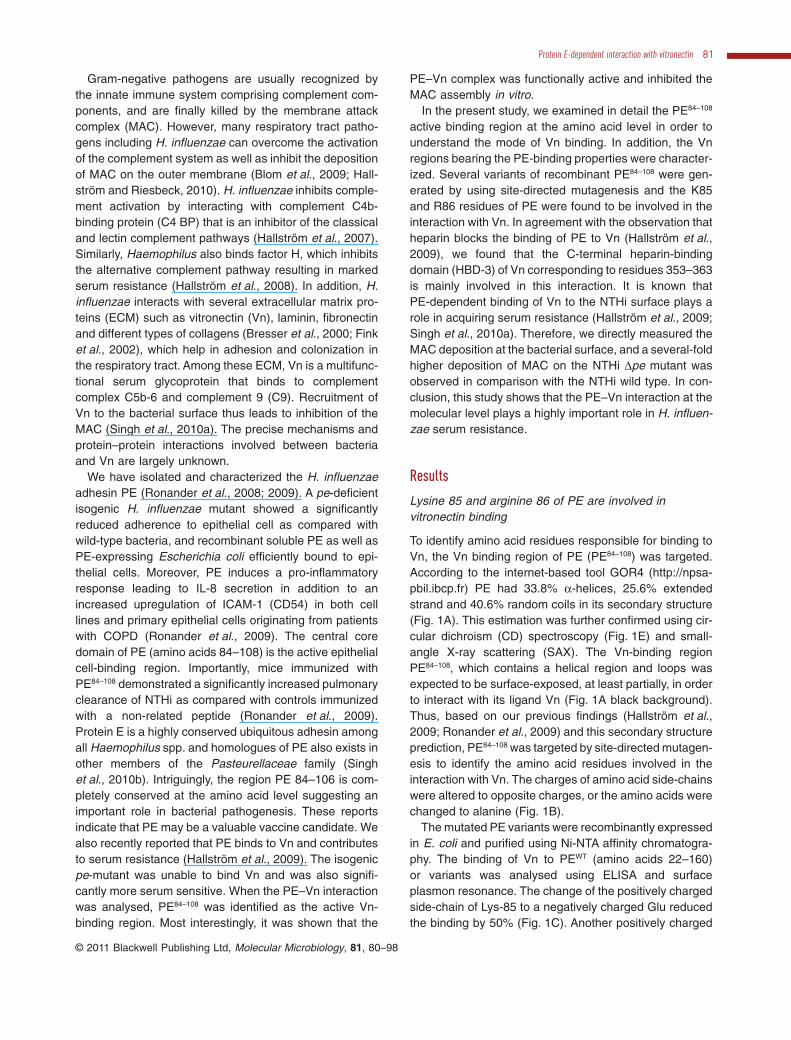
To identify amino acid residues responsible for binding toVn, the Vn binding region of PE (PE84–108) was targeted.According to the internet-based tool GOR4 (http://npsa-pbil.ibcp.fr) PE had 33.8% a-helices, 25.6% extendedstrand and 40.6% random coils in its secondary structure(Fig. 1A). This estimation was further confirmed using cir-cular dichroism (CD) spectroscopy (Fig. 1E) and small-angle X-ray scattering (SAX). The Vn-binding regionPE84–108, which contains a helical region and loops wasexpected to be surface-exposed, at least partially, in orderto interact with its ligand Vn (Fig. 1A black background).Thus, based on our previous findings (Hallström et al.,2009; Ronander et al., 2009) and this secondary structureprediction, PE84–108 was targeted by site-directed mutagen-esis to identify the amino acid residues involved in theinteraction with Vn. The charges of amino acid side-chainswere altered to opposite charges, or the amino acids werechanged to alanine (Fig. 1B).
The mutated PE variants were recombinantly expressedin E. coli and purified using Ni-NTA affinity chromatogra-phy. The binding of Vn to PEWT (amino acids 22–160)or variants was analysed using ELISA and surfaceplasmon resonance. The change of the positively chargedside-chain of Lys-85 to a negatively charged Glu reducedthe binding by 50% (Fig. 1C). Another positively charged
Protein E-dependent interaction with vitronectin 81
© 2011 Blackwell Publishing Ltd, Molecular Microbiology, 81, 80–98
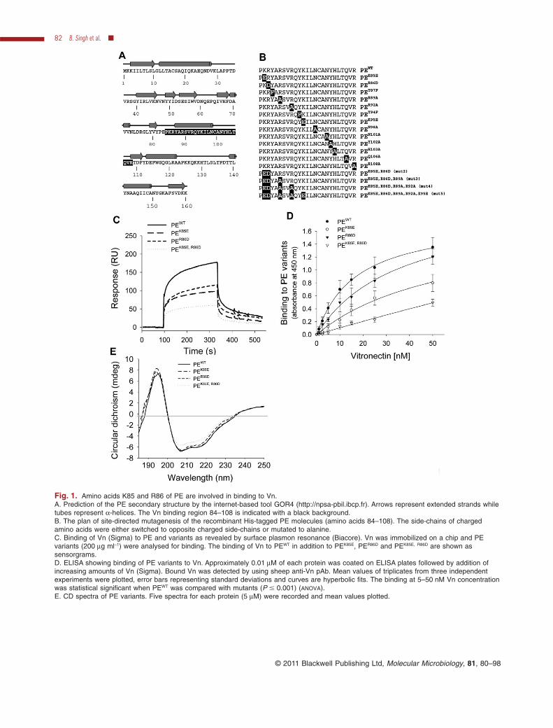
Fig. 1. Amino acids K85 and R86 of PE are involved in binding to Vn.A. Prediction of the PE secondary structure by the internet-based tool GOR4 (http://npsa-pbil.ibcp.fr). Arrows represent extended strands whiletubes represent a-helices. The Vn binding region 84–108 is indicated with a black background.B. The plan of site-directed mutagenesis of the recombinant His-tagged PE molecules (amino acids 84–108). The side-chains of chargedamino acids were either switched to opposite charged side-chains or mutated to alanine.C. Binding of Vn (Sigma) to PE and variants as revealed by surface plasmon resonance (Biacore). Vn was immobilized on a chip and PEvariants (200 mg ml-1) were analysed for binding. The binding of Vn to PEWT in addition to PEK85E, PER86D and PEK85E, R86D are shown assensorgrams.D. ELISA showing binding of PE variants to Vn. Approximately 0.01 mM of each protein was coated on ELISA plates followed by addition ofincreasing amounts of Vn (Sigma). Bound Vn was detected by using sheep anti-Vn pAb. Mean values of triplicates from three independentexperiments were plotted, error bars representing standard deviations and curves are hyperbolic fits. The binding at 5–50 nM Vn concentrationwas statistical significant when PEWT was compared with mutants (P � 0.001) (ANOVA).E. CD spectra of PE variants. Five spectra for each protein (5 mM) were recorded and mean values plotted.
82 B. Singh et al. �
© 2011 Blackwell Publishing Ltd, Molecular Microbiology, 81, 80–98

side-chain containing amino acid residue Arg-86 waschanged to Asp, and also showed a 48% reduction inbinding to Vn. Alteration of the charged side-chains of K85and R86 in the double mutant PEK85E, R86D (Fig. 1B: mut2)showed defective Vn binding (Fig. 1C). In addition, similarexperiments were performed with all PE variants usingELISA, where PE variants were coated on microtitre platesand Vn binding was detected using anti-Vn pAb. Ourresults revealed that PEK85E and PER86D decreased Vnbinding by 30–50% and that the PEK85E, R86D double mutantof PE lost 70% Vn binding (Fig. 1D). PEWT, PEK85E, PER86D
and PEK85E, R86D were analysed for folding pattern by usingCD, and these analyses suggested that PE variants werefolded (Fig. 1E). All the PE variants shown in Fig. 1Bwere tested for Vn binding by Biacore and ELISA. Othermultiple mutations (Fig. 1B: mut3, mut4, mut5) in thePE molecule showed similar binding pattern to the Vnmolecule as shown by PE K85, R86 (data not shown).
Lysine 85 and arginine 86 residues of PE on NTHisurface recognize Vn
The specificity of the binding of Vn to non-typeable H.influenzae (NTHi) 3655 was also verified by a competitionassay, where [125I]–Vn binding was blocked with unla-belled PE variants. Vn binding to NTHi was inhibited byunlabelled PEWT in a dose-dependent manner (Fig. 2A).Single mutants of the PE molecule, i.e. PEK85E and PER86D
competed with Vn (Fig. 2B), whereas the double mutantPEK85E, R86D did not inhibit Vn binding to NTHi 3655. Theseresults suggested that K85 and R86 were crucial for Vnbinding and were involved in the PE–Vn interaction at thebacterial surface. Moreover, since recombinant proteinswith alterations of the K85 and R86 side-chains to acidiccharged counterparts lost PE–Vn binding, indicating thatthe interaction may be electrostatic. However, these datasolely cannot prove the nature of this interaction.
To analyse the effect of mutated K85 and R86 whenexpressed at the surface (similarly as expression in NTHi
3655), PE variants were expressed in E. coli. Expressionof PE was monitored by flow cytometry (Fig. 3D) followedby binding of recombinant Vn80–396 that was analysed bytransmission electron microscopy (TEM) (Fig. 3A). E. coli(PEWT) displayed a similar binding pattern as comparedwith the single mutants expressing PEK85E and PER86D
(857–945 gold particles mm-2), whereas the double mutant(PEK85E, R86D) had a five- to sixfold reduced binding (248particles mm-2) to recombinant Vn (Fig. 3A). The compila-tion of several fields of vision of Vn80–396 binding to E. colivariants is shown in Fig. 3B.
In addition to experiments with recombinant Vn80–396, thebinding of native serum Vn to E. coli expressing PE wildtype and variants at the surface was analysed by semi-quantitative Western blotting. Bacteria were treated withnormal human serum (NHS) diluted 1:25 to 1:500 in PBScontaining 2.5% BSA followed by analysis of Vn bound tothe bacterial surface using anti-Vn pAb (Fig. 3C). Theseresults demonstrated that E. coli expressing PEWT at thesurface acquired Vn binding capacity, whereas E. coli(PEK85E, R86D) lost Vn binding and showed a similar Vnbinding as the control E. coli devoid of PE (Fig. 3B). Thus,these two data sets indicated that K85 and R86 are bothnecessary for Vn binding when PE was present at the E.coli surface. This experimental model with E. coli alsomimicked the functional role of PE as compared withexpression in NTHi 3655.
The C-terminal of Vn (amino acids 353–363) is a majorPE binding region
Vitronectin is a 65–75 kDa glycoprotein that containsa 43-amino-acid-long somatomedin-B (SMB) domain,which binds plasminogen activator inhibitor-1 (PAI-1)(Arroyo De Prada et al., 2002; Schroeck et al., 2002). Inaddition, Vn has four putative haemopexin-like regionsand three heparin-binding domains (HBDs) (Singh et al.,2010a), and in the case of PE, heparin is known to inhibitbinding to Vn (Hallström et al., 2009). To exclude that the
Fig. 2. PE blocks the Vn interaction withNTHi 3655.A. Unlabelled PEWT blocks binding of[125I]-labelled Vn to NTHi 3655 in a directbinding assay as described in detail inExperimental procedures.B. Binding of [125I]–Vn to NTHi 3655 isblocked with PEWT, PEK85E, PER86D, whereasthe double mutant PEK85E, R86D does not block.Mean values of triplicates from threeindependent experiments were plotted andstandard deviations were shown as errorbars. Differences between the control (withoutany competitor) and PEK85E, R86D werestatistically significant with PEWT, PEK85E,PER86D (P � 0.001). When PEWT, PEK85E andPER86D were compared these were notsignificant (ANOVA).
Protein E-dependent interaction with vitronectin 83
© 2011 Blackwell Publishing Ltd, Molecular Microbiology, 81, 80–98
SMB domain was involved in the PE–Vn binding, wepre-incubated PE with PAI-1 followed by addition of Vn.Binding was analysed by ELISA and dot blot, but noinhibitory effect of PAI-1 was observed (data not shown).We have recently shown that Moraxella catarrhalis Ubiq-uitous surface protein (Usp) A2 binds to the C-terminus(HBD-3) of the Vn molecule (Singh et al., 2010c). In addi-tion to recombinant Vn molecules used in that particular
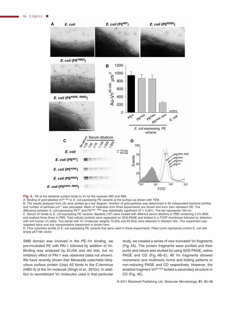
study, we created a series of new truncated Vn fragments(Fig. 4A). The protein fragments were purified and theirpurity and nature was studied by using SDS-PAGE, nativePAGE and CD (Fig. 4B–E). All Vn fragments showedmonomeric and multimeric forms and folding patterns innon-reducing PAGE and CD respectively. However, thesmallest fragment Vn80–229 lacked a secondary structure inCD (Fig. 4E).
Fig. 3. PE at the bacterial surface binds to Vn by the residues K85 and R86.A. Binding of gold-labelled Vn80–396 to E. coli expressing PE variants at the surface as shown with TEM.B. The results analysed from (A) were plotted as a bar diagram. Number of gold particles was determined in 60 independent bacterial profilesand number of particles mm-2 was calculated. Mean of triplicates from three experiments are shown and error bars represent SD. Thedifference between E. coli expressing PEWT and PEK85E, R86D was statistically significant (P � 0.001). The bar represents 100 nm.C. Serum Vn binds to E. coli expressing PE variants. Bacteria (108) were treated with different serum dilutions in PBS containing 2.5% BSAand washed three times in PBS. Total cellular proteins were separated on SDS-PAGE and blotted to a PVDF membrane followed by detectionwith anti-human Vn pAbs. Two bands with Vn (molecular weights 75 kDa and 65 kDa) were detected in Western blot. This experiment wasrepeated twice and one representative experiment is shown here.D. Flow cytometry profile of E. coli expressing PE variants that were used in these experiments. Filled curve represents control E. coli withempty pET16b vector.
84 B. Singh et al. �
© 2011 Blackwell Publishing Ltd, Molecular Microbiology, 81, 80–98

The purified recombinant Vn proteins (Fig. 4B) wereused for binding to whole bacteria and different solublePE variants. NTHi 3655 wild type (108 bacterial cells) wereincubated with Vn fragments (0.5 mg) and Vn bound to thebacterial surface was analysed by whole-cell proteinWestern blotting (Fig. 5A). Only Vn80–396, Vn80–379, Vn80–373
and Vn80–363 bound to the NTHi surface, whereas Vn80–353
and further shorter truncated fragments of Vn did notinteract with NTHi. In the next series of experiments weanalysed binding of Vn fragments to purified PE in ELISA.Microtitre plates were coated with PEWT and Vn fragments(0–100 nM) were allowed to bind. Resulting bound Vnfragments were quantified by anti-Vn pAb. The resultsrevealed that Vn80–396 and Vn80–379 displayed similarbinding saturation kinetics, while Vn80–373 and Vn80–363
showed a 20–30% reduced PE binding in comparisonwith Vn80–396 and Vn80–379 (Fig. 5B). Further truncation ofVn reduced binding to the PE molecule, indicated that Vn353–363 is a major PE binding region while a minorinteraction may also be involved in Vn 363-373. Acomparative binding analysis of PE variants to all Vnfragments was also performed (Fig. 5C). Vn80–396, Vn80–379
bound PEWT, PEK85E and PER86D stronger than Vn80–373 andVn80–363. In contrast, the double mutant PEK85E, R86D signifi-cantly lost Vn binding with all Vn fragments (Fig. 5C). Ourdata thus confirmed that further truncation after aminoacids Vn80–363 resulted in lost binding to the PE molecule.
Binding of the Vn80–396 fragments to the NTHi 3655surface was also analysed for binding specificity andcompetition with other smaller fragments. NTHi 3655 was
Fig. 4. Expression of recombinant Vn fragments and analyses for folding and multimerization.A. Schematic representation of the different truncated Vn molecules that were expressed in a mammalian expression system.B. Quality control of purified recombinant Vn molecules expressed in HEK293T cells. All protein fragments were purified by Ni-NTAchromatography and subjected to 15% SDS-PAGE (approximately 2 mg per lane) that was stained with Coomassie blue R250.C. Non-reducing SDS-PAGE showing the Vn fragments monomers and multimers. Each fragment (5 mg) was loaded in a 4–12% gradient SDSpolyacrylamide gel and stained with Coomassie blue R250.D. A similar gel (as shown in C) was analysed by Western blotting by using anti-Vn antibodies.E. CD spectra of Vn fragments at a concentration of 5 mM. Five spectra for each Vn fragment were recorded and mean values are shownhere.
Protein E-dependent interaction with vitronectin 85
© 2011 Blackwell Publishing Ltd, Molecular Microbiology, 81, 80–98

Fig. 5. Vn 353–363 binds to NTHi PE.A. Binding of Vn fragments to PE-expressing NTHi 3655 wild type as revealed by Western blot. Bacteria (108) were incubated with 0.5 mg ofeach Vn fragment and washed twice with PBS. Thereafter, total cellular proteins were separated on an SDS-PAGE and blotted onto a PVDFmembrane. This experiment was repeated three times, and one representative blot is shown here.B. Binding of Vn fragments to wild-type PE. Recombinant PE (0.1 mM) was coated in microtitre plates and increasing amounts of all Vnfragments were added, followed by detection of bound Vn using anti-human Vn pAbs. Data shown are the means of triplicates from threeindependent experiments and curves shown are hyperbolic fits. The binding of Vn to PEWT at 1.5–100 nM was statistically significant(P � 0.001) when Vn80–396 or Vn80–379 and other fragments were compared. Differences between Vn80–396 and Vn80–379, and between Vn80–373 andVn80–363 were not significant (ANOVA).C. Comparison of PE variants bound to different Vn fragments. Equimolar amounts (0.1 mM) of PE variants were coated on microtitre platesand 20 nM of each Vn fragment was allowed to bind. Data represent means of triplicates from three independent experiments and error barsindicate standard deviations. In the Vn groups Vn80–396, Vn80–379, Vn80–373 and Vn80–363 the binding differences between PEWT and PE mutantswere statistically significant (P � 0.01) (ANOVA).
86 B. Singh et al. �
© 2011 Blackwell Publishing Ltd, Molecular Microbiology, 81, 80–98

allowed to bind [125I]–Vn80–396 and the other smaller unla-belled fragments (Vn80–363 and Vn80–353) were used forcompetition. Results showed that Vn80–396 and Vn80–363
blocked 40–50% binding of the [125I]–Vn80–396 molecule,whereas Vn80–353 did not inhibit binding (Fig. 6A). Similarresults were also observed when PEWT was coated on amicrotitre plate and [125I]–Vn80–396 binding to immobilizedPEWT was analysed. Here also Vn80–396 and Vn80–363
blocked 60–70% of the binding to the [125I]–Vn80–396
fragment, whereas Vn80–353 did not significantly block(Fig. 6B). These both experiments directly indicated thatthe major PE binding region of Vn is located betweenamino acids 353 and 363.
In our next set of experiments, Vn fragments werelabelled with gold particles and their interaction with wholeNTHi 3655 and NTHi 3655Dpe in addition to E. coliexpressing PE at the surface was analysed by TEM. Asexemplified in Fig. 7A, only the gold-labelled fragmentsVn80–396, Vn80–379, Vn80–373 and Vn80–363 interacted with NTHi3655, whereas the other fragments were unable to bind(all data not shown). As can be seen in the bar diagram(Fig. 7C), 636 gold-Vn80–396 particles, 798 gold-Vn80–363
particles mm-2 were bound to the surface of NTHi 3655,and this was in comparison with Vn80–353 and Vn80–320 thatonly bound 125 and 61 particles mm-2 respectively. NTHi3655Dpe mutants showed a sixfold reduced Vn80–396
binding with 106 particles mm-2. The observed minor Vnbinding to the PE-deficient NTHi 3655Dpe could be due to
other weak Vn-binding surface proteins (Fig. 7A). More-over, PE-expressing E. coli had a similar Vn-bindingpattern as compared with NTHi 3655 (Fig. 7B). E. coli(PEWT) bound 881 Vn80–396 gold particles mm-2, whereasthe E. coli control without PE bound considerably lessVn80–396 (45 particles mm-2). Regarding the other Vn frag-ments, a similar pattern as with NTHi 3655 was observedwith E. coli (PEWT) (Fig. 7B and D). The TEM data werethus comparable and in agreement with results obtainedin vitro (Fig. 5B), or in vivo at the bacterial surface(Figs 5A and 6A). These results clearly indicated that theC-terminal domain in Vn corresponding to residues Vn353–363 is a major region for the interaction with PE.
Non-typeable H. influenzae is more sensitive to humanserum that is pre-treated with PE
Vitronectin binds C9 and thus inhibits its insertion into thebacterial membrane during MAC assembly (Singh et al.,2010a). When Vn is bound to the bacterial surface, theMAC is inhibited and bacteria have a prolonged survivalwhen exposed to NHS. The PE-expressing wild-typeNTHi 3655 was resistant to 10% NHS, and compared withthe PE-deficient mutant NTHi 3655Dpe that was signifi-cantly less serum resistant (Fig. 8A). We recently reportedthat Vn-depleted serum markedly increased H. influenzaekilling, and that addition of Vn to depleted NHS restoredthe bacterial serum resistance (Hallström et al., 2009).
Fig. 6. Vn 353–363 amino acids interact to PE at the NTHi surface.A. Binding of [125I]–Vn80–396 (approximately 300 000 kcpm) at the NTHi 3655 surface and inhibition by using cold Vn fragments. [125I]–Vn80–396
was added to 108 bacterial cells, after extensive washing bound fraction was counted in a gamma counter. In blocking experiments, bacteriawere treated with Vn80–396, Vn80–363 and Vn80–353 prior to addition of labelled Vn. [125I]–Vn80–396 binding without any competitor was compared withtreated samples.B. PEWT protein was coated on MaxiSorp ELISA plates (strip plates), [125I]–Vn80–396 (approximately 300 000 kcpm) was added, the unboundfractions were washed and bound fractions were counted. In competitive experiments Vn80–396, Vn80–363 and Vn80–353 were added to PE-coatedwells prior to addition of the [125I]–Vn80–396.In both experiments means of triplicates from three independent experiments (A and B) were plotted and standard deviations are shown aserror bars. Differences between control (without any competitor) or Vn80–353 were statistically significant with Vn80–396 and Vn80–363 treatment(P � 0.01). Differences between Vn80–396 and Vn80–363 were not significant (ANOVA).
Protein E-dependent interaction with vitronectin 87
© 2011 Blackwell Publishing Ltd, Molecular Microbiology, 81, 80–98

Hypothetically, if NHS is incubated with soluble PE, anincreased C9 deposition at the bacterial surface mayoccur due to quenching of Vn, and will be followed by anaccelerated MAC-mediated killing. Hence, the Vn-bindingcapacity of recombinant PEWT or PE variants was analy-
sed by pre-incubation of these recombinant proteins withNHS followed by exposure to NTHi 3655. When purifiedPEWT was added to NHS, a dose-dependent decrease inbacterial survival was seen in comparison with the recom-binant control protein MID962–1200 (Fig. 8B). This effect of
Fig. 7. Transmission electron microscopy of NTHi 3655 and E. coli expressing PEWT.A. Binding of gold-labelled recombinant Vn80–396, Vn80–363, Vn80–353 and Vn80–320 to the surface of NTHi 3655.B. Binding of similar gold-labelled Vn fragments to E. coli expressing PEWT at the surface.C and D. Bar diagrams based upon the experiments delineated in (A) and (B). Vn molecules (gold particles mm-2) that were bound to NTHi3655 and E. coli expressing PEWT were determined in 60 independent bacterial profiles and number of particles mm-2 was calculated. Mean oftriplicates from three experiments are shown and error bars represent SD. Differences were statistically significant when PE binding to Vn80–396
and Vn80–363 were compared with the other truncated Vn fragments (P � 0.001).Bars in (A) and (B) indicate 100 nm.
88 B. Singh et al. �
© 2011 Blackwell Publishing Ltd, Molecular Microbiology, 81, 80–98

Vn neutralization could be replenished if NTHi 3655 wastreated with Vn prior to addition of PE treated serum(Fig. 8C). We also tested whether it was possible toquench Vn by incubating NHS with the different mutatedPE variants followed by incubation with whole bacteria.Interestingly, pre-incubation of NHS with PEK85E andPER86D resulted in significantly higher bacterial killing,whereas experiments with PEK85E, R86D did not reveal anydifference in bactericidal serum activity (Fig. 8D). Pre-treatment of NHS with recombinant PEWT and mutatedvariants thus indicated that PE was able to bind Vnderived from serum, and that in particular residues K85and R86 were responsible for the binding. These resultswere in parallel with findings obtained with the directbinding assays (Figs 2, 3 and 5).
Vitronectin bound to PE prevents MAC deposition at thesurface of H. influenzae
It is well known that Vn inhibits the MAC complex and thisproperty of Vn is successfully utilized by many pathogensto evade the host innate immune response (Singh et al.,2010a). Recently we found that NTHi 3655Dpe signifi-
cantly lost Vn binding (as also shown in Fig. 7A and C),and that this PE-dependent Vn interaction is involved inserum resistance (Hallström et al., 2009). To analyse theeffect of the PE–Vn interaction on MAC formation, weperformed a serum resistance assay and observed theconcentrations of C9 polymerized, and measured C9monomers consumed during MAC deposition. Westernblotting using a specific anti-C9 pAb that recognizes bothmonomeric and polymeric C9, and subsequent densito-metric analyses were performed. Under these experimen-tal conditions, MAC deposition on bacteria occurred in atime-dependent manner (Fig. 9A and B). C9 is a 71 kDamonomer that may exist in a monomeric or oligomericstate, associated as a C5b-9 complex in NHS undernormal physiological conditions (Podack and Tschopp,1982). After complete polymerization of C9 (ring closure),a high-molecular-weight band of approximately 400–500 kDa (a C9 oligomer) appears (Podack and Tschopp,1982; Deng et al., 2007). The disappearance of the C9monomer in the presence of NTHi 3655Dpe (Fig. 9B) wasmore evident in comparison with the NTHi 3655 wild type,and an increased C9 oligomerization directly revealedthat MAC formation was more efficient in reactions
Fig. 8. H. influenzae is more sensitive toserum that has been pre-treated with PE.A. Serum resistance of NTHi 3655 and NTHi3655Dpe when incubated with 10% NHS.B. Pre-incubation of NHS with increasingconcentrations (1–5 mM) of recombinant PEWT
quenches Vn and hence NTHi appears moreserum sensitive. MID962–1200 is a proteinfragment from Moraxella catarrhalisIgD-binding protein (MID) that was used as anegative control (Nordström et al., 2006).C. NTHi 3655 was incubated with NHS, NHStreated with PE (2 mM) and PE-treated NHSwas added to Vn (2 mM)-treated bacterialcells. Differences were statistically significant(5–10 min) when NHS only and NHS+PE orNHS+PE+Vn were compared (P � 0.05), aswell as differences between NHS+PE andNHS+PE+Vn (P � 0.001) (ANOVA).D. PEWT, PEK85E, PER86D showed decrease inserum resistance, whereas PEK85E, R86D did notshow any decrease of resistance when addedto NHS. PEWT, PEK85E and PER86D weresignificantly different (P � 0.001) whencompared with control NHS or PEK85E, R86D
(ANOVA). No significant difference wasobserved between control NHS andPEK85E, R86D.In (A)–(D) means of triplicates from threeindependent experiments are shown andstandard deviations are indicated as errorbars.
Protein E-dependent interaction with vitronectin 89
© 2011 Blackwell Publishing Ltd, Molecular Microbiology, 81, 80–98

containing PE-deficient NTHi mutants (Fig. 9A and B).Densitometric measurements of C9 when incubatedwith the PE-expressing NTHi 3655 revealed that within10–15 min of incubation 75–90% of C9 was a monomer(Fig. 9C). In contrast, only 40–45% of C9 was remainingas a monomer when exposed to NTHi 3655Dpe.
We also performed TEM of these serum-treatedsamples. Samples at 5 and 10 min (as described in
Fig. 9A and B) were chosen and analysed by gold-labelled anti-C9 pAbs. Data obtained by TEM also sug-gested a time-dependent deposition of C9. The number ofgold particles reflecting C9 was counted from 60 indepen-dent bacterial profiles. At 5 min, 82 gold particles mm-2
were found on the NTHi 3655 wild type, whereas 974 goldparticles mm-2 were detected on the NTHi 3655Dpemutant. Similarly, 114 particles mm-2 and 1224 particles
90 B. Singh et al. �
© 2011 Blackwell Publishing Ltd, Molecular Microbiology, 81, 80–98

mm-2 were seen after 10 min on the NTHi wild type andthe mutant surface, respectively (Fig. 9D and E). Theseresults directly explained why NTHi 3655Dpe mutantswere more serum-sensitive as compared with the wildtype, and also that the PE–Vn interaction was highlyimportant for NTHi serum resistance.
In our next set of experiments we analysed the capacityof the PEWT and PEK85E, R86D variant for Vn binding andsubsequent effect in MAC formation at the NTHi 3655
surface. PEWT and PEK85E, R86D were immobilized on CNBragarose beads to simulate PE as a surface bound proteinand checked on a Western blot (Fig. 10A). Control beadswere treated similarly without any added protein. Thepull-down capacity of these beads was measured bysemi-quantitative Western blotting at different serumdilutions. PEWT bound Vn more intensely in comparisonwith PEK85E, R86D (Fig. 10B). To bind Vn, beads coated withPE (equivalent to 2 mM protein) and control beads were
Fig. 9. MAC deposition at the H. influenzae surface is regulated by PE-dependent Vn binding.A and B. NTHi 3655 and NTHi 3655Dpe (108 cells) were resuspended in GVB++ buffer containing 10% NHS and incubated at 37°C. MACdeposition is seen as increase in C9 oligomer and disappearance of the C9 monomer. The complement reaction was terminated at differenttime points (5–60 min) by snap freezing of samples. Samples were subjected to 4–12% gradient SDS-PAGE, blotted onto PVDF membranefollowed by detection using an anti-human C9 pAb. The experiment was repeated twice. One of the representative blots from triplicates of asingle experiment is shown here.C. The C9 monomer density was measured from triplicate blots of a single experiment. Error bars indicate standard deviations. Statisticalsignificant differences were observed between 10 and 40 min (ANOVA). The experiment was repeated twice.D. TEM analysis that demonstrates C9 deposition on the surface of NTHi 3655 and NTHi 3655Dpe. Samples obtained at 5 and 10 min from(A) and (B) were probed with gold labelled anti-human C9 pAb.E. Estimation of gold-labelled Vn bound to the wild type and PE-deficient NTHi mutant. The curve shows the number of particles mm-2
evaluated from 60 independent bacterial profiles. Mean of triplicates from three experiments are shown and error bars represent SD.Bar represents 100 nm. *P � 0.05; **P � 0.01; ***P � 0.001.
Fig. 10. PE-treated serum increases MACdeposition at the bacterial surface.A. CNBr agarose beads were immobilizedwith PEWT and PEK85E, R86D proteins (seeExperimental procedures). One set of beadswere treated similarly without protein togenerate control beads. Agarose beads boundPE (5 mg) along with control were resolved ina 12% SDS-PAGE and stained withCoomassie blue R250.B. Pull-down assay showing binding of Vn byPE-coated beads. PE-coated beads (1 mg)and control beads were added to 100 ml ofdifferent serum dilutions in PBS containing2.5% BSA. Beads were washed extensivelywith PBS and proteins were separated inSDS-PAGE followed by Western blotting anddetection of Vn by anti-Vn antibodies. Thisexperiment was repeated twice; onerepresentative blot is shown here.C–E. Normal human serum was treated withcontrol, PEWT- and PEK85E, R86D-coated beads(2 mM) prior to adding to bacteria. C9deposition experiments were performed asdescribed in Fig. 9. This experiment wasrepeated twice. From a single experimentsamples, each blot was performed intriplicate; one representative blot out oftriplicate is shown here.F. The C9 monomer density was measuredfrom triplicate blot of a single experiment. Thedata for 5–15 min are shown. The differences(5–15 min) between control beads and PEWT
were statistically significant (P � 0.01). Acomparison between PEWT and PEK85E, R86D
beads revealed P � 0.05 (ANOVA). Anystatistical significance was not observedbetween control beads and PEK85E, R86D-coatedbeads.
Protein E-dependent interaction with vitronectin 91
© 2011 Blackwell Publishing Ltd, Molecular Microbiology, 81, 80–98

added to serum and incubated for 1 h. This serum wassubsequently tested for C9 deposition at the NTHi 3655surface. The results shown in Fig. 10C–E represent C9deposition on control, PEWT and PEK85E, R86D beads whenincubated with serum. Three blots from a typical experi-ment were scanned for C9 band intensity and are shownin the bar chart (Fig. 10F). The serum incubated withcontrol beads showed a similar C9 disappearance astreatment with beads coated with PEK85E, R86D, whereasserum incubated with PEWT-containing beads had a com-paratively faster disappearance of C9 (Fig. 10F). Theseresults directly suggested the involvement of K85 andR86 during Vn interaction.
Discussion
Vitronectin plays a crucial role in many biological systemsincluding cell migration, adhesion, repair angiogenesis(Singh et al., 2010a) and microbial pathogenesis (Leroy-Dudal et al., 2004; Attia et al., 2006; Hallström et al.,2006; Bergmann et al., 2009; Hallström and Riesbeck,2010; Singh et al., 2010a). It has a multi-domain structuralarrangement, where the N-terminal SMB domain is wellcharacterized for its role in several biological processes.The C-terminal HBD-1 (amino acids 175–219), HBD-2(amino acids 175–219) and HBD-3 (amino acids 348–361) are known to bind to bacterial pathogens (Singhet al., 2010a). Being one of the major regulators of thecomplement system, Vn has a crucial impact on MACdeposition on the membrane of Gram-negative respira-tory pathogens that successfully exploit the MAC-inhibitory role of Vn (Hallström et al., 2009; Singh et al.,2010a). On the other hand, Gram-positive bacteria havebeen shown to use the Vn–integrin interaction for cellularinvasion and internalization (Bergmann et al., 2009).
Recently we reported that the PE84–108 domain is com-pletely conserved in all Haemophilus spp. including 186NTHi clinical isolates, Hib and other Haemophilus spp.The conserved property of PE among NTHi makes thisprotein valuable as a future vaccine candidate (Singhet al., 2010b). PE interacts with Vn at a KD = 4 ¥ 10-7 Mand protects NTHi from serum-mediated lysis. Vn boundto PE at the NTHi surface has a functional capacity toinhibit the MAC assembly, and this observation supportsthe involvement of a PE–Vn interaction in NTHi-dependent serum resistance. Furthermore, a peptidemapping analysis indicated that PE84–108 is the active Vnbinding region (Hallström et al., 2009). In the presentarticle, we focused on the properties of PE involved inbinding to Vn. The PE84–108 region was studied in detail byusing a site-directed mutagenesis approach. Interestingly,the PE84–108 region harbours K85 and R86, which aresignificantly involved in the function of PE. Mutation ofK85 to the opposite charged side-chain E85, and R86 to
D86 resulted in loss of PE binding to Vn. The mutated PEvariants were also verified for any defective protein foldingand tertiary structures by using CD spectroscopy, wherePEWT, PEK85E, PER86D and PEK85, R86D showed similar helixand beta-sheet patterns (Fig. 1E).
Previously it has been demonstrated that Vn binds toStreptococcus pneumoniae and contributes to bacterialinternalization. This interaction can be efficiently blockedin the presence of heparin, indicating that the HBD(s) ofVn are involved in this interaction. Moreover, addition ofVn to epithelial cells significantly increases the adhesionof S. pneumoniae (Bergmann et al., 2009). Anotherpathogenic organism, Pseudomonas aeruginosa, has apotential binding capacity with Vn and binding of Vn withavb5 plays a key role in P. aeruginosa internalization byA549 cells (Leroy-Dudal et al., 2004; Leduc et al., 2007).H. influenzae binds Vn by the two outer membrane pro-teins Hsf and PE, which both contribute serum resistance(Hallström et al., 2006; 2009). However, Haemophilusducreyi binds Vn via the trimeric autotransporter DsrA andthus exhibits serum resistance (Leduc et al., 2009). Manyother research reports suggest that Vn potentially playsimportant roles in the pathogenesis of Yersinia pseudotu-berculosis (Gustavsson et al., 2002), Neisseria meningiti-dis (Duensing and Putten, 1998; Sa et al., 2010) andCandida albicans (Limper and Standing, 1994). In light ofthe important role of Vn in microbial pathogenesis, there isnot much data published that in detail characterize theinteraction between Vn and its microbial ligands. The lackof structural data on the HBD(s) of Vn also limits the clearviews about the Vn–bacterial interactions. In this study, Vn353–363 amino acids region was identified as the majorPE binding region. We designed a peptide library for thesequence Vn 312–396 and analysed for PE binding, butwere not successful in defining the binding site (data notshown). Thus, there might be a secondary structure withinthe Vn 353–363 region that is involved in the interactionwith PE that could not be mimicked by short syntheticpeptides. Previously, it has been reported by us thatheparin binding region (HBD-3) comprising Vn aminoacids 341–368 might have an interaction with the PEmolecule (Hallström et al., 2009). These data are inagreement with our previous findings indicating that Vn353–363 are involved in binding to PE. The detailed align-ment of Vn 320–396 has been published by us (Singhet al., 2010c) and reveals that the 353–363 amino acidregion of Vn is variable among different mammalianspecies. This variation might be responsible for selectivebinding of Vn to pathogens, as reported recently with S.pneumoniae. Interestingly, mouse Vn binds several-foldless efficiently in comparison with human Vn (Bergmannet al., 2009).
Recently we reported that Vn plays a significant role inthe survival of NTHi. The Vn-depleted serum decreases
92 B. Singh et al. �
© 2011 Blackwell Publishing Ltd, Molecular Microbiology, 81, 80–98

the survival, whereas, addition of Vn restores serum resis-tance capacity (Hallström et al., 2009). M. catarrhalis alsoshowed similar Vn-dependent serum survival, whereasVn-depleted serum kills bacteria faster than NHS andaddition of Vn restores serum resistance. UspA2 wasfound as the major Vn-binding protein of M. catarrhalisand UspA2-deficient mutants were highly sensitive toNHS (Attia et al., 2005; 2006). Based upon those earlierfindings, we recently examined the UspA2–Vn interactionin detail. Interestingly, UspA2-treated human serum moreefficiently killed bacteria as compared with control serum,and this was directly linked to quenching of Vn (Singhet al., 2010c). In parallel to Moraxella UspA2, our resultson Haemophilus PE in the present study suggested thatthe enhanced killing of H. influenzae when NHS waspre-incubated with PEWT as well with the single mutatedvariants PEK85E or PER86D was due to the interaction andconsequently quenching of the biological activity of Vn.
Previously we have shown that NTHi 3655Dpe isserum-sensitive and that the Vn-binding capacity signifi-cantly decreased in these PE-deficient mutants. The Vndepleted serum killed NTHi 3655 more efficiently in com-parison with NHS and addition of Vn restored serum resis-tance of NTHi 3655 (Hallström et al., 2009). In support ofthat study we showed in the present study that MACdeposition is significantly higher in NTHi 3655Dpemutants in comparison with wild-type counterparts(Fig. 9A–E). Thus, the decrease in Vn binding to NTHi3655Dpe is responsible for a considerably higher deposi-tion of MAC.
In conclusion, this study suggests that the role of H.influenzae PE is important for Vn binding and conse-quently serum resistance. Lysine 85 and arginine 86 arelocated within the active binding region PE84–108 and arenecessary for the Vn interaction. However, it is not com-pletely evident that PE interacts with Vn by only K85 andR86. A future ultrastructure of the PE–Vn complex mayprove details on the interaction. A detailed analysis of theVn molecule and characterization of the precise aminoacids that are responsible for the interaction with PE alsoremains to be done.
Experimental procedures
Bacterial strains, reagents and cell culture
The NTHi 3655 and an isogenic mutant (NTHi 3655Dpe) weregrown in brain heart infusion (BHI) liquid broth, supplementedwith 10 mg ml-1 nicotinamide adenine dinucleotide (NAD) andhaemin, or streaked on chocolate agar plates and incubatedat 37°C in a humid atmosphere with 5% CO2. The NTHi3655Dpe mutant was grown in the presence of 20 mg ml-1
kanamycin (Ronander et al., 2009). E. coli BL21 (DE3) andDH5a were cultured in Luria–Bertani (LB) broth or on LB agarplates at 37°C in a humid atmosphere containing 5% CO2.
The vectors pET26bpe and pET16bpe were as described(Hallström et al., 2009). pET26bpe and variants were supple-mented with 50 mg ml-1 kanamycin and pET16bpe and vari-ants were supplemented with 100 mg ml-1 ampicillin in LBmedium. Human embryo kidney (HEK293T) cells were grownin advanced DMEM (Gibco; Invitrogen, Stockholm, Sweden)with 2 mM L-glutamine, 100 mg ml-1 streptomycin and 100 Uml-1 penicillin.
Recombinant DNA techniques and site-directedmutagenesis
The construct pET26bpe as described in Hallströmet al. (2009) was used for expression and mutation of PE.The nucleotide region (250–324) of the pe gene(CGSHi3655_04936) encodes for PE84–108 and was manipu-lated at several amino acids by using a site-directedmutagenesis approach. The QuikChange site-directedmutagenesis was performed using high fidelity PfuTurbo ®
DNA polymerase (Stratagene, La Jolla, CA). The primersused for mutagenesis are listed in Table 1. The mutagenesisprocedure including PCR and DpnI digestion was performedas described previously (Singh and Röhm, 2008). Mutantvectors were sequenced and finally transformed into E. coliBL21 (DE3) cells. The E. coli cells containing pET26bpe andvariants were used for expression and purification, while E.coli containing pET16bpe was used for surface expression ofPE (Ronander et al., 2009). Truncated Moraxella IgD-bindingprotein (MID962–1200) was prepared as described (Nordströmet al., 2006).
Recombinant protein expression and purification
The E. coli BL21 (DE3) containing recombinant pET26bvectors were grown in 500 ml of LB medium with kanamycinat 37°C until OD600 to 0.8–1. Expression was induced by1 mM IPTG and cultivation continued for another 3 h at 37°C.The cells were harvested at 5000 g for 15 min at 4°C. Theperiplasmic fraction was separated according to a protocoldescribed in Singh and Röhm (2008). Purified proteins weredialysed by several exchanges of HNET buffer (50 mMHEPES, pH 7.5, 150 mM NaCl, 3 mM EDTA and 0.005%Tween-20) for Biacore assay and concentrated by using Cen-tricon cartridge (3 kDa molecular weight cut-off; Millipore,Bedford, MA). Protein concentrations were measured by UVabsorbance using a Nano-drop spectrophotometer (ThermoScientific, Wilmington, DE) in addition to verification by aBicinchoninic acid (BCA) assay (Pierce, Rockford, IL). Thepurity and concentrations of expressed proteins was verifiedon SDS-PAGE stained with Coomassie blue R250.
Expression of Vn fragments in HEK293 cell lines andpurification
Vitronectin domains harbouring heparin binding sites havebeen expressed in HEK293T cells as Vn amino acids80–396, 80–320 and 80–229 as described in Singh et al.(2010c). Furthermore, new constructs corresponding toamino acids 80–379, 80–373, 80–363, 80–353, 80–339 and
Protein E-dependent interaction with vitronectin 93
© 2011 Blackwell Publishing Ltd, Molecular Microbiology, 81, 80–98

80–330, all containing C-terminal 6x-His tags (Fig. 4A) wereamplified by using Vn 80–396 as template. The forwardprimer was inserted with BshT1 (AgeI) and the reverse primerwith Acc65I (KpnI) restriction enzyme sites. Amplified anddigested inserts were ligated into the pHLsec vector (28).Protein expression and purification was performed asdescribed elsewhere (Singh et al., 2010c).
Circular dichroism (CD)
Purified PE variants and Vn fragments were dialysed against20 mM phosphate buffer and the concentrations were mea-sured by using Nano-drop at (280 nm) and BCA method(Pierce, Rockford, IL). Finally, 5.0 mM proteins were trans-ferred to a cuvette and CD spectrum was recorded betweenthe wavelengths of 190 nm to 250 nm by using a CD spec-tropolarimeter J815 (Jasco, Essex, UK).
SDS-PAGE and non-reducing PAGE
Routine SDS-PAGE was performed according to standardlaboratory protocols. In gradient separations NuPAGE Gradi-ent 4–12% Bis-tris readymade gels were used (Invitrogen,Carlsbad, CA) and separation was performed according toprotocol suggested by the manual. In non-reducing condi-tions protein samples were loaded with SDS-PAGE loadingdye without b-mercaptoethanol.
Flow cytometry (FACS)
E. coli cells were grown in LB medium containing appropriateantibiotics and induced by 1 mM IPTG for 1 h at 37°C. Bac-teria (108) were resuspended in 100 ml of PBS containing2.5% BSA and treated with affinity-purified rabbit polyclonalanti-PE antibodies for 1 h at room temperature. After onewashing with PBS-BSA, FITC-conjugated anti-rabbit anti-bodies (Dakopatts) were added (diluted according to the
manufacturer’s instructions) and incubated for 1 h at roomtemperature. Additionally three washes performed with PBScontaining 2.5% BSA and finally cells were resuspended in100 ml of PBS containing 2.5% BSA. E. coli harbouring emptyvector was used as a control and treated similarly. Sampleswere analysed in a flow cytometer (EPICS XL_MCL;Beckman Coulter).
Immobilization of PE on CNBr-activated agarose beads
Cyanogen bromide-activated SepharoseTM 4 Fast Flow (GEHealthcare Biosciences, Uppsala, Sweden) was used forimmobilization of PE variants. Sepharose powder (330 mgml-1) was washed several times in 1 mM HCl and resus-pended in coupling buffer (0.1 M NaHCO3, pH 8.3 containing0.5 M NaCl). Protein solutions were also dialysed againstcoupling buffer and added to the resin (5–10 mg protein ml-1
of resin) and incubated overnight at 4°C in a rolling shaker.Unbound ligands were washed with 5 gel volume of couplingbuffer and free groups were blocked by blocking buffer (0.1 MTris-HCl, pH 8.0) for 2 h at room temperature. Beads werethen washed three times by alternating buffers, NaAc, pH 4.0containing 500 mM NaCl and 0.1 M Tris-HCl, pH 8.0 contain-ing 500 mM NaCl. Finally beads were washed with PBS andstored at 4°C. The protein ligands immobilized were mea-sured by Nano-Drop and BCA methods.
ELISA
Purified PE variants were coated on PolySorp microtitreplates (Nunc-Immuno, Roskilde, Denmark). Coating of PE(0.1 mM) was performed in 100 mM Tris-HCl, pH 9.0 for 15 hat 4°C. Plates were washed three times with PBS to removeexcess unbound protein and blocked with PBS-2.5% BSA for1 h at 25°C. The Vn was added to the wells in PBS-2.5% BSAand incubated for 1 h at 25°C. In competitive ELISA, increas-ing concentration of competitor ligand was added to thebinding reactions and allowed to bind for 1 h at room
Table 1. Primers used in site-directed mutagenesis of PE amino acids 84-108.
Primer name Sequence 5′–3′ PE variants
K85E _For GTTTATCCTGAGCCTGAACGTTATGCACGTTC K85ER86D _For GTTTATCCTGAGCCTAAAGATTATGCACGTTCTGTTCG R86DY87F _For CCTGAGCCTAAACGTTTTGCACGTTCTGTTCGTC Y87FR89A_For GAGCCTAAACGTTATGCAGCTTCTGTTCGTCAGTATAAG R89AR92A_For GCACGTTCTGTTGCTCAGTATAAGATTTTGAATTGTGC R92AY94F_For GCACGTTCTGTTCGTCAGTTTAAGATTTTGAATTGTGC Y94FK95E_For GTTCTGTTCGTCAGTATGAGATTTTGAATTGTGC K95EN98A_For TCTGTTCGTCAGTATAAGATTTTGGCTTGTGCAAATTATCATTTAACTC N98AN101A _For GTATAAGATTTTGAATTGTGCAGCTTATCATTTAACTCAAATACGAACTG N101AY102A_For GTATAAGATTTTGAATTGTGCAAATGCTCATTTAACTCAAATACGAACTG Y102AH103A_For ATAAGATTTTGAATTGTGCAAATTATGCTTTAACTCAAATACGAACTGAT H103AQ106A_For GAATTGTGCAAATTATCATTTAACTGCAATACGAACTGATTTCTATGATG Q106AR108A_For GCAAATTATCATTTAACTCAAATAGCAACTGATTTCTATGATGAATTTTG R108AMut 2_For GTTTATCCTGAGCCTGAAGATTATGCACGTTC K85E, R86D (mut 2)Mut 3_For GAGCCTGAAGATTATGCAGCTTCTGTTCGTCAGTATAAG K85E, R86D, R89A (mut 3)Mut 4_For GCAGATTCTGTTGCTCAGTATAAGATTTTGAATTGTGC K85E, R86D, R89A, R92A (mut 4)Mut 5_For ATTCTGTTGATCAGTATGAGATTTTGAATTGTGC K85E, R86D, R89A, R92A, K95E (mut 5)
The mutated bases are underlined in all primers. The forward primer for each mutation is shown here, while reverse primer of same sequence wasalso used.
94 B. Singh et al. �
© 2011 Blackwell Publishing Ltd, Molecular Microbiology, 81, 80–98

temperature. The unbound Vn fraction was removed bywashing with PBS containing 0.05% Tween-20, and boundVn was detected by polyclonal sheep anti-human Vn antibod-ies (pAb) (AbD Serotec, Kidlington, Oxford, UK) and second-ary horseradish peroxidase (HRP)-conjugated donkey anti-sheep pAb (AbD Serotec). After four additional washingsteps, plates were developed and read at 450 nm in a micro-plate reader (Multiskan® plus, Labsystems, Helsinki, Finland).
Surface plasmon resonance (Biacore)
The interaction between PE variants and Vn was analysedusing surface plasmon resonance (Biacore 2000, Uppsala,Sweden). Four flow cells of a CM5 sensor chip were acti-vated, each with 20 ml of a mixture of 0.2 M 1-ethyl-3-(3-dimethylaminopropyl) carbodiimide and 0.05 M N-hydroxy-sulphosuccinimide at a flow rate of 10 ml min-1, after which Vn(10 ml ml-1 in 10 mM sodium acetate buffer, pH 4.0) wasinjected over flow cell 2 to reach 4000 resonance units (RU).Unreacted groups were blocked with 20 ml of 1 M ethanola-mine (pH 8.5). A negative control was prepared by activatingand subsequently blocking the surface of flow cell 1. Thecomparative association kinetics were studied for variousconcentrations of the PE variants. The flow buffer (50 mMHEPES, pH 7.5 containing 150 mM NaCl, 3 mM EDTA and0.005% Tween-20) was used for binding. Protein solutionswere injected for 600 s during the association phase at aconstant flow rate of 30 ml min-1. The sample was first injectedover the negative control surface and then over immobilizedVn. The signal from the control surface was subtracted. Thedissociation was followed for 200 s at the same flow rate. Inall experiments, 30 ml of 2 M NaCl was used to remove boundligands during a regeneration step. BiaEvaluation 3.0 soft-ware (Biacore) was used for data analysis and SigmaPlot 8.0to generate final figures.
[125I] labelling of Vn fragments and binding assay
The labelling of Vn was performed by the Chloramine-Tmethod (25). For labelling, 0.05 M [125I] isotope (AmershamBiosciences, Buckinghamshire, UK) was used per mole ofprotein. The labelled proteins were purified from unincorpo-rated [125I] using PD10 columns (GE Healthcare Biosciences,Uppsala, Sweden).
NTHi and mutants were grown in BHI medium toODA600 = 1.0 and washed with PBS-2.5% BSA. Approxi-mately, 108 bacterial cells per well were added to microtitreplates and increasing concentrations of [125I]–Vn were added.For competition experiments, cold ligands were added to thebinding reactions. Bacteria were incubated for 30 min at 37°Cand were pelleted at 4200 g for 10 min, followed by twowashing steps of PBS-2.5% BSA. The unbound radiolabelledprotein was removed by three washes with PBS, harvested ina 96-well plate harvester (Tomtec, Hamden, CT), and finallycounted in a liquid scintillation counter (Trilux, Microbeta1450, Perkin Elmer).
In the radioactive ligand direct binding assay, PE (0.1 mM)was coated on ELISA strip plate (MaxiSorp) and washedthree times with PBS to remove excess unbound protein andblocked with PBS-2.5% BSA for 1 h at room temperature.
[125I]–Vn was added to the wells in PBS-2.5% BSA and incu-bated for 1 h at 25°C. In the competitive assay, competitorligands were added to the binding reaction prior to addition oflabelled Vn and allowed to bind for 1 h at room temperature.The unbound [125I]–Vn fraction was removed by washing withPBS containing 0.005% Tween-20. Each single well wasseparated from strip by mechanical breaking and total radio-activity was counted in a scintillation counter.
Serum resistance assay
NTHi 3655 wild type was tested for impact of PE variants inNHS-mediated killing. Approximately, 105 cells were resus-pended in 100 ml of dextrose-GVB (DGVB++) buffer, pH 7.3,containing 140 mM glucose, 0.1% (w/v) gelatin, 1 mM MgCl2,0.15 mM CaCl2 and finally 10% NHS. Bacteria were incu-bated at 37°C and samples (10 ml) were collected at differ-ent time intervals and plated on chocolate agar plates. Theeffect of PE variants on serum proteins was analysed bypre-incubating increasing concentrations of PE variants(0.5–5 mM) with 10% serum at room temperature for 1 h andsubsequently used in the serum resistance assay. Similarly,the effect of Vn was evaluated by treating bacterial cells withVn for 1 h at room temperature, prior to addition of NHS. Theviable bacterial cells were determined by counting colony-forming units (cfu) after incubation overnight at 37°C.
Transmission electron microscopy (TEM)
Proteins were directly labelled with colloidal gold asdescribed (Roth, 1996). The wild-type H. influenzae andmutants were grown in BHI for 3 h at 37°C. Bacterial solu-tions were incubated with gold-labelled Vn fragments (100 mgml-1), fixed in PBS containing 4% paraformaldehyde and0.1% glutaraldehyde and prepared for electron microscopyas described (Carlemalm, 1990). TEM was thereafter per-formed as described (Bengtson et al., 2008), and specimenswere examined in a JEOL JEM 1230 transmission electronmicroscope (JEOL, Peabody, MA) at 60 kV acceleratingvoltage. Images were recorded with a Gatan Multiscan791 CCD camera (Gatan, Pleasanton, CA). Only particlesobserved within a distance of 15 nm or less adjacent to thecell surface were counted. This corresponds to the estab-lished maximum distance between an IgG and its antigen, orbetween a protein labelled with gold of this size, and itstarget. Differences between groups concerning Vn–PE inter-action, and complement activation were analysed by the one-tailed test for differences between means. The following apriori null hypotheses were tested: full-length Vn does notbind to PE (Fig. 3), or full-length Vn binds to PE double pointmutation with the same affinity (Fig. 3), or different Vn con-structs exhibit the same affinity to PE (Fig. 7), or NTHi 3655and NTHi Dpe show the same C9 deposition (Fig. 9). Thenull hypothesis was rejected and statistical significance wasassumed when P � 0.05.
Western blotting and MAC deposition assay
After processing, bacterial cells were boiled with 1¥ SDS-PAGE (containing b-mercaptoethanol) loading buffer for
Protein E-dependent interaction with vitronectin 95
© 2011 Blackwell Publishing Ltd, Molecular Microbiology, 81, 80–98

10 min at 95°C and centrifuged for 5 min at 20 000 g. Super-natants (10–15 ml) were loaded in 12% or 15% SDS-polyacrylamide gels, or 4–12% gradient SDS-polyacrylamidegels (Invitrogen, Carlsbad, CA). The gels were subjectedto run at particular current as recommended in standardprotocols. Gels were blotted to PVDF membranes. Mem-branes were blocked with PBS containing 5% milk for 1 h atroom temperature. All reactions were performed in PBS con-taining 5% milk and washing was done with PBS containing0.05% Tween-20. Vn detection was performed with anti-human Vn antibodies (pAb) (AbD Serotec, Kidlington, Oxford,UK) and HRP-conjugated donkey anti-sheep secondary pAb(AbD Serotec).
Membrane attack complex deposition at the bacterialsurface was estimated by C9 polymerization. Usually C9monomer is a 71 kDa and it polymerizes into 14–16 mertubules to make a pore at bacterial surface. In NHS C9 existsas a monomer or is associated in an oligomeric form, butwhen the complement reaction is initiated C9 is polymerizedto make the ring closure (cytolytic pore). This C9 oligomer isstable and not dissociated in SDS containing buffer andreducing conditions, thus can be easily detected by SDS-PAGE (Podack and Tschopp, 1982; Deng et al., 2007). Bac-terial cells (108) were taken from chocolate agar platesand resuspended into GVB++ buffer followed by addition of10% NHS and incubation at 37°C. The effect of PEWT andPEK85E, R86D was evaluated by addition of 2 mM protein coatedbeads in 10% NHS and incubation at room temperature for1 h. This treated serum was analysed for bactericidal activityand compared with non-coated control beads. The comple-ment activity was stopped at indicated time points by coolingin an ice bath. Samples were boiled with SDS loading buffer(containing b-mercaptoethanol) for 10 min at 95°C and cen-trifuged for 5 min at 20 000 g. Supernatants were resolved in4–12% gradient SDS-PAGE gels and blotted on to PVDFmembranes. The detection of C9 was performed by usinganti-human C9 pAbs (Complement Technology, Texas) andHRP-conjugated donkey anti-sheep secondary pAb asdescribed above. Blots were finally developed with an ECLWestern blotting kit (Pierce, Thermo Scientific, Rockford, IL).
Statistical analysis
Statistical analysis was performed by using Student’s t-testfor paired or unpaired data in SigmaPlot 8.0. ANOVA (one-way/two-way) analysis was performed by GraphPad Prism 5(GraphPad Software, San Diego, CA). P-values � 0.05 wereconsidered statistically significant. P-values are presented as*P � 0.05, **P � 0.01, ***P � 0.001 and n.s., not significant.
Acknowledgements
This work was supported by grants from the Alfred Österlund,the Anna and Edwin Berger, the Marianne and MarcusWallenberg, Knut and Alice Wallenberg, Inga-Britt and ArneLundberg, the Söderberg, and the Greta and Johan KockFoundations, the Swedish Medical Research Council, theSwedish Foundation for Strategic Research, the CancerFoundation at the University Hospital in Malmö, and Skanecounty council’s research and development foundation.
ReferencesArroyo De Prada, N., Schroeck, F., Sinner, E.K., Muehlen-
weg, B., Twellmeyer, J., Sperl, S., et al. (2002) Interactionof plasminogen activator inhibitor type-1 (PAI-1) withvitronectin. Eur J Biochem 269: 184–192.
Attia, A.S., Lafontaine, E.R., Latimer, J.L., Aebi, C., Syrogi-annopoulos, G.A., and Hansen, E.J. (2005) The UspA2protein of Moraxella catarrhalis is directly involved in theexpression of serum resistance. Infect Immun 73: 2400–2410.
Attia, A.S., Ram, S., Rice, P.A., and Hansen, E.J. (2006)Binding of vitronectin by the Moraxella catarrhalis UspA2protein interferes with late stages of the complementcascade. Infect Immun 74: 1597–1611.
Bengtson, S.H., Eddleston, J., Morgelin, M., Zuraw, B.L., andHerwald, H. (2008) Regulation of kinin B(2) receptors bybradykinin in human lung cells. Biol Chem 389: 1435–1440.
Bergmann, S., Lang, A., Rohde, M., Agarwal, V., Renne-meier, C., Grashoff, C., et al. (2009) Integrin-linked kinaseis required for vitronectin-mediated internalization of Strep-tococcus pneumoniae by host cells. J Cell Sci 122: 256–267.
Blom, A.M., Hallström, T., and Riesbeck, K. (2009) Comple-ment evasion strategies of pathogens-acquisition of inhibi-tors and beyond. Mol Immunol 46: 2808–2817.
Bresser, P., Virkola, R., Jonsson-Vihanne, M., Jansen, H.M.,Korhonen, T.K., and van Alphen, L. (2000) Interaction ofclinical isolates of nonencapsulated Haemophilus influen-zae with mammalian extracellular matrix proteins. FEMSImmunol Med Microbiol 28: 129–132.
Carlemalm, E. (1990) Lowicryl resins in microbiology. J StructBiol 104: 189–191.
Dawid, S., Barenkamp, S.J., and St Geme, J.W., 3rd (1999)Variation in expression of the Haemophilus influenzaeHMW adhesins: a prokaryotic system reminiscent ofeukaryotes. Proc Natl Acad Sci USA 96: 1077–1082.
Deng, J., Gold, D., LoVerde, P.T., and Fishelson, Z. (2007)Mapping of the complement C9 binding domain inparamyosin of the blood fluke Schistosoma mansoni. Int JParasitol 37: 67–75.
Duensing, T.D., and Putten, J.P. (1998) Vitronectin binds tothe gonococcal adhesin OpaA through a glycosaminogly-can molecular bridge. Biochem J 334 (Part 1): 133–139.
Fink, D.L., and St Geme, J.W., 3rd (2003) Chromosomalexpression of the Haemophilus influenzae Hap autotrans-porter allows fine-tuned regulation of adhesive potential viainhibition of intermolecular autoproteolysis. J Bacteriol 185:1608–1615.
Fink, D.L., Green, B.A., and St Geme, J.W., 3rd (2002) TheHaemophilus influenzae Hap autotransporter binds tofibronectin, laminin, and collagen IV. Infect Immun 70:4902–4907.
Forsgren, A., Riesbeck, K., and Janson, H. (2008) Protein Dof Haemophilus influenzae: a protective nontypeable H.influenzae antigen and a carrier for pneumococcal conju-gate vaccines. Clin Infect Dis 46: 726–731.
Gilsdorf, J.R., Chang, H.Y., McCrea, K.W., Forney, L.J., andMarrs, C.F. (1992) Comparison of hemagglutinating pili oftype b and nontypeable Haemophilus influenzae. J InfectDis 165 (Suppl. 1): S105–S106.
96 B. Singh et al. �
© 2011 Blackwell Publishing Ltd, Molecular Microbiology, 81, 80–98

Giufre, M., Carattoli, A., Cardines, R., Mastrantonio, P., andCerquetti, M. (2008) Variation in expression of HMW1 andHMW2 adhesins in invasive nontypeable Haemophilusinfluenzae isolates. BMC Microbiol 8: 83.
Gross, J., Grass, S., Davis, A.E., Gilmore-Erdmann, P.,Townsend, R.R., and St Geme, J.W., 3rd (2008) The Hae-mophilus influenzae HMW1 adhesin is a glycoprotein withan unusual N-linked carbohydrate modification. J BiolChem 283: 26010–26015.
Gustavsson, A., Armulik, A., Brakebusch, C., Fassler, R.,Johansson, S., and Fallman, M. (2002) Role of the beta1-integrin cytoplasmic tail in mediating invasin-promotedinternalization of Yersinia. J Cell Sci 115: 2669–2678.
Hallström, T., and Riesbeck, K. (2010) Haemophilus influen-zae and the complement system. Trends Microbiol 18:258–265.
Hallström, T., Trajkovska, E., Forsgren, A., and Riesbeck, K.(2006) Haemophilus influenzae surface fibrils contribute toserum resistance by interacting with vitronectin. J Immunol177: 430–436.
Hallström, T., Jarva, H., Riesbeck, K., and Blom, A.M. (2007)Interaction with C4b-binding protein contributes to non-typeable Haemophilus influenzae serum resistance.J Immunol 178: 6359–6366.
Hallström, T., Zipfel, P.F., Blom, A.M., Lauer, N., Forsgren, A.,and Riesbeck, K. (2008) Haemophilus influenzae interactswith the human complement inhibitor factor H. J Immunol181: 537–545.
Hallström, T., Blom, A.M., Zipfel, P.F., and Riesbeck, K.(2009) Nontypeable Haemophilus influenzae protein Ebinds vitronectin and is important for serum resistance.J Immunol 183: 2593–2601.
Jurcisek, J.A., Bookwalter, J.E., Baker, B.D., Fernandez, S.,Novotny, L.A., Munson, R.S., Jr, and Bakaletz, L.O. (2007)The PilA protein of non-typeable Haemophilus influenzaeplays a role in biofilm formation, adherence to epithelialcells and colonization of the mammalian upper respiratorytract. Mol Microbiol 65: 1288–1299.
Leduc, D., Beaufort, N., de Bentzmann, S., Rousselle, J.C.,Namane, A., Chignard, M., and Pidard, D. (2007) ThePseudomonas aeruginosa LasB metalloproteinase regu-lates the human urokinase-type plasminogen activatorreceptor through domain-specific endoproteolysis. InfectImmun 75: 3848–3858.
Leduc, I., Olsen, B., and Elkins, C. (2009) Localization of thedomains of the Haemophilus ducreyi trimeric autotrans-porter DsrA involved in serum resistance and binding to theextracellular matrix proteins fibronectin and vitronectin.Infect Immun 77: 657–666.
Leroy-Dudal, J., Gagniere, H., Cossard, E., Carreiras, F.,and Di Martino, P. (2004) Role of alphavbeta5 integrinsand vitronectin in Pseudomonas aeruginosa PAK interac-tion with A549 respiratory cells. Microbes Infect 6: 875–881.
Limper, A.H., and Standing, J.E. (1994) Vitronectin interactswith Candida albicans and augments organism attachmentto the NR8383 macrophage cell line. Immunol Lett 42:139–144.
Look, D.C., Chin, C.L., Manzel, L.J., Lehman, E.E., Humlicek,A.L., Shi, L., et al. (2006) Modulation of airway inflamma-tion by Haemophilus influenzae isolates associated with
chronic obstructive pulmonary disease exacerbation. ProcAm Thorac Soc 3: 482–483.
Meats, E., Feil, E.J., Stringer, S., Cody, A.J., Goldstein, R.,Kroll, J.S., et al. (2003) Characterization of encapsulatedand noncapsulated Haemophilus influenzae and determi-nation of phylogenetic relationships by multilocussequence typing. J Clin Microbiol 41: 1623–1636.
Murphy, T.F. (2003) Respiratory infections caused by non-typeable Haemophilus influenzae. Curr Opin Infect Dis 16:129–134.
Murphy, T.F. (2009) Current and future prospects for avaccine for nontypeable Haemophilus influenzae. CurrInfect Dis Rep 11: 177–182.
Nordström, T., Jendholm, J., Samuelsson, M., Forsgren, A.,and Riesbeck, K. (2006) The IgD-binding domain of theMoraxella IgD-binding protein MID (MID962–1200) acti-vates human B cells in the presence of T cell cytokines.J Leukoc Biol 79: 319–329.
Podack, E.R., and Tschopp, J. (1982) Circular polymerizationof the ninth component of complement. Ring closure of thetubular complex confers resistance to detergent dissocia-tion and to proteolytic degradation. J Biol Chem 257:15204–15212.
Poolman, J.T., Bakaletz, L., Cripps, A., Denoel, P.A., Fors-gren, A., Kyd, J., and Lobet, Y. (2000) Developing a non-typeable Haemophilus influenzae (NTHi) vaccine. Vaccine19 (Suppl. 1): S108–S115.
Ronander, E., Brant, M., Janson, H., Sheldon, J., Forsgren,A., and Riesbeck, K. (2008) Identification of a novel Hae-mophilus influenzae protein important for adhesion to epi-thelial cells. Microbes Infect 10: 87–96.
Ronander, E., Brant, M., Eriksson, E., Morgelin, M., Hallgren,O., Westergren-Thorsson, G., et al. (2009) NontypeableHaemophilus influenzae adhesin protein E: characteriza-tion and biological activity. J Infect Dis 199: 522–531.
Roth, J. (1996) The silver anniversary of gold: 25 years of thecolloidal gold marker system for immunocytochemistry andhistochemistry. Histochem Cell Biol 106: 1–8.
Sa, E.C.C., Griffiths, N.J., and Virji, M. (2010) Neisseria men-ingitidis Opc invasin binds to the sulphated tyrosines ofactivated vitronectin to attach to and invade human brainendothelial cells. PLoS Pathog 6: e1000911.
Schroeck, F., Arroyo de Prada, N., Sperl, S., Schmitt, M.,and Viktor, M. (2002) Interaction of plasminogen activatorinhibitor type-1 (PAI-1) with vitronectin (Vn): mapping thebinding sites on PAI-1 and Vn. Biol Chem 383: 1143–1149.
Sethi, S., Wrona, C., Grant, B.J., and Murphy, T.F. (2004)Strain-specific immune response to Haemophilus influen-zae in chronic obstructive pulmonary disease. Am J RespirCrit Care Med 169: 448–453.
Sheets, A.J., Grass, S.A., Miller, S.E., and St Geme, J.W.,3rd (2008) Identification of a novel trimeric autotransporteradhesin in the cryptic genospecies of Haemophilus.J Bacteriol 190: 4313–4320.
Singh, B., and Röhm, K.H. (2008) A new subfamily of bacte-rial glutamate/aspartate receptors. Biol Chem 389: 33–36.
Singh, B., Su, Y.C., and Riesbeck, K. (2010a) Vitronectin inbacterial pathogenesis: a host protein used in comple-ment escape and cellular invasion. Mol Microbiol 78:545–560.
Protein E-dependent interaction with vitronectin 97
© 2011 Blackwell Publishing Ltd, Molecular Microbiology, 81, 80–98

Singh, B., Brant, M., Kilian, M., Hallström, B., and Riesbeck,K. (2010b) Protein E of Haemophilus influenzae is a ubiq-uitous highly conserved adhesin. J Infect Dis 201: 414–419.
Singh, B., Blom, A.M., Unal, C., Nilson, B., Morgelin, M., andRiesbeck, K. (2010c) Vitronectin binds to the head regionof Moraxella catarrhalis ubiquitous surface protein A2 andconfers complement-inhibitory activity. Mol Microbiol 75:1426–1444.
Watt, J.P., Wolfson, L.J., O’Brien, K.L., Henkle, E., Deloria-Knoll, M., McCall, N., et al. (2009) Burden of diseasecaused by Haemophilus influenzae type b in childrenyounger than 5 years: global estimates. Lancet 374: 903–911.
Winter, L.E., and Barenkamp, S.J. (2009) Antibodies specificfor the Hia adhesion proteins of nontypeable Haemophilusinfluenzae mediate opsonophagocytic activity. ClinVaccine Immunol 16: 1040–1046.
98 B. Singh et al. �
© 2011 Blackwell Publishing Ltd, Molecular Microbiology, 81, 80–98


