Embed Size (px)
Citation preview

TLR4 Recognizes Pseudallescheria boydii Conidia and PurifiedRhamnomannans*□S
Received for publication, September 3, 2010, and in revised form, October 14, 2010 Published, JBC Papers in Press, October 19, 2010, DOI 10.1074/jbc.M110.181255
Rodrigo T. Figueiredo‡§1, Patrícia L. Fernandez‡¶1, Fabianno F. Dutra‡, Yissett Gonzalez¶, Lívia Cristina Lopes�,Vera Carolina B. Bittencourt�**, Guilherme L. Sassaki‡‡, Eliana Barreto-Bergter�, and Marcelo T. Bozza‡2
From the ‡Departamento de Imunologia, Laboratorio de Inflamacao e Imunidade, Instituto de Microbiologia, UniversidadeFederal do Rio de Janeiro, Rio de Janeiro, 941-902 Brazil, the §Polo de Xerem, Instituto de Ciencias Biomedicas, UniversidadeFederal do Rio de Janeiro, Rio de Janeiro, 941-902 Brazil, ¶INDICASAT, Institute of Scientific Investigations and High TechnologyServices, Ciudad de Panama, 843-01103 Panama, the �Departamento de Microbiologia Geral, Instituto de Microbiologia,Universidade Federal do Rio de Janeiro, Rio de Janeiro, 941-902 Brazil, the **Universidade Federal do Estado do Rio de Janeiro, Riode Janeiro, 20211-040 Brazil, and the ‡‡Departamento de Bioquímica, Universidade Federal do Parana, Curitiba, 81531-990 Brazil
Pseudallescheria boydii (Scedosporium apiospermum) is asaprophytic fungus widespread in the environment, and hasrecently emerged as an agent of localized as well as dissemi-nated infections, particularly mycetoma, in immunocompro-mised and immunocompetent hosts. We have previouslyshown that highly purified �-glucan from P. boydii activatesmacrophages through Toll-like receptor TLR2, however, themechanism of P. boydii recognition by macrophage is largelyunknown. In this work, we investigated the role of innate im-mune receptors in the recognition of P. boydii. Macrophagesresponded to P. boydii conidia and hyphae with secretion ofproinflammatory cytokines. The activation of macrophages byP. boydii conidia required functionalMyD88, TLR4, and CD14,whereas stimulation by hyphae was independent of TLR4 andTLR2 signaling. Removal of peptidorhamnomannans from P.boydii conidia abolished induction of cytokines bymacrophages.A fraction highly enriched in rhamnomannans was obtained andcharacterized byNMR, high performance TLC, andGC-MS.Preparation of rhamnomannans derived from P. boydii triggeredcytokine release bymacrophages, as well asMAPKs phosphoryla-tion and I�B� degradation. Cytokine release induced by P. boy-dii-derived rhamnomannans was dependent on TLR4 recogni-tion and required the presence of non-reducing end units ofrhamnose of the rhamnomannan, but notO-linked oligosaccha-rides from the peptidorhamnomannan. These results imply thatTLR4 recognizes P. boydii conidia and this recognition is at leastin part due to rhamnomannans expressed on the surface ofP. boydii.
Fungal infections are escalating recently, especially as aconsequence of growing incidence in the population of im-
munocompromised individuals (1). Strong risk factors for thedevelopment of invasive fungal infections are therapy withcorticoids, cytotoxic chemotherapy, transplant followed byimmunosuppressive therapy, and TNF neutralization (2–4).These conditions strongly delineate the essential role of im-munity, mainly innate immunity mediated by phagocytosisand recruitment of polymorphonuclear leukocytes, in thecontrol of fungal infections (5, 6). Toll-like receptors (TLRs)3are pattern recognition receptors homologues to the Toll re-ceptor of Drosophila melanogaster (7). The Drosophila Tollreceptor was initially characterized as a molecule involved inthe immunity during the infection with the filamentous fun-gus Aspergillus fumigatus, leading to the induction of an anti-fungal peptide, drosomycin, and resistance against this patho-gen (8). Mammalian TLRs recognize pathogen-associatedmolecular patterns, for example, lipopolysaccharides fromGram-negative bacteria, bacterial lipoproteins, flagellin, andviral and bacterial non-methylated CpG motifs are recog-nized, respectively, by TLR4, TLR2, TLR5, and TLR9 (7).TLRs also have been implicated in the recognition and trig-gering of immunity during fungal infections in mammals (9,10). TLR2 and TLR4 mediate cytokine release and NF�B acti-vation in response to different developmental stages of A. fu-migatus (11–13). Leukocyte activation induced by Candidaalbicans also involves TLR2 and TLR4 triggering (14–16).The relevance of TLR2 and TLR4 for the recognition of theseimportant fungal pathogens is demonstrated by the observa-tions that Tlr2�/� and Tlr4�/� mice show a higher suscepti-bility to C. albicans and A. fumigatus infections (14–18).Pseudallescheria boydii is a saprophytic fungus, extremely
widespread in the environment, that presents different devel-opmental stages (19, 20). Hyphal forms of P. boydii grow asbranching septated structures producing structures of disper-sion, the conidia. P. boydii infections present a large spectrumof manifestations varying from localized mycetomas, sinusitis,and pulmonary infections to disseminated infections, espe-cially in immunodeficient patients. P. boydii is one of themost common pathogenic fungi that cause mycetoma, whose
* The work was supported by the Conselho Nacional de DesenvolvimentoCientífico e Tecnologico (CNPq), Coordenacao de Aperfeicoamento dePessoal de Ensino Superior (CAPES), Fundacao de Amparo a Pesquisa doEstado do Rio de Janeiro (FAPERJ), Programa de Nucleos de Excelencia(Pronex), and Universidade Federal do Rio de Janeiro.
□S The on-line version of this article (available at http://www.jbc.org) con-tains supplemental Fig. S1.
1 Both authors contributed equally to this work.2 To whom correspondence should be addressed: CCS Bloco I, UFRJ, Avenida
Carlos Chagas Filho, 373 Cidade Universitaria, Rio de Janeiro, RJ, 21941–902Brazil. Tel.: 55-21-22700990; Fax: 55-21-25608344; E-mail: [email protected].
3 The abbreviations used are: TLR, Toll-like receptors; MyD88, myeloid differ-entiation protein-88; Pam3Cys, (S)-(2,3-bis(palmitoyloxy)-(2RS)-propyl)-N-palmitoyl-(R)-Cys-(S)-Ser(S)-Lys(4)-OH, trihydrochloride.
THE JOURNAL OF BIOLOGICAL CHEMISTRY VOL. 285, NO. 52, pp. 40714 –40723, December 24, 2010© 2010 by The American Society for Biochemistry and Molecular Biology, Inc. Printed in the U.S.A.
40714 JOURNAL OF BIOLOGICAL CHEMISTRY VOLUME 285 • NUMBER 52 • DECEMBER 24, 2010
by guest on July 7, 2016http://w
ww
.jbc.org/D
ownloaded from

incidence extends from subtropical to temperate areas. Thisinfection is a major cause of morbidity, particularly in ruralareas, where treatment and diagnosis of infections are ex-tremely difficult (19, 20).In the absence of an adequate clearance by phagocytic cells,
P. boydii conidia that have reached deep tissues can differenti-ate in hyphal forms and promote tissue dissemination. Al-though innate immunity clearly plays an essential role in re-sistance against P. boydii infection, the mechanisms ofrecognition of this pathogen by the innate immune cells arelargely uncharacterized (21). We have recently observed thathighly purified �-glucan from P. boydii activates macrophagesand dendritic cells through TLR2, thus indicating a role forTLRs on P. boydii recognition (22). In this work we investi-gated the role of innate immune receptors on recognition ofP. boydii developmental forms. We provide evidence that P.boydii conidia are recognized by TLR4, our results also sug-gest that rhamnomannans isolated from this fungus inducemacrophage activation through TLR4 signaling.
EXPERIMENTAL PROCEDURES
Mice—C57BL/6 (wild-type) mice were obtained from theUniversidade Federal do Rio de Janeiro Breeding Unit (Rio deJaneiro, Brazil). Tlr4�/�, Tlr2�/�, Cd14�/�, andMyd88�/�
on a C57BL/6 background were provided by Drs. ShizuoAkira (Osaka University, Japan), Douglas Golenbock (Univer-sity of Massachusetts), and Ricardo Gazzinelli (UFMG, Bra-zil). The animals were kept at constant temperature (25 °C)with free access to chow and water in a room with a 12-hlight/dark cycle. The experiments were approved by the Insti-tutional Animal Welfare Committee.Reagents—LPS O111:B4 from Escherichia coli was obtained
from Sigma. Bacterial lipoprotein, Pam3Cys-Ser-(Lys)4(Pam3Cys), was obtained from EMC collections. Polymixin Bwas purchased from Bedford Laboratories. RPMI medium formacrophage culture was obtained from Sigma and was sup-plemented with fetal calf serum (FCS) and penicillin-strepto-mycin (Invitrogen).P. boydii Growth and Isolation of Conidial and Hyphal
Forms for in Vitro Stimulation Assays—P. boydii strain HLPB,isolated from eumycotic mycetoma, was kindly supplied byBodo Wanke from Evandro Chagas Hospital, Instituto Os-waldo Cruz, Rio de Janeiro, Brazil. The P. boydii identity wasconfirmed by sequencing performed by Dr. Kathrin Tintelnot(Robert Koch-Institut, Berlin, Germany). The sequencing ofthe ITS regions revealed that this strain belongs to clade 4(Scedosporium apiospermum sensu stricto) according to thetaxonomy proposed by Gilgado et al. (23). Cells were grownon Sabouraud solid slants, inoculated in liquid culture me-dium, and incubated for 7 days at 25 °C with shaking. Cul-tures were then transferred to the same medium and incu-bated for 7 days at the same temperature with shaking; themycelium was filtered, washed with distilled water, and storedat �20 °C. Conidial forms of P. boydii were grown on agar-Sabouraud for 7 days. The culture plates were washed withphosphate-buffered saline and filtered through sterile gauzeto remove hyphae fragments and debris. Conidial suspensionswere counted in a hemocytometer, washed three times with
apyrogenic saline, and heat killed at 115 °C for 15 min. For theextraction of peptidopolysaccharides, P. boydii conidia wereextracted with 0.05 M phosphate buffer, pH 7.2, at 100 °C for2 h. Conidia were recovered by centrifugation at 1160 � g for5 min, washed three times with apyrogenic saline, andcounted in a hemocytometer. P. boydiimycelia was washedthree times with saline apyrogenic, and then hyphal fragmentswere prepared by mechanical disruption and sonication ofmycelia for 10 min, amounts of hyphae employed in the ex-periments were normalized by wet weight. For inactivatingthe hyphae, stock preparations of hyphae were heat killed at115 °C for 15 min. Because hyphal preparations constituteextremely heterogeneous suspensions with filaments varyinggreatly in morphology and length, the experiments of macro-phage stimulation were performed taking into account thewet weight of hyphal suspensions.Purification of Rhamnomannans and Chemical Treatments—
P. boydiimycelia (120 g) were submitted to an alkaline extrac-tion (KOH 2% w/v, 2 h, 100 °C), then neutralized with glacialacetic acid and centrifuged, polysaccharides in supernatantwere precipitated with 3 volumes of ethanol, suspended indistilled water, dialyzed, and lyophilized. Polysaccharideswere then fractionated by gel filtration in a Superdex 200 col-umn (30 cm x 10 cm), previously equilibrated, and fractionswere eluted in a sodium phosphate buffer (0.01 M, pH 7.0)with 0.15 M NaCl, at a flow rate of 0.5 ml/min using a FPLCsystem with a AKTA device (GE Healthcare). Eluted fractionswere monitored by A280 for protein and colorimetrically(A490) for carbohydrate (24). Fractions containing the po-lysaccharide were pooled, dialyzed against distilled water, andlyophilized. Neutral carbohydrates were determined by thephenol/sulfuric acid method (24), protein was determined bythe Lowry method (25), phosphate by the procedure of Ames(26), and hexosamines by the method of Belcher et al. (27).Partial acid hydrolysis was performed by the treatment of rh-amnomannans (2 mg) with trifluoroacetic acid (0.1 M TFA at100 °C for 20 min), degraded rhamnomannans were then dia-lyzed against distilled water and then lyophilized. Peptidorh-amnomannans (5 mg) was chemically de-O-glycosylated bymild reductive alkaline treatment under reducing conditions,and the liberated O-linked oligosaccharide alditol fraction wasrecovered on dialysis (28).Monosaccharide Analysis—For qualitative analysis of car-
bohydrates, fractions were hydrolyzed with TFA (3 M, 100 °C,3 h), and samples were analyzed by high performance TLC, incomparison to a standard mix of known sugars (25 �g/�l).High performance TLC plates were treated with 0.3 M
KH2PO4, dried at room temperature, developed two times in1-butanol/acetone/H2O (4:5:1, v/v/v) and stained with orcin-ol/sulfuric acid. To quantify the carbohydrates, the hydro-lyzed polysaccharide was reduced with NaBH4 for 1 h, neu-tralized with glacial acetic acid, and acetylated with aceticanhydride/pyridine (1:1, v/v) for 1 h at 100 °C. The resultingalditol acetates were examined by GC using a capillary col-umn of DB-225 (25 m � 0.22 mm) at a temperature of 170–210 °C with a variation of 20 °C/min.NMR Spectroscopy—For NMR experiments, the samples
were deuterium exchanged by repeated dissolution in D2O
TLR4 Recognizes P. boydii Conidia and Rhamnomannans
DECEMBER 24, 2010 • VOLUME 285 • NUMBER 52 JOURNAL OF BIOLOGICAL CHEMISTRY 40715
by guest on July 7, 2016http://w
ww
.jbc.org/D
ownloaded from

and freeze drying. Spectra were obtained from solutions inD2O at 30 °C, using sodium-3-trimethylsilyl propionate asstandard (� � 0). All spectra were obtained with a Bruker 400MHz AVANCE III NMR spectrometer with a 5-mm inversegradient probe. Signal assignments in the one-dimensional 1H(zgpr) and 13C NMR (zgpg decoupled) spectra were carriedout using edited HSQC (hsqcedetgp), COSY (cosygpprqf),and TOCSY (mlevphpr.2) programs. The two-dimensionalexperiments were recorded for quadrature detection in theindirect dimension, COSY spectra were acquired using 8scans per series of 2 K � 256 W data points, and two-dimen-sional TOCSY spectra were acquired using 16 scans per seriesof 2 � 512 W data points, edited HSQC, COSY, and TOCSYspectra were acquired using 8, 4, and 8 scans, respectively, perseries of 2 K � 512 W data points with zero filling in F1 (4 K)prior to Fourier transformation.Macrophage Culture and Stimulation—Elicited peritoneal
macrophages were obtained by intraperitoneal injection of 2ml of 3% sterile thioglycollate (Sigma). After 4 days, mice weresacrificed; peritoneal cells were harvested with chilled Hanks’balanced salt solution and plated at a density of 2 � 105 cells/well, in 96-well plates. Non-adherent cells were washed andmacrophages were cultured in RPMI medium for the stimula-tion. For the culture of bone marrow-derived macrophages,bone marrow cells were harvested from murine femur andtibia, and cultured in RPMI, FCS (20%), L929 supernatant(30%), antibiotics, and �-marcaptoethanol. After 3 days, me-dium was exchanged, and on the sixth day, cells were plated at2 � 105 cells/well in 96-well plates. The following day, differ-entiated macrophages were stimulated. Macrophages werestimulated with live conidia, inactivated hyphae, or heat-killedconidia, as indicated in the figure legends. In some experi-ments, LPS and Pam3Cys were included in the stimuli as posi-tive controls for TLR4 and TLR2 activation, respectively.Polymixin B (10 �g/ml) was included in the stimuli withconidia and hyphae to exclude possible endotoxin contamina-tion. Stimulations were also performed in the absence of poly-mixin B with similar results.Cytokine Quantification by ELISA—IL-6, IP-10/CXCL10,
IL-12p40, and IL-10 ELISA were obtained from R&D Systemsand performed according to the manufacturer’s instructions,the TNF ELISA was obtained from Peprotech and performedfollowing the manufacturer’s instructions.Western Blot Analysis for MAPKs Phosphorylation and
I�B� Degradation—To evaluate ERK1/2 and p38 phosphory-lation, elicited peritoneal macrophages were plated in 6-wellplates at a density of 2.5 � 106 cells/well, non-adherent cellswere removed by washing with medium, and adherent cellswere stimulated as indicated in the figure legends. After 15,30, or 60 min, cells were lysed in a buffer consisting of Tris-HCl (50 mM), NaCl (150 mM), Nonidet P-40 (1%), sodium de-oxycholate (0.25%), EDTA (1 mM), aprotinin (5 �g/ml), leu-peptin (5 �g/ml), pepstatin (5 �g/ml), PMSF (1 mM), sodiumorthovanadate (1 mM), and NaF (1 mM), pH 7.5. Cell lysateswere centrifuged and the supernatants were boiled and sub-jected to electrophoresis in SDS-polyacrylamide gel (12%) inreducing conditions. The proteins were transferred to a nitro-cellulose membrane at 4 °C for 2 h. Then the membranes
were blocked with Tris-buffered saline solution with 0.05%Tween 20 (TBS-T) and 5% fat-free milk. The membraneswere incubated overnight with anti-phospho-ERK1/2(1/1000) (Santa Cruz Biotechnology) or anti-phospho-p38(1/1000) (Cell Signaling), diluted in blocking solution. Themembranes were washed in TBS-T and incubated for 2 h withhorseradish peroxidase-conjugated goat anti-rabbit (1/5000)or goat anti-mouse (1/5000) IgG polyclonal antibodies (SantaCruz Biotechnology). The bands were revealed by chemilumi-nescence, using ECL substrate. The normalization of MAPKphosphorylation was performed by stripping membranes dur-ing 30 min at 50 °C in stripping buffer (�-mercaptoethanol100 mM, SDS 2%, 62.5 mM Tris-HCl, pH 6.7). After stripping,membranes were washed with TBS-T, blocked with 5% fat-free milk TBS-T, incubated with rabbit anti-ERK2 (1/1000)overnight, and detected as described above. The degradationof I�B protein was analyzed by Western blot with polyclonalrabbit anti-I�B (Sigma) diluted (1/1000) in block solution.Statistical Analysis—Data are presented as mean � S.E.
Results were analyzed using a statistical software package(GraphPad Prism 4). Statistical differences among the experi-mental groups were evaluated by means of Student’s t test.Values of p � 0.05 were regarded as significant.
RESULTS
P. boydii Conidia Induced Cytokine Secretion byMacrophages—The mechanisms by which the innate immunesystem recognizes P. boydii, as well the induction of cytokinesby the morphological stages of this fungus, are unknown.Thus to evaluate macrophage activation by P. boydii, macro-phages were stimulated with live conidia, the developmentalform responsible for initiating host colonization (19–21). P.boydii live conidia induced the secretion of substantialamounts of TNF, IL-12, IP-10/CXCL10, and IL-10, but lowamounts of IL-6 (Fig. 1A). Filamentous fungal pathogens pres-ent different developmental phases, such as conidia and hy-phae, and the morphological transition promotes a distinctrecognition of these fungal structures, which induce differentleukocyte responses (13, 29–32). During stimulation ofmacrophages with P. boydii live conidia, these fungal struc-tures differentiated into hyphae. Thus, to investigate the dif-ferential recognition of P. boydii conidia and hyphae bymacrophages, resting heat-inactivated conidia and hyphaewere used. Stimulation of macrophages with hyphae and heat-killed conidia resulted in cytokine induction, with hyphae in-ducing a maximal TNF and IL-6 release at 5 mg/ml (no IL-10release), whereas heat-killed conidia were more effective at 5conidia per macrophage and induced TNF, IL-10, IL-6, andIP-10/CXCL10 release (Fig. 1B and data not shown). To eval-uate if conidia viability affected macrophage activation, peri-toneal macrophages were stimulated with these two conidialpreparations. Induction of TNF secretion by heat-killedconidia was similar to that of live conidia (Fig. 1C). Further-more, the same pattern of cytokine induction was obtained byconidia inactivated with thimerosal and heat-killed conidia,which also stimulated TNF production from mouse bonemarrow-derived macrophages and human macrophages (datanot shown). These results indicate that recognition of P. boy-
TLR4 Recognizes P. boydii Conidia and Rhamnomannans
40716 JOURNAL OF BIOLOGICAL CHEMISTRY VOLUME 285 • NUMBER 52 • DECEMBER 24, 2010
by guest on July 7, 2016http://w
ww
.jbc.org/D
ownloaded from
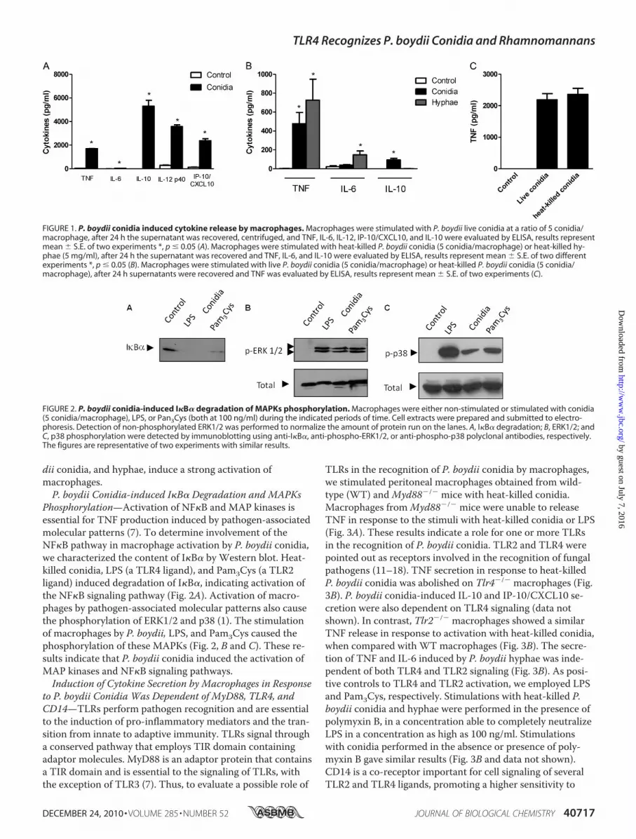
dii conidia, and hyphae, induce a strong activation ofmacrophages.P. boydii Conidia-induced I�B� Degradation and MAPKs
Phosphorylation—Activation of NF�B and MAP kinases isessential for TNF production induced by pathogen-associatedmolecular patterns (7). To determine involvement of theNF�B pathway in macrophage activation by P. boydii conidia,we characterized the content of I�B� by Western blot. Heat-killed conidia, LPS (a TLR4 ligand), and Pam3Cys (a TLR2ligand) induced degradation of I�B�, indicating activation ofthe NF�B signaling pathway (Fig. 2A). Activation of macro-phages by pathogen-associated molecular patterns also causethe phosphorylation of ERK1/2 and p38 (1). The stimulationof macrophages by P. boydii, LPS, and Pam3Cys caused thephosphorylation of these MAPKs (Fig. 2, B and C). These re-sults indicate that P. boydii conidia induced the activation ofMAP kinases and NF�B signaling pathways.Induction of Cytokine Secretion by Macrophages in Response
to P. boydii Conidia Was Dependent of MyD88, TLR4, andCD14—TLRs perform pathogen recognition and are essentialto the induction of pro-inflammatory mediators and the tran-sition from innate to adaptive immunity. TLRs signal througha conserved pathway that employs TIR domain containingadaptor molecules. MyD88 is an adaptor protein that containsa TIR domain and is essential to the signaling of TLRs, withthe exception of TLR3 (7). Thus, to evaluate a possible role of
TLRs in the recognition of P. boydii conidia by macrophages,we stimulated peritoneal macrophages obtained from wild-type (WT) andMyd88�/� mice with heat-killed conidia.Macrophages fromMyd88�/� mice were unable to releaseTNF in response to the stimuli with heat-killed conidia or LPS(Fig. 3A). These results indicate a role for one or more TLRsin the recognition of P. boydii conidia. TLR2 and TLR4 werepointed out as receptors involved in the recognition of fungalpathogens (11–18). TNF secretion in response to heat-killedP. boydii conidia was abolished on Tlr4�/� macrophages (Fig.3B). P. boydii conidia-induced IL-10 and IP-10/CXCL10 se-cretion were also dependent on TLR4 signaling (data notshown). In contrast, Tlr2�/� macrophages showed a similarTNF release in response to activation with heat-killed conidia,when compared with WT macrophages (Fig. 3B). The secre-tion of TNF and IL-6 induced by P. boydii hyphae was inde-pendent of both TLR4 and TLR2 signaling (Fig. 3B). As posi-tive controls to TLR4 and TLR2 activation, we employed LPSand Pam3Cys, respectively. Stimulations with heat-killed P.boydii conidia and hyphae were performed in the presence ofpolymyxin B, in a concentration able to completely neutralizeLPS in a concentration as high as 100 ng/ml. Stimulationswith conidia performed in the absence or presence of poly-myxin B gave similar results (Fig. 3B and data not shown).CD14 is a co-receptor important for cell signaling of severalTLR2 and TLR4 ligands, promoting a higher sensitivity to
FIGURE 1. P. boydii conidia induced cytokine release by macrophages. Macrophages were stimulated with P. boydii live conidia at a ratio of 5 conidia/macrophage, after 24 h the supernatant was recovered, centrifuged, and TNF, IL-6, IL-12, IP-10/CXCL10, and IL-10 were evaluated by ELISA, results representmean � S.E. of two experiments *, p � 0.05 (A). Macrophages were stimulated with heat-killed P. boydii conidia (5 conidia/macrophage) or heat-killed hy-phae (5 mg/ml), after 24 h the supernatant was recovered and TNF, IL-6, and IL-10 were evaluated by ELISA, results represent mean � S.E. of two differentexperiments *, p � 0.05 (B). Macrophages were stimulated with live P. boydii conidia (5 conidia/macrophage) or heat-killed P. boydii conidia (5 conidia/macrophage), after 24 h supernatants were recovered and TNF was evaluated by ELISA, results represent mean � S.E. of two experiments (C).
FIGURE 2. P. boydii conidia-induced I�B� degradation of MAPKs phosphorylation. Macrophages were either non-stimulated or stimulated with conidia(5 conidia/macrophage), LPS, or Pan3Cys (both at 100 ng/ml) during the indicated periods of time. Cell extracts were prepared and submitted to electro-phoresis. Detection of non-phosphorylated ERK1/2 was performed to normalize the amount of protein run on the lanes. A, I�B� degradation; B, ERK1/2; andC, p38 phosphorylation were detected by immunoblotting using anti-I�B�, anti-phospho-ERK1/2, or anti-phospho-p38 polyclonal antibodies, respectively.The figures are representative of two experiments with similar results.
TLR4 Recognizes P. boydii Conidia and Rhamnomannans
DECEMBER 24, 2010 • VOLUME 285 • NUMBER 52 JOURNAL OF BIOLOGICAL CHEMISTRY 40717
by guest on July 7, 2016http://w
ww
.jbc.org/D
ownloaded from

small concentrations of agonists (33). Moreover, CD14 hasbeen shown to participate in the recognition of fungal mole-cules (34–36). Thus we investigated the role of CD14 in therecognition of conidia by macrophages. These experimentswere conducted in the absence of serum to avoid any exoge-nous source of soluble CD14. Macrophages from Cd14�/�
mice stimulated with conidia presented an impaired produc-tion of TNF as compared with WT macrophages (Fig. 3C). Asexpected, at the concentration tested (100 ng/ml), LPS wasunable to induce an optimal TNF release from Cd14�/�
macrophages (Fig. 3C).Isolation and Characterization of P. boydii
Rhamnomannans—Mannans are molecular patterns ex-pressed by pathogenic fungi like C. albicans, and triggersTLR4 activation, as well as Dectin-2 and mannose receptor,promoting cytokine secretion by macrophages (14, 37). Be-cause our results pointed to a role for TLR4 in P. boydiiconidia recognition, we hypothesized that polysaccharidessimilar to mannans could be the molecular patterns expressedin P. boydii conidia involved in TLR4 activation. Thus, weisolated and characterized rhamnomannans from P. boydii
using a hot alkaline extraction. Polysaccharides were thenfractioned by gel filtration, and further analyses were carriedout using fraction II that consisted predominantly of rhamno-mannans with a low amount of protein (Fig. 4, Tables 1 and2). This fraction showed the presence of rhamnose, man-nose, glucose, and traces of galactose, whereas fractions Iand III presented only glucose (data not shown). To pre-cisely determine the composition of monosaccharides inthe rhamnomannan fraction purified from P. boydii, weanalyzed the alditol acetates by GC-MS. As indicated inTable 1, fraction II present rhamnose (23.5%), mannose(45.5%), glucose (31.0%), and traces of galactose. In con-trast, fractions I and III were constituted essentially by glu-cose and minor traces of mannose (data not shown). Wealso performed quantification of total sugars, protein,phosphate, and hexosamine, and all fractions were free ofphosphate and hexosamine (Table 2).
FIGURE 4. Purification of rhamnomannans from P. boydii. Polysaccha-rides from P. boydii were extracted by hot alkaline treatment and successiveprecipitation with ethanol, polysaccharides were submitted to gel filtrationin a Superdex 200 column with a phosphate/NaCl buffer. Carbohydrateswere quantified by reaction in the presence of 5% phenol and sulfuric acidand reading at 490 nm. The result represents the pattern of elution of differ-ent fractions in relationship to their carbohydrate contents.
TABLE 1Chemical composition (%) of rhamnomannan fraction of P. boydii
Percentual of componentsUnfractionated polysaccharides Fraction II
Total sugara 90 90Proteinb 7 TracesPhosphatec Traces Not detectedHexosamined Traces Not detected
a Data from Ref. 24.b Data from Ref. 25.c Data from Ref. 26.d Data from Ref. 27.
TABLE 2Quantitative composition of monosaccharides present in thepolysaccharides obtained from P. boydii by hot alkaline extraction asevaluated by gas chromatographyMonosaccharides were analyzed by detection of their alditol-acetate derivativesusing a capillary column DB-225 (25 � 0.22 mm) programmed for a temperatureof 170–210 °C with a variation of 20 °C/min.
Percentual of monosaccharidesUnfractionated polysaccharides Fraction II
Rhamnose 7.9 26.5Mannose 16.5 38.5Glucose 72.4 28.2Galactose 3.2 7.7
FIGURE 3. The P. boydii conidia-induced TNF release by macrophagesrequires functional MyD88, TLR4, and CD14. Peritoneal macrophageswere obtained from WT, Myd88�/� (A), Tlr2�/�, Tlr4�/� (B), and Cd14�/� (C)mice and stimulated with heat-killed P. boydii conidia (5 conidia/cell). Ascontrols, LPS and Pam3Cys (both at 100 ng/ml) were included in the experi-mental settings. Polymyxin B (10 �g/ml) was added during stimulation ofmacrophages with conidia. Supernatants were collected and TNF was eval-uated by ELISA. Results represent mean � S.E. and are representative of twoor three experiments. * p � 0.05.
TLR4 Recognizes P. boydii Conidia and Rhamnomannans
40718 JOURNAL OF BIOLOGICAL CHEMISTRY VOLUME 285 • NUMBER 52 • DECEMBER 24, 2010
by guest on July 7, 2016http://w
ww
.jbc.org/D
ownloaded from

RMNAnalysis of PreparationsContainingRhamnomannans—One dimensional and two-dimensional NMR analysis con-firmed the structures of the polysaccharides present in Frac-tion II, suggesting that it contains typical signals of �-glucanand rhamnomannan (22, 38). Edited HSQC spectrum showedsubstituted C-1 and C-4 signals at � 101.2/5.400, 101.5/5.359,and 78.8/3.667 of �-D-Glcp units (Fig. 5A). The �-glucan wasconfirmed by a TOCSY experiment using a mixing time of120 ms, which allows observing the glucopyranosyl connectiv-ity (Fig. 5B). Total 1H-1H axial correlations were observed forthe three �-D-Glcp units at � 5.400-A, 5.359-B, and 4.976-C,corroborating a glycogen-like structure. These features areshown in the partial TOCSY spectrum in the diagonal and its
cross-peaks (Fig. 5B). Rhamnomannan identification was de-termined by one-dimensional (1H and 13C) and two-dimen-sional COSY, TOCSY, and HSQC experiments. The NMRdata of fraction II showed at C-1 signals at � 97.9/4.981,101.0/4.967, 102.2/5.228, and 103.9/5.060, typical of terminal�-rhamnose units, O-3,6-substituted-�-mannopyranose (�-D-Manp-(13 3,6)), O-2-substituted-�-mannopyranose(�-D-Manp-(13 2)), and �-Manp-3-O-substituted units,respectively (38, 39). The signal at � 79.9/4.127 confirms the3-O-substituted �-Manp units (Fig. 5A). The phase-sensitiveedited HSQC gave inverted signals of CH2 at � 62.0/3878,62.4/3.785, and 67.0/4.013; 3.771, which correspond to non-substituted C-6 units of Glcp and Manp and O-substituted
FIGURE 5. Partial two-dimensional NMR spectra (edited HSQC and TOCSY) of fraction II. A, partial edited HSQC spectrum, assignment of the main sig-nals from the anomeric region and carbohydrate linkages, the positive phase (blue) correspond to CH and CH3 carbons, and the negative phase (red) corre-spond to CH2 carbons. B, partial TOCSY from the anomeric region showing the main cross-peaks of the rhamnomannan and �-glucan.
TLR4 Recognizes P. boydii Conidia and Rhamnomannans
DECEMBER 24, 2010 • VOLUME 285 • NUMBER 52 JOURNAL OF BIOLOGICAL CHEMISTRY 40719
by guest on July 7, 2016http://w
ww
.jbc.org/D
ownloaded from
C-6 of Manp units. These signals were observed on negativephase (red), and the C-6 units of Rhap were observed at �18.4/1.300 on positive phase (blue) (see Fig. 5A). COSY andTOCSY complemented the identification of the rhamnoman-nan and showed the characteristic low connectivity of theRhap and Manp units, easily visualized when compared withhigher connectivity of the Glcp units (Fig. 5B).Rhamnomannan Preparations from P. boydii-induced Cyto-
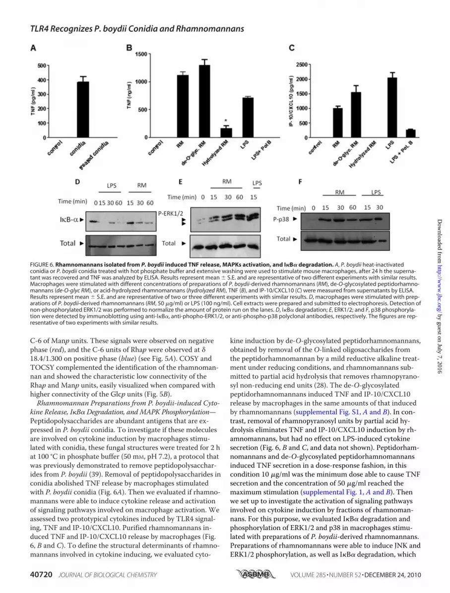
kine Release, I�B� Degradation, andMAPK Phosphorylation—Peptidopolysaccharides are abundant antigens that are ex-pressed in P. boydii conidia. To investigate if these moleculesare involved on cytokine induction by macrophages stimu-lated with conidia, these fungal structures were treated for 2 hat 100 °C in phosphate buffer (50 mM, pH 7.2), a protocol thatwas previously demonstrated to remove peptidopolysacchar-ides from P. boydii (39). Removal of peptidopolysaccharides inconidia abolished TNF release by macrophages stimulatedwith P. boydii conidia (Fig. 6A). Then we evaluated if rhamno-mannans were able to induce cytokine release and activationof signaling pathways involved on macrophage activation. Weassessed two prototypical cytokines induced by TLR4 signal-ing, TNF and IP-10/CXCL10. Purified rhamnomannans in-duced TNF and IP-10/CXCL10 release by macrophages (Fig.6, B and C). To define the structural determinants of rhamno-mannans involved in cytokine inducing, we evaluated cyto-
kine induction by de-O-glycosylated peptidorhamnomannans,obtained by removal of the O-linked oligosaccharides fromthe peptidorhamnomannan by a mild reductive alkaline treat-ment under reducing conditions, and rhamnomannans sub-mitted to partial acid hydrolysis that removes rhamnopyrano-syl non-reducing end units (28). The de-O-glycosylatedpeptidorhamnomannans induced TNF and IP-10/CXCL10release by macrophages in the same amounts of that inducedby rhamnomannans (supplemental Fig. S1, A and B). In con-trast, removal of rhamnopyranosyl units by partial acid hy-drolysis eliminates TNF and IP-10/CXCL10 induction by rh-amnomannans, but had no effect on LPS-induced cytokinesecretion (Fig. 6, B and C, and data not shown). Peptidorham-nomannans and de-O-glycosylated peptidorhamnomannansinduced TNF secretion in a dose-response fashion, in thiscondition 10 �g/ml was the minimum dose able to cause TNFsecretion and the concentration of 50 �g/ml reached themaximum stimulation (supplemental Fig. 1, A and B). Thenwe set up to investigate the activation of signaling pathwaysinvolved on cytokine induction by fractions of rhamnoman-nans. For this purpose, we evaluated I�B� degradation andphosphorylation of ERK1/2 and p38 in macrophages stimu-lated with preparations of P. boydii-derived rhamnomannans.Preparations of rhamnomannans were able to induce JNK andERK1/2 phosphorylation, as well as I�B� degradation, which
FIGURE 6. Rhamnomannans isolated from P. boydii induced TNF release, MAPKs activation, and I�B� degradation. A, P. boydii heat-inactivatedconidia or P. boydii conidia treated with hot phosphate buffer and extensive washing were used to stimulate mouse macrophages, after 24 h the superna-tant was recovered and TNF was analyzed by ELISA. Results represent mean � S.E. and are representative of two different experiments with similar results.Macrophages were stimulated with different concentrations of preparations of P. boydii-derived rhamnomannans (RM), de-O-glycosylated peptidorhamno-mannans (de-O-glyc RM), or acid-hydrolyzed rhamnomannans (hydrolyzed RM), TNF (B), and IP-10/CXCL10 (C) were measured from supernatants by ELISA.Results represent mean � S.E. and are representative of two or three different experiments with similar results. D, macrophages were stimulated with prep-arations of P. boydii-derived rhamnomannans (RM, 50 �g/ml) or LPS (100 ng/ml). Cell extracts were prepared and submitted to electrophoresis. Detection ofnon-phosphorylated ERK1/2 was performed to normalize the amount of protein run on the lanes. D, I�B� degradation; E, ERK1/2; and F, p38 phosphoryla-tion were detected by immunoblotting using anti-I�B�, anti-phopho-ERK1/2, or anti-phospho-p38 polyclonal antibodies, respectively. The figures are rep-resentative of two experiments with similar results.
TLR4 Recognizes P. boydii Conidia and Rhamnomannans
40720 JOURNAL OF BIOLOGICAL CHEMISTRY VOLUME 285 • NUMBER 52 • DECEMBER 24, 2010
by guest on July 7, 2016http://w
ww
.jbc.org/D
ownloaded from

are essential components of signaling pathways triggered byTLRs. LPS, a TLR4 activator and a positive control to our ex-perimental conditions, was able to induce a similar pattern ofsignal transduction, with JNK, ERK1/2 phosphorylation, andI�B degradation (Fig. 6D).Cytokine Release Induced by Rhamnomannans Derived
from P. boydii Required TLR4 Recognition—Cytokine releaseby macrophages in response to P. boydii conidia is dependenton TLR4 signaling, and removal of peptidopolysaccharidesfrom conidia reduces cytokine release by macrophages. Basedon these results, the role of TLR4 on macrophage activationby rhamnomannan was investigated. P. boydii-derived rham-nomannans induced TNF, IL-6, IL-10, and IP-10/CXCL10production by WT macrophages, but not by macrophagesfrom Tlr4�/� mice (Fig. 7, A and B, and data not shown). Ac-tivation of bone marrow-derived macrophages by P. boydii-derived rhamnomannans was also dependent on TLR4 signal-ing, as observed by the reduced TNF and IL-10 release byTlr4�/� macrophages (data not shown). All experiments wereperformed in the presence of polymyxin B (10 �g/ml), used ina concentration able to completely neutralize LPS at 100ng/ml (data not shown). Induction of TNF and IP-10/CXCL10 release by de-O-glycosylated rhamnomannans alsorequired TLR4 signaling, as demonstrated by the impairedTNF release by Tlr4�/� macrophages in comparison to theWT macrophages (data not shown).
DISCUSSION
Although P. boydii represents an emergent pathogen with aubiquitous distribution in the environment, there is a greatgap in the knowledge about the mechanisms of resistancetriggered by the immune recognition of this pathogen. In thiswork we investigated the activation of innate immunity bydifferent developmental stages of P. boydii. Our results indi-cate that P. boydii conidia and hyphae induce macrophageactivation, as observed by cytokine release, but use distinctmechanisms with conidia inducing TLR4 but not TLR2 sig-naling, whereas hyphae recognition is independent of bothTLR2 and TLR4.Macrophages responded to live conidia stimulation secret-
ing TNF, IL-12, IP-10/CXCL10, and IL-10. Because the differ-entiation of live conidia to hyphae occurred during stimula-tion, recognition of P. boydii by macrophages could involveone or both of these two different developmental forms. Both
P. boydii conidia and hyphae induced TNF secretion bymacrophages, but heat-killed conidia promoted IL-10 secre-tion by macrophages, whereas hyphae did not induce IL-10release. The mechanisms involved in the differences of IL-10secretion by conidia and hyphae are not clear. It is possiblethat a different expression of molecules could result in trig-gering of distinct receptors. C-type lectin receptors like man-nose receptor or DC-SIGN and its mouse counterpartSIGNR1 are involved in the recognition of several pathogens.These receptors bind mannosylated structures and are stronginducers of IL-10 (40, 41). Our results demonstrate that IL-10induction by P. boydii conidia is dependent of TLR4 signaling,thus it is possible that TLR4 and lectin receptors could coop-erate in the recognition of P. boydii conidia and IL-10 induc-tion by this developmental stage, whereas recognition of hy-phae would involve different pattern recognition receptorsthat would not induce IL-10 release by macrophages. IL-10 isan anti-inflammatory cytokine and is involved on inhibitionof macrophage activation, so differences in IL-10 induction byconidia and hyphae could contribute for pathogenesis of in-fection promoting an initial anti-inflammatory response thatcould allow conidia to establish infection and germinate intissues.Previous studies described that A. fumigatus germinating
conidia, but not resting conidia, were able to induce a pro-inflammatory macrophage response, being this property is aconsequence of the exposure of �-glucans and possibly un-known TLR ligands during the swelling of conidia (29–31).Our results indicated that P. boydii conidia induce cytokinerelease by macrophages, but we cannot exclude the possibilitythat the P. boydii conidial preparations used in our experi-ments contain germinating conidia.We observed that P. boydii conidia induced the activation
of intracellular signaling pathways typical of TLRs, includingdegradation of I�B� and phosphorylation of MAPKs. Conidiaalso induced TNF and IP-10/CXCL10. These cytokines areinduced by LPS through MyD88 and TRIF pathways uponTLR4 activation (7). The secretion of TNF by macrophagesafter challenge with P. boydii conidia required MyD88, thusindicating a role for TLRs. Recognition of P. boydii conidiawas dependent on TLR4 signaling, whereas TLR2 was dispen-sable for induction of cytokine release. In contrast, our datademonstrated that macrophage activation by P. boydii hyphae
FIGURE 7. Preparations of rhamnomannans derived from P. boydii-induced cytokine release by macrophages through TLR4 signaling. Macrophageswere obtained from WT or Tlr4�/� mice and stimulated with preparations of P. boydii-derived rhamnomannans (RM, 50 �g/ml), LPS (100 ng/ml), Pam3Cys(100 ng/ml), and polymyxin B (Pol. B) (10 �g/ml) were added during stimuli with preparations of rhamnomannans and LPS in some wells. Supernatantswere recovered and TNF (A) and IL-10 (B) were quantified by ELISA, results represent mean � S.E. and are representative of two or three different experi-ments with similar results. *, p � 0.05.
TLR4 Recognizes P. boydii Conidia and Rhamnomannans
DECEMBER 24, 2010 • VOLUME 285 • NUMBER 52 JOURNAL OF BIOLOGICAL CHEMISTRY 40721
by guest on July 7, 2016http://w
ww
.jbc.org/D
ownloaded from

is independent of TLR2 and TLR4 signaling. The Saccharo-myces cerevisiae cell wall particle, zymosan, requires TLR2 forinduction of cytokines by macrophages (42–44). Similarly C.albicans and A. fumigatus induce activation of immune cellsby TLR2 triggering (11, 12, 15, 16). We have previously dem-onstrated that a highly purified �-glucan obtained from P.boydii is a TLR2 activator (22). Our present results do notdiscard the possibility that TLR2 participates in P. boydii rec-ognition, but indicate that TLR4 is the major receptor in-volved on P. boydii conidia recognition. These results alsoimply that molecules distinct from �-glucans are the majoractivators of innate immunity induced by P. boydii conidia,and suggest that �-glucans of the cell wall might be inaccessi-ble for recognition by TLR2 or are minor components of thecell wall in P. boydii conidia. Using Cd14�/� macrophages, weobserved that these cells also had impaired TNF secretionwhen compared with WT macrophages, thus indicating animportant role for CD14 in macrophage activation by P. boy-dii conidia. Although CD14 lacks an intracellular signalingtail, it participates on binding of pathogen molecules, increas-ing responses triggered by TLR2 and TLR4 (33). Possibly,CD14 promotes binding and transference of molecules ex-pressed in the P. boydii conidial surface to TLR4.Rhamnomannan-enriched preparations isolated from P.
boydii induced cytokine release by macrophages, as well asdegradation of I�B� and phosphorylation of MAPKs. Ourresults showed that P. boydii-derived rhamnomannans alsorequired TLR4 signaling for cytokine induction by macro-phages. The requirement for TLR4 on macrophage activationby P. boydii-derived rhamnomannans seems to mirror therole of TLR4 in recognition of P. boydii conidia by macro-phages. This possibility is supported by our results demon-strating that P. boydii conidia present a strong expression ofrhamnomannans on the cell surface, as analyzed by immuno-fluorescence using labeling with monoclonal antibodies ofanti-rhamnomannans from P. boydii (46).A putative contamination of endotoxin in the preparations
of conidia and rhamnomannans as responsible for the ob-served cytokine secretion is unlikely. We used polymyxin B ina dose that abrogated LPS-induced cytokine production andthis treatment had no effect on conidia or rhamnomannans-induced cytokine secretion. Conversely, removal of pepti-dopolysaccharides in conidia and hydrolysis of rhamnoman-nans abrogated the production of cytokine, although had noeffect on LPS-induced TNF secretion.Although our preparations of rhamnomannans presented a
significant amount of �-glucans (about 28% of total polysac-charides of fraction II), activation of TLR4 seems to be a con-sequence of the recognition of rhamnomannans, as shown bythe following evidence: 1) we have previously demonstratedthat highly purified �-glucans are activators of TLR2 but notTLR4; 2) removing terminal rhamnoses from rhamnoman-nans, by partial acid hydrolysis, abolished cytokine inductionby these polysaccharides, whereas �-glucans do not presentcaps with rhamnoses; and 3) concentrations of contaminant�-glucans are unable to induce the same level of cytokine re-lease observed by a concentration of 50 �g/ml of polysacchar-
ides enriched with rhamnomannans (less than 15 �g/ml of�-glucans).
We also demonstrated that O-linked oligosaccharides fromthe peptidorhamnomanan are not involved on macrophageactivation by P. boydii-derived rhamnomannans, becausepreparations of peptidorhamnomannans that were submittedto �-elimination, a process that removes O-linked but notN-linked carbohydrates, showed a similar induction of cyto-kines by macrophages and this effect was still dependent onTLR4 activation. In contrast, removal of terminal rhamnopyr-anosyl units impaired cytokine release by macrophages in re-sponse to P. boydii-derived rhamnomannans, implying thatstructures with terminal rhamnose and/or mannose are struc-tural motifs involved on TLR4 recognition. It has recentlybeen described that a soluble form of TLR4-Fc is able to bindcomplex mixtures of fungal polysaccharides and the bindingis blocked by soluble mannans or fucose (45), a deoxy-monosaccharide-like rhamnose, suggesting that TLR4 couldrecognize structural patterns like fungal polysaccharidesthrough direct interaction with terminal deoxycarbohydratesexpressed in complex polysaccharides like rhamnomannans.TLR4 has been described as a receptor involved in C. albicansand A. fumigatus recognition (15–17). Mannans obtainedfrom yeasts like S. cerevisiase and Candida sp. activate macro-phages through TLR4 (35). Moreover, O-linked mannansfrom C. albicans and glucuroxylomannan obtained fromCryptococcus neoformans are also able to induce innate im-mune activation through TLR4 (14, 36). In these works, TLR4recognizes C. albicans-derived O-linked mannans, but notN-linked, a result different from ours, that demonstrate thatremoving O-linked oligosaccharides from P. boydii-derivedrhamnomannans does not affect TLR4-mediated recognition.The differences observed could be due to the different experi-mental settings. The experimental approach employed byNetea et al. (14) was based on genetic deficiency of pathwaysinvolved in the N-linked and O-linked formation of oligosac-charide chains in surface proteins. It is possible that the ap-proach used by Netea et al. (14) resulted in an anomalous pat-tern of glycosylation or in differences on the expression ofTLR4 ligands, instead of reflecting a simple absence of recog-nition of N-linked mannans by TLR4. Another possibility isthat the patterns of glycosylation in P. boydii- and C. albicans-derived mannans are distinct in relation to TLR4 activation,with different requirements for N-linked or O-linked man-nans/rhamnomannans in these two different fungalpathogens.Our results indicate that recognition of conidial forms of P.
boydii by the innate immune system requires functional TLR4and CD14 and that P. boydii-derived rhamnomannans aremolecular patterns recognized by TLR4. These results addnew information on the role of mannan-containing polymersin innate recognition of fungal pathogens. It is possible thatthese polymers expressed in other filamentous fungi (like A.fumigatus) could be involved in innate immune recognition,possibly by triggering TLR4. Thus, modulation of TLR4 sig-naling could be an important therapy for inducing resistancein individuals with invasive infections caused by P. boydii.Otherwise, antagonism of TLR4 signaling in association with
TLR4 Recognizes P. boydii Conidia and Rhamnomannans
40722 JOURNAL OF BIOLOGICAL CHEMISTRY VOLUME 285 • NUMBER 52 • DECEMBER 24, 2010
by guest on July 7, 2016http://w
ww
.jbc.org/D
ownloaded from

effective antifungal drugs could control the infection and re-duce tissue damage associated with the immune responsedirected to selected fungal infections including P. boydii.
Acknowledgments—We are grateful to Shizuo Akira and DouglasGolenbock for providing genetically deficient mice strains (Myd88,Tlr2, Tlr4, and Cd14), and Patricia Bozza, Maria Bellio, andRicardo Gazzinelli for providing mice and reagents. We thank Clau-dia Paiva for the critical reading the manuscript.
REFERENCES1. Beck-Sague, C., and Jarvis, W. R. (1993) J. Infect. Dis. 167, 1247–12512. Baddley, J. W., Stroud, T. P., Salzman, D., and Pappas, P. G. (2001) Clin.
Infect. Dis. 32, 1319–13243. Warris, A., Bjorneklett, A., and Gaustad, P. (2001) N. Engl. J. Med. 344,
1099–11004. Peter, E., Bakri, F., Ball, D. M., Cheney, R. T., and Segal, B. H. (2002)
Clin. Infect. Dis. 35, e54–565. Zelante, T., Montagnoli, C., Bozza, S., Gaziano, R., Bellocchio, S., Boni-
fazi, P., Moretti, S., Fallarino, F., Puccetti, P., and Romani, L. (2007) Adv.Exp. Med. Biol. 590, 209–221
6. Hohl, T. M., Rivera, A., and Pamer, E. G. (2006) Curr. Opin. Immunol.18, 465–472
7. Akira, S., and Takeda, K. (2004) Nat. Rev. Immunol. 4, 499–5118. Lemaitre, B., Nicolas, E., Michaut, L., Reichhart, J. M., and Hoffmann,
J. A. (1996) Cell 86, 973–9839. Netea, M. G., Van der Graaf, C., Van der Meer, J. W., and Kullberg, B. J.
(2004) Eur. J. Clin. Microbiol. Infect. Dis. 23, 672–67610. Levitz, S. M. (2010) PLoS Pathog. 6, e100075811. Mambula, S. S., Sau, K., Henneke, P., Golenbock, D. T., and Levitz, S. M.
(2002) J. Biol. Chem. 277, 39320–3932612. Meier, A., Kirschning, C. J., Nikolaus, T., Wagner, H., Heesemann, J.,
and Ebel, F. (2003) Cell. Microbiol. 5, 561–57013. Netea, M. G., Warris, A., Van der Meer, J. W., Fenton, M. J., Verver-
Janssen, T. J., Jacobs, L. E., Andresen, T., Verweij, P. E., and Kullberg,B. J. (2003) J. Infect. Dis. 188, 320–326
14. Netea, M. G., Gow, N. A., Munro, C. A., Bates, S., Collins, C., Ferwerda,G., Hobson, R. P., Bertram, G., Hughes, H. B., Jansen, T., Jacobs, L.,Buurman, E. T., Gijzen, K., Williams, D. L., Torensma, R., McKinnon,A., MacCallum, D. M., Odds, F. C., Van der Meer, J. W., Brown, A. J.,and Kullberg, B. J. (2006) J. Clin. Invest. 116, 1642–1650
15. Villamon, E., Gozalbo, D., Roig, P., O’Connor, J. E., Fradelizi, D., and Gil,M. L. (2004)Microbes Infect. 6, 1–7
16. Netea, M. G., Van Der Graaf, C. A., Vonk, A. G., Verschueren, I., VanDer Meer, J. W., and Kullberg, B. J. (2002) J. Infect. Dis. 185, 1483–1489
17. Balloy, V., Si-Tahar, M., Takeuchi, O., Philippe, B., Nahori, M. A., Tan-guy, M., Huerre, M., Akira, S., Latge, J. P., and Chignard, M. (2005) In-fect. Immun. 73, 5420–5425
18. Bellocchio, S., Montagnoli, C., Bozza, S., Gaziano, R., Rossi, G., Mam-bula, S. S., Vecchi, A., Mantovani, A., Levitz, S. M., and Romani, L.(2004) J. Immunol. 172, 3059–3069
19. O’Bryan, T. A. (2005) Expert Rev. Anti-infect. Ther. 3, 765–77320. Cortez, K. J., Roilides, E., Quiroz-Telles, F., Meletiadis, J., Antachopou-
los, C., Knudsen, T., Buchanan, W., Milanovich, J., Sutton, D. A.,Fothergill, A., Rinaldi, M. G., Shea, Y. R., Zaoutis, T., Kottilil, S., andWalsh, T. J. (2008) Clin. Microbiol. Rev. 21, 157–197
21. Roilides, E., Simitsopoulou, M., Katragkou, A., and Walsh, T. J. (2009)Med. Mycol. 47, 433–440
22. Bittencourt, V. C., Figueiredo, R. T., da Silva, R. B., Mourao-Sa, D. S.,Fernandez, P. L., Sassaki, G. L., Mulloy, B., Bozza, M. T., and Barreto-Bergter, E. (2006) J. Biol. Chem. 281, 22614–22623
23. Gilgado, F., Cano, J., Gene, J., and Guarro, J. (2005) J. Clin. Microbiol. 43,4930–4942
24. Dubois, M., Gilles, K. A., Hamilton, J. K., Rebers, P. A., and Swith, E.(1956) Anal. Chem. 28, 350–356
25. Lowry, O. H., Rosebrough, N. J., Farr, A. L., and Randall, R. J. (1951)J. Biol. Chem. 193, 265–275
26. Ames, B. N. (1966)Methods Enzymol. 8, 115–11827. Belcher, R. A., Nutten, A. J., and Sambrook, C. M. (1954) Analyst 79,
201–20828. Leitao, E. A., Bittencourt, V. C., Haido, R. M., Valente, A. P., Peter-
Katalinic, J., Letzel, M., de Souza, L. M., and Barreto-Bergter, E. (2003)Glycobiology 13, 681–692
29. Gersuk, G. M., Underhill, D. M., Zhu, L., and Marr, K. A. (2006) J. Im-munol. 176, 3717–3724
30. Hohl, T. M., Van Epps, H. L., Rivera, A., Morgan, L. A., Chen, P. L.,Feldmesser, M., and Pamer, E. G. (2005) PLoS Pathog. 1, e30
31. Steele, C., Rapaka, R. R., Metz, A., Pop, S. M., Williams, D. L., Gordon,S., Kolls, J. K., and Brown, G. D. (2005) PLoS Pathog. 1, e42
32. Gantner, B. N., Simmons, R. M., and Underhill, D. M. (2005) EMBO J.24, 1277–1286
33. Pugin, J., Heumann, I. D., Tomasz, A., Kravchenko, V. V., Akamatsu, Y.,Nishijima, M., Glauser, M. P., Tobias, P. S., and Ulevitch, R. J. (1994)Immunity 1, 509–516
34. Barbosa, F. M., Fonseca, F. L., Figueiredo, R. T., Bozza, M. T., Casadevall,A., Nimrichter, L., and Rodrigues, M. L. (2007) Clin. Vaccine Immunol.14, 94–98
35. Tada, H., Nemoto, E., Shimauchi, H., Watanabe, T., Mikami, T., Mat-sumoto, T., Ohno, N., Tamura, H., Shibata, K., Akashi, S., Miyake, K.,Sugawara, S., and Takada, H. (2002)Microbiol. Immunol. 46, 503–512
36. Shoham, S., Huang, C., Chen, J. M., Golenbock, D. T., and Levitz, S. M.(2001) J. Immunol. 166, 4620–4626
37. Saijo, S., Ikeda, S., Yamabe, K., Kakuta, S., Ishigame, H., Akitsu, A., Fu-jikado, N., Kusaka, T., Kubo, S., Chung, S. H., Komatsu, R., Miura, N.,Adachi, Y., Ohno, N., Shibuya, K., Yamamoto, N., Kawakami, K., Ya-masaki, S., Saito, T., Akira, S., and Iwakura, Y. (2010) Immunity 32,681–691
38. Gorin, P. A. (1981) Adv. Carbohydr. Chem. Biochem. 38, 13–10439. Pinto, M. R., Mulloy, B., Haido, R. M., Travassos, L. R., and Barreto
Bergter, E. (2001)Microbiology 147, 1499–150640. Chieppa, M., Bianchi, G., Doni, A., Del Prete, A., Sironi, M., Laskarin, G.,
Monti, P., Piemonti, L., Biondi, A., Mantovani, A., Introna, M., and Al-lavena, P. (2003) J. Immunol. 171, 4552–4560
41. Wieland, C. W., Koppel, E. A., den Dunnen, J., Florquin, S., McKenzie,A. N., van Kooyk, Y., van der Poll, T., and Geijtenbeek, T. B. (2007)Mi-crobes Infect. 9, 134–141
42. Brown, G. D., Herre, J., Williams, D. L., Willment, J. A., Marshall, A. S.,and Gordon, S. (2003) J. Exp. Med. 197, 1119–1124
43. Gantner, B. N., Simmons, R. M., Canavera, S. J., Akira, S., and Underhill,D. M. (2003) J. Exp. Med. 197, 1107–1117
44. Underhill, D. M., Ozinsky, A., Hajjar, A. M., Stevens, A., Wilson, C. B.,Bassetti, M., and Aderem, A. (1999) Nature 401, 811–815
45. Hsu, T. L., Cheng, S. C., Yang, W. B., Chin, S. W., Chen, B. H., Huang,M. T., Hsieh, S. L., and Wong, C. H. (2009) J. Biol. Chem. 284,34479–34489
46. Lopes, L. C., Rollin-Pinheiro, R., Guimaraes, A. J., Bittencourt, V. C.,Martinez, L. R., Koba, W., Farias, S. E., Nosanchuk, J. D., and Barreto-Bergter, E. (2010) PLoS Negl. Trop. Dis. 4, e853
TLR4 Recognizes P. boydii Conidia and Rhamnomannans
DECEMBER 24, 2010 • VOLUME 285 • NUMBER 52 JOURNAL OF BIOLOGICAL CHEMISTRY 40723
by guest on July 7, 2016http://w
ww
.jbc.org/D
ownloaded from
Barreto-Bergter and Marcelo T. BozzaLívia Cristina Lopes, Vera Carolina B. Bittencourt, Guilherme L. Sassaki, Eliana
Rodrigo T. Figueiredo, Patrícia L. Fernandez, Fabianno F. Dutra, Yissett González, Conidia and Purified RhamnomannansPseudallescheria boydiiTLR4 Recognizes
doi: 10.1074/jbc.M110.181255 originally published online October 19, 20102010, 285:40714-40723.J. Biol. Chem.
10.1074/jbc.M110.181255Access the most updated version of this article at doi:
Alerts:
When a correction for this article is posted•
When this article is cited•
to choose from all of JBC's e-mail alertsClick here
http://www.jbc.org/content/285/52/40714.full.html#ref-list-1
This article cites 46 references, 21 of which can be accessed free at
by guest on July 7, 2016http://w
ww
.jbc.org/D
ownloaded from


