Embed Size (px)
Citation preview

REVIEW ARTICLEpublished: 28 October 2014
doi: 10.3389/fphys.2014.00415
Haptoglobin, hemopexin, and related defensepathways—basic science, clinical perspectives, and drugdevelopmentDominik J. Schaer1*, Francesca Vinchi2, Giada Ingoglia2, Emanuela Tolosano2 and Paul W. Buehler3†
1 Division of Internal Medicine, University of Zurich, Zurich, Switzerland2 Department of Molecular Biotechnology and Health Sciences, University of Torino, Torino, Italy3 Division of Hematology, Laboratory of Biochemistry and Vascular Biology, Center for Biologics Evaluation and Research, Food and Drug Administration, Bethesda,
MD, USA
Edited by:
Magnus Gram, Lund University,Sweden
Reviewed by:
Sruti Shiva, University of Pittsburgh,USAJeak Ling Ding, National Universityof Singapore, SingaporeJoseph M. Rifkind, National Instituteon Aging, USASoren Kragh Moestrup, AarhusUniversity, Denmark
*Correspondence:
Dominik J. Schaer, Division ofInternal Medicine, UniversityHospital, Ramistrasse 100,Zurich CH-8091, Switzerlande-mail: [email protected]
†The findings and conclusions inthis article have not been formallydisseminated by the Food and DrugAdministration and should not beconstrued to represent any Agencydetermination or policy.
Hemolysis, which occurs in many disease states, can trigger a diverse pathophysiologiccascade that is related to the specific biochemical activities of free Hb and its porphyrincomponent heme. Normal erythropoiesis and concomitant removal of senescent redblood cells (RBC) from the circulation occurs at rates of approximately 2 × 10⁶
RBCs/second. Within this physiologic range of RBC turnover, a small fraction ofhemoglobin (Hb) is released into plasma as free extracellular Hb. In humans, there isan efficient multicomponent system of Hb sequestration, oxidative neutralization andclearance. Haptoglobin (Hp) is the primary Hb-binding protein in human plasma, whichattenuates the adverse biochemical and physiologic effects of extracellular Hb. The cellularreceptor target of Hp is the monocyte/macrophage scavenger receptor, CD163. FollowingHb-Hp binding to CD163, cellular internalization of the complex leads to globin and hememetabolism, which is followed by adaptive changes in antioxidant and iron metabolismpathways and macrophage phenotype polarization. When Hb is released from RBCswithin the physiologic range of Hp, the potential deleterious effects of Hb are prevented.However, during hyper-hemolytic conditions or with chronic hemolysis, Hp is depleted andHb readily distributes to tissues where it might be exposed to oxidative conditions. In suchconditions, heme can be released from ferric Hb. The free heme can then accelerate tissuedamage by promoting peroxidative reactions and activation of inflammatory cascades.Hemopexin (Hx) is another plasma glycoprotein able to bind heme with high affinity. Hxsequesters heme in an inert, non-toxic form and transports it to the liver for catabolismand excretion. In the present review we discuss the components of physiologic Hb/hemedetoxification and their potential therapeutic application in a wide range of hemolyticconditions.
Keywords: hemolysis, haptoglobin, hemopexin, CD163, vascular diseases, transfusion, sickle cell disease
INTRODUCTIONHemolysis with release of free hemoglobin (Hb) and heme occursin a wide range of disease states and clinical interventions, includ-ing genetic and acquired anemias such as sickle cell disease,burns, extracorporeal circulation and massive blood transfusion(Schaer et al., 2013c). Additionally, chemical or recombinantmodified Hbs have been evaluated as oxygen therapeutics in sev-eral disease states or when blood is not available (Silverman andWeiskopf, 2009). The study of these therapeutic candidates hasalso provided relevant information regarding the concentration-dependent pathophysiology associated with extracellular Hb incirculation and tissue compartments. Critical reviews providegreater detail concerning their dose dependent toxicity (Buehleret al., 2010).
The primary pathophysiologic effects associated with freeHb/heme are acute hemodynamic instability and acute or chronic
tissue injury (Schaer et al., 2013c). The underlying biochem-istry of these adverse effects appears to be related to the nitricoxide and oxidant reactivity of free Hb and heme. The ability tocounteract extracellular Hb resulting from normal red blood cellturnover and mild hemolysis is one function of haptoglobin (Hp),an α2-sialoglycoprotein, which is the primary Hb-binding pro-tein in plasma. Hb-bound Hp targets a specific cellular pathwayof clearance through the monocyte/macrophage surface recep-tor CD163 (Kristiansen et al., 2001). Depending on the extentand frequency of hemolysis, Hp may become depleted, render-ing this pathway ineffective. Previous reports from patients withsickle cell disease, spherocytosis, autoimmune hemolytic anemia,erythropoietic protoporphyria and pyruvate kinase deficiencysuggest that Hp depletion in plasma occurs prior to the decline ofhemopexin (Hx) concentrations (Muller-Eberhard et al., 1968).The disease related consumption of the plasma Hp pool during
www.frontiersin.org October 2014 | Volume 5 | Article 415 | 1

Schaer et al. Haptoglobin, hemopexin, and associated pathways
hemolysis makes Hp a specific clinical marker for intravascularhemolysis (Kormoczi et al., 2006).
Heme released following oxidation of Hb to met-Hb or fromheme saturated hepatocytes is bound by albumin and rapidlytransferred to Hx, the plasma protein with the highest bind-ing affinity for heme. Hx is another glycoprotein produced bythe liver with a plasma concentration of 1–2 mg/ml (Muller-Eberhard et al., 1968). Hx prevents heme’s pro-oxidant andpro-inflammatory effects and promotes its detoxification, partic-ularly when Hp concentrations are low or depleted in cases ofsevere or prolonged hemolysis. Both Hp and Hx are acute-phaseproteins, induced during infection and inflammatory states inorder to minimize tissue injury and facilitate tissue repair.
The present review will describe the primary mechanismsby which Hp and Hx prevent Hb/heme toxicity prior tomonocyte/macrophage-hepatocyte clearance, critically evaluatethe difference in genetic phenotype function and describe therationale for exogenous Hp and Hx as therapeutic proteins foruse in hemolytic disease states.
MECHANISMS BY WHICH Hp AND Hx PROTECT AGAINSTHb/heme TOXICITYMechanistic research on the in vitro and in vivo function of Hpand Hx has suggested well defined mechanisms of protection(Schaer et al., 2013c). The best-characterized function of Hp isintravascular sequestration of extracellular Hb following forma-tion of large Hb-Hp protein complexes, a process that preventsextravasation of free Hb into tissues. This effect is particularlyevident in the kidneys where oxidative reactions at the heme moi-ety of Hb lead to globin deposition (hyaline casts), iron overload,lipid peroxidation and renal tubular injury (Qian et al., 2010;Ballarin et al., 2011; Billings et al., 2011) (Figure 1). Similarly, bysequestering heme within a protein complex, Hx prevents heme’sability to intercalate into cell membranes and plasma lipopro-teins, where it participates in reactions with organic peroxides,the result of which is the generation of free radicals in local-ized tissue spaces followed by lipid, protein and DNA oxidation.Additionally, Hx blocks heme activation of immune receptors andvascular inflammatory processes (Belcher et al., 2014). The com-bined antioxidant and Hb/heme binding characteristics of Hpand Hx allow Hb-Hp and heme-Hx complexes to circulate inthe blood in a less toxic form until they can be cleared by theirrespective scavenger receptors. In the following sections, Hp andHx will be discussed with an emphasis on tissue protection fromthe deleterious effects of extracellular Hb and heme.
Hp AND Hx SEQUESTER EXTRACELLULAR Hb/heme WITHIN THEPLASMA COMPARTMENT UNTIL CELLULAR CLEARANCEWithin red blood cells (RBCs), Hb exists as a tetrameric hemecontaining protein with a molecular weight of 64 kD. Followingrelease from RBCs during intravascular hemolysis, extracellularHb is in a dynamic tetramer ↔ dimer equilibrium (Ackers andHalvorson, 1974). The dimer fraction of Hb is favored by lowHb concentration and an oxygenated Hb (Fe2+) state. The 32-kDHb dimer readily passes through the glomerulus—if not boundby Hp—and is rapidly cleared by the kidneys (Murray et al.,
1961; Andersen et al., 1966; Boretti et al., 2009). Renal filtra-tion is therefore a primary route of clearance for extracellular Hbfollowing Hp depletion. In guinea pigs and dogs, we have pre-viously characterized plasma clearance of extracellular Hb witha circulatory half-life of approximately 10–60 min (Boretti et al.,2009, 2014). This is consistent with earlier findings using radio-labeled Hb in rabbit and dog (Bunn et al., 1969). Hb can alsobe filtered into the lymphatic system after intravascular injec-tion, and translocates across intact endothelial monolayers bothin vitro and in vivo (Nakai et al., 1998; Faivre-Fiorina et al.,1999; Matheson et al., 2000). The mechanisms underlying this“translocation/extravasation” phenomenon remain unclear, butmay involve active transport mechanisms—for example, throughthe caveolar system, which is also responsible for the controlledtransendothelial transport of other plasma proteins such as albu-min (Faivre-Fiorina et al., 1999; Komarova and Malik, 2010).
Within the plasma compartment, the abundance of anti-oxidants (i.e., ascorbic acid and urate) stabilizes Hb in a reducedstate (Butt et al., 2010). This physiologic function of plasmamakes it less likely for Hb to participate in oxidative reactionsthat lead to rapid met-Hb formation and release of free heme.However, once dimerization of Hb occurs, autoxidation of dimersproceeds rapidly with a rate constant (kox = 0.24 h−1) 16-foldfaster than tetrameric Hb (kox = 0.015 h−1) (Griffon et al., 1998).Furthermore, after translocation into the extravascular space,Hb may encounter much harsher oxidative conditions than inplasma. This may be particularly true in diseased and inflamedtissues or after an ischemic insult when oxidants accumulateand an acidic pH is created in local tissue sites. A disconnectof intravascular and extravascular Hb oxidation was observedin animal models of intravascular hemolysis or following infu-sion of ferrous (Fe2+) Hb. While oxidized ferric (Fe3+) Hbusually remains below the level of detection in plasma, a largefraction of excreted Hb ultimately appears in the urine in theoxidized ferric state (Boretti et al., 2009). This observation couldbe extrapolated to other tissues, such as the vascular wall, whereHb(Fe3+) may accumulate. The large Hb-Hp complex cannot befiltered by the kidney and in vitro translocation across endothe-lial monolayers is almost completely blocked (Lipiski et al., 2013).The most evident protective function of Hp is therefore thesequestration of extracellular Hb within the antioxidant richplasma compartment (Figure 2). Following binding, Hp lim-its Hb’s access to susceptible environments where heme release,oxidative processes and NO-consuming reactions are much morelikely to occur and may finally lead to the sequelae of Hb tox-icity, such as renal failure and vascular injury (Gladwin et al.,2012).
A similar sequestering function is observed with Hx, whichis capable of controlling the reactivity of free heme followingorientation into Hx’s “heme pockets.” By scavenging free heme,Hx prevents heme-mediated oxidative reactions with lipids, pro-teins, nucleic acids and other biological molecules. Along withHx, several plasma proteins are able to bind heme, includ-ing albumin, lipoproteins, α1-microglobulin, but none of theseproteins can efficiently prevent heme intercalation into lipidmembranes or block heme’s pro-oxidant and pro-inflammatoryeffects.
Frontiers in Physiology | Oxidant Physiology October 2014 | Volume 5 | Article 415 | 2
Schaer et al. Haptoglobin, hemopexin, and associated pathways
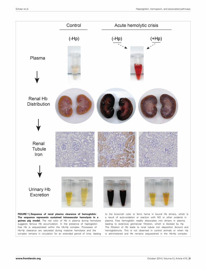
FIGURE 1 | Sequence of renal plasma clearance of hemoglobin:
The sequence represents sustained intravascular hemolysis in a
guinea pig model. The red color of Hb in plasma during hemolysissuggests ferrous Hb accumulation. In the presence of haptoglobinfree Hb is sequestrated within the Hb-Hp complex. Processes ofHb-Hp clearance are saturated during massive hemolysis and thecomplex remains in circulation for an extended period of time, leading
to the brownish color or ferric heme in bound Hb dimers, which isa result of auto-oxidation or reaction with NO or other oxidants inplasma. Free hemoglobin readily dissociates into dimers in plasma,leading to extensive glomerular filtration, which is blocked by Hp.The filtration of Hb leads to renal tubule iron deposition (brown) andhemoglobinuria. This is not observed in control animals or when Hpis administered and Hb remains sequestered in the Hb-Hp complex.
www.frontiersin.org October 2014 | Volume 5 | Article 415 | 3

Schaer et al. Haptoglobin, hemopexin, and associated pathways
FIGURE 2 | Diastolic (A) and systolic (B) systemic arterial blood
pressure response after bolus infusion of free Hb followed by a bolus
infusion of purified human Hp in guinea pigs. (C) HPLC analysis ofguinea pig plasma after infusion of free Hb followed by Hp. The tracesshow free Hb co-eluting with an Hb standard (red). The Hb-Hp complexhas a larger molecular weight, as indicated by the earlier elution time. TheHb-Hp complex has a long circulation half-life with detectable levels up to
50 h after infusion in this species. (D) NO reactions with free Hb in thevascular wall. Ferrous Hb (Fe2+) can react with the vasodilator NO via tworeactions: (1) NO dioxygenation of oxy-Hb that generates nitrate (NO−
3 ) andferric Hb (Fe3+), and (2) iron nitrosylation of deoxy-Hb that occurs by directiron binding of NO to non-liganded ferrous Hb (Fe2+). Both reactions leadto depletion of NO and explain the acute vasoactivity of extracellular Hb,which is attenuated by Hp.
Hp AND Hx PROMOTE FREE Hb AND HEME CLEARANCE VIA RECEPTORTARGETED PATHWAYSHaptoglobinHb toxicity largely depends on the rate of hemolysis, tissue oxi-dant status and clearance capacity. In this network, Hp acts in
concert with the plasma’s small molecular reducing agents thatmaintain Hb in a reduced, less reactive ferrous (Fe2+) oxidationstate (Buehler et al., 2007).
In addition to keeping Hb within the antioxidant environmentof plasma, Hp plays an essential role in the clearance of Hb and
Frontiers in Physiology | Oxidant Physiology October 2014 | Volume 5 | Article 415 | 4

Schaer et al. Haptoglobin, hemopexin, and associated pathways
metabolic detoxification of heme (Figure 3). In humans, the scav-enger receptor CD163, previously assigned as the macrophagescavenger receptor M130 (Law et al., 1993), has been identifiedas an endocytosis receptor for Hb-Hp complexes (Kristiansenet al., 2001). CD163 is exclusively expressed by cells of themonocyte-macrophage lineage, with particularly high expressionlevels in red pulp macrophages of the spleen and liver Kupffercells (Zwadlo et al., 1987; Bachli et al., 2006). Within the Hb-Hp complex, a high-affinity binding epitope has been identifiedwithin the interface region of the Hp β-chain and the Hb α-chain(Madsen et al., 2004; Andersen et al., 2012). Tetrameric Hb canalso bind to and is internalized by CD163 in the absence of Hp(Schaer et al., 2006; Buehler et al., 2008). It is unknown whetherthis lower affinity interaction involves similar regions within Hband CD163, or whether separate binding structures have inde-pendently evolved for Hb and the Hb-Hp complex, respectively(Buehler et al., 2008). Experimental evidence suggests that otherHb and Hb-Hp clearance pathways exist in addition to the CD163scavenger receptor, and that efficient non-renal Hb clearancemechanisms may be active in the absence of Hp (Murray et al.,1961; Schaer et al., 2006; Etzerodt et al., 2013). However, thenature of these mechanisms requires further studies to be morethoroughly understood.
Inflammatory and anti-inflammatory processes modulate theactivity of the Hb-Hp scavenger pathway at multiple levels.Systemic inflammation, particularly if it involves the inter-leukin (IL)-6 effector pathway, increases expression of Hp inthe liver and many parenchymal and non-parenchymal cells.In many species, Hp is one of the most extensively respond-ing acute-phase plasma proteins. IL-6 has also been reportedto enhance expression of CD163 on macrophages, suggestingthat enhanced Hb sequestration and clearance capacity are gen-eral adaptive responses to infection and tissue injury (Buechleret al., 2000). Intriguingly, however, some inflammatory medi-ators, such as endotoxin and other Toll-like receptor (TLR)agonists or tumor necrosis factor-α (TNF-α) trigger protease-mediated shedding of CD163 from the cell surface of mono-cytes and macrophages (Droste et al., 1999; Hintz et al., 2002;Moller et al., 2002; Weaver et al., 2006; Etzerodt et al., 2010).This shedding acutely blocks clearance of Hb-Hp complexes bymonocytes (Schaer et al., 2007). High levels of soluble CD163are consequently found in patients with sepsis or more specificmacrophage activation syndromes (Schaer et al., 2005; Molleret al., 2006). The physiologic role of this process is unknown,but it implies that complex regulatory mechanisms control Hbdetoxification and cellular exposure. Additionally, sCD163 mayitself contribute the oxidative detoxification of extracellular Hband Fcγ-receptor-mediated endocytosis of sCD163-Hb-IgG com-plexes by monocytes/macrophages, and endothelial cells havebeen proposed to be an alternative route for Hb clearance(Subramanian et al., 2013).
Of special consideration is the regulation of the Hb clearancesystem by anti-inflammatory glucocorticoids. In global proteomeand transcriptome exploration studies, CD163 was found to beone of the principal glucocorticoid-induced genes in human andmouse macrophages, and patients treated with high-dose pulseglucocorticoids had a significantly enhanced Hb-Hp clearance
capacity of their peripheral blood monocytes (Schaer et al.,2001, 2002; Vallelian et al., 2010). Glucocorticoids also act as atranscriptional (co-)activator on Hp synthesis in many species,including human (Marinkovic and Baumann, 1990). In a dogmodel, a glucocorticoid-supported high Hp level was completelyprotective against hemoglobinuria and hypertension during an8-h infusion of free Hb (Boretti et al., 2009). Cumulatively,inflammation and glucocorticoids appear to enhance toleranceagainst extracellular Hb. In contrast, other drugs such as theantimalarial chloroquine can negatively affect Hb/heme detox-ification in macrophages and potentially enhance free Hb tox-icity (Schaer et al., 2013b). The potential clinical relevance ofthese observations—for example, for the treatment of hemolyticanemias—remains to be resolved.
HemopexinThe heme scavenging function of Hx primarily attenuates hemetoxicity on the vascular endothelium (Vinchi et al., 2013). Whileheme easily enters into endothelial cells when bound to albu-min, this translocation is completely blocked in the presence ofHx. Heme sequestration within the Hx complex further ensuresprotection against heme-driven oxidative processes in the extra-cellular space and it prevents heme-triggered inflammation andadhesion molecule expression.
In vivo, the heme-Hx complex is primarily cleared by hepa-tocytes via receptor-mediated endocytosis (Vinchi et al., 2013).To date, the only known heme-Hx complex receptor is theLDL receptor-related protein 1 (LRP 1), a multi-ligand scav-enger receptor (Hvidberg et al., 2005). In addition to hepato-cytes, LRP1 is expressed on several other cell types, includingmacrophages, neurons, and syncytiotrophoblasts. The role ofthese LRP1 expressing cell types and the possible contribution ofalternative receptors in heme-Hx complex clearance remain to bestudied.
Some studies have suggested that Hx could be recycled as anintact molecule to the extracellular milieu. However, Hvidberget al., have shown that most Hx is degraded in lysosomes(Hvidberg et al., 2005). This agrees with the observation thatplasma Hx level usually deplete in disorders associated with freeheme exposures in both humans and mice.
Hp AND Hx SUPPORT LOCAL NO HOMEOSTASISDepletion of NO, which occurs when the gaseous vasodilatorreacts with Hb, explains the acute vasoactivity of extracellularHb, which is characterized by an acute systemic and, to someextent, pulmonary hypertensive response (Figure 2) (Dohertyet al., 1998; Reiter et al., 2002; Gladwin et al., 2004; Olson et al.,2004; Minneci et al., 2005). In experimental animals, consistentacute hypertensive responses can be observed at extracellular Hblevels exceeding approximately 40 μM (heme). The observationthat infusion of an Hb-Hp complex lacks the vasoactivity of anequimolar quantity of extracellular Hb has initially suggested thatHp may interfere with Hb’s reactions with NO. However, whenthe NO reactivity of the Hb-Hp complex was measured in vitroor in animal plasma, no differences in NO-driven heme oxidationand/or NO consumption were found between free Hb and theHb-Hp complex, respectively (Boretti et al., 2009). Additionally,
www.frontiersin.org October 2014 | Volume 5 | Article 415 | 5

Schaer et al. Haptoglobin, hemopexin, and associated pathways
FIGURE 3 | Summary of intravascular/extravascular Hb/heme clearance
and cellular/tissue distribution. The main components of the Hb/hemedetoxification system are haptoglobin (Hp), which sequesters Hb in theoxidatively protected Hb-Hp complex and hemopexin (Hx). The Hb-Hpcomplex is cleared and metabolized by CD163+ macrophages. Alternatively,during more severe hemolysis, free heme—after release from ferric Hb—can
be detoxified by the hemopexin (Hx) rescue pathway. In all scenarios heme isfinally detoxified by the heme oxygenases, which provide free iron for exportby ferroportin or, alternatively, storage in the ferritin complex. The physiologicclearance and detoxification organs for Hb/heme are the liver and spleen,respectively, while the primary targets of Hb/heme toxic activities are thevascular endothelium and the kidney.
Frontiers in Physiology | Oxidant Physiology October 2014 | Volume 5 | Article 415 | 6
Schaer et al. Haptoglobin, hemopexin, and associated pathways
Hb-triggered changes in NO metabolite plasma concentrationswere not attenuated by Hp treatment at a dose that com-pletely suppressed Hb’s hypertensive response (Baek et al., 2012).Therefore, it was speculated that Hp supports local NO bioavail-ability by keeping the small Hb molecule away from sites of NOproduction or NO effector functions. Such vulnerable anatomicsites might be the endothelial caveolar system, which representsthe primary localization of endothelial NO synthase, the suben-dothelial space, or the NO responsive contractile smooth musclecell layers within the wall of arterial resistance vessels (Buehleret al., 2010).
Hx also appears to support vascular NO homeostasis asdemonstrated by studies in heme-overloaded Hx-null mice andmouse models of hemolytic disorders (Chiabrando et al., 2014).It is also likely that the beneficial effect of Hx on NO bioavail-ability is due to heme sequestration away from sites of NOproduction. Several scenarios could contribute to heme-drivenreduction of NO availability. The increased production of reactiveoxygen species (ROS) induced by heme may promote NO con-sumption and formation of peroxynitrite (ONOO-) through thereaction of NO with superoxide (O·−
2 ). Additionally, NO synthase(NOS), may be uncoupled due to oxidation of the essential cofac-tor BH4, thus leading to the generation of O·−
2 in place of NO.Accumulation of ONOO- may further contribute to the reductionof NOS activity and the disruption of the NOS dimer.
In summary, although a sparing effect of Hp and Hx on localvascular NO homeostasis is strongly supported, an experimentalproof of this concept is still warranted.
Hp CONTROLS OXIDATIVE REACTIONS OF HbIncreased tissue concentrations of peroxides [i.e., hydrogen per-oxide (H2O2) or lipid peroxides] occur in many disease states,particularly within the context of inflammatory reactions andduring cycles of ischemia followed by reperfusion (Niethammeret al., 2009). The biochemical reactions of Hb with peroxides havebeen extensively studied in vitro and can be summarized as fol-lows: deoxy-Hb reacts with H2O2 via the following steps: (1) oxo-ferryl Hb [Hb(Fe4+ = O)] generation, (2) ferric Hb [Hb(Fe3+)]generation, and (3) protein radical generation [·Hb(Fe4+ = O)](Boutaud et al., 2010).
Hb(Fe2+) + H2O2 → Hb(Fe4+=O) + H2O (1)
Hb(Fe4+=O) + H+ → Hb(Fe3+)OH− (2)
Hb(Fe3+) + H2O2 → ·Hb(Fe4+=O) + H2O (3)
The globin chain free radical shown in reaction 3 can partic-ipate in localized amino acid oxidations within Hb (“intrinsicreactions”) or it can transfer to susceptible external moleculessuch as lipoproteins (“extrinsic reactions”). During the “intrin-sic” free radical reactions within Hb, a defined peptide hotspotthat is located at the α-globin/β-globin interface becomes theprimary site where amino acid oxidation occurs. Among theseamino acids is the highly susceptible β-chain Cys93, which canbe oxidized to cysteic acid, as well as the α-chain Tyr42, whichappears to be an important radical hub within Hb (Jia et al.,
2007; Reeder et al., 2008; Pimenova et al., 2010). The cumula-tive result of these oxidative processes becomes apparent as anunfolding, intermolecular crosslinking and progressive degrada-tion of the Hb molecule (Jia et al., 2007; Vallelian et al., 2008).The oxidized protein can escape clearance by the Hp-CD163 scav-enger pathway, and it appears that some Hb degradation productsmay eventually have direct pro-inflammatory and cytotoxic activ-ities (Buehler et al., 2009; Schaer et al., 2013a). The result of the“extrinsic” radical reactions may be the generation of oxidizedlipoproteins, damage to other (non-Hb) proteins and modifica-tion of cell membrane components (Miller et al., 1997; Schaeret al., 2013a). Hp binding does not reduce the primary reactivityof Hb with peroxides, a reaction that is generally measured as theinitial change in the heme-iron oxidation states (i.e., oxidation ofFe2+ → Fe3+ → Fe4+) during the peroxidative reaction sequence.However, Hp binding limits secondary reactions of these com-pounds with internal (i.e., globin amino acids) and/or externalsubstrates (Miller et al., 1997; Cooper et al., 2013).
The structural basis of this protection is not yet understood.Hp complex formation may physically limit the transfer of rad-icals located on the surface of Hb toward external acceptormolecules. Additionally, Hp may limit direct heme pocket accessfor external substrates. Furthermore, Hb dimerization that isforced by Hb-Hp complex formation may interfere with intra-molecular free radical translocation pathways that normally existacross the Hb dimer interface. Lastly, it has been proposed thatHp may function as a suicidal free radical scavenger within thecomplex (Pimenova et al., 2010). In vitro, the primary result ofthis oxidative protection becomes apparent as a stabilization ofHb’s protein structure (Pimenova et al., 2010).
Hp PREVENTS HEME RELEASE FROM HbHeme is an extremely hydrophobic compound that cannot persistas a monomeric soluble molecule under physiologic pH condi-tions. The term “free heme” is therefore a misnomer, which in factrefers to heme that is not stably located within the heme pocketof a classic heme protein such as Hb or myoglobin. Instead, hemecan readily be transferred from ferric (but not from ferrous) Hb tolow-affinity heme acceptors (Bunn and Jandl, 1968; Gattoni et al.,1996). The most extensively studied low-affinity heme acceptorsare lipoproteins and albumin; however, the group of low-affinityheme acceptors likely involves a very diverse group of soluble andmembrane-based proteins and lipids. In the case of lipoproteins,the transferred heme can trigger a lipid-peroxidation cascade,which ultimately results in structural modification of the parti-cle (Balla et al., 1991). Oxidized lipoproteins are cytotoxic andrepresent one of the principal mediators of Hb toxicity (Nagyet al., 2010; Schaer et al., 2013a). Less understood effects of hemetransfer (or “free heme”) are its direct cytotoxic and inflammatoryeffects (Balla et al., 1993; Belcher et al., 2000, 2003; Wagener et al.,2001; Jeney et al., 2002; Larsen et al., 2010; Fortes et al., 2012).Proposed mechanisms involve stimulation of Toll-like receptors,such as the endotoxin receptor TLR-4 and inhibition of the pro-teasome, among others (Figueiredo et al., 2007; Lin et al., 2012a;Vallelian et al., 2014).
It is in this context that the fourth known protective activ-ity of Hp appears: the prevention of heme release from ferric
www.frontiersin.org October 2014 | Volume 5 | Article 415 | 7

Schaer et al. Haptoglobin, hemopexin, and associated pathways
(Fe3+) Hb (Bunn and Jandl, 1968; Lipiski et al., 2013; Mollanet al., 2014). We have previously learned that ferric Hb (Fe3+)—the substrate for heme transfer—does not usually accumulatein plasma, even under severe hemolytic conditions (Baek et al.,2012). It is therefore unlikely that heme transfer from Hb is a rel-evant process that occurs during hemolysis within the plasma.However, unlike extracellular Hb, the Hb-Hp complex has aslower and saturable clearance, which allows ferric (Fe3+) Hb-Hp to accumulate and to reach significant plasma concentrations.In a hemolysis model in guinea pigs, we detected up to 50% offerric Hb-Hp 24 h after infusion of an Hp bolus for treatmentof severe blood transfusion-associated hemolysis. Conceptually,the Hb-Hp complex in plasma may therefore be better comparedwith a polymerized Hb such as a hemoglobin-based oxygen car-rier (HBOC). The intentionally slow clearance rates of HBOCsalso allow these substances for significant accumulation of fer-ric species in vivo. Following infusion of most HBOCs there isno barrier against heme release and the oxidative damage thatis associated with heme loss may be a significant component ofvascular toxicity that accompanies administration of these ther-apeutic candidates (Natanson et al., 2008). As an evolutionaryhypothesis, it is possible that the protective strategy of Hb seques-tration within the plasma compartment was only achievable incombination with the sophisticated heme retention and oxida-tion control capacities of Hp. Only these functions may allowthe large Hb-Hp complex to circulate in a less toxic form untilcleared. Heme retention and oxidative stability may therefore dis-criminate the Hb-Hp complex from the adverse event profile ofsome HBOCs with comparable molecular size and long intravas-cular retention times (Pimenova et al., 2009; Silverman et al.,2009).
Hx BLOCKS INTERACTIONS OF FREE HEME WITH BIOLOGICALMOLECULESHx has the highest binding affinity for heme (Kd < 10−13 M).Hx sequesters heme in an oxidatively inert conformation in acomplex with a 1:1 stoichiometry, until it is cleared by the liver.Heme transfer to more reactive biomolecules is therefore sup-pressed by Hx. Accordingly, heme-overloaded Hx-null mice showincreased oxidative stress in blood vessels, endothelial activa-tion, enhanced inflammation and decreased NO bioavailability(Vinchi et al., 2013). Moreover, Hx-null mice display defectiveheme accumulation and catabolism in hepatocytes and reducedheme excretion in the bile (Tolosano et al., 1999; Vinchi et al.,2008, 2013).
The work of Belcher et al. (2014) has provided additionalinsight into how heme sequestration within the heme-Hx com-plex could attenuate heme-triggered inflammation. Heme is aweak activator of TLR4 and in endothelial cells heme-mediatedTLR4 activation promotes Weibel-Palade body (WPB) degranu-lation, enhanced expression of adhesion molecules and activationof NF-κB (Belcher et al., 2014). The heme-triggered activationof the TLR4 pathway is blocked by Hx. By similar mechanisms,Hp and Hx may interrupt the synergistic pro-inflammatory andtoxic activities of Hb that occur in synergy with HMGB1 andLPS, respectively (Liang et al., 2009; Baek et al., 2014). Otheranti-inflammatory effects of Hx may also represent a direct
macrophage suppressor effect, which is independent of Hb/heme-triggered signaling (Lin et al., 2012b).
HAPTOGLOBIN AND HEMOPEXIN IN PATHOLOGY ANDDRUG DEVELOPMENTCARDIOVASCULAR DISEASESDue to its unique anatomic location, the vascular wall appearsto be the principal target of free Hb and heme exposure dur-ing hemolysis. The diverse Hb/heme-triggered disease processes(i.e., NO consumption, lipid peroxidative processes) are there-fore likely to modulate pathologies of the cardiovascular sys-tem such as atherosclerosis or typical vascular complications ofhemolytic anemias. Oxidized lipoproteins are an established pro-moter of atherosclerosis and both Hb and heme can promoteLDL oxidation (Balla et al., 1991; Belcher et al., 2010; Nagy et al.,2010). Likewise, free heme appears to be an endogenous trig-ger of endothelium and monocyte/macrophage inflammation,which are both essential in the progression of atheroscleroticlesions (Belcher et al., 2014). Furthermore, heme that is releasedduring microhemorrhage in the vascular wall appears to be a fun-damental modulator of the resident macrophage phenotype inatherosclerotic plaques (Boyle et al., 2009, 2012; Kaempfer et al.,2011; Finn et al., 2012). However, despite all this biochemical andcell biologic evidence there is so far no direct experimental orepidemiologic proof that chronic hemolysis or microhemorrhagerelated Hb release into the vascular wall could directly aggravateatherosclerosis. Accordingly, we do not know whether substitu-tion of Hp or Hx at supra-physiologic levels could attenuate thisprocess. More evidence supports a direct contribution of Hb andheme-triggered reactions to the cardiovascular complication ofsickle cell disease, primarily in relation to pulmonary hyperten-sion and the acute chest syndrome. Nitric oxide consumption byfree Hb and heme-triggered endothelial inflammatory activationare the principal pathophysiologic components (Rother et al.,2005; Ghosh et al., 2013; Belcher et al., 2014).
Hp POLYMORPHISM AND CARDIOVASCULAR DISEASE ASSOCIATIONSIn humans, an Hp gene polymorphism exists that determines thethree major phenotypes: 1-1, 2-1, and 2-2 (Levy et al., 2010).As a result of a partial intragenic duplication within the Hb α-chain coding region on chromosome 16 (16q22.2), Hp α-chain2, as opposed to Hp α-chain 1, has two cysteines for disulfidebonding with two other α-chains. Therefore, Hp 2-2 is secretedas a heterogeneous mixture of Hp αβ-chain polymers, whereasthe Hp 1-1 phenotype is produced as a homogeneous αβ-dimer.Phenotype 2-1 is a mixed phenotype composed of a range ofdimers and polymers. The phenotype distribution varies accord-ing to ethnicity; however, in the Western world, it approaches15% for Hp 1-1, 50% for Hp 2-1, and 35% for Hp 2-2 (Langloisand Delanghe, 1996). The crystal structure of the Hb-Hp complexconfirmed earlier biochemical studies predicting that only thenon-polymorphic β-chain subunit of Hp interacts with boundHb αβ-dimer, although no functional role of the α-chain could beinferred from these studies (Andersen et al., 2012). Therefore, permilligram, Hp protein, Hb binding and the physiologic “neutral-ization” capacity of the three phenotypes should be comparable.The assumption of equal binding has also been confirmed by
Frontiers in Physiology | Oxidant Physiology October 2014 | Volume 5 | Article 415 | 8

Schaer et al. Haptoglobin, hemopexin, and associated pathways
independent studies in human plasma and with purified Hb-Hpcomplexes (Delanghe et al., 2000; Lipiski et al., 2013).
In the past, multiple functions of Hp, such as its anti-oxidativeand CD163 adaptor functions, have been reported to be phe-notype dependent (Asleh et al., 2003, 2005; Levy et al., 2007).Cumulatively, these studies implied that the Hp 2-2 pheno-type might be dysfunctional compared to Hp 1-1, with lessprotective capacity against Hb-triggered pathologies. However,more recently, larger quantities of more standardized and better-characterized Hp proteins became available for experimentalstudies, including in vivo administration studies. These experi-mental Hp products resulted from industrial efforts to developphenotype-specific therapeutic Hp products from pooled humanplasma. Comparative investigations of two Hp products with pre-dominant Hp 1-1 or Hp 2-2 composition found no significantdifferences in the intravascular sequestration or renal and vas-cular short term protection provided by dimeric and multimericHp phenotypes in a therapeutic setting of acute intravascular Hbexposure in guinea pigs and dogs (Lipiski et al., 2013; Borettiet al., 2014). Additionally, a range of biochemical studies examin-ing oxidative Hb reactions, prevention of low-density lipoproteinoxidation, and heme retention in the Hb-Hp complex providedevidence of equal protective functions of the dimeric and multi-meric phenotypes (Lipiski et al., 2013). Also, NO consumptionappears to be identical with the Hb:Hp 1-1 and 2-2 complexes(Azarov et al., 2008; Lipiski et al., 2013).
Epidemiologic studies have also found controversial associa-tions of Hp genotypes with the prevalence and clinical sequelaeof atherosclerosis in the general population and in specific patientpopulations, particularly those with diabetes mellitus. However,more stratified analysis of several large observational studies nowconvincingly revealed that the Hp 2-2 genotype/phenotype isassociated with increased relative risk of coronary artery diseaseof up to 10-fold in diabetic patients with a glycosylated HbA1c >
6.5 (Cahill et al., 2013). Accompanying mechanistic studies havefound higher tissue iron accumulation, additional signs of oxida-tive damage and a higher number of apoptotic macrophagesin aortic atherosclerotic plaques from patients with the Hp 2-2genotype (Moreno et al., 2008; Purushothaman et al., 2012).
Thus, far, the pathophysiology underlying these associationsis unknown. According to studies discussed above, the primaryHb detoxifying functions of Hp 1-1 and Hp 2-2 are comparableand can therefore not easily explain the epidemiologic differences.However, the biologic functions of Hp that have been investi-gated so far are more representative of the short term protectivefunctions of Hp during acute hemolysis and may not fully reflectlonger term antioxidant or immune modulatory functions of theprotein that might be more relevant for chronic vascular diseasedevelopment.
Most epidemiologic studies of phenotype association withcardiovascular disease have been controlled for overall popula-tion heterogeneity and for established cardiovascular risk factors;however, these studies were not systematically controlled fordifferences in Hp plasma concentrations (Cahill et al., 2013).Generally, individuals with the Hp 2-2 phenotype have lowerplasma Hp concentrations. Therefore, besides the postulatedphenotype dependent molecular functions of Hp, lower Hp
concentrations may have an additional impact on vascular dis-ease development in individuals with the Hp 2-2 phenotype (Cidet al., 1993; Shen et al., 2012).
The controversial findings related to Hp phenotype-specificfunctions and associated disease state risks should certainly stim-ulate more extensive investigations. However, it now appears that,for therapeutic considerations, the Hp phenotype selection ofa product will likely not be a primary determinant of clinicalsuccess (Lipiski et al., 2013).
DISEASE RISK AND HxA genetic Hx polymorphism has been reported in populationsof African ancestry (Kamboh et al., 1993). The biological sig-nificance of this polymorphism in hemolytic disorders is merelyspeculative.
The only available data suggesting a strong association betweenplasma Hx level and disease risk come from studies evaluatingthe mouse model of Hx deficiency (Tolosano et al., 1999). Severalstudies reported a higher susceptibility of Hx-null mice to hemol-ysis triggered organ damage and heme-driven vascular injury,as discussed below. In other diseases, the impact of Hx has notbeen directly related to its function as a heme scavenger, likelydue to the technical difficulties in quantifying small amounts of“free” heme or possibly due to heme-independent functions ofHx. One such example is related to a regulatory function of Hxin neuroinflammation. Hx deficient mice are more susceptible tothe development of experimental autoimmune encephalomyeli-tis (EAE), the murine model of multiple sclerosis (Rolla et al.,2013). In mice, Hx deficiency favors the differentiation of naïveCD4+ T cells toward Th17 lineage and enhances the stabiliza-tion and expansion of memory Th17 cells by IL-23. This is dueto a higher disposition of naïve T cells to differentiate toward theTh17 lineage and to a higher production of Th17 differentiatingcytokines IL-6 and IL-23 by APCs. These data indicate that Hxmay have a negative regulatory role in Th17-mediated inflamma-tion. Interestingly, Hp-null mice subjected to EAE also develop anexacerbated disease and display an increased expression of inflam-matory cytokines in the central nervous system, suggesting thatHp and Hx share some anti-inflammatory effects (Galicia et al.,2009). The potential role of the common Hb-heme axis remainsto be established.
PRE-CLINICAL STUDIES OF SCAVENGER PROTEIN THERAPEUTICSMouse models of sickle cell disease have a phenotype of chronicheme-driven endothelial activation and dysfunction that could berecovered by repeated Hx administration (Vinchi et al., 2013).These findings highlight a causative role of the free Hb-hemeaxis in SCD associated vasculopathy. Acute increases in plasmaHb or extracellular heme concentrations can trigger acute vasco-occlusion as well as the acute chest syndrome in SCD mice. Twoindependent studies have shown that both vaso-occlusion and theacute chest syndrome can be prevented by the infusion of Hp orHx, respectively (Ghosh et al., 2013; Belcher et al., 2014).
Hemolysis has also been recognized to exacerbate some renaland vascular complications related to the transfusion of storedred blood cells. Retrospective clinical observation studies sug-gested a link between the storage duration of red blood cells and
www.frontiersin.org October 2014 | Volume 5 | Article 415 | 9

Schaer et al. Haptoglobin, hemopexin, and associated pathways
the incidence of cardiovascular complications such as myocar-dial infarction, stroke and death. One component of the so-calledred blood cell storage lesion is an increased RBC fragility, whichmay lead to hemolysis post-transfusion. In a guinea pig transfu-sion model, increased fragility of older RBC could be linked toincreased hemolysis in the post-transfusion period, appearance offree Hb in the circulation and Hb-triggered injury in the kidneyand vasculature. All these pathologic changes could be preventedby Hp treatment at the time of old blood transfusion (Baek et al.,2012; Lipiski et al., 2013).
Hemolysis with increased free Hb/heme levels and accompa-nying Hp/Hx depletion can be observed in some patients withsevere sepsis. In a mouse sepsis model, Hx administration signif-icantly reduced organ injury and mortality (Larsen et al., 2010),presumably by blocking free heme-mediated oxidative processesand inflammation. In contrast, other mouse studies provided evi-dence that Hx could even accelerate uncontrolled infection by apoorly defined activity that impairs leukocyte recruitment to thefocus of primary infection (Spiller et al., 2011). Observationalstudies in human patients provided evidence for a positive asso-ciation of mortality with free Hb release in adults with sepsis(Janz et al., 2013). In this patient cohort, preserved plasma Hpconcentrations were significantly correlated with reduced mor-tality. Whether these data could point to a causative role of freeHb/heme in the progression of human sepsis remains specula-tive and further laboratory research and better-controlled clinicalstudies are needed to resolve the role of the Hb/heme-Hp/Hx axisin the control of severe infection and sepsis.
POTENTIAL CLINICAL USES OF PLASMA-DERIVED HUMAN Hp AND HxThe rationale for the use of Hp and Hx as therapeutic agentsis based on the idea that they act by scavenging circulating Hband heme, with particular relevance to pathological conditionsassociated with hemolysis. Central to this concept is the notionthat acute and chronic hemolytic conditions are characterizedby depletion of the Hb and heme scavengers. Pre-clinical proof-of-concept animal models have demonstrated that Hp and Hxeffectively attenuate Hb- and heme-induced vascular and renalpathologies. Based on these preliminary observations, it wouldappear rational to develop Hp and Hx for therapeutic use (Borettiet al., 2009; Dalton and Podmore, 2011). Plasma-purified Hp hasbeen marketed in Japan since 1985 with primary indications foruse in conjunction with extracorporeal circulation, massive trans-fusion and thermal injury (Schaer et al., 2013c). The primarytherapeutic effect in these disease states is protection of the kid-neys from Hb-induced toxicity (Hashimoto et al., 1993). Dosingapproaches to minimize overall renal exposure to Hb in medi-cally compromised patients are described in a few case reports(Homann et al., 1977; Tanaka et al., 1991; Imaizumi et al., 1994).However, the primary dosing strategy employed in these stud-ies is consistent with a dose-to-effect approach, whereby the Hpdose is increased until amelioration of hemoglobinuria occurs.The Japanese medical communities’ use of Hp encompasses awide range of hemolytic events in approved and off-label diseasestates in which hemolysis occurs. A new therapeutic protein beingconsidered for approval in the United States and European mar-kets would require safety and efficacy trials in a specific disease
state with measurable efficacy end points. For example, in the cir-cumstance of treating a chronic hemolytic anemia, such as sicklecell disease, with repeated Hp and/or Hx doses, a measurableoutcome such as significant reduction in vascular complications,reduced hospitalization or duration of hospitalization may berequired. As a result, selection of the appropriate disease state(s)to demonstrate measurable improvements in morbidity remainsa critical and challenging decision for the drug development pro-cess. Pre-clinical studies are still ongoing with the aim of betterdefining the common and protein-specific functions of Hp andHx, their disease specific modulatory functions and potential off-target effects. This research may further support well-designedclinical trials translating the protective effects of heme scavengersinto clinical progress.
ACKNOWLEDGMENTSThis work was supported by the Swiss National ScienceFoundation (grants 310030/120658 and 31003A/138500),University of Zurich Research Priority Program “IntegrativeHuman Physiology,” Swiss Federal Commission for Technologyand Innovation (CTI), FDA Internal Funding and the TelethonGrant GGP12082.
REFERENCESAckers, G. K., and Halvorson, H. R. (1974). The linkage between oxygenation and
subunit dissociation in human hemoglobin. Proc. Natl. Acad. Sci. U.S.A. 71,4312–4316. doi: 10.1073/pnas.71.11.4312
Andersen, C. B., Torvund-Jensen, M., Nielsen, M. J., De Oliveira, C.L., Hersleth, H. P., Andersen, N. H., et al. (2012). Structure of thehaptoglobin-haemoglobin complex. Nature 489, 456–459. doi: 10.1038/nature11369
Andersen, M. N., Mouritzen, C. V., and Gabrielli, E. R. (1966). Mechanisms ofplasma hemoglobin clearance after acute hemolysis in dogs: serum haptoglobinlevels and selective deposition in liver and kidney. Ann. Surg. 164, 905–912. doi:10.1097/00000658-196611000-00019
Asleh, R., Guetta, J., Kalet-Litman, S., Miller-Lotan, R., and Levy, A. P.(2005). Haptoglobin genotype- and diabetes-dependent differences in iron-mediated oxidative stress in vitro and in vivo. Circ. Res. 96, 435–441. doi:10.1161/01.RES.0000156653.05853.b9
Asleh, R., Marsh, S., Shilkrut, M., Binah, O., Guetta, J., Lejbkowicz, F., et al.(2003). Genetically determined heterogeneity in hemoglobin scavenging andsusceptibility to diabetic cardiovascular disease. Circ. Res. 92, 1193–1200. doi:10.1161/01.RES.0000076889.23082.F1
Azarov, I., He, X., Jeffers, A., Basu, S., Ucer, B., Hantgan, R. R., et al. (2008). Rate ofnitric oxide scavenging by hemoglobin bound to haptoglobin. Nitric Oxide 18,296–302. doi: 10.1016/j.niox.2008.02.006
Bachli, E. B., Schaer, D. J., Walter, R. B., Fehr, J., and Schoedon, G. (2006).Functional expression of the CD163 scavenger receptor on acute myeloidleukemia cells of monocytic lineage. J. Leukoc. Biol. 79, 312–318. doi:10.1189/jlb.0605309
Baek, J. H., D’agnillo, F., Vallelian, F., Pereira, C. P., Williams, M. C., Jia, Y., et al.(2012). Hemoglobin-driven pathophysiology is an in vivo consequence of thered blood cell storage lesion that can be attenuated in guinea pigs by haptoglobintherapy. J. Clin. Invest. 122, 1444–1458. doi: 10.1172/JCI59770
Baek, J. H., Zhang, X., Williams, M. C., Schaer, D. J., Buehler, P. W., and D’agnillo, F.(2014). Extracellular Hb enhances cardiac toxicity in endotoxemic guinea pigs:protective role of haptoglobin. Toxins (Basel). 6, 1244–1259. doi: 10.3390/tox-ins6041244
Balla, G., Jacob, H. S., Eaton, J. W., Belcher, J. D., and Vercellotti, G. M.(1991). Hemin: a possible physiological mediator of low density lipoproteinoxidation and endothelial injury. Arterioscler. Thromb. 11, 1700–1711. doi:10.1161/01.ATV.11.6.1700
Balla, J., Jacob, H. S., Balla, G., Nath, K., Eaton, J. W., and Vercellotti, G.M. (1993). Endothelial-cell heme uptake from heme proteins: induction of
Frontiers in Physiology | Oxidant Physiology October 2014 | Volume 5 | Article 415 | 10

Schaer et al. Haptoglobin, hemopexin, and associated pathways
sensitization and desensitization to oxidant damage. Proc. Natl. Acad. Sci. U.S.A.90, 9285–9289. doi: 10.1073/pnas.90.20.9285
Ballarin, J., Arce, Y., Torra Balcells, R., Diaz Encarnacion, M., Manzarbeitia, F.,Ortiz, A., et al. (2011). Acute renal failure associated to paroxysmal nocturnalhaemoglobinuria leads to intratubular haemosiderin accumulation and CD163expression. Nephrol. Dial. Transplant. 26, 3408–3411. doi: 10.1093/ndt/gfr391
Belcher, J. D., Beckman, J. D., Balla, G., Balla, J., and Vercellotti, G. (2010). Hemedegradation and vascular injury. Antioxid. Redox Signal. 12, 233–248. doi:10.1089/ars.2009.2822
Belcher, J. D., Bryant, C. J., Nguyen, J., Bowlin, P. R., Kielbik, M. C., Bischof, J.C., et al. (2003). Transgenic sickle mice have vascular inflammation. Blood 101,3953–3959. doi: 10.1182/blood-2002-10-3313
Belcher, J. D., Chen, C., Nguyen, J., Milbauer, L., Abdulla, F., Alayash, A. I.,et al. (2014). Heme triggers TLR4 signaling leading to endothelial cell activa-tion and vaso-occlusion in murine sickle cell disease. Blood 123, 377–390. doi:10.1182/blood-2013-04-495887
Belcher, J. D., Marker, P. H., Weber, J. P., Hebbel, R. P., and Vercellotti, G. M. (2000).Activated monocytes in sickle cell disease: potential role in the activation ofvascular endothelium and vaso-occlusion. Blood 96, 2451–2459.
Billings, F. T. T., Ball, S. K., Roberts, L. J. 2nd., and Pretorius, M. (2011).Postoperative acute kidney injury is associated with hemoglobinemia and anenhanced oxidative stress response. Free Radic. Biol. Med. 50, 1480–1487. doi:10.1016/j.freeradbiomed.2011.02.011
Boretti, F. S., Baek, J. H., Palmer, A. F., Schaer, D. J., and Buehler, P. W.(2014). Modeling hemoglobin and hemoglobin:haptoglobin complex clearancein a non-rodent species-pharmacokinetic and therapeutic implications. Front.Physiol. 5:385. doi: 10.3389/fphys.2014.00385
Boretti, F. S., Buehler, P. W., D’agnillo, F., Kluge, K., Glaus, T., Butt, O. I., et al.(2009). Sequestration of extracellular hemoglobin within a haptoglobin com-plex decreases its hypertensive and oxidative effects in dogs and guinea pigs.J. Clin. Invest. 119, 2271–2280. doi: 10.1172/JCI39115
Boutaud, O., Moore, K. P., Reeder, B. J., Harry, D., Howie, A. J., Wang, S., et al.(2010). Acetaminophen inhibits hemoprotein-catalyzed lipid peroxidation andattenuates rhabdomyolysis-induced renal failure. Proc. Natl. Acad. Sci. U.S.A.107, 2699–2704. doi: 10.1073/pnas.0910174107
Boyle, J. J., Harrington, H. A., Piper, E., Elderfield, K., Stark, J., Landis,R. C., et al. (2009). Coronary intraplaque hemorrhage evokes a novelatheroprotective macrophage phenotype. Am. J. Pathol. 174, 1097–1108. doi:10.2353/ajpath.2009.080431
Boyle, J. J., Johns, M., Kampfer, T., Nguyen, A. T., Game, L., Schaer, D. J.,et al. (2012). Activating transcription factor 1 directs Mhem atheroprotectivemacrophages through coordinated iron handling and foam cell protection. Circ.Res. 110, 20–33. doi: 10.1161/CIRCRESAHA.111.247577
Buechler, C., Ritter, M., Orso, E., Langmann, T., Klucken, J., and Schmitz, G.(2000). Regulation of scavenger receptor CD163 expression in human mono-cytes and macrophages by pro- and antiinflammatory stimuli. J. Leukoc. Biol.67, 97–103.
Buehler, P. W., Abraham, B., Vallelian, F., Linnemayr, C., Pereira, C. P., Cipollo,J. F., et al. (2009). Haptoglobin preserves the CD163 hemoglobin scavengerpathway by shielding hemoglobin from peroxidative modification. Blood 113,2578–2586. doi: 10.1182/blood-2008-08-174466
Buehler, P. W., D’agnillo, F., Hoffman, V., and Alayash, A. I. (2007). Effects ofendogenous ascorbate on oxidation, oxygenation, and toxicokinetics of cell-free modified hemoglobin after exchange transfusion in rat and guinea pig.J. Pharmacol. Exp. Ther. 323, 49–60. doi: 10.1124/jpet.107.126409
Buehler, P. W., D’agnillo, F., and Schaer, D. J. (2010). Hemoglobin-based oxygencarriers: from mechanisms of toxicity and clearance to rational drug design.Trends Mol. Med. 16, 447–457. doi: 10.1016/j.molmed.2010.07.006
Buehler, P. W., Vallelian, F., Mikolajczyk, M. G., Schoedon, G., Schweizer, T.,Alayash, A. I., et al. (2008). Structural stabilization in tetrameric or polymerichemoglobin determines its interaction with endogenous antioxidant scavengerpathways. Antioxid. Redox Signal. 10, 1449–1462. doi: 10.1089/ars.2008.2028
Bunn, H. F., Esham, W. T., and Bull, R. W. (1969). The renal handlingof hemoglobin. I. Glomerular filtration. J. Exp. Med. 129, 909–923. doi:10.1084/jem.129.5.909
Bunn, H. F., and Jandl, J. H. (1968). Exchange of heme among hemoglobins andbetween hemoglobin and albumin. J. Biol. Chem. 243, 465–475.
Butt, O. I., Buehler, P. W., and D’agnillo, F. (2010). Differential induction ofrenal heme oxygenase and ferritin in ascorbate and nonascorbate producing
species transfused with modified cell-free hemoglobin. Antioxid. Redox Signal.12, 199–208. doi: 10.1089/ars.2009.2798
Cahill, L. E., Levy, A. P., Chiuve, S. E., Jensen, M. K., Wang, H., Shara, N. M.,et al. (2013). Haptoglobin genotype is a consistent marker of coronary heartdisease risk among individuals with elevated glycosylated hemoglobin. J. Am.Coll. Cardiol. 61, 728–737. doi: 10.1016/j.jacc.2012.09.063
Chiabrando, D., Vinchi, F., Fiorito, V., Mercurio, S., and Tolosano, E. (2014). Hemein pathophysiology: a matter of scavenging, metabolism and trafficking acrosscell membranes. Front. Pharmacol. 5:61. doi: 10.3389/fphar.2014.00061
Cid, M. C., Grant, D. S., Hoffman, G. S., Auerbach, R., Fauci, A. S., andKleinman, H. K. (1993). Identification of haptoglobin as an angiogenic factorin sera from patients with systemic vasculitis. J. Clin. Invest. 91, 977–985. doi:10.1172/JCI116319
Cooper, C. E., Schaer, D. J., Buehler, P. W., Wilson, M. T., Reeder, B. J., Silkstone,G., et al. (2013). Haptoglobin binding stabilizes hemoglobin ferryl iron and theglobin radical on tyrosine beta145. Antioxid. Redox Signal. 18, 2264–2273. doi:10.1089/ars.2012.4547
Dalton, J., and Podmore, A. (2011). Enriched Haptoglobin Polymers for theTreatment of Disease. US Patent Application 20110021418.
Delanghe, J., Allcock, K., Langlois, M., Claeys, L., and De Buyzere, M. (2000). Fastdetermination of haptoglobin phenotype and calculation of hemoglobin bind-ing capacity using high pressure gel permeation chromatography. Clin. Chim.Acta 291, 43–51. doi: 10.1016/S0009-8981(99)00194-1
Doherty, D. H., Doyle, M. P., Curry, S. R., Vali, R. J., Fattor, T. J., Olson, J. S., et al.(1998). Rate of reaction with nitric oxide determines the hypertensive effect ofcell-free hemoglobin. Nat. Biotechnol. 16, 672–676. doi: 10.1038/nbt0798-672
Droste, A., Sorg, C., and Hogger, P. (1999). Shedding of CD163, a novel regula-tory mechanism for a member of the scavenger receptor cysteine-rich family.Biochem. Biophys. Res. Commun. 256, 110–113. doi: 10.1006/bbrc.1999.0294
Etzerodt, A., Kjolby, M., Nielsen, M. J., Maniecki, M., Svendsen, P., and Moestrup,S. K. (2013). Plasma clearance of hemoglobin and haptoglobin in miceand effect of CD163 gene targeting disruption. Antioxid. Redox Signal. 18,2254–2263. doi: 10.1089/ars.2012.4605
Etzerodt, A., Maniecki, M. B., Moller, K., Moller, H. J., and Moestrup, S. K. (2010).Tumor necrosis factor alpha-converting enzyme (TACE/ADAM17) mediatesectodomain shedding of the scavenger receptor CD163. J. Leukoc. Biol. 88,1201–1205. doi: 10.1189/jlb.0410235
Faivre-Fiorina, B., Caron, A., Fassot, C., Fries, I., Menu, P., Labrude, P., et al. (1999).Presence of hemoglobin inside aortic endothelial cells after cell-free hemoglobinadministration in guinea pigs. Am. J. Physiol. 276, H766–H770.
Figueiredo, R. T., Fernandez, P. L., Mourao-Sa, D. S., Porto, B. N., Dutra, F. F., Alves,L. S., et al. (2007). Characterization of heme as activator of Toll-like receptor 4.J. Biol. Chem. 282, 20221–20229. doi: 10.1074/jbc.M610737200
Finn, A. V., Nakano, M., Polavarapu, R., Karmali, V., Saeed, O., Zhao, X., et al.(2012). Hemoglobin directs macrophage differentiation and prevents foam cellformation in human atherosclerotic plaques. J. Am. Coll. Cardiol. 59, 166–177.doi: 10.1016/j.jacc.2011.10.852
Fortes, G. B., Alves, L. S., De Oliveira, R., Dutra, F. F., Rodrigues, D., Fernandez, P.L., et al. (2012). Heme induces programmed necrosis on macrophages throughautocrine TNF and ROS production. Blood 119, 2368–2375. doi: 10.1182/blood-2011-08-375303
Galicia, G., Maes, W., Verbinnen, B., Kasran, A., Bullens, D., Arredouani, M.,et al. (2009). Haptoglobin deficiency facilitates the development of autoimmuneinflammation. Eur. J. Immunol. 39, 3404–3412. doi: 10.1002/eji.200939291
Gattoni, M., Boffi, A., Sarti, P., and Chiancone, E. (1996). Stability of the heme-globin linkage in alphabeta dimers and isolated chains of human hemoglobin. Astudy of the heme transfer reaction from the immobilized proteins to albumin.J. Biol. Chem. 271, 10130–10136. doi: 10.1074/jbc.271.17.10130
Ghosh, S., Adisa, O. A., Chappa, P., Tan, F., Jackson, K. A., Archer, D. R., et al.(2013). Extracellular hemin crisis triggers acute chest syndrome in sickle mice.J. Clin. Invest. 123, 4809–4820. doi: 10.1172/JCI64578
Gladwin, M. T., Kanias, T., and Kim-Shapiro, D. B. (2012). Hemolysis and cell-freehemoglobin drive an intrinsic mechanism for human disease. J. Clin. Invest. 122,1205–1208. doi: 10.1172/JCI62972
Gladwin, M. T., Sachdev, V., Jison, M. L., Shizukuda, Y., Plehn, J. F., Minter, K., et al.(2004). Pulmonary hypertension as a risk factor for death in patients with sicklecell disease. N. Engl. J. Med. 350, 886–895. doi: 10.1056/NEJMoa035477
Griffon, N., Baudin, V., Dieryck, W., Dumoulin, A., Pagnier, J., Poyart, C., et al.(1998). Tetramer-dimer equilibrium of oxyhemoglobin mutants determined
www.frontiersin.org October 2014 | Volume 5 | Article 415 | 11

Schaer et al. Haptoglobin, hemopexin, and associated pathways
from auto-oxidation rates. Protein Sci. 7, 673–680. doi: 10.1002/pro.5560070316
Hashimoto, K., Nomura, K., Nakano, M., Sasaki, T., and Kurosawa, H. (1993).Pharmacological intervention for renal protection during cardiopulmonarybypass. Heart Vessels 8, 203–210. doi: 10.1007/BF01744743
Hintz, K. A., Rassias, A. J., Wardwell, K., Moss, M. L., Morganelli, P. M., Pioli, P.A., et al. (2002). Endotoxin induces rapid metalloproteinase-mediated shed-ding followed by up-regulation of the monocyte hemoglobin scavenger receptorCD163. J. Leukoc. Biol. 72, 711–717.
Homann, B., Kult, J., and Weis, K. H. (1977). [On the use of concentratedhaptoglobin in the treatment of a haemolytic transfusion accident of theABO-system (author’s transl)]. Anaesthesist 26, 485–488.
Hvidberg, V., Maniecki, M. B., Jacobsen, C., Hojrup, P., Moller, H. J., and Moestrup,S. K. (2005). Identification of the receptor scavenging hemopexin-heme com-plexes. Blood 106, 2572–2579. doi: 10.1182/blood-2005-03-1185
Imaizumi, H., Tsunoda, K., Ichimiya, N., Okamoto, T., and Namiki, A. (1994).Repeated large-dose haptoglobin therapy in an extensively burned patient: casereport. J. Emerg. Med. 12, 33–37. doi: 10.1016/0736-4679(94)90009-4
Janz, D. R., Bastarache, J. A., Sills, G., Wickersham, N., May, A. K., Bernard, G.R., et al. (2013). Association between haptoglobin, hemopexin and mortality inadults with sepsis. Crit. Care 17, R272. doi: 10.1186/cc13108
Jeney, V., Balla, J., Yachie, A., Varga, Z., Vercellotti, G. M., Eaton, J. W., et al. (2002).Pro-oxidant and cytotoxic effects of circulating heme. Blood 100, 879–887. doi:10.1182/blood.V100.3.879
Jia, Y., Buehler, P. W., Boykins, R. A., Venable, R. M., and Alayash, A. I.(2007). Structural basis of peroxide-mediated changes in human hemoglobin:a novel oxidative pathway. J. Biol. Chem. 282, 4894–4907. doi: 10.1074/jbc.M609955200
Kaempfer, T., Duerst, E., Gehrig, P., Roschitzki, B., Rutishauser, D., Grossmann,J., et al. (2011). Extracellular hemoglobin polarizes the macrophage proteometoward Hb-clearance, enhanced antioxidant capacity and suppressed HLA class2 expression. J. Proteome Res. 10, 2397–2408. doi: 10.1021/pr101230y
Kamboh, M. I., Bunker, C. H., Nwankwo, M. U., and Ferrell, R. E. (1993).Hemopexin: a unique genetic polymorphism in populations of African ancestry.Hum. Biol. 65, 655–660.
Komarova, Y., and Malik, A. B. (2010). Regulation of endothelial permeabilityvia paracellular and transcellular transport pathways. Annu. Rev. Physiol. 72,463–493. doi: 10.1146/annurev-physiol-021909-135833
Kormoczi, G. F., Saemann, M. D., Buchta, C., Peck-Radosavljevic, M., Mayr, W. R.,Schwartz, D. W., et al. (2006). Influence of clinical factors on the haemolysismarker haptoglobin. Eur. J. Clin. Invest. 36, 202–209. doi: 10.1111/j.1365-2362.2006.01617.x
Kristiansen, M., Graversen, J. H., Jacobsen, C., Sonne, O., Hoffman, H. J., Law, S.K., et al. (2001). Identification of the haemoglobin scavenger receptor. Nature409, 198–201. doi: 10.1038/35051594
Langlois, M. R., and Delanghe, J. R. (1996). Biological and clinical significance ofhaptoglobin polymorphism in humans. Clin. Chem. 42, 1589–1600.
Larsen, R., Gozzelino, R., Jeney, V., Tokaji, L., Bozza, F. A., Japiassu, A. M., et al.(2010). A central role for free heme in the pathogenesis of severe sepsis. Sci.Transl. Med. 2, 51ra71. doi: 10.1126/scitranslmed.3001118
Law, S. K., Micklem, K. J., Shaw, J. M., Zhang, X. P., Dong, Y., Willis, A. C.,et al. (1993). A new macrophage differentiation antigen which is a memberof the scavenger receptor superfamily. Eur. J. Immunol. 23, 2320–2325. doi:10.1002/eji.1830230940
Levy, A. P., Asleh, R., Blum, S., Levy, N. S., Miller-Lotan, R., Kalet-Litman, S.,et al. (2010). Haptoglobin: basic and clinical aspects. Antioxid. Redox Signal.12, 293–304. doi: 10.1089/ars.2009.2793
Levy, A. P., Purushothaman, K. R., Levy, N. S., Purushothaman, M., Strauss, M.,Asleh, R., et al. (2007). Downregulation of the hemoglobin scavenger recep-tor in individuals with diabetes and the Hp 2-2 genotype: implications for theresponse to intraplaque hemorrhage and plaque vulnerability. Circ. Res. 101,106–110. doi: 10.1161/CIRCRESAHA.107.149435
Liang, X., Lin, T., Sun, G., Beasley-Topliffe, L., Cavaillon, J. M., and Warren, H.S. (2009). Hemopexin down-regulates LPS-induced proinflammatory cytokinesfrom macrophages. J. Leukoc. Biol. 86, 229–235. doi: 10.1189/jlb.1208742
Lin, S., Yin, Q., Zhong, Q., Lv, F. L., Zhou, Y., Li, J. Q., et al. (2012a). Heme acti-vates TLR4-mediated inflammatory injury via MyD88/TRIF signaling pathwayin intracerebral hemorrhage. J. Neuroinflammation 9, 46. doi: 10.1186/1742-2094-9-46
Lin, T., Sammy, F., Yang, H., Thundivalappil, S., Hellman, J., Tracey, K. J.,et al. (2012b). Identification of hemopexin as an anti-inflammatory factorthat inhibits synergy of hemoglobin with HMGB1 in sterile and infectiousinflammation. J. Immunol. 189, 2017–2022. doi: 10.4049/jimmunol.1103623
Lipiski, M., Deuel, J. W., Baek, J. H., Engelsberger, W. R., Buehler, P. W., and Schaer,D. J. (2013). Human Hp1-1 and Hp2-2 phenotype-specific haptoglobin thera-peutics are both effective in vitro and in guinea pigs to attenuate hemoglobintoxicity. Antioxid. Redox Signal. 19, 1619–1633. doi: 10.1089/ars.2012.5089
Madsen, M., Moller, H. J., Nielsen, M. J., Jacobsen, C., Graversen, J. H.,Van Den Berg, T., et al. (2004). Molecular characterization of the hap-toglobin.hemoglobin receptor CD163. Ligand binding properties of the scav-enger receptor cysteine-rich domain region. J. Biol. Chem. 279, 51561–51567.doi: 10.1074/jbc.M409629200
Marinkovic, S., and Baumann, H. (1990). Structure, hormonal regulation, andidentification of the interleukin-6- and dexamethasone-responsive element ofthe rat haptoglobin gene. Mol. Cell. Biol. 10, 1573–1583.
Matheson, B., Razynska, A., Kwansa, H., and Bucci, E. (2000). Appearance of disso-ciable and cross-linked hemoglobins in the renal hilar lymph. J. Lab. Clin. Med.135, 459–464. doi: 10.1067/mlc.2000.106458
Miller, Y. I., Altamentova, S. M., and Shaklai, N. (1997). Oxidation of low-density lipoprotein by hemoglobin stems from a heme-initiated globin rad-ical: antioxidant role of haptoglobin. Biochemistry 36, 12189–12198. doi:10.1021/bi970258a
Minneci, P. C., Deans, K. J., Zhi, H., Yuen, P. S., Star, R. A., Banks, S. M., et al.(2005). Hemolysis-associated endothelial dysfunction mediated by acceleratedNO inactivation by decompartmentalized oxyhemoglobin. J. Clin. Invest. 115,3409–3417. doi: 10.1172/JCI25040
Mollan, T. L., Jia, Y., Banerjee, S., Wu, G., Kreulen, R. T., Tsai, A. L., et al. (2014).Redox properties of human hemoglobin in complex with fractionated dimericand polymeric human haptoglobin. Free Radic. Biol. Med. 69, 265–277. doi:10.1016/j.freeradbiomed.2014.01.030
Moller, H. J., Moestrup, S. K., Weis, N., Wejse, C., Nielsen, H., Pedersen, S. S.,et al. (2006). Macrophage serum markers in pneumococcal bacteremia: pre-diction of survival by soluble CD163. Crit. Care Med. 34, 2561–2566. doi:10.1097/01.CCM.0000239120.32490.AB
Moller, H. J., Peterslund, N. A., Graversen, J. H., and Moestrup, S. K.(2002). Identification of the hemoglobin scavenger receptor/CD163 as anatural soluble protein in plasma. Blood 99, 378–380. doi: 10.1182/blood.V99.1.378
Moreno, P. R., Purushothaman, K. R., Purushothaman, M., Muntner, P., Levy, N.S., Fuster, V., et al. (2008). Haptoglobin genotype is a major determinant of theamount of iron in the human atherosclerotic plaque. J. Am. Coll. Cardiol. 52,1049–1051. doi: 10.1016/j.jacc.2008.06.029
Muller-Eberhard, U., Javid, J., Liem, H. H., Hanstein, A., and Hanna, M. (1968).Plasma concentrations of hemopexin, haptoglobin and heme in patients withvarious hemolytic diseases. Blood 32, 811–815.
Murray, R. K., Connell, G. E., and Pert, J. H. (1961). The role of haptoglobinin the clearance and distribution of extracorpuscular hemoglobin. Blood17, 45–53.
Nagy, E., Eaton, J. W., Jeney, V., Soares, M. P., Varga, Z., Galajda, Z., et al. (2010).Red cells, hemoglobin, heme, iron, and atherogenesis. Arterioscler. Thromb.Vasc. Biol. 30, 1347–1353. doi: 10.1161/ATVBAHA.110.206433
Nakai, K., Sakuma, I., Ohta, T., Ando, J., Kitabatake, A., Nakazato, Y., et al. (1998).Permeability characteristics of hemoglobin derivatives across cultured endothe-lial cell monolayers. J. Lab. Clin. Med. 132, 313–319. doi: 10.1016/S0022-2143(98)90045-2
Natanson, C., Kern, S. J., Lurie, P., Banks, S. M., and Wolfe, S. M.(2008). Cell-free hemoglobin-based blood substitutes and risk of myocar-dial infarction and death: a meta-analysis. JAMA 299, 2304–2312. doi:10.1001/jama.299.19.jrv80007
Niethammer, P., Grabher, C., Look, A. T., and Mitchison, T. J. (2009). A tissue-scalegradient of hydrogen peroxide mediates rapid wound detection in zebrafish.Nature 459, 996–999. doi: 10.1038/nature08119
Olson, J. S., Foley, E. W., Rogge, C., Tsai, A. L., Doyle, M. P., andLemon, D. D. (2004). NO scavenging and the hypertensive effect ofhemoglobin-based blood substitutes. Free Radic. Biol. Med. 36, 685–697. doi:10.1016/j.freeradbiomed.2003.11.030
Pimenova, T., Pereira, C. P., Gehrig, P., Buehler, P. W., Schaer, D. J., and Zenobi,R. (2010). Quantitative mass spectrometry defines an oxidative hotspot in
Frontiers in Physiology | Oxidant Physiology October 2014 | Volume 5 | Article 415 | 12

Schaer et al. Haptoglobin, hemopexin, and associated pathways
hemoglobin that is specifically protected by haptoglobin. J. Proteome Res. 9,4061–4070. doi: 10.1021/pr100252e
Pimenova, T., Pereira, C. P., Schaer, D. J., and Zenobi, R. (2009). Characterizationof high molecular weight multimeric states of human haptoglobin andhemoglobin-based oxygen carriers by high-mass MALDI MS. J. Sep. Sci. 32,1224–1230. doi: 10.1002/jssc.200800625
Purushothaman, K. R., Purushothaman, M., Levy, A. P., Lento, P. A., Evrard, S.,Kovacic, J. C., et al. (2012). Increased expression of oxidation-specific epitopesand apoptosis are associated with haptoglobin genotype: possible implica-tions for plaque progression in human atherosclerosis. J. Am. Coll. Cardiol. 60,112–119. doi: 10.1016/j.jacc.2012.04.011
Qian, Q., Nath, K. A., Wu, Y., Daoud, T. M., and Sethi, S. (2010).Hemolysis and acute kidney failure. Am. J. Kidney Dis. 56, 780–784. doi:10.1053/j.ajkd.2010.03.025
Reeder, B. J., Grey, M., Silaghi-Dumitrescu, R. L., Svistunenko, D. A., Bulow, L.,Cooper, C. E., et al. (2008). Tyrosine residues as redox cofactors in humanhemoglobin: implications for engineering nontoxic blood substitutes. J. Biol.Chem. 283, 30780–30787. doi: 10.1074/jbc.M804709200
Reiter, C. D., Wang, X., Tanus-Santos, J. E., Hogg, N., Cannon, R. O. 3rd., Schechter,A. N., et al. (2002). Cell-free hemoglobin limits nitric oxide bioavailability insickle-cell disease. Nat. Med. 8, 1383–1389. doi: 10.1038/nm1202-799
Rolla, S., Ingoglia, G., Bardina, V., Silengo, L., Altruda, F., Novelli, F., et al. (2013).Acute-phase protein hemopexin is a negative regulator of Th17 response andexperimental autoimmune encephalomyelitis development. J. Immunol. 191,5451–5459. doi: 10.4049/jimmunol.1203076
Rother, R. P., Bell, L., Hillmen, P., and Gladwin, M. T. (2005). The clinical sequelaeof intravascular hemolysis and extracellular plasma hemoglobin: a novel mech-anism of human disease. JAMA 293, 1653–1662. doi: 10.1001/jama.293.13.1653
Schaer, C. A., Deuel, J. W., Bittermann, A. G., Rubio, I. G., Schoedon, G., Spahn, D.R., et al. (2013a). Mechanisms of haptoglobin protection against hemoglobinperoxidation triggered endothelial damage. Cell Death Differ. 20, 1569–1579.doi: 10.1038/cdd.2013.113
Schaer, C. A., Laczko, E., Schoedon, G., Schaer, D. J., and Vallelian, F. (2013b).Chloroquine interference with hemoglobin endocytic trafficking suppressesadaptive heme and iron homeostasis in macrophages: the paradox of an anti-malarial agent. Oxid. Med. Cell. Longev. 2013, 870472. doi: 10.1155/2013/870472
Schaer, C. A., Vallelian, F., Imhof, A., Schoedon, G., and Schaer, D. J. (2007).CD163-expressing monocytes constitute an endotoxin-sensitive Hb clearancecompartment within the vascular system. J. Leukoc. Biol. 82, 106–110. doi:10.1189/jlb.0706453
Schaer, D. J., Boretti, F. S., Hongegger, A., Poehler, D., Linnscheid, P.,Staege, H., et al. (2001). Molecular cloning and characterization of themouse CD163 homologue, a highly glucocorticoid-inducible member of thescavenger receptor cysteine-rich family. Immunogenetics 53, 170–177. doi:10.1007/s002510100304
Schaer, D. J., Boretti, F. S., Schoedon, G., and Schaffner, A. (2002). Induction ofthe CD163-dependent haemoglobin uptake by macrophages as a novel anti-inflammatory action of glucocorticoids. Br. J. Haematol. 119, 239–243. doi:10.1046/j.1365-2141.2002.03790.x
Schaer, D. J., Buehler, P. W., Alayash, A. I., Belcher, J. D., and Vercellotti, G. M.(2013c). Hemolysis and free hemoglobin revisited: exploring hemoglobin andhemin scavengers as a novel class of therapeutic proteins. Blood 121, 1276–1284.doi: 10.1182/blood-2012-11-451229
Schaer, D. J., Schaer, C. A., Buehler, P. W., Boykins, R. A., Schoedon, G., Alayash, A.I., et al. (2006). CD163 is the macrophage scavenger receptor for native andchemically modified hemoglobins in the absence of haptoglobin. Blood 107,373–380. doi: 10.1182/blood-2005-03-1014
Schaer, D. J., Schleiffenbaum, B., Kurrer, M., Imhof, A., Bachli, E., Fehr, J.,et al. (2005). Soluble hemoglobin-haptoglobin scavenger receptor CD163 asa lineage-specific marker in the reactive hemophagocytic syndrome. Eur. J.Haematol. 74, 6–10. doi: 10.1111/j.1600-0609.2004.00318.x
Shen, H., Song, Y., Colangelo, C. M., Wu, T., Bruce, C., Scabia, G., et al. (2012).Haptoglobin activates innate immunity to enhance acute transplant rejection inmice. J. Clin. Invest. 122, 383–387. doi: 10.1172/JCI58344
Silverman, T. A., and Weiskopf, R. B. (2009). Hemoglobin-based oxygen car-riers: current status and future directions. Transfusion 49, 2495–2515. doi:10.1111/j.1537-2995.2009.02356.x
Silverman, T. A., Weiskopf, R. B., Planning, C., and The, S. (2009). Hemoglobin-based oxygen carriers: current status and future directions. Anesthesiology 111,946–963. doi: 10.1097/ALN.0b013e3181ba3c2c
Spiller, F., Costa, C., Souto, F. O., Vinchi, F., Mestriner, F. L., Laure, H. J., et al.(2011). Inhibition of neutrophil migration by hemopexin leads to increasedmortality due to sepsis in mice. Am. J. Respir. Crit. Care Med. 183, 922–931.doi: 10.1164/rccm.201002-0223OC
Subramanian, K., Du, R., Tan, N. S., Ho, B., and Ding, J. L. (2013).CD163 and IgG codefend against cytotoxic hemoglobin via autocrine andparacrine mechanisms. J. Immunol. 190, 5267–5278. doi: 10.4049/jimmunol.1202648
Tanaka, K., Kanamori, Y., Sato, T., Kondo, C., Katayama, Y., Yada, I., et al. (1991).Administration of haptoglobin during cardiopulmonary bypass surgery. ASAIOTrans. 37, M482–M483.
Tolosano, E., Hirsch, E., Patrucco, E., Camaschella, C., Navone, R., Silengo, L., et al.(1999). Defective recovery and severe renal damage after acute hemolysis inhemopexin-deficient mice. Blood 94, 3906–3914.
Vallelian, F., Deuel, J. W., Opitz, L., Schaer, C. A., Puglia, M., Lönn, M., et al. (2014).Proteasome inhibition and oxidative reactions disrupt cellular homeostasis dur-ing heme stress. Cell Death Differ. doi: 10.1038/cdd.2014.154. [Epub ahead ofprint].
Vallelian, F., Pimenova, T., Pereira, C. P., Abraham, B., Mikolajczyk, M. G.,Schoedon, G., et al. (2008). The reaction of hydrogen peroxide with hemoglobininduces extensive alpha-globin crosslinking and impairs the interaction ofhemoglobin with endogenous scavenger pathways. Free Radic. Biol. Med. 45,1150–1158. doi: 10.1016/j.freeradbiomed.2008.07.013
Vallelian, F., Schaer, C. A., Kaempfer, T., Gehrig, P., Duerst, E., Schoedon, G.,et al. (2010). Glucocorticoid treatment skews human monocyte differentiationinto a hemoglobin-clearance phenotype with enhanced heme-iron recyclingand antioxidant capacity. Blood 116, 5347–5356. doi: 10.1182/blood-2010-04-277319
Vinchi, F., De Franceschi, L., Ghigo, A., Townes, T., Cimino, J., Silengo, L.,et al. (2013). Hemopexin therapy improves cardiovascular function by pre-venting heme-induced endothelial toxicity in mouse models of hemolyticdiseases. Circulation 127, 1317–1329. doi: 10.1161/CIRCULATIONAHA.112.130179
Vinchi, F., Gastaldi, S., Silengo, L., Altruda, F., and Tolosano, E. (2008). Hemopexinprevents endothelial damage and liver congestion in a mouse model of hemeoverload. Am. J. Pathol. 173, 289–299. doi: 10.2353/ajpath.2008.071130
Wagener, F. A., Eggert, A., Boerman, O. C., Oyen, W. J., Verhofstad, A.,Abraham, N. G., et al. (2001). Heme is a potent inducer of inflammationin mice and is counteracted by heme oxygenase. Blood 98, 1802–1811. doi:10.1182/blood.V98.6.1802
Weaver, L. K., Hintz-Goldstein, K. A., Pioli, P. A., Wardwell, K., Qureshi, N., Vogel,S. N., et al. (2006). Pivotal advance: activation of cell surface Toll-like receptorscauses shedding of the hemoglobin scavenger receptor CD163. J. Leukoc. Biol.80, 26–35. doi: 10.1189/jlb.1205756
Zwadlo, G., Voegeli, R., Schulze Osthoff, K., and Sorg, C. (1987). A monoclonalantibody to a novel differentiation antigen on human macrophages associatedwith the down-regulatory phase of the inflammatory process. Exp. Cell Biol. 55,295–304.
Conflict of Interest Statement: The authors declare that the research was con-ducted in the absence of any commercial or financial relationships that could beconstrued as a potential conflict of interest.
Received: 28 June 2014; accepted: 08 October 2014; published online: 28 October 2014.Citation: Schaer DJ, Vinchi F, Ingoglia G, Tolosano E and Buehler PW (2014)Haptoglobin, hemopexin, and related defense pathways—basic science, clinical per-spectives, and drug development. Front. Physiol. 5:415. doi: 10.3389/fphys.2014.00415This article was submitted to Oxidant Physiology, a section of the journal Frontiers inPhysiology.Copyright © 2014 Schaer, Vinchi, Ingoglia, Tolosano and Buehler. This is an open-access article distributed under the terms of the Creative Commons Attribution License(CC BY). The use, distribution or reproduction in other forums is permitted, providedthe original author(s) or licensor are credited and that the original publication in thisjournal is cited, in accordance with accepted academic practice. No use, distribution orreproduction is permitted which does not comply with these terms.
www.frontiersin.org October 2014 | Volume 5 | Article 415 | 13