Embed Size (px)
Citation preview
w.sciencedirect.com
b i om e d i c a l j o u r n a l 4 0 ( 2 0 1 7 ) 8 0e9 3
Available online at ww
ScienceDirect
Biomedical Journaljournal homepage: www.elsevier .com/locate/bj
Review Article
Harnessing the properties of dendritic cells in thepursuit of immunological tolerance
Christopher Horton, Kumaran Shanmugarajah, Paul J. Fairchild*
Sir William Dunn School of Pathology, University of Oxford, UK
a r t i c l e i n f o
Article history:
Received 11 January 2017
Accepted 16 January 2017
Available online 26 April 2017
Keywords:
Dendritic cell
Tolerance
Allograft rejection
Autoimmunity
Regulatory T cell
Immunotherapy
* Corresponding author. Sir William Dunn ScE-mail address: [email protected]
Peer review under responsibility of Chanhttp://dx.doi.org/10.1016/j.bj.2017.01.0022319-4170/© 2017 Chang Gung University. Plicense (http://creativecommons.org/licenses
a b s t r a c t
The acquisition of self-perpetuating, immunological tolerance specific for graft alloanti-
gens has long been described as the “holy grail” of clinical transplantation. By removing the
need for life-long immunosuppression following engraftment, the adverse consequences
of immunosuppressive regimens, including chronic infections and malignancy, may be
avoided. Furthermore, autoimmune diseases and allergy are, by definition, driven by
aberrant immunological responses to ordinarily innocuous antigens. The re-establishment
of permanent tolerance towards instigating antigens may, therefore, provide a cure to
these common diseases. Whilst various cell types exhibiting a tolerogenic phenotype have
been proposed for such a task, tolerogenic dendritic cells (tol-DCs) are exquisitely adapted
for antigen presentation and interact with many facets of the immune system: as such,
they are attractive candidates for use in strategies for immune intervention. We review
here our current understanding of tol-DC mediated induction and maintenance of
immunological tolerance. Additionally, we discuss recent in vitro findings from animal
models and clinical trials of tol-DC immunotherapy in the setting of transplantation,
autoimmunity and allergy which highlight their promising therapeutic potential, and
speculate how tol-DC therapy may be developed in the future.
Immunologic tolerance is the specific absence of a destructive
immune response to a specific antigen. Due to the inherently
random nature of somatic recombination of T cell receptor
(TCR) geneswithin developing thymocytes, a small population
of mature thymocytes with self-reactive specificities persists
following negative selection within the thymus. Mechanisms
of self-tolerance, therefore, allow control of these hazardous
autoreactive lymphocytes. The deliberate induction of toler-
ance to specific antigens may have important implications
across a number of fields. Recently, exciting progress has been
made in the use of tol-DCs in transplantation, autoimmunity
and allergy. This review will describe the mechanisms of
hool of Pathology, Unive.uk (P.J. Fairchild).
g Gung University.
ublishing services by Else/by-nc-nd/4.0/).
action of these tol-DCs and outline their use in the pre-clinical
and clinical setting.
Mechanisms of DC-mediated tolerance
Since their discovery by Steinman and Cohn over 40 years ago,
DCs have been predominantly viewed as immunogenic leu-
kocytes, responsible for the coordination of powerful, antigen-
specific immune responses distant from the site of antigen
acquisition. Inmore recent history, these professional antigen
presenting cells (APCs) have been shown to play a critical role
rsity of Oxford, South Parks Rd., Oxford, OX1 3RE, UK.
vier B.V. This is an open access article under the CC BY-NC-ND
b i om e d i c a l j o u r n a l 4 0 ( 2 0 1 7 ) 8 0e9 3 81
in both the induction and maintenance of immunological
tolerance. The extraordinary plasticity of phenotypes DCs can
display, in addition to numerous DC subsets that have been
described, are the predominant factors underlying their abil-
ity to produce apparently diametrically-opposed effects on the
immune system.
The detection of pathogen- or damage-associated signa-
tures by DCs during classical immune responses triggers
substantial upregulation of gene products required for effec-
tive antigen presentation and effector T cell (Teff) activation,
including MHC Class II, CD80/86 and pro-inflammatory cyto-
kines. Such changes are required to fulfil the three-stage
activation of naıve Teffs: TCR engagement of cognate peptide-
MHC (signal 1), ligation of costimulatory receptors (CD28) by
costimulatory molecules (CD80 and CD86) (signal 2) and liga-
tion of receptors with T cell stimulating cytokines (signal 3).
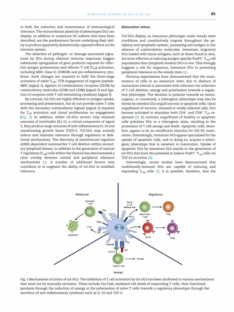
By contrast, tol-DCs are highly effective in antigen uptake,
processing and presentation, but do not provide naıve T cells
with the necessary costimulatory signals (signal 2) required
for Teff activation and clonal proliferation on engagement
[Fig. 1]. In addition, whilst tol-DCs secrete only minimal
amounts of interleukin (IL)-12, a critical component of signal
3, they produce large amounts of anti-inflammatory IL-10 and
transforming growth factor (TGF)-b. Tol-DCs may actively
induce and maintain tolerance through regulatory or dele-
tional mechanisms. The discovery of autoimmune regulator
(AIRE)-dependent autoreactive T cell deletion within second-
ary lymphoid tissues, in addition to the generation of natural
T regulatory (Treg) cells within the thymus has demonstrated a
clear overlap between central and peripheral tolerance
mechanisms [1]. A number of additional factors may
contribute to or augment the ability of tol-DCs to establish
tolerance.
Fig. 1 Mechanisms of action of tol-DCs. The inhibition of T cell acti
that need not be mutually exclusive. These include Fas-FasL-med
paralysis through the induction of anergy or the polarisation of n
secretion of anti-inflammatory cytokines such as IL-10 and TGF-b
Maturation status
Tol-DCs display an immature phenotype under steady state
conditions and constitutively migrate throughout the pe-
riphery and lymphatic system, presenting self-antigen in the
absence of costimulatory molecules. Immature, migratory
DCs loaded with tissue antigens, such as those found in skin,
aremore effective at inducing antigen-specific FoxP3þ Treg cell
populations than lymphoid resident DCs in vivo. This strongly
suggests a role for migratory, immature DCs in promoting
peripheral tolerance in the steady state [2].
Previous experiments have demonstrated that the main-
tenance of cells in an immature state, due to absence of
maturation stimuli is associated with tolerance via induction
of T cell deletion, anergy and polarisation towards a regula-
tory phenotype. The decision to polarize towards an immu-
nogenic, or conversely, a tolerogenic phenotype may also be
driven bywhether DCs engulf necrotic or apoptotic cells. Upon
engulfment of necrotic, stressed or virally infected cells, DCs
become activated to stimulate both CD4þ and CD8þ Teff re-
sponses [3]. In contrast, engulfment of healthy or apoptotic
cells polarizes DCs to a tolerogenic state, resulting in the
promotion of T cell anergy and death. Apoptotic cells, there-
fore, appear to be an insufficient stimulus for full DC matu-
ration. Interestingly, immature DCs appear specialised for the
uptake of apoptotic cells, and in doing so, acquire a tolero-
genic phenotype that is resistant to maturation. Uptake of
apoptotic DCs by immature DCs results in the generation of
tol-DCs that have the potential to induce FoxP3þ Treg cells via
TGF-b1 secretion [4].
Interestingly, recent studies have demonstrated that
traditionally-matured DCs are capable of inducing and
expanding Treg cells [5]. It is possible, therefore, that the
Re noic acid
vation by tol-DCs has been attributed to various mechanisms
iated cell death of responding T cells, their functional
aıve T cells towards a regulatory phenotype through the
.
b i om e d i c a l j o u r n a l 4 0 ( 2 0 1 7 ) 8 0e9 382
maturation status of DCs is not an absolute determinant of
immunogenic/regulatory phenotypes.
Anergic tolerance
Whilst true T cell anergy can be achieved in vitro simply in the
absence of co-stimulation, more active suppressing mecha-
nisms are required for T cell anergy in vivo. Tol-DCs actively
induce anergy among T cells via binding of CTLA-4 on acti-
vated T cells [Fig. 1], which functions as a potent inhibitor of T
cell activation, via a suppressive effect on IL-2 production, IL-
2R upregulation and cell cycle progression [6]. DC-induced
anergic T cells perpetuate ongoing tolerance through the
acquisition of suppressor activity, largely due to CTLA-4
upregulation [7]. In this way, a single tolerogenic DC may
trigger a cascade of events, culminating in a significantly
amplified pro-tolerogenic signal, with potential implications
in the future development of therapeutic strategies.
Deletional tolerance
In order to maintain immunological homoeostasis and pre-
vent deleterious autoreactive responses, tol-DCs assist with
the active removal of potentially autoreactive, naıve T cells
from the body, both within the thymus and in the peripheral
tissues. Certain subclasses of splenic DC induce extensive T
cell apoptosis in a manner dependent on an interaction be-
tween DC Fas ligand (FasL) and Fas expressed by the target
lymphocyte [Fig. 1] [8]. Ex vivo generated 1a,25-
dihydroxyvitamin D3 (VitD3)-cultured tol-DCs also demon-
strate the ability to induce autoreactive T cell apoptosis in
culture [9]. A number of mechanisms may underlie tol-DC
induced apoptosis, including interactions between FasL and
Fas [8,10,11], tryptophan catabolism through indoleamine 2,3-
dioxygenase (IDO) expression [12e14] and TRAIL interactions
with TRAIL receptors [15].
More recently, ligation of Fas on tol-DCs themselves has
been shown to significantly improve their ability to inhibit
CD4þ T cell proliferation and enhance IL-10 secretion [16].
Whilst this has been demonstrated in co-cultures between
FasLþ activated T cells and Fasþ regulatory DCs, it is
conceivable that FasL presented by regulatory DCs may also
promote enhanced tolerogenic phenotypes in neighbouring
DCs, acting via a feed-forward mechanism.
In addition to Teffs, long lived memory T cells represent a
further threat to the induction and maintenance of tolerance
[17e19]. However, DCs presenting cognate antigen to such
lymphocytes are capable of triggering substantial deletion and
inactivation of CD4 and CD8 memory T cells, inhibiting sub-
sequent recall responses [20e23]. Given that memory
lymphocyte responses are frequently resistant to endogenous
and pharmacological tolerance-inducing mechanisms to
which naıve T cells are susceptible, this may prove to be
particularly useful for the treatment of disease states
perpetuated by memory T cell activation, such as Type I dia-
betes or transplantation [24]. Furthermore, memory T cell
populations are poorly controlled by immunosuppressant
medication [25]. The difficulty of overcoming memory T cell
responses is demonstrated in transplantation studies in
which Tregs are poorly equipped to suppress memory T cell
proliferation and cytokine production [26] and those capable
of suppressing naıve T cell mediated grafts fail to suppress
memory T cell mediated rejection [27]. The ability for tol-DCs
to induce deletional tolerance in naıve and memory lympho-
cyte populations may, therefore, permit more robust toler-
ance than alternative methods.
Regulatory tolerance
As the major bridge between the non-specific innate response
and highly-targeted adaptive response, the key role of DCs is
to prime naıve T cells to generate a range of effector lym-
phocytes. In the presence of tolerogenic signals, including
TGF-b and retinoic acid, and the absence of strong co-
stimulation, presentation of peptide-MHC complexes by DCs
to naıve CD4þFoxP3� T cells may result in their differentiation
to induced Tregs (iTregs) [Fig. 1]. This subset functions to main-
tain tolerance to innocuous foreign antigens. It appears that
tissue specific subsets of DCs, such as CD8aþ DEC-205þ splenic
DCs and CD103þ intestinal DCs in the mouse, are highly spe-
cialised for this purpose [28e32]. Furthermore, mature DCs
exhibit the ability to expand ordinarily non-proliferative nat-
ural Tregs (nTregs), a key population maintaining tolerance to
self-antigens, in a CD80/86 and IL-2 dependent manner [5,33].
IL-10 plays a significant role in the generation of iTregs
through conditioning CD4þ T cells to become unresponsive to
antigens and express a suppressive phenotype [34,35]. DCs
differentiated in the presence of IL-10 secrete significant
quantities of IL-10 and minimal IL-12 on activation. In both
in vitro and in vivo studies, this has been shown to induce the
differentiation of naıve T cells to a regulatory phenotype [36,37].
In addition to IL-10, presentation of antigen by DC in the
presence of TGF-b, a regulatory polypeptide cytokine, pro-
motes differentiation of naıve T cells into Tregs. Transgenic
murine studies of a DC-selective loss of TGF-b indicate that
DCs are an important source of TGF-b in vivo, as transgenic
animals suffer severe autoimmunity and colitis, indicative of
poor Treg induction [38]. Furthermore, DC-driven differentia-
tion of FoxP3� precursors into FoxP3þ Tregs in vitro is blocked
with the addition of neutralizing antibodies to TGF-b [30].
Tol-DCs may also polarize T cells towards a regulatory
phenotype through the surface expression of the immuno-
regulatory molecule PD-L1, which, when blocked, redirects T
cells to an immunogenic, interferon (IFN)-g secreting pheno-
type [39]. Some evidence suggests that the ligation of surface
PD-L1 triggers IL-10 production, which consequently polarizes
naıve T cells to Tregs [40].
Although previously viewed as a one-way flow of infor-
mation (from dendritic cell to naıve T cell), the DC phenotype
and function also critically depend on Tregs signals. Indeed,
early in vitro studies of CD4þCD25þ Treg demonstrated their
ability to downregulate CD80 and CD86 co-stimulatory mole-
cules, via both transcriptional and non-transcriptional
mechanisms [41], resulting in poor subsequent T cell prolif-
erative responses [42]. Further, production of IL-10 by Tregs has
been shown to induce tol-DCs, which are themselves capable
of secreting TGF-b, IL-10 and IL-27 and generating regulatory
cells [43].
In addition to Treg induction, in vitro evidence suggests that
dexamethasone and vitD3-induced tol-DCs promote the
b i om e d i c a l j o u r n a l 4 0 ( 2 0 1 7 ) 8 0e9 3 83
induction of regulatory B cells [Fig. 1] [44,45] which function to
suppress inflammatory lymphocyte differentiation [46], pro-
mote the differentiation of FoxP3þ Tregs [47,48] and attenuate
neonatal DC immunogenicity [49].
Infectious tolerance
The naturally-occurring phenomenon of infectious tolerance
is one of the key, theoretical benefits to the use of cellular
therapy in establishing long term tolerance to self- and allo-
antigens in the clinic. Infectious tolerance may be defined as
the transmission of a regulatory phenotype from one
lymphocyte population (Tregs) to another, either directly
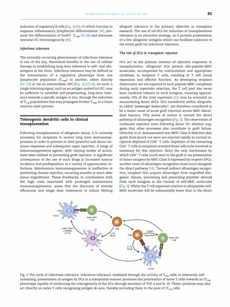
[50e52] or via an intermediate APC [Fig. 2] [53,54]. As such, a
single tolerising signal, such as an antigen-pulsed tol-DC,may
be sufficient to establish self-perpetuating, long-term toler-
ance towards a specific antigen in vivo, through the induction
of Treg populations that may propagate further Tregs in a chain
reaction-style process.
Tolerogenic dendritic cells in clinicaltransplantation
Following transplantation of allogeneic tissue, it is currently
necessary for recipients to receive long term immunosup-
pression in order to prevent or limit powerful anti-donor im-
mune responses and subsequent organ rejection. A range of
immunosuppressive agents, with varying modes of action,
have been utilised in preventing graft rejection. A significant
consequence of the use of such drugs is increased tumour
incidence and predisposition to a variety of opportunistic in-
fections. Maintenance immunosuppression is ineffective at
preventing chronic rejection, occurring months or years after
tissue engraftment. These drawbacks, in combination with
the high costs associated with prolonged maintenance
immunosuppression, mean that the discovery of entirely
efficacious and single dose treatments to induce lifelong
Fig. 2 The cycle of infections tolerance. Infectious tolerance, med
sustaining: presentation of antigen by DCs in a suboptimal mann
phenotype capable of reinforcing the tolerogenicity of the DCs thr
act directly on naıve T cells recognising antigen de novo, thereby
allograft tolerance is the primary objective in transplant
research. The use of tol-DCs for induction of transplantation
tolerance is an attractive strategy, as it permits presentation
of a few allogeneic antigens which can facilitate tolerance to
the entire graft via infectious tolerance.
The role of DCs in transplant rejection
DCs act as the primary initiator of rejection responses in
transplantation. Allogeneic DCs present allo-peptide-MHC
molecules, accompanied by costimulation and appropriate
cytokines, to recipient T cells, resulting in T cell clonal
expansion and effector function. As developing recipient
thymocytes are not exposed to such peptide-MHC complexes
during early repertoire selection, the T cell pool has never
been rendered tolerant to such antigens, meaning approxi-
mately 10% of the total repertoire [55] may be activated on
encountering donor APCs. DCs transferred within allografts,
so called “passenger leukocytes”, are therefore considered to
be a major cause of acute graft rejection across MHC discor-
dant barriers. This series of events is termed the direct
pathway of alloantigen recognition [Fig. 3]. The observation of
continued rejection even following donor DC ablation sug-
gests that other processes also contribute to graft failure.
Glimcher et al. demonstrated that MHC Class II-deficient skin
grafts from knock out mice are rejected rapidly in normal re-
cipients depleted of CD8þ T cells. Depletion of the remaining
CD4þ T cells in recipients revealed these cells to be involved or
necessary for this rejection. Since the only mechanism by
which CD4þ T cells could react to the graft is via presentation
of donor antigens by MHC Class II expressed by recipient APCs,
another route of alloantigen recognitionmust occur alongside
the direct pathway [56]. Termed indirect alloantigen recogni-
tion, recipient DCs acquire alloantigen from engrafted allo-
geneic tissues, processing and presenting peptides derived
from such antigens in the context of self-MHC molecules
[Fig. 3].Whilst the T cell repertoire reactive to allopeptide-self-
MHC molecules will be substantially lower than in the direct
iated through the activity of Treg cells, is inherently self-
er promotes the polarisation of naıve T cells towards an iTreg
ough secretion of TGF-b and IL-10. These cytokines may also
recruiting them to the pool of iTreg cells.
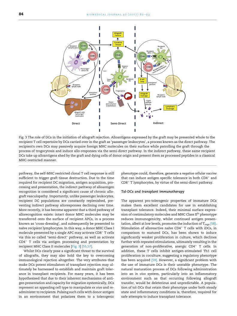
Fig. 3 The role of DCs in the initiation of allograft rejection. Alloantigens expressed by the graft may be presented whole to the
recipient T cell repertoire by DCs carried over in the graft as ‘passenger leukocytes’, a process known as the direct pathway. The
recipient's own DCs may passively acquire foreign MHC molecules on their surface while patrolling the graft through the
process of trogocytosis and induce allo-responses via the semi-direct pathway. In the indirect pathway, these same recipient
DCs take up alloantigens shed by the graft and dying cells of donor origin and present them as processed peptides in a classical
MHC-restricted manner.
b i om e d i c a l j o u r n a l 4 0 ( 2 0 1 7 ) 8 0e9 384
pathway, the self-MHC restricted clonal T cell response is still
sufficient to trigger graft tissue destruction. Due to the time
required for recipient DC migration, antigen acquisition, pro-
cessing and presentation, the indirect pathway of alloantigen
recognition is considered a significant cause of chronic allo-
graft vasculopathy. Importantly, unlike passenger leukocytes,
recipient DC populations are constantly replenished, pre-
venting indirect pathway alloresponses declining over time.
More recently, it has become apparent that a third pathway of
allorecognition exists: intact donor MHC molecules may be
transferred onto the surface of recipient APCs, in a process
known as ‘cross-dressing’, and subsequently be presented to
naıve recipient lymphocytes. In this way, a donor MHC Class I
molecule presented by a single APC may activate CD8þ T cells
via this so called “semi-direct” pathway, as well as activate
CD4þ T cells via antigen processing and presentation by
recipient MHC Class II molecules [Fig. 3] [55,57].
Whilst DCs clearly pose a significant threat to the survival
of allografts, they may also hold the key to overcoming
immunological rejection altogether. The very attributes that
make DCs potent stimulators of transplant rejection may ul-
timately be harnessed to establish and maintain graft toler-
ance in transplant recipients. For many years, it has been
hypothesised that due to their inherent mechanisms of anti-
gen presentation and capacity formigration systemically, DCs
represent an appealing cell type to manipulate ex vivo and re-
administer to recipients. Pulsing such cells with donor antigen
in an environment that polarizes them to a tolerogenic
phenotype could, therefore, generate a negative cellular vaccine
that can induce antigen specific tolerance in both CD4þ and
CD8þ T lymphocytes, by virtue of the semi-direct pathway.
Tol-DCs and transplant immunotherapy
The apparent pro-tolerogenic properties of immature DCs
makes them excellent candidates for use in establishing
transplant tolerance. Indeed, their minimal surface expres-
sion of costimulatory molecules andMHC Class IIlo phenotype
reduces immunogenicity, whilst continued antigen presen-
tation, albeit at low levels, promotes the induction of Tregs [58].
Stimulation of alloreactive naıve CD4þ T cells with iDCs, in
comparison to matured DCs, has been shown to induce
significantly weaker proliferation in culture, which declines
further with repeated stimulations, ultimately resulting in the
generation of non-proliferative, anergic CD4þ T cells. In
addition, these T cells inhibit antigen-stimulated Th1 cell
proliferation in coculture, suggesting a regulatory phenotype
has been acquired [59]. However, a significant problem with
the use of immature DCs is their unstable phenotype. The
natural maturation process of DCs following administration
into an in vivo system, particularly into an inflammatory
environment such as that occurring following allograft
transfer, would be deleterious and unpredictable. A popula-
tion of tol-DCs that retain their phenotype under both steady
state and inflammatory conditions is, therefore, required for
safe attempts to induce transplant tolerance.
b i om e d i c a l j o u r n a l 4 0 ( 2 0 1 7 ) 8 0e9 3 85
The use of both recipient (autologous) and donor tol-DCs
has been evaluated in small animal models of trans-
plantation tolerance. In these studies, both sources have been
efficacious in promoting graft tolerance. However, donor and
recipient tol-DCs operate via different mechanisms. Morelli
et al. have indicated that intravenously-administered, matu-
ration-resistant allogeneic DCs as a source of donor antigen
can prolong cardiac allograft survival without direct interac-
tion with recipient T cells. These allogeneic cells were short
lived, and functioned as stores of donor antigen which were
subsequently reprocessed by recipient DCs, presented via the
indirect pathway and utilised to upregulate FoxP3þ regulatory
T cells. A similar outcome can also be achieved by adminis-
tering apoptotic donor DCs [60]. In contrast, administration of
recipient DCs, which might be expected to survive for longer
periods of time (up to two weeks), are able to acquire, process
and present donor antigen to endogenous T cells and promote
antigen specific tolerance. When compared side-by-side, it
appears that syngeneic DCs have a greater effect in length-
ening cardiac allograft survival than allogeneic DCs (median
22.5 days vs. median 16.5 days) [61].
In mouse models, tol-DCs generated from gene modifica-
tion, pharmacological modification and cytokine induction
have all improved graft survival. Pharmacological modifica-
tions to enhance DC tolerogenicity, including rapamycin,
mitomycin-C and dexamethasone, have been the most
consistent and effective method to extend graft survival [62].
For example, a single infusion of rapamycin-treated allo-Ag
pulsed DCs prior to transplantation significantly prolonged
cardiac allograft survival, whilstmultiple infusions led to graft
survival of longer than 100 days in 40% of recipients [63].
Cuturi's group has previously shown a combination treatment
of recipient immature bone marrow DCs (bmDCs) with sub-
optimal LF 15-0195 immunosuppression induces definitive
cardiac allograft acceptance in 92% of recipients [64]. Other
strategies to induce tol-DCs have included use of viral trans-
fection. DCs transfected with IDO administered to mice prior
to cardiac allografting resulted in prolonged graft survival [65],
whilst adenoviral vector delivery of CTLA4-Ig to DCs, in
combination with NF-kB blockade results in promotion of T
cell apoptosis and prolongation of heart allograft survival in
MHC-mismatched rodents [66]. Administration of tolerogenic
IDOþ DCs generated by a1-antitrypsin priming, a potent
immunoregulatory serpin, also significantly lengthened kid-
ney allograft survival in rats, and is associated with an
expansion of Tregs in vivo [67]. Concomitant treatment with 1-
methyltryptophan (1-MT), an IDO inhibitor, abolishes this
graft-specific tolerance.
In small animal models, tol-DCs have been used to induce
donor-specific non-responsiveness in various tissue allo-
grafts, including intestinal [68,69], liver [70e72], islet [73] and
skin transplants [74,75] and kidney allografts in rhesus ma-
caques [76]. Tol-DCs may have particular advantages in in-
duction of transplant tolerance towards pancreatic islets. A
key obstacle in achieving pancreatic islet graft tolerance is the
reservoir of islet-specific CD4þ and CD8þ memory T cells that
is established at the onset of Type 1 diabetes and may be
reactivated to become a significant source of graft rejection
following islet transplantation. However, DCs may be
uniquely equipped to inactivate these deleterious populations
and have terminated anti-islet CD8þ T cell responses in mu-
rine studies [77]. Therefore, in the context of graft rejection
driven by memory T cells, tol-DC immunotherapy may be
more efficacious in attenuating rejection than other regula-
tory immunotherapies, such as Tregs.
Despite success in small animal models, few clinical
studies have investigated the use of autologous or allogeneic
tol-DCs in establishing tolerance following transplantation.
The One Study, an ongoing multicenter trial evaluating a
range of immunoregulatory cell therapies in solid organ
transplantation, is a trial investigating the efficacy of autolo-
gous tol-DCs in establishing andmaintaining renal transplant
tolerance. In this study, tol-DCs are derived from CD14þ
monocytes isolated from peripheral blood by leukapheresis
and elutriation, which are subsequently cultured with low
concentrations of GM-CSF in the absence of other cytokines or
immunosuppressive drugs. This previously verified protocol,
in both rodent and human, generates maturation-resistant,
phenotypically-immature DCs. The study will determine the
incidence of biopsy-confirmed acute graft rejection over the
course of sixty weeks post-surgery, following intravenous
injection of tol-DCs and concomitant tapering of immuno-
suppression medication in sixteen patients. As secondary
outcome measures, the study is also measuring the total
immunosuppressive burden exerted on the patient, incidence
of post-transplant dialysis and graft loss and incidence of
neoplasia. In addition to tol-DC efficacy, the study is also
examining the efficacy of Treg and macrophage sub-
populations at other centres. Results from the trial are ex-
pected during the autumn of 2018. Whilst high DC numbers
are being injected into patients (1 � 106 cells/kg), the systemic
nature of administration (via slow peripheral venous access)
may perhaps restrict the number of tol-DCs that extravasate
into the engrafted organ and ultimately process and present
donor antigen via the semi-direct pathway. Methods by which
administered DCs could be targeted to and retainedwithin the
transplanted organ, associated lymphatics and spleen could,
therefore, offer enhanced efficacy over standard intravenous
injection.
Autoimmunity
The exact aetiology of autoimmune diseases, which are
thought to affect more than 5% of the global population, re-
mains unclear, but is thought to result from failure of initial
tolerance induction (central tolerance) or a breakdown in
maintenance of established peripheral tolerance. This may be
due to pathogenic insults, such asmicrobial infection, chronic
inflammation or neoantigen generation through mutation.
Whilst many autoimmune diseases can be effectively
managedwith the use of immunosuppressive therapies, there
are significant deleterious consequences associated with the
use of immunotherapy. For example, due to the critical
involvement of tumour necrosis factor (TNF)-a in co-
ordinating effective host immune responses against micro-
bial infection, anti-TNF therapy, utilised for rheumatoid
arthritis, Crohn's disease and psoriasis, predisposes patients
to severe bacterial infections [78] and promotes reactivation of
latent tuberculosis [79,80]. Generalised immunosuppression
b i om e d i c a l j o u r n a l 4 0 ( 2 0 1 7 ) 8 0e9 386
achieved by such treatments also increases the incidence of
solid organ and haematological neoplasms [81]. Since these
immunomodulatory therapies do not eliminate the cause of
autoimmunity, treatment must be sustained indefinitely. As
such, it is of significant interest to develop a therapy that can
re-establish tolerance to the offending antigen with minimal
treatments and maintain this tolerance indefinitely.
Tol-DCs and autoimmunity immunotherapy
Mouse studies have identified that tol-DCs have potential for
clinical application for the treatment of inflammatory
arthritis. Using immature DCs transfected with IL-4, murine
collagen induced arthritis was effectively treated in vivo.
Intravenous injection of the modified cells resulted in rapid
migration to the liver and lymphatics and ultimately near
complete suppression of the disease for four weeks post-
treatment [82]. In vitro data suggest these effects to be as a
result of IFN-g suppression from splenocytes following
collagen challenge and a reduction in IgG2 isotype antibodies
produced against type II collagen. DCs genetically modified to
express FasL are similarly effective in suppressing disease
establishment and promoting disease amelioration [83,84].
This amelioration coincides with the upregulation of
CD4þCD25þ FoxP3þ iTreg cells [85]. Rosiglitazone, a PPAR-g
selective agonist, has recently been utilised to generate tol-
DCs that demonstrate therapeutic effects in the treatment of
murine collagen induced arthritis. Subcutaneous injections of
antigen loaded, rosiglitazone cultured immature DCs reduced
the clinical scoring and histological severity of arthritis in
mice, and was associated with a highly significant reduction
in ex vivo IFN-g production by isolated splenocytes [86]. These
studies have identified that tol-DCs can treat inflammatory
arthritis by suppressing humoral and cell-mediated arms of
the immune response.
Antigen-pulsed tol-DCs may also be a useful cell based
therapy for systemic lupus erythematosus (SLE), which is
mediated predominantly by antinuclear antibodies. Recent
in vitro data have shown that knockdown of RelB, a key
member of the NF-kB family involved in DC maturation, re-
sults in the generation of DCs with a semi-mature phenotype
and reduced costimulatory molecule expression. RelB-
modified DCs derived from lupus prone mice produce mini-
mal IL-12p70 and induce a hyporesponsive state in autor-
eactive splenic T cells [87]. In human studies, VitD3 and
dexamethasone conditioned tol-DCs derived from SLE pa-
tients have been shown to attenuate T cell activation and
proliferation in mixed leukocyte reactions, and are capable of
inducing Tregs from naıve T cells [88]. Future clinical studies
are required to determine whether tol-DCs can effectively
treat SLE.
Tol-DCs have also displayed promising results inmodels of
multiple sclerosis (MS). Using VitD3 cultured tol-DCs pulsed
with myelin oligodendrocyte glycoprotein (MOG), a critical
autoantigen, Mansilla et al. have demonstrated reduced dis-
ease incidence, induction of Tregs and IL-10, and consequently
reduction in the severity of signs of disease [89]. These results
have since been repeatedwith the use of cryopreserved VitD3-
treated tol-DCs pulsed with MOG peptide [90]. The main-
tenance of the tolerogenic phenotype following thawing is a
significant finding for clinical application, as the ability to
freezeethaw cell stockswould remove the need for patients to
undergo repeated leukapheresis procedures for freshly-
isolated DCs and allow treatments to be immediately avail-
able, avoiding prolonged in vitro monocyte-to-DC differentia-
tion cultures. The therapeutic effects of endogenous tol-DC in
models of MS have also been demonstrated in depletion ex-
periments. Selective depletion of CD11cþCD11bþ DCs and
immature DCs with the use of clodronate-loaded liposomes
significantly blocks the disease suppressing effects of intra-
venous soluble MOG administration, as measured by clinical
scoring. Depletion of these tol-DCs was associated with a loss
ofMOG-induced T cell tolerance, normally characterised by an
increase in prevalence of FoxP3þ cells and decreased pro-
duction of the inflammatory cytokines IL-2, IFN-g and IL-17
[91].
In vitro studies of human cells derived from multiple scle-
rosis patients suggest that tol-DC therapy may hold promise
for inducing antigen specific tolerance in the clinical setting.
Tol-DCs have been successfully generated from monocytes of
relapsing-remitting MS (RR-MS) patients, using VitD3
enhanced cultures, and have displayed the desired stable
semi-mature phenotype and anti-inflammatory properties
in vitro. Furthermore, tol-DCs loadedwithmyelin peptidewere
shown to efficiently inhibit antigen-specific responses among
autoreactive T cells derived from RR-MS patients [92]. The
first-in-man clinical trials of myelin-derived peptide pulsed
tol-DCs in MS patients, to determine safety and tolerability,
are due to begin this year [93,94].
Lymphocytic infiltration of the pancreas and subsequent
autoimmune attack of beta islet cells is considered the major
pathological process driving Type I diabetes (T1D) mellitus. As
such, therapies promoting pancreas-specific antigen toler-
ance, particularly when provided in early childhood, may
be sufficient to impede further islet cell loss and the devel-
opment of diabetes. In support of these findings, in vitro
studies of T1D patient-derived lymphocytes and DCs have
shown that insulin and GAD65 loaded monocyte-derived tol-
DCs induced T cell hyporesponsiveness in mixed leukocyte
reactions and reduced IFN-g and IL-2 secretion in comparison
to control, conventional DCs (cDCs). In addition, lymphocytes
previously stimulated with tol-DCs and subsequently rechal-
lenged with antigen loaded cDCs exhibited stable hypores-
ponsiveness in a subset of patients, indicative of T cell
tolerisation [95].
Tol-DCs have also shown promise in models of inflam-
matory bowel disease. Severe combined immune deficient
(SCID) mice adoptively transferred with CD4þCD25� T cells,
leading to the development of wasting disease and colitis,
exhibit attenuated weight loss and gut pathology following
administration of dexamethasone/VitD3-conditioned tol-DCs
[96]. IL-10-treated tol-DCs have demonstrated a similar ther-
apeutic effect in the SCID model [97]. Tol-DCs pulsed with
carbonic anhydrase I, a caecal bacterial antigen implicated to
inflammatory bowel disease (IBD) pathogenesis, ameliorated
macroscopic and histological signs of experimental colitis in
mice and was associated with raised FoxP3þ Treg numbers in
mesenteric lymph nodes [98]. These data support the sug-
gestion that tol-DCs may provide therapeutic benefit in in-
flammatory bowel disease patients.
b i om e d i c a l j o u r n a l 4 0 ( 2 0 1 7 ) 8 0e9 3 87
Clinical trials of tol-DCs for treatment of autoimmunity
In contrast to transplantation, a number of Phase I clinical
trials have been completed, investigating the use of tol-DCs
for immune intervention in autoimmunity.
The identification of highly disease specific, circulating
autoreactive T cells and autoantibodies against citrullinated
peptide antigens in the serum of around 76% of rheumatoid
arthritis (RA) patients [99e101] indicates such individuals may
be receptive to tolerising immunotherapy, such as tol-DC
administration. The recently developed “Rheumavax” ther-
apy consists of autologous DCs rendered tolerogenic through
NF-kB inhibitor exposure and subsequently pulsed with four
citrullinated peptide antigens [102]. In an open-label, pro-
spective Phase I clinical trial, a single intradermal injection of
up to 4.5 � 106 DCs was shown to be well tolerated in cit-
rullinated peptide-specific RA patients, and was associated
with an increase in the Treg/Teff cell ratio by 25% or more in 11
of the 15 treated patients [103]. A similar autologous, antigen-
pulsed DC therapy, designated CreaVax-RA, was well-
tolerated in RA patients and demonstrated preliminary signs
of efficacy by significantly reducing antigen-specific autoan-
tibody levels in 55.6% of autoantibody positive patients [104].
A Phase I, dose escalation clinical trial of autologous tol-DC
therapy for inflammatory arthritis involved injection of tol-
DCs, differentiated in vitro from CD14þ monocytes, isolated
via leukapheresis, and loaded with autologous synovial fluid
antigens. No worsening of symptoms was recorded in the
target joints during days 1e5 following administration, con-
firming short-term therapeutic safety. Synovitis improved in
one out of three participants in both the 1 � 106 and 3 � 106
cells dosage cohorts, and in both patients receiving 10 � 106
cells. No improvement was observed in the three control
participants [105]. These safety and early efficacy data indi-
cate that tol-DC therapy may be a promising treatment
strategy for inflammatory arthritis patients, warranting
further studies involving a greater number of participants.
Phase I clinical trials for tol-DC therapy in Type I diabetes
has also been investigated. Intradermal administration of
10 � 106 DCs, either unmanipulated or engineered towards a
tolerogenic phenotype ex vivo, was safely tolerated by all T1D
patients, with no adverse events recorded over the course of 2
months. Administration of engineered DCs was associated
with a statistically significant increase in suppressive
B220þCD11c� B cells [106] during the administration period
[107]. A multicentre Phase II RCT evaluating the efficacy of
autologous tol-DCs in recent onset T1Dhasbeenplanned [108].
A Phase I trial of monocyte-derived autologous tol-DC
therapy has recently been completed in patients with re-
fractory Crohn's disease. Therapeutic safety was confirmed
for both single and three biweekly intraperitoneal injections
of tol-DCs [109]. A clinical response, defined as a decrease in
the Clinical Activity Score CDAI of �100, was observed in two
patients (22%) and clinical remission (CDAI below 150 points)
was observed in another, yet the mean decrease in CDAI was
nonsignificant (274e222, p ¼ 0.3). Currently ongoing trials
include a Phase I evaluation of highly localised, intralesional
administration of tol-DCs in patients with refractory Crohn'sdisease [110]. Future trials should investigate the therapeutic
benefit of tol-DCs in a larger cohort of Crohn's patients, as well
as in patients with related pathologies, including ulcerative
colitis.
Allergy
In addition to autoimmunity, excessive immune responses
specific for otherwise innocuous antigens may result in
allergic reactions. Such immune responses can lead to the
development of a number of commonly occurring diseases,
including asthma, urticaria and dermatitis. The key involve-
ment of Th2 cells and IgE secreting B cells in the allergic re-
actions mean that therapies that can establish tolerance to
the offending allergen can potentially provide curative treat-
ment. At present, the only such approved therapy is allergen-
specific immunotherapy (AIT), involving exposure to esca-
lating concentrations of allergen. This “low zone tolerance”,
describing the repetitive exposure of individuals to low doses
of allergen, has been shown to critically depend on in-
teractions between FoxP3þ Treg cells and tol-DCs in mouse
experiments [111,112]. By extension, it is thought that the
administration of allergen-pulsed tol-DCs could provide a
stimulus for allergen-specific tolerance induction. The switch
from a Th2 to a Treg-based allergen specific response, driven
by tol-DC administration, may result in the suppression of
other immune cells involved in the allergic pathology,
including eosinophils and IgE-secreting B cells.
Tol-DCs and allergy immunotherapy
Immunomodulation of allergic asthma is a particularly active
area of current tol-DC therapy research. Whilst immunogenic
DCs play a key role in the priming of allergen-specific Teffs and
induction of airway hyperresponsiveness in allergic asthma
[113], tol-DCs have been demonstrated to suppress clinical
features of asthma [114]. In vitro studies confirm the ability of
tol-DCs to downregulate proallergic Th2 proliferative and
cytokine responses [115], whilst IL-10 treated tol-DCs have
demonstrated potent suppression of airway hyper-
responsiveness in mouse models of asthma [116e118]. Single
tol-DC treatments were sufficient for long-lived suppression
of Th2 responses [119]. Recent studies using human tol-DCs to
induce antigen specific Treg cells and suppress patient-
derived, allergen-specific Teff responses in vitro have pro-
vided the basis for future clinical trials. Other forms of allergy,
such as contact dermatitis, may also be amenable to tol-DC
therapy. Dexamethasone-treated tol-DCs derived from pe-
ripheral blood samples of individuals with IgE-mediated latex
allergy inhibit allergen-specific T cell proliferation and IgE
production in vitro and induce IL-10 competent Tregs [120]. Tol-
DCs, therefore, demonstrate potential therapeutic effects in
models of allergic disease and further investigation and future
clinical trials will determine whether these findings can be
translated to a clinical setting.
Future challenges
Whilst the pre-clinical and early clinical trial data for tol-DC
therapy is encouraging, there are some significant questions
b i om e d i c a l j o u r n a l 4 0 ( 2 0 1 7 ) 8 0e9 388
that must be addressed to ensure the development of
consistent, safe and efficacious treatments. One of the major
concerns surrounding tol-DC therapy is the potential for
generating cells with an unstable tolerogenic phenotype. The
potentially unstable phenotype of tol-DCs has been demon-
strated in experimental models. For example, TNF-induced
semi-mature DCs that lack the capacity to secrete inflamma-
tory cytokines have been found to be capable of differentiating
further, under the influence of lipopolysaccharide, into
immunogenic DCs capable of stimulating Th1 and Th2 medi-
ated immune responses [121]. Furthermore, semi-mature DCs
which are ordinarily tolerogenic at low doses, and therapeutic
in collagen-induced arthritis (CIA) mouse models, exhibit
immunogenic properties when inoculated at higher doses
with reduced capacity for FoxP3þ Treg cell induction [122]. A
valid concern is, therefore, the risk of inducing an immune
response to the target antigen, rather than inducing tolerance.
This concern is particularly acute in the setting of trans-
plantation. Here, the presence of a pro-inflammatory envi-
ronment mediated by the surgery, ischaemia-reperfusion
injury and the high burden of necrotic tissue may result in
APC upregulation of costimulatory molecules and secretion
of inflammatory cytokines [3]. Strategies to maintain the
stability of tol-DCs or limit the pro-inflammatory environ-
ment that accompanies transplantation may, thus, facilitate
induction of tolerance using tol-DCs in the transplantation
setting.
Another key question relates to the strategy utilised to
generate DCs with a truly tolerogenic phenotype. Whilst DCs
may be isolated from a number of anatomical sites, including
bone marrow and the blood via leukapheresis, it is likely that
the most suitable source of DCs for therapeutic use is from
peripheral blood derived monocytes, isolated from a single
blood draw, and subsequently differentiated ex vivo under
specific culture conditions. A variety of strategies could be
employed during this differentiation process to polarize these
DCs towards a tolerogenic phenotype, including addition of
VitD3 or the recombinant cytokines IL-10, TGF-b1 and VEGF,
withdrawal of GM-CSF, co-culture with apoptotic cells or
immunosuppressive agents such as mycophenolate mofetil
(MMF), tacrolimus, rapamycin and dexamethasone, or the use
of costimulatory blockade, such as CTLA-4-Ig or monoclonal
antibodies specific for CD40L or OX40L. Whilst a range of
methods have been described in the literature, few studies
have directly compared the stability and overall efficacy of tol-
DCs generated by different means. Further work is therefore
warranted to establish which method, or perhaps combina-
tion of methods, is most suitable to generate tol-DCs in the
clinical setting.
Quality control of immunotherapies is also of critical
importance. A recent study has indicated that certain surface
phenotypes of tol-DCs may, in fact, promote autoimmune
disease [123]. Interestingly, purified CD11cþ VitD3-treated
DCs, which, in in vitro tests, showed reduced capacity to
prime T cells, were equally capable of stimulating murine
experimental autoimmune encephalomyelitis (EAE) as fully
immunogenic, antigen-pulsed CD11cþ DCs. In vitro analysis of
DC functionality alone is therefore likely to be insufficient to
predict in vivo behaviour, and robust assays should be per-
formed prior to therapeutic use.
In the transplantation setting, it is currently unclear
whether the most effective method of achieving tolerance
would be to administer antigen-pulsed tol-DCs or administer
antigen-naıve tol-DCs that subsequently take up alloantigens
following donor engraftment. The former, involving ex vivo
pulsing of cultured tol-DCs with selected donor antigens,
would maximize the chance of administered DCs processing
and presenting appropriate antigen to the recipient'slymphocyte repertoire. However, this method does not mimic
normal physiological conditions of antigen acquisition and
limits the range of donor antigens that can be presented. In
addition, this strategy bypasses the semi-direct pathway of
antigen presentation. In contrast, administration of antigen-
naıve tol-DCs is likely to be a far less efficient method of
generating donor antigen-presenting tol-DCs, due to the
inevitable systemic dispersion of cells following inoculation.
However, more natural conditions of antigen acquisition and
presentation in vivo will permit antigen presentation through
the semi-direct pathway, influencing both CD4 and CD8 line-
ages from a single DC.
Establishing safe and ethically tolerable protocols for clinical
trials of tol-DC immunotherapy, particularly in the transplant
setting, is also challenging. In order to demonstrate efficacy of
tol-DC therapy in isolation, patients in intervention groups
must relinquish standard-of-care therapies. Patients weaned
off treatmentsmust be carefullymonitored on a regular basis to
identify early signs of disease recurrence and graft failure. Due
to the efficacy of current medications used for autoimmune
disease and transplantation, it may be challenging to satisfy
ethical standards and implement tol-DC strategies.
Future opportunities
Genetic modification of tol-DCs to over-express molecules
associated with a tolerogenic phenotype and/or to silence
immunostimulatory molecules represents a promising
approach to maximize potency and ensure consistency
among DC immunotherapies. Classically, DCs are seen to be
relatively resistant to genetic modification, often losing
viability following manipulation due to their sustained
maturation. As such, modification of the genome must usu-
ally occur at an earlier stage of the differentiation pathway,
typically at the level of the monocyte. The arrival of novel,
highly targeted genome editing tools such as CRISPR-Cas9
promises to alleviate many of the issues associated with ge-
netic modification. Suggested targets for knock-out include
costimulatory molecules CD40, CD80 and CD86, ensuring DCs
can never provide a “signal 2” required for T cell activation,
but instead be inclined to promote tolerance. In addition,
over-expression of apoptosis inducing molecules, such as
FasL, has been shown to generate so called “killer” DCs with a
tolerance-inducing phenotype [124,125]. At present, minimal
literature exists on the generation and functionality of
CRISPR-Cas9 modified DCs, but this will likely change in the
coming years due to rapid improvements and uptake of such
techniques.
The short in vitro and in vivo lifespan of terminally differ-
entiated DCs, combined with their inherent resistance to ge-
netic modification and the transient effects of RNA
b i om e d i c a l j o u r n a l 4 0 ( 2 0 1 7 ) 8 0e9 3 89
modification mean that less differentiated “source cells” are
likely needed for culturing and cryopreservation. Such “source
cells” could then be differentiated to tol-DCs when required
for treatment, to maximize cell viability and cell functionality.
Although monocytes could perform this function of “source
cell”, they too have a limited lifespan in culture and patients
would require repeated blood draws to maintain monocyte
cell cultures for long periods. A superior “source cell” might,
therefore, be pluripotent stem cells, either embryonic stem
cells [126] or induced pluripotent stem cells (iPSC) [127], typi-
cally derived from reprogrammed patient dermal fibroblasts.
There are a number of advantages to using iPSC for the gen-
eration of tol-DCs [128]. Firstly, DCs differentiated from iPSC
display an unusual, naturally tolerogenic phenotype, remi-
niscent of the phenotype expressed by DCs isolated from
foetal tissue. This would be clearly advantageous for the
clinical applications described here and may mean minimal
pharmacological or genetic modifications would be required
to achieve true tol-DCs. Due to the indefinite replicative po-
tential of iPSC, a single biopsy is all that would be required to
establish a cell line capable of significant expansion, to a level
required for immunotherapy. iPSC, unlike DCs, are also
amenable to genetic modification, allowing investigators to
derive clonal populations of genetically-modified cells that
would retain these changes throughout the differentiation
process to DCs. Finally, cell viability of iPSC following cryo-
preservation is far higher than that of terminally differenti-
ated DCs, which would permit patient's cell lines to be
cryogenically stored for extended periods, until required [128].
Conclusions
As themajor professional APC of the immune system, DCs are
uniquely placed at the heart of the immune response, func-
tioning to link the innate and adaptive systems. As a conse-
quence, DCs are highly specialised to interact with and control
a vast range of immune cells. Such characteristics make DCs a
target for modification, to permit the induction and mainte-
nance of antigen-specific tolerance. This would permit the
development of “negative cellular vaccines” for trans-
plantation, autoimmune disease and allergy. Both in vitro and
in vivo studies of rodent and human cells, in addition to early
data from clinical trials indicate significant promise for clinical
tol-DC therapy. New technologies, including the development
of CRISPR-Cas9 genome editing and iPSC, may prove fruitful
future avenues for overcoming the current obstacles and
advancing pre-clinical work from the bench to the bedside.
Conflicts of interest
The authors declare that they have no conflicts of interest.
Acknowledgements
We are grateful to Tim Davies, Herman Waldmann and Ste-
phen Cobbold for helpful discussions. Research into dendritic
cells in the authors' laboratory is funded by the Rosetrees
Trust (Grant A1372), the Medical Research Council Confidence
in Concept Fund (Grant MC-PC-15029), the CRUK Oxford
Centre Development Fund (Grant CRUKDF-0716-PF) and the
Guy Newton Translational Research Fund (Grant GN-05).
r e f e r e n c e s
[1] Gardner JM, Devoss JJ, Friedman RS, Wong DJ, Tan YX,Zhou X, et al. Deletional tolerance mediated by extrathymicAire-expressing cells. Science 2008;321:843e7.
[2] Idoyaga J, Fiorese C, Zbytnuik L, Lubkin A, Miller J,Malissen B, et al. Specialized role of migratory dendriticcells in peripheral tolerance induction. J Clin Invest2014;4:1e11.
[3] Sauter B, Albert ML, Francisco L, Larsson M, Somersan S,Bhardwaj N. Consequences of cell death: exposure tonecrotic tumor cells, but not primary tissue cells orapoptotic cells, induces the maturation ofimmunostimulatory dendritic cells. J Exp Med2000;191:423e34.
[4] Kushwah R, Wu J, Oliver JR, Jiang G, Zhang J,Siminovitch KA, et al. Uptake of apoptotic DC convertsimmature DC into tolerogenic DC that inducedifferentiation of Foxp3þ Treg. Eur J Immunol2010;40:1022e35.
[5] Yamazaki S, Iyoda T, Tarbell K, Olson K, Velinzon K,Inaba K, et al. Direct expansion of functional CD25þ CD4þregulatory T cells by antigen-processing dendritic cells.J Exp Med 2003;198:235e47.
[6] Walunas TL, Bakker CY, Bluestone JA. CTLA-4 ligationblocks CD28-dependent T cell activation. J Exp Med1996;183:2541e50.
[7] Steinbrink K, Graulich E, Kubsch S, Knop J, Enk AH. CD4þand CD8þ anergic T cells induced by interleukin-10-treatedhuman dendritic cells display antigen-specific suppressoractivity. Blood 2002;99:2468e76.
[8] Suss G, Shortman K. A subclass of dendritic cells kills CD4 Tcells via Fas/Fas-ligand-induced apoptosis. J Exp Med1996;183:1789e96.
[9] Van Halteren AGS, Tysma OM, Van Etten E, Mathieu C,Roep BO. 1,25-Dihydroxyvitamin D3 or analogue treateddendritic cells modulate human autoreactive T cells via theselective induction of apoptosis. J Autoimmun2004;23:233e9.
[10] Kurts C, Heath WR, Kosaka H, Miller JF, Carbone FR. Theperipheral deletion of autoreactive CD8þ T cells induced bycross-presentation of self-antigens involves signalingthrough CD95 (Fas, Apo-1). J Exp Med 1998;188:415e20.
[11] Lu L, Qian S, Hershberger PA, Rudert WA, Lynch DH,Thomson AW. Fas ligand (CD95L) and B7 expression ondendritic cells provide counter- regulatory signals for T cellsurvival and proliferation. J Immunol 1997;158:5676e84.
[12] Fallarino F, Vacca C, Orabona C, Belladonna ML, Bianchi R,Marshall B, et al. Functional expression of indoleamine 2,3-dioxygenase by murine CD8 alpha(þ) dendritic cells. IntImmunol 2002;14:65e8.
[13] Grohmann U, Fallarino F, Silla S, Bianchi R, Belladonna ML,Vacca C, et al. CD40 ligation ablates the tolerogenicpotential of lymphoid dendritic cells. J Immunol2001;166:277e83.
[14] Grohmann U, Fallarino F, Bianchi R, Belladonna ML,Vacca C, Orabona C, et al. IL-6 inhibits the tolerogenicfunction of CD8 alphaþ dendritic cells expressingindoleamine 2,3-dioxygenase. J Immunol 2001;167:708e14.
b i om e d i c a l j o u r n a l 4 0 ( 2 0 1 7 ) 8 0e9 390
[15] Izawa T, Kondo T, Kurosawa M, Oura R, Matsumoto K,Tanaka E, et al. Fas-independent T-cell apoptosis bydendritic cells controls autoimmune arthritis in MRL/lprmice. PLoS One 2012;7:e48798.
[16] Qian C, Qian L, Yu Y, An H, Guo Z, Han Y, et al. Fas signalpromotes the immunosuppressive function of regulatorydendritic cells via the ERK/??-catenin pathway. J Biol Chem2013;288:27825e35.
[17] Schenk AD, Nozaki T, Rabant M, Valujskikh A, Fairchild RL.Donor-reactive CD8 memory T cells infiltrate cardiacallografts within 24-h posttransplant in naive recipients.Am J Transplant 2008;8:1652e61.
[18] Donckier V, Craciun L,Miqueu P, Troisi RI, Lucidi V, Rogiers X,et al. Expansionofmemory-typeCD8þTcells correlateswiththe failure of early immunosuppression withdrawal aftercadaver liver transplantation using high-doseATG inductionand rapamycin. Transplantation 2013;96:306e15.
[19] Koyama I, Nadazdin O, Boskovic S, Ochiai T, Smith RN,Sykes M, et al. Depletion of CD8 memory t cells forinduction of tolerance of a previously transplanted kidneyallograft. Am J Transplant 2007;7:1055e61.
[20] Nasreen M, Waldie TM, Dixon CM, Steptoe RJ. Steady-stateantigen-expressing dendritic cells terminate CD4þ memoryT-cell responses. Eur J Immunol 2010;40:2016e25.
[21] Kenna TJ, Thomas R, Steptoe RJ. Steady-state dendritic cellsexpressing cognate antigen terminate memory CD8þ T-cellresponses. Blood 2008;111:2091e100.
[22] Kenna TJ, Waldie T, McNally A, Thomson M, Yagita H,Thomas R, et al. Targeting antigen to diverse APCsinactivates memory CD8þ T cells without eliciting tissue-destructive effector function. J Immunol 2010;184:598e606.
[23] Torres-Aguilar H, Aguilar-Ruiz SR, Gonz�alez-P�erez G,Munguıa R, Baja~na S, Meraz-Rıos M a, et al. Tolerogenicdendritic cells generated with differentimmunosuppressive cytokines induce antigen-specificanergy and regulatory properties in memory CD4þ T cells.J Immunol 2010;184:1765e75.
[24] Ehlers MR, Rigby MR. Targeting memory T cells in Type 1diabetes. Curr Diab Rep 2015;15:84.
[25] Page AJ, Ford ML, Kirk AD. Memory T-cell-specifictherapeutics in organ transplantation. Curr Opin OrganTransplant 2009;14:643e9.
[26] Afzali B, Mitchell PJ, Scott�a C, Canavan J, Edozie FC,Fazekasova H, et al. Relative resistance of human CD4 þmemory T cells to suppression by CD4 þCD25 þ regulatoryT cells. Am J Transplant 2011;11:1734e42.
[27] Yang J, Brook MO, Carvalho-Gaspar M, Zhang J, Ramon HE,Sayegh MH, et al. Allograft rejection mediated by memory Tcells is resistant to regulation. Proc Natl Acad Sci U S A2007;104:19954e9.
[28] Luo X, Tarbell KV, Yang H, Pothoven K, Bailey SL, Ding R,et al. Dendritic cells with TGF-beta1 differentiate naiveCD4þCD25- T cells into islet-protective Foxp3þ regulatory Tcells. Proc Natl Acad Sci U S A 2007;104:2821e6.
[29] Yamazaki S, Bonito AJ, Spisek R, Dhodapkar M, Inaba K,Steinman RM. Dendritic cells are specialized accessory cellsalong with TGF-b for the differentiation of Foxp3þ CD4þregulatory T cells from peripheral Foxp3- precursors. Blood2007;110:4293e302.
[30] Yamazaki S, Dudziak D, Heidkamp GF, Fiorese C, Bonito AJ,Inaba K, et al. CD8þ CD205þ splenic dendritic cells arespecialized to induce Foxp3þ regulatory T cells. J Immunol2008;181:6923e33.
[31] Coombes JL, Siddiqui KRR, Arancibia-C�arcamo CV, Hall J,Sun C-M, Belkaid Y, et al. A functionally specializedpopulation of mucosal CD103þ DCs induces Foxp3þregulatory T cells via a TGF-beta and retinoic acid-dependent mechanism. J Exp Med 2007;204:1757e64.
[32] Sun C-M, Hall JA, Blank RB, Bouladoux N, Oukka M, Mora JR,et al. Small intestine lamina propria dendritic cells promotede novo generation of Foxp3 T reg cells via retinoic acid.J Exp Med 2007;204:1775e85.
[33] Feh�erv�ari Z, Sakaguchi S. Control of Foxp3þ CD25þCD4þregulatory cell activation and function by dendritic cells. IntImmunol 2004;16:1769e80.
[34] Gru G. New insights into the molecular mechanism ofinterleukin-10- mediated immunosuppression. J LeukocBiol 2005;77:3e15.
[35] Hsu P, Santner-Nanan B, Hu M, Skarratt K, Lee CH,Stormon M, et al. IL-10 potentiates differentiation of humaninduced regulatory T cells via STAT3 and Foxo1. J Immunol2015;195:3665e74.
[36] Wakkach A, Fournier N, Brun V, Breittmayer JP, Cottrez F,Groux H. Characterization of dendritic cells that inducetolerance and T regulatory 1 cell differentiation in vivo.Immunity 2003;18:605e17.
[37] Gregori S, Tomasoni D, Pacciani V, Scirpoli M, Battaglia M,Magnani CF, et al. Differentiation of type 1 T regulatory cells(Tr1) by tolerogenic DC-10 requires the IL-10-dependentILT4/HLA-G pathway. Blood 2010;116:935e44.
[38] Travis MA, Reizis B, Melton AC, Masteller E, Tang Q,Proctor JM, et al. Loss of integrin alpha(v)beta8 on dendriticcells causes autoimmunity and colitis in mice. Nature2007;449:361e5.
[39] Unger WWJ, Laban S, Kleijwegt FS, Van Der Slik AR,Roep BO. Induction of Treg by monocyte-derived DCmodulated by vitamin D3 or dexamethasone: differentialrole for PD-L1. Eur J Immunol 2009;39:3147e59.
[40] Kuipers H, Muskens F, Willart M, Hijdra D, van Assema FBJ,Coyle AJ, et al. Contribution of the PD-1 ligands/PD-1signaling pathway to dendritic cell-mediated CD4þ cellactivation. Eur J Immunol 2006;36:2472e82.
[41] Cederbom L, Hall H, Ivars F. CD4þCD25þ regulatory T cellsdown-regulate co-stimulatory molecules on antigen-presenting cells. Eur J Immunol 2000;30:1538e43.
[42] Oderup C, Cederbom L, Makowska A, Cilio CM, Ivars F.Cytotoxic T lymphocyte antigen-4-dependent down-modulation of costimulatory molecules on dendritic cells inCD4þ CD25þ regulatory T-cell-mediated suppression.Immunology 2006;118:240e9.
[43] Awasthi A, Carrier Y, Peron JP, Bettelli E, Kamanaka M,Flavell RA, et al. A dominant function for interleukin 27 ingenerating interleukin 10-producing anti-inflammatory Tcells. Nat Immunol 2007;8:1380e9.
[44] Rosser EC, Mauri C. Regulatory B cells: origin, phenotype,and function. Immunity 2015;42:607e12.
[45] Volchenkov R, Karlsen M, Jonsson R, Appel S. Type 1regulatory T cells and regulatory B cells induced bytolerogenic dendritic cells. Scand J Immunol2013;77:246e54.
[46] Parekh VV, Prasad DVR, Banerjee PP, Joshi BN, Kumar A,Mishra GC. B cells activated by lipopolysaccharide, but notby anti-Ig and anti-CD40 antibody, induce anergy in CD8þ Tcells: role of TGF-beta 1. J Immunol 2003;170:5897e911.
[47] Carter NA, Vasconcellos R, Rosser EC, Tulone C, Munoz-Suano A, Kamanaka M, et al. Mice lacking endogenousIL-10-producing regulatory B cells develop exacerbateddisease and present with an increased frequency ofTh1/Th17 but a decrease in regulatory T cells. J Immunol2011;186:5569e79.
[48] Flores-Borja F, Bosma A, Ng D, Reddy V, Ehrenstein MR,Isenberg DA, et al. CD19þCD24hiCD38hi B cells maintainregulatory T cells while limiting TH1 and TH17differentiation. Sci Transl Med 2013;5:173ra23.
[49] Sun CM, Deriaud E, Leclerc C, Lo-Man R. Upon TLR9signaling, CD5þ B cells control the IL-12-dependent
b i om e d i c a l j o u r n a l 4 0 ( 2 0 1 7 ) 8 0e9 3 91
Th1-priming capacity of neonatal DCs. Immunity2005;22:467e77.
[50] Walker MR, Kasprowicz DJ, Gersuk VH, B�enard A,Van Landeghen M, Buckner JH, et al. Induction of FoxP3 andacquisition of T regulatory activity by stimulated humanCD4 þ CD25 e T cells. J Clin Invest 2003;112:1437e43.
[51] Dieckmann D, Bruett CH, Ploettner H, Lutz MB, Schuler G.Human CD4(þ)CD25(þ) regulatory, contact-dependent Tcells induce interleukin 10-producing, contact-independenttype 1-like regulatory T cells. J Exp Med 2002;196:247e53.
[52] Andersson J, Tran DQ, Pesu M, Davidson TS, Ramsey H,O'Shea JJ, et al. CD4þ FoxP3þ regulatory T cells conferinfectious tolerance in a TGF-beta-dependent manner. J ExpMed 2008;205:1975e81.
[53] DiPaolo RJ, Brinster C, Davidson TS, Andersson J, Glass D,Shevach EM. Autoantigen-specific TGFbeta-induced Foxp3þregulatory T cells prevent autoimmunity by inhibitingdendritic cells from activating autoreactive T cells.J Immunol 2007;179:4685e93.
[54] Lan Q, Zhou X, Fan H, Chen M, Wang J, Ryffel B, et al.Polyclonal CD4þFoxp3þ Treg cells induce TGFbeta-dependent tolerogenic dendritic cells that suppress themurine lupus-like syndrome. J Mol Cell Biol 2012;4:409e19.
[55] Benichou G, Yamada Y, Yun SH, Lin C, Fray M, Tocco G.Immune recognition and rejection of allogeneic skin grafts.Immunotherapy 2011;3:757e70.
[56] Auchincloss H, Lee R, Shea S, Markowitz JS, Grusby MJ,Glimcher LH. The role of “indirect” recognition in initiatingrejection of skin grafts from major histocompatibilitycomplex class II-deficient mice. Proc Natl Acad Sci U S A1993;90:3373e7.
[57] Herrera OB, Golshayan D, Tibbott R, Ochoa FS, James MJ,Marelli-Berg FM, et al. A novel pathway of alloantigenpresentation by dendritic cells. J Immunol2004;173:4828e37.
[58] Yates SF, Paterson AM, Nolan KF, Cobbold SP, Saunders NJ,Waldmann H, et al. Induction of regulatory T cells anddominant tolerance by dendritic cells incapable of fullactivation. J Immunol 2007;179:967e76.
[59] Jonuleit H, Schmitt E, Schuler G, Knop J, Enk AH. Inductionof interleukin 10-producing, nonproliferating CD4(þ) T cellswith regulatory properties by repetitive stimulation withallogeneic immature human dendritic cells. J Exp Med2000;192:1213e22.
[60] Divito SJ, Wang Z, Shufesky WJ, Liu Q, Tkacheva OA,Montecalvo A, et al. Endogenous dendritic cells mediate theeffects of intravenously injected therapeuticimmunosuppressive dendritic cells in transplantation.Blood 2010;116:2694e705.
[61] Peche H, Trinit�e B, Martinet B, Cuturi MC. Prolongation ofheart allograft survival by immature dendritic cellsgenerated from recipient type bone marrow progenitors.Am J Transplant 2005;5:255e67.
[62] Wu W, Shan J, Li Y, Luo L, Sun G, Zhou Y, et al. Adoptivetransfusion of tolerance dendritic cells prolongs thesurvival of cardiac allograft: a systematic review of 44 basicstudies in mice. J Evid Based Med 2012;5:139e53.
[63] Taner T, Hackstein H, Wang Z, Morelli AE, Thomson AW.Rapamycin-treated, alloantigen-pulsed host dendritic cellsinduce Ag-specific T cell regulation and prolong graftsurvival. Am J Transplant 2005;5:228e36.
[64] B�eriou G, Peche H, Guillonneau C, Merieau E, Cuturi MC.Donor-specific allograft tolerance by administration ofrecipient-derived immature dendritic cells andsuboptimal immunosuppression. Transplantation2005;79:969e72.
[65] Li C, Liu T, Zhao N, Zhu L, Wang P, Dai X. Dendritic cellstransfected with indoleamine 2,3-dioxygenase gene
suppressed acute rejection of cardiac allograft. IntImmunopharmacol 2016;36:31e8.
[66] Bonham CA, Peng L, Liang X, Chen Z, Wang L, Ma L, et al.Marked prolongation of cardiac allograft survival bydendritic cells genetically engineered with NF-kappa Boligodeoxyribonucleotide decoys and adenoviral vectorsencoding CTLA4-Ig. J Immunol 2002;169:3382e91.
[67] Chen G, Li J, Chen L, Lai X, Qiu J. a1-Antitrypsin-primedtolerogenic dendritic cells prolong allograft kidneytransplants survival in rats. Int Immunopharmacol2016;31:216e21.
[68] Xu X, Gao X, Zhao X, Liao Y, Ji W, Li Q, et al. PU.1-Silenceddendritic cells induce mixed chimerism and alleviateintestinal transplant rejection in rats via a Th1 to Th2 shift.Cell Physiol Biochem 2016;38:220e8.
[69] Chen T, Xu H, Wang HQ, Zhao Y, Zhu CF, Zhang YH, et al.Prolongation of rat intestinal allograft survival byadministration of triptolide-modified donor bone marrow-derived dendritic cells. Transpl Proc 2008;40:3711e3.
[70] Xie J, Wang Y, Bao J, Ma Y, Zou Z, Tang Z, et al. Immunetolerance induced by RelB short-hairpin RNA interferencedendritic cells in liver transplantation. J Surg Res2013;180:169e75.
[71] Wang G-Y, Yang Y, Li H, Zhang J, Li M-R, Zhang Q, et al.Rapamycin combined with donor immature dendritic cellspromotes liver allograft survival in association with CD4(þ)CD25(þ) Foxp3(þ) regulatory T cell expansion. Hepatol Res2012;42:192e202.
[72] Li L, Zhang S, Ran J, Liu J, Li Z, Li L. Mechanism of immunehyporesponsiveness induced by recipient- derivedimmature dendritic cells in liver transplantation rat. ChinMed Sci J 2011;26:28e35.
[73] Ali A, Garrovillo M, Jin M-X, Hardy MA, Oluwole SF.Major histocompatibility complex class I peptide-pulsedhost dendritic cells induce antigen-specific acquiredthymic tolerance to islet cells 1,2. Transplantation2000;69:221e6.
[74] Segovia M, Louvet C, Charnet P, Savina A, Tilly G,Gautreau L, et al. Autologous dendritic cells prolongallograft survival through Tmem176b-dependent antigencross-presentation. Am J Transplant 2014;14:1021e31.
[75] Chen Y, Lai HS, Chiang BL, Tseng SH, Chen WJ. Tetrandrineattenuates dendritic cell-mediated alloimmune responsesand prolongs graft survival in mice. Planta Med2010;76:1424e30.
[76] Ezzelarab MB, Zahorchak AF, Lu L, Morelli AE, Chalasani G,Demetris AJ, et al. Regulatory dendritic cell infusionprolongs kidney allograft survival in nonhuman primates.Am J Transplant 2013;13:1989e2005.
[77] Coleman MA, Jessup CF, Bridge JA, Overgaard NH, Penko D,Walters S, et al. Antigen-encoding bone marrow terminatesislet-directed memory CD8þ T-cell responses to alleviateislet transplant rejection. Diabetes 2016;65:1328e40.
[78] Galloway J, Hyrich K, Mercer L, Dixon W, Fu B,Ustianowski A, et al. Anti-TNF therapy is associated with anincreased risk of serious infections in patients withrheumatoid arthritis especially in the first 6 months oftreatment: updated results from the British Society forRheumatology Biologics Register with special emph.Rheumatology 2011;50:124e31.
[79] Keane J, Gershon S, Wise RP, Mirabile-Levens E, Kasznica J,Schwieterman WD, et al. Tuberculosis associated withinfliximab, a tumor necrosis factor alpha-neutralizingagent. N Engl J Med 2001;345:1098e104.
[80] Martinez ON, Noiseux CR, Martin JAC, Lara VG, GregorioMara~n�on HG. Reactivation tuberculosis in a patient withanti-TNF-alpha treatment. Am J Gastroenterol2001;96:1665e6.
b i om e d i c a l j o u r n a l 4 0 ( 2 0 1 7 ) 8 0e9 392
[81] Bongartz T, Sutton AJ, Sweeting MJ, Buchan I, Matteson EL,Montori V. Anti-TNF antibody therapy in rheumatoidarthritis and the risk of serious infections andmalignancies: systematic review and meta-analysis of rareharmful effects in randomized controlled trials. JAMA2006;295:2275e85.
[82] Kim SH, Kim S, Evans CH, Ghivizzani SC, Oligino T,Robbins PD. Effective treatment of established murinecollagen-induced arthritis by systemic administration ofdendritic cells genetically modified to express IL-4.J Immunol 2001;166:3499e505.
[83] Liu Z, Xu X, Hsu HC, Tousson A, Yang PA, Wu Q, et al. CII-DC-AdTRAIL cell gene therapy inhibits infiltration of CII-reactive T cells and CII-induced arthritis. J Clin Invest2003;112:1332e41.
[84] Kim SH, Kim S, Oligino TJ, Robbins PD. Effective treatmentof established mouse collagen-induced arthritis by systemicadministration of dendritic cells genetically modified toexpress fasL. Mol Ther 2002;6:584e90.
[85] Ning B, Wei J, Zhang A, Gong W, Fu J, Jia T, et al. Antigen-specific tolerogenic dendritic cells ameliorate the severity ofmurine collagen-induced arthritis. PLoS One2015;10:e0131152.
[86] Byun SH, Lee JH, Jung NC, Choi HJ, Song JY, Seo HG, et al.Rosiglitazone-mediated dendritic cells ameliorate collagen-induced arthritis in mice. Biochem Pharmacol2016;115:85e93.
[87] Wu H, Lo Y, Chan A, Law KS, Mok MY. Rel B-modifieddendritic cells possess tolerogenic phenotype and functionson lupus splenic lymphocytes in vitro. Immunology2016;149:48e61.
[88] Wu HJ, Lo Y, Luk D, Lau CS, Lu L, Mok MY. Alternativelyactivated dendritic cells derived from systemic lupuserythematosus patients have tolerogenic phenotype andfunction. Clin Immunol 2015;156:43e57.
[89] Mansilla MJ, Sell�es-Moreno C, F�abregas-Puig S, Amoedo J,Navarro-Barriuso J, Teniente-Serra A, et al. Beneficial effectof tolerogenic dendritic cells pulsed with MOG autoantigenin experimental autoimmune encephalomyelitis. CNSNeurosci Ther 2015;21:222e30.
[90] Mansilla MJ, Contreras-Cardone R, Navarro-Barriuso J,Cools N, Berneman Z, Ramo-Tello C, et al. Cryopreservedvitamin D3-tolerogenic dendritic cells pulsed withautoantigens as a potential therapy for multiple sclerosispatients. J Neuroinflammation 2016;13:113.
[91] Wang L, Li Z, Ciric B, Safavi F, Zhang G-X, Rostami A.Selective depletion of CD11cþ CD11bþ dendriticcells partially abrogates tolerogenic effects ofintravenous MOG in murine EAE. Eur J Immunol2016;46:2454e66.
[92] Raıch-Regu�e D, Grau-L�opez L, Naranjo-G�omez M,Ramo-Tello C, Pujol-Borrell R, Martınez-C�aceres E, et al.Stable antigen-specific T-cell hyporesponsiveness inducedby tolerogenic dendritic cells from multiple sclerosispatients. Eur J Immunol 2012;42:771e82.
[93] Cools N, Berneman Zwi. A “Negative” dendritic cell-basedvaccine for the treatment of multiple sclerosis: a first-in-human clinical trial. https://clinicaltrials.gov/ct2/show/NCT02618902 [accessed 16.12.06].
[94] Ramo C, Martinez-Caceres E. Tolerogenic dendritic cells as atherapeutic strategy for the treatment of multiple sclerosispatients (TOLERVIT-MS) (TOLERVIT-MS) 2016. https://clinicaltrials.gov/ct2/show/NCT02903537 [accessed16.12.06].
[95] Segovia-Gamboa N, Rodriguez-Arellano ME, Rangel-Cruz R,Sanchez-Diaz M, Ramirez-Reyes JC, Faradji R, et al.Tolerogenic dendritic cells induce antigen-specifichyporesponsiveness in insulin- and glutamic acid
decarboxylase 65-autoreactive T lymphocytes from type 1diabetic patients. Clin Immunol 2014;154:72e83.
[96] Pedersen AE, Schmidt EGW, Gad M, Poulsen SS,Claesson MH. Dexamethasone/1a-25-dihydroxyvitaminD3-treated dendritic cells suppress colitis in the SCID T-celltransfer model. Immunology 2009;127:354e64.
[97] Pedersen AE, Gad M, Kristensen NN, Haase C, Nielsen CH,Claesson MH. Tolerogenic dendritic cells pulsed withenterobacterial extract suppress development of colitis inthe severe combined immunodeficiency transfer model.Immunology 2007;121:526e32.
[98] Yamanishi H, Murakami H, Ikeda Y, Abe M, Kumagi T,Hiasa Y, et al. Regulatory dendritic cells pulsed withcarbonic anhydrase I protect mice from colitis induced byCD4þCD25- T cells. J Immunol 2012;188:2164e72.
[99] Law SC, Street S, Yu CH, Capini C, Ramnoruth S, Nel HJ,et al. T cell autoreactivity to citrullinated autoantigenicpeptides in rheumatoid arthritis patients carrying HLA-DRB1 shared epitope alleles. Arthritis Res Ther2012;14:R118.
[100] James EA, Rieck M, Pieper J, Gebe JA, Yue BB, Tatum M, et al.Citrulline-specific Th1 cells are increased in rheumatoidarthritis and their frequency is influenced by diseaseduration and therapy. Arthritis Rheum 2014;66:1712e22.
[101] Schellekens GA, De Jong BAW, Van Den Hoogen FHJ, Van DePutte LBA, Van Venrooij WJ. Citrulline is an essentialconstituent of antigenic determinants recognized byrheumatoid arthritis-specific autoantibodies. J Clin Invest1998;101:273e81.
[102] Thomas R, Street S, Ramnoruth N, Pahau H, Law S,Brunck M, et al. Feasibility, safety and clinical effects of asingle intradermal administration of autologous tolerisingdendritic cells exposed to citrullinated peptides in patientswith rheumatoid arthritis. Arthritis Rheum 2011;63:S946.
[103] Benham H, Nel HJ, Law SC, Mehdi AM, Street S,Ramnoruth N, et al. Citrullinated peptide dendritic cellimmunotherapy in HLA risk genotype-positive rheumatoidarthritis patients. Sci Transl Med 2015;7:290ra87.
[104] Joo YB, Park JE, Choi CB, Choi J, Jang J, Heo M, et al. Phase 1study of immunotherapy using autoantigenloadeddendritic cells in patients with anti-citrullinated peptideantigen positive rheumatoid arthritis. ACR MeetingAbstracts. 2014 ACR/ARHP Annual Meeting. AbstractNumber: 946
[105] Bell G, Anderson A, Diboll J, Reece R, Eltherington O,Harry R, et al. Autologous tolerogenic dendritic cells inrheumatoid and inflammatory arthritis. Ann Rheum Dis2017;76:227e34.
[106] Iwata Y, Matsushita T, Horikawa M, DiLillo DJ, Yanaba K,Venturi GM, et al. Characterization of a rare IL-10-competent B-cell subset in humans that parallels mouseregulatory B10 cells. Blood 2011;117:530e41.
[107] Giannoukakis N, Phillips B, Finegold D, Harnaha J, Trucco M.Phase I (safety) study of autologous tolerogenic dendriticcells in type 1 diabetic patients. Diabetes Care2011;34:2026e32.
[108] Trucco M. Autologous immunoregulatory dendritic cells forType 1 diabetes therapy. https://clinicaltrials.gov/ct2/show/NCT02354911 [accessed 16.12.06].
[109] Jauregui-Amezaga A, Cabez�on R, Ramırez-Morros A,Espa~na C, Rimola J, Bru C, et al. Intraperitonealadministration of autologous tolerogenic dendritic cells forrefractory Crohn's disease: a Phase I study. J Crohns Colitis2015;9:1071e8.
[110] Benitez D, Varea S, Ricart E. Intralesional tolerogenicdendritic cells in Crohn's disease treatment. https://clinicaltrials.gov/ct2/show/NCT02622763 [accessed16.12.06].
b i om e d i c a l j o u r n a l 4 0 ( 2 0 1 7 ) 8 0e9 3 93
[111] Luckey U, Maurer M, Schmidt T, Lorenz N, Seebach B,Metz M, et al. T cell killing by tolerogenic dendritic cellsprotects mice from allergy. J Clin Invest 2011;121:3860e71.
[112] Luckey U, Schmidt T, Pfender N, Romer M, Lorenz N,Martin SF, et al. Crosstalk of regulatory T cells andtolerogenic dendritic cells prevents contact allergy insubjects with low zone tolerance. J Allergy Clin Immunol2012;130:781e97.
[113] Burchell JT, Strickland DH, Stumbles PA. The role ofdendritic cells and regulatory T cells in the regulation ofallergic asthma. Pharmacol Ther 2010;125:1e10.
[114] Takagi T, Taguchi O, Toda M, Ruiz DB, Bernabe PG,D'Alessandro-Gabazza CN, et al. Inhibition of allergicbronchial asthma by thrombomodulin is mediated bydendritic cells. Am J Respir Crit Care Med 2011;183:31e42.
[115] Bellinghausen I, Brand U, Steinbrink K, Enk AH, Knop J,Saloga J. Inhibition of human allergic T-cell responses byIL-10-treated dendritic cells: differences fromhydrocortisone-treated dendritic cells. J Allergy ClinImmunol 2001;108:242e9.
[116] Koya T, Matsuda H, Takeda K, Matsubara S, Miyahara N,Balhorn A, et al. IL-10-treated dendritic cells decreaseairway hyperresponsiveness and airway inflammation inmice. J Allergy Clin Immunol 2007;119:1241e50.
[117] Nayyar A, Zhang X, Gordon JR. Tolerogenic bone marrowderived dendritic cells modulate allergic reactivity of lungcells from mice with severe allergic lung disease. http://docslide.us/documents/tolerogenic-bone-marrow-derived-dendritic-cells-modulate-allergic-reactivity.html.
[118] Henry E, Desmet CJ, Garze V, Fievez L, Bedoret D,Heirman C, et al. Dendritic cells genetically engineered toexpress IL-10 induce long-lasting antigen-specific tolerancein experimental asthma. J Immunol 2008;181:7230e42.
[119] Nayyar A, Dawicki W, Huang H, Lu M, Zhang X, Gordon JR.Induction of prolonged asthma tolerance by IL-10-differentiated dendritic cells: differential impact on airwayhyperresponsiveness and the Th2 immunoinflammatoryresponse. J Immunol 2012;189:72e9.
[120] Escobar A, Aguirre A, Guzm�an MA, Gonz�alez R, Catal�an D,Acu~na-Castillo C, et al. Tolerogenic dendritic cells derivedfrom donors with natural rubber latex allergy modulateallergen-specific T-cell responses and IgE production. PLoSOne 2014;9:e85930.
[121] Voigtl€ander C, R€ossner S, Cierpka E, Theiner G, Wiethe C,Menges M, et al. Dendritic cells matured with TNF can befurther activated in vitro and after subcutaneous injectionin vivo which converts their tolerogenicity intoimmunogenicity. J Immunother 2006;29:407e15.
[122] Lim DS, Kang MS, Jeong JA, Bae YS. Semi-mature DC areimmunogenic and not tolerogenic when inoculated at ahigh dose in collagen-induced arthritis mice. Eur J Immunol2009;39:1334e43.
[123] Besusso D, Saul L, Leech MD, O'Connor RA, MacDonald AS,Anderton SM, et al. 1,25-dihydroxyvitamin D3-conditionedCD11cþ dendritic cells are effective initiators of CNSautoimmune disease. Front Immunol 2015;6:575.
[124] Matsue H, Matsue K, Walters M, Okumura K, Yagita H,Takashima A. Induction of antigen-specificimmunosuppression by CD95L cDNA-transfected “killer”dendritic cells. Nat Med 1999;5:930e7.
[125] Min WP, Gorczynski R, Huang XY, Kushida M, Kim P,Obataki M, et al. Dendritic cells genetically engineered toexpress Fas ligand induce donor-specifichyporesponsiveness and prolong allograft survival. JImmunol 2000;164:161e7.
[126] Tseng S-Y, Nishimoto KP, Silk KS, Majumdar AS, Dawes GN,Waldmann H, et al. Generation of immunogenic dendriticcells from human embryonic stem cells without serum andfeeder cells. Regen Med 2009;4:513e26.
[127] Silk KM, Silk JD, Ichiryu N, Davies TJ, Carpenter L, Watt SM,et al. Cross-presentation of tumor antigens by inducedpluripotent stem cell-derived CD141þ XCR1þ dendriticcells. Gene Ther 2012;19:1035e40.
[128] Fairchild PJ, Leishman A, Sachamitr P, Telfer C, Hackett S,Davies TJ. Dendritic cells and pluripotency: unlikely allies inthe pursuit of immunotherapy. Regen Med 2015;10:275e86.