Embed Size (px)
Citation preview

Volume 11 Number 11 November 2009 pp. 1216–1225 1216
www.neoplasia.com
HE3235 Inhibits Growth ofCastration-ResistantProstate Cancer1,2
Theodore D. Koreckij*, Richard J. Trauger†,Robert Bruce Montgomery‡, Tiffany E.M. Pitts*,Ilsa Coleman§, Holly Nguyen*, Chris L. Reading†,Peter S.Nelson§, Robert L. Vessella*,¶ andEvaCorey*
*Department of Urology, University of Washington, Seattle,WA, 98195, USA; †Hollis Eden Pharmaceutical, San Diego,CA 92121, USA; ‡Department of Medicine, University ofWashington, Seattle, WA, USA; §Division of Human Biology,Fred Hutchinson Cancer Research Center, Seattle, WA,USA; ¶Puget Sound VA Administrations, Seattle, WA, USA
AbstractTreatments for advanced prostate cancer (CaP) typically involve androgen deprivation therapy. However, mostpatients eventually develop castration-resistant CaP (CRPC) for which highly effective therapies are limited. Weexplored the efficacy of a novel agent, HE3235, in inhibiting growth of CRPC in preclinical models. Castratedmale mice were implanted subcutaneously with LuCaP35V CaP xenografts in the presence and absence of5′-androstenediol (AED) and treated with HE3235. To investigate the effect of HE3235 on CaP tumor in the bone,castrated mice were injected intratibially with C4-2B CaP cells and treated with HE3235. Serum prostate-specificantigen (PSA) levels, tumor volume, immunohistochemistry, gene expression, and levels of intratumoral androgenswere analyzed. HE3235 significantly prolonged the tumor doubling time of LuCaP35V, decreased androgen recep-tor expression, and lowered levels of intratumoral testosterone by ∼89% and dihydrotestosterone by ∼63% inboth the presence and the absence of AED. HE3235 inhibited tumor growth in the bone environment. Weightsof tumored tibiae of HE3235-treated animals were lower than those of control (P = .031), and normalized PSAlevels were also significantly decreased at the end of study by HE3235 treatment (P = .0076). HE3235 inhibitsthe growth of subcutaneous CRPC as well as CRPC in the bone environment. Our data show that HE3235 exhibitsa wide range of effects, including alteration of androgen receptor signaling and reductions in levels of intratumoralandrogens. Our results support ongoing clinical investigations into the effectiveness of HE3235 in the setting ofCRPC and warrants further studies into the mechanisms behind the effects of HE3235.
Neoplasia (2009) 11, 1216–1225
Abbreviations: AED, 5′-androstenediol; AR, androgen receptor; ARE, androgen re-sponse element; BMD, bone mineral density; CaP, prostate cancer; CRPC, castration-resistant prostate cancer; DHT, dihydrotestosterone; IHC, immunohistochemistry; IP,intraperitoneal; PSA, prostate-specific antigen; T, testosteroneAddress all correspondence to: Eva Corey, PhD, Department of Urology, Box 356510,University of Washington, Seattle, WA 98195. E-mail: [email protected] was provided by Hollis Eden Pharmaceuticals, San Diego, CA, and thereagents for prostate-specific antigen determinations were provided by Abbott Labora-tories, Abbott Park, IL. The project was funded in part by Hollis Eden Pharmaceuticals.T.K. was supported by the Ruth L. Kirschstein National Research Training Grant.2This article refers to supplementary materials, which are designated by Tables W1 toW3 and are available online at www.neoplasia.com.Received 5 June 2009; Revised 23 July 2009; Accepted 23 July 2009
Copyright © 2009 Neoplasia Press, Inc. All rights reserved 1522-8002/09/$25.00DOI 10.1593/neo.09960
IntroductionCurrent treatment of advanced prostate cancer (CaP) involves androgendeprivation therapy, but unfortunately, most patients eventually prog-ress to castration-resistant CaP (CRPC). The current criterion standardtreatment involves the use of docetaxel-based regimens. Although theseregimens offer an improvement in survival over previous therapies forCRPC, the overall survival for these patients still remains only 18 to20 months [1,2]. Thus, a great deal of emphasis is placed on the devel-opment of new, more effective therapies for CRPC.
Although the mechanisms behind development of CRPC are notfully understood, a central theme is that continued activity of theandrogen receptor (AR), despite castrate levels of circulating androgens,is critical for tumor growth. There exists a plethora of literature focusing

Neoplasia Vol. 11, No. 11, 2009 HE3235 Inhibits Growth of Prostate Cancer Koreckij et al. 1217
on the role of AR in CRPC: AR levels, AR transcriptional complexes,AR mutations, and ligand-independent mechanisms of AR activation(reviewed in [3–5]).Initially, androgen deprivation therapy focuses on suppression of
testicular production of androgens, testosterone (T) and its metabolite,dihydrotestosterone (DHT). The adrenal gland, however, represents anadditional source of androgens in humans (i.e., 5′-androstenediol[AED], dehydroepiandrosterone). Adrenal androgens can directly acti-vate AR or be converted to its cognate ligand DHT. Because the syn-thesis of adrenal androgens is unaffected by treatment methods usedto decrease testicular androgen production (i.e., luteinizing hormone-releasing hormone agonists), they are thought to be involved in thegrowth of CRPC. In support of this hypothesis, secondary hormonaltherapies aimed at blocking production of adrenal androgens (i.e.,ketoconazole) have resulted in prostate-specific antigen (PSA) declinesand delays in disease progression [6]. Further complicating the picturesurrounding continued activity of AR in CRPC are the results of recentinvestigations showing significant levels of intratumoral Tand DHT inpatients with CRPC. We and others have reported on the continuedpresence of intratumoral androgens in both xenograft models of CRPCand in metastatic CaP patient samples despite castrate levels of cir-culating testicular androgens [7,8]. In addition, CaP cells have recentlybeen shown to have de novo androgen synthesis capability through up-regulation of enzymes involved in steroidogenesis [9]. These data, to-gether with the marginal success of the available secondary hormonaltherapies [6], clearly demonstrate the critical need for development ofagents that will be more effective at the inhibition of AR signaling. Manynew inhibitors of AR signaling are being tested for their efficacy in treatingCRPC in preclinical and clinical settings along with several new drugsaimed at blocking specific enzymes in the steroidogenesis pathway [10,11].We have previously demonstrated that HE3235, a synthetic ana-
log of a naturally occurring androstenediol, inhibits AED-stimulatedgrowth of LNCaP cells in vitro and in vivo [12]. In the present study,we have evaluated the effects of HE3235 in two models of CRPC:LuCaP35V xenografts grown subcutaneously and C4-2B xenograftsgrown in the bone environment. Our data show that treatment withHE3235 inhibits the growth rate of subcutaneous CRPC tumors by25% in animals stimulated with AED and by 43% in animals notstimulated with AED. In addition, HE3235 inhibited the growthof CRPC in the bone environment as demonstrated by a 17% reduc-tion in tibiae weight along with an approximately 50% reduction innormalized PSA levels. The effects of HE3235 include reductions inAR expression, alterations in the AR signaling pathway, and decreasesin the levels of intratumoral androgens.
Material and Methods
Cell LinesLuCaP35V, a CRPC subline of LuCaP 35 CaP xenograft, was
derived from the lymph node metastasis of a patient who had previ-ously undergone an orchiectomy [13]. It is maintained by serial pas-sage in castrated severe combined immunodeficient (SCID) malemice. C4-2B, a CRPC subline of LNCaP cells (a gift from Dr. Chung,Emory University), was derived from a bone metastasis [14], and ismaintained in vitro under standard tissue culture conditions.
Transient Transfection Reporter AssaysFor transient transfection, C4-2B cells grown in RPMI 1640
(Invitrogen, Grand Island, NY) supplemented with 10% fetal bovine
serum (Atlanta Biological, Atlanta, GA) under standard tissue cultureconditions and a single suspension of LuCaP35V cells prepared fromtumors [15] were used. Cells were transiently transfected with an an-drogen response element (ARE) reporter (provided by Dr. Plymate,University of Washington, Seattle, WA) or with a 5.8-kb PSA luciferaseplasmid (provided by Dr. Kim, Fred Hutchinson Cancer ResearchCenter, Seattle, WA) using the Amaxa Nucleofector with solution Von program 27 as per the manufacturer’s instructions (Amaxa Bio-systems, Inc, Gaithersburg, MD). The hTK renilla-luciferase plasmidwas transfected under the same conditions to allow for normalizationof transfection efficiencies. Control cells were mock-transfected. Aftertransfection, cells were placed in RPMI 1640with 5% charcoal-strippedserum (Invitrogen) and treated with 0, 10, and 50 nM HE3235. Cellswere incubated for 48 hours, and luciferase activity was detected with aDual-Luciferase Reporter Assay (Promega, Madison,WI) using a TecanGENios Plus luminometer (Phenix Research Products, Hayward, CA).
Proliferation StudyC4-2B cells were plated in a six-well plate (200,000 cells per well)
in RPMI 1640 medium with 5% charcoal-stripped serum, andHE3235 was added at 0, 10, and 50 nM concentrations. After threedays, viable cells were counted using the Trypan blue exclusion assay.Experiments were performed in triplicate and repeated twice. Prolif-eration studies with LuCaP35V were not possible as because this linedoes not proliferate in vitro.
In Vivo StudiesAll animal procedures were performed in compliance with the
University of Washington Institutional Animal Care and Use Com-mittee and National Institutes of Health guidelines. For subcutane-ous studies, 50 male CB-17 SCID mice (Charles River Laboratories,Wilmington, MA) were castrated, and after a 2-week recovery period,half of the mice then received subcutaneous AED pellets (5 mg, 60-daytime release; IRA, Sarasota, FL). The other half of animals receivedplacebo pellets. LuCaP35V was implanted subcutaneously (∼20 mgtumor bits) 3 days after implantation of the pellets. Animals were ran-domized into the following study groups when tumor volume exceeded100 mm3: 1) Control LuCaP35V, group receiving a placebo pellet +placebo treatment (HERF202), n = 12; 2) LuCaP35V + HE3235,group receiving the placebo pellet + HE3235, n = 12; 3) AED-LuCaP35V, group receiving the AED pellet + HERF202, n = 12;and 4) AED-LuCaP35V + HE3235, group receiving the AED pellet +HE3235, n = 11. HE3235, 17α-ethynyl-5α-androstan-3α, 17β-diol(Hollis Eden Pharmaceuticals, Inc, San Diego, CA) was administeredthrough intraperitoneal (IP) injection once daily, 5 d/wk for 4 weeksthrough IP injection at dose of 160 mg/kg. HE3235 was suspendedin Captisol (CyDex, Lenexa, KS) for IP injections to increase bio-availibily. Placebo consisted of Captisol vehicle (HERF202). Tumor vol-umes were measured twice weekly, and blood samples were drawnweekly for PSA determinations (IMx Total PSA Assay; Abbott Labo-ratories, Abbott Park, IL). Exponential growth equations were usedfor calculations of tumor doubling times. Animals were killed after4 weeks of treatment when tumors exceeded 1000 mm3 or if other-wise compromised. Sacrifice PSA index was calculated by dividing theserum PSA levels by the tumor volume. At sacrifice, half of each tumorwas processed for paraffin embedding and immunohistochemistry(IHC), and the other half was flash frozen for gene expression analysisand determinations of intratumoral androgen levels. Statistical analysesof HE3235 effects were performed using Student’s t tests (Prism

1218 HE3235 Inhibits Growth of Prostate Cancer Koreckij et al. Neoplasia Vol. 11, No. 11, 2009
GraphPad; GraphPad Software, San Diego, CA). Results with differ-ences yielding P ≤ .05 were considered significant.
For intratibial studies, 30 male CB-17 SCID mice were castrated,and after 2 weeks of recovery, the intratibial injections were performedas previously described [16]. Blood samples were drawn weekly fordetermination of serum PSA levels, which was used to evaluate tumorgrowth. When tumors were established in the bone (PSA >0.6 and<5 ng/ml), animals were randomized into two study groups: 1) ControlC4-2B, group receiving HERF202, n = 9; and 2) C4-2B + HE3235,group receiving HE3235 (160 mg/kg IP), n = 10. Animals were doseddaily (IP injections as previously mentioned), 7 d/wk for 4 weeks. Ani-mals were killed after 4 weeks or if otherwise compromised. Tibiae wereexcised, weighed, decalcified in EDTA, and embedded in paraffin foranalyses. Effects of tumor growth and HE3235 treatment on bone wasexamined using radiographs (Faxitron Specimen Radiography System,Model MX-20; Faxitron x-ray Corporation, Wheeling, IL) and bonemineral density (BMD) measurements (PIXImus Lunar densitometer;GE Healthcare, Waukesha, WI), which were obtained before sacrifice.Statistical analyses were performed using Student’s t tests (Prism Graph-Pad; GraphPad Software). Results with differences yielding P ≤ .05 wereconsidered significant.
Determinations of Levels of Intratumoral AndrogensFour animals from each group with LuCaP35V tumors were used
for analyses of intratumoral androgens. Levels of intratumoral androgenwere determined by mass spectrometry as previously described [8].Levels of intratumoral androgens were compared using Student’s t tests,and results yielding P ≤ .05 were considered significant.
RNA ExtractionTumor fragments (50-100 mg) were placed into 1 ml of STAT-60
solution (Tel-Test, Inc, Friendswood, TX) and homogenized usingOmni Tips (Omni International, Marietta, GA). The RNA extrac-tion was carried out as recommended by the manufacturer’s protocol.
Gene Expression Profiling and Analysis fromSubcutaneous Tumors
For oligo array analyses, we used four pools of RNA from group 1(control LuCaP35V) and four pools of RNA from group 2 (LuCaP35V +HE3235). Each pool contained an equal amount of RNA from threedifferent tumors from the specific group. A reference standard RNAfor use in two-color oligo arrays was prepared as described previously[15]. Total RNA was amplified using the Ambion MessageAmp aRNAKit (Ambion, Inc, Austin, TX). Amplified amino-allyl RNA from eachpooled sample was labeled with Cy3 fluorescent dye (reference amino-allyl RNA was labeled with Cy5) and hybridized to 44K whole humangenome expression oligo microarray slides (Agilent Technologies, Inc,Santa Clara, CA).
Fluorescence array images were collected (Agilent DNA oligo arrayscanner G2565BA, Agilent Technologies, Inc), and Agilent FeatureExtraction software was used to grid, extract, and normalize the data.Spots of poor quality or average intensity levels (<300) were removedfrom further analysis. The Statistical Analysis of Microarray (SAM)program (http://www-stat.stanford.edu/~tibs/SAM/) was used to ana-lyze expression differences between treated and untreated specimens.Unpaired, two-sample t tests were calculated for all probes passing filtersand controlled for multiple testing by estimation of q values using thefalse discovery rate method. These results were then reduced to uniquegenes by eliminating all but the highest scoring probe for each gene.
Gene set enrichment analysis of GO (http://www.geneontology.org/)categories was performed using the EASE (http://david.abcc.ncifcrf.gov/) software.
Quantitative Reverse Transcription–PolymeraseChain Reaction
Real-time polymerase chain reaction (PCR) confirmation of se-lected gene expression from the oligo array results was performedas previously described [15]. The same pools of RNA were used asfor the oligo array analysis. See Table W1 for primers used to deter-mine expression of genes of interest.
ImmunohistochemistryFive-micrometer sections of paraffin-embedded subcutaneous and
intratibial tumors were used. IHC was performed by standard pro-cedure [17], using an anti–human AR mouse monoclonal antibody(1:60 dilution; BioGenex, San Ramon, CA). For analysis, a quasi-continuous score was created bymultiplying each nuclear intensity level(1 indicates no stain; 2, faint stain; 3, intense stain) by the correspondingoverall percentage of cells at that intensity for the entire tumor, and thentotaling the results [18]. All evaluations were blinded, and statisticalanalysis was performed using Student’s t tests. IHC for PSA expressionin intratibial tumors was done using an anti–human PSA rabbit poly-clonal antibody (3 μg/ml; Dako, Carpinteria, CA).
Results
In Vitro StudiesOur previous studies with LNCaP cells demonstrated that while
HE3235 (Figure 1A) inhibits proliferation it stimulates AR-mediatedtranscription [12]. In the present study, we used castration-resistantC4-2B cells, which express the same mutated AR as LNCaP cells,and LuCaP35V cells, which express wild-type AR. First, we deter-mined if HE3235 alters AR-mediated transcription in these cellsusing ARE- and PSA-promoter-reporter constructs. Our results showthat HE3235 increases AR-mediated transcription using both of thereporter plasmids in C4-2B cells that express mutated AR. AR-mediated transcription was also increased in LuCaP35V that expresswild-type AR, but this activation did not reach significance (Fig-ure 1, B–E ). This might be due to a lower transfection efficiencyof these cells in vitro. There are differences in the magnitude ofthe increases in AR-mediated transcription between the C4-2B andLuCaP35V cells, and we hypothesize that they might be attributed tothe differences of HE3235 effects on mutated and wild-type AR. Im-portant to the intratibial studies, our results also show that treatmentwith HE3235 causes a significant decrease in the proliferation of C4-2Bcells in a dose-dependent fashion (ANOVA, P < .0001; Figure 1F ).
Effect of HE3235 on AED-Supplemented LuCaP35VSubcutaneous Tumors
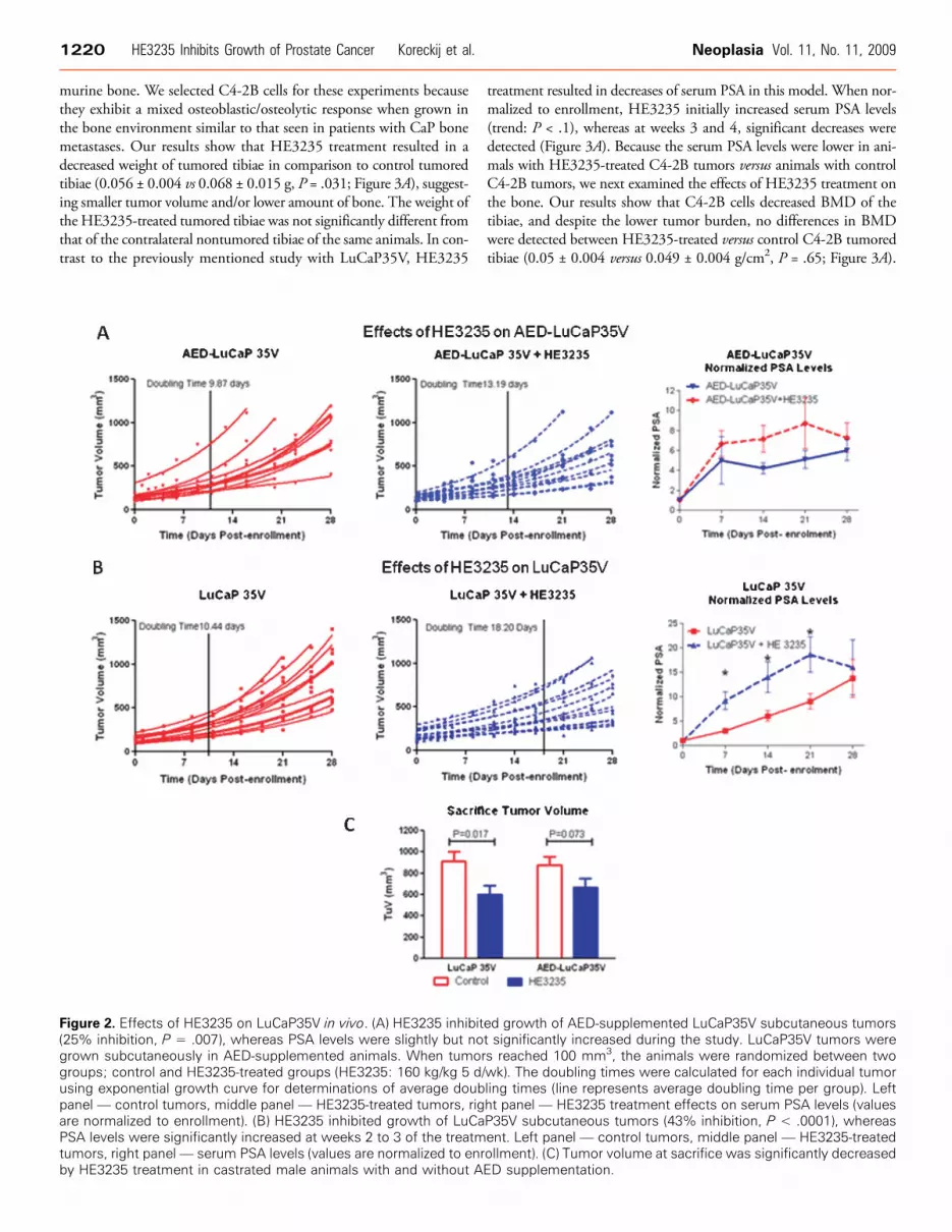
In the preclinical setting, we first evaluated effects of HE3235 onLuCaP35V in the presence of AED. Castrated male mice were sup-plemented with AED pellets to closer mimic the human adrenalgland secreting AED because the adrenal gland in mice lacks the en-zymes necessary for steroidogenesis [19]. Treatment with HE3235significantly inhibited growth of LuCaP35V in mice supplementedwith AED; doubling time of AED-LuCaP35V + HE3235 was13.15 ± 2.96 days (mean ± SD) in comparison to 9.87 ± 1.64 daysof AED-LuCaP35V (P = .007; Figure 2A). Treatment with HE3235

Figure 1. Effects of HE3235 on C4-2B and LuCaP35V cells in vitro. (A) Chemical structure of HE3235 and AED. (B and C) HE3235 in-creased AR-mediated transcription in C4-2B cells in vitro as demonstrated by ARE-luc and PSA-promoter-luc reporter assays. C4-2B cellswere transfected with ARE-luc (B) or PSA-promoter-luc (C) using Amaxa. After 2 days, the luciferase activity was measured. The lucsignal was normalized to renilla luciferase. The results are plotted as fold change over the untreated C4-2B cells. (D and E) Single-cellsuspensions of LuCaP 35V cells were prepared by dissociation of subcutaneous tumors. The cells were transfected with ARE-luc (D) orPSA-promoter-luc (E) using Amaxa. Treatment with HE3235 increases AR-mediated transcription in LuCaP35V CaP cells. (F) HE3235effect on AR-mediated transcription in C4-2B cells. C4-2B cells were grown under standard tissue culture conditions (see Materialsand Methods) and treated with HE3235. HE3235 significantly inhibited C4-2B proliferation. RLU indicates relative light units.
Neoplasia Vol. 11, No. 11, 2009 HE3235 Inhibits Growth of Prostate Cancer Koreckij et al. 1219
also resulted in slight increases in serum PSA levels versus the controlAED-LuCaP35V animals, but these differences did not reach signifi-cance. Similarly, the PSA index at the end of the study was also higherin HE3235-treated animals, and this difference did not reach signifi-cance (AED-LuCaP35V, 0.09 ± 0.04 ng/ml per cubic millimeter;and AED-LuCaP35V + HE3235, 0.13 ± 0.09 ng/ml per cubic milli-meter, P = .17). However, these elevations in PSA are in concordancewith the above in vitro studies demonstrating increases in AR-mediatedtranscription by HE3235 treatment.
Effect of HE3235 on LuCaP35V Subcutaneous TumorsOur previous data showed that HE3235 inhibits proliferation of
LNCaP cells in the presence and absence of AED in vitro [12].Therefore, in this study, we set out to investigate whether HE3235inhibits growth of CaP tumors in castrated male mice in the absenceof AED. These experimental conditions mimic the clinical scenarioof patients treated with agents aimed at blocking adrenal synthesis of
androgens (e.g., ketoconazole). In this setting, HE3235 significantlyinhibited the tumor doubling times of LuCaP35V (LuCaP35V +HE3235, 18.2 ± 6.28 days; untreated LuCaP35V, 10.44 ± 1.8 days;P < .0001; Figure 2B). HE3235 treatment resulted in significant in-creases in serum PSA levels in the treated animals versus control animalsbearing LuCaP35V tumors in the period of 1 to 3 weeks after treat-ment initiation (P < .0001; Figure 2B). PSA levels were not significantlyhigher in the treated animals at the end of the study. We hypothesizethat this is due to the decreases in tumor volume. This explanation issupported by results showing a significantly higher PSA index in theLuCaP35V + HE3235 animals than PSA index in the LuCaP35Vanimals, 0.18 ± 0.07 versus 0.07 ± 0.02 ng/ml per cubic millimeter,respectively (P < .0001).
Effects of HE3235 on the Growth of CaP in BoneTo investigate the effect of HE3235 on CRPC tumors growing in
the bone environment, we used C4-2B cells directly injected into
1220 HE3235 Inhibits Growth of Prostate Cancer Koreckij et al. Neoplasia Vol. 11, No. 11, 2009
murine bone. We selected C4-2B cells for these experiments becausethey exhibit a mixed osteoblastic/osteolytic response when grown inthe bone environment similar to that seen in patients with CaP bonemetastases. Our results show that HE3235 treatment resulted in adecreased weight of tumored tibiae in comparison to control tumoredtibiae (0.056 ± 0.004 vs 0.068 ± 0.015 g, P = .031; Figure 3A), suggest-ing smaller tumor volume and/or lower amount of bone. The weight ofthe HE3235-treated tumored tibiae was not significantly different fromthat of the contralateral nontumored tibiae of the same animals. In con-trast to the previously mentioned study with LuCaP35V, HE3235
Figure 2. Effects of HE3235 on LuCaP35V in vivo. (A) HE3235 inhibit(25% inhibition, P = .007), whereas PSA levels were slightly but nogrown subcutaneously in AED-supplemented animals. When tumorgroups; control and HE3235-treated groups (HE3235: 160 kg/kg 5 d/using exponential growth curve for determinations of average doubpanel — control tumors, middle panel — HE3235-treated tumors, rigare normalized to enrollment). (B) HE3235 inhibited growth of LuCaPSA levels were significantly increased at weeks 2 to 3 of the treatmtumors, right panel— serum PSA levels (values are normalized to enrby HE3235 treatment in castrated male animals with and without A
treatment resulted in decreases of serum PSA in this model. When nor-malized to enrollment, HE3235 initially increased serum PSA levels(trend: P < .1), whereas at weeks 3 and 4, significant decreases weredetected (Figure 3A). Because the serum PSA levels were lower in ani-mals with HE3235-treated C4-2B tumors versus animals with controlC4-2B tumors, we next examined the effects of HE3235 treatment onthe bone. Our results show that C4-2B cells decreased BMD of thetibiae, and despite the lower tumor burden, no differences in BMDwere detected between HE3235-treated versus control C4-2B tumoredtibiae (0.05 ± 0.004 versus 0.049 ± 0.004 g/cm2, P = .65; Figure 3A).
ed growth of AED-supplemented LuCaP35V subcutaneous tumorst significantly increased during the study. LuCaP35V tumors weres reached 100 mm3, the animals were randomized between twowk). The doubling times were calculated for each individual tumorling times (line represents average doubling time per group). Leftht panel — HE3235 treatment effects on serum PSA levels (valuesP35V subcutaneous tumors (43% inhibition, P < .0001), whereasent. Left panel — control tumors, middle panel — HE3235-treatedollment). (C) Tumor volume at sacrifice was significantly decreasedED supplementation.

Figure 3. Effect of HE3235 on growth of C4-2B in bone. (A) C4-2B cells were injected into tibiae of castrated male mice and when tumorswere established half of the animals were treated with HE3235 (160 mg/kg) daily for 28 days. Left panel— HE3235 treatment resulted indecreases in weight of tumored tibiae suggesting inhibition of tumor growth while not exhibiting any significant effects on nontumoredtibiae. Furthermore, HE3235-treated tumored tibiae weight was not significantly different as contralateral nontumored tibiae. Middlepanel — HE3235 treatment caused alterations in serum PSA levels, which were significantly decreased at weeks 3 and 4 of the treat-ment (values are normalized to enrollment). *P< .001, #P< .10. Right panel— HE3235 treatment did not have any significant effects onBMD of tumored and nontumored tibiae. BMD was measured at the site of tumor-cell injection adjacent to the growth place. (B) Rep-resentative examples of H&E, PSA immunoreactivity, and radiographs of HE3235-treated and control tibiae are shown. Left panel — H&Estaining show tumor growth in the bone marrow cavity next to the growth plate (located on the right-hand side). Multiple tumor foci arepresent between bone trabeculae secondary to the growth of this tumor in bone (magnification, ×10). Middle panel — HE3235-treatedtibiae show intensive PSA immunoreactivity (brown staining) in comparison to untreated tumored tibiae. Right panel — representativeradiographs of tibia from each group.
Neoplasia Vol. 11, No. 11, 2009 HE3235 Inhibits Growth of Prostate Cancer Koreckij et al. 1221
Also, no differences in BMD were detected between normal contralat-eral tibiae of the two groups (Figure 3A).Representative radiographs and hematoxylin and eosin (H&E)
staining of the tumored tibiae are shown in Figure 3B. These imagesshow an osteoblastic reaction associated with growth of C4-2B tumorsin bone and tumor foci between newly formed woven bone in bothHE3235-treated and control tibiae (Figure 3B). We have also per-formed IHC analysis of PSA expression. Our results show thatHE3235-treated tumor exhibited stronger PSA immunoreactivity de-spite lower levels of serum PSA (Figure 3B).
Analysis of Intratumoral Androgens in LuCaP35VSubcutaneous Tumors
We have previously shown that there are detectable levels of intra-tumoral androgens in CRPC xenografts and in human CRPC samplesdespite anorchid levels of circulating androgens [8]. Thus, we set outto determine whether the HE3235 inhibitory effects involve alterationsin levels of intratumoral androgens. Treatment with HE3235 signifi-cantly lowered levels of T and DHT in LuCaP35V tumors both inthe presence and in the absence of AED when compared with respec-tive control animals (Table 1). Animals receiving HE3235 with AED

1222 HE3235 Inhibits Growth of Prostate Cancer Koreckij et al. Neoplasia Vol. 11, No. 11, 2009
supplementation had a 63% reduction in T levels in tumors versuscontrol AED-supplemented tumors (P < .001), and a 40% reductionin DHT levels (P = .015). Interestingly, HE3235 caused even largerreductions in levels of intratumoral androgens in tumors growing incastrated animals not supplemented with AED. T levels were 93%lower in LuCaP35V + HE3235 versus control LuCaP35V tumors (P =.006), and DHT levels were reduced by 85% in HE3235-treatedLuCaP35V in comparison to control LuCaP35V tumors (P < .001).Owing to the small size of intratibial tumors, we were unable to deter-mine the intratumoral androgen levels in these specimens.
Gene Expression Analysis of LuCaP35V Subcutaneous TumorsIn an attempt to delineate in more detail the mechanisms of
HE3235 effects on CRPC, we set out to investigate alteration inRNA expression levels in LuCaP35V tumors, which resulted fromHE3235 treatment. We performed gene expression analysis on con-trol LuCaP35V and LuCaP35V + HE3235 groups because HE3235proved most efficacious in this experimental setting. To compare theoverall expression patterns of control LuCaP35V and HE3235-treated LuCaP35V tumors on oligo arrays, log2 ratio measurementswere analyzed using the SAM software to perform an unpaired two-sample t test (http://www-stat.stanford.edu/_tibs/SAM/). A false dis-covery rate of q values less than 5% was considered significant and setthe threshold for differential expression at greater than a 1.6-foldchange with treatment. At this level of significance, there were 355unique genes upregulated and 30 unique genes downregulated inHE3235-treated LuCaP35V versus control LuCaP35V. An EASEgene set enrichment revealed significant changes in a multitude ofcellular pathways associated with various biologic processes. Owingto our previous findings that HE3235 stimulates AR-mediated tran-scription as determined by reporter assay [9], and our present resultsdemonstrating the effects of HE3235 on levels of intratumoral andro-gens, we focused further investigations on alterations associated withAR-mediated transcription and on enzymes involved in steroidogenesis.Our results show that 67 of 355 HE3235-upregulated and 8 of 30HE3235-downregulated messages have been previously reported tobe regulated by androgens (Table W2), whereas the analysis of expres-sion levels of selected enzymes associated with the steroidogenesis path-way (Table W3) did not reveal any significant changes fitting thepreviously mentioned criteria. The oligo array analysis also showed thatAR messenger RNA (mRNA) levels were significantly decreased byHE3235 treatment (−3.1-fold).
We used real-time PCR to validate some of the oligo array results.We have elected two of the most downregulated genes, AR and AK5;five highly upregulated AR-regulated genes with suggested roles inCaP progression; and we also included PSA and hK2, two well-known AR-regulated genes that did not show altered expression onoligo arrays. Real-time PCR confirmed the oligo array analysis re-sults showing significant down-regulation of AR and AK5 mRNAin HE3235-treated tumors as well as the up-regulation of several
AR-regulated genes, although not all genes examined resulted in sta-tistically significant changes. In animals supplemented with AED, weobserved a diminished and in some instances, an abolished effect ongene expression in response to HE3235 (Figure 4). Despite the factthat the oligo array analysis did not show any significant alteration inexpression of 13 enzymes associated with steroidogenesis, we examinedlevels of three of these, HSD17B3, AKR1C3, and SRD5A1 by PCR.Even this analysis did not reveal any significant alterations in mRNAlevels of these enzymes after HE3235 treatment (data not shown).
Effects of HE3235 on AR ImmunoreactivityThe results of the oligo arrays and real-time PCR results showed that
levels of AR transcript were decreased by treatment with HE3235.Therefore, the effects of HE3235 on levels of AR protein were inves-tigated. IHC analysis showed significant decreases in the nuclear local-ization of AR after HE3235 treatment; the AR score of LuCaP35V +HE3235 was 248 ± 8 versus 198 ± 28 of control (P = .0048), whereasalteration of the AR score in AED-LuCaP35V + HE3235 versus AED-LuCaP35V did not reach significance (228 ± 36 vs 208 ± 8, P = .28;Figure 5). The AR score of HE3235-treated C4-2B intratibial tumorswas also significantly lower versus control C4-2B tumors (236 ± 15 vs182 ± 15, P < .001; Figure 5).
DiscussionWe have recently reported that HE3235 inhibits growth of AED-stimulated LNCaP cells in vitro and in vivo [12]. However, LNCaPcells are androgen-sensitive and express a mutated AR. Despite the rel-atively high frequency of mutations in AR reported in CRPC, whichcan alter AR responses to various steroids, most CRPC tumors expresswild-type AR [20]. Therefore, to further support the clinical potentialof this compound, our first objective was to determine whetherHE3235 inhibits growth of CRPC LuCaP35V cells that expresswild-type AR. Our results clearly demonstrate that HE3235 inhibitsgrowth of CRPC cells that express wild-type AR and that HE3235 in-hibits growth of LuCaP35V not only in the presence of AED but alsoin the absence of AED. Interestingly, the inhibition was even more pro-nounced when AED was absent. Although the mechanisms behindthese differences in tumor growth are not known at present, our resultsindicate that in a clinical setting, HE3235 might have more beneficialeffects when used in men on secondary androgen ablation regimensthat include inhibitors of adrenal androgen synthesis. However, despitethe inhibition of growth, HE3235 did not eradicate the tumors, butmerely slowed down tumor growth. Further studies are needed to de-termine if higher doses of HE3235 or more frequent administrationwould result in the elimination of the tumors.
Serum PSA levels are used to monitor disease progression as wellas effectiveness of treatment. HE3235 treatment of animals bearingLuCaP35V tumors resulted in increases of serum PSA and in PSAindex despite the inhibition of tumor growth. In concordance with
Table 1. Intratumoral Androgen Levels in LuCaP35V Subcutaneous Tumors.
Androgens
AED-LuCaP35V AED-LuCaP35V + HE3235 Control LuCaP35V LuCaP35V + HE3235T (pg/mg)
2.90 ± 0.31 1.06 ± 0.35 1.47 ± 0.67 0.10 ± 0.05 DHT (pg/mg) 3.03 ± 0.65 1.81 ± 0.31 4.28 ± 1.04 0.64 ± 0.17Values are mean ± SD.

Neoplasia Vol. 11, No. 11, 2009 HE3235 Inhibits Growth of Prostate Cancer Koreckij et al. 1223
these in vivo results, we also show that AR-mediated transcriptionwas increased by HE3235 in vitro using ARE- and PSA-promoter-reporter assays, further supporting stimulation of AR-mediated tran-scription despite growth inhibition. Because inhibition of tumor growthalong with increases in AR-mediated transcription is not well docu-mented in the literature, we further evaluated this negative associationby oligo array analysis, which confirmed increases in AR-mediated tran-scription based on higher levels of messages which expression is regu-lated by AR. To further complicate the situation of decreased tumorvolume, increased serum PSA, and increased levels of multiple AR-regulated genes, HE3235 treatment decreased the expression of theAR mRNA and lowered levels of nuclear AR in the HE3235-treatedtumors.More detailed investigations of the effects of HE3235 on tumorgrowth and AR-mediated transcription will be required to fully under-stand this interplay regulation of tumor growth and AR signaling. How-
Figure 4. Real-time PCR validation of the androgen-regulated genes thanalysis. We confirmed AR down-regulation concomitantly with AR agenes. The addition of AED seems to attenuate the effect of HE3GPR64, G protein–coupled receptor 64; ARG2, anterior gradient homoltranscription factor 1; hK2, human glandular kallikrein; AK5, adenylateanalysis. Significant differences were detected in number of mRNA in
ever, we currently have two working hypotheses: 1) levels of HE3235in the tumors are sufficient to cause increases in AR-mediated transcrip-tion despite lower levels of AR and lower intratumoral androgens and2) other mechanisms, independent of AR, are involved in stimulationof expression of AR-regulated genes. For example, it has recently beenreported that mitogen-activated protein kinase pathway stimulatesARE-reporter activity in PC-3 and DU 145 cells that lack AR [21].
One of the important recent findings in CaP research is that thereare measurable levels of intratumoral androgens in CRPC tumors de-spite anorchid levels of serum androgens [8]. Abiraterone, a novelinhibitor of steroidogenesis currently evaluated in preclinical andclinical settings, reduces serum androgens in patients and may func-tion by suppressing intratumoral androgens in CRPC [10,22].Therefore, we examined whether the inhibition of tumor growthby HE3235 is also associated with altered levels of intratumoral
at demonstrated response to treatment with HE3235 on oligo arrayctivation as demonstrated by differential changes in AR-regulated235 on these genes. ODC1 indicates ornithine decarboxylase 1;og 2 (Xenopus laevis); SAA2, serum amyloid A2; RUNX1, runt-relatedkinase 5. Numbers in parentheses show fold change in oligo arrayLuCaP35V treated with HE3235 versus control tumors. *P < .05.

Figure 5. Effects of HE3235 on AR immunoreactivity. Sections of treated and control tumors (AED-LuCaP35V and LuCaP35V subcu-taneous tumors and C4-2B intratibial tumors) were stained with anti-AR antibody, and nuclear staining was quantified based on intensityof immunoreactivity and percentage of positive nuclei. HE3235 significantly decreased the AR score in LuCaP 35V and C4-2B tumors,although the decreases were not significant in AED-LuCaP35V. Original magnification: AED-LuCaP35V and LuCaP 35V, ×20; C4-2B, ×10.
1224 HE3235 Inhibits Growth of Prostate Cancer Koreckij et al. Neoplasia Vol. 11, No. 11, 2009
androgens. In both non-AED and AED-supplemented animals,HE3235 treatment resulted in significant reductions in intratumoralT and DHT. A greater suppression of intratumoral androgens was de-tected in LuCaP35V + HE3235 tumors versus tumors grown in thepresence of AED. These differences might be instrumental in the lesspronounced inhibition of growth in AED-supplemented tumors. Onepotential mechanism resulting in decreased levels of intratumoral an-drogen might be HE3235 inhibition of the transcription of enzymesinvolved in steroidogenesis because increased messages have been de-tected in CRPC metastases [8]. However, despite decreased levels ofintratumoral androgens, HE3235 did not alter the transcription of se-lected enzymes associated with steroidogenesis. Nonetheless, transcrip-tional control does not represent the only means by which to controlintratumoral androgen production. HE3235 suppression of T andDHT in all tumors may be the result of either a blockade on importof androgenic precursors or inhibition of enzymatic activity of thecomponents of the steroid synthesis pathway. The exact mechanism be-hind this suppression of intratumoral androgens is not known, anddelineation of the mechanism(s) will require further investigations onHE3235’s effect on enzyme activity in vitro and on transporters ofandrogens and their precursors (e.g., SLCO1B3 and SLCO2B1).
Even with dramatic reductions in the levels of intratumoral andro-gens, LuCaP35V tumors were not eliminated. Their growth wasmerely slowed. This may indicate either of the following: 1) thelow levels of intratumoral androgens still present in LuCaP35V +HE3235 tumors are sufficient to drive CRPC tumor growth andgreater reductions in levels of intratumoral androgens will be neces-sary for complete inhibition of growth; or 2) a subset of truly castration-resistant tumor cells exist in these tumors that do not require androgensfor growth. Therefore, as new agents aimed at blocking the intracrinesynthesis of androgens become available, improvements in patient out-comes must be tempered against howmuch suppression of intratumoralandrogen production is achieved, and drugs aimed at decreasing expres-sion of AR as well as agents that will kill AR-negative cells need to bedeveloped and evaluated.
One of the most frequent sites of CaP metastases is bone [16]. It isimportant that new pharmaceuticals aimed at treating advanced CaPare tested against CaP growing within the bone environment. Ourresults show that HE3235 inhibits the growth of CRPC in boneas demonstrated by decreased tibiae weight as well as decreased serumPSA levels in HE3235-treated animals. The decreases in serum PSAlevels are interesting. We have shown in vitro that HE3235 increasesAR-mediated transcription in both C4-2B and LuCaP35V cells usingreporter assay, and we show increased serum PSA levels in vivo withsubcutaneous LuCaP35V tumors treated with HE3235. However, se-rum PSA levels in our intratibial studies were decreased by the HE3235treatment. We hypothesize that because treated tumors are producingmore PSA, an even greater reduction in tumor volume is necessary be-fore serum PSAvalues decrease in comparison to control tumors. SerumPSA increases caused by HE3235 treatment of larger subcutaneoustumors initially overshadows the concomitant inhibition of tumorgrowth. In the intratibial study, treatment is started on considerablysmaller enrollment tumor volumes that are more susceptible to growthinhibition. With smaller enrollment tumor volumes in the intratibialstudy, the balance between HE3235 effects on AR-mediated transcrip-tion and inhibition of tumor growth is reached sooner as is demon-strated by the initial elevation of serum PSA levels in HE3235-treatedanimals followed by lower levels thereafter. Our in vitro experiments,demonstrating HE3235 inhibition of C4-2B proliferation taken to-gether with our IHC analysis showing activity of HE3235 within thebone environment, further support the premise that HE3235 inhibitsCRPC growth within the bone.
In conclusion, our results show that HE3235 inhibits growth ofCRPC in multiple models of CaP, including inhibition of CRPC inthe bone environment. It invokes a variety of cellular alterations con-tributing to its ability to inhibit growth of CaP cells, including de-creases in levels of AR messages and AR nuclear localization as well asinhibition of the production of intratumoral androgens. Our resultswarrant further investigation into mechanisms of HE3235 actionand clinical investigation of HE3235 in the treatment of CRPC.

Neoplasia Vol. 11, No. 11, 2009 HE3235 Inhibits Growth of Prostate Cancer Koreckij et al. 1225
AcknowledgmentsThe authors thank Lisha Brown and Ted Kalhoun for their tech-nical expertise.
References[1] Petrylak DP, Tangen CM, Hussain MH, Lara PN Jr, Jones JA, Taplin ME,
Burch PA, Berry D, Moinpour C, Kohli M, et al. (2004). Docetaxel and estra-mustine compared with mitoxantrone and prednisone for advanced refractoryprostate cancer. N Engl J Med 351, 1513–1520.
[2] Tannock IF, de WR, Berry WR, Horti J, Pluzanska A, Chi KN, Oudard S,Theodore C, James ND, Turesson I, et al. (2004). Docetaxel plus prednisoneor mitoxantrone plus prednisone for advanced prostate cancer. N Engl J Med351, 1502–1512.
[3] Burnstein K (2005). Regulation of androgen receptor levels: implications forprostate cancer progression and therapy. J Cell Biochem 95, 657–669.
[4] Culig Z, Comuzzi B, Steiner H, Bartsch G, and Hobisch A (2004). Expressionand function of androgen receptor coactivators in prostate cancer. J Steroid Bio-chem Mol Biol 92, 265–271.
[5] Scher HI, Buchanan G, Gerald W, Butler LM, and Tilley WD (2004). Target-ing the androgen receptor: improving outcomes for castration-resistant prostatecancer. Endocr Relat Cancer 11, 459–476.
[6] Lam JS, Leppert JT, Vemulapalli SN, Shvarts O, and Belldegrun AS (2006).Secondary hormonal therapy for advanced prostate cancer. J Urol 175, 27–34.
[7] Titus MA, Schell MJ, Lih FB, Tomer KB, and Mohler JL (2005). Testosteroneand dihydrotestosterone tissue levels in recurrent prostate cancer. Clin Cancer Res11, 4653–4657.
[8] Montgomery RB, Mostaghel EA, Vessella R, Hess DL, Kalhorn TF, Higano CS,True LD, and Nelson PS (2008). Maintenance of intratumoral androgens inmetastatic prostate cancer: a mechanism for castration-resistant tumor growth.Cancer Res 68, 4447–4454.
[9] Locke JA, Guns ES, Lubik AA, Adomat HH, Hendy SC, Wood CA, EttingerSL, Gleave ME, and Nelson CC (2008). Androgen levels increase by intra-tumoral de novo steroidogenesis during progression of castration-resistant prostatecancer. Cancer Res 68, 6407–6415.
[10] Attard G, Reid AH, Yap TA, Raynaud F, Dowsett M, Settatree S, Barrett M,Parker C, Martins V, Folkerd E, et al. (2008). Phase I clinical trial of aselective inhibitor of CYP17, abiraterone acetate, confirms that castration-resistant prostate cancer commonly remains hormone driven. J Clin Oncol 26,4563–4571.
[11] Vasaitis T, Belosay A, Schayowitz A, Khandelwal A, Chopra P, Gediya LK, Guo Z,Fang HB, Njar VC, and Brodie AM (2008). Androgen receptor inactivation
contributes to antitumor efficacy of 17α-hydroxylase/17,20-lyase inhibitor 3β-hydroxy-17-(1H -benzimidazole-1-yl)androsta-5,16-diene in prostate cancer.Mol Cancer Ther 7, 2348–2357.
[12] Trauger R, Corey E, Bell D, White S, Garsd A, Stickney D, Reading C, andFrincke J (2009). Inhibition of androstenediol-dependent LNCaP tumourgrowth by 17α-ethynyl-5α-androstane-3α, 17β-diol (HE3235). Br J Cancer 100,1068–1072.
[13] Corey E, Quinn JE, Buhler KR, Nelson PS, Macoska JA, True LD, and VessellaRL (2003). LuCaP 35: a new model of prostate cancer progression to androgenindependence. Prostate 55, 239–246.
[14] Thalmann GN, Sikes RA, Wu TT, Degeorges A, Chang SM, Ozen M, Pathak S,and Chung LW (2000). LNCaP progression model of human prostate cancer:androgen-independence and osseous metastasis. Prostate 44, 91–103.
[15] Coleman IM, Kiefer JA, Brown LG, Pitts TE, Nelson PS, Brubaker KD,Vessella RL, and Corey E (2006). Inhibition of androgen-independent prostatecancer by estrogenic compounds is associated with increased expression ofimmune-related genes. Neoplasia 8, 862–878.
[16] Corey E, Quinn JE, Bladou F, Brown LG, Roudier MP, Brown JM, Buhler KR,and Vessella RL (2002). Establishment and characterization of osseous prostatecancer models: intra-tibial injection of human prostate cancer cells. Prostate 52,20–33.
[17] Kiefer JA, Vessella RL, Quinn JE, Odman AM, Zhang J, Keller ET, KostenuikPJ, Dunstan CR, and Corey E (2004). The effect of osteoprotegerin adminis-tration on the intra-tibial growth of the osteoblastic LuCaP 23.1 prostate cancerxenograft. Clin Exp Metastasis 21, 381–387.
[18] Lai JS, Brown LG, True LD, Hawley SJ, Etzioni RB, Higano CS, Ho SM,Vessella RL, and Corey E (2004). Metastases of prostate cancer express estrogenreceptor-beta. Urology 64, 814–820.
[19] van Weerden WM, Bierings HG, van Steenbrugge GJ, de Jong FH, and SchroderFH (1992). Adrenal glands of mouse and rat do not synthesize androgens. Life Sci50, 857–861.
[20] Marcelli M, Ittmann M, Mariani S, Sutherland R, Nigam R, Murthy L, Zhao Y,DiConcini D, Puxeddu E, Esen A, et al. (2000). Androgen receptor mutationsin prostate cancer. Cancer Res 60, 944–949.
[21] Carey AM, Pramanik R, Nicholson LJ, Dew TK, Martin FL, Muir GH, andMorris JD (2007). Ras-MEK-ERK signaling cascade regulates androgen recep-tor element–inducible gene transcription and DNA synthesis in prostate cancercells. Int J Cancer 121, 520–527.
[22] Montgomery RB, Mostaghel E, Nelson PS, Marck B, Nguyen H, and VessellaRL (2009). Abiraterone suppresses castration resistant human prostate cancergrowth in the absence of testicular and adrenal androgens. Advances in ProstateCancer, AACR Conference, January 2009. San Diego, CA, Abstract.

Table W1. Real-time PCR Primers.
17BHSD3
Hydroxysteroid (17-beta) dehydrogenase 3 5′: CTGAAGCTCAACACCAAGGTCA NM_000197 3′: CTGCTCCTCTGGTCCTCTTCAGAR
Androgen receptor 5′: GGACTTGTGCATGCGGTACTCA NM_000044 3′: CCTGGCTTCCGCAACTTACACAGR2
Anterior gradient homolog 2 (Xenopus laevis) 5′: GTCCCCAGGATTATGTTTGTTGACC NM_006408 3′: AGTCTTCTCACACTTCTTCTGGTTTCAK5
Adenylate kinase 5 5′: GGAGGTGAAGCAAGGGGAAGAGTT NM_174858.1 3′: TCCTTTGGAGAAGGCGGTTGGTCAKR1C3
Aldo-keto reductase family 1, member C3 5′: GAGAAGTAAAGCTTTGGAGGTCACA NM_003739 3′: CAACCTGCTCCTCATTATTGTATAAGPR64
G protein–coupled receptor 64 5′: CCAAAGAAA ATGTCAGGAAGCAATGG NM_001079858.1 3′: AGCAGTGTGGTGGAGTTAGTGGAGhK2
Human glandular kallikrein 2 5′: ATGTTGTGTGCTGGGCTCTGGAC NM_005551 3′: GGTTGGC TGCGATCCTGTCCTTGODC 1
Ornithine decarboxylase 1 5′: ATGTGGGTGATTGGATGCTCTTTGA NM_002539 3′: CAGGCTGCTCTGTGGCGTTTCATPSA
Prostate-specific antigen 5′: CCCCAGAATCACCCGAGCAG NM_001648 3′: ACCAGAGGAGTTCTTGACCCCAAAASAA2
Serum amyloid A2 5′: TGCTCGGGGGAACTATGATGCTG NM_030754 3′: GTCGGAAGTGATTGGGGTCTCTGSRD5A1
Steroid-5-alpha-reductase, alpha polypeptide 1 5′: CCTGTTGAATGCTTCATGACTTG NM_001047 3′: TAAGGCAAAGCAATGCCAGATGRUNX1
Runt-related transcription factor 1 5′: CTCCCTGAACCACTCCACTGCCT NM_001001890 3′: GACCCACATTCTGCCTTCCTCATAA
Table W2. Changes in Androgen-Regulated Genes by HE3235.
Gene Symbol
Gene Name Fold ChangeIncreased expression
SAA2 Serum amyloid A2 5.8 FER1L3 fer-1-like 3, myoferlin (C. elegans) 4.1 RUNX1 Runt-related transcription factor 1(acute myeloid leukemia 1; aml1 oncogene)
3.9GPR64
G protein–coupled receptor 64 3.0 LOX Lysyl oxidase 2.5 RIS1 Ras-induced senescence 1 2.5 PKNOX2 PBX/knotted 1 homeobox 2 2.3 RBM24 RNA binding motif protein 24 2.3 ABCG1 ATP-binding cassette, subfamily G (WHITE),member 1
2.2GDF15
Growth differentiation factor 15 2.2 ODC1 Ornithine decarboxylase 1 2.2 TFF1 Trefoil factor 1 (breast cancer, estrogen-induciblesequence expressed in)
2.2ABCC4
ATP-binding cassette, subfamily C (CFTR/MRP),member 42.1
CDH26
Cadherin-like 26 2.0 GDA Guanine deaminase 2.0 CDC42EP2 CDC42 effector protein (Rho GTPase binding) 2 2.0 IL1R1 Interleukin 1 receptor, type I 2.0 FADS1 Fatty acid desaturase 1 2.0 PXDN Peroxidasin homolog (Drosophila) 2.0 ChGn Chondroitin beta1,4 N -acetylgalactosaminyltransferase 2.0 AZGP1 alpha-2-Glycoprotein 1, zinc 2.0 FABP5 Fatty acid binding protein 5 (psoriasis-associated) 1.9 S100A11 S100 calcium binding protein A11 (calgizzarin) 1.9 LOC114984 Hypothetical protein BC014089 1.9 MAF v-maf musculoaponeurotic fibrosarcomaoncogene homolog (avian)
1.9LGALS3
Lectin, galactoside-binding, soluble, 3 (galectin 3) 1.9 GPX3 Glutathione peroxidase 3 (plasma) 1.9 LOC645904 Similar to mitotic spindle assembly checkpointprotein MAD1 (mitotic arrest deficient-like protein 1)(MAD1-like 1) (mitotic checkpoint MAD1protein-homolog) (HsMAD1) (hMAD1)(Tax-binding protein 181)
1.9
SAT
Spermidine/spermine N1-acetyltransferase 1.9 CASP10 Caspase 10, apoptosis-related cysteine peptidase 1.8 CRISP3 Cysteine-rich secretory protein 3 1.8 PPM1E Protein phosphatase 1E (PP2C domain containing) 1.8 CTBP1 C-terminal binding protein 1 1.8 C15orf48 Chromosome 15 open reading frame 48 1.8 PGC Progastricsin (pepsinogen C) 1.8 PVRL3 Poliovirus receptor-related 3 1.8 C21orf122 Chromosome 21 open reading frame 122 1.7 RPL36A Ribosomal protein L36a 1.7 SLC26A2 Solute carrier family 26 (sulfate transporter), member 2 1.7 NFKBIZ Nuclear factor of kappa light polypeptidegene enhancer in B-cells inhibitor, zeta
1.7AGR2
Anterior gradient 2 homolog (Xenopus laevis) 1.7 MICAL1 Microtubule-associated monoxygenase,calponin and LIM domain containing 1
1.7LFNG
Lunatic fringe homolog (Drosophila) 1.7 MRPL33 Mitochondrial ribosomal protein L33 1.7 AMPD3 Adenosine monophosphate deaminase (isoform E) 1.7 MGC33839 Hypothetical protein MGC33839 1.7 RPLP1 Ribosomal protein, large, P1 1.7 ZNF33A Zinc finger protein 33A 1.6 AMACR alpha-Methylacyl-CoA racemase 1.6 RPL21 Ribosomal protein L21 1.6 GRHL2 Grainyhead-like 2 (Drosophila) 1.6 SUMO2 SMT3 suppressor of mif two 3 homolog 2 (yeast) 1.6 RAB32 RAB32, member RAS oncogene family 1.6 C6orf166 Chromosome 6 open reading frame 166 1.6 MT1G Metallothionein 1G 1.6 GNE Glucosamine (UDP-N -acetyl)-2-epimerase/N -acetylmannosamine kinase
1.6POPDC3
Popeye domain containing 3 1.6 TIMP2 TIMP metallopeptidase inhibitor 2 1.6 CLDN8 Claudin 8 1.6 LIPG Lipase, endothelial 1.6Table W2. (continued )
Gene Symbol
Gene Name Fold ChangeRFPL1
Ret finger protein-like 1 1.6 LONPL Peroxisomal LON protease like 1.6 ARHGEF10 Rho guanine nucleotide exchange factor(GEF) 10
1.6RHOBTB3
Rho-related BTB domain containing 3 1.6 C14orf78 Chromosome 14 open reading frame 78 1.6 SAA3 Serum amyloid A3 1.6 FER1L4 Fer-1-like 3, myoferlin (C. elegans) 1.6Decreased expression
JUN v-jun sarcoma virus 17 oncogene homolog (avian) −1.6 IRX5 Iroquois homeobox protein 5 −1.6 SLC3A1 Solute carrier family 3 (cystine, dibasic andneutral amino acid transporters, activator ofcystine, dibasic and neutral amino acid transport),member 1
−1.7
TFPI
Tissue factor pathway inhibitor (lipoprotein-associatedcoagulation inhibitor)−1.7
JAG1
Jagged 1 (Alagille syndrome) −1.9 CD44 CD44 antigen (Indian blood group) −2.0 AR Androgen receptor (dihydrotestosterone receptor;testicular feminization; spinal and bulbarmuscular atrophy; Kennedy disease)
−3.1
AK5
Adenylate kinase 5 −3.7Table W3. Steroidogenesis Enzymes.
Gene Symbol
Gene NameAKR1C1
Aldo-keto reductase family 1, member C1 (dihydrodiol dehydrogenase 1;20-alpha (3-alpha)-hydroxysteroid dehydrogenase)AKR1C3
Aldo-keto reductase family 1, member C3 (3-alpha hydroxysteroiddehydrogenase, type II)CYP11A1
Cytochrome P450, family 11, subfamily A, polypeptide 1 CYP17A1 Cytochrome P450, family 17, subfamily A, polypeptide 1 FASN Fatty acid synthase HSD17B1 Hydroxysteroid (17-beta) dehydrogenase 1 HSD17B2 Hydroxysteroid (17-beta) dehydrogenase 2 HSD17B3 Hydroxysteroid (17-beta) dehydrogenase 3 HSD17B4 Hydroxysteroid (17-beta) dehydrogenase 4 HSD17B6 Hydroxysteroid (17-beta) dehydrogenase 6 SRD5A1 Steroid-5-alpha-reductase, alpha polypeptide 1 (3-oxo-5 alpha-steroiddelta 4-dehydrogenase alpha 1)
UGT2B15 UDP glucuronosyltransferase 2 family, polypeptide B15 UGT2B17 UDP glucuronosyltransferase 2 family, polypeptide B17


