Embed Size (px)
Citation preview
1030
Review
Heme, heme oxygenase and ferritin in vascularendothelial cell injury
J�zsef Balla1, GregoryM. Vercellotti2, Vikt�ria Jeney1, Akihiro Yachie3, Zsuzsa Varga1,JohnW. Eaton4 andGy�rgy Balla1
1Departments of Medicine and Neonatology, University of Debrecen, Debrecen, Hungary2Department of Medicine, University of Minnesota, Minneapolis, MN, USA3Department of Laboratory Sciences, Kanazawa University, Kanazawa, Japan4James Graham Brown Cancer Center, University of Louisville, Louisville, KY, USA
Iron-derived reactive oxygen species are implicated in the pathogenesis of numerous vascular disor-ders including atherosclerosis, microangiopathic hemolytic anemia, vasculitis, and reperfusion injury.One abundant source of redox active iron is heme, which is inherently dangerous when released fromintracellular heme proteins. The present review concerns the involvement of heme in vascular endo-thelial cell damage and the strategies used by endothelium to minimize such damage. Exposure ofendothelium to heme greatly potentiates cell killing mediated by polymorphonuclear leukocytes andother sources of reactive oxygen. Free heme also promotes the conversion of low-density lipoprotein(LDL) into cytotoxic oxidized products. Only because of its abundance, hemoglobin probably repre-sents the most important potential source of heme within the vascular endothelium; hemoglobin inplasma, when oxidized, transfers heme to endothelium and LDL, thereby enhancing cellular suscepti-bility to oxidant-mediated injury. As a defense against such toxicity, upon exposure to heme orhemoglobin, endothelial cells up-regulate heme oxygenase-1 and ferritin. Heme oxygenase-1 is aheme-degrading enzyme that opens the porphyrin ring, producing biliverdin, carbon monoxide, andthe most dangerous product – free redox active iron. The latter can be effectively controlled by ferri-tin via sequestration and ferroxidase activity. Ferritin serves as a protective gene by virtue of antioxi-dant, antiapoptotic, and antiproliferative actions. These homeostatic adjustments have been showneffective in the protection of endothelium against the damaging effects of exogenous heme and oxi-dants. The central importance of this protective system was recently highlighted by a child diagnosedwith heme oxygenase-1 deficiency, who exhibited extensive endothelial damage.
Keywords:Atherosclerosis / Endothelial cell injury / Ferritin / Heme / Heme oxygenase /
Received:May 24, 2005; revised: July 8, 2005; accepted: July 10, 2005
1 Introduction
Heme is absolutely required for aerobic life. However, freeheme can be quite cytotoxic, particularly in the presence ofoxidants or activated phagocytes. Of all sites in the body,the vasculature – and in particular the endothelial lining –may be at greatest risk of exposure to free heme. This isbecause erythrocytes contain heme in a concentration of20 mmol/L and are vulnerable to unexpected lysis. Theextracellular hemoglobin is easily oxidized to ferrihemo-globin which, in turn, will readily release heme. Given the
hydrophobic nature of heme, it is no surprise that it easilycrosses cell membranes and can synergistically enhancecellular oxidant damage. Here, we present a brief review ofthe nature of heme-mediated cytotoxicity and of the strate-gies by which normal endothelium manages to protect itselffrom this clear and present danger (Fig. 1).
2 Direct heme and hemoglobin toxicity tovascular endothelial cells
Damage caused by reactive oxygen species can be greatlyamplified by “free” redox active iron [1]. For example,iron-rich Staphylococcus aureus are three orders of magni-tude more susceptible to killing by hydrogen peroxide thanare iron-poor staphylococci [2]. Conversely, depletion of
i 2005WILEY-VCH Verlag GmbH &Co. KGaA,Weinheim
DOI 10.1002/mnfr.200500076 Mol. Nutr. Food Res. 2005, 49, 1030–1043
Correspondence: Professor J�zsef Balla, Pf. 19., Nagyerdei krt. 98.,4012 Debrecen, HungaryE-mail: [email protected]: +36-52-413-653

Mol. Nutr. Food Res. 2005, 49, 1030–1043 Heme, heme oxygenase and ferritin in vascular endothelial cell injury
cellular iron powerfully protects eukaryotic and prokaryoticcells against oxidant challenge [3]. We have shown that onecritical feature of highly damaging iron to endothelium isdue to permeation of the metal into cells. Chelation of ironby certain lipophilic chelators, such as 8-hydroxyquinoline,results in the accumulation of catalytically active lipophiliciron chelates in endothelial lipid compartments; endothe-lium pretreated with 8-hydroxyquinoline-iron chelate wasexquisitely sensitive to both endogenous and exogenousoxidant stress [4].
One abundant source of potentially toxic iron is iron proto-porphyrin IX or heme with its hydrophobic nature. Heme, aubiquitous iron-containing compound, is present in largeamounts in many cells [5] and is also inherently dangerous,particularly when it escapes from intracellular sites [6–9].Heme greatly amplifies cellular damage arising from acti-vated oxygen [6–8].
The potential toxicity of free heme derives from the easewith which this highly hydrophobic compound can enterand cross cell membranes, and therefore readily concen-trates within the hydrophobic milieu of intact cells [6, 7].Both in vitro and in vivo, cells will accumulate exogenous
heme and synergistically amplify the cytotoxic effects ofoxidants of reagent, enzymatic, or cellular origin. Hemeuptake by endothelial cells can exacerbate their damage bypolymorphonuclear leukocytes (PMNs) – cells that tend tomarginate along endothelial surfaces in the presence ofdiverse inflammatory mediators [6, 8]. Intriguingly, hemewas shown by Graca-Souza et al. [10] to induce PMN acti-vation as well. Moreover, Wagener et al. [11, 12] revealedthat heme can enhance endothelial cell adhesion moleculeexpression, which regulates PMN adhesion and provokesinflammation.
The uptake of heme is required for this synergistic toxicityand the hydrophobicity of heme is critical for entry intoendothelial cells. The spontaneous uptake of heme and theassociated amplification of cellular oxidant sensitivity areboth inhibited by hemopexin [6, 7]. The plasma heme-bind-ing protein, hemopexin, can block catalytic activity ofheme [13, 14]. Hemopexin is certainly not the sole factor inplasma that protects against heme-amplified oxidantdamage to endothelium. Albumin may also limit the intru-sion of extracellular heme and its pro-oxidant effects. Oncewithin the cell, heme can promote oxidative damage eitherdirectly or, perhaps more importantly, via the release of
1031
i 2005WILEY-VCH Verlag GmbH &Co. KGaA,Weinheim www.mnf-journal.de
Hemoglobin
H2O2
O2- •
Methemoglobin
Fe
Fe
FeFe
CO
Fe
Fe Fe
Fe Fe
H2O2
O2- •
Sensitization
Oxidative damage
Stress adaptation
Fe
Oxidative resistance
Biliverdin
Fe
HO-1
FerritinFerroxidase
Iron storage
Fe Fe
Fe
Fe
Heme
degradation
HaptoglobinHemopexin
FeRed blood cell
Mononuclear cell
Polymorphonuclear cell
Endothelium
Heme
Heme degradation
Low-density lipoprotein
1
2a
2b
1
3
4
5
6
7a
8
FeFe Fe
7b
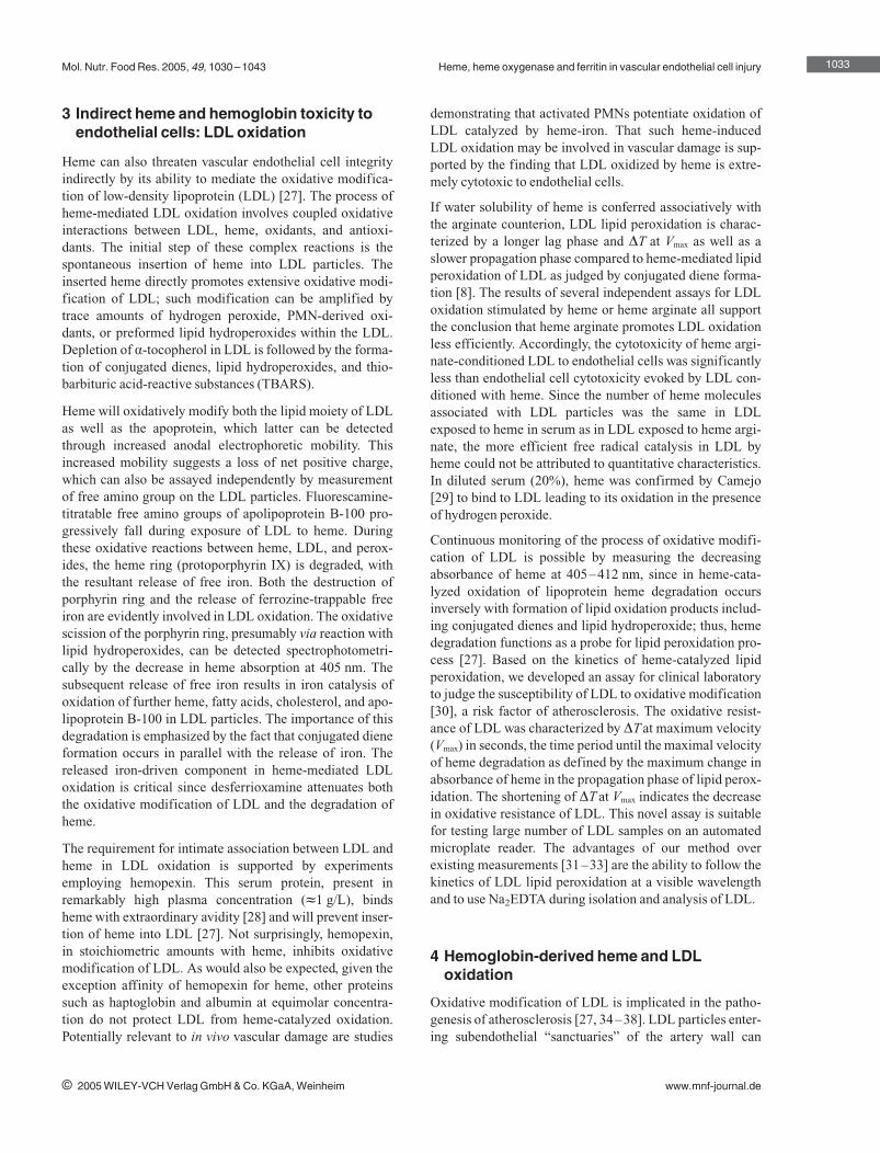
Figure 1. Oxidative stress and adaptation induced by heme and hemoglobin. 1: Leakage of hemoglobin from red blood cells; 2aand b: oxidation of hemoglobin; 3: heme release from methemoglobin; 4: heme uptake and sensitization of endothelium to oxidativestress; 5: heme uptake by LDL; 6: oxidative modification of LDL; 7a and b: oxidative stress induced by oxidized LDL and oxidantsderived from leukocytes; and 8: induction of adaptation to oxidative stress via up-regulating heme oxygenase-1 and ferritin.

J. Balla et al. Mol. Nutr. Food Res. 2005, 49, 1030–1043
iron which can occur through either nonenzymatic oxida-tive degradation of heme [6, 7] or enzymatic, heme oxyge-nase-catalyzed heme cleavage. In either case, the iron mayinitially lodge within the hydrophobic interstices of thephospholipid bilayer; within this highly oxidizable matrix,iron acts as an especially active catalyst of oxidation of cellmembrane constituents [6].
We asked: Could heme sensitize endothelial cells to oxida-tive challenge in the presence of plasma [8]? After all,plasma is enriched with binding proteins, such as albuminand hemopexin, known to inhibit heme-mediated celldamage. Exposure of endothelium to heme in the presenceof whole human plasma synergizes cellular oxidant damagefor added oxidants, with an optimal heme-exposure durationof 60 min. Intriguingly, cytotoxicity studies showed littleadded toxicity to endothelium if water solubility of heme isconferred associatively with the arginate counterion (hemearginate). Even with efficient permeation heme arginatedoes not amplify oxidant-induced cytotoxicity. In support,exposure of endothelium to heme arginate in plasma-freemedium increases endothelial cell heme content to an extentsimilar to what is observed after heme treatment. Compar-able heme uptake can be obtained in the presence of humanplasma although at two orders of magnitude greater concen-tration for both hemearginate and heme.
The hydrophobicity of various heme-analogs, ferriporphyr-ins, is critical for entry into cells and required for the syner-gistic oxidative toxicity. Substitution of vinyl side chains ofheme with hydrogen does not alter the hydrophobicity ofthe resultant ferriporphyrin, iron deuteroporphyrin IX;accordingly, hypersusceptibility is similarly provoked. Onthe contrary, if water solubility of heme is conferred asso-ciatively with the arginate counterion or the vinyl sidechains of heme are substituted by sulfonate, propionate, orglycol leading to hydrophilic ferriporphyrins (iron deutero-porphyrin IX,2,4-bis-sulfonate, iron coproporphyrin III,and iron deuteroporphyrin IX,2,4-bis-glycol), these ferri-porphyrins failed to sensitize cells to oxidants or activatedPMNs.
Although free heme is rapidly incorporated into hydropho-bic domains of cells and serves as a source of highly damag-ing iron, the question remains as to whether intact hemeliganded to proteins, as in hemoglobin, might also transferheme to vascular endothelium. Whereas reduced (ferro- oroxy-) hemoglobin is relatively innocuous to endothelialcells, oxidized (ferri- or met-) hemoglobin greatly amplifiesoxidant-mediated endothelial injury [15, 16]. This isbecause ferrihemoglobin readily releases its heme moietiesas first demonstrated by Bunn and Jandl [17]. Releasedheme from ferrihemoglobin can indeed be rapidly incorpo-rated into hydrophobic domains of cultured endotheliumand serve as a source of highly damaging iron. Although
ferrohemoglobin itself is not capable of sensitizing vascularendothelial cells to oxidant injury, we and others haveshown that it can readily be oxidized to heme-releasingmethemoglobin in the presence of inflammatory-cell-derived oxidants [15, 18, 19]. For instance, PMNs, whenactivated with the phorbol ester PMA, markedly oxidizeferrohemoglobin to ferrihemoglobin within 30 min [15].Accordingly, ferrohemoglobin in the presence of activatedPMNs can provide heme to endothelium, which greatlyenhances cellular susceptibility to oxidant-mediated cellinjury [15, 16]. The oxidation of ferrohemoglobin to ferri-hemoglobin is essential for this deleterious effect. Anothercandidate for generating methemoglobin is nitric oxide.Reaction of nitric oxide with free hemoglobin producesmethemoglobin and leads to decreased nitric oxide bio-availability, causing pulmonary hypertension, vasculardamage, and end-organ injury as reviewed by Gladwin etal. [20].
The initial release of heme from ferrihemoglobin can beinhibited by complexing with the hemoglobin-binding pro-tein, haptoglobin [17]. If metheme binding to globin isstrengthened by haptoglobin or if released heme is reli-ganded to hemopexin, ferrihemoglobin loses much of itscapacity to sensitize endothelium to reactive oxygen [15].Hemoglobin :haptoglobin complex is eliminated from thecirculation through the recently characterized CD163receptor [21], which is expressed exclusively by cells of themonocyte-macrophage lineage.
The importance of heme release from ferrihemoglobin insuch toxicity is emphasized by the fact that ferrohemoglo-bin or other heme proteins, such as metmyoglobin and cyto-chrome c, all of which avidly bind heme [22], do not alterendothelial integrity. At higher concentrations of freemethemoglobin in plasma (such as might occur in certainhemolytic diseases, atherosclerosis, and malaria infections)the normal mechanisms for control of hemoglobin (hapto-globin/hemopexin) can be overwhelmed and released hemewill enter the endothelial cells.
These previous studies and those that revealed that hemo-globin behaves as a biologic Fenton reagent [23, 24] madeus wonder whether hemoglobin in plasma could provideheme-iron to endothelium in vivo. We demonstrated thatoxyhemoglobin does not serve as a source of damagingheme-iron to endothelium. In contrast, oxidation of hemo-globin to ferrihemoglobin by phagocyte-mediated oxida-tion fosters transfer of heme moieties to the vessel wall andaggravates endothelial cell damage in the short term. Ferri-hemoglobin present in plasma increases the level ofendothelial cell associated heme in lung [25], indicatingthat protective effects of haptoglobin [26], hemopexin [6,13, 14], and albumin can be overwhelmed and the deliveryof heme-iron to the endothelium occurs in vivo [25].
1032
i 2005WILEY-VCH Verlag GmbH &Co. KGaA,Weinheim www.mnf-journal.de
Mol. Nutr. Food Res. 2005, 49, 1030–1043 Heme, heme oxygenase and ferritin in vascular endothelial cell injury
3 Indirect heme and hemoglobin toxicity toendothelial cells: LDL oxidation
Heme can also threaten vascular endothelial cell integrityindirectly by its ability to mediate the oxidative modifica-tion of low-density lipoprotein (LDL) [27]. The process ofheme-mediated LDL oxidation involves coupled oxidativeinteractions between LDL, heme, oxidants, and antioxi-dants. The initial step of these complex reactions is thespontaneous insertion of heme into LDL particles. Theinserted heme directly promotes extensive oxidative modi-fication of LDL; such modification can be amplified bytrace amounts of hydrogen peroxide, PMN-derived oxi-dants, or preformed lipid hydroperoxides within the LDL.Depletion of a-tocopherol in LDL is followed by the forma-tion of conjugated dienes, lipid hydroperoxides, and thio-barbituric acid-reactive substances (TBARS).
Heme will oxidatively modify both the lipid moiety of LDLas well as the apoprotein, which latter can be detectedthrough increased anodal electrophoretic mobility. Thisincreased mobility suggests a loss of net positive charge,which can also be assayed independently by measurementof free amino group on the LDL particles. Fluorescamine-titratable free amino groups of apolipoprotein B-100 pro-gressively fall during exposure of LDL to heme. Duringthese oxidative reactions between heme, LDL, and perox-ides, the heme ring (protoporphyrin IX) is degraded, withthe resultant release of free iron. Both the destruction ofporphyrin ring and the release of ferrozine-trappable freeiron are evidently involved in LDL oxidation. The oxidativescission of the porphyrin ring, presumably via reaction withlipid hydroperoxides, can be detected spectrophotometri-cally by the decrease in heme absorption at 405 nm. Thesubsequent release of free iron results in iron catalysis ofoxidation of further heme, fatty acids, cholesterol, and apo-lipoprotein B-100 in LDL particles. The importance of thisdegradation is emphasized by the fact that conjugated dieneformation occurs in parallel with the release of iron. Thereleased iron-driven component in heme-mediated LDLoxidation is critical since desferrioxamine attenuates boththe oxidative modification of LDL and the degradation ofheme.
The requirement for intimate association between LDL andheme in LDL oxidation is supported by experimentsemploying hemopexin. This serum protein, present inremarkably high plasma concentration (L1 g/L), bindsheme with extraordinary avidity [28] and will prevent inser-tion of heme into LDL [27]. Not surprisingly, hemopexin,in stoichiometric amounts with heme, inhibits oxidativemodification of LDL. As would also be expected, given theexception affinity of hemopexin for heme, other proteinssuch as haptoglobin and albumin at equimolar concentra-tion do not protect LDL from heme-catalyzed oxidation.Potentially relevant to in vivo vascular damage are studies
demonstrating that activated PMNs potentiate oxidation ofLDL catalyzed by heme-iron. That such heme-inducedLDL oxidation may be involved in vascular damage is sup-ported by the finding that LDL oxidized by heme is extre-mely cytotoxic to endothelial cells.
If water solubility of heme is conferred associatively withthe arginate counterion, LDL lipid peroxidation is charac-terized by a longer lag phase and DT at Vmax as well as aslower propagation phase compared to heme-mediated lipidperoxidation of LDL as judged by conjugated diene forma-tion [8]. The results of several independent assays for LDLoxidation stimulated by heme or heme arginate all supportthe conclusion that heme arginate promotes LDL oxidationless efficiently. Accordingly, the cytotoxicity of heme argi-nate-conditioned LDL to endothelial cells was significantlyless than endothelial cell cytotoxicity evoked by LDL con-ditioned with heme. Since the number of heme moleculesassociated with LDL particles was the same in LDLexposed to heme in serum as in LDL exposed to heme argi-nate, the more efficient free radical catalysis in LDL byheme could not be attributed to quantitative characteristics.In diluted serum (20%), heme was confirmed by Camejo[29] to bind to LDL leading to its oxidation in the presenceof hydrogen peroxide.
Continuous monitoring of the process of oxidative modifi-cation of LDL is possible by measuring the decreasingabsorbance of heme at 405–412 nm, since in heme-cata-lyzed oxidation of lipoprotein heme degradation occursinversely with formation of lipid oxidation products includ-ing conjugated dienes and lipid hydroperoxide; thus, hemedegradation functions as a probe for lipid peroxidation pro-cess [27]. Based on the kinetics of heme-catalyzed lipidperoxidation, we developed an assay for clinical laboratoryto judge the susceptibility of LDL to oxidative modification[30], a risk factor of atherosclerosis. The oxidative resist-ance of LDL was characterized by DT at maximum velocity(Vmax) in seconds, the time period until the maximal velocityof heme degradation as defined by the maximum change inabsorbance of heme in the propagation phase of lipid perox-idation. The shortening of DT at Vmax indicates the decreasein oxidative resistance of LDL. This novel assay is suitablefor testing large number of LDL samples on an automatedmicroplate reader. The advantages of our method overexisting measurements [31–33] are the ability to follow thekinetics of LDL lipid peroxidation at a visible wavelengthand to use Na2EDTA during isolation and analysis of LDL.
4 Hemoglobin-derived heme and LDLoxidation
Oxidative modification of LDL is implicated in the patho-genesis of atherosclerosis [27, 34–38]. LDL particles enter-ing subendothelial “sanctuaries” of the artery wall can
1033
i 2005WILEY-VCH Verlag GmbH &Co. KGaA,Weinheim www.mnf-journal.de

J. Balla et al. Mol. Nutr. Food Res. 2005, 49, 1030–1043
become trapped and exposed to oxidative stresses. LDLoxidation has been shown to foster recruitment of macro-phages, and by binding to scavenger receptors on the sur-face of macrophages, oxidized LDL can ultimately generatefoam cells. Oxidized LDL is also directly cytotoxic, parti-cularly to vascular endothelial cells. Such damage wouldpresumably exacerbate atheroma formation both by allow-ing LDL to freely enter the artery wall and by promotingplatelet adherence and growth factor liberation.
Although beyond heme [27, 39] a number of heme proteins– such as hemoglobin [40], myoglobin [41], horseradishperoxidase [42], myeloperoxidase [43], and lipoxygenase[44, 45] – have been reported to act as oxidants of LDL, themechanisms involved are by no means clear. In a plasma-free model, hemoglobin reacting with hydrogen peroxidewas shown to induce lipid peroxidation of LDL accompa-nied by oxidative cross-linking of apolipoprotein B-100 viathe formation of ferryl hemoglobin and the subsequent gen-eration of radicals on the globin surface [46]. The authorsof that study concluded that negligible heme transfer fromhemoglobin to LDL, or none at all, occurred under the oxi-dative conditions they employed. Oxidation of hemoglobinto the ferryl state by peroxides has been reported to beaccompanied by tyrosyl radical formation [47, 48]. In end-stage renal failure patients on chronic hemodialysis therapy,a high degree of apolipoprotein B-100 modification result-ing from covalent association of hemoglobin with LDL wasobserved [49]. Authors postulated that tyrosyl radical spe-cies of hemoglobin that forms by oxidation of methemoglo-bin with hydrogen peroxide to ferryl hemoglobin inducescross-linking of LDL accompanied by an increase in dityro-sine formation, and the modification of lipoprotein occursthrough a mechanism independent of lipid peroxidation.
Our studies offer an alternative pathway for modification ofLDL by hemoglobin in plasma involving heme release fromferrihemoglobin. The results reported [50] generally sup-port such a mechanism insofar as maneuvers which restrictheme transfer to LDL uniformly diminish or block LDLoxidation. We hypothesized that oxidation of free hemoglo-bin in plasma could threaten vascular endothelial cell integ-rity via oxidative modification of LDL by heme. Indeed,LDL isolated from plasma incubated with either ferrihemo-globin or heme was found to be markedly cytotoxic. In con-trast, LDL isolated from plasma incubated with ferrohemo-globin or other heme proteins such as metmyoglobin orcytochrome c, all of which avidly bind heme, failed to harmendothelial cell monolayers. These results suggest that therelease of heme from ferrihemoglobin is an important pre-cedent event in generating toxic (presumably oxidized)LDL. Therefore, we conducted similar experiments usingvarious strategies to stabilize the heme moiety. Haptoglobinor cyanide was shown to strengthen heme–globin ligand-ing, preventing heme release from ferrihemoglobin. Prein-
cubation of ferrihemoglobin with sodium cyanide or stoi-chiometric amounts of haptoglobin prevented the genera-tion of oxidized LDL. Our findings might explain why hap-toglobin polymorphisms were found in clinical studies to bea risk factor in the pathogenesis of atherosclerosis [51].
In elegant studies Shaklai’s group [52] recently revealedthat haptoglobin phenotypes differ in their ability to inhibitheme transfer from hemoglobin to LDL. Heme transferfrom methemoglobin to LDL was demonstrated to bealmost completely omitted by haptoglobin 1-1 and only par-tially by haptoglobin 2-2. Accordingly, haptoglobin 1-1 wasshown to inhibit hemoglobin-induced oxidation of lipopro-tein more vigorously compared to haptoglobin 2-2. Thesefindings might explain why individuals with haptoglobin2-2 have more atherosclerotic incidences as compared tothose with haptoglobin 1-1 [51].
Although ferrohemoglobin in plasma does not itself pro-voke oxidation of LDL, hemoglobin can readily be oxidizedto heme-releasing methemoglobin in the presence ofinflammatory-cell-derived oxidants [8, 15, 18]. Concor-dantly, if endothelial cells are exposed to LDL isolatedfrom plasma containing ferrohemoglobin and activatedPMNs, oxidative endothelial damage develops [50]. Impor-tantly, neither activated PMNs alone nor ferrohemoglobinalone causes the generation of cytotoxic LDL. Oxidation offerrohemoglobin by activated PMNs in plasma can beinhibited by catalase; concomitantly, LDL isolated fromplasma containing ferrohemoglobin, activated PMNs, andcatalase leads to reduced endothelial cell cytotoxicity.
In a recent study, we have shown that LDL-associated lipidhydroperoxides convert ferrohemoglobin to methemoglo-bin in a dose-dependent manner as well [53]. Reduction oflipid hydroperoxide content of LDL with GSH peroxidaseprevents the formation of methemoglobin. Interestingly,haptoglobin, a hemoglobin-binding protein, could not inhi-bit this oxidation, but it can prevent heme release from theresultant methemoglobin.
The results of several independent assays for LDL lipid per-oxidation support the conclusion that ferrihemoglobin-derived heme promotes LDL oxidation [50]. Shortening ofDT at Vmax by ferrihemoglobin is paralleled by a rapiddecrease in the a-tocopherol content of LDL, which is fol-lowed by the formation of conjugated dienes, lipid hydro-peroxides (LOOHs), and TBARS. In contrast, ferrihemo-globin complexed with haptoglobin or cyanomethemoglo-bin did not alter either DT at Vmax or the a-tocopherol con-tent of LDL. This also prevents the generation of conju-gated dienes, lipid hydroperoxides, and TBARS in LDL.Finally, ferrohemoglobin in plasma does not have the capa-city to increase the susceptibility of LDL to oxidative mod-ification.
1034
i 2005WILEY-VCH Verlag GmbH &Co. KGaA,Weinheim www.mnf-journal.de

Mol. Nutr. Food Res. 2005, 49, 1030–1043 Heme, heme oxygenase and ferritin in vascular endothelial cell injury
The release of free heme from ferrihemoglobin is an impor-tant precedent event in generating toxic LDL. Once heme islodged within the LDL, spontaneous oxidative reactionsinvolving small amounts of lipid hydroperoxides or otheroxidizing equivalent will lead to oxidative lysis of hemegroup and release of heme-iron within the LDL particle.Most likely, it is the hemoglobin derived heme-iron that cat-alyzes the further breakdown of heme as well as the oxida-tion of polyunsaturated fatty acids and other components ofthe LDL.
These observations raised the question of the nature of thetoxic substance(s), which might arise from hemoglobin/heme-iron-mediated LDL oxidation. Oxidation of LDLleads to formation of a wide range of biologically activeproducts, and some of these, such as 7b-hydroperoxycho-lesterol [54] and 7-oxysterols [55] have been reported to behighly cytotoxic. Moreover, ebselen, a seleno organic com-pound, which has hydroperoxide reducing activity, protectsagainst oxidized LDL-induced cell death in human fibro-blast cells [56]. Reaction of LDLwith heme derives a mark-edly toxic LDL in less than 2 h [27]. Our results suggestthat an accumulation of LOOH is the predominant toxicspecies within oxidized LDL catalyzed by heme becausespecific enzymatic reduction of LOOH to LOH yields LDLwith minimal toxic effects [50]. Furthermore, we find that,on an equimolar basis, LOOH within oxidized LDL and anorganic hydroperoxide, cumene hydroperoxide, have verysimilar toxic effects on endothelial cells.
5 Adaptation to oxidative stress in vascularendothelial cells: heme oxygenase-1 andferritin
First correlative support for the notion of heme oxygenaseand ferritin being an antioxidant cytoprotective stratagemof endothelium derived from the time-dependent dichoto-mous effects of heme exposure on endothelial cells [57].Within cells, heme can directly mediate damaging oxida-tion reactions, and undergo oxidative breakdown, releasingfree iron which is well known to catalyze oxidant degrada-tion of a large variety of biologic substances such as fattyacids, proteins, and nucleic acids. The foregoing considera-tions prompted us to hypothesize that endothelial cellsmight synthesize a natural iron “chelator” to limit the reac-tivity of heme-derived, intracellular iron, although thisnotion was triggered by our observation that endothelialcells exposed to heme for a longer period of time convertfrom hypersusceptibility to hyperresistance to oxidativechallenge [57]. Namely (1) heme after rapid incorporationinto endothelial cells, as little as 1 h, markedly aggravatescytotoxicity engendered by PMN oxidants or various formsof reactive oxygen; (2) intriguingly, if vascular endothelial
cells are briefly pulsed with heme and then allowed to incu-bate for a more prolonged period (12–72 h), the cellsbecome highly resistant to oxidant-mediated injury and tothe accumulation of endothelial lipid peroxidation prod-ucts. As in our studies with heme itself, the effects of ferri-hemoglobin on endothelial susceptibility to oxidant damagewere found to be also dichotomous [15, 16]. Brief exposureto ferrihemoglobin produced an endothelium hypersuscep-tible to oxidant damage, more prolonged exposure renderedit highly resistant.
We were extremely curious about the molecular basis ofthis cellular protection against oxidant damage. Since hemeand various stimuli were shown to cause the induction ofboth heme oxygenase-1 [58–62] and ferritin [63–65] invarious cell types, we wondered whether one or both ofthese constituents might be expressed in endothelial cells inresponse to heme. Endothelial cells exposed to heme wereshown to be also rapidly induced to increase heme oxyge-nase-1 mRNA level and enzyme activity as well as both Hand L ferritin synthesis [57]. Heme oxygenase is thought toserve as a provider of intracellular iron from heme; thisiron, in turn, drives the synthesis of ferritin. Alternatively,heme itself might enhance ferritin synthesis directly byincreasing RNA translation [65]. Indeed, exposure ofendothelial cells to the combination of heme and tin meso-porphyrin IX causes substantial increases in intracellularferritin with significant decrement in heme oxygenaseactivity, or subcutaneous injection of Sn-protoporphyrin IXslightly but insignificantly decreased ferrihemoglobin-induced ferritin accumulation in rat lungs [25]. Althoughwe demonstrated that free heme can be rapidly incorporatedinto hydrophobic domains of endothelium and serve as aninducer for both heme oxygenase-1 and ferritin, the ques-tion remained as to whether intact hemoglobin would alsobe similarly assimilated. Hemoglobin was demonstrated toup-regulate vascular endothelial cell heme oxygenase-1and ferritin genes [15, 16]. Oxidation of ferrohemoglobin toferrihemoglobin is essential for this effect [15], presumablybecause ferrihemoglobin readily releases its heme moieties;ferrohemoglobin or other heme proteins, such as metmyo-globin and cytochrome c, all of which avidly bind heme, donot alter the expression of heme oxygenase-1 and ferritin.Furthermore, if heme binding to globin is strengthened byhaptoglobin or cyanide [17], or if released heme is ligandedto hemopexin [28], ferrihemoglobin loses much of its capa-city to induce both heme oxygenase-1 and ferritin inendothelial cells [15]. Endothelium can successfully com-pete for heme derived from ferrihemoglobin in vivo [25] inspite of the fact that two plasma proteins, haptoglobin andhemopexin, may act to depress endothelial heme-iron load-ing by the mechanism through their ability to tightly bindfree hemoglobin and heme, respectively. We note that thisdepression is not absolute however, since slight but signifi-cant increases in ferritin content continue to occur in ferri-
1035
i 2005WILEY-VCH Verlag GmbH &Co. KGaA,Weinheim www.mnf-journal.de

J. Balla et al. Mol. Nutr. Food Res. 2005, 49, 1030–1043
hemoglobin-treated endothelium, despite the addition ofthese binding proteins. This might reflect the previouslyreported capacity, shown with other mammalian tissues, ofcells to incorporate and metabolize iron derived from hemeor heme-proteins bound to hemopexin or haptoglobin [66,67]. However, the blunted response of endothelium sug-gests endothelial cells have relatively few haptoglobin orhemopexin receptors [15].
Endothelial cells were shown to exhibit increased hemeoxygenase-1 and ferritin synthesis in lungs in an in vivomodel in which hemoglobin was present in plasma [25];methemoglobin, but not ferrohemoglobin, increases theexpression of total lung heme oxygenase-1 mRNA that isaccompanied by a marked enhancement of total lung hemeoxygenase enzyme activity in rats. In situ hybridization forheme oxygenase-1 mRNA revealed endothelial cell-asso-ciated accumulation of heme oxygenase-1 mRNA. Theheme-iron uptake in endothelium was also supported bydetection of immunoreactive ferritin. We have reasonedthat heme might derive from damaged circulating red cellsin close contact with vascular lining cells and since oxida-tion of hemoglobin to ferrihemoglobin is essential forendothelial perturbation, we sought to model oxidant condi-tions which might be relevant to vascular pathophysiology[15, 16]. Activated inflammatory cells (PMNs and mono-cytes) can efficiently oxidize hemoglobin contained in redcells to ferrihemoglobin. We demonstrated that solublehemoglobin in plasma is more rapidly oxidized to ferrihe-moglobin when exposed to activated PMNs; moreover,endothelial cells are induced to increase the expression ofheme oxygenase-1 and ferritin. Although the released hemeis the critical component in these inductions, some caveatsare acknowledged: for instance, activated PMNs candirectly release inorganic iron from heme, so that free ironmay itself contribute to the demonstrated ferritin accumula-tion. In addition, activated PMNs alone (with no hemoglo-bin present) were also shown to induce endothelial hemeoxygenase-1 [15]. This might reflect the production of reac-tive oxidant species, such as superoxide anion and hydrogenperoxide, since heme oxygenase-1 has been shown to beinduced in other cells by reagent oxidants [62], or insteadmight be due to other heme-containing PMN constituents,such as myeloperoxidase.
Heme or hemoglobin amplifies oxidant-mediated endothe-lial cytotoxicity during brief exposure, yet markedlyinduces adaptation and protects oxidant-exposed targetcells if provided for longer duration. This resistance paral-lels with the synthesis and accumulation of large amountsof heme oxygenase-1 and ferritin [15, 57]. In order to iden-tify the ultimate cytoprotectant we developed experimentalconditions [57] that increase endothelial ferritin level, butnot heme oxygenase-1 activity. Preincubation of endothe-lial cells with a cell-permeant iron-pyridoxal isonicotinoyl
hydrazone chelate, or with a combination of heme and tinmesoporphyrin IX causes substantial increases in intracel-lular ferritin without any increment in heme oxygenaseactivity. In both these cases, an associated marked protec-tion against subsequent oxidant challenge was noted. Inorder to further examine the consequences of elevated intra-cellular ferritin, we directly loaded preformed apoferritininto cultured endothelial cells. Ferritin-loaded cells becomeresistant, in a dose-responsive fashion, to oxidant stressimposed by various forms of reactive oxygen. Accompany-ing this resistance there was a parallel reduction in the per-oxidation products.
In our studies of endothelial cells, the protection providedby ferritin is evidently attributable to either iron storageand/or the intrinsic ferroxidase activity of the heavy (H)subunit [57]. Ferroxidase activity of ferritin is located onlyon the H but not the light subunit [68–70]. This activity cat-alyzes the oxidation of ferrous iron under aerobic condi-tions to ferric iron to allow intracellular iron storage in bio-logical systems. Ferroxidase activity in human H-chain fer-ritin has been widely studied by Arosio’s group with the aidof site-directed mutagenesis [68]. A site discovered by hisgroup employing X-ray crystallography was identified asthe ferroxidase center. The critical role of ferroxidase activ-ity in ferritin-mediated cytoprotection against oxidant chal-lenge is supported by the fact that human recombinantwild-type H ferritin loaded into endothelium also preventedendothelial damage provoked by oxidants [57].
Furthermore, we found that the recombinant ferritin,mutant 222 [70], which lacks ferroxidase activity and isunable to take up iron is an ineffectual protectant [57]. Fer-ritin is unlikely to protect cells by indiscriminant iron chela-tion. Ferritin’s protective function is possibly linked to itsaffinity for ferrous (Fe2+) iron formed by superoxide orendogenous reductants, and although its ferroxidase activ-ity stores and inactivates iron in a ferric form, it is thereduced form of iron that can fuel production of the toxichydroxyl radical. In effect, increased cellular ferritin maycompete for ferrous iron, thus reducing the pool of catalyti-cally active iron species.
There is disagreement concerning the ability of ferritin tomaintain iron in a safe state incapable of catalyzing oxida-tive injury. All of these studies were performed in cell-freesystems and suggested that ferritin can amplify oxidativephenomena [71–73]. For example, in vitro superoxide [72]and nitric oxide [74] have been shown to displace iron fromferritin in the presence of strong chelators. These conflict-ing observations likely relate to the iron content of the ferri-tin used in the cell-free experiments; excess apoferritinkinetically would be expected to bind excess iron whereasiron-laden ferritin would be expected to release it underharsh experimental conditions. Endothelial cells induced to
1036
i 2005WILEY-VCH Verlag GmbH &Co. KGaA,Weinheim www.mnf-journal.de

Mol. Nutr. Food Res. 2005, 49, 1030–1043 Heme, heme oxygenase and ferritin in vascular endothelial cell injury
produce ferritin by iron compounds were protected despitethe increased cellular iron content, suggesting that suscept-ibility to oxidative injury is not dependent on cellular ironcontent per se, but rather on the presence of apoferritin cap-able of ferroxidase activity and storing iron [57].
To examine the relevance of our in vitro data on endothelialcells, we collected specimens of coronary arteries of car-diac explants from patients with atherosclerotic or idio-pathic cardiomyopathy and determined whether there wasferritin present at sites of atherosclerotic disease [75].Immunoperoxidase studies revealed abundant immuno-reactive ferritin specifically in coronary atheroscleroticlesions with active inflammation in all specimens with vir-tually no detectable staining seen in normal coronaryarteries of the dilated cardiomyopathy hearts. Of note, noferritin was seen extracellularly or in coronary artery ather-osclerotic lesions where there was little cellular infiltrate orinflammation. The identity of the cell types within thelesion was supported by their immunoreactive characteris-tics on serial sections. Pronounced ferritin reactivity waspresent in large amounts in Ulex and factor VIII-positiveendothelial cells, mainly in the inflamed shoulder areas bor-dering the fibrous caps. Ferritin was also found within thecytoplasm of SMA-positive myofibroblasts in the neoin-tima of the fibrous cap and in foamy macrophages. Simi-larly to ferritin, up-regulation of heme oxygenase-1 alsooccurs in the atherosclerotic plaques [76, 77]. The increasedexpression of ferritin and heme oxygenase-1 in the athero-sclerotic lesions might reflect cellular response to heme orheme-iron-generated lipid peroxidation products.
Since oxidative modification of LDL has been suggested tobe a key event in atherosclerosis [27, 34–38] and hemeoxygenase-1 was revealed to be inducible by oxidants [62],we tested whether LDL oxidation alters the expression ofendothelial heme oxygenase-1. Indeed, we demonstratedthat oxidative modification of LDL, catalyzed by eitherheme or copper, elicits massive induction of heme oxyge-nase-1 in endothelial cells accompanied by significantincrements in ferritin content [78]. Such induction corre-lated with the oxidative insult imposed by LDLox, sincepreconditioning of LDL or the endothelium by selectedantioxidants markedly diminished the expression of bothheme oxygenase-1 and ferritin. Our results suggest that anaccumulation of LOOH within oxidized LDL is mainlyresponsible for the induction of heme oxygenase-1 and fer-ritin in endothelium exposed to LDLox because specificenzymatic reduction of LOOH to LOH yields LDL withminimal effects [50]. Agarwal et al. recently investigatedthe mechanism by which oxidized LDL regulates theexpression of heme oxygenase-1. They found that amongthe components of oxLDL, the most potent inducer of hemeoxygenase-1 is a lipid-hydroperoxide, the 13-hydroperoxy-octadecadienoic acid (13-HPODE) which transcriptionally
regulates the heme oxygenase-1 through a 13-HPODE-spe-cific regulatory element in the human heme oxygenase-1promoter [79].
Pretreatment of endothelial cells by the iron chelator desfer-rioxamine B also prevented such induction, thereby unco-vering the fundamental role of intracellular iron in theresponse to LDL oxidation [78]. We observed an intracellu-lar GSH depletion in endothelium [50] known to enhanceheme oxygenase-1 expression [80]. In experiments inwhich oxidation of LDL is catalyzed by heme, heme per secannot be ascribed as the cause for the induction of hemeoxygenase-1 and ferritin, since in the course of catalyzingthe oxidation of LDL, heme itself undergoes degradation.Furthermore, treatment of LDL with antioxidants prior toits exposure to heme prevents the oxidation of lipoproteinsand the induction of both heme oxygenase-1 and ferritin, inspite of the fact that heme content of LDL remains high.
As in our studies with heme and hemoglobin, the effects ofLDL oxidation on endothelial susceptibility to oxidantdamage were found to be also dichotomous. Exposure toLDLox produced substantial cytotoxicity; a prolongedexposure at sublethal concentration rendered endotheliumresistant to oxidant damage. This prompted us to hypothe-size that ferritin might provide resistance to LDLox in thevasculature. Apoferritin loading of endothelium provides ameans by which intracellular ferritin can be increased with-out an inducing stimulus [57]. Indeed, cells incubated withapoferritin are protected in a dose-dependent fashion fromLDLox-mediated toxicity [75]. To test whether the ferroxi-dase activity of ferritin is necessary for its protective roleagainst LDLox, we employed a recombinant human H ferri-tin and a mutant H ferritin, 222, formed by site-directedmutagenesis. Mutant 222, containing two amino acid sub-stitutions, lacks ferroxidase activity and is devoid of iron-chelating capability [70] but is taken up by endothelial cellsshown by ELISA [57]. Apoferritin or recombinant H ferri-tin efficiently protects endothelial cells from LDLoxwhereas similar loading with mutant 222 is without signifi-cant effect. That induction of heme oxygenase and ferritinresults in adaptation to LDL-mediated oxidative injury issupported by the finding that endothelial cells treated withheme or other cell-permeant iron chelate (iron-pyridoxalisonicotinoyl hydrazone) are also protected. Importantly, ithas been shown by Van Lenten et al. [81] that intracellularferritin abolishes iron-induced LDLmodification.
The role of iron in cell proliferation is thought to representan important factor in the clonal expansion of cancer cells;thus, in a variety of tumors, including breast cancer andcolon cancer, transferrin receptors are increased relative totheir minimal expression on surfaces of nonmalignant cellsin the same tissue [82–84]. This has led to the speculationthat such receptor up-regulation may be advantageous for
1037
i 2005WILEY-VCH Verlag GmbH &Co. KGaA,Weinheim www.mnf-journal.de

J. Balla et al. Mol. Nutr. Food Res. 2005, 49, 1030–1043
tumor proliferation by supplying iron for DNA synthesis[85]. Indeed, neoplastic cells have an increased tendency toincorporate iron into metabolically active compounds [86].Chelation of cellular iron by desferri-exochelin was shownto induce death by apoptosis in human breast cancer cells[87]. Since diverse chemotherapeutic agents and tumor-engaging immune cells act via oxidant-mediated toxicity, itseems reasonable to question whether the quantity andquality of cellular iron in tumor cells might modulate theirsusceptibility to oxidants, or to prototype oxidant che-motherapeutic agents, for instance, bleomycin.
Similarly to endothelial cells, colon and breast cancer lines(Caco-2 and BT-20 tumor cells) undergo dose-related lysiswhen challenged by heme followed by various forms of oxi-dants, but the former cells are generally less sensitive to oxi-dant injury [88]. To assess the relevance of cellular iron onoxidant-dependent chemotherapeutic efficacy, we exam-ined the effect of pretreating BT-20 cells with heme on cyto-toxicity induced by bleomycin, an agent known to damageDNA through iron-driven oxygen radicals. Augmentedcytotoxicity and cellular DNA strand scission were noted incells treated with heme added a few hours before bleomycinexposure.
In contrast to endothelial cells – keeping with a post-tran-scriptional control of ferritin synthesis, ferritin light andheavy chain mRNA levels are not affected by cellular labileiron pool in endothelium – in both BT-20 and Caco-2 tumorcell lines induced by heme or FeSO4 there is a significantincrease in the level of H ferritin mRNA [57, 88]. Anincreased synthesis of H ferritin in different tumor cells hasalso been described by others [89] who, like us, have specu-lated that it might protect rapidly proliferating cells fromtoxicity of free ferrous iron [90] otherwise necessary fornew cell metabolism. Beaumont et al. [91] described a five-fold increase in H ferritin mRNA in DMSO-induced Frienderythroleukemia cells. Gurner et al. [92] demonstrated thatadvanced breast cancer cells (stage II or III) had five timeshigher cytosolic ferritin levels than stage I cancers, imply-ing that cellular ferritin may confer protection againstimmune cell or chemotherapy-derived oxidants.
Regardless of the mechanism by which tumor cell ferritin isincreased, once synthesized its ability to modulate oxidant-cytotoxic susceptibility may have important ramificationsfor chemotherapy efficacy. Our results using bleomycinvalidate this suggestion; i. e., this chemotherapeutic agent,known to oxidatively degrade cellular DNA, requires freeFe2+ and O2 for its toxic effects. The ultimate agent of DNAdamage is probably a form of reduced oxygen producedduring tertiary complex formation between DNA, bleomy-cin, Fe, and O2 [93, 94]. Sequestration of available cellularreactive iron by its incorporation into newly synthesizedferritin endows reduced sensitivity of the cancer cells to
bleomycin, as measured by both cytotoxicity and DNA-scission assays. The latter results are reminiscent of data ofothers demonstrating inhibition of bleomycin-induced cel-lular DNA strand scission in bleomycin-treated Ehrlichascites tumor cells that were simultaneously incubated withthe Fe2+ complexer, 1,10-phenanthroline [94].
Catabolism of heme by heme oxygenase may rid the cellsof a membrane-permeant form of iron, but the resultantnonheme iron would represent a potential hazard unlesssequestered by ferritin [57]. The cytoprotective nature ofheme oxygenase and ferritin has been confirmed in variousmodels. Keyse and Tyrell [62] discovered that heme oxyge-nase-1 is the major 32-kDa stress protein inducible by oxi-dative stress such as UVA radiation and hydrogen peroxide.This very important observation prompted us to study therole of heme oxygenase in heme-catalyzed oxidativedamage of endothelial cells. In their subsequent work Vileet al. [95] demonstrated that heme oxygenase-1 inductionmediates an adaptive response to oxidative stress via ferritinsynthesis in human skin fibroblast. Maines’s group [96]raised the notion that increases in heme oxygenase-1 tran-script and protein reflect a means to elevate levels of anti-oxidants in cells with compromised defense mechanismscaused by stress. Abraham et al. [97] described thatincreased expression of heme oxygenase-1 in endothelium– via transfection of the human heme oxygenase-1 geneinto rabbit coronary microvessel endothelial cells – pro-vides protection against heme and hemoglobin toxicity.Overexpression of heme oxygenase-1 in human pulmonaryepithelial cells was shown by Lee et al. [98] to result in cellgrowth arrest and increased resistance to hyperoxia. In aseries of studies Lin and Girotti [99–101] revealed thatheme-enhanced resistance to oxidative killing is mediatedby H ferritin chain in human leukemia cells confirming theantioxidant role of ferritin. Increased ferroxidase activityvia overexpression of wild-type ferritin H-chain wasdemonstrated by Cozzi et al. [102] and Broxmeyer et al.[70] to reduce cell growth and increase resistance to hydro-gen peroxide toxicity in HeLa cells [102]. Furthermore,heavy chain ferritin was nicely shown by Berberat et al.[103] to act as an antiapoptotic gene that protects liversfrom ischemia/reperfusion injury.
In a cell culture oxidative stress model carried out by Roth-fuss and Speit [104], where lymphocytes were treated byhyperbaric oxygen, the cells became resistant to oxygentoxicity by increasing their own cellular ferritin levels. Inthe same experiments exogenous CO did not protect cellsfrom hyperbaric oxygen-induced oxidative DNA damage.The interaction of H ferritin with DNA explored by Surgu-ladze et al. [105] may be the responsible step to modulatethe iron-mediated gene damage. Ferritin alone or togetherwith heme oxygenase reduces oxidative stress providingcytoprotection for dividing T cells and may set a favorable
1038
i 2005WILEY-VCH Verlag GmbH &Co. KGaA,Weinheim www.mnf-journal.de

Mol. Nutr. Food Res. 2005, 49, 1030–1043 Heme, heme oxygenase and ferritin in vascular endothelial cell injury
environment for T cell growth and survival in a red bloodcell–T cell in vitro cell culture system. The cytoprotectiveferritin counteracts the toxic effect of the intracellular labileiron pool as nicely shown by Fonseca et al. [106] in theseexperiments. The importance of the labile iron pool and Hferritin interaction is underlined by Epsztejn’s observationwhere murine erythroleukemia cells transfected with H fer-ritin subunits show lower production of reactive oxygenspecies, and the long-term cell damage induced by freeradicals was significantly lower in the H ferritin over-expressing clones [107].
Brain is especially rich in lipoproteins, which can be the tar-get of heme-catalyzed free radical toxicity. In an in vitrocortical astrocyte model the induced intracellular ferritinwas explored by Regan et al. [108] to provide defenseagainst heme-mediated injury. In cases of human intracer-ebral and subarachnoid hemorrhages, Wu et al. [109] andSuzuki et al. [110] suggested that ferritin plays a protectiverole against heme-iron-mediated pathological reactions.Hemoglobin was shown by Taylor et al. [111] to induce pro-tection against hyperoxia-mediated lung injury in rats. Thesurface of human body can suffer injuries from radiations.Employing in vitro tissue culture experiments Goralska etal. [112] and Applegate et al. [113] revealed that lensepithelial and skin cells are protected by endogenous ferri-tin against photooxidative stress induced by UV and infra-red radiation. Furthermore, the aspirin-induced endothelialcytoprotection against hydrogen peroxide toxicity wasrevealed by Oberle et al. [114] to depend upon the up-regu-lation of ferritin synthesis. Importantly, it has been foundthat intracellular ferritin attenuates iron-catalyzed LDL oxi-dation [81]. These studies emphasize the paramount role offerroxidase activity and iron sequestration by ferritin toserve as a protective gene by virtue of antioxidant, anti-apoptotic, and antiproliferative actions.
Increased coexpression of heme oxygenase and ferritin canbe observed in humans and in animal models. Nath et al.[115] provided the first in vivo evidence in an animal modelthat induction of heme oxygenase-1 coupled to ferritinsynthesis is a rapid protective antioxidant response; induc-tion of heme oxygenase-1 and ferritin protects rats againstrhabdomyolysis-induced renal failure. Otterbein et al. dis-covered that hemoglobin-induced protection against lethalendotoxemia in rats [116] is mediated by a pathway inde-pendent from ferritin [117]. Furthermore, they also demon-strated that heme oxygenase-1 can provide protectionagainst hyperoxia-induced lung injury in vivo by modula-tion of neutrophil inflammation and lung apoptosis [118].Dennery [119] described that heme oxygenase-2 is alsoessential for protection against hyperoxia-induced lunginjury since heme oxygenase-2-deficient mice were shownto be sensitive to hyperoxia-induced oxidative lung injurywith the absence of ferritin induction. Poss and Tonegawa
provided further in vivo evidence – employing mice lackingfunctional heme oxygenase-1 [120] – that up-regulation ofheme oxygenase-1 serves as an adaptive mechanism to pro-tect cells from oxidative damage during stress [121]. Hemeoxygenase-1 was explored by Soares et al. [122] to rescuecardiac xenograft from rejection, as transplanted heartsfrom heme oxygenase-1 knockout mice were rapidly reject-ed. Bak et al. [123] provided evidence that up-regulation ofheme oxygenase-1 in myocardium prevents arrhythmia;hearts from heme oxygenase-1 knockout mice exposed toischemia/reperfusion rapidly develop ventricular fibrilla-tion.
By demonstrating the protective effect of carbon monoxidein a model of hyperoxic lung injury [124] and mouse car-diac xenotransplantation model [122, 125], Otterbein et al.and Soares et al. opened a new chapter in heme oxygenase-1 research. It has been suggested by Motterlini’s group[126] that transition metal carbonyls should be utilized fortherapeutic delivery of carbon monoxide. Conversion ofheme by heme oxygenase to biliverdin and bilirubin wasdemonstrated by Dore et al. and Clark et al. to protect neu-rons against oxidative stress [127] and to ameliorate postis-chemic myocardial dysfunction [128]. In fact, Stocker[129] demonstrated that bilirubin is an important antioxi-dant. Description of a cycle by Baranano et al. [130], inwhich bilirubin, acting as an antioxidant, is itself oxidizedto biliverdin and then recycled by biliverdin reductase backto bilirubin, suggests a mechanism to amplify the antioxi-dant effect. It was suggested by Ferris et al. [131] that hemeoxygenase-1 prevents cell death by regulating cellular iron;Fe2+ up-regulates an iron-transporter pump that mightremove Fe2+ from cells. Carbon monoxide and bilirubin pro-duced during the degradation of heme have attracted a greatdeal of interest on its potential function in regulating vascu-lar tone and hemostasis, as well as antiinflammatory, anti-apoptotic, and antiproliferative responses.
Since the main interest of our group has been the pathologi-cal roles of heme, heme proteins in human diseases, and theendogenous protective mechanisms of different cells andorgans, the last 10 years provided us a great satisfaction see-ing the scientific effort to study the details of the heme-heme oxygenase-ferritin system. The presence and theincreased levels of heme oxygenase and ferritin alone arenot enough for cell and organ protection; the availability ofheme as substrate is also necessary to produce carbon mon-oxide and bilirubin. During these processes a decrease inheme concentration, “labile heme pool”, also occurs,together with the consumption of reducing substances andoxygen. This complexity is the reason why in various mod-els and pathological states ferritin, heme oxygenase, or bothtogether are required for the protective phenomenon.
1039
i 2005WILEY-VCH Verlag GmbH &Co. KGaA,Weinheim www.mnf-journal.de

J. Balla et al. Mol. Nutr. Food Res. 2005, 49, 1030–1043
6 Heme oxygenase-1 deficiency
The toxic effects of heme may be important in a number ofpathologies. These include not only acute conditions suchas intravascular hemolysis, which can lead to renal failure,but also more insidious processes such as atherogenesis.Intriguingly, intralesional deposits of iron [132, 133] per-haps derived from heme of erythrocytes have beenobserved. Studies from our laboratory have revealed thatferritin is highly expressed in human atherosclerotic lesions[75] coupled to heme oxygenase-1 [76] while Wang et al.[77] have shown that there is increased expression of hemeoxygenase-1 in atherosclerotic plaques. The up-regulationof heme oxygenase-1 and ferritin genes in endothelium inthe early phase of progression of atherosclerosis possiblyreflects a cellular response to heme- or iron-generated lipidperoxidation products.
The central importance of heme oxygenase-1 in vascularbiology was highlighted by the discovery of a child withheme oxygenase-1 deficiency diagnosed by Yachie et al.[134]. In the heme oxygenase-1 deficient child, both intra-vascular hemolysis and endothelial cell injury wererevealed. Lymphoblastoid cell line derived from the patientwas demonstrated to have increased sensitivity to hemetoxicity. Fatty streaks and fibrous plaques in the aortareported by Kawashima et al. [135], as sign of severe ather-osclerosis, as well as mesangioproliferative glomerularchanges in the kidney, were also prominent features [134,135]. Similar damage to endothelium, as well as hepaticand renal tubular cytotoxicity, has been observed in trans-genic knockout mice deficient in heme oxygenase-1 [120].
We hypothesized that oxidation of free hemoglobin inplasma could threaten vascular endothelial cell integrity viaoxidative modification of LDL in vivo and that oxidizedLDL might also induce cytoprotectants such as heme oxy-genase-1 and ferritin. Evidence that toxic species of LDLaccumulate in vivo derived from experiments involvingLDL isolated from the plasma of the heme oxygenase-1deficient child reported earlier [50]. Whereas heme, or fer-rihemoglobin, may be directly cytotoxic to vessel walls,investigations presented earlier [50] determined that hemo-globin in plasma in heme oxygenase-1 deficiency might, atleast in part, represent an indirect process. Specifically,extensive hemoglobin/heme-iron-mediated oxidation ofLDL produces oxidized forms of LDL with appreciablecytotoxicity.
Importantly, spectral analysis of the heme oxygenase-1deficient child’s plasma revealed that oxidation of hemo-globin to ferrihemoglobin occurred in his plasma. Hemo-globin was predominantly ferrihemoglobin, the proportionof total hemoglobin present as ferrihemoglobin was around80% (l60 lM).
Intriguingly, LDL of the heme oxygenase-1 deficient childhad increased electrophoretic mobility suggesting the lossof net positive charge, which was confirmed by measure-ment of fluorescamine-titratable free amino groups on theLDL particle; fluorescamine-reactive amino group contentfell to 732 mol/mol apolipoprotein B-100 compared to con-trol (978 mol/mol apolipoprotein B-100). Similar altera-tions in the anodal mobility of LDL occur when normalplasma is exposed to ferrihemoglobin in vitro whereas fer-rohemoglobin has no effect.
Iron accumulated in the heme oxygenase-1 deficient child’sLDL (8 mol/mol apolipoprotein B-100). A comparableamount of heme is taken up by LDL if plasma samples fromhealthy subjects are exposed to ferrihemoglobin for a fewhours (3.2 l 0.2 heme molecules/LDL particle), and within2–3 days the heme in LDL particles is degraded while theiron content of LDL is increasing to 2.9 l 0.3 mol/mol apo-lipoprotein B-100. Heme was not detectable in the child’sLDL. The oxidative resistance of the heme oxygenase-1deficient boy’s LDLwas found to be virtually zero and therewas only tiny amount of a-tocopherol in his LDL particles.The oxidative modification of the child’s LDL was demon-strated by several independent assays for lipid peroxidation.Conjugated dienes, LOOHs, and TBARS accumulated inhis lipoprotein.
One of the biological features of the heme oxygenase-1deficient child’s LDL was shown to induce endothelial cellcytotoxicity. These results raised the question of what kindsof cytotoxic materials might be present in the child’s LDL.LDL is a complex mixture which includes triglycerides,cholesterol esters, phospholipids, unesterified cholesterols,lysophosphatidylcholine, phosphatidylethanolamine, di-acylglycerol, ceramide, and some phosphatidylinositol.Oxidation leads to formation of a wide range of biologicallyactive products, and some of these, such as 7-oxysterols,have been reported to be highly cytotoxic [55]. However,we suspected that the majority of the toxicity of child’s LDLmight derive from the high concentrations of LOOH, whichis chemically very similar to organic hydroperoxides. Insupport, preincubation of the heme oxygenase-1 deficientchild’s LDL with reduced glutathione/glutathione peroxi-dase (which will relatively specifically reduce the LOOH tothe alcohol) abolished almost 100% of the cytotoxic effects.In further support of the toxicity of the LOOH per se, whenendothelial cells were exposed to a concentration ofcumene hydroperoxide approximately equal to the LOOHcontent of the toxic LDL, almost identical cytotoxicity wasobserved. If endothelial cells were exposed to either hemeoxygenase-1 deficient child’s LDL or cumene hydroperox-ide, we observed a precipitous decline in intracellular GSHcontent.
1040
i 2005WILEY-VCH Verlag GmbH &Co. KGaA,Weinheim www.mnf-journal.de

Mol. Nutr. Food Res. 2005, 49, 1030–1043 Heme, heme oxygenase and ferritin in vascular endothelial cell injury
Finally, we hypothesized that cells of the heme oxygenase-1deficient child were prone to oxidative damage arising fromheme-mediated oxidation of LDL. Indeed, we found ele-vated cytotoxicity induced by heme-catalyzed oxidation ofLDL in lymphoblastoid cells derived from the heme oxyge-nase-1 deficient patient [52].
A vicious circle could be formed by heme-iron catalysis ofLDL peroxidation in the heme oxygenase-1 deficient child.These complex reactions include (1) oxidation of ferrohe-moglobin to methemoglobin, (2) spontaneous insertion ofthe heme released from methemoglobin into LDL, (3) sub-sequent oxidative scission of the porphyrin ring, (4) releaseof free iron from the porphyrin ring, (5) iron catalysis ofoxidation of further heme molecules, LDL fatty acids, andproteins, and (6) LDL-associated lipid hydroperoxidesfurther convert ferrohemoglobin to methemoglobin. Cer-tainly, Fe2+ reacts very readily with lipid hydroperoxides ata rate several orders of magnitude greater than the reactionbetween Fe2+ and hydrogen peroxide (1.56103 vs. 76/M6s, respectively) yielding alkoxyl radicals (R–O9) andFe3+. Fe3+, although less reactive, can catalyze the formationof both alkoxyl and peroxy (RO29 ) radicals and the coupledformation of Fe2+. The continued reduction of iron may beimportant because maximal rates of lipid peroxidationrequire the presence of both ferrous and ferric species.
Since endothelial cytolysis was induced by the heme oxyge-nase-1 deficient child’s LDL, we wondered if it is also cap-able of enhancing the expression of heme oxygenase-1 andferritin in endothelial cells of healthy subjects. Exposure ofendothelial cells derived from healthy subjects to sublethalstress of the child’s LDL led to marked increase in enzymeactivity for heme oxygenase and doubled ferritin content.Inhibition of heme oxygenase enzyme activity in endothe-lium blunted the rapid ferritin response to the child’s LDL,suggesting that the induction of ferritin synthesis was inpart due to iron liberated from endogenous heme. Reduc-tion of LDL-associated lipid hydroperoxide of heme oxyge-nase-1 deficient child by glutathione/glutathione peroxi-dase prevented the up-regulation of both heme oxygenase-1and ferritin, indicating that this induction probably involvesLDL-associated hydroperoxides or secondary oxidationevents caused by these peroxides.
Oxidation of hemoglobin in plasma leading to hemerelease, as it occurs in heme oxygenase-1 deficiency inhuman, represents a hazard to vascular endothelial cells bynot only sensitizing endothelium to oxidant damage butalso catalyzing the oxidation of LDL. Rise of adaptationagainst such an insult relies on the response of heme oxyge-nase and ferritin. Endothelial cell damage and progressionof atherosclerosis in the heme oxygenase-1 deficient childmight be explained, at least in part, by heme-catalyzed oxi-dation of LDL and the lack of adaptation. The up-regulation
of heme oxygenase-1 and ferritin genes in atheroscleroticlesions possibly reflects a response to heme and heme-iron-generated lipid peroxidation products.
7 References
[1] Halliwell, B., Gutteridge, J. M. C., Biochem. J. 1984, 219, 1–14.
[2] Repine, J. E., Fox, R. B., Berger, E. M., J. Biol. Chem. 1981,256, 7094–7096.
[3] Gannon, D. E., Varani, J., Phan, S. H., Ward, J. H., Kaplan, J.,Lab. Invest. 1987, 57, 37–44.
[4] Balla, G., Vercellotti, G. M., Eaton, J. W., Jacob, H. S., J. Lab.Clin. Med. 1990, 116, 546–554.
[5] Ponka, P., Am. J. Med. Sci. 1999, 318, 241–256.
[6] Balla, G., Vercellotti, G. M., Muller-Eberhard, U., Eaton, J.,Jacob, H. S., Lab. Invest. 1991, 64, 648–655.
[7] Balla, G., Vercellotti, G., Eaton, J. W., Jacob, H. S., Trans.Assoc. Am. Physicians 1990, 103, 174–917.
[8] Balla, J., Balla, G., Jeney, V., Kakuk, G., Jacob, H. S., Blood2000, 95, 3442–3450.
[9] Paller, M. S., Jacob, H. S., Proc. Natl. Acad. Sci. USA 1994,91, 7002–7006.
[10] Graca-Souza, A. V., Arruda, M. A., de Freitas, M. S., Barja-Fidalgo, C., Oliveira, P. L., Blood 2002, 99, 4160–4165.
[11] Wagener, F. A., Feldman, E., de Witte, T., Abraham, N. G.,Proc. Soc. Exp. Biol. Med. 1997, 216, 456–463.
[12] Wagener, F. A., Eggert, A., Boerman, O. C., Oyen, W. J., Ver-hofstad, A., Blood 2001, 98, 1802–1811.
[13] Gutteridge, J. M., Smith, A., Biochem. J. 1988, 256, 861–865.
[14] Eskew, J. D., Vanacore, R. M., Sung, L., Morales, P. J., Smith,A., J. Biol. Chem. 1999, 274, 638–648.
[15] Balla, J., Jacob, H. S., Balla, G., Nath, K., Eaton, J. W., Proc.Natl. Acad. Sci. USA 1993, 90, 9285–9289.
[16] Balla, J., Jacob, H. S., Balla, G., Nath, K., Vercellotti, G. M.,Trans. Assoc. Am. Physicians 1992, 105, 1–6.
[17] Bunn, H. F., Jandl, J. H., J. Biol. Chem. 1968, 243, 465–475.
[18] Weiss, S. J., J. Biol. Chem. 1982, 257, 2947–2953.
[19] Dallegri, F., Ballestrero, A., Frumento, G., Patrone, F., Blood1987, 70, 1743–1749.
[20] Gladwin, M. T., Crawford, J. H., Patel, R. P., Free Radic. Biol.Med. 2004, 36, 707–717.
[21] Kristiansen, M., Graversen, J. H., Jacobsen, C., Sonne, O.,Hoffman, H. J.,Nature 2001, 409, 198–201.
[22] Smith, M. L., Paul, J., Ohlosson, P. I., Hjortsberg, K., Paul, K.G., Proc. Natl. Acad. Sci. USA 1991, 88, 882–886.
[23] Sadrzadeh, S. M., Graf, E., Panter, S. S., Hallaway, P. E.,Eaton, J. W., J. Biol. Chem. 1984, 259, 14354–14356.
[24] Sadrzadeh, S. M., Eaton, J. W., J. Clin. Invest. 1988, 82,1510–1515.
[25] Balla, J., Nath, K., Balla, G., Juckett, M. B., Jacob, H. S., Am.J. Physiol. 1995, 268, 321–327.
[26] Gutteridge, J. M., Biochim. Biophys. Acta 1987, 917, 219–223.
[27] Balla, G., Jacob, H. S., Eaton, J. W., Belcher, J. D., Vercellotti,G. M., Arterioscler. Thromb. 1991, 11, 1700–1711.
[28] Smith, A., Antioxid. Redox. Signal. 2000, 2, 157–175.
1041
i 2005WILEY-VCH Verlag GmbH &Co. KGaA,Weinheim www.mnf-journal.de

J. Balla et al. Mol. Nutr. Food Res. 2005, 49, 1030–1043
[29] Camejo, G., Halberg, C., Manschik-Ludin, A., Hurt-Camejo,E., Rosengren, B., J. Lipid Res. 1998, 39, 755–766.
[30] Ujhelyi, L., Balla, J., Muszbek, L., Kakuk, G., Belcher, J.,Clin. Chem. 1998, 44, 1762–1764.
[31] Esterbauer, H., Striegl, G., Puhl, H., Rotheneder, M., FreeRadic. Res. Commun. 1989, 6, 67–75.
[32] el-Saadani, M., Esterbauer, H., el-Sayed, M., Goher, M., Nas-sar, A. Y., J. Lipid Res. 1989, 30, 627–630.
[33] Regnstrom, L., Nilsson, L., Tornvall, P., Landou, C., Hams-ten, A., Lancet 1992, 339, 1183–1186.
[34] Steinberg, D., Parthasarathy, S., Carew, T. E., Khoo, J. C.,Witztum, I. L.,N. Engl. J. Med. 1989, 320, 915–924.
[35] Esterbauer, H., Gebicki, J., Puhl, H., Jurgens, G., Free Radic.Biol. Med. 1992, 13, 341–390.
[36] Witztum, J., Steinberg, D., J. Clin. Invest. 1991, 88, 1785–1792.
[37] Ross, R.,N. Engl. J. Med. 1999, 340, 115–126.
[38] Hennig, B., Chow, C. K., Free Radic. Biol. Med. 1988, 4, 99–105.
[39] Miller, Y. I., Shaklai, N., Biochem. Mol. Biol. Int. 1994, 34,1121–1129.
[40] Paganga, G., Rice-Evans, C., Rule, R., Leake, D., FEBS Lett.1992, 303, 154–158.
[41] Hogg, N., Rice-Evans, C., Darley-Usmar, V., Wilson, M. T.,Paganga, G., Arch. Biochem. Biophys. 1994, 314, 39–44.
[42] Wieland, E., Parthasarathy, S., Steinberg, D., Proc. Natl.Acad. Sci. USA 1993, 90, 5929–5933.
[43] Savenkova, M. L., Mueller, D. M., Heinecke, J. W., J. Biol.Chem. 1994, 269, 20394–20400.
[44] Sparrow, C. P., Parthasarathy, S., Steinberg, D., J. Lipid Res.1988, 29, 745–753.
[45] Kuhn, H., Belkner, J., Zaiss, S., Fahrenklemper, T., Wohlfeil,S., J. Exp. Med. 1994, 179, 1903–1911.
[46] Miller, Y. I., Altamentova, S. M., Shaklai, N., Biochemistry1997, 36, 12189–12198.
[47] Giulivi, C., Cadenas, E., Free Radic. Biol. Med. 1998, 24,269–279.
[48] Maples, K. R., Kennedy, C. H., Jordan, S. J., Mason, R. P.,Arch. Biochem. Biophys. 1990, 277, 402–409.
[49] Ziouzenkova, O., Asatryan, L., Akmal, M., Tetta, C., Wratten,M. L., J. Biol. Chem. 1999, 274, 18916–18924.
[50] Jeney, V., Balla, J., Yachie, A., Varga, Z., Vercellotti, G. M.,Blood 2002, 100, 879–887.
[51] Delanghe, J., Cambier, B., Langlois, M., De Buyzere, M.,Neels, H., Atherosclerosis 1997, 132, 215–219.
[52] Bamm, V. V., Tsemakhovich, V. A., Shaklai, M., Shaklai, N.,Biochemistry 2004, 43, 3899–3906.
[53] Nagy, E., Jeney, V., Yachie, A. P., Szab�, R., Wagner, O., Cell.Mol. Biol. 2005 (in press).
[54] Irwin, K. C., Martin, L. L., Gunderson, K. G., Linberg, L. F.,Morel, D. W., Proc. Natl. Acad. Sci. USA 1994, 91, 11452–11456.
[55] Li, W., Dalen, H., Eaton, J. W., Yuan, X. M., Atheroscler.Thromb. Vasc. Biol. 2001, 21, 1124–1130.
[56] Coffey, M. D., Cole, R. A., Colles, S. M., Chisolm, G. M., J.Clin. Invest. 1995, 96, 1866–1873.
[57] Balla, G., Jacob, H. S., Balla, J., Rosenberg, M., Nath, K., J.Biol. Chem. 1992, 267, 18148–18153.
[58] Tenhunen, R., Marver, H. S., Schmid, R., Proc. Natl. Acad.Sci. USA 1968, 61, 748–755.
[59] Maines, M. D., Kappas, A., Proc. Natl. Acad. Sci. USA 1974,71, 4293–4297.
[60] Shibahara, S., Yoshida, T., Kikuchi, G., Arch. Biochem. Bio-phys. 1978, 188, 243–250.
[61] Abraham, N. G., Mitrione, S. M., Hodgson, W. J., Levere, R.D., Shibahara, S., Adv. Exp. Med. Biol. 1988, 241, 97–116.
[62] Keyse, S. M., Tyrell, R. M., Proc. Natl. Acad. Sci. USA 1989,86, 99–103.
[63] White, K., Munro, H. N., J. Biol. Chem. 1988, 263, 8938–8942.
[64] Dickey, L. F., Wang, Y. H., Shull, G. E., Wortman, I. A., Theil,E. C., J. Biol. Chem. 1988, 263, 3071–3074.
[65] Lin, J.-J., Daniels-McQueen, S., Patino, M. M., Gaffield, L.,Walden, W. E., Science 1990, 247, 74–77.
[66] Alam, J., Smith, A., J. Biol. Chem. 1992, 267, 16379–16384.
[67] Kino, K., Tsunoo, H., Higa, Y., Takami, M., Hamaguchi, H.,J. Biol. Chem. 1980, 255, 9616–9620.
[68] Lawson, D. M., Treffry, A., Artymiuk, P. J., Harrison, P. M.,Yewdall, S. J., FEBS Lett. 1989, 254, 207–210.
[69] Harrison, P. M., Arosio, P., Biochim. Biophys. Acta 1996,1275, 161–203.
[70] Broxmeyer, H. E., Cooper, S., Levi, S., Arosio, P., Proc. Natl.Acad. Sci. USA 1991, 88, 770–774.
[71] Thomas, C. E., Aust, S. D., J. Biol. Chem. 1986, 261, 13064–13070.
[72] Thomas, C. E., Morehouse, L. E., Aust, S. D., J. Biol. Chem.1985, 260, 3275–3280.
[73] Reif, D.W., Free Radic. Biol. Med. 1992, 12, 417–427.
[74] Reif, D. W., Simmons, R. D., Arch. Biochem. Biophys. 1990,283, 537–541.
[75] Juckett, M. B., Balla, J., Balla, G., Jessurun, J., Jacob, H. S. etal., Am. J. Pathol. 1995, 147, 782–789.
[76] Balla, J., Balla, G., Kakuk, G., Agarwal, A., Nath, K. A., J.Invest. Med. 1995, 43, 376A.
[77] Wang, L., Lee, T., Lee, F., Pai, R., Chau, L., Am. J. Pathol.1998, 152, 711–720.
[78] Agarwal, A., Balla, J., Balla, G., Croatt, A. J., Vercellotti, G.M., Am. J. Physiol. 1996, 271, 814–823.
[79] Hill-Kapturczak, N., Voakes, C., Garcia, J., Visner, G., Nick,H. S., Arterioscler. Thromb. Vasc. Biol. 2003, 23, 1416–1422.
[80] Nath, K. A., Balla, J., Croatt, A. J., Vercellotti, G. M., KidneyInt. 1995, 47, 592–602.
[81] Van Lenten, B. J., Prieve, J., Navab, M., Hama, S., Lusis, A.J., J. Clin. Invest. 1995, 95, 2104–2110.
[82] Hamilton, T. A., Wada, H. G., Sussman, H. H., Proc. Natl.Acad. Sci. USA 1979, 76, 6406–6410.
[83] Faulk, W. P., Hsi, B. L., Stevens, P. J., Lancet 1980, 2, 390–392.
[84] Vandewalle, B., Granier, A. M., Peyrat, J. P., Bonneterre, J.,Lefebvre, J., J. Cancer Res. Clin. Oncol. 1985, 110, 71–76.
[85] Laskey, J., Webb, I., Schulman, H. M., Ponka, P., Exp. CellRes. 1988, 176, 87–95.
[86] Weinberg, E. D., Biol. Trace Elem. Res. 1992, 34, 923–940.
[87] Pahl, P. M., Horwitz, M. A., Horwitz, K. B., Horwitz, L. D.,Breast Cancer Res. Treat. 2001, 69, 69–79.
[88] Cermak, J., Balla, J., Jacob, H. S., Balla, G., Enright, H., Can-cer Res. 1993, 53, 5308–5313.
[89] Coccia, E. M., Profita, V., Fioruccu, G., Romeo, G., Affabris,E.,Mol. Cell Biol. 1992, 12, 3015–3022.
1042
i 2005WILEY-VCH Verlag GmbH &Co. KGaA,Weinheim www.mnf-journal.de

Mol. Nutr. Food Res. 2005, 49, 1030–1043 Heme, heme oxygenase and ferritin in vascular endothelial cell injury
[90] Cazzola, M., Bergamaschi, G., Dezza, L., Arosio, P., Blood1990, 75, 1903–1919.
[91] Beaumont, C., Dugast, I., Renaudie, F., Souroujon, M.,Grandchamp, B., J. Biol. Chem. 1989, 264, 7498–7504.
[92] G�ner, G., Kirkali, G., T�re, I. R., Cancer Lett. 1992, 67,103–112.
[93] Takeshita, M., Grollman, A. P., Ohtsubo, E., Ohtsubo, H.,Proc. Natl. Acad. Sci. USA 1978, 75, 5983–5987.
[94] Byrnes, R. W., Petering, D. H., Biochem. Pharmacol. 1991,141, 1241–1248.
[95] Vile, G. F., Basu-Modak, S., Waltner, C., Tyrell, R. M., Proc.Natl. Acad. Sci. USA 1994, 91, 2607–2610.
[96] Ewing, J. F., Haber, S. N., Maines, M. D., J. Neurochem.1992, 58, 1140–1149.
[97] Abraham, N. G., Lavrovsky, Y., Schwartzman, M. L., Stoltz,R. A., Levere, R. D., Proc. Natl. Acad. Sci. USA 1995, 92,6798–6802.
[98] Lee, P. J., Alam, J., Wiegand, G. W., Choi, A. M., Proc. Natl.Acad. Sci. USA 1996, 93, 10393–10398.
[99] Lin, F., Girotti, A. W.,Cancer Res. 1996, 56, 4636–4643.
[100] Lin, F., Girotti, A. W., Arch. Biochem. Biophys. 1997, 346,131–141.
[101] Lin, F., Girotti, A. W., Arch. Biochem. Biophys. 1998, 352,51–58.
[102] Cozzi, A., Corsi, B., Levi, S., Santambrogio, P., Albertini,A., J. Biol. Chem. 2000, 275, 25122–25129.
[103] Berberat, P. O., Katori, M., Kaczmarek, E., Anselmo, D.,Lassman, C., FASEB J. 2003, 17, 1724–1726.
[104] Rothfuss, A., Speit, G.,Mutation Res. 2002, 508, 157–165.
[105] Surguladze, N., Thompson, K. M., Beard, J. L., Connor, J.R., Fried, M. G., J. Biol. Chem. 2004, 279, 14694–14702.
[106] Fonseca, A. M., Pereira, C. F., Porto, G. P., Arosa, F. A., FreeRadic. Biol. Med. 2003, 35, 1404–1416.
[107] Epsztejn, S., Glickstein, H., Picard, V., Slotki, I. N., Breuer,W., Blood 1999, 94, 3593–3603.
[108] Regan, R. F., Kumar, N., Gao, F., Guo, Y., Neuroscience2002, 113, 985–994.
[109] Wu, J., Hua, Y., Keep, R. F., Nakamura, T., Hoff, J. T., et al.,Stroke 2003, 34, 2964–2969.
[110] Suzuki, H., Muramutsu, M., Kojima, T., Taki, W., Stroke2003, 34, 2796–2800.
[111] Taylor, J. L., Carraway, M. S., Piantadosi, C. A., Am. J. Phy-siol. 1998, 274, 582–905.
[112] Goralska, M., Holley, B. L., McGahan, M. C., Invest.Ophthalmol. Vis. Sci. 2001, 42, 1721–1727.
[113] Applegate, L. A., Scaletta, C., Panizzon, R., Frenk, E., Hohl-feld, P., Int. J. Mol. Med. 2000, 5, 247–251.
[114] Oberle, S., Polte, T., Abate, A., Podhaisky, H. P., Schroder,H.,Circ. Res. 1998, 82, 1016–1020.
[115] Nath, K. A., Balla, G., Vercellotti, G. M., Balla, J., Jacob, H.S., J. Clin. Invest. 1992, 90, 267–270.
[116] Otterbein, L., Sylvester, S. L., Choi, A. M., Am. J. Respir.Cell Mol. Biol. 1995, 13, 595–601.
[117] Otterbein, L., Chin, B. Y., Otterbein, S. L., Lowe, V. C., Fess-ler, H. E., Am. J. Physiol. 1997, 272, 268–275.
[118] Otterbein, L. E., Kolls, J. K., Mantell, L. L., Cook, J. L.,Alam, J., J. Clin. Invest. 1999, 103, 1047–1054.
[119] Dennery, P. A., Spitz, D. R., Yang, G., Tatarov, A., Lee, C. S.,J. Clin. Invest. 1998, 101, 1001–1011.
[120] Poss, K. D., Tonegawa, S., Proc. Natl. Acad. Sci. USA 1997,94, 10919–10924.
[121] Poss, K. D., Tonegawa, S., Proc. Natl. Acad. Sci. USA 1997,94, 10925–10930.
[122] Soares, M. P., Lin, Y., Anrather, J., Csizmadia, E., Takigami,K.,Nat. Med. 1998, 4, 1073–1077.
[123] Bak, I., Szendrei, L., Turoczi, T., Papp, G., Joo, F., FASEB J.2003, 17, 2133–2135.
[124] Otterbein, L. E., Mantell, L. L., Choi, A. M., Am. J. Physiol.1999, 276, 688–694.
[125] Sato, K., Balla, J., Otterbein, L., Smith, R. N., Brouard, S., J.Immunol. 2001, 166, 4185–4194.
[126] Motterlini, R., Clark, J. E., Foresti, R., Sarathchandra, P.,Mann, B. E., Circ. Res. 2002, 90, 17–24.
[127] Dore, S., Takahashi, M., Ferris, C. D., Hester, L. D., Guas-tella, D., Proc. Natl. Acad. Sci. USA 1999, 96, 2445–2450.
[128] Clark, J. E., Foresti, R., Sarathchandra, P., Kaur, H., Green,C. J., Am. J. Physiol. Heart Circ. Physiol. 2000, 278, 643–651.
[129] Stocker, R., Yamamoto, Y., McDonagh, A. F., Glazer, A. N.,Ames, B. N., Science 1987, 235, 1043–1046.
[130] Baranano, D. E., Rao, M., Ferris, C. D., Snyder, S. H., Proc.Natl. Acad. Sci. USA 2002, 99, 16093–16098.
[131] Ferris, C. D., Jaffrey, S. R., Sawa, A., Takahashi, M., Brady,S. D.,Nat. Cell Biol. 1999, 1, 152–157.
[132] Hunter, G. C., Dubick, M. A., Keen, C. L., Eskelson, C. D.,Proc. Soc. Exp. Biol. Med. 1991, 196, 273–279.
[133] Smith, C., Mitchinson, M. J., Aruoma, O. I., Halliwell, B.,Biochem. J. 1992, 286, 901–905.
[134] Yachie, A., Niida, Y., Wada, T., Igarashi, N., Kaneda, H., J.Clin. Invest. 1999, 103, 129–135.
[135] Kawashima, A., Oda, Y., Yachie, A., Koizumi, S., Nakanishi,I.,Hum. Pathol. 2002, 33, 125–130.
1043
i 2005WILEY-VCH Verlag GmbH &Co. KGaA,Weinheim www.mnf-journal.de


