Embed Size (px)
Citation preview

1
Hemispheric asymmetry emerges at distinct parts of the occipitotemporal
cortex for objects, logograms and phonograms:
A functional MRI study.
Kimihiro Nakamura1, 5, Tatsuhide Oga1, Tomohisa Okada2, Norihiro Sadato2, Yoshihiro
Takayama5, Taeko Wydell3, Yoshiharu Yonekura4, Hidenao Fukuyama1.
1Human Brain Research Center, Kyoto University Graduate School of Medicine, Japan,
2Laboratory of Cerebral Integration, National Institute for Physiological Sciences, Japan,
3Department of Human Sciences, Brunel University, UK, 4Fukui Medical University, Japan,
5Department of Speech Physiology, Graduate School of Medicine, University of Tokyo, Japan.
Running title: Brain asymmetry for objects, logograms and phonograms
Address correspondence to: Kimihiro Nakamura, MD
Department of Speech Physiology, Graduate School of Medicine,
University of Tokyo, 7-3-1 Hongo, Tokyo 113-0033, Japan
Tel: +81-3-5841-3656
Fax: +81-3-5800-3251
E-mail: [email protected]

2
Abstract
Behavioral and neuropsychological studies have suggested that the right hemisphere has a special
advantage in the visual recognition of logograms. While this long-standing ‘right hemisphere
hypothesis’ has never been investigated systematically by previous neuroimaging studies, a
candidate neural substrate of such asymmetry might be found within the occipitotemporal cortex
that is known to exhibit lateralized response to a certain class of stimuli, such as letters and faces.
To address the issue, we examined the hemispheric specialization of brain activation during
naming of objects, logograms and phonograms using functional magnetic resonance imaging.
The three types of stimuli overall produced left-predominant activation of the perisylvian and
inferior parietal regions relative to the resting baseline. This inter-hemispheric difference was
significant irrespective of the stimuli type. In the occipitotemporal cortex, six subregions showing
lateralized response were identified. That is, the three stimuli commonly produced left-lateralized
response in the posterior fusiform and superior temporal gyri and right-lateralized response in the
extrastriate cortex. Only logograms and objects produced a distinct cluster showing right-
lateralized activation in the medial anterior fusiform gyrus associated with semantic knowledge,
whereas only phonograms produced a left-lateralized activation in the posterior middle temporal
cortex close to the site associated with visual perception of alphabetical letters. These findings
suggest that while these stimuli similarly recruit the left perisylvian language area as a common
neural component for naming, processing of objects and logograms becomes left-lateralized only
in the downstream of the occipitotemporal cortex. By contrast, visual processing of phonograms
is specialized to the left hemisphere in earlier stages of the area. The present data provide further
evidence suggesting that both the left-right and anterior-posterior axes of the occipitotemporal
cortex are differentially tuned according to the specific features of visual stimuli.

3
Introduction
Printed words constitute a specialized class of visual objects that can be rapidly recognized by
human observers. Several lines of evidence indicate that the occipitotemporal cortex is
differentially sensitive to visual objects of various categories, including faces and letters (Allison,
et al., 1994, Gauthier, 2000), and that such perceptual categorization occurs approximately
150~200 ms after the onset of the stimuli (Tarkiainen, et al., 1999, Gros, et al., 2002). Notably,
faces and letterstrings each have been consistently associated with the right and left side of the
region, suggesting that the inter-hemispheric difference already emerges at this part of the ventral
temporal region for visual object recognition.
It remains unknown, however, whether such hemispheric specialization of object categories
occurs similarly in visual perception of logographic characters. For instance, it has been
postulated that the right hemisphere plays a special role in the visual recognition of Japanese
kanji (Hatta, 1977, Sasanuma, et al., 1977, Sugishita, et al., 1986, Nakagawa, 1994). In fact, it is
possible that neural representation of logograms is formed more like that of objects, because (1)
this script system has evolved from the ancient pictograms and (2) reading of logograms and
naming of objects have been thought to involve similar cognitive processes in the sense that both
require the semantic access for phonological retrieval.
The main aim of the present study was to compare the pattern of hemispheric asymmetry in the
occipitotemporal cortex among logograms, phonograms and objects. Although a few past studies
have attempted to differentiate the brain activation during reading between logographic and
phonographic writing systems (Chee, et al., 2000, Sakurai, et al., 2000, Fu, et al., 2002), the
possible inter-hemispheric difference of their respective neural representation has not been
examined systematically using functional brain imaging. More importantly, several recent studies
suggest that this region is composed of several subdivisions storing increasingly more abstract
representations from posterior to anterior, not only for objects (Henson, et al., 2000, Vuilleumier,

4
et al., 2002) but also for letterstrings (Dehaene, et al., in press). In this context, it will be expected
that the lateralized neural response should appear at different stages of the ventral visual stream
among the three types of stimuli, given the fact that they each should be associated with different
kinds of representations (i.e., phonology for phonograms, phonology and semantics for
logograms and visual semantics for objects).
Materials and Methods
Participants
Eleven normal adults (age range 20-36 years) volunteered to participate in the present study. All
of them were strongly right handed, native Japanese speakers without history of neurological or
psychiatric illness. Informed consent was obtained from each participant prior to the experiment.
The protocol of this study was approved by the Committee of Medical Ethics, Fukui Medical
University.
Behavioral tasks
Visual stimuli consisted of 30 line drawings of common objects, 30 kanji characters of high
frequency and 30 kana characters. The objects were selected from the standardized set of
Snodgrass and Vanderwart (1980). The kanji characters, each denoting a noun or morpheme of
various categories, are pronounced most commonly in On-reading (a loaned pronunciation from
Chinese), whereas the kana characters each represented a syllabic unit in Japanese. Participants
named the objects or characters presented for 500 ms every 3 s in a pseudo-random order, and
pressed an optical key with the right index finger immediately after the oral response (For kanji
stimuli, participants were requested explicitly to pronounce them in On-reading). As a baseline
condition, visual fixation of a central cross was used commonly for the three tasks.

5
fMRI procedure
Scanning was conducted with a 3 Tesla whole-body MRI system (GE Signa, Horizon CX) using
a standard head coil optimized for whole-brain echo-planar imaging (EPI). The blocked-design
experiment for the naming tasks used a gradient echo EPI sequence with the following
parameters: TR 3000 ms, TE 30 ms, flip angle 90º, field-of-view 22 x 22 cm2, and pixel matrix
64 x 64. Thirty-six contiguous 3.5 mm thick slices with 0.5 mm gap were obtained in the axial
plane for each participant. The three naming tasks each were performed in a separate scanning
session. Each session included nine epochs alternating every ~30 s (four for task and five for
baseline) and lasted 258 s, yielding 86 functional images per task per participant. Participants
were reminded not to open the mouth wide to minimize the head motion during scanning. This
same procedure has been employed in our previous studies (Nakamura, et al., 2000, Nakamura, et
al., 2001, Nakamura, et al., 2002). The visual stimuli for each task were presented in a pseudo-
random order for each participant, while the task order was counterbalanced among participants.
Data analysis
After image reconstruction, the functional images were processed using the SPM99 software
(Wellcome Department of Cognitive Neurology, London, UK). Three initial images were
discarded to eliminate non-equilibrium effects of magnetization. Images were corrected for head
motion, resampled every 2 mm using sinc interpolation, normalized to the standard brain space
(Friston, et al., 1995), spatially smoothed with an isotropic Gaussian filter (7 mm full width at
half maximum). The time-series for each voxel were high-pass filtered at 120 s and smoothed
with a 4 s Gaussian kernel.
Random effect analysis (Friston, et al., 1999) was used for the group-based statistical inference.
For each participant, images of the weighted-sum of the parameter estimates were computed for
each task relative to the baseline by fitting the fMRI time-series with a boxcar reference

6
waveform convolved with a canonical hemodynamic response function. The contrast images per
task per participant were subject to the second-level analysis using one sample t-test to examine
overall brain regions activated by each task relative to the baseline (thresholded at voxel-level p <
0.001; spatial extent > 20 contiguous voxels). Also, the same contrast images were pooled across
tasks and submitted to a one-sample t-test to generate a masking image for subsequent analyses
(voxelwise at p = 0.05).
For inter-hemispheric comparisons, the contrast images were flipped horizontally to create their
respective left-right inverted images. A difference image for brain asymmetry was then computed
per task per participant by subtracting these left-right inversions from the original contrast images.
These difference images were submitted to one-sample t-test to examine the overall pattern of
hemispheric lateralization for each task (voxel-level p = 0.001; spatial extent > 20 voxels). For
the specific purpose of the study, clusters showing the hemispheric difference were searched in
the occipitotemporal cortex using a volume of interest (VOI) covering the most of the region (40
x 60 x 40 mm in dimension, centered at x = ±40 mm, y = -60 mm, z = 0 mm), for which a more
lenient statistical cut-off was used to increase the sensitivity for detecting voxels showing the
asymmetric response (voxelwise p = 0.005; spatial extent > 20 voxels). To protect against false
positive errors, the inter-hemispheric difference at the locations identified in voxel-based analysis
was further examined by comparing the percent signal change at the local maxima and 19
adjacent voxels (i.e., those located within 3 mm around the peak) obtained from the t-statistics for
each task. Only the regions surviving both voxelwise and VOI-based analyses were interpreted.
Furthermore, for each of these regions, between-task difference in hemispheric asymmetry was
confirmed voxelwise using one-way analysis of variance (ANOVA) in which the difference in
brain asymmetry was measured as an interaction term between the three types of stimuli (i.e.,
object, logogram and phonogram). Brain regions were reported according to the stereotaxic atlas
of Talairach and Tournoux (1988).

7
Results
Three naming tasks commonly activated the bilateral fronto-temporal and occipito-temporal areas,
although these activation sites spread more broadly for logograms and objects than phonograms
(Figure 1). In the parietal regions, logograms and phonograms produced a larger activation site in
the left supramarginal/angular gyrus than objects. Activation of the left inferior temporal/fusiform
area was observed consistently across the three types of stimuli, while that of the right
homologous area extended more anteriorly for objects and logograms than phonograms.
The inter-hemispheric comparison revealed that the brain activity during naming was overall
lateralized to the left hemisphere regardless of the stimulus type (Figure 2 and Table 1). This left-
predominant response was distributed mainly in the perisylvian region and inferior parietal lobe,
including the supramarginal gyrus. By contrast, right-predominant activation was found in the
medial frontal area across the three types of stimuli. Additionally, objects and logograms showed
right-lateralized response in the middle temporal and middle occipital gyri.
Asymmetric activations within the occipitotemporal areas are summarized in Table 2 and
displayed in Figures 3 and 4. Three types of stimuli commonly produced two distinct clusters
showing left-lateralized activation in the middle portion of the superior temporal gyrus and the
posterior fusiform gyrus. Right-predominant activation was observed in the lateral occipital area
regardless of the stimulus type. In addition, objects produced two separate clusters showing right-
predominant activation, one in a lateral part of the middle temporal gyrus and the other in a
medial part of the anterior fusiform gyrus. The latter site exhibited a right-lateralized response
similarly for logograms. These two stimuli produced no other cluster showing left-predominant
activation. In contrast, phonograms produced a distinct cluster showing left-predominant
activation in the posterior part of the middle/inferior temporal gyri, whereas they yielded no other
cluster exhibiting right-predominant response.

8
As illustrated in Figure 5, a paired t-test for percent signal changes revealed a greater activation
of the left hemisphere at the superior temporal gyrus and posterior fusiform area across the three
types of stimuli (p = 0.002 for objects, p < 0.001 for logograms and p < 0.001 for phonograms; p
= 0.03 for objects, p = 0.006 for logograms and p < 0.001 for phonograms, each respectively).
The signal response from the lateral occipital area was significantly greater in the right
hemisphere, irrespective of the stimulus type (p = 0.005 for objects, p < 0.001 for logograms and
p = 0.001 for phonograms). The lateral middle temporal gyrus and medial anterior fusiform gyrus
both exhibited greater response for objects and logograms (p = 0007 for objects p = 0.006 for
logograms; p = 0.02 for objects and p = 0.006 for logograms, each respectively). The former area
was inactive relative to the baseline during naming of phonograms, showing no inter-hemispheric
difference (p = 0.61), while the latter area showed greater activation in the right side for
phonograms (p = 0.03). By contrast, the posterior middle temporal gyrus responded more greatly
in the left hemisphere only for phonograms (p = 0.22 for objects, p = 0.75 for logograms and p =
0.001 for phonograms). These results overall were in good accordance with those obtained from
the voxel-based statistics.
Lastly, for the regions identified in the analyses above, the degree of hemispheric asymmetry
was further compared among the three different stimuli. At the anterior part of the medial
fusiform gyrus, objects and logograms showed greater trend of rightward lateralization than
phonograms (Z =1.88, p =0.03 and Z =2.58, p =0.005, respectively), whereas the hemisphere
lateralization did not differ between the former two stimuli (Z=0.93, p =0.18). The posterior
middle temporal gyrus showed greater trend of leftward asymmetry for phonograms than the
other two stimuli (Z = 2.32, p = 0.010 compared to objects and Z = 2.11, p = 0.018 compared to
logograms, respectively). The inter-hemispheric asymmetry at this location did not differ between
objects and logograms (Z = 1.18, p = 0.12). None of the other regions showed a significant
hemispheric difference between the three types of stimuli (p > 0.05).

9
Discussion
Asymmetric activations common across the stimuli
The present data suggest that naming produces a similar, left-predominant activation of the
fronto-parieto-temporal areas commonly across the three types of stimuli. This neural network
comprises the inferior frontal gyrus, superior temporal gyrus and inferior parietal area in the left
hemisphere that have been shown to be active during word production, including reading and
naming (Price, 1998). In the occipitotemporal region, two different regions, one in the posterior
end of the superior temporal cluster mentioned above and the other in the posterior fusiform
gyrus, also exhibited left-lateralized response irrespective of the stimulus type.
Previous neuropsychological and brain imaging studies have consistently suggested that this
left basal temporal area plays an important role in reading and naming (Fiez and Petersen, 1998,
Foundas, et al., 1998, Price, 1998). The present finding that the area showed a left-predominant
response irrespective of the type of stimuli seems in accordance with the view that the area
participates in word production by mediating the retrieval of phonology (Price, 1998). It is of
note, however, that there is currently a controversy as to the precise nature of representation
associated with this area (Cohen, et al., 2000, Price and Devlin, 2003).
On the other hand, three types of stimuli commonly produced right-predominant activation in
the medial frontal area and lateral occipital cortex. The former area is generally thought to be
involved in motor aspect of speech production (Fiez and Petersen, 1998), of which it seems rather
difficult at this point to give a physiological or psychological account for the observed right-
dominance. In contrast, the rightward asymmetry of the latter area is consistent with a few
neuroimaging studies suggesting that the lateral extrastriate cortex in the right hemisphere is
sensitive to certain physical features of stimuli, such as changes of view-point for objects
(Vuilleumier, et al., 2002) and those of typographic case for letterstrings (Dehaene, et al., 2001).

10
Therefore the observed asymmetry in this latter area is likely to reflect the process of computing
the object shape that should work commonly across the three different stimuli.
Stimulus-specific asymmetries in the temporal cortex
We found that both objects and logograms produce right-predominant response in the medial
anterior fusiform gyrus whereas phonograms did not exhibit this pattern of hemispheric
asymmetry. Most neuroimaging studies to date have reported left-lateralized activation during
reading of non-alphabetical orthographies such as Chinese (Tan, et al., 2000, Kuo, et al., 2001)
and Japanese (Sakurai, et al., 2000). However, the advantage of the right hemisphere in visual
recognition of logograms has long been suggested by several lines of evidence, including
behavioral (Hatta, 1977, Sasanuma, et al., 1977, Nakagawa, 1994), neuropsychological (Sugishita,
et al., 1986, Sugishita and Yoshioka, 1987) and electrophysiological (Hatta, et al., 1983, Hayashi,
et al., 1998, Yamaguchi, et al., 2002) and magnetoencephalographic studies (Kamada, et al.,
1998), in which the right-predominant activity has been located in variously different brain
regions, including fronto-central (Yamaguchi, et al., 2002), parietal (Hayashi, et al., 1998) and
occipitotemporal cortices (Kamada, et al., 1998). The present result therefore provides new
evidence suggesting that logograms elicit a right-predominant activation at least in the medial
part of the fusiform gyrus. It is interesting to note that this finding is comparable to a previous
fMRI study suggesting the rightward asymmetry of this and adjacent area in the perception of
logographic symbols such as ‘!’ and ‘♂’(Henson et al., 2000).
Activation of the right medial fusiform gyrus has been reported by several neuroimaging
studies using behavioral tasks requiring semantic processing of visual objects, including naming
(Chao, et al., 1999, Martin and Chao, 2001, Tyler, et al., 2003). The notion that the anterior part
of the ventral temporal cortex stores increasingly more abstract representations such as semantics
has been suggested for different categories of visual stimuli, including faces (Henson, et al., 2000,

11
Vuilleumier, et al., 2002) and words (Dehaene, et al., in press). In this context, the right
predominant activation of the anterior medial fusiform gyrus can be interpreted as reflecting the
activation of semantic knowledge that both logograms and objects induce in the process of visual
recognition, while the absence of such asymmetry for phonograms can be explained by the fact
that this latter script is not directly associated with semantic properties. It is likely, however, that
this right predominant activation at this location represents a rather incidental phenomenon and
not a mandatory process required for naming, since damage to this area usually causes no visible
effect on this particular ability.
By contrast, phonograms produced a left-predominant response in the inferior occipitotemporal
cortex. At this location, neither logograms nor objects exhibited the trend of lateralization. This
finding suggests that the Japanese phonograms produce a similar pattern of hemispheric
asymmetry as alphabetical letters, including single letters, non-word letterstrings and words
(Dehaene, et al., 2002, Gros, et al., 2002, Tarkiainen, et al., 2002). It is interesting to note that the
observed focus showing the left-lateralized response is located in a part of the posterior temporal
cortex approximately 15 mm superior to the region associated with the perception of alphabetical
words (Cohen, et al., 2000). This spatial discrepancy may relate to the fact that the phonographic
characters of Japanese each represent a syllabic, that is, more coarse phonetic unit than phonemes,
because it has been suggested that the left temporal cortex is differentially tuned according to the
specific cognitive demand placed by the writing systems used (Paulesu, et al., 2000, Cohen, et al.,
2002). In fact, neurolinguistic studies of Japanese have suggested that this dorsal lateral
occipitotemporal area is important for reading of phonograms as a pathway linking the visual
cortex and the inferior parietal area involved in orthography-to-phonology conversion (Iwata,
1986, Sakurai, et al., 2001)
.

12
Conclusion
The present data suggest that naming of objects, logograms and phonograms activates the left
perisylvian area quite similarly but recruits the bilateral occipitotemporal cortex to a different
degree. In the latter region, logograms and objects showed a similar pattern of hemispheric
asymmetry that is distinct from the one observed for phonograms. In this sense, it might be said
that logograms act like objects or symbols within this region associated with more stimulus-
specific cognitive processes whereas the leftward-lateralization common to the three types of
stimuli appears only at more global or later processing stages associated with the perisylvian
cortex.
Given the recent evidence that different neuronal clusters of the occipitotemporal area respond
to various categories of visual objects, including faces, letters, animals, tools, houses and so on
(Gauthier, 2000, Martin and Chao, 2001) and that the organization of brain systems for object
naming changes as a function to perceptual experience (van Turennout, et al., 2000), it is not very
surprising that the processing stream of objects, logograms and phonograms is shaped differently
in this region. In fact, normal readers are exposed to these materials to a variously different
degree, as inferred from the fact that phonograms are acquired at an earlier stage of life and used
extremely frequently in everyday life than logograms. Such differences might eventually lead to
the differential, script-specific organization of the occipitotemporal region.

13
References
Allison, T., McCarthy, G., Nobre, A., Puce, A. and Belger, A. 1994. Human extrastriate visual
cortex and the perception of faces, words, numbers, and colors. Cereb Cortex 4: 544-554
Chao, L. L., Haxby, J. V. and Martin, A. 1999. Attribute-based neural substrates in temporal
cortex for perceiving and knowing about objects. Nat Neurosci 2: 913-919.
Chee, M. W., Weekes, B., Lee, K. M., Soon, C. S., Schreiber, A., Hoon, J. J. and Chee, M. 2000.
Overlap and dissociation of semantic processing of Chinese characters, English words, and
pictures: evidence from fMRI. Neuroimage 12: 392-403.
Cohen, L., Dehaene, S., Naccache, L., Lehericy, S., Dehaene-Lambertz, G., Henaff, M. A. and
Michel, F. 2000. The visual word form area: spatial and temporal characterization of an initial
stage of reading in normal subjects and posterior split-brain patients. Brain 123: 291-307.
Cohen, L., Lehericy, S., Chochon, F., Lemer, C., Rivaud, S. and Dehaene, S. 2002. Language-
specific tuning of visual cortex? Functional properties of the Visual Word Form Area. Brain 125:
1054-1069.
Dehaene, S., Jobert, A., Naccache, L., Ciuciu, P., Poline, J. B., Le Bihan, D. and Cohen, L. in
press. Behavioral and cerebral bases of unconscious invariant word recognition and letter binding.
Psychological Science
Dehaene, S., Le Clec, H. G., Poline, J. B., Le Bihan, D. and Cohen, L. 2002. The visual word
form area: a prelexical representation of visual words in the fusiform gyrus. Neuroreport 13: 321-
325.
Dehaene, S., Naccache, L., Cohen, L., Bihan, D. L., Mangin, J. F., Poline, J. B. and Riviere, D.
2001. Cerebral mechanisms of word masking and unconscious repetition priming. Nat Neurosci
4: 752-758.

14
Fiez, J. A. and Petersen, S. E. 1998. Neuroimaging studies of word reading. Proc Natl Acad Sci U
S A 95: 914-921
Foundas, A. L., Daniels, S. K. and Vasterling, J. L. 1998. Anomia: case stuides with lesion
localization. Neurocase 4: 35-43
Friston, K. J., Ashburner, J., Poline, J. B., Frith, C. D., Heather, J. D. and Frackowiak, R. S. J.
1995. Spatial Registration and Normalization of Images. Human Brain Mapping 2: 165-189.
Friston, K. J., Holmes, A. P. and Worsley, K. J. 1999. How many subjects constitute a study?
Neuroimage 10: 1-5
Fu, S., Chen, Y., Smith, S., Iversen, S. and Matthews, P. M. 2002. Effects of word form on brain
processing of written Chinese. Neuroimage 17: 1538-1548
Gauthier, I. I. 2000. What constrains the organization of the ventral temporal cortex? Trends
Cogn Sci 4: 1-2.
Gros, H., Doyon, B., Rioual, K. and Celsis, P. 2002. Automatic grapheme processing in the left
occipitotemporal cortex. Neuroreport 13: 1021-1024.
Hatta, T. 1977. Recognition of Japanese kanji in the left and right visual fields. Neuropsychologia
15: 685-688
Hatta, T., Honjoh, Y. and Mito, H. 1983. Event-related potentials and reaction times as measures
of hemispheric differences for physical and semantic Kanji matching. Cortex 19: 517-528.
Hayashi, M., Kayamoto, Y., Tanaka, H. and Yamada, J. 1998. Semantic activation by Japanese

15
kanji: evidence from event-related potentials. Percept Mot Skills 86: 375-382.
Henson, R., Shallice, T. and Dolan, R. 2000. Neuroimaging evidence for dissociable forms of
repetition priming. Science 287: 1269-1272.
Iwata, M. 1986. Neural mechanism of reading and writing in the Japanese language. Funct
Neurol 1: 43-52.
Kamada, K., Kober, H., Saguer, M., Moller, M., Kaltenhauser, M. and Vieth, J. 1998. Responses
to silent Kanji reading of the native Japanese and German in task subtraction
magnetoencephalography. Brain Res Cogn Brain Res 7: 89-98.
Kuo, W. J., Yeh, T. C., Duann, J. R., Wu, Y. T., Ho, L. T., Hung, D., Tzeng, O. J. and Hsieh, J. C.
2001. A left-lateralized network for reading Chinese words: a 3 T fMRI study. Neuroreport 12:
3997-4001
Martin, A. and Chao, L. L. 2001. Semantic memory and the brain: structure and processes. Curr
Opin Neurobiol 11: 194-201.
Nakagawa, A. 1994. Visual and semantic processing in reading Kanji. J Exp Psychol Hum
Percept Perform 20: 864-875.
Nakamura, K., Honda, M., Hirano, S., Oga, T., Sawamoto, N., Hanakawa, T., Inoue, H., Ito, J.,
Matsuda, T., Fukuyama, H. and Shibasaki, H. 2001. Modulation of the visual word retrieval
system in writing: A functional MRI study on the Japanese orthographies. Journal of Cognitive
Neuroscience 14: 104-115.
Nakamura, K., Honda, M., Hirano, S., Oga, T., Sawamoto, N., Hanakawa, T., Inoue, H., Ito, J.,
Matsuda, T., Fukuyama, H. and Shibasaki, H. 2002. Modulation of the visual word retrieval

16
system in writing: A functional MRI study on the Japanese orthographies. Journal of Cognitive
Neuroscience 14: 104-115.
Nakamura, K., Honda, M., Okada, T., Hanakawa, T., Toma, K., Fukuyama, H., Konishi, J. and
Shibasaki, H. 2000. Participation of the left posterior inferior temporal cortex in writing and
mental recall of kanji orthography: A functional MRI study. Brain 123: 954-967
Paulesu, E., McCrory, E., Fazio, F., Menoncello, L., Brunswick, N., Cappa, S. F., Cotelli, M.,
Cossu, G., Corte, F., Lorusso, M., Pesenti, S., Gallagher, A., Perani, D., Price, C., Frith, C. D. and
Frith, U. 2000. A cultural effect on brain function. Nat Neurosci 3: 91-96
Price, C. J. 1998. The functional anatomy of word comprehension and production. Trends in
Cognitive Sciences 2: 281-288.
Price, C. J. and Devlin, J. T. 2003. The myth of the visual word form area. Neuroimage 19: 473-
481
Sakurai, Y., Ichikawa, Y. and Mannen, T. 2001. Pure alexia from a posterior occipital lesion.
Neurology 56: 778-781.
Sakurai, Y., Momose, T., Iwata, M., Sudo, Y., Ohtomo, K. and Kanazawa, I. 2000. Different
cortical activity in reading of Kanji words, Kana words and Kana nonwords. Brain Res Cogn
Brain Res 9: 111-115.
Sasanuma, S., Itoh, M., Mori, K. and Kobayashi, Y. 1977. Tachistoscopic recognition of kana
and kanji words. Neuropsychologia 15: 547-553
Sugishita, M. and Yoshioka, M. 1987. Visual processes in a hemialexic patient with posterior
callosal section. Neuropsychologia 25: 329-339

17
Sugishita, M., Yoshioka, M. and Kawamura, M. 1986. Recovery from hemialexia. Brain Lang
29: 106-118
Tan, L.-H., Spinks, J. A., Gao, J.-H., Liu, H.-L., Perfetti, C. A., Xiong, J., Stofer, K. A., Pu, Y.,
Pu., Liu, Y., Liu. and Fox, P., T. 2000. Brain Activation in the Processing of Chinese Characters
and Words: A Functional MRI Study. Human Brain Mapping 10: 16-27
Tarkiainen, A., Cornelissen, P. L. and Salmelin, R. 2002. Dynamics of visual feature analysis and
object-level processing in face versus letter-string perception. Brain 125: 1125-1136.
Tarkiainen, A., Helenius, P., Hansen, P. C., Cornelissen, P. L. and Salmelin, R. 1999. Dynamics
of letter string perception in the human occipitotemporal cortex. Brain 122: 2119-2132.
Tyler, L. K., Bright, P., Dick, E., Tavares, P., Pilgrim, L., Fletcher, P., Greer, M. and Moss, H.
2003. Do semantic categories activate distinct cortical regions? Evidence for a distributed neural
semantic system. Cognitive Neuropsychology 20: 541-559
van Turennout, M., Ellmore, T. and Martin, A. 2000. Long-lasting cortical plasticity in the object
naming system. Nat Neurosci 3: 1329-1334.
Vuilleumier, P., Henson, R. N., Driver, J. and Dolan, R. J. 2002. Multiple levels of visual object
constancy revealed by event-related fMRI of repetition priming. Nat Neurosci 5: 491-499.
Yamaguchi, S., Toyoda, G., Xu, J., Kobayashi, S. and Henik, A. 2002. Electroencephalographic
activity in a flanker interference task using Japanese orthography. J Cogn Neurosci 14: 971-979.

18
Table 1 Brain regions showing asymmetric activation for each type of the stimuli.
Coordinate Brain region # voxels Z score
x y z Objects Left-predominant
insula 48 3.45 -40 4 9 superior temporal gyrus 271 4.38 -46 -36 15 supramarginal/angular gyri 385 4.38 -20 -62 36 middle/inferior frontal gyri 1041 4.28 -44 28 13 24 3.26 -32 6 49 21 3.18 -30 4 40
Right-predominant medial frontal gyrus 69 3.19 8 14 47 middle temporal gyrus 24 3.34 36 -29 -7 middle occipital gyrus 74 3.36 36 -76 28 121 4.15 34 -81 6 cerebellum 90 3.17 28 -58 -29
Logograms Left-predominant
superior temporal gyrus 232 4.03 -46 -36 15 middle/inferior frontal gyri 1089 3.93 -44 21 27 supramarginal/angular gyri 501 3.64 -20 -62 38
Right-predominant medial frontal area 165 3.86 8 12 47 mid occipital gyrus 26 3.78 34 -81 6
Phonograms Left-predominant
superior temporal gyrus 135 4.11 -46 -36 15 supramarginal/angular gyri 59 3.29 -22 -64 35 32 2.89 -50 18 16 27 2.81 -55 -33 35
Right-predominant medial frontal gyrus 77 3.52 10 12 47

19
Table 2 Regions showing asymmetric activation in the occipitotemporal cortex.
Coordinate Brain region # voxels Z score
x y z Objects Left-predominant
superior temporal gyrus 170 3.47 -44 -34 15 posterior fusiform gyrus 393 4.56 -42 -63 -14
Right-predominant middle / inferior occipital gyri 206 3.51 40 -75 13 anterior fusiform gyrus, medial 54 2.92 32 -35 -7 middle temporal gyrus, lateral 26 2.86 59 -33 -3
Logograms Left-predominant
superior temporal gyrus 202 4.12 -42 -36 17 posterior fusiform gyrus 182 3.58 -40 -69 -13
Right-predominant anterior fusiform gyrus, medial 49 2.95 34 -35 -8 middle occipital gyrus 126 3.60 32 -77 8
Phonograms Left-predominant
superior temporal gyrus 142 4.39 -42 -36 17 posterior middle/inferior temporal gyri 56 3.43 -50 -68 0 posterior fusiform gyrus 334 3.86 -42 -67 -17
Right-predominant middle occipital gyrus 133 3.45 32 -75 7

20
Figure Legends
Figure 1 Brain areas activated by the three naming tasks relative to their respective baseline
(thresholded at voxel-level p < 0.001; spatial extent > 20 voxels). Irrespective of the stimulus
type, activation of the fronto-parieto-temporal area spread more broadly in the left hemisphere.
Figure 2 Brain regions showing significant inter-hemispheric difference (thresholded at voxel-
level p < 0.001; spatial extent > 20 voxels). Left and right panels each illustrate regions showing
left- and right-predominant activations. The lateralized activations were overall distributed in the
left fronto-temporo-parietal regions across the three tasks.
Figure 3 Clusters showing left-predominant activation in the occipitotemporal cortex. The three
types of stimuli commonly produced left-predominant activation in the superior temporal (blue)
and posterior fusiform (red) gyri, while phonograms yielded an additional cluster in the posterior
middle temporal region (white).
Figure 4 Clusters showing right-predominant activation in the occipitotemporal cortex. The three
stimuli produced right-lateralized response in the middle occipital gyrus (yellow). Only objects
and logograms exhibited rightward lateralization in the anterior medial fusiform gyrus (green).
Figure 5 Plots of percent signal change in the occipitotemporal cortex (interhemispheric
difference significant at p < 0.001***, p < 0.01** and p < 0.05*). The superior temporal and
posterior fusiform gyri showed left-predominant activation across the three types of stimuli,
whereas the response of the middle occipital gyrus was greater in the right hemisphere
irrespective of the stimulus type. Objects and logograms produced right-predominant response in
the middle temporal and medial fusiform gyri, while phonograms left-predominant response in
the posterior middle temporal gyrus.

Figure 1
Object
Phonogram
Logogram
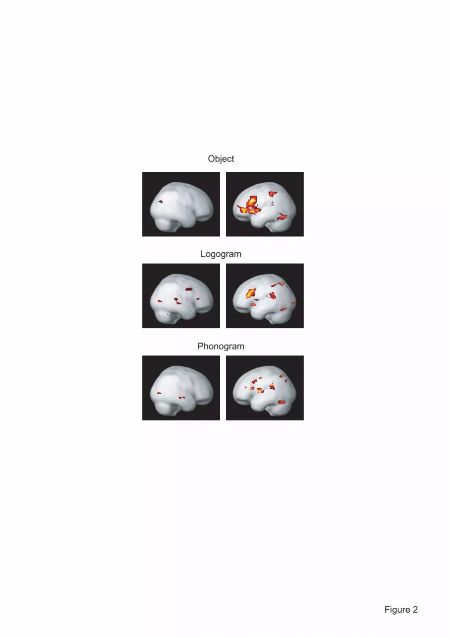
Figure 2
Object
Phonogram
Logogram
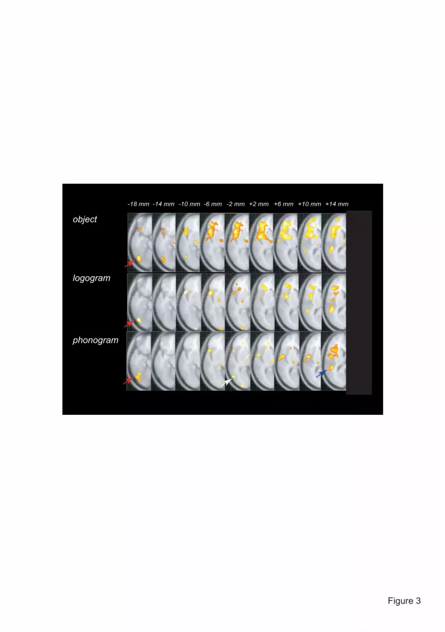
Figure 3
phonogram
object
logogram
-18 mm -14 mm -10 mm -6 mm -2 mm +2 mm +6 mm +10 mm +14 mm

Figure 4
-18 mm -14 mm -10 mm -6 mm -2 mm +2 mm +6 mm +10 mm +14 mm
phonogram
object
logogram

Figure 5
sup temporal gyrusx= ±42, y= -38, z = +16
% s
igna
l cha
nge
obje
ct
logo
gram
phon
ogra
m
-.2
-.1
0
.1
.2
.3
.4
*******
post fusiform gyrusx= ±42, y= -64, z = -24
0
.2
.4
.6
.8
1
1.2
** *****
% s
igna
l cha
nge
obje
ct
logo
gram
phon
ogra
m
% s
igna
l cha
nge
obje
ct
logo
gram
phon
ogra
m
post mid temporal gyrus x= ±50, y= -70, z = -4
0
.05
.1
.15
.2
.25
.3
**
mid occipital gyrusx= ±32, y= -80, z = +4
% s
igna
l cha
nge
obje
ct
logo
gram
phon
ogra
m-.1
0
.1
.2
.3
.4
.5
****
***
medial fusiform gyrusx= ±34, y= -36, z = -12
% s
igna
l cha
nge
obje
ct
logo
gram
phon
ogra
m
** **
-.1
-.05
0
.05
.1
.15
.2
.25
mid temporal gyrus x= ±60, y= -34, z = -6
% s
igna
l cha
nge
obje
ct
logo
gram
phon
ogra
m
0
.1
.2
.3
.4
.5
.6
.7
****
Left hemisphere Right hemisphere


