Embed Size (px)
Citation preview

doi:10.1182/blood-2002-05-1459Prepublished online October 3, 2002;2003 101: 837-845
Georgine E. de Greef, Bob Löwenberg and Ruud DelwelSmit, H. Berna Beverloo, Gregor Verhoef, Leo F. Verdonck, Gert J. Ossenkoppele, Pieter Sonneveld,J. M. Valk, Sonja van der Poel-van de Luytgaarde, Ronald Hack, Rosalyn Slater, Elisabeth M. E. Sahar Barjesteh van Waalwijk van Doorn-Khosrovani, Claudia Erpelinck, Wim L. J. van Putten, Peter a study of 319 de novo AML patientsHigh EVI1 expression predicts poor survival in acute myeloid leukemia:
http://bloodjournal.hematologylibrary.org/content/101/3/837.full.htmlUpdated information and services can be found at:
(4217 articles)Neoplasia � (3716 articles)Clinical Trials and Observations �
Articles on similar topics can be found in the following Blood collections
http://bloodjournal.hematologylibrary.org/site/misc/rights.xhtml#repub_requestsInformation about reproducing this article in parts or in its entirety may be found online at:
http://bloodjournal.hematologylibrary.org/site/misc/rights.xhtml#reprintsInformation about ordering reprints may be found online at:
http://bloodjournal.hematologylibrary.org/site/subscriptions/index.xhtmlInformation about subscriptions and ASH membership may be found online at:
Copyright 2011 by The American Society of Hematology; all rights reserved.Washington DC 20036.by the American Society of Hematology, 2021 L St, NW, Suite 900, Blood (print ISSN 0006-4971, online ISSN 1528-0020), is published weekly
For personal use only. by guest on June 6, 2013. bloodjournal.hematologylibrary.orgFrom

CLINICAL OBSERVATIONS, INTERVENTIONS, AND THERAPEUTIC TRIALS
High EVI1 expression predicts poor survival in acute myeloid leukemia:a study of 319 de novo AML patientsSahar Barjesteh van Waalwijk van Doorn-Khosrovani, Claudia Erpelinck, Wim L. J. van Putten, Peter J. M. Valk,Sonja van der Poel-van de Luytgaarde, Ronald Hack, Rosalyn Slater, Elisabeth M. E. Smit, H. Berna Beverloo, Gregor Verhoef,Leo F. Verdonck, Gert J. Ossenkoppele, Pieter Sonneveld, Georgine E. de Greef, Bob Lowenberg, and Ruud Delwel
The proto-oncogene EVI1 encodes a DNAbinding protein and is located on chromo-some 3q26. The gene is aberrantly ex-pressed in acute myeloid leukemia (AML)patients carrying 3q26 abnormalities. TwomRNAs are transcribed from this locus:EVI1 and a fusion of EVI1 with MDS1(MDS1-EVI1), a gene located 5� of EVI1.The purpose of this study was to investi-gate which of the 2 gene products isinvolved in transformation in human AML.To discriminate between EVI1 and MDS1-EVI1 transcripts, distinct real-time quanti-tative polymerase chain reaction (PCR)
assays were developed. Patients with3q26 abnormalities often showed highEVI1 and MDS1-EVI1 expression. In acohort of 319 AML patients, 4 subgroupscould be distinguished: EVI1� and MDS1-EVI1� (6 patients; group I), EVI1� andMDS1-EVI1� (26 patients; group II), EVI1�
and MDS1-EVI1� (12 patients; group III),and EVI1� and MDS1-EVI1� (275 patients;group IV). The only 4 patients with a 3q26aberration belonged to groups I and II.Interestingly, high EVI1 and not MDS1-EVI1 expression was associated with un-favorable karyotypes (eg, �7/7q�) or com-
plex karyotypes. Moreover, a significantcorrelation was observed between EVI1expression and 11q23 aberrations (mixedlineage leukemia [MLL] gene involve-ment). Patients from groups I and II hadsignificantly shorter overall and event-free survival than patients in groups IIIand IV. Our data demonstrate that highEVI1 expression is an independent poorprognostic marker within the intermediate-risk karyotypic group. (Blood. 2003;101:837-845)
© 2003 by The American Society of Hematology
Introduction
The EVI1 proto-oncogene is located on human chromosome 3q26and is involved in pathogenesis of human acute myeloid leukemia(AML) or myelodysplastic syndrome (MDS) carrying 3q26 rear-rangements.1,2 Although these rearrangements are infrequent inAML, they are of remarkable prognostic value. Patients with thesekaryotypes often do not respond to therapy, even when the mostactive antileukemic therapeutic options are used.3
EVI1 encodes a nuclear DNA binding protein with 2 zinc fingerdomains, an N-terminal domain containing 7 zinc fingers and amore C-terminal domain with 3 zinc fingers. Both domainsrecognize and bind to specific DNA consensus sequences.4,5 Whilemost reports indicate that EVI1 gene expression is not detectable innormal blood or bone marrow,6-9 other studies suggest low butdetectable expression of EVI1 in normal bone marrow cells.10 Highexpression of EVI1 has been observed in developing oocytes and inthe kidney.11 Although the exact mechanism of transformation byEVI1 is still obscure, several studies have shown that inappropriateexpression of EVI1 in immature hematopoietic cells interferes witherythroid and granulocytic development.12 It has become evidentthat EVI1 may form a fusion transcript with the MDS1 gene. MDS1is a 4-exon gene located upstream of EVI1. Splicing may occurfrom exon 2 of MDS1 to the second exon of EVI1 to form the fusiontranscript MDS1-EVI1. This intergenic splicing may occur in
normal tissues as well as in myeloid leukemia.1,13 MDS1-EVI1encodes a longer protein containing the entire EVI1 protein butwith an additional, unique N-terminal extension. Althoughrelated, the 2 proteins EVI1 and MDS1-EVI1 may have oppositeproperties.14,15
Previous studies showed that EVI1 may be expressed in patientswithout 3q268,9,16,17; however, the sets of polymerase chain reaction(PCR) primers that were chosen in these studies did not discrimi-nate between EVI1 and MDS1-EVI1. We designed different primerand probe combinations to discriminate between EVI1 and MDS-EVI1 and quantify the transcript levels by means of real-time PCRanalysis. To provide an answer to the question, which of thesetranscripts are expressed in patients with 3q26 rearrangements, wefirst screened 7 patients carrying 3q26 abnormalities. Using thesame technique, we studied the expression levels of these tran-scripts in the bone marrow samples of healthy volunteers. Toinvestigate how frequently EVI1, MDS1-EVI1, or MDS1 may beexpressed in de novo AML, we determined expression levels ofthese transcripts in a cohort of 319 AML patients at diagnosis. Theresults were analyzed in relation to hematologic, cytogenetic, andclinical characteristics as well as outcome of therapy. Our datademonstrate that expression of EVI1 and not of MDS1-EVI1 isassociated with highly aggressive AML. High EVI1 expression
From the Institute of Hematology, Erasmus Medical Centre, Rotterdam,Netherlands; the Department of Cell Biology and Genetics, Erasmus MedicalCentre, Rotterdam, Netherlands; the Department of Clinical Genetics,Erasmus Medical Centre, Rotterdam, Netherlands; the Department ofHematology, Vrije Universiteit Medical Centre, Amsterdam, Netherlands; theDepartment of Hematology, University Medical Centre, Utrecht, Netherlands;and the Department of Hematology, University Hospitals, KatholiekeUniversiteit Leuven, Leuven, Belgium.
Submitted May 20, 2002; accepted September 6, 2002. Prepublished online asBlood First Edition Paper, October 3, 2002; DOI 10.1182/blood-2002-05-1459.
Supported by grants from the Dutch Cancer Society (Koningin WilhelminaFonds) and the Erasmus University Medical Centre (Revolving Fund).
Reprints: Ruud Delwel, Institute of Hematology, Erasmus Medical Centre, POBox 1738, 3000 DR Rotterdam, Netherlands; e-mail: [email protected].
The publication costs of this article were defrayed in part by page chargepayment. Therefore, and solely to indicate this fact, this article is herebymarked ‘‘advertisement’’ in accordance with 18 U.S.C. section 1734.
© 2003 by The American Society of Hematology
837BLOOD, 1 FEBRUARY 2003 � VOLUME 101, NUMBER 3
For personal use only. by guest on June 6, 2013. bloodjournal.hematologylibrary.orgFrom

occurs with high frequency in patients without 3q26 abnormalities,suggesting other mechanisms of aberrant EVI1 expression.
Patients, materials, and methods
Patients and healthy volunteers
Bone marrow samples of AML patients at diagnosis and healthy volunteers(n � 9) were obtained after informed consent. Blasts from AML patientsand mononucleated fractions from healthy bone marrow specimens wereisolated from the samples by Ficoll-Hypaque (Nygaard, Oslo, Norway)centrifugation.18 The cells were then cryopreserved as described in Delwelet al.19 After thawing cells were washed with Hanks Blanced Salt Solution(HBSS) and further processed for RNA isolation. AML samples treatedaccording to these procedures usually contain more than 90% blasts afterthawing.19 Seven patients with 3q26 rearrangements (4 patients with AML,2 with refractory anemia with excess blasts in transformation [RAEB-t],and 1 with chronic myelogenous leukemia) were selected that had not beenincluded in a clinical trial. A total of 319 de novo AML patients who hadbeen referred to our institution and collaborating centers between 1987 and2000 were chosen for analysis. Of these patients, 229 were treatedaccording to the HOVON-29 (Dutch-Belgian Haematology-OncologyGroup) protocol, 66 according to the HOVON-4 protocol, and 13 accordingto the HOVON-31 protocol. These treatment protocols have been describedelsewhere.20 Eleven patients received other forms of treatment. The clinicaland hematologic characteristics of the 319 patients at diagnosis are shownin Table 1. AML samples were classified according to FAB nomenclature.21
RNA isolation, cDNA synthesis, and real-time PCR
Total RNA was extracted with guanidium thiocyanate followed by centrifu-gation in cesium chloride solution. Then 1 �L RNA was transcribed intocDNA using Superscript (Life Technologies, Merelbeke, Belgium) andrandom hexamers in a 40-�L reaction under standard conditions.
An aliquot of one 20th of the resulting cDNA was used for quantitativePCR amplification. Real-time PCR amplification was performed with theABI PRISM 7700 Sequence Detector (Applied Biosystems, Nieuwerkerkaan den IJssel, Netherlands), using 50 �L mix containing 2 �L cDNA
sample; 250 �M deoxyribonucleoside triphosphates (dNTPs; AmershamPharmacia Biotech, Roosendaal, Netherlands); 15 pmol forward andreverse primer (Life Technologies); 3 mM MgCl2 (5 mM for porphobilino-gen deaminase [PBGD] reaction); 200 nM probe, labeled at the 5� end withthe reporter dye molecule FAM (6-carboxy-fluorescein) for EVI1, MDS1-EVI1, and MDS1 or with JOE (carboxyrhodamine) for PBGD and at the 3�end with the quencher dye molecule TAMRA (6-carboxy-tetramethyl-rhodamine; Eurogentec, Maastricht, Netherlands); 5 �L 10 � buffer A; 30�L water; and 1.25 U AmpliTaq Gold (Applied Biosystems). The thermalcycling conditions included 10 minutes at 95°C followed by 45 cycles ofdenaturation for 15 seconds at 95°C and annealing/extension at 60°C for 30seconds. The primer/probe combinations were chosen such that we coulddiscriminate between EVI1, MDS1/EVI1, and MDS1 transcripts (Figure 1).The oligonucleotide sequences of the primers and probes are shown inTable 2.
Using 7 different dilutions of a cDNA sample (equal to 0.0064-100 ngtotal RNA) prepared from AML cells that were positive for each of thetranscripts (patient 2, Table 3), standard curves were made for EVI1 andMDS1-EVI1. As the expression levels of MDS1 transcripts were too low tomeasure the efficiency of amplification, MDS1 expression was consideredpositive (�) when the threshold cycle (Ct) value was below 35.
To determine the expression levels in AML, all samples were tested induplicate and the average values were used for quantification. To quantifythe relative expression of EVI1 and MDS1-EVI1, the Ct values werenormalized for endogenous reference (�Ct � Cttarget � CtPBGD) and com-pared with a calibrator, using the �� Ct method (��Ct � �Ct Sample � �CtCalibrator).As calibrator we used the average Ct value of EVI1 and MDS1-EVI1 in the 9bone marrow samples of the healthy volunteers. We used the ��Ct value tocalculate relative expression (2��� Ct). As the ��Ct method is applicableonly when the amplification efficiencies of the target and the reference areessentially equal, we analyzed the efficiencies for another 4 patients(patients 25, 28, 29, and 30; Table 4), using the same dilutions indicatedabove. The mean �Ct values (Cttarget � CtPBGD) were plotted against theconcentrations of total RNA (log). The slope of the fitted line was thendetermined. A slope of less than 0.1 is indicative of equal efficiencies.
To define high EVI1 and MDS1-EVI1 expression, a cutoff value of 50(relative expression 2�� �Ct) was chosen. This value was chosen to avoid theinfluence of particle distribution statistics, particularly in those cases with aslightly higher EVI1 and MDS1-EVI1 expression. To prevent bias, thesurvival analysis was also performed at cutoff points of 10, 25, and 100.
Figure 1. Schematic representation of EVI1 and MDS1-EVI1 and the primer andprobes used for real-time PCR. Primers 1 and 2 plus probe A were used to determineMDS1-EVI1 expression levels. EVI1 transcript levels were determined using primers3 and 2 plus probe A. MDS1 expression was measured using primers 1 and 4 plusprobe B. Probe C was used for Northern blot analysis.
Table 1. Demographic and clinical characteristics of 319 de novo AML patients
Sex, no.
Male 167
Female 152
Age, median (range), y 45.1 (15.2-76.8)
Age group, no.
Younger than 35 y 89
35-50 y 112
Older than 50 y 118
FAB, no.
M0 10
M1 68
M2 74
M3 33
M4 56
M5 67
M6 4
Unclassified 7
Cytogenetic risk group, no.
Favorable 57
Intermediate/unknown* 212
Unfavorable 50
WBC count, median (range), 109/L 23.4 (0.3-282)
Blast count, median (range), % 69 (0-98)
Platelet count, median (range), 109/L 49 (3-931)
FAB indicates French-American-British classification21; and WBC, white blood cell.*For 6 patients of this group, no cytogenetic information was available at
diagnosis.
Table 2. Oligonucleotide primer and probe sequences used forquantitative real-time PCR
Oligonucleotide sequence (5�-3�)
Primer 1 GAAAGACCCCAGTTATGGATGG
Primer 2 GTACTTGAGCCAGCTTCCAACA
Primer 3 CTTCTTGACTAAAGCCCTTGGA
Primer 4 TCTCTTCCCCAAATACAACCAAG
Probe A TCTTAGACGAATTTTACAATGTGAAGTTCTGCATAGA.TG
Probe B TCTTAGACGAATTTTACAATGTGAAGTTCTGCATAGATG
PBGD forward primer GGCAATGCGGCTGCAG
PBGD reverse primer GGGTACCCACGCGAATCAC
PBGD probe CATCTTTGGGCTGTTTTCTTCCGCC
838 BARJESTEH VAN WAALWIJK VAN DOORN-KHOSROVANI et al BLOOD, 1 FEBRUARY 2003 � VOLUME 101, NUMBER 3
For personal use only. by guest on June 6, 2013. bloodjournal.hematologylibrary.orgFrom

Northern blotting
Northern blotting was carried out on mRNA isolated from healthy bonemarrow samples as well as AML samples. A portion (20 �g) of total RNAfrom each sample was separated on a 1% agarose, 6% formaldehyde gel andblotted with 10 � SSC (sodium chloride/sodium citrate; Amersham) ontoHybond-N� nylon membrane (Amersham). The blot was hybridized in 1NNaH2PO4 buffer containing 7% sodium dextran sulfate and 1 mM EDTA(ethylenediaminetetraacetic acid), pH 8.0. As probe, human EVI1 (600-bpHindIII-Ncol fragment) and murine GAPDH (777-bp HindIII-EcoRI frag-ment)23 were 32P-labeled by random priming (Boehringer, Mannheim,Germany). The blot was hybridized at 65°C overnight and washed for 15minutes at 65°C in 2 � SSC/0.5% sodium dodecyl sulfate (SDS) and for 15minutes at 65°C in 1 � SSC/0.5% SDS. It was then analyzed byautoradiography.
Cytogenetic analysis and stratification accordingto karyotype risk group
Cytogenetic analysis was carried out according to standard techniques, andthe abnormalities were categorized in 3 cytogenetic groups. Patients withinv(16)/t(16;16), t(8;21), and t(15;17) abnormalities were considered asbeing in the favorable-risk category. The unfavorable-risk category wasdefined by the presence of �5/del(5q), �7del(7q), t(6;9), t(9;22), 3q26abnormality or complex karyotype (more than 3 abnormalities). All otherpatients were classified as intermediate risk. Karyotypes were describedaccording to the International System for Human Cytogenetic Nomenclature.22
Analysis of FLT3 internal tandem duplication mutations in AML
The internal tandem duplications in exon 11 of the human FLT3 gene weredetermined as described previously.24 Briefly, cDNA (derived from 50 ngtotal RNA) and genomic DNA (1 �g) were subjected to PCR using primers11F 5�-CAATTTAGGTAT-3� and 11R 5�-CAAACTCTAAATTTTCTCT-3�. The PCR cycling conditions were as follows: 3 minutes at 94°C; 30cycles of 1 minute at 94°C, 1 minute at 54°C, 1 minute at 72°C; and a finalstep of 10 minutes at 72°C. PCR products were resolved on a 2.5%agarose gel.
Statistical analysis
Statistical analysis was performed with Stata Statistical Software, Release7.0 (Stata, College Station, TX). Spearman rank correlation, Pearson �2 test,and Kruskal-Wallis test were used to assess the association between EVI1and MDS1-EVI1 expression and the clinical and hematologic characteristicsof patients. Actuarial probabilities of overall survival (OS, with failuredeath due to any cause) and event-free survival (EFS, with failure in case ofno complete remission (CR) at day 1 or at relapse or at death in first CR)were estimated by the method of Kaplan and Meier. The Cox proportionalhazards model was applied to determine the association of high EVI1expression with OS and EFS, without and with adjustment for other factorssuch as age, cytogenetic risk, and FLT3 internal tandem duplication
(FLT3-ITD). All tests were 2-sided, and a P of less than .05 was consideredstatistically significant.
Results
Quantification of EVI1, MDS1-EVI1, and MDS1 by real-time PCR
EVI1, MDS1-EVI1, and MDS1 expression levels were analyzed byreal-time PCR employing specific primer/probe combinations(Figure 1; Table 2). Efficiency of the quantification method forEVI1 and MDS1-EVI1 was examined by standard curves madeusing mRNA isolated from AML samples that were positive forEVI1 or MDS1-EVI1. Linear correlation between Ct values andcopy numbers was obtained for EVI1 and MDS1-EVI1, withcorrelation coefficients of 0.94 and 0.98, respectively. The effi-ciency of amplification, determined in cDNA obtained from a bonemarrow sample from patient 2, was approximately 1.00 for EVI1,0.95 for MDS1-EVI1, and 0.96 for PBGD. To evaluate whether the��Ct method used in our study was indeed applicable, we verifiedthe differences in efficiencies of amplification in another 4 AMLsamples ( samples 25, 28, 29, and 30). The slopes of the fitted linesfor the mean �Ct values at different mRNA concentrations were�0.089 for EVI1 and 0.036 for MDS1-EVI1, indicating that the��Ct method was indeed applicable. The expression levels ofMDS1 transcripts were too low to measure the efficiency ofamplification. Therefore we decided not to quantify the MDS1expression level but to show whether it was expressed (�) ornot (�).
As calibrator, we used the average expression of EVI1 andMDS1-EVI1 in 9 bone marrow samples from healthy volunteers.The mean Ct values of EVI1 and MDS1-EVI1 in these normalsamples were 38.4 1.5 and 38.3 2.6, respectively. The valuesobtained were normalized for the internal reference, PBGD. Themean PBGD value for normal bone marrow samples was 23.2 0.9. MDS1 expression was undetectable in bone marrow samplesfrom healthy volunteers.
EVI1, MDS1-EVI1, and MDS1 expression in AML patientscarrying 3q26 abnormalities
The relative expression of EVI1 and MDS1-EVI1 transcripts inpatients with 3q26 abnormalities is shown in Table 3. In 3 AMLpatients carrying an inv(3)(q22;q26) and in 1 AML patient with atranslocation t(3;3)(q22;q26), high expression of EVI1 as well asMDS1-EVI1 transcripts was observed. One of the 2 RAEB-tpatients with t(3;12) showed high EVI1 levels and a moderateincrease in MDS1-EVI1 expression, while in the other RAEB-t
Table 3. EVI1, MDS1-EVI1, and MDS1 expression in 7 AML patients with 3q26 abnormalities
Patient FAB Cytogenetic abnormalities*Relative expression
of EVI1†Relative expression
of MDS1-EVI1†Expression of
MDS1‡
1 M5 inv(3)(q22q26),�7 1 618 104 �
2 M1 inv(3)(q22q26) 4 390 4 390 �
3 M0 inv(3)(q21q26) 4 771 416 �
4 M4 t(3;3)(q22;q26) 2 353 1 448 �
5 RAEB-t t(3;12)(q26;p13) 1 951 35 �
6 RAEB-t t(3;12)(q25a26;p12), del(7)(q22) 1 11 585 �
7 CML t(3;17)(q26;q22),t(1;17;9;22)
(p36;q12;q34;q11)/del(11)(p11.1p14)
30 1 �
*According to the ISCN.22
†Values represent expression levels of EVI1 and MDS1-EVI1 as compared with the average values determined in 9 healthy bone marrow samples 2�� �Ct (see “Patients,materials, and methods”).
‡MDS1 expression was considered positive (�) when the Ct value was below 35.
EVI1 AND MDS1-EVI1 IN DE NOVO AML 839BLOOD, 1 FEBRUARY 2003 � VOLUME 101, NUMBER 3
For personal use only. by guest on June 6, 2013. bloodjournal.hematologylibrary.orgFrom
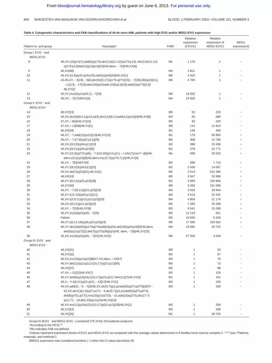
Table 4. Cytogenetic characteristics and FAB classifications of 44 de novo AML patients with high EVI1 and/or MDS1-EVI1 expression
Patient no. and group Karyotype* FAB†
Relativeexpressionof EVI1‡
Relativeexpression ofMDS1-EVI1‡
MDS1expression§
Group I, EVI1� and
MDS1-EVI1�
8 46,XX,r(2)(p?q?),add(5)(q1?3),der(11)t(11;12)(q1?4;p13), der(12)t(11;12)
(q1?4;p13)del(12)(p13p13)[23]/45,idem, �7[3]/46,XX[6]
Mx 1 176 1 �
9 46,XX[68] M4 3 821 1 �
10 46,XX,t(1;6)(p32;q24or25),del(2)(q34)[33]/46,XX[1] M0 4 420 1 �
11 43-45,XY,�3[19],�5[4],der(5)t(5;17)(q1?3;q2?1)[15], �7[19],i(8)(q10)[11],
�11[13],�17[19],ider(19)(q10)add (19)(q13)[18],add(2)(q1?3)[13]/
46,XY[2]
M6 4 705 1 �
12 45,XY,inv(3)(q12q26.2),�7[20] M0 19 552 1 �
13 46,XX,�7[27]/46XX[3] M4 24 920 1 �
Group II, EVI1� and
MDS1-EVI1�
14 46,XY[23] M5 53 220 �
15 46,XX,der(6)t(6;11)(p12;q23),der(11)t(6;11)add(11)(p15)[4]/46,XX[6] M4 55 685 �
16 47,XY,�8[4]/46,XY[16] M5 93 220 �
17 47,XX,�13[58]/46,XX[1] M5 141 12 810 �
18 46,XX[34] M1 149 340 �
19 46,XY,�7,add(12)(p12)[13]/46,XY[15] M1 179 95 950 �
20 45,XY,�7,t(7;8)(q22;p11)[25] M5 308 12 766 �
21 46,XX,t(9;22)(q34;q11)[10] M2 389 15 936 �
22 46,XX,t(9;11)(q34;q23)[5] M1 578 15 771 �
23 46,XY,t(3;3)(q2?3;q26),�7,t(10;20)(p13;q11),�r.ishr(7)(cen7�)[5]/46,
idem,i(21)(q10)[8]/46,idem,ins(12;?)(q1?5;?) [2]/46,XY[4]
Mx 699 25 532 �
24 45,XY,�7[5]/46XY[4] M2 690 1 716 �
25 46,XY,t(9;22)(q34;q11)[22] M2 2 436 14 067 �
26 46,XX,del(7)(q22)[41],46,XX[1] M2 2 513 231 395 �
27 46,XX[10] M5 2 947 32 996 �
28 46,XY,t(6;11)(q25;q23)[38] M1 3 083 156 956 �
29 46,XY[40] M5 3 456 151 085 �
30 45,XY,�7,t(9;11)(p21;q23)[33] M4 3 916 29 944 �
31 46,XY,t(11;19)(q23;p13)[11] M4 4 513 31 542 �
32 46,XX,t(2;9;11)(p13;p22;q23)[20] M4 4 804 21 174 �
33 46,XX,t(9;11)(p21;q23)[10] M5 7 383 25 268 �
34 45,XY,�7[33]/46,XY[8] M3 9 541 15 286 �
35 45,XY,inv(3)(q22q26),�7[25] M5 12 119 551 �
36 Failure M5 16 845 5 349 �
37 46,XY,t(6;11;18)(q26;q23;q23)[23] M5 17 560 150 562 �
38 46,XY,der(3)del(3)(p1?4q2?4)add(3)(q24),del(18)(q22q24)[5]/46,idem,
del(6)(q11q27)[2],del(7)(q2?2q36)[cp3]/45, idem,�7[9]/46,XY[10]
M0 19 082 28 725 �
39 45,XX,inv(3)(q22q26),�7[31]/46,XX[1] M5 27 554 3 040 �
Group III, EVI1� and
MDS1-EVI1�
40 46,XX[31] M5 1 53 �
41 46,XY[32]. M2 1 67 �
42 46,XX,inv(16)(p13q22)[8]/47,XX,idem,�22[47] M4 1 70 �
43 46,XY,del(12)(q11q21),t(15;17)(q22;q11)[65] M3 1 73 �
44 46,XX[37] M2 1 88 �
45 47,XX,�21[22]/46,XX[7] M2 1 129 �
46 46,XY,del(6)(q14q16),t(10;17)(p15;q21),?der(11)[7]/46,XY[3] M1 1 151 �
47 45,X,�Y,t(8;21)(q22;q22),�13[27]/46,XY[1] M2 1 165 �
48 44,XX,add(2),�5,�7[2]/45,XX,dic(5;7)(p1;p1)del(5)(q3?1q3?3)[2]/37–
42,XX,der(1)t(1;4)(q1?;p1?),�4,dic(5;7)(p1;p1)del(5)(q3?1q3?3),
del(8)(p?21;p2?2),inv(10)(p12q?23),�15,add(15)(q2?3),dic(17;?)
(p11;?),�18,del( 20)(q11q13)/46,XX[13]
M3 1 209 �
49 46,XX,inv(11)(p15q13),t(15;17)(q22;q12)[28]/46,XX[1] M3 1 209 �
50 46,XY[52] M3 1 296 �
51 46,XX[26] M1 1 28 725 �
Group IV (EVI1� and MDS1-EVI1�) contained 275 of the 319 patients analyzed.*According to the ISCN.22
†Mx indicates FAB not defined.‡Values represent expression levels of EVI1 and MDS1-EVI1 as compared with the average values determined in 9 healthy bone marrow samples 2�� �Ct (see “Patients,
materials, and methods”).§MDS1 expression was considered positive (�) when the Ct value was below 35.
840 BARJESTEH VAN WAALWIJK VAN DOORN-KHOSROVANI et al BLOOD, 1 FEBRUARY 2003 � VOLUME 101, NUMBER 3
For personal use only. by guest on June 6, 2013. bloodjournal.hematologylibrary.orgFrom

patient MDS1-EVI1 expression was high and EVI1 expression wascomparable to that of normal bone marrow. A CML patient witht(3;17) showed weak expression of EVI1 and no detectableexpression of MDS1-EVI1. Expression of MDS1 was observed inpatient 6 only.
Northern blot analysis was carried out using an EVI1 cDNAprobe on total mRNA isolated from 2 patients (patients 2 and 3)carrying inv(3)(q22q26) and a CML patient (patient 7) with atranslocation t(3;17) (Figure 2). Although this probe does notdiscriminate between the EVI1 and MDS1-EVI1 transcripts, highexpression as observed by real-time PCR was confirmed. Noexpression was observed by Northern blot analysis in a normalbone marrow sample (N1) or in a sample from an AML patientwithout 3q26 abnormality (C1); these 2 samples were negative fordifferent transcripts as determined by real-time PCR (Figure 2).
EVI1, MDS1-EVI1, and MDS1 expression in a cohortof 319 de novo AML patients
The expression levels of EVI1, MDS1-EVI1, and MDS1 were nextinvestigated by real-time PCR in bone marrow samples of 319patients newly diagnosed with AML. This cohort did not includethe 7 patients from Table 3. Of these 319 patients, 44 expressedEVI1, MDSI-EVI1, or both (Table 4): 6 expressed EVI1 only (groupI), 26 expressed EVI1 as well as MDS1-EVI1 (group II), and 12expressed MDS1-EVI1 only (group III). In the remaining 275patients (group IV), neither EVI1 nor MDS1-EVI1 was expressed.In 15 patients the MDS1 gene expression (�) was detectable.MDS1 expression was always associated with high levels of EVI1plus MDS1-EVI1 (Table 4).
To confirm the results obtained by real-time PCR, Northern blotanalysis was carried out in 6 cases. Patients who appeared to behighly positive for EVI1 and/or MDS1-EVI1 by real-time PCR(patients 8, 9, and 34) also showed the proper size transcripts byNorthern blotting analysis (Figure 2). Two patients who were negativeby real-time PCR (C2 and C3) and a normal bone marrow sample (N2)showed no transcripts by Northern blot analysis.
3q26 abnormalities in de novo AML samples expressingEVI1 or MDS1-EVI1
Cytogenetic analysis among the 44 patients who were positive forEVI1 and/or MDS1-EVI1 revealed that only 4 AML patients(patients 12, 23, 35, and 39) carried a 3q26 abnormality (Table 4).None of the patients within group IV (EVI1� and MDS1-EVI1�)carried 3q26 aberrations.
EVI1 expression correlates with unfavorable karyotypes
3q26 defects in AML are frequently accompanied by additionalunfavorable cytogenetic abnormalities (eg, �7/7q�). In fact, in 2cases shown in Table 3 (patients 1 and 6) and all 4 cases with a 3q26aberration shown in Table 4 (patients 12, 23, 35, and 39),chromosome 7 abnormalities were observed. We next investigatedwhether EVI1 and/or MDS1-EVI1 expression in de novo AMLwithout a 3q26 abnormality also correlated with the presence ofpoor-risk karyotypes (Tables 4 and 5). In 67% of patients (4 of 6)from group I (EVI1 only) and 42% of patients (11 of 26) from groupII (EVI1� and MDS1-EVI1�), unfavorable karyotypic abnormali-ties (ie, �7/7q�, �5/5q�, t(9;22), t(6;9)) or complex karyotypes( 3 abnormalities) were present. In contrast, only 8% (1 of 12) ofthe patients from group III (EVI1� and MDS1-EVI1�) and 12% (33of 275) of the patients from group IV (EVI1� and MDS1-EVI1�)carried unfavorable karyotypes. Thus EVI1 expression (groups Iand II) correlated with unfavorable karyotypes (P � .0001), whereasthe presence of MDS1-EVI1 alone (group III) did not. Moreover,favorable-risk karyotypes, that is, t(8;21), t(15;17) or inv(16), werenot noted in any of the EVI1-expressing AML patients from group Ior group II. In contrast, 33% (4 of 12) of the patients in group III(EVI1� and MDS1-EVI1�) and 19% (53 of 275) of the patients ingroup IV (EVI1� and MDS1-EVI1�) exhibited favorable-riskkaryotypes (Table 5).
Among the 212 cases that belong to the leukemias withintermediate-risk karyotypes, 14 showed an 11q23 translocation(MLL rearrangement). Interestingly, 8 of these 14 patients (57%)were found in group II, that is, patients expressing EVI1� andMDS1-EVI1� (Table 4). These data suggest a strong correlationbetween EVI1 expression and the presence of MLL rearrangements.
Correlation of EVI1 and MDS1/EVI1 expression with otherprognostic indicators
We next investigated whether EVI1 and/or MDS1/EVI1 expressionshowed any correlation with other known prognostic indicators.Internal tandem duplication in the FLT3 receptor tyrosine kinasegene has been observed in approximately 20% to 30% of patientswith AML. Moreover, this mutation appears to confer a poorprognosis in many studies carried out in the past 5 years.25,26 In 85(27%) of the 319 cases investigated, an FLT3-ITD was found. Asshown in Table 5, high EVI1 and MDS1-EVI1 expression rarely
Figure 2. EVI1 mRNA expression in AML samples as determined by Northernblotting. Human 600-bp HindIII-Ncol EVI1 probe (see Figure 1) was used, which doesnot discriminate between EVI1 and MDS1-EVI1. Murine GAPDH fragment was usedas control. Patients 2, 3, and 7 carry a 3q26 abnormality (Table 3); patients 8, 9, and34 have high EVI1 expression but no 3q26 abnormality (Table 4). C1, C2, C3represent AML patients without EVI1 expression. N1 and N2 represent normal bonemarrow samples.
Table 5. Distribution of different karyotypic risk categories and Flt3-ITD mutations among groups of AML patients
n Flt3-ITD, no.
Risk karyotype, no.
Favorable* Unfavorable† Intermediate/unknown‡
Group I, EVI1� and MDS1-EVI1� 6 1 0 4 2
Group II, EVI1� and MDS1-EVI1� 26 1 0 12 14
Group III, EVI1� and MDS1-EVI1� 12 1 4 1 7
Group IV, EVI1� and MDS1-EVI1� 275 82 53 33 189
*t(8;21), t(15;17) or inv(16).†�7/7q-, �5/5q-, t(9;22), t(6;9) or complex karyotype (3 abnormalities).‡Other cytogenetic aberrations or normal karyotype.
EVI1 AND MDS1-EVI1 IN DE NOVO AML 841BLOOD, 1 FEBRUARY 2003 � VOLUME 101, NUMBER 3
For personal use only. by guest on June 6, 2013. bloodjournal.hematologylibrary.orgFrom

coincided with FLT3-ITD mutation. Almost all the patients with anFLT3-ITD mutation belonged to group IV, and 82% (70 of 85)carried an intermediate-risk karyotype. No significant correlationwas found between EVI1 and/or MDS1-EVI1 expression and sex,age, WBC count, platelet count, blast counts in blood or bonemarrow, or FAB classification (data not shown).
EVI1 an independent unfavorable prognostic marker in theintermediate-risk karyotypic group
All patients received induction therapy and were included in thesurvival analysis. Clinical outcome was investigated in the distinctgroups of patients based on their EVI1 and MDS1-EVI1 expression.Survival analysis was performed using a cutoff value of 50. Theremission rates for patients in groups I, II, III, and IV were 50%,77%, 67%, and 79%, respectively (Table 6). All patients in group Idied within 12 months, and 25 of 26 patients in group II (EVI1� andMDS1-EVI1�) died within 30 months (Table 6; Figure 3A,B).
The actuarial survival probabilities at 60 months were 33% ingroups III and IV and only 0% for group I and 8% for group II(Table 6; Figure 3A,B). Thus patients in groups I and II had asignificantly shorter OS (P � .002). The EFS probabilities at 60months for groups I and II (0% and 4%, respectively) were muchlower than those for groups III and IV (25% and 27%, respec-tively). A significant difference (P � .002) was also observedbetween EFS in patients with high EVI1 expression (groups I andII) and that in AML patients without EVI1 expression (groups III
and IV). We also analyzed survival using alternative cutoff values(ie, 10, 25, and 100). Although the numbers of patients within the 4different subgroups were slightly different, changing the cutoffvalues did not alter the conclusions drawn from the analysis atcutoff value 50 (data not shown).
Cox regression analysis was applied to assess the prognosticsignificance of high EVI1 expression for OS and EFS (Table 7).High EVI1 expression was associated with an increased hazardratio for death (OS; HR � 1.85) or failure (EFS; no CR, death inCR, or relapse, HR � 1.82), which was statistically significant inunivariable analysis. After adjustment for karyotypic risk factors,age, and FLT3 in a multivariable analysis, high EVI1 expressionwas still associated with an increased hazard ratio (P � .09). Ashigh EVI1 expression is often associated with unfavorable karyo-types, its prognostic value seemed to be overshadowed. To excludethe effect of unfavorable cytogenetics, we decided to investigatethe prognostic value of EVI1 in the intermediate-risk group.
We investigated whether high EVI1 expression would be ofprognostic value for patients carrying intermediate-risk karyotypes,as half of the patients with EVI1 expression (groups I and II)belonged to this risk group. As shown in Figure 4A-B, intermediate-risk patients with high EVI1 expression (n � 16) had significantlyshorter OS and EFS (P � .05 and P � .03) than their EVI1-negative counterparts (n � 196). Furthermore, the disease-freesurvival was significantly shorter in patients with high EVI1expression (P � .007). None of the EVI1-overexpressing patientscarried an FLT3-ITD, whereas FLT3-ITD was observed in 36% (70of 196) of the patients without high EVI1 expression. Univariableand multivariable analysis revealed that high EVI1 expressionserves as an independent prognostic marker for EFS and OS in theintermediate-risk group (Table 8).
Table 6. Therapy response and actuarial probability of survival at 60 months in relation to EVI1 and MDS1-EVI1 expression
n
Completeremission, no.
(%)
Actuarial probability ofsurvival at 60 mo, %
Relapse, no.(%)EFS OS
Group I, EVI1� and MDS1-EVI1� 6 3 (50) 0 0 1 (33)
Group II, EVI1� and MDS1-EVI1� 26 20 (77) 4 8 15 (75)
Group III, EVI1� and MDS1-EVI1� 12 8 (67) 25 33 3 (38)
Group IV, EVI1� and MDS1-EVI1� 275 218 (79) 27 33 100 (46)
Figure 3. Overall survival and event-free survival in AML patients based on EVI1and MDS1-EVI1 expression. (A) Overall survival; (B) event-free survival.
Table 7. Univariable and multivariable analysis of high EVI1 expression asprognostic factor for survival
EFS OS
HR (95% CI) P HR (95% CI) P
Univariable analysis
High EVI1 expression 1.82 (1.25-2.67) .002 1.85 (1.25-2.73) .002
Multivariable analysis
Cytogenetics risk �.0001 �.0001
Favorable 1 (—) 1 (—)
Intermediate 2.29 (1.47-3.57) 2.85 (1.71-4.77)
Unfavorable 3.10 (1.78-5.41) 4.20 (2.26-7.83)
Age, y .64 .56
Younger than 35 1 (—) 1 (—)
35-50 0.98 (0.69-1.38) 1.12 (0.78-1.61)
Older than 50 1.12 (0.81-1.57) 1.21 (0.85-1.73)
FLT3 mutation vs no
mutation 1.48 (1.11-1.98) .01 1.53 (1.12-2.09) .009
High EVI1 expression 1.47 (0.96-2.25) .09 1.48 (0.96-2.29) .09
— indicates not applicable.
842 BARJESTEH VAN WAALWIJK VAN DOORN-KHOSROVANI et al BLOOD, 1 FEBRUARY 2003 � VOLUME 101, NUMBER 3
For personal use only. by guest on June 6, 2013. bloodjournal.hematologylibrary.orgFrom

Discussion
We developed a sensitive method to quantify EVI1 and its fusiontranscript MDS1-EVI1 in AML. Our data demonstrate that EVI1rather than MDS1-EVI1 is a strong indicator for poor treatmentresponse and survival. MDS1 expression was seen in 5% (15 of319) of the AML patients and was always associated with highEVI1 and MDS1-EVI1 expression. High EVI1 mRNA expressionwas observed in 10% (32 of 319) of the patients with newlydiagnosed AML. Poor clinical outcome of patients with 3q26abnormality has previously been reported.3,27-29 As we demon-strated in this study, patients with 3q26 abnormality represent aminor subgroup of patients with high EVI1 expression. In fact, only12.5% (4 of 32) of the patients with high EVI1 expression carried a3q26 abnormality. High EVI1 expression was significantly corre-lated with the presence of unfavorable cytogenetic abnormalities.Favorable-risk karyotypes were not present among theEVI1-expressing groups.
Evi1 was first discovered as a proto-oncogene in retrovirallyinduced myeloid leukemias in the mouse. Retroviral insertions inthe Evi1 locus in those tumors mostly occurred in close vicinity ofthe first 2 or 3 exons of Evi1, causing Evi1 overexpression.30 Thesedata underline that EVI1 rather than MDS1-EVI1 is the transform-ing gene in AML. This conclusion is further strengthened by thefact that in the cohort of de novo AML patients investigated in thepresent study, EVI1 and not MDS1-EVI1 expression correlated withunfavorable-risk leukemias.
In contrast to what has been published previously, we demon-strate that EVI1 expression in de novo AML may be an importantparameter to define a subgroup of poor-risk AML patients. Langa-beer et al31 studied 197 de novo AML patients but did not find anyprognostic value for high EVI1 expression. This is not surprising,however, as the investigators did not discriminate between EVI1and MDS1-EVI1 expression. A number of EVI1-positive patients in
this study carried a favorable-risk karyotype, indicating that thesepatients are most likely expressing MDS1-EVI1 rather than EVI1.Furthermore, previous studies were carried out using classicalreverse transcriptase–PCR, while we performed quantitative real-time PCR to be able to determine high EVI1 expression levelsbased on a defined cutoff value.
Cytogenetic analysis provides a powerful approach to dis-criminate between favorable-risk and unfavorable-risk groupsof AML patients. However, using karyotyping, only 30% to 40%of the AML patients can be classified within these 2 subgroups.In other words, a majority of AML patients belong to intermedi-ate or unknown karyotypic risk groups. A major challenge willbe discriminating favorable-risk from unfavorable-risk patientswithin this heterogeneous group of patients by means ofmolecular biologic approaches. Among the patients with interme-diate-risk karyotype studied, 33% (70 of 212) harbored anFLT3-ITD mutation that predicts poor prognosis. Sixteen (8%)of the 212 patients had high EVI1 expression and showed verypoor survival. Interestingly, none of the EVI1-positive patientsharbored an FLT3-ITD, indicating that the EVI1-expressinggroup represents a distinct subclass of poor-response leukemias.Within the same group of patients with intermediate-riskkaryotype, a subpopulation of poor responders has been definedwith very low mRNA levels of the CEBP� gene.32 Again, nooverlap was found with the other 2 molecularly defined classes.Moreover, mutation analysis revealed another subset of interme-diate-risk AML patients that harbored 3� mutations within theCEBP� gene. These cases could be categorized as leukemiaswith a good prognosis.32 These observations encourage addi-tional gene expression studies and mutation analyses to furtherunravel different classes of AML, particularly within theintermediate-risk karyotypic subgroup of AML.
In 8 of 14 patients with an 11q23 abnormality, we observed highEVI1 and MDS1-EVI1 expression. In previous studies, the correla-tion between EVI1 and 11q23 has been overlooked, as the cohortswere not large enough and the methods used did not discriminatebetween EVI1 and MDS1-EVI1. Two of 16 EVI1-positive patientsscreened by Ohyashiki et al17 and 1 of 29 EVI1-positive patientsstudied by Langabeer et al31 carried an 11q23 abnormality.Translocations involving chromosome band 11q23 disrupt the MLLgene. MLL is a putative transcription regulator that may formcomplexes with other transcription factors. It contains both a strongactivation domain and a repression domain.33 In de novo leukemia,75% of the breakpoints in MLL are mapped to the centromeric halfof the breakpoint cluster region (BCR),34 which is located in therepression domain. Disruption of the repression domain in theseparticular 11q23 translocations might lead to an alteration of the
Table 8. Univariable and multivariable analysis of high EVI1 expression asprognostic factor for survival in intermediate-risk karyotype
EFS OS
HR (95% CI) P HR (95% CI) P
Univariable analysis
High EVI1 expression 1.76 (1.05-2.97) .05 1.69 (1.01-2.87) .06
Multivariable analysis
Age, y .61 .91
Younger than 35 1 (—) 1 (—)
35-50 0.84 (0.56-1.26) 0.97 (0.64-1.49)
Older than 50 0.99 (0.67-1.47) 1.05 (0.69-1.61)
Flt3 mutation vs no mutation 1.71 (1.22-2.38) .002 1.62 (1.14-2.30) .009
High EVI1 expression 2.09 (1.22-3.60) .01 2.01 (1.16-3.47) .02
— indicates not applicable.
Figure 4. Overall survival and event-free survival in AML patients with immediate-risk karyotype based on EVI1 expression. (A) Overall survival; (B) event-freesurvival.
EVI1 AND MDS1-EVI1 IN DE NOVO AML 843BLOOD, 1 FEBRUARY 2003 � VOLUME 101, NUMBER 3
For personal use only. by guest on June 6, 2013. bloodjournal.hematologylibrary.orgFrom

repressor function of MLL, leading to an up-regulation of down-stream target genes. A possible explanation for high EVI1 andMDS1-EVI1 expression in a large proportion of patients with 11q23defects could therefore be that transcription of those genes is underthe control of MLL. We hypothesize that MLL normally repressesEVI1 and MDS1-EVI1 expression. This repression might then bedisrupted as a result of a chimeric protein generated by 11q23translocation, as has been suggested for Hox genes.5 Cloning andnucleotide sequencing analysis of the MLL fusion genes inEVI1-positive versus EVI1-negative patients may provide criticalinformation on this issue.
One of the goals of this study was to investigate whether EVI1,MDS1-EVI1, or both transcripts were expressed in AML patientswith 3q26 aberrations. All of the 8 AML patients with a classicalt(3;3) or an inv(3) (Tables 3 and 4) showed high expression ofEVI1. High EVI1 expression in 7 patients was associated with highMDS1-EVI1. Thus, although our data suggest that EVI1 rather thanMDS1-EVI1 is the critical gene involved in transformation ofmyeloid precursors, it is noteworthy that MDS1-EVI1 is alsofrequently expressed. Since the 3q26 breakpoints are often locatedbetween the MDS1 and EVI1 loci, the normal allele is responsiblefor MDS1-EVI1 expression. MDS1-EVI1 expression might bedirectly or indirectly up-regulated by EVI1 expression. Anotherpossible explanation is that aberrantly expressed EVI1 as a result of3q26 aberration mainly transforms progenitor cells that normallyhave high EVI1 and/or MDS1-EVI1 levels. Fractionated CD34�
progenitor cell populations indeed show high EVI1, MDS1-EVI1,and MDS1 expression (S.B.v.W.v.D.-K. et al, unpublished data,January 2001). Previous studies10,35 also confirmed EVI1 expres-sion in early CD34� progenitor cells. In fact, transformation inthese progenitors may be a result of a disturbance in the tightlycontrolled balance between EVI1 and its fusion transcript.
The mechanism by which EVI1 is expressed in patients without3q26 is unknown. It is conceivable that defects in the EVI1
promoter or in EVI1-regulatory genes affect expression. It is alsopossible that CD34� progenitor cells naturally expressing EVI1and/or MDS1-EVI1 are transformed by other mechanisms andarrested at that particular stage of differentiation. It should be notedthat high EVI1 expression is often associated with the presence ofpoor-risk abnormalities. For example, complex karyotypes are seentwice as often in patients with high EVI1 expression than in patientswithout EVI1 expression. Our data point to a critical role that EVI1may play in genomic instability in AML patients expressingthis gene.
The data presented here demonstrate that EVI1 overexpressionin AML patients correlates with poor treatment outcome. Wepropose that EVI1 gene expression measurements with real-timePCR should be incorporated into the diagnostic procedures for denovo AML patients, especially in the subpopulation with intermedi-ate- or unknown-risk karyotypes. Determination of EVI1 expres-sion levels in this particular category of AML patients will beuseful in distinguishing a subgroup of patients with poor prognosis.Identification and classification of subtypes of AML with specificmolecular defects will be of great value in designing uniquestratified treatment approaches.
Acknowledgments
A. Hagemeijer of the Center for Human Genetics, CatholicUniversity of Leuven, Belgium, is acknowledged for the cytoge-netic analysis performed on patient samples obtained from thatinstitute. We thank our colleagues from the bone marrow transplan-tation group and the molecular diagnostics laboratory, Institute ofHematology, Erasmus Medical Centre, for preparation and storageof AML samples. We wish to thank Karola van Rooijen forpreparation of the figures.
References
1. Jolkowska J, Witt M. The EVI-1 gene—its role inpathogenesis of human leukemias. Leuk Res.2000;24:553-558.
2. Morishita K, Parganas E, William CL, et al. Acti-vation of EVI1 gene expression in human acutemyelogenous leukemias by translocations span-ning 300-400 kilobases on chromosome band3q26. Proc Natl Acad Sci U S A. 1992;89:3937-3941.
3. Reiter E, Greinix H, Rabitsch W, et al. Low cura-tive potential of bone marrow transplantation forhighly aggressive acute myelogenous leukemiawith inversion inv (3)(q21q26) or homologoustranslocation t(3;3) (q21;q26). Ann Hematol.2000;79:374-377.
4. Nucifora G. The EVI1 gene in myeloid leukemia.Leukemia. 1997;11:2022-2031.
5. Kawagoe H, Kawagoe R, Sano K. Targeteddown-regulation of MLL-AF9 with antisense oli-godeoxyribonucleotide reduces the expression ofthe HOXA7 and -A10 genes and induces apopto-sis in a human leukemia cell line, THP-1. Leuke-mia. 2001;15:1743-1749.
6. Russell M, Thompson F, Spier C, Taetle R. Ex-pression of the EVI1 gene in chronic myelog-enous leukemia in blast crisis. Leukemia. 1993;7:1654-1657.
7. Xi ZF, Russell M, Woodward S, Thompson F,Wagner L, Taetle R. Expression of the Zn fingergene, EVI-1, in acute promyelocytic leukemia.Leukemia. 1997;11:212-220.
8. Russell M, List A, Greenberg P, et al. Expressionof EVI1 in myelodysplastic syndromes and other
hematologic malignancies without 3q26 translo-cations. Blood. 1994;84:1243-1248.
9. Dreyfus F, Bouscary D, Melle J, Ribrag V, GuesnuM, Varet B. Expression of the Evi-1 gene in my-elodysplastic syndromes. Leukemia. 1995;9:203-205.
10. Privitera E, Longoni D, Brambillasca F, Biondi A.EVI-1 gene expression in myeloid clonogeniccells from juvenile myelomonocytic leukemia(JMML). Leukemia. 1997;11:2045-2048.
11. Morishita K, Parganas E, Parham DM, Matsugi T,Ihle JN. The Evi-1 zinc finger myeloid transform-ing gene is normally expressed in the kidney andin developing oocytes. Oncogene. 1990;5:1419-1423.
12. Kreider BL, Orkin SH, Ihle JN. Loss of erythropoi-etin responsiveness in erythroid progenitors dueto expression of the Evi-1 myeloid-transforminggene. Proc Natl Acad Sci U S A. 1993;90:6454-6458.
13. Fears S, Mathieu C, Zeleznik-Le N, Huang S,Rowley JD, Nucifora G. Intergenic splicing ofMDS1 and EVI1 occurs in normal tissues as wellas in myeloid leukemia and produces a newmember of the PR domain family. Proc Natl AcadSci U S A. 1996;93:1642-1647.
14. Sitailo S, Sood R, Barton K, Nucifora G. Forcedexpression of the leukemia-associated gene EVI1in ES cells: a model for myeloid leukemia with3q26 rearrangements. Leukemia. 1999;13:1639-1645.
15. Soderholm J, Kobayashi H, Mathieu C, RowleyJD, Nucifora G. The leukemia-associated gene
MDS1/EVI1 is a new type of GATA-binding trans-activator. Leukemia. 1997;11:352-358.
16. Ogawa S, Mitani K, Kurokawa M, et al. Abnormalexpression of Evi-1 gene in human leukemias.Hum Cell. 1996;9:323-332.
17. Ohyashiki JH, Ohyashiki K, Shimamoto T, et al.Ecotropic virus integration site-1 gene preferen-tially expressed in post-myelodysplasia acute my-eloid leukemia: possible association with GATA-1,GATA-2, and stem cell leukemia gene expres-sion. Blood. 1995;85:3713-3718.
18. Boyum A. Separation of leukocytes from bloodand bone marrow. Introduction. Scand J Clin LabInvest Suppl. 1968;97:7.
19. Delwel R, Touw I, Bot F, Lowenberg B. Fucosebinding lectin for characterizing acute myeloidleukemia progenitor cells. Blood. 1986;68:41-45.
20. Rombouts WJ, Lowenberg B, van Putten WL,Ploemacher RE. Improved prognostic signifi-cance of cytokine-induced proliferation in vitro inpatients with de novo acute myeloid leukemia ofintermediate risk: impact of internal tandem dupli-cations in the Flt3 gene. Leukemia. 2001;15:1046-1053.
21. Bennett JM, Catovsky D, Daniel MT, et al. Pro-posed revised criteria for the classification ofacute myeloid leukemia. A report of the French-American-British Cooperative Group. Ann InternMed. 1985;103:620-625.
22. Mitelman F, ed. ISCN 1995: International Systemfor Human Cytogenetic Nomenclature (1995).Basel, Switzerland: Karger; 1995.
844 BARJESTEH VAN WAALWIJK VAN DOORN-KHOSROVANI et al BLOOD, 1 FEBRUARY 2003 � VOLUME 101, NUMBER 3
For personal use only. by guest on June 6, 2013. bloodjournal.hematologylibrary.orgFrom

23. Jenkins JR, Ayton P, Jones T, et al. Isolation ofcDNA clones encoding the beta isozyme ofhuman DNA topoisomerase II and localisation ofthe gene to chromosome 3p24. Nucleic AcidsRes. 1992;20:5587-5592.
24. Nakao M, Yokota S, Iwai T, et al. Internal tandemduplication of the flt3 gene found in acute myeloidleukemia. Leukemia. 1996;10:1911-1918.
25. Kondo M, Horibe K, Takahashi Y, et al. Prognosticvalue of internal tandem duplication of the FLT3gene in childhood acute myelogenous leukemia.Med Pediatr Oncol. 1999;33:525-529.
26. Meshinchi S, Woods WG, Stirewalt DL, et al.Prevalence and prognostic significance of Flt3internal tandem duplication in pediatric acute my-eloid leukemia. Blood. 2001;97:89-94.
27. Grigg AP, Gascoyne RD, Phillips GL, HorsmanDE. Clinical, haematological and cytogenetic fea-tures in 24 patients with structural rearrange-ments of the Q arm of chromosome 3. Br JHaematol. 1993;83:158-165.
28. Bitter MA, Neilly ME, Le Beau MM, Pearson MG,
Rowley JD. Rearrangements of chromosome 3involving bands 3q21 and 3q26 are associatedwith normal or elevated platelet counts in acutenonlymphocytic leukemia. Blood. 1985;66:1362-1370.
29. Pintado T, Ferro MT, San Roman C, Mayayo M,Larana JG. Clinical correlations of the 3q21;q26 cytogenetic anomaly. A leukemic or myelo-dysplastic syndrome with preserved or in-creased platelet production and lack ofresponse to cytotoxic drug therapy. Cancer.1985;55:535-541.
30. Morishita K, Parker DS, Mucenski ML, JenkinsNA, Copeland NG, Ihle JN. Retroviral activationof a novel gene encoding a zinc finger protein inIL-3-dependent myeloid leukemia cell lines. Cell.1988;54:831-840.
31. Langabeer SE, Rogers JR, Harrison G, et al.EVI1 expression in acute myeloid leukaemia. Br JHaematol. 2001;112:208-211.
32. Barjesteh van Waalwijk van Doorn-Khosravani S,Meijer J, Erpelinck C, et al. Mutations in the ba-
sic-fork-leucine zipper and low C/EBP� expres-sion levels are prognostic markers in intermedi-ate-risk AML. Hematol J. In press.
33. Zeleznik-Le NJ, Harden AM, Rowley JD. 11q23translocations split the “AT-hook” cruciform DNA-binding region and the transcriptional repressiondomain from the activation domain of the mixed-lineage leukemia (MLL) gene. Proc Natl Acad SciU S A. 1994;91:10610-10614.
34. Broeker PL, Super HG, Thirman MJ, et al. Distri-bution of 11q23 breakpoints within the MLL break-point cluster region in de novo acute leukemiaand in treatment-related acute myeloid leukemia:correlation with scaffold attachment regions andtopoisomerase II consensus binding sites. Blood.1996;87:1912-1922.
35. Gerhardt TM, Schmahl GE, Flotho C, Rath AV,Niemeyer CM. Expression of the Evi-1 gene inhaemopoietic cells of children with juvenile my-elomonocytic leukaemia and normal donors. Br JHaematol. 1997;99:882-887.
EVI1 AND MDS1-EVI1 IN DE NOVO AML 845BLOOD, 1 FEBRUARY 2003 � VOLUME 101, NUMBER 3
For personal use only. by guest on June 6, 2013. bloodjournal.hematologylibrary.orgFrom


